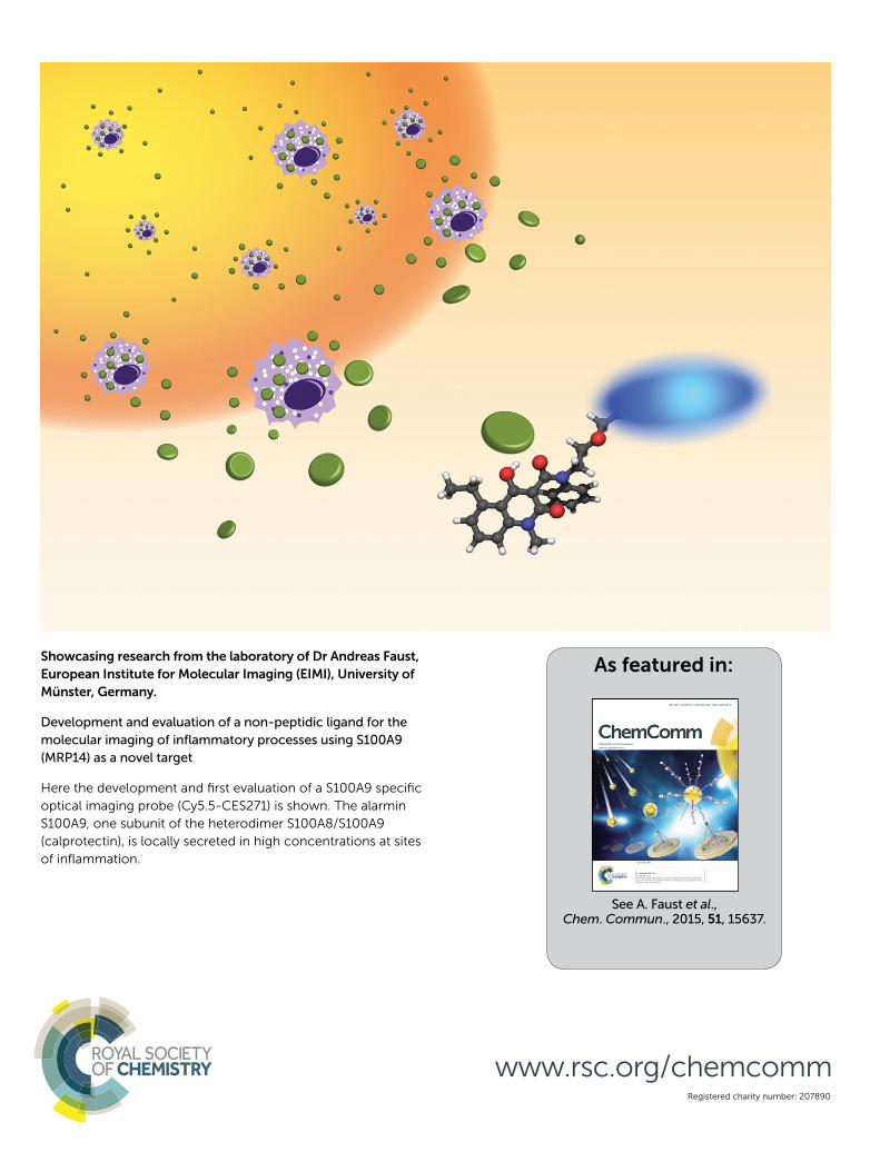
As featured in: See A. Faust et al., Chem. Commun., 2015, 51, 15637. Showcasing research from the laboratory of Dr Andreas Faust, European Institute for Molecular Imaging (EIMI), University of Münster, Germany. Development and evaluation of a non-peptidic ligand for the molecular imaging of inflammatory processes using S100A9 (MRP14) as a novel target Here the development and first evaluation of a S100A9 specific optical imaging probe (Cy5.5-CES271) is shown. The alarmin S100A9, one subunit of the heterodimer S100A8/S100A9 (calprotectin), is locally secreted in high concentrations at sites of inflammation. Registered charity number: 207890 www.rsc.org/chemcomm

Welcome message from author
This document is posted to help you gain knowledge. Please leave a comment to let me know what you think about it! Share it to your friends and learn new things together.
Transcript
As featured in:
See A. Faust et al.,Chem. Commun., 2015, 51, 15637.
Showcasing research from the laboratory of Dr Andreas Faust,
European Institute for Molecular Imaging (EIMI), University of
Münster, Germany.
Development and evaluation of a non-peptidic ligand for the
molecular imaging of infl ammatory processes using S100A9
(MRP14) as a novel target
Here the development and fi rst evaluation of a S100A9 specifi c
optical imaging probe (Cy5.5-CES271) is shown. The alarmin
S100A9, one subunit of the heterodimer S100A8/S100A9
(calprotectin), is locally secreted in high concentrations at sites
of infl ammation.
Registered charity number: 207890
www.rsc.org/chemcomm

This journal is©The Royal Society of Chemistry 2015 Chem. Commun., 2015, 51, 15637--15640 | 15637
Cite this:Chem. Commun., 2015,
51, 15637
Development and evaluation of a non-peptidicligand for the molecular imaging of inflammatoryprocesses using S100A9 (MRP14) as a noveltarget†
A. Faust,‡*ab T. Voller,‡c F. Busch,a M. Schafers,abde J. Roth,bce S. Hermannabe andT. Voglbce
The establishment of novel molecular imaging tools to monitor the
local activity of inflammation remains an interdisciplinary challenge.
Our target, the alarmin S100A9, one subunit of the heterodimer
S100A8/S100A9 (calprotectin), is locally secreted in high concen-
trations from immigrated and activated phagocytes at local sites of
inflammation. Calprotectin is already a well established serum
biomarker for many inflammatory disorders. Here we show the
development and first evaluation of the novel S100A9 specific
molecular imaging probe Cy5.5-CES271 for optical imaging of local
inflammatory activity in vivo.
Inflammatory reactions like autoimmune and infectious diseases1
as well as cardiovascular diseases such as atherosclerosis2 ormyocardial ischemia3 characteristically feature local immunecell activation and inflammation. These inflammatory reactionsthat initially serve the host by avoiding local tissue damage andby clearing infections, can in fact harm the patient duringoverwhelming immune responses or under chronic inflamma-tory conditions such as rheumatoid arthritis. The disease activityand progression, e.g. in atherosclerosis correlate well with thenumber of immigrated immune cells.1b,4 Activated phagocytesand epithelial cells express and locally secrete high levels of theS100 protein complex S100A8/S100A9, which acts as a so calledalarmin or Danger Associated Molecular Pattern (DAMP) mole-cule with potent pro-inflammatory capacities.1a,4 By binding to
both, the extracellular matrix and Toll-like receptor 4 (TLR4) orRAGE expressing immune cells, S100A8/S100A9 exhibits a highlocal concentration gradient amplifying local inflammatory reac-tions. Recently, S100A8/S100A9 has been established as a biomarkerin many inflammatory diseases.1a,5 S100A8/S100A9 serum levelscould for example be characterized as an accurate predictor forthe risk of cardiovascular events in patients. Moreover, the knockoutof S100A9 significantly reduced disease severity in ApoE�/�
mice, a mouse model for atherosclerosis.6
Previously, we reported the use of antibody-based opticalprobes such as Cy5.5t-labelled antibodies targeting the S100A9subunit (anti-S100A9-Cy5.5) of the S100A8/S100A9 heterodimerin vivo. We could observe excellent correlations between signalintensity and local S100A9 expression. Furthermore, we couldpredict the individual disease course of a single mouse in differentmouse models for inflammatory and infectious diseases.7 However,translation of this imaging strategy into patients is challenging dueto known limitations of antibody-based approaches. Therefore, wedeveloped the first S100A9 affine optical imaging tracer based on asmall non-peptidic structure, which can also be used for manyother imaging modalities such as SPECT or PET when radio-labelled. We chose linomide 1 as lead compound, a syntheticimmuno-modulator based on 3-quinolinecarboxamides whichis firstly described in prostate 1996.8 A modified substance of 1,laquinimod 2 was already selected for clinical studies in manand its efficacy has been demonstrated in animal models ofseveral autoimmune diseases, including multiple sclerosis.9 Thedrug was granted a fast track review by the FDA in 2009.10
Multivariate analytical tools were used to derive the SAR forthe binding activity of a series of laquinimod analogues towardsS100A9 with the assumption that similar analogs bind to thesame binding site in a similar binding mode.
Recently, we could show that the most potent ligands are basedon 3-quinolinecarboxamides with an ethyl group in 5-positioninstead of the chloride (Fig. 1).11 We propose that the b-hydroxy-carboxamide unit is responsible for binding to the protein. Incomparison the quinoline moiety, where slight variations arepossible, the N-phenyl or N-methylamine used for forming the
a European Institute for Molecular Imaging (EIMI), University of Munster,
Waldeyerstr. 15, 48149 Munster, Germany. E-mail: [email protected] Cells-in-Motion Cluster of Excellence (EXC 1003–CiM), University of Munster,
48149 Munster, Germanyc Institute of Immunology, University Hospital of Munster, Rontgenstr. 21,
48149 Munster, Germanyd Department of Nuclear Medicine, University Hospital of Munster,
Albert-Schweitzer-Campus 1, 48149 Munster, Germanye Interdisciplinary Center for Clinical Research (IZKF Munster),
University of Munster, Domagkstr. 3, 48149 Munster, Germany
† Electronic supplementary information (ESI) available: Experimental proceduresand analytical data of all compounds, details on ELISA, binding studies, mice andbiodistribution. See DOI: 10.1039/c5cc07019h‡ Authors contributed equally.
Received 20th August 2015,Accepted 9th September 2015
DOI: 10.1039/c5cc07019h
www.rsc.org/chemcomm
ChemComm
COMMUNICATION
Ope
n A
cces
s A
rtic
le. P
ublis
hed
on 1
6 Se
ptem
ber
2015
. Dow
nloa
ded
on 1
/6/2
022
4:12
:07
AM
. T
his
artic
le is
lice
nsed
und
er a
Cre
ativ
e C
omm
ons
Attr
ibut
ion-
Non
Com
mer
cial
3.0
Unp
orte
d L
icen
ce.
View Article OnlineView Journal | View Issue

15638 | Chem. Commun., 2015, 51, 15637--15640 This journal is©The Royal Society of Chemistry 2015
amide bond is free for attaching different linker systems. Inaddition a connection of a quinolinecarboxamide on a goldsurface is possible without loss of inhibition.11
The synthesis of the target compound Cy5.5-CES271 for opticalimaging of S100A9 is outlined in Scheme 1. Starting from thecommercially available 3-nitrophthalic acid anhydride, diethylmalonate was condensed and the resulting lactone was treatedwith hydrochloric acid to give 2-acetyl-6-nitrobenzoic acid.12 Theanthranilic acid was synthesized in a two-step hydrogenationprocedure and treated with phosgene to give the correspondingisatoic anhydride in good yield.13 After N-alkylation with iodo-methane the anhydride was opened with diethyl malonate underdecarboxylation and reclosed yielding the correspondingb-hydroxyester. Acidic cleavage yielded the 3-quinolinecarboxylicacid 3 as key intermediate for functionalization for differentlabelling techniques.14 For the first S100A9 affine imaging probethe cyanine dye Cy5.5TM label was coupled to intermediate 3through a PEG (polyethylene glycol) linker in order to avoid inter-actions between the Cy5.5TM label and the protein. After the
amination of the mesyl-PEG-azide15 4 with aniline gave low yieldsthe mesylate was converted into the corresponding bromide andwe got the secondary amine in satisfactory amounts. The keyintermediate 3 was then coupled to the secondary amine and theresulting carboxamide was then reduced to give the free amine asprecursor for coupling with the cyanine dye. An activated NHS-ester derivative of Cy5.5TM was added under basic conditionsyielding Cy5.5-CES271 suitable for optical imaging of S100A9.16
For verification the HPLC-purified tracer was analyzed by highresolution mass spectrometry showing the fourfold anionicspecies (Scheme 1B and C).
Studies were then conducted to verify strong binding affinityof Cy5.5-CES271 for S100A9 (Fig. 2A). In this regard, we modi-fied our specific S100-ELISA to analyse S100A9 binding to TLR4/MD2. Instead of the capturing anti-S100A9 antibody we coatedTLR4/MD2 (3146-TM-050/CF, R&D Systems) to the wells of a96-well plate which served as capturing molecule.7 After blockingof the unspecific binding sites by PBS/5% skim milk powder,plates were washed three times. S100A9 protein was added at aconcentration of 1 mg ml�1 in the presence or absence of 100 mMCy5.5-CES271 and incubated for two hours at room temperature.Unbound S100 protein was removed by washing the plates forthree times, followed by the addition of a primary anti-S100A9-antibody (1 mg ml�1, polyclonal, rabbit). After a washing step, thesecondary anti-rabbit-IgG-antibody coupled to HRP (1 mg ml�1,Cell Signalling) was added. TMB was used as substrate for HRPto quantify binding efficiency by absorbance readings at 450 nmin an ELISA reader (Anthos Mikrosysteme). Addition of the non-peptidic S100A9 ligand (Cy5.5-CES271) markedly blocked bind-ing of S100A9 to TLR4/MD2, as indicated by a decrease of signalgiven by the TLR4-S100A9 ELISA. These results confirm that the
Scheme 1 (A) Synthesis of Cy5.5-CES271: (a) Ac2O, NEt3, diethyl malonate, 40 1C, 4 h, 88%; (b) HCl, H2O, toluene, 100 1C, 14 h, 80%; (c) 1. PtO2, H2O,NaOH, 90 1C 3 h then rt, 14 h; 2. RANEYs-Ni, H2O, NaOH, 110 1C, 3 h then rt, 24 h, 90% (two steps); (d) diphosgene, THF, toluene, rt, 5 h, 86%; (e) NaH,MeI, DMF, rt, 14 h; (f) NaH, diethyl malonate, DMF, 85 1C, 14 h, 53% (two steps); (g) HCl/HOAc 60 1C, 6 h, 50%; (h) LiBr, CH3CN, reflux, 16 h, 80%; (i) aniline,H2O, reflux, 16 h, 42%; ( j) NEt3, SOCl2, CH2Cl2, 0 1C, 4 h, 82%; (k) H2, Pd/C, THF, rt, 16 h, 79%; (l) Cy5.5-NHS, NEt3, DMF, rt, 16 h, 50%. (B) Analysis andverification of Cy5.5-CES271 (here M4� species) by high resolution ESI-MS; (C) quality control (RP-HPLC) of Cy5.5-CES271.
Fig. 1 Lead compound (top) and the established synthetic immuno-modulators linomide 1 and laquinimod 2.
Communication ChemComm
Ope
n A
cces
s A
rtic
le. P
ublis
hed
on 1
6 Se
ptem
ber
2015
. Dow
nloa
ded
on 1
/6/2
022
4:12
:07
AM
. T
his
artic
le is
lice
nsed
und
er a
Cre
ativ
e C
omm
ons
Attr
ibut
ion-
Non
Com
mer
cial
3.0
Unp
orte
d L
icen
ce.
View Article Online

This journal is©The Royal Society of Chemistry 2015 Chem. Commun., 2015, 51, 15637--15640 | 15639
attachment of the dye Cy5.5 does not interfere with the bindingof Cy5.5-CES271 to S100A9. This blocking study also provestarget specificity of Cy5.5-CES271, and shows that binding ofS100A9 to TLR4/MD2 can be efficiently blocked in the presenceof Cy5.5-CES271 (resulting in low absorption).
Next, we determined the binding constants of Cy5.5-CES271to murine and human S100A9 by fluorimetric measurements(Fig. 2B). Briefly, 0,3788 mM S100A9 (5 mg ml�1 of homodimerS100A9) solved in 50 ml PBS was coated to the bottom of a 96-wellplate and served as capturing molecule. For each S100A9 coatedwell a control well was used with 50 ml PBS alone. After a washingstep, unspecific binding sites were blocked by PBS/5% skim milkpowder. Cy5.5-CES271 was added at increasing concentrations.After 1 h incubation at 4 1C, the supernatants were removed andthe fluorescence intensity was measured with a fluorimeter.Non-linear regression analysis was performed with a one sitesaturation model, to calculate the binding constant of Cy5.5-CES271 to either murine or human S100A9. The Kd-values of2.66 mM (murine) and 2.06 mM (human, Fig. 2B) confirm thatCy5.5-CES271 is eligible for imaging purposes. We observed astrong binding affinity of the tracer to S100A9 that is not affectedby the attachment of Cy5.5 to CES271.
Finally, we analysed the biodistribution of Cy5.5-CES271injected to healthy Balb/c mice by the measurement of fluores-cence intensity in various organs. Fig. 3 shows the tracer accumu-lation 1 and 3 h after injection. The tracer was injected at a dose of2 nmol per mouse. We could observe a good tissue availability ofCy5.5-CES271 and an elimination that was mainly driven by renalexcretion, as indicated by the high renal uptake and the increasingconcentrations of the tracer in the urinary bladder urine incomparison to the relatively low hepatic uptake. This kinetics,that is different to our previously published antibody based traceranti-S100A9-Cy5.57 kinetics favors Cy5.5-CES271 for imaging oforgans neighboring the liver like the lung or the heart.
In conclusion, we developed the first optical imaging probeCy5.5-CES271 based on non-peptidic 3-quinolinecarboxamidefor specific imaging of extracellularly released S100A9 proteinindicating local phagocyte activity. The specificity was confirmedby a modified S100-ELISA and the binding potency was
determined as sufficient for both murine and human S100A9for in vivo optical imaging techniques. Ongoing in vivo studiesin the inflamed tissue in models of ear inflammation, arthritisor myocardial infarction will give us information about thefeasibility as optical probe for specific S100A9 targeted imaging.The established synthesis and imaging strategy allows furtherlabelling methods (e.g. radionuclides) and is therefore suitablefor clinical translation.
We gratefully acknowledge financial support from the DFG(SFB656 A9 and Z5), the Interdisciplinary Centre of ClinicalResearch (IZKF Munster, core unit PIX) and the Medical CollegeMunster.
Notes and references1 (a) T. Vogl, K. Tenbrock, S. Ludwig, N. Leukert, C. Ehrhardt,
M. A. van Zoelen, W. Nacken, D. Foell, T. van der Poll, C. Sorgand J. Roth, Nat. Med., 2007, 13, 1042; (b) P. van Lent, L. C. Grevers,A. B. Blom, O. J. Arntz, F. A. van de Loo, P. van der Kraan,S. Abdollahi-Roodsaz, G. Srikrishna, H. Freeze, A. Sloetjes,W. Nacken, T. Vogl, J. Roth and W. B. van den Berg, Arthritis Rheum.,2008, 58, 3776; (c) P. van Lent, A. B. Blom, R. F. Schelbergen,A. Sloetjes, F. P. Lafeber, W. F. Lems, H. Cats, T. Vogl, J. Roth andW. B. van den Berg, Arthritis Rheum., 2012, 64, 1466; (d) K. Loser,
Fig. 2 (A) Analysis of the binding of Cy5.5-CES271 to S100A9. S100A9 binding to TLR4/MD2 was quantified by an ELISA-based assay. Specific binding ofCES271-Cy5.5 to S100A9 inhibits formation of the S100A9–TLR4/MD2 complex and thereby diminishes the ELISA signal (n = 5; p o 0.05). (B) Estimationof the binding constant of Cy5.5-CES271 to human and murine S100A9. The constant was calculated using the one site saturation regression model.Each dot represents the mean value of four independent experiments.
Fig. 3 Biodistribution of Cy5.5-CES271 in healthy Balb/c mice. The tracerwas intravenously injected at a dose 2 nmol per mouse. The tracer accu-mulation was measured 1 and 3 h post injection (n = 5 for each time point).
ChemComm Communication
Ope
n A
cces
s A
rtic
le. P
ublis
hed
on 1
6 Se
ptem
ber
2015
. Dow
nloa
ded
on 1
/6/2
022
4:12
:07
AM
. T
his
artic
le is
lice
nsed
und
er a
Cre
ativ
e C
omm
ons
Attr
ibut
ion-
Non
Com
mer
cial
3.0
Unp
orte
d L
icen
ce.
View Article Online
15640 | Chem. Commun., 2015, 51, 15637--15640 This journal is©The Royal Society of Chemistry 2015
T. Vogl, M. Voskort, A. Lueken, V. Kupas, W. Nacken, L. Klenner,A. Kuhn, D. Foell, L. Sorokin, T. A. Luger, J. Roth and S. Beissert,Nat. Med., 2010, 16, 713; (e) B. Petersen, M. Wolf, J. Austermann,P. van Lent, D. Foell, M. Ahlmann, V. Kupas, K. Loser, C. Sorg,J. Roth and T. Vogl, EMBO J., 2013, 9, 100.
2 (a) S. Cagnin, M. Biscuola, C. Patuzzo, E. Trabetti, A. Pasquali,P. Laveder, G. Faggian, M. Iafrancesco, A. Mazzucco, P. F. Pignattiand G. Lanfranchi, BMC Genomics, 2009, 10, 13; (b) L. A. Altwegg,M. Neidhart, M. Hersberger, S. Muller, F. R. Eberli, R. Corti,M. Roffi, G. Sutsch, S. Gay, A. von Eckardstein, M. B.Wischnewsky, T. F. Luscher and W. Maier, Eur. Heart J., 2007,28, 941; (c) K. Croce, H. Gao, Y. Wang, T. Mooroka, M. Sakuma,C. Shi, G. K. Sukhova, R. R. Packard, N. Hogg, P. Libby and D. I.Simon, Circulation, 2009, 120, 427; (d) K. Yonekawa, M. Neidhart,L. A. Altwegg, C. A. Wyss, R. Corti, T. Vogl, M. Grigorian, S. Gay,T. F. Luscher and W. Maie, Atherosclerosis, 2011, 218, 486.
3 (a) J. H. Boyd, B. Kan, H. Roberts, Y. Wang and K. R. Walley, Circ.Res., 2008, 102, 1239; (b) D. A. Morrow, Y. Wang, K. Croce,M. Sakuma, M. S. Sabatine, H. Gao, A. D. Pradhan, A. M. Healy,J. Buros, C. H. McCabe, P. Libby, C. P. Cannon, E. Braunwald andD. I. Simon, Am. Heart J., 2008, 155, 49.
4 (a) J. Chan, J. Roth, J. Oppenheim, K. Tracey, T. Vogl, M. Feldmann,N. Horwood and J. Nanchahal, J. Clin. Invest., 2012, 122, 2711;(b) M. G. Ionita, A. Vink, I. E. Dijke, J. D. Laman, W. Peeters,P. H. van der Kraak, F. L. Moll, J. P. de Vries, G. Pasterkamp andD. P. de Kleijn, Arterioscler., Thromb., Vasc. Biol., 2009, 29, 1220.
5 D. Foell and J. Roth, Arthritis Rheum., 2004, 50, 3762.6 M. M. Averill, C. Kerkhoff and K. E. Bornfeldt, Arterioscler., Thromb.,
Vasc. Biol., 2012, 32, 223.7 T. Vogl, M. Eisenblatter, T. Voller, S. Zenker, S. Hermann, P. van Lent,
A. Faust, C. Geyer, B. Petersen, K. Roebrock, M. Schafers, C. Bremerand J. Roth, Nat. Commun., 2014, 5, 4593.
8 I. B. Joseph and J. T. Isaacs, Prostate, 1996, 29, 183.
9 C. Brunmark, A. Runstrom, L. Ohlsson, B. Sparre, T. Brodin,M. Astrom and G. Hedlund, J. Neuroimmunol., 2002, 130, 163.
10 J. Preiningerova, Expert Opin. Invest. Drugs, 2009, 18, 985.11 P. Bjork, A. Bjork, T. Vogl, M. Stenstrom, D. Liberg, A. Olsson,
J. Roth, F. Ivars and T. Leanderson, PLoS Biol., 2009, 7, e1000097.12 C. Luthy, H. Zondler, T. Rapold, G. Seifert, B. Urwyler, T. Heinis,
C. Steinrucken and J. Allen, Pest Manage. Sci., 2001, 57, 205.13 K. Jansson, EP2316818A1, 2009.14 S. Jonsson, G. Andersson, T. Fex, T. Fristedt, G. Hedlund, K. Jansson,
L. Abramo, L. Fritzson, A. Pekarski, A. Runstrom, H. Sandin,I. Thuvesson and A. Bjork, J. Med. Chem., 2004, 47, 2075.
15 A. Faust, B. Waschkau, J. Waldeck, C. Holtke, H. J. Breyholz,S. Wagner, K. Kopka, O. Schober, W. Heindel, M. Schafers andC. Bremer, Bioconjugate Chem., 2009, 20, 904.
16 Labelling procedure: The amino-functionalized precursor (3.0 mg,4.2 mmol) was dissolved in 400 mL dry dimethylformamide providedwith 10 mL triethylamine. To this solution, Cy5.5-NHS ester (GE)(1 mg, 0.9 mmol) was added. The reaction mixture was vortexed for16 h at room temperature in the dark. Purification of Cy5.5-CES271was performed by gradient-HPLC using a Knauer system with twoK-1800 pumps, an S-2500 UV detector and a RP-HPLC Nucleosil100–5 C18 column (250 mm � 4.6 mm). Eluent A: water (0.1% TFA).Eluent B: Acetonitrile (0.1% TFA). Gradient from 95% A to 40% Aover 19 minutes, holding for 5 minutes and back to 95% in oneminute at a flow rate of 5.5 ml min�1, detection at l = 254 nm. Theappropriate fractions (tR = 16.5 min) were collected, lyophilized,redissolved in 1 mL water and finally stored at �20 1C. The averagecontent of Cy5.5-CES271 was 0.45 � 0.02 mmol ml�1 (E50%) asdetermined by photometric measurements with labs = 678 nm ande678 = 250000 M�1 cm�1. MS (ES�): m/z = 464.1 (100%), 464.5, 464.8 [M]3�;696.7, 697.2, 697.7 [M + H]2�. HRMS (ES�): m/z = 347.84493,348.09560, 348.34570, 348.59556, 348.84580 [M]4� calculated:347.84511, 348.09589, 348.34575, 348.59593, 348.84592 [M]4�.
Communication ChemComm
Ope
n A
cces
s A
rtic
le. P
ublis
hed
on 1
6 Se
ptem
ber
2015
. Dow
nloa
ded
on 1
/6/2
022
4:12
:07
AM
. T
his
artic
le is
lice
nsed
und
er a
Cre
ativ
e C
omm
ons
Attr
ibut
ion-
Non
Com
mer
cial
3.0
Unp
orte
d L
icen
ce.
View Article Online
Related Documents