http://informahealthcare.com/ddi ISSN: 0363-9045 (print), 1520-5762 (electronic) Drug Dev Ind Pharm, Early Online: 1–14 ! 2015 Informa Healthcare USA, Inc. DOI: 10.3109/03639045.2015.1019355 RESEARCH ARTICLE Development and characterization of folate anchored Saquinavir entrapped PLGA nanoparticles for anti-tumor activity Ruchi Singh 1 , Prashant Kesharwani 2 , Neelesh Kumar Mehra 3,4 , Shashank Singh 5 , Smita Banerjee 1 , and N. K. Jain 3 1 Department of Zoology, Dr. H. S. Gour University, Sagar, India, 2 Department of Pharmaceutical Sciences, Eugene Applebaum College of Pharmacy and Health Sciences, Wayne State University, Detroit, MI, USA, 3 Pharmaceutics Research Laboratory, Department of Pharmaceutical Sciences, Dr. H. S. Gour Central University, Sagar, India, 4 Pharmaceutical Nanotechnology Research Laboratory, ISF College of Pharmacy, Moga, Punjab, India, and 5 Indian Institute of Integrative Medicine (IIIM), Jammu, India Abstract Objective: Saquinavir (SQV) is a US-FDA approved HIV protease inhibitor (HPI) for HIV cure. The purpose of the present investigation was to develop and characterize the anticancer potential of the SQV-loaded folic acid (FA) conjugated PEGylated and non-PEGylated poly(D,L-lactide- co-glycolide) (PLGA) nanoparticles (NPs) (SQV–Fol–PEG–PLGA and SQV–Fol–PLGA) employing PC-3 (human prostate) and MCF-7 (human breast) cancer cell lines. Materials and methods: Developed NPs were characterized by IR, NMR, DSC, XRD, size, charge and further tested for drug loading and cellular uptake properties. Result: The entrapment efficiency was found to be 56 ± 0.60 and 58 ± 0.80 w/v for SQV– Fol–PEG–PLGA and SQV–PLGA NPs, respectively. The obtained results of SQV–Fol–PEG–PLGA showed enhanced cytotoxicity and cellular uptake and were most preferentially taken up by the cancerous cells via folate receptor-mediated endocytosis (RME) mechanism. At 260 mM concentration, SQV–PLGA NPs and SQV–Fol–PEG–PLGA NPs showed 20%, 20% and 23% cell growth inhibition in PC-3 cells, respectively whereas in MCF-7 cells it was 12%, 15% and 14% cell growth inhibition, respectively. Conclusions: Developed targeted SQV–Fol–PEG–PLGA NPs were superior anticancer potential as compared to non-targeted SQV–PLGA NPs. Thus, these targeted NPs provide another option for anticancer drug delivery scientists. Keywords Anti-tumor activity, drug targeting, folic acid, nanoparticles, Saquinavir History Received 12 November 2014 Revised 10 January 2015 Accepted 26 January 2015 Published online 4 March 2015 Introduction Protease inhibitors (PIs) are a new class of drugs including Saquinavir (SQV), Ritonavir, Nelfinavir, mesylate and Indinavir sulfate used for the treatment of human immunodeficiency virus (HIV) infection. The PIs are peptide-like substrate analogues that bind to the active site resulting in the inhibition of the enzyme activity. SQV mesylate is the first US-FDA approved HIV PI 1 . Recent studies have shown that PIs possess antitumor activity, which is independent from their ability to inhibit HIV protease. It was found that SQV, Ritonavir and Indinavir induced growth arrest and differentiation of NB 4 and HL-60 human myeloid leukemia cells, and enhanced the ability of all-trans retinoic acid (ATRA) to decrease proliferation and increase differentiation of these cells 2–4 . SQV was selected to be used as the effective antiviral drug as it has shown significant antitumor activity against various cancer cell lines, such as ovarian, prostate and malignant glioma cells. Currently, nanoparticles (NPs) mediated targeted drug delivery have received great deal of attention owing to unique accumulation at the target site with enhanced permeability and retention (EPR) with minimized adverse side effects 5,6 . In the past two decades polymers have been used as drug delivery vectors with controlled drug release and increased aqueous solubility 7 , because they have higher affinity for preferential accumulation in certain solid tumors through leaky endothelial tissue surrounding the tumor through EPR effect 8,9 . Poly(D,L-lactide-co-glycolide) (PLGA) has been chosen to design NPs as drug delivery system due to its attractive properties. PLGA is a biodegradable, biocompatible and FDA approved polymer 10–15 . We have chosen folic acid (FA) as targeting moiety because it participates in the biosynthesis of nucleotide bases and are available in pteroyl-L-glutamic acid, pteroyl-L-glutamate and pteroyl mono glutamic acid forms. Folate receptors (FRs) have been frequently over-expressed in a wide range of tumors and generally present in caveolae membrane protein. FRs participate in cellular uptake through endocytosis mechanism covalently linked to active g-carboxyl functional group of FA 7,16–18 . Our main aim in the present investigation was to develop and characterize the cancer targeting propensity of the SQV-loaded FA anchored PEGylated PLGA NPs for tumor targeting employ- ing PC-3 (human prostate) and MCF-7 (human breast) cancer cell lines. To the best of our knowledge, exploring the anticancer properties of SQV-loaded FA anchor PEGylated PLGA NPs is a debut report. Address for correspondence: Dr. Prashant Kesharwani, C/O Prof. N. K. Jain, Pharmaceutics Research Laboratory, Department of Pharmaceutical Sciences, Dr. H. S. Gour Central University, Sagar 470 003, India. E-mail: [email protected] Drug Dev Ind Pharm Downloaded from informahealthcare.com by 35.16.92.48 on 03/04/15 For personal use only.

Welcome message from author
This document is posted to help you gain knowledge. Please leave a comment to let me know what you think about it! Share it to your friends and learn new things together.
Transcript

http://informahealthcare.com/ddiISSN: 0363-9045 (print), 1520-5762 (electronic)
Drug Dev Ind Pharm, Early Online: 1–14! 2015 Informa Healthcare USA, Inc. DOI: 10.3109/03639045.2015.1019355
RESEARCH ARTICLE
Development and characterization of folate anchored Saquinavirentrapped PLGA nanoparticles for anti-tumor activity
Ruchi Singh1, Prashant Kesharwani2, Neelesh Kumar Mehra3,4, Shashank Singh5, Smita Banerjee1, and N. K. Jain3
1Department of Zoology, Dr. H. S. Gour University, Sagar, India, 2Department of Pharmaceutical Sciences, Eugene Applebaum College of Pharmacy
and Health Sciences, Wayne State University, Detroit, MI, USA, 3Pharmaceutics Research Laboratory, Department of Pharmaceutical Sciences,
Dr. H. S. Gour Central University, Sagar, India, 4Pharmaceutical Nanotechnology Research Laboratory, ISF College of Pharmacy, Moga, Punjab,
India, and 5Indian Institute of Integrative Medicine (IIIM), Jammu, India
Abstract
Objective: Saquinavir (SQV) is a US-FDA approved HIV protease inhibitor (HPI) for HIV cure. Thepurpose of the present investigation was to develop and characterize the anticancer potentialof the SQV-loaded folic acid (FA) conjugated PEGylated and non-PEGylated poly(D,L-lactide-co-glycolide) (PLGA) nanoparticles (NPs) (SQV–Fol–PEG–PLGA and SQV–Fol–PLGA) employingPC-3 (human prostate) and MCF-7 (human breast) cancer cell lines.Materials and methods: Developed NPs were characterized by IR, NMR, DSC, XRD, size, chargeand further tested for drug loading and cellular uptake properties.Result: The entrapment efficiency was found to be 56 ± 0.60 and 58 ± 0.80 w/v for SQV–Fol–PEG–PLGA and SQV–PLGA NPs, respectively. The obtained results of SQV–Fol–PEG–PLGAshowed enhanced cytotoxicity and cellular uptake and were most preferentially taken up bythe cancerous cells via folate receptor-mediated endocytosis (RME) mechanism. At 260 mMconcentration, SQV–PLGA NPs and SQV–Fol–PEG–PLGA NPs showed 20%, 20% and 23% cellgrowth inhibition in PC-3 cells, respectively whereas in MCF-7 cells it was 12%, 15% and 14%cell growth inhibition, respectively.Conclusions: Developed targeted SQV–Fol–PEG–PLGA NPs were superior anticancer potential ascompared to non-targeted SQV–PLGA NPs. Thus, these targeted NPs provide another option foranticancer drug delivery scientists.
Keywords
Anti-tumor activity, drug targeting, folic acid,nanoparticles, Saquinavir
History
Received 12 November 2014Revised 10 January 2015Accepted 26 January 2015Published online 4 March 2015
Introduction
Protease inhibitors (PIs) are a new class of drugs includingSaquinavir (SQV), Ritonavir, Nelfinavir, mesylate and Indinavirsulfate used for the treatment of human immunodeficiency virus(HIV) infection. The PIs are peptide-like substrate analogues thatbind to the active site resulting in the inhibition of the enzymeactivity. SQV mesylate is the first US-FDA approved HIV PI1.
Recent studies have shown that PIs possess antitumor activity,which is independent from their ability to inhibit HIV protease.It was found that SQV, Ritonavir and Indinavir induced growtharrest and differentiation of NB4 and HL-60 human myeloidleukemia cells, and enhanced the ability of all-trans retinoic acid(ATRA) to decrease proliferation and increase differentiationof these cells2–4. SQV was selected to be used as the effectiveantiviral drug as it has shown significant antitumor activityagainst various cancer cell lines, such as ovarian, prostate andmalignant glioma cells.
Currently, nanoparticles (NPs) mediated targeted drug deliveryhave received great deal of attention owing to unique
accumulation at the target site with enhanced permeability andretention (EPR) with minimized adverse side effects5,6. In the pasttwo decades polymers have been used as drug delivery vectorswith controlled drug release and increased aqueous solubility7,because they have higher affinity for preferential accumulation incertain solid tumors through leaky endothelial tissue surroundingthe tumor through EPR effect8,9. Poly(D,L-lactide-co-glycolide)(PLGA) has been chosen to design NPs as drug delivery systemdue to its attractive properties. PLGA is a biodegradable,biocompatible and FDA approved polymer10–15.
We have chosen folic acid (FA) as targeting moiety because itparticipates in the biosynthesis of nucleotide bases and areavailable in pteroyl-L-glutamic acid, pteroyl-L-glutamate andpteroyl mono glutamic acid forms. Folate receptors (FRs) havebeen frequently over-expressed in a wide range of tumors andgenerally present in caveolae membrane protein. FRs participatein cellular uptake through endocytosis mechanism covalentlylinked to active g-carboxyl functional group of FA7,16–18.
Our main aim in the present investigation was to develop andcharacterize the cancer targeting propensity of the SQV-loadedFA anchored PEGylated PLGA NPs for tumor targeting employ-ing PC-3 (human prostate) and MCF-7 (human breast) cancercell lines. To the best of our knowledge, exploring theanticancer properties of SQV-loaded FA anchor PEGylatedPLGA NPs is a debut report.
Address for correspondence: Dr. Prashant Kesharwani, C/O Prof. N. K.Jain, Pharmaceutics Research Laboratory, Department of PharmaceuticalSciences, Dr. H. S. Gour Central University, Sagar 470 003, India. E-mail:[email protected]
Dru
g D
ev I
nd P
harm
Dow
nloa
ded
from
info
rmah
ealth
care
.com
by
35.1
6.92
.48
on 0
3/04
/15
For
pers
onal
use
onl
y.
Materials and methods
Materials
SQV was obtained as a benevolent gift from m/s Hoffman LaRoche (Mannheim, Germany). Pluronic F-68, PLGA (lactide:gly-colide 50:50, molecular weight 40 000–75 000) and poly ethyleneglycol bisamine (3 KDa) were purchased from Sigma Aldrich Pvt.Ltd. (Mumbai, India). Hi-Anticlot vials, RPMI-1640, dialysismembrane (MWCO, 5–6 and 12–14 KDa), FA, N-hydroxysucci-namide (NHS) and sodium azide were purchased from HiMediaPvt Ltd. (Mumbai, India). All reagents and solvents were ofanalytical grades and used as received.
Synthesis of Fol–PEG–PLGA copolymer
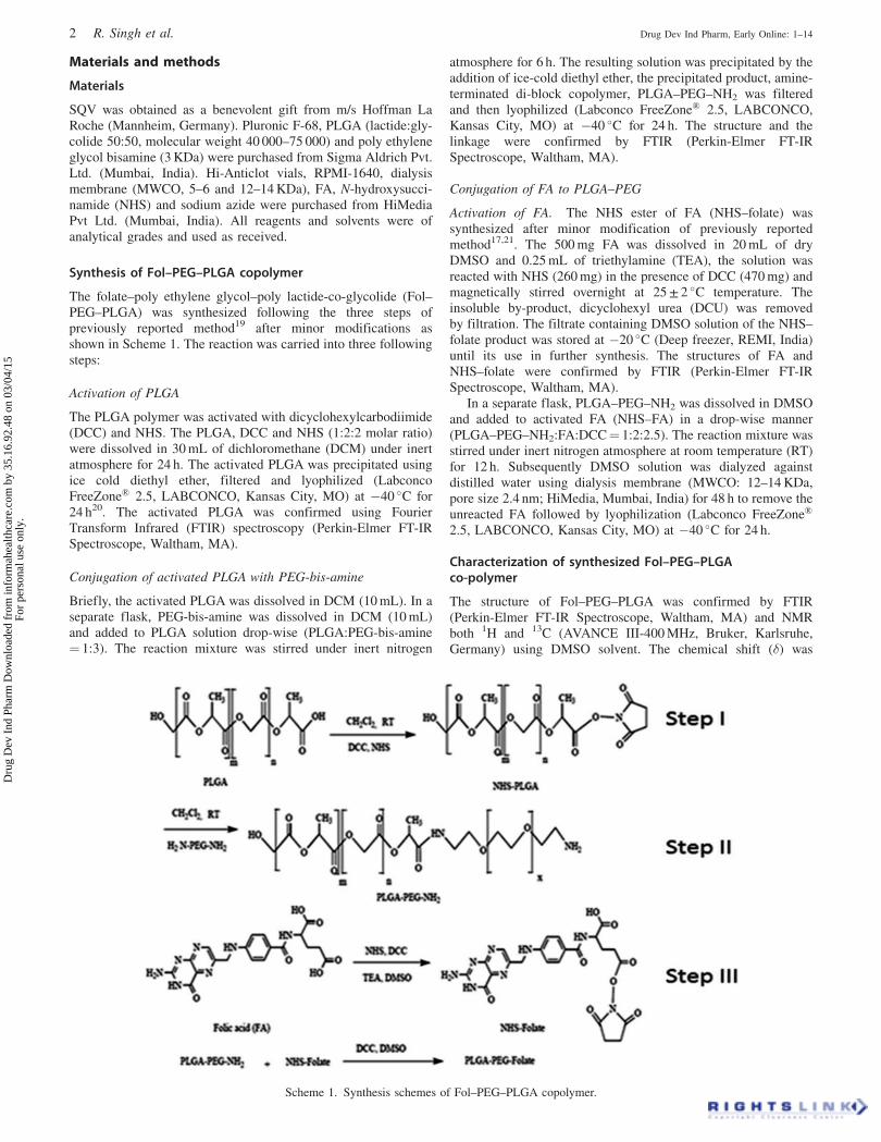
The folate–poly ethylene glycol–poly lactide-co-glycolide (Fol–PEG–PLGA) was synthesized following the three steps ofpreviously reported method19 after minor modifications asshown in Scheme 1. The reaction was carried into three followingsteps:
Activation of PLGA
The PLGA polymer was activated with dicyclohexylcarbodiimide(DCC) and NHS. The PLGA, DCC and NHS (1:2:2 molar ratio)were dissolved in 30 mL of dichloromethane (DCM) under inertatmosphere for 24 h. The activated PLGA was precipitated usingice cold diethyl ether, filtered and lyophilized (LabconcoFreeZone� 2.5, LABCONCO, Kansas City, MO) at �40 �C for24 h20. The activated PLGA was confirmed using FourierTransform Infrared (FTIR) spectroscopy (Perkin-Elmer FT-IRSpectroscope, Waltham, MA).
Conjugation of activated PLGA with PEG-bis-amine
Briefly, the activated PLGA was dissolved in DCM (10 mL). In aseparate flask, PEG-bis-amine was dissolved in DCM (10 mL)and added to PLGA solution drop-wise (PLGA:PEG-bis-amine¼ 1:3). The reaction mixture was stirred under inert nitrogen
atmosphere for 6 h. The resulting solution was precipitated by theaddition of ice-cold diethyl ether, the precipitated product, amine-terminated di-block copolymer, PLGA–PEG–NH2 was filteredand then lyophilized (Labconco FreeZone� 2.5, LABCONCO,Kansas City, MO) at �40 �C for 24 h. The structure and thelinkage were confirmed by FTIR (Perkin-Elmer FT-IRSpectroscope, Waltham, MA).
Conjugation of FA to PLGA–PEG
Activation of FA. The NHS ester of FA (NHS–folate) wassynthesized after minor modification of previously reportedmethod17,21. The 500 mg FA was dissolved in 20 mL of dryDMSO and 0.25 mL of triethylamine (TEA), the solution wasreacted with NHS (260 mg) in the presence of DCC (470 mg) andmagnetically stirred overnight at 25 ± 2 �C temperature. Theinsoluble by-product, dicyclohexyl urea (DCU) was removedby filtration. The filtrate containing DMSO solution of the NHS–folate product was stored at �20 �C (Deep freezer, REMI, India)until its use in further synthesis. The structures of FA andNHS–folate were confirmed by FTIR (Perkin-Elmer FT-IRSpectroscope, Waltham, MA).
In a separate flask, PLGA–PEG–NH2 was dissolved in DMSOand added to activated FA (NHS–FA) in a drop-wise manner(PLGA–PEG–NH2:FA:DCC¼ 1:2:2.5). The reaction mixture wasstirred under inert nitrogen atmosphere at room temperature (RT)for 12 h. Subsequently DMSO solution was dialyzed againstdistilled water using dialysis membrane (MWCO: 12–14 KDa,pore size 2.4 nm; HiMedia, Mumbai, India) for 48 h to remove theunreacted FA followed by lyophilization (Labconco FreeZone�
2.5, LABCONCO, Kansas City, MO) at �40 �C for 24 h.
Characterization of synthesized Fol–PEG–PLGAco-polymer
The structure of Fol–PEG–PLGA was confirmed by FTIR(Perkin-Elmer FT-IR Spectroscope, Waltham, MA) and NMRboth 1H and 13C (AVANCE III-400 MHz, Bruker, Karlsruhe,Germany) using DMSO solvent. The chemical shift (�) was
Scheme 1. Synthesis schemes of Fol–PEG–PLGA copolymer.
2 R. Singh et al. Drug Dev Ind Pharm, Early Online: 1–14
Dru
g D
ev I
nd P
harm
Dow
nloa
ded
from
info
rmah
ealth
care
.com
by
35.1
6.92
.48
on 0
3/04
/15
For
pers
onal
use
onl
y.

expressed in parts per million (ppm) relative to the NMR solventsignal (DMSO-d6).
Determination of folate content in synthesized Fol–PEG–PLGA co-polymer
The amount of FA conjugated with Fol–PEG–PLGA wasdetermined spectrophotometrically (UV mini 1240, Shimadzu,Kyoto, Japan) using UV/visible calibration curve of FA generatedin DMSO at lmax wavelength¼ 350 nm.
Drug loading
In present study, SQV was loaded into the NPs by reportedliterature with minor modifications22. The SQV (2.5 mg) wasdissolved in 500 mL acetone and added to 1.5 mL of DCMcontaining PLGA polymer (25 mg). The mixture was vortexedvigorously for 2–3 min. The organic phase was slowly mixed withan aqueous solution (20 mL) containing 0.1% Pluronic F-68solution. An emulsion was obtained by sonication using probesonicator (Soniweld, Mumbai, India) for 4 min at a constant poweroutput of 50 W. The sample was kept in an ice bath duringsonication to prevent any overheating of the emulsion. It was gentlystirred magnetically at RT to evaporate the organic solvent for 4 h.The NPs were recovered by centrifugation (UltracentrifugeZ36HK, HERMLE Labor Tchrhk GmbH, Wehingen, Germany)at 20 000 rpm for 30 min at 4 �C. The NPs’ mass was washed twicewith water, lyophilized (Labconco FreeZone� 2.5, LABCONCO,Kansas City, MO ) and stored in refrigerator at 4 �C.
Characterization of SQV–PLGA NPs and SQV–Fol–PEG–PLGA NPs
Electron microscopy
The shape and surface morphology of the developed NPs (SQV–PLGA NPs and SQV-Fol-PEG-PLGA NPs) were characterized byScanning Electron Microscopy (JEOL 5400, Tokyo, Japan) andTransmission Electron Microscopy (Morgagni 268 D, FEI,Netherland).
Particle size and surface charge measurement
Particle size and surface charge of the developed NPs formula-tions (SQV–PLGA NPs and SQV–Fol–PEG–PLGA NPs) weredetermined using Zetasizer (Nano ZS; Malvern Instruments Ltd,Malvern, UK), which evaluates mean diameter and size distribu-tion profiles of NPs by light scattering based on laser diffraction.A dilute suspension of NPs (20mg/mL) was prepared in deionizeddistilled water and measurements were taken in clear disposableZeta cells and recorded.
Differential scanning calorimeter (DSC)
The transition and melting temperatures of SQV, polymers PLGAand co-polymers Fol–PEG–PLGA, SQV–PLGA NPs, Sag–Fol–PEG–PLGA NPs and physical mixture of SQV, FA, PEG-bis-amine and PLGA were measured using DSC (SIIO 6300, Tokyo,Japan) under a flow of nitrogen at 50 mL/min and heating rate of10 �C/min in the temperature range 50–450 �C and thermogramswere obtained.
X-ray diffraction measurement (XRD)
The X-ray diffraction (XRD) of SQV, PLGA polymer, SQV–PLGA NPs, SQV–Fol–PEG–PLGA NPs and physical mixture ofSQV, FA, PEG-bis amine and PLGA were obtained with an X-raydiffractometer (D8 Advance, BRUKER, Karlsruhe, Germany)operated at 2� range from 10 to 40 �C using 2.2 KW sourceequipped with Cu anode, dermic X-ray tube, lynux eye detectorand Ni filter23.
Drug entrapment efficiency (DEE) of nanoparticles
The supernatant of NPs suspension was collected after centrifu-gation (Ultracentrifuge Z36HK, HERMLE Labor Tchrhk GmbH,Wehingen, Germany) at 20 000 rpm for 30 min at 4 �C. Onemilliliter of nanosuspension was taken and subsequent dilutionswere made with methanol. The concentration of drug wasdetermined at 239 nm using UV/visible spectrophotometer (UVmini 1240, Shimadzu, Kyoto, Japan) and the percent DEE wascalculated using the following equation24
Drug entrapment efficiency %
¼ Amount of drug remained in nanoparticles
Initial amount of drug used in formulation� 100:
Determination of folate content on the surface of NPs
The amount of folate present on the surface of NPs prepared bysingle emulsification solvent evaporation (SESE) method wasdetermined in a UV spectrophotometer (UV mini 1240,Shimadzu, Kyoto, Japan). The analysis was carried out inCH2Cl2/DMSO (1:4) solvent by dissolving 2 mg of NPs inCH2Cl2/DMSO in 1:4 ratio solvent and vortexed vigorously for 3–4 min. The obtained dispersion was centrifuged (UltracentrifugeZ36HK, HERMLE Labor Tchrhk GmbH, Wehingen, Germany) at20 000 rpm for 30 min and the supernatant was diluted suitablywith DMSO and determined spectrophotometrically at 350 nm24
Folate amount on surface of NPs ðmgÞ
¼ Amount of folate in supernatant ðmgÞAmount of NPs ðmgÞ :
In vitro drug release study
The in vitro release study was performed in the three differentmedia (pH 2.0, PBS; pH 4.5, PBS; pH 7.4), simulating variousregions of GIT. The dialysis bags with molecular weight cut-off(MWCO) of 12 kDa (HiMedia, Mumbai, India) were filled withNPs formulation, immersed in 25 mL each of 0.1 N HCl, PBS (pH4.5 and 7.4) separately. The pH of the media was measured andmaintained using pH meter (Microprocessor pH meter 1012,ESICO, USA). The in vitro release medium was continuouslystirred at optimum speed and maintained at 37 ± 0.5 �C in a waterbath shaker (YORCO, Scientific Industries, Delhi, India). Thesamples were withdrawn at different time points and immediatelyreplenished with fresh solution added to the receptor compartmentin order to maintain sink condition. The withdrawn samples wereanalyzed spectrophotometrically (UV mini 1240, Schimadzu,Japan) at 239 nm against respective diffusion medium as blank.
In vitro study
Hemocompatibility assessment in vitro
The whole human blood was collected using heparin asanticoagulant in Hi-Anticlot blood clotting vials (HiMedia,Mumbai, India) and centrifuged (Ultracentrifuge Z36HK,HERMLE Labor Tchrhk GmbH, Germany) at 1500 rpm for10 min to acquire human red blood cells (RBCs). After removingthe serum by suction, RBCs were washed five times with sterilephysiological saline solution by centrifugation and diluted up to10-fold volume to obtain the absorbance value of the positivecontrol supernatant located in the range 0.7 at 541 nm. Then threegroups of experiments were set up.
In every group, seven aliquots of 100mL of the diluted RBCssuspension were added in centrifuge tubes containing (i) 1.9 mLof physiological saline solution as a negative control; (ii) 1.9 mL
DOI: 10.3109/03639045.2015.1019355 Saquinavir entrapped PLGA nanoparticles 3
Dru
g D
ev I
nd P
harm
Dow
nloa
ded
from
info
rmah
ealth
care
.com
by
35.1
6.92
.48
on 0
3/04
/15
For
pers
onal
use
onl
y.

of deionized water as a positive control; (iii) 1.9 mL offormulations (SQV, SQV–PLGA, SQV–Fol–PEG–PLGA) atconcentration of 31.25, 62.5, 125, 250 and 500mg/mL.
After shaking gently and placing at a standstill for 3 h at RT,the eight aliquots of mixture in every group were centrifuged at1500 rpm for 10 min, and the absorbance of the supernatant wasrecorded from 500 to 650 nm in a UV visible spectrophotometer(UV mini 1240, Shimadazu, Kyoto, Japan).
In vitro cytotoxicity (MTT) assay
The cytotoxic activity of the developed NPs formulations wasevaluated using MTT cytotoxic assay. The 1� 104 cells/well wereseeded in 96-well micro-culture plates in 200mL DMEM,supplemented with 10% FBS and incubated for 24 h at 37 �C inCO2 incubator. The PC-3, folate receptor bearing human prostatecarcinoma cell line is one of the cell lines used in prostate cancerresearch23,25. The PC-3 and MCF-7 cells were maintained at 1640RPMI and DMEM medium, respectively and supplemented with10% Fetal Calf Serum, 100mg/mL streptomycin and 100 units/mLpenicillin in a CO2 incubator (New Brunswick, USA) at 37 �C.MTT reagent yields low background absorbance of cells. Cellswere trypsinized and seeded at 1� 104 cells/well in a 96-wellplate (Coster, Chicago, IL) and incubated at 37 �C in 5% CO2
humidified incubator for 24 h to allow exponential growth of cells.The medium was replaced on every alternative day. The cells wereexposed at various concentrations of free SQV, SQV–PLGA NPsand Sag–Fol–PEG–PLGA NPs formulations ranging from 10 to200mM for 24 and 48 h. After incubation, 20mL MTT dyesolution was added and the plates were incubated further foranother 4 h in 5% CO2 humidified chamber, then medium wasdecanted and 100mL DMSO was added to dissolve formazancrystals. The absorbance was taken at 541 nm in a spectropho-tometer (UV mini 1240, Shimadzu, Japan).
Cell uptake study of formulations
Preparation of dye-loaded NPs for cellular uptake study. TheFITC-loaded SQV–Fol–PEG–PLGA and SQV–PLGA NPs wereprepared to investigate the uptake by MCF-7 and PC-3 cancercells. To prepare fluorescently labeled PLGA NPs, FITC (Sigma-Aldrich) was dissolved in 2.5 mL ethyl alcohol (0.5 mg/mL),which was added to the solution of PLGA polymer and Fol–PEG–PLGA copolymer in DCM. For the preparation of thesefluorescently labeled particles, the o/w emulsion was left over-night to allow evaporation of the solvent. The pellet recoveredafter centrifugation was then lyophilized (Labconco FreeZone�
2.5, LABCONCO, Kansas City, MO) at �40 �C23,26.
Dye loading for cellular uptake study. The dye-loaded Fol–PEG–PLGA NPs and PLGA NPs were purified from unloadeddye by centrifugation. One milliliter of dye-loaded SQV andSQV–Fol–PEG–PLGA NPs and SQV–PLGA NPs suspensionswere taken. The PC-3 and MCF 7 were seeded in six-well plate at2.0� 105 cells/mL and 0.8� 105 cells/mL density, respectively.Cells were incubated for 24 h at 37 �C with 5% CO2, and then themedium in each well was replaced with 2 mL of phenol red-,serum- and antibiotic-free medium containing 100mg/mL offormulation concentration labeled with FITC. The cells wereincubated for 4 h, then the medium was removed and cells weretrypsinized using trypsin (0.05% in PBS containing 0.02% EDTA)solution leaving it for 1 min. Subsequently cells were washedthree times with PBS (pH 7.4) to remove endogenous folatesbound to folate receptors (FRs) on the cell surface and re-suspended in 500mL of PBS (pH 7.4). Fluorescence of the dyewas measured by acquisition of cells on fluorescence-activatedcell sorting analysis (FACS Becton Dickinson Biosciences,Franklin Lakes, NJ). The data of time kinetics of fluorescence-
related cell uptake were analyzed using BD FACS Diva software(Becton Dickinson Biosciences, Franklin Lakes, NJ). PercentageP1 denotes the unstained cell population and percentage P2represents the shift in fluorescence denoting the FITC labeledSQV, SQV–PLGA NPs and SQV–Fol–PEG–PLGA NPs uptakeby the cells23.
Results and discussion
Currently, multifunctional NPs play pivotal role in the deliveryof therapeutics for the treatment of various dreadful diseases.The Fol–PEG–PLGA (folate–poly ethylene glycol–poly lactide-co-glycolide) was synthesized in three steps as discussed inmaterials and method section above. The FTIR spectroscopy wasused to investigate the chemical bonds of synthesized copolymers(Fol–PEG–PLGA) at each step.
In the first step, the PLGA polymer was activated with DCCand NHS. The FTIR spectra of PLGA and activated NHS-PLGAare shown in Figure 1(A) and (B). The characteristics peaks at2948 and 2998 cm�1 were due to C–H, C–H3 and C–H2 functionalgroup stretching vibration of PLGA while the peaks at 1397, 1426and 1454 cm�1 were ascribed to bending vibration in relation tothe spectrum connected with C¼O groups (Figure 1A). The FTIRspectrum of activated PLGA shown in Figure 1(B) depicts clearlythe characteristic peak at 1627 cm�1 –COO stretching of PLGA.
In second step, PLGA–PEG diblock copolymer was synthe-sized by coupling reaction between primary amine group of PEGdiamine and terminal carboxylic end group of PLGA via amidelinkage. The carboxylic group end of PLGA was first activatedwith NHS in the presence of DCC, which was then conjugated toprimary amine group of PEG diamine. A linear type PEG diaminehas NH2 groups at both the ends of the molecule. An excessamount of PEG diamine was used to prevent the formation ofPLGA–PEG–PLGA triblock copolymers22. The chemical struc-ture of the synthesized diblock copolymer was confirmed by FTIRas displayed in Figure 1(C). The obtained characteristic peaks werefound more specifically and the bands at 1535 and 1627 cm�1 wereassigned to C–O–C ether stretching vibration of NH2–PEG–NH2
and –COO stretching vibration of PLGA, respectively.In third step, NHS ester of FA (NHS-folate) was synthesized
and then conjugated with PEG–PLGA diblock copolymer.The chemical structure of FA and NHS-folate were confirmedby FTIR (Figures 1D and 2A).
The most important characteristic peaks obtained in theFTIR spectrum of FA (Figure 1D) were 3544, 3417 cm�1 (N–Hstretch of primary amine and amide); 3323 cm�1 (alkyl C–H andC¼C stretch); 1693 cm�1 (aromatic bending C¼C); 1484 cm�1
(CH–NH–C¼O amides cm�1); 839 cm�1 (aromatic C–H bendingand benzene 1,4-distribution). These peaks were found similarto our previous reports7,18,23,27.
It should be noted that FA has both a and g carboxyl functionalgroup and both of them could be activated using DCC/NHSchemistry but it is also known that the g-carboxylic acid isprimarily conjugated due to its higher reactivity28. In FTIRspectrum of the NHS conjugated FA (Figure 2B) found thecharacteristic bands at 3337, 2928 and 1626 cm�1 were attributedto the stretching mode of primary aliphatic amine N–H stretching,carboxylic acid C¼O and O–H stretching unconjugated andaromatic C¼C bending, respectively. The obtained characteristicpeaks were found similar as reported by Gupta et al. and Thakuret al.7,18 all of which confirmed the synthesis of NHS ester of FA.
The principle of the chemical reactions in synthesis of Fol–PEG–PLGA is the formation of amide bonds. The structure ofconjugate was checked and confirmed by FTIR (Figure 2A and B)and 1HNMR spectroscopy shown in Figure 3 and 13C NMR asshown in Figure 4. FTIR spectra shown in Figure 3 were carried
4 R. Singh et al. Drug Dev Ind Pharm, Early Online: 1–14
Dru
g D
ev I
nd P
harm
Dow
nloa
ded
from
info
rmah
ealth
care
.com
by
35.1
6.92
.48
on 0
3/04
/15
For
pers
onal
use
onl
y.

out to confirm the presence of amide linkage in polymer bands offolate control or polymer is observed at 1459 and 1606 cm�1.These obtained characteristic peaks are found similar to otherreports24,29 may be due to the stretching vibrations of C¼C in thebackbone of the aromatic ring present in FA. The band around1573 cm�1 was attributed to characteristic absorption band of thephenyl ring in FA, which is similar as reported by Liang et al30.
More specifically, the bands at 1535 and 1628 cm�1 wereassigned to C–O–C ether stretching vibration of NH2–PEG–NH2
and –COO stretching vibration of PLGA, respectively. Obtainedbands were found similar as reported by Liang et al30. The mostsignificant FTIR absorption peak in Fol–PEG–PLGA polymer isdue to the presence of –CO–NH– linkage. The carbonyl (C¼O)and amine (N�H) groups present in the amide linkage exhibitedbands at 1628 and 1574 cm�1. Obtained bands were found similarto that reported by Akasaka et al. and Yang et al31,32.
The synthesized copolymer Fol–PEG–PLGA was fur-ther characterized by 1H NMR spectroscopy and 13C NMR.
The 1H NMR analysis (Figure 3) showed principal peaks (in ppm)related to benzene of folate moiety [d¼ 6.64, 6.90, 7.65, 8.7 ppm].It reveals the coupling of folate to PLGA–PEG via NHS/DCCmediated reaction. The PEG moiety [d¼ 3.43 ppm, 3.53] and thePLGA moiety [d¼ 4.9 ppm] are similar to the earlierreports19,30,33. The peaks at 1.6 and 5.2 ppm were due to –CH3
and –CH– protons of PLA block. The peak at 4.9 ppm belongs tothe –CH2– protons of PGA block. More confirmation for thepolymerization of the block copolymer (Fol–PEG–PLGA) wasobtained by 13C NMR analysis.
The characteristic peaks in 13C NMR for PLGA moiety(58.68 ppm for –CH2, 69.71 ppm for –CH) were 89.91 and111.21 ppm (Figure 4). One broad peak at 69.71 ppm and anothernarrow peak at 56.83 ppm were assigned reported to the CH3 andCH2 moiety of PEG macromere. A peak was observed at148.54 ppm for CH¼N, which confirmed the FA ring insynthesized Fol–PEG–PLGA copolymer. Obtained peaks werematched with earlier reports7,34,
Figure 1. FTIR spectra of (A) poly lactide-co-glycolide (PLGA), (B) NHS-poly lactide-co-glycolide (PLGA), (C) PEG conjugated PLGA and (D) folicacid (FA).
Figure 2. FTIR spectra of (A) NHS ester of FA (NHS–FA) and (B) folic acid conjugated PEG–PLGA copolymer.
DOI: 10.3109/03639045.2015.1019355 Saquinavir entrapped PLGA nanoparticles 5
Dru
g D
ev I
nd P
harm
Dow
nloa
ded
from
info
rmah
ealth
care
.com
by
35.1
6.92
.48
on 0
3/04
/15
For
pers
onal
use
onl
y.

The amount of FA conjugated to Fol–PEG–PLGA wasdetermined using a UV–visible calibration curve of FA generatedin DMSO at 350 nm, using equation of line Y¼ 0.226X� 0.182concentration and amount of FA in suspension were found to be12.22mg/mL, 12.92 mg, respectively. On molar ratio basis theconjugation percentage of folate to PLGA–PEG was found to be53.00 ± 0.53%.
The physicochemical characteristics, such as size and morph-ology of NPs were confirmed by SEM and TEM. The sphericalshape and smooth surface was observed in SEM photomicro-graphs of NPs (Figure 5A and B). From TEM photomicrographs(Figure 6A and B) spherical shape was observed for bothSQV–PLGA and SQV–Fol–PEG–PLGA NPs.
The particle size of the developed NPs mainly depends uponsonication and sonication time. A longer sonication time isassociated with a release of high energy leading to rapiddispersion of the polymer organic phase as nano-droplets ofsmaller size and monomodal distribution profile as reportedearlier35.
The optimum particle size and polydispersity index (PDI)of the SQV–PLGA NPs and SQV–Fol–PEG–PLGA NPs werefound to be 118.8 ± 2.52, 223.8 ± 5.42 nm and 0.15 ± 0.002,0.18 ± 0.005, respectively. The optimum particle size and PDIclearly revealed that the developed SQV-loaded targeted and non-targeted NPs had narrow particle size distribution and monodispersion modal pattern. The increase in the particle size of NPs
probably was due to the conjugation of FA–PEG–PLGA polymerwith NPs.
The surface charge measurement with the help of zetapotential is another important criterion for the stability of thenanoparticulate formulation. Since most cellular membranes arenegatively charged, zeta potential can affect NPs tendency topermit membranes, with cationic particles generally displayingmore toxicity associated with cell wall disruption. Both SQV–PLGA and SQV–Fol–PEG–PLGA NPs exhibited negative zetapotential due to the presence of terminal carboxylic functionalgroups in the polymer. Higher negative value of zeta potential�27.961 mV with zeta deviation 3.43 mV was obtained for SQV–PLGA NPs due to the presence of uncapped end carboxyl groups.SQV–Fol–PEG–PLGA NPs had smaller negative zeta potentialvalue of �8.967 mV with zeta deviation 5.09 mV. The value ofzeta potential is affected by the presence of positively chargedamine group of FA. Similar charges in zeta potential wereobserved in case of both the drug-loaded NPs formulation. Theseresults suggest the absence of SQV drug on the surface of NPs.
The DSC studies confirm the chemical interaction betweenPLGA and SQV, PLGA with folate moiety and SQV. Theemphasis was put on the comparison of the thermal behavior ofSQV in SQV-loaded NPs to allow conclusions about structuraldifferences between the SQV and SQV-loaded NPs.
The DSC curves of SQV and lyophilized SQV-loadedNPs (SQV–PLGA and SQV–Fol–PEG–PLGA) are shown in
Figure 4. 13C NMR spectrum of synthesizedfolate conjugated polyethylene glycol–PLGAcopolymer.
Figure 3. 1H NMR spectrum of synthesizedfolate conjugated polyethylene glycol–PLGAcopolymer.
6 R. Singh et al. Drug Dev Ind Pharm, Early Online: 1–14
Dru
g D
ev I
nd P
harm
Dow
nloa
ded
from
info
rmah
ealth
care
.com
by
35.1
6.92
.48
on 0
3/04
/15
For
pers
onal
use
onl
y.

Figure 7(A–D). The DSC curves of SQV with a sharp peakshowed melting endotherms at 252.9 �C (data not shown), whichwas similar to those reported by Dodiya et al36. In Figure 7(A),which shows the thermogoram of pure PLGA, no distinct meltingpoint was observed because PLGA is amorphous in nature. Thecharacteristic peak at 369.4 �C was related to the thermaldecomposition of the polymer. The DSC curves of pure PLGAshow that the polymer is thermally stable up to 200 �C and theresult was similar with Mukerjee & Vishwanatha37.
The similar melting transition properties of loaded andunloaded NPs show that the PLGA polymer and Fol–PEG–PLGA copolymer remained unaffected during encapsulation. TheDSC thermograms of pure PLGA polymer, SQV-loaded PLGANPs, Fol–PEG–PLGA synthesized copolymer and SQV-loadedFol–PEG–PLGA NPs (SQV–Fol–PEG–PLGA) are shown inFigures 7(A–D), respectively. It can be observed that the NPspreparation method did not affect the polymer structure. The DSCstudy did not detect any crystalline drug material in the SQV-loaded NPs formulations (Figure 7D), as the sharp peak of SQVwas absent. Absence of endothermic peak of SQV in the
thermogram of SQV-loaded PLGA NPs (SQV–PLGA & SQV–Fol–PEG–PLGA) indicates that the drug might be entrapped inthe PLGA matrix. Thus, the drug incorporated into NPs was in anamorphous or disordered-crystalline phase of molecular disper-sion or solid solution state within the polymer matrix. Similarmelting transition properties of loaded and unloaded NPs showthat the PLGA polymer remained unaffected duringencapsulation.
X-ray diffraction (XRD) was used to define crystalline/amorphous phases of NPs. XRD analysis of SQV, PLGA, SQV–PLGA NPs, SQV–Fol–PEG–PLGA NPs and physical mixture(SQV, FA, PEG-bis-amine and PLGA) were performed by X-raydiffractometry. Figure 8 presents the XRD data recorded in the 2�range (10�–80�). XRD of SQV exhibited high intensity peaksat 2� angle values of 14.558�, 15.363�, 17.481� and 21.925�
(Figure 8A–C), which are similar to those reported by Dodiyaet al36. Sharp intense peaks might be due to the presence ofcrystalline form of the drug.
In XRD spectrum of PLGA, no peak but a broad amorphousband between 10� and 80� was observed, which indicates that
Figure 6. Transmission electron photomicrographs of (A) SQV–PLGA NPs and (B) SQV–Fol–PLGA NPS.
Figure 5. Scanning electron photomicrographs of (A) SQV–PLGA NPs at magnification 5000� and (B) SQV–Fol–PLGA NPS at magnification3500�.
DOI: 10.3109/03639045.2015.1019355 Saquinavir entrapped PLGA nanoparticles 7
Dru
g D
ev I
nd P
harm
Dow
nloa
ded
from
info
rmah
ealth
care
.com
by
35.1
6.92
.48
on 0
3/04
/15
For
pers
onal
use
onl
y.

PLGA is an amorphous copolymer. The diffraction pattern ofSQV-loaded PLGA NPs (SQV–PLGA NPs) in Figure 8(B)showed only a single characteristic peak at 19.279�.
In the XRD of SQV-loaded Fol–PEG–PLGA NPs (SQV–Fol–PEG–PLGA) shown in Figure 8(C), principal peaks of SQV wereobserved at 15.363�, 17.263� and 21.909�. Two more peaks at19.097� and 23.167� were also observed for the organic moiety ofPEG (poly ethylene glycol), which indicated crystalline propertiesof PEG peaks similar to that reported by Bolourchian et al. andPatel & Patel38,39. Diffraction peaks of FA were reported to be at2� between 10� and 30� with a centered peak at �22� by Floreaet al.40 and Haidary et al41. In the present study, peaks were alsofound at 21.392�.
XRD patterns of the physical mixture of SQV, FA, PEG bisamine and PLGA are shown in Figure 8(D). The characteristicpeaks at 14.589�, 17.581� for SQV, 19.934� and 23.335� forPEG moiety and 21.963� for FA were found. It may bepresumed that the free drug, SQV exhibited sharp specificcrystal peaks but the same drug due to molecular dispersioninside the NPs (SQV–PLGA & SQV–Fol–PEG–PLGA) exhib-ited lack of numerous and sharp peaks. The results of XRDspectra of the above formulations indicated decrease in crystal-linity due to nanosizing.
The entrapment efficiency of SQV using single emulsionsolvent evaporation method was found to be 56.00 ± 0.68% and58.00 ± 0.80% for SQV–PLGA and SQV–Fol–PEG–PLGA NPs,respectively. The SQV–Fol–PEG–PLGA NPs formulation had alarger size with higher drug content. The higher size is due to theoutside orientation of the folate moiety and the higher drugcontent is mainly due to the larger particle size.
Folate present on the surface of NPs was quantitativelyestimated by UV spectroscopy. The ghost NPs are used forquantitative analysis of folate. The amount of folate on the surfaceof NPs prepared by SESE method was found to be 8.16 mg offolate/mg of NPs.
The in vitro release studies of SQV were examined in the threedifferent media 0.1 N HCl (pH 2) and phosphate buffers (4.5 and7.4). The pH of medium influenced for release of SQV from bothNPs SQV–PLGA and SQV–Fol–PEG–PLGA NPs in threedifferent media. The drug release rate was found to be decreasingupon increasing the pH of media. The release rate was found to behigher in acidic media, which may be attributed to the acidfacilitated degradation of polymer. The burst release was observedin initial first hour. Approximately 30% of SQV was releasedin all three media within 1 h as seen from Figure 9. The SQV–Fol–PEG–PLGA NPs showed the higher drug release rate than
Figure 7. Differential scanning thermograms (DSC) of (A) poly lactide-co-glycolide (PLGA), (B) SQV-loaded poly lactide-co-glycolide (SQV–PLGA)NPs, (C) folate conjugated poly lactide coglycolide copolymer (Fol–PEG–PLGA) and (D) SQV-loaded folate conjugated poly lactide-co-glycolidepolymer (SQV–Fol–PEG–PLGA) NPs.
8 R. Singh et al. Drug Dev Ind Pharm, Early Online: 1–14
Dru
g D
ev I
nd P
harm
Dow
nloa
ded
from
info
rmah
ealth
care
.com
by
35.1
6.92
.48
on 0
3/04
/15
For
pers
onal
use
onl
y.

SQV–PLGA NPs in all three media. Gradually the release rateslowed down after 1 h and prolonged release can be attributed toslow diffusion of drug from the polymer at later stage (p50.005).
The PLGA part of the copolymer is mainly located inrelatively hydrophobic core of the NPs and lactic–lactic bondsare relatively less available to the water22. However, the overalleffect of release rate may be ascribed to the copolymercompositions, drug loading and particle size of NPs. Since thedrug is insoluble in selected medium, 0.5% (w/v) of Tween 80(100mL) was added to enhance the solubility and 0.025% (w/v)
sodium azide to avoid microbial growth. Tween 80 also preventsadsorption of the drug to the container surface.
The hemolytic study describes the in vitro assay specificallydeveloped for the analysis of NPs in term of erythrocytes–formulations interactions. In hemolytic toxicity study, synthesizedNPs formulations were incubated with blood as shown inFigure 10. It was observed that the hemoglobin was released bydamaged cells. The NPs and undamaged red blood corpuscles(RBCs) were removed by centrifugation, and the absorbanceof supernatant was measured spectrophotometerically42.
Figure 8. X-ray diffraction (XRD) analysis of (A) poly lactide-co-glycolide (PLGA), (B) SQV-loaded poly lactide-co-glycolide (SQV–PLGA) NPs,(C) SQV-loaded folate conjugated poly lactide-co-glycolide polymer (SQV–Fol–PEG–PLGA) NPs and (D) physical mixture (SQV, FA, PEG-bisamine, PLGA).
Figure 9. Cumulative amount of SQVreleased from SQV–PLGA NPs and SQV–Fol–PEG–PLGA NPs in different media (pH2, 7.4 and 4.5) at 37 �C (n¼ 3).
DOI: 10.3109/03639045.2015.1019355 Saquinavir entrapped PLGA nanoparticles 9
Dru
g D
ev I
nd P
harm
Dow
nloa
ded
from
info
rmah
ealth
care
.com
by
35.1
6.92
.48
on 0
3/04
/15
For
pers
onal
use
onl
y.

The percentage hemolysis was detected for free SQV, SQV–PLGA and SQV–Fol–PEG–PLGA NPs.
The investigation showed that there was no hemolysis effectfor the blank whereas the hemolysis percentage of SQV, SQV–PLGA NPs and SQV–Fol–PEG–PLGA NPs were found withinthe experimental concentration in the range 31.25–500mg/mL. At31.25mg/mL lower concentration the SQV, SQV–PLGA NPs andSQV–Fol–PEG–PLGA NPs showed 10.67%, 3.315% and 1.20%hemolytic toxicity, respectively and it increased with increasingconcentration of the formulation. Surprisingly the FA conjugationon the SQV-loaded PLGA NPs surfaces drastically reducedthe RBC hemolysis. At a high concentration of 500 mg/mL,the free SQV exhibited 49%, while SQV–PLGA NPs and SQV–Fol–PEG–PLGA NPs displayed 21.60% and 17.3% RBChemolysis, respectively.
The present copolymers were nonporous sphere shaped NPswith particle size distribution of 118.8 ± 2.52 (SQV–PLGA) and223.8 ± 5.42 nm (SQV–Fol–PEG–PLGA) and are less likely toachieve direct transmembrane transport accompanied by RBCsmembrane damage. Moreover, sphere shaped NPs have difficultyrupturing the membrane of RBCs to cause the subsequent releaseof hemoglobin43.
To determine the lethality and cancer targeting propensity ofSQV and developed nanoformulations, the PC-3 (human prostatecancer) and MCF-7 (human breast cancer) cell lines were selectedas folate receptor positive (high level of folate receptor expression)and folate receptor negative cell lines (low level of folate receptorexpression) for in vitro experiments. The selection of human cancercell lines was also based on their availability in the laboratory.Literature suggests that the MCF-7 as a folate receptor negative celllines44–46, while PC-3 as a folate receptor positive cell lines47.
The MTT cytotoxicity studies showed the absorbancevalues of treated cells were lower than control cells. Thisindicates a reduction in the rate of cell proliferation. Thein vitro time dependent cytotoxicity activity of the free SQV,SQV-loaded PLGA NPs (SQV–PLGA) and SQV-loaded folateconjugated PLGA (SQV–Fol–PEG–PLGA) NPs on PC-3and MCF-7 cell lines were studied according to percent-age reduction in cell viability (% cell growth inhibition)(Figure 11A–D) (p50.005).
In both cell lines, SQV–Fol–PEG–PLGA NPs showed higherpercentage cell growth inhibition than non-targeted drug-loadedPLGA (SQV–PLGA) NPs due to folate receptor-mediated endo-cytosis (RME) on the tumor cell membrane. The time dependentcytotoxicity on cells was observed post-24 and 48 h incubationwith the formulations. After 24 h, no obvious cytotoxicity wasdetected. At 260mM SQV, SQV–PLGA NPs and SQV–Fol–PEG–PLGA NPs showed 20%, 20% and 23% cell growth inhibition inPC-3 cells, respectively whereas in MCF-7 cells it was 12%, 15%and 14% cell growth inhibition, respectively (Figure 11A and C)(p50.005).
In contrast, cell viability drastically decreased at 48 h incuba-tion of the developed NPs formulations. The obtained resultsshowed that cell growth inhibition increased upon increasingconcentration of NPs formulations in both cell lines. In PC-3cells, 53%, 76% and 78% cell growth inhibition was observedin SQV, SQV–PLGA, SQV–Fol–PEG–PLGA, respectively at130mM concentration. Doniya and co-workers reported that SQVinhibits the cancer cell growth in a concentration-dependentmanner. In another study48 reported that SQV caused a concen-tration-dependent induction of apoptosis in LnCP, DU-145 andPC-3 prostate cancer cells.
Figure 10. Haemocompatibility assessmentin vitro (A) HRBCs suspension was added tocentrifuge tubes containing positive control,negative control and formulations. (B) Afterplacing above centrifuge tubes at a stand tillfor 3 h at RT.
10 R. Singh et al. Drug Dev Ind Pharm, Early Online: 1–14
Dru
g D
ev I
nd P
harm
Dow
nloa
ded
from
info
rmah
ealth
care
.com
by
35.1
6.92
.48
on 0
3/04
/15
For
pers
onal
use
onl
y.

The sensitivity of human breast cancer cell line to SQV isunknown. To examine the effect of the SQV and SQV-loadednanoformulations on breast cancer cells; MCF-7 breast carcinomacell line was used as it has been investigated several times in anumber of studies44–46. In MCF-7 cells after 48 h treatment withformulations the cell growth inhibition was found to be 36%, 41%and 47% at 130mM concentration of SQV, SQV–PLGA andSQV–Fol–PEG–PLGA, respectively. The results demonstrate thatboth NP formulations possessed higher growth inhibition proper-ties for cancer cells than free SQV drug. Neither free drug nordrug-loaded NPs showed considerable inhibition effects on cellgrowth when drug concentration was 13 mM.
From the MTT cytotoxicity study we concluded that the SQVhas antineoplastic effect and induces cell death in prostate andbreast cancer cells in a dose and time dependent manner.Moreover, SQV was shown to be an inhibitor of the breastcancer resistance protein (BCRP1, gene product of ABCG2) butnot a substrate49,50. Maksimovic and co-workers synthesized andinvestigated the anticancer activity of nitric oxide-modifiedderivative, SQV–NO51.
Sgadari et al.52 have shown that PIs including SQV have directanti-angiogenic and antitumor effect; the inhibitory effect of PIs issimilar to that observed with paclitaxel, a microtubule-bindingdrug with cytotoxic and angiogenic activity used in the treatmentof solid tumor. Upon comparison of the cytotoxic potential, SQV–
Fol–PEG–PLGA NPs are found more cytotoxic than SQV–PLGANPs and drug solution probably due to RME mechanism in thecells. The SQV–Fol–PEG–PLGA NPs are internalized via folateRME on MCF-7 and PC-3 resulted in greater cytotoxicity,whereas SQV–PLGA NPs are internalized via simple endocytosismechanism.
Furthermore, the treatment of cells with PIs leads toaggresome formation and accumulation of polyubiquitinatedproteins, implying proteasome inhibition. Thus, their resultssupport a model whereby PIs, such as SQV used in this studycause tumor cell death via triggering of the ESR, inhibition ofproteasome activity and subsequent accumulation of misfoldedproteins. Inhibition of glioma growth via ESR takes place inin vivo setting53.
The FITC labeled SQV–PLGA and SQV–Fol–PEG–PLGANPs formulations were further used for cell uptake study in PC-3and MCF-7 cell lines using FACS. SQV, PI inhibits the tumorgrowth and induces regression of tumors. The obtained percent-age fluorescence intensity of the NPs formulations is shown inFigures 12 and 13 and compared with control. The SQV–Fol–PEG–PLGA NPs exhibited higher fluorescence intensity relativeto free SQV and SQV–PLGA NPs and internalized via folateRME. The fluorescence intensity was found to be higher in PC-3cell compared to that in MCF-7 cells may be due to higher folatereceptor over-expression in PC-3 cell.
Figure 11. In Vitro cell cytotoxicity: MTT assay result of PC-3 (prostate cancer cell line) and MCF-7 (human breast cancer cell line) post-24 and 48 htreatment of SQV, SQV–PLGA NPs and SQV–Fol–PEG–PLGA NPs (n¼ 3).
DOI: 10.3109/03639045.2015.1019355 Saquinavir entrapped PLGA nanoparticles 11
Dru
g D
ev I
nd P
harm
Dow
nloa
ded
from
info
rmah
ealth
care
.com
by
35.1
6.92
.48
on 0
3/04
/15
For
pers
onal
use
onl
y.

Holm and co-workers reported the high affinity of FAtowards folate-binding protein employing human prostate cancercells54. However, it is not clear what kind of receptors enhancethe uptake of folate linked NPs. The type of folate-bindingprotein expressed in human prostate has not been reported. Thepossibility is that the uptake of folate conjugated NPs in PC-3cells is through folate transporter mediated47. Our study suggeststhat target effect detected in PC-3 cells might be due to thepresence of carrier mediated folate transporter, that enhances theuptake of folate conjugated NPs. In contrast to higher uptakeby PC-3 cells, tumor cells with lower FR density on thesurface (MCF 7) showed relatively lesser uptake of the folate-targeted drug and lower efficacy. Fluorescence percentage of P1
and P2 after 24 h treatment with each formulations andpercentage P1 denotes the unstained cell population.Percentage P2 represents the shift in fluorescence denoting theFITC labeled NPs formulation uptake by PC-3 and MCF-7 cells(Figures 12 and 13).
Chen et al. and Mansoori et al. indicated that the MCF-7 cellsshowed lower folate receptors density and less uptake of the folatemediated targeted drug delivery45,46. Similarly, Boddu andco-workers also reported that the folate conjugated NPs exhibitapproximately four times higher cellular uptake compared to freeanticancer drug19. The HIV PI SQV shows anticancer activity.
Although its nitric oxide-modified derivative SQV–NO(SQV–NO) was less toxic to normal cells, it exerted strongerinhibition of B16 melanoma growth in syngeneic C57BL/6 micethan SQV did55.
Conclusion
The present investigation reveals the anticancer activity of theSQV-loaded targeted and non-targeted NPs formulation employ-ing two different cell lines (MCF-7 and PC-3). HIV-1 PIspossibly become another class of cytotoxic drugs, alone or incombination with radiation or chemotherapy. SQV is known toinduce cell death in chemo-sensitive and chemo-resistant cancercells in a time- and dose-dependent manner. Moreover, SQV is afirst FDA approved agent of HIV and earlier data suggests that itmay also have clinical application in the treatment of cancer.Thus it may be concluded that the targeted SQV–Fol–PEG–PLGA NPs is superior compared to non-targeted SQV–PLGANPs. The SQV–Fol–PEG–PLGA NPs is used in selectivetargeted delivery of SQV to the FRs-over expressed cancercells, has unlock a new vista for site-specific targeting ofanticancer chemotherapeutics. Thus SQV and other PIs appear tobe promising anti-angiogenic and anti-tumor compounds inaddition to being AIDS treatment agent.
Figure 12. In vitro cell uptake study: fluorescence percentage of P1 & P2 after 24 h treatment with each formulation. Percentage P1 denotes theunstained cell population. Percentage P2 represents the shift in fluorescence denoting the FITC labeled formulation uptake by the PC3 cells.(A) Negative control, (B) labeled SQV treated cells, (C) labeled SQV–PLGA NPs treated cells and (D) labeled SQV–Fol–PEG–PLGA NPs.
12 R. Singh et al. Drug Dev Ind Pharm, Early Online: 1–14
Dru
g D
ev I
nd P
harm
Dow
nloa
ded
from
info
rmah
ealth
care
.com
by
35.1
6.92
.48
on 0
3/04
/15
For
pers
onal
use
onl
y.

Declaration of interest
The authors report no conflicts of interest. The authors alone areresponsible for the content and writing of this article.
The authors (Ruchi Singh) would like to acknowledge Department ofScience and Technology (DST), New Delhi, India for providing theFellowship to carry out this research work. Additionally, authors wouldalso like to acknowledge F. Hoffman-La Roche, Mannheim (Germany) forproviding the gift sample of Saquinavir.
References
1. Navia MA, Fitzgerald PM, McKeever BM, et al. Three-dimensionalstructure of the aspartyl protease from human immunodeficiencyvirus HIV-1. Nature 1989;337:615–20.
2. Ikezoe T, Daar ES, Hisatake J, et al. HIV-1 protease inhibitorsdecrease proliferation and induce differentiation of human myelo-cytic leukemia cells. Blood 2000;96:3553–9.
3. Pati S, Pelser CB, Dufraine J, et al. Antitumorigenic effects of HIVprotease inhibitor ritonavir: inhibition of Kaposi sarcoma. Blood2002;99:3771–9.
4. Sgadari C, Barillari G, Toschi E, et al. HIV protease inhibitors arepotent anti-angiogenic molecules and promote regression of Kaposisarcoma. Nat Med 2002;8:225–32.
5. Brannon-Peppas L, Blanchette JO. Nanoparticle and targetedsystems for cancer therapy. Adv Drug Deliv Rev 2004;56:1649–59.
6. Jain S, Kesharwani P, Tekade RK, Jain NK. One platformcomparison of solubilization potential of dendrimer with somesolubilizing agents. Drug Dev Ind Pharm 2014. [Epub ahead ofprint]. DOI: 10.3109/03639045.2014.900077.
7. Gupta U, Dwivedi SK, Bid HK, et al. Ligand anchored dendrimersbased nanoconstructs for effective targeting to cancer cells. Int JPharm 2010;393:185–96.
8. Kesharwani P, Tekade RK, Gajbhiye V, et al. Cancer targetingpotential of some ligand-anchored poly(propylene imine)dendrimers: a comparison. Nanomed Nanotech Biol Med 2011;7:295–304.
9. Kesharwani P, Tekade RK, Jain NK. Dendrimer generationalnomenclature: the need to harmonize. 2015. [Epub ahead of print].doi:10.1016/j.drudis.2014.12.015.
10. Niu X, Zou W, Liu C, et al. Modified nanoprecipitation method tofabricate DNA-loaded PLGA nanoparticles. Drug Dev Ind Pharm2009;35:1375–83.
11. Barichello JM, Morishita M, Takayama K, Nagai T. Encapsulationof hydrophilic and lipophilic drugs in PLGA nanoparticles by thenanoprecipitation method. Drug Dev Ind Pharm 1999;25:471–6.
12. Kesarwani P, Jain K, Tekade RK, et al. Spectrophotometricestimation of Paclitaxel. Int J Adv Pharm Sci 2011;2:29–32.
13. Mishra V, Kesharwani P, Jain NK. Functionalized polymericnanoparticles for delivery of bioactives. Nanobiomed 2014;3:91–123.
14. Dubey M, Kesharwani P, Tiwari A, et al. Formulation andevaluation of floating microsphere containing anti diabetic drug.Int J Pharm Chem Sci 2012;1:475–9.
15. Kesharwani P, Tekade RK, Jain K, Jain NK. Generation dependentcancer targeting potential of poly(propyleneimine) dendrimer.Biomaterials 2014;35:5539–48.
16. Birdhariya B, Kesharwani P, Jain NK. Effect of surface capping ontargeting potential of folate decorated poly (propylene imine)dendrimers. Drug Dev Ind Pharm 2014. [Epub ahead of print].DOI: 10.3109/03639045.2014.954584.
Figure 13. In vitro cell uptake study: fluorescence percentage of P1 and P2 after 24 h treatment with each formulation. Percentage P1 denotes theunstained cell population. Percentage P2 represents the shift in fluorescence denoting the FITC labeled formulation uptake by the MCF 7 cells. (A)Negative control, (B) labeled SQV treated cells, (C) labeled SQV–PLGA NPs treated cells and (D) labeled SQV–Fol–PEG–PLGA NPs.
DOI: 10.3109/03639045.2015.1019355 Saquinavir entrapped PLGA nanoparticles 13
Dru
g D
ev I
nd P
harm
Dow
nloa
ded
from
info
rmah
ealth
care
.com
by
35.1
6.92
.48
on 0
3/04
/15
For
pers
onal
use
onl
y.
17. Mehra NK, Jain NK. Development, characterization and cancertargeting potential of surface engineered carbon nanotubes. J DrugTarget 2013;21:745–58.
18. Thakur S, Kesharwani P, Tekade RK, Jain NK. Impact of pegylationon biopharmaceutical properties of dendrimers. Polymer 2015;59:67–92.
19. Boddu SH, Vaishya R, Jwala J, et al. Preparation and characteriza-tion of folate conjugated nanoparticles of doxorubicin using PLGA–PEG–FOL polymer. Med Chem 2012;2:68–75.
20. Mody N, Tekade RK, Mehra NK, et al. Dendrimer, liposomes,carbon nanotubes and PLGA nanoparticles: one platform assessmentof drug delivery potential. AAPS PharmSciTech 2014;15:388–99.
21. Kesharwani P, Iyer AK. Recent advances in dendrimer-basednanovectors for tumor-targeted drug and gene delivery. DrugDiscov Today 2014. [Epub ahead of print]. doi:10.1016/j.drudis.2014.12.012.
22. Muruganeshan SK, Subramaniyan G, Averineni RK, et al.PEGylated PLGA nanoparticulate delivery of docetaxel: synthesisof diblock copolymers, optimization of preparation variable formu-lation characteristics and in vitro release studies. J BiomedNanotechnol 2007;3:52–60.
23. Mehra NK, Verma AK, Mishra PR, Jain NK. The cancer targetingpotential of d-a-tocopheryl polyethylene glycol 1000 succinatetethered multi walled carbon nanotubes. Biomaterials 2014;35:4573–88.
24. Boddu SH, Jwala J, Chowdhury MR, Mitra AK. In vitro evaluationof a targeted and sustained release system for retinoblastoma cellsusing Doxorubicin as a model drug. J Ocul Pharmacol Ther 2010;26:459–68.
25. Kesharwani P, Tekade RK, Jain NK. Generation dependent safetyand efficacy of folic acid conjugated dendrimer based anticancerdrug formulations. Pharm Res 2014. [Epub ahead of print]. DOI:10.1007/s11095-014-1549-2.
26. Zhang L, Chan JM, Gu FX, et al. Self-assembled lipid-polymerhybrid nanoparticles: a robust drug delivery platform. ACS Nano2008;2:1696–702.
27. Kesharwani P, Tekade RK, Jain NK. Formulation development andin vitro–in vivo assessment of the fourth-generation PPI dendrimeras a cancer-targeting vector. Nanomedicine (Lond) 2014;9:2291–308.
28. Yoo HS, Park TG. Folate-receptor-targeted delivery of doxorubicinnano-aggregates stabilized by doxorubicin–PEG–folate conjugate.J Control Rel 2004;100:247–56.
29. Choy JH, Jung JS, Oh JM, et al. Layered double hydroxide as anefficient drug reservoir for folate derivatives. Biomaterials 2004;25:3059–64.
30. Liang C, Yang Y, You L, et al. Improved therapeutic effect of folate-decorated PLGA–PEG nanoparticles for endometrial carcinoma.Bioorg Med Chem 2011;19:4057–66.
31. Akasaka K, Gyimesi-Forras K, Lammerhofer M, et al. Investigationsof molecular recognition aspects related to the enantiomer separ-ation of 2-methoxy-2-(1-naphthyl) propionic acid using quininecarbamate as chiral selector: an NMR and FT-IR spectroscopic aswell as X-ray crystallographic study. Chirality 2005;17:544–55.
32. Yang SJ, Lin FH, Tsai KC, et al. Folic acid-conjugate chitosannanoparticles enhanced protoporphyrin IX accumulation in colo-rectal cancer cells. Bioconj Chem 2010;21:679–89.
33. Dube D, Francis M, Leroux JC, Winnik FM. Preparation and tumorcell uptake of poly(N-isopropylacrylamide) folate conjugates.Bioconj Chem 2002;13:685–92.
34. Ganji F, Abdekhodaie MJ. Synthesis and characterization of a newthermosensitive chitosan–PEG diblock copolymer. CarbohydrPolym 2008;74:435–41.
35. Rubiana MM, Raul CF. PLGA nanoparticles contain praziquantel:effect of formulation variables on size distribution. Int J Pharm2005;290:137–44.
36. Dodiya SS, Chavhan SS, Sawant KK, Korde AG. Solid lipidnanoparticles and nanosuspension formulation of saquinavir: prep-aration, characterization, pharmacokinetics and biodistributionstudies. J Microencapsul 2011;28:515–27.
37. Mukerjee A, Vishwanatha JK. Formulation, characterization andevaluation of curcumin-loaded PLGA nanospheres for cancertherapy. Anticancer Res 2009;29:3867–76.
38. Bolourchian N, Mahboobian MD, Dadashzadeh S. The effect ofPEG molecular weights on dissolution behavior of simvastatin insolid dispersions. Iran J Pharm Res 2013;12:11–20.
39. Patel R, Patel M. Preparation, characterization, and dissolutionbehavior of a solid dispersion of simvastatin with polyethyleneglycol 4000 and polyvinylpyrrolidone k30. J Disper Sci Tech 2008;29:193–204.
40. Florea GM, Ficai A, Oprea O, et al. Drug delivery systems based onsilica with prolonged delivery of folic acid. Roman J Mater 2012;42:313–16.
41. Haidary SM, Corcoles EP, Ali NK. Folic acid delivery device basedon porous silicon nanoparticles synthesized by electrochemicaletching. Int J Electrochem Sci 2013;8:9956–66.
42. McNeil SE. Unique benefits of nanotechnology to drug delivery anddiagnostics. Methods Mol Biol 2011;697:3–8.
43. Sun YN, Wang CD, Zhang XM, et al. Shape dependence of goldnanoparticles on in vivo acute toxicological effects and biodistribu-tion. J Nanosci Nanotechnol 2011;11:1210–16.
44. Elbayoumi TA, Torchilin VP. Enhanced cytotoxicity of monoclonalanticancer antibody 2C5-modified doxorubicin-loaded PEGylatedliposomes against various tumor cell lines. Euro J Pharm Sci 2007;32:159–68.
45. Chen H, Ahn R, Bossche VJ, et al. Folate-mediated intracellulardrug delivery increases the anticancer efficacy of nanoparticulateformulation of arsenic trioxide. Mol Cancer Ther 2009;8:1955–63.
46. Mansoori GA, Brandenburg KS, Shakeri-Zadeh A. A comparativestudy of two folate-conjugated gold nanoparticles for cancernanotechnology applications. Cancers 2010;2:1911–28.
47. Hattori Y, Maitani Y. Folate-linked nanoparticles-mediated suicidegene therapy in human prostate cancer and nasopharyngeal cancerwith herpes simplex virus thymidine kinase. Cancer Gene Ther2005;12:796–809.
48. Pajonk F, Himmelsbach J, Riess K, et al. The humanimmunodeficiency virus (HIV)-1 protease inhibitor saquinavirinhibits proteasome function and causes apoptosis and radio-sensitization in non-HIV-associated human cancer cells. CancerRes 2002;62:5230–5.
49. Gupta A, Zhang Y, Unadkat JD, Mao Q. HIV protease inhibitorsare inhibitors but not substrates of the human breast cancerresistance protein (BCRP1/ABCG2). J Pharmacol Exp Ther2004;310:334–41.
50. Weiss J, Rose J, Storch CH, et al. Modulation of human BCRP1(ABCG2) activity by anti-HIV drugs. J Antimicrob Chemother2007;59:238–45.
51. Maksimovic-Ivanic D, Mijatovic S, Miljkovic D, et al. Theantitumor properties of a nontoxic, nitric oxide-modified versionof saquinavir independent of Akt. Mol Cancer Ther 2009;8:1169–78.
52. Sgadari C, Toschi E, Palladino C, et al. Mechanism of paclitaxelactivity in Kaposi’s sarcoma. J Immunol 2000;165:509–17.
53. Pyrko P, Kardosh A, Wang W, et al. HIV-1 protease inhibitorsnelfinavir and atazanavir induce malignant glioma death bytriggering endoplasmic reticulum stress. Cancer Res 2007;67:10920–8.
54. Holm J, Ingemann Hansen S, Hoier-Madsen M. High-affinity folatebinding in human prostate. Biosci Reports 1993;13:99–105.
55. Rothweiler F, Michaelis M, Brauer P, et al. Anticancer effects of thenitric oxide-modified saquinavir derivative saquinavir-NO againstmultidrug-resistant cancer cells. Neoplasia 2010;12:1023–30.
14 R. Singh et al. Drug Dev Ind Pharm, Early Online: 1–14
Dru
g D
ev I
nd P
harm
Dow
nloa
ded
from
info
rmah
ealth
care
.com
by
35.1
6.92
.48
on 0
3/04
/15
For
pers
onal
use
onl
y.
Related Documents