This supplement was not sponsored by outside commercial interests. It was funded entirely by the publisher. of the 81 th Annual Meeting March 10 – 12, 2015 Kiel, Germany Abstracts Deutsche Gesellschaft für Experimentelle und Klinische Pharmakologie und Toxikologie e.V. DOI 10.1007/s00210-015-1087-4 Naunyn-Schmiedeberg´s Arch Pharmacol (201 ) 3 (Suppl 1):S1– 88 5 S98

Welcome message from author
This document is posted to help you gain knowledge. Please leave a comment to let me know what you think about it! Share it to your friends and learn new things together.
Transcript

This supplement was not sponsored by outside commercial interests.It was funded entirely by the publisher.
of the 81th Annual MeetingMarch 10 – 12, 2015 Kiel, Germany
Abstracts
Deutsche Gesellschaft fürExperimentelle und KlinischePharmakologie und Toxikologie e.V.
DOI 10.1007/s00210-015-1087-4 Naunyn-Schmiedeberg´s Arch Pharmacol (201 ) 3 (Suppl 1):S1–885 S98

S2
001
Interhelical interaction and receptor phosphorylation regulate activation kinetics of different human beta1-adrenoceptor variants Ahles A.1,2, Rodewald F.1, Hinz L.1, Bünemann M.3, Engelhardt S.1,2 1Technische Universität München, Institut für Pharmakologie und Toxikologie, Germany 2DZHK (Deutsches Zentrum für Herz-Kreislauf-Forschung), Standort Munich Heart Alliance, München, Germany 3Philipps-Universität Marburg, Institut für Pharmakologie und Klinische Pharmazie, Germany Genetic variation within G protein-coupled receptors compromises the therapeutic application of drugs targeting these receptors. One of the most intensely studied variation is p.Arg389Gly in the human beta1-adrenoceptor. Arginine at position 389 in helix 8 is a hyperfunctional receptor variant, yet the molecular basis for the differences between the individual beta1-adrenoceptor variants (Arg389-ADRB1 and Gly389-ADRB1) is poorly understood. Despite its hyperfunctionality, we found the Arg389-variant of the ADRB1 to be hyperphosphorylated upon continuous stimulation with norepinephrine when compared to the Gly389-variant. Using ADRB1 sensors to monitor activation kinetics by fluorescence resonance energy transfer, the Arg389-ADRB1 exerted faster activation speed and arrestin recruitment than the Gly389-variant. Both depended on phosphorylation of the receptor as shown by knockdown of G protein-coupled receptor kinases and phosphorylation-deficient ADRB1 mutants. Futhermore, structural modeling of the human beta1-adrenoceptor suggested interaction of the side chain of Arg389 with opposing amino acid residues in helix 1. Site-directed mutagenesis of Lys85 and Thr86 in helix 1 to unpolar residues revealed that this interaction indeed determined ADRB1 activation kinetics, as both the Gly389- and the Arg389-variant with mutation of Lys85/Thr86 to Leu85/Val86 displayed a significant slowing of their activation kinetics. Taken together, these findings suggest that differences in interhelical interaction und receptor phosphorylation regulate the different activation speed and efficacy of ADRB1 variants.
002
A dual calcium/DAG sensor reports on ligand efficacy: validation for the muscarinic M3ACh receptor Alonso Cañizal M. C.1,2, Winkler C.1,2, Ziegler N.1,2, Hoffmann C.1,2 1Rudolf-Virchow-Zentrum, Bio-Imaging-Center, Würzburg, Germany 2University of Würzburg, Institut für Pharmakologie und Toxikologie, Germany Many GPCR activate the heterotrimeric protein Gq, which leads to the subsequent activation of the phospholipase C (PLC) pathway, producing an increase of calcium due to its release from the intracellular stores and the translocation of the protein kinase C (PKC) to the membrane for binding diacylglycerol (DAG). To study the PKC/Ca2+ signaling pathway due to the specific activation of the receptor, we have validated a probe that allows the simultaneous detection of both second messengers in real time. The system consists of two sensors fused in frame with a 2A peptide sequence between them3. The first one is a DAG sensor, consisting of a green fluorescent protein (cpGFP) fused to the C domains of PKC, which translocates to the membrane and binds DAG when it is generated. The second one is the red calcium sensor R-GECO, a fluorescent protein based on Ca2+ sensors GCaMP3. Using confocal microscopy analysis, we validated this sensor system for the M3AChR, a well know GPCR that couples to the Gq-protein and up-regulates PLC activity. The sensor was expressed in HEK293 cells which were co-transfected using a CFP-tagged muscarinic M3AChR. In parallel, analysis of a M3AChR receptor FRET-sensor construct was performed. The receptor FRET-sensor was based on insertion of the specific FlAsH binding sequence (CCPGCC) located within the third intracellular loop and cyan fluorescent protein fused to the C-terminus. Using this approach, we tested five well established receptor agonists with different efficacy: acetylcholine, carbachol, oxotremorine M, oxotremorine and pilocarpine at saturating ligand concentrations. After ligand addition, a fast response was observed, with an increase in the R-GECO signal due to the release of calcium and a decrease in the green signal caused by the movement of the sensor to the membrane. The increase of calcium produced by the different compounds directly correlates with the degree of conformational changes that the different ligands produced as measured by change in FRET at the M3AChR receptor FRET-sensor. The dual sensor not only allows to observe the signalling pathway specifically induced by the activation of the receptor, but also produces a precise and differential response that correlates with the structural changes produced in the receptor upon its activation. 3 Tewson P, Westenberg M, Zhao Y, Campbell RE, Quinn AM, Hughes TE. Simultaneous detection of Ca2+ and diacylglycerol signaling in living cells. PLoS One. 2012, 7, e42791.
003
Effects of Activin receptor-like kinase 7 (Alk7) activation by Activins in brown adipocytes Balkow A., Jagow J., Kilic A., Pfeifer A. University of Bonn, Institute of Pharmacology and Toxicology, Germany The activation of brown adipose tissue (BAT) is one suggested solution to fight the growing prevalence of obesity. Uncoupling protein 1 (UCP1) expressed in BAT is responsible for heat production. An activation of BAT leads to an increased energy expenditure, which has a positive effects on metabolic homeostasis. Alk7 is a Type I receptor belonging to the TGFß-superfamily with highest expression in human and murine adipose tissue. However, the role of Alk7 in adipocytes and especially in regulation of metabolism is not clear. We could show that Alk7 mRNA expression increases during brown adipogenesis reaching highest levels in mature adipocytes. The predicted ligands for Alk7 –Activin AB and Activin B- are also expressed in brown pre- and mature adipocytes on mRNA levels which do not change during differentiation. Treatment of brown adipocytes with Activin AB or Activin B (10 ng/ml each) leads to an increased phosphorylation of SMAD3, i.e. increased activation of the canonical downstream pathway of Alk7 with Activin AB being more the potent ligand. This SMAD3 phosphorylation was further increased in cells overexpressing Alk7 through lentiviral transduction. Treatment with Activin AB or Activin B in the late phase of differentiation (starting at day 4) decreased protein expression of the adipogenic markers PPARγ and aP2. Decreased PPARy expression was due to the inhibitory effect of Alk7 on PPARy promoter activity as demonstrated by luciferase assays. In addition, expression of lipases (HSL and ATGL) was diminished by Activin treatment which led to decreased lipolysis. In conclusion, we are able to show that the endogenously expressed ligands Activin AB and Activin B activate the downstream effector of Alk7 (SMAD3) in brown adipocytes. “Overactivation” of Alk7 leads to depression of adipogenic differentiation in brown adipocytes as well as diminished lipolysis. Due to its adipocyte-enriched expression, modulation of Alk7 expression and activity could act as a possible target for treatment of obesity.
004
The allosteric core region of the M2 muscarinic acetylcholine receptor differentially regulates allosteric, orthosteric and dualsteric ligand activity Chirinda B.1, Bock A.2, Mohr K.1 1University of Bonn, Pharmacology and Toxicology Section, Germany 2University of Wuerzburg, Institute of Pharmacology and Toxicology, Germany Muscarinic acetylcholine receptors (mAChRs) are seven transmembrane domain-spanning proteins belonging to the family A of G protein-coupled receptors (GPCRs). In addition to the orthosteric binding site (i.e. the site where the endogenous ligand acetylcholine (ACh) binds and activates the receptor), mAChRs possess a common allosteric binding site which is located directly above the orthosteric binding site. The amino acids Y177, W422 and T423 located in the extracellular loop 2 and the upper part of transmembrane domain 7, respectively have been previously found to line the core region of the allosteric binding site of the M2 mAChR1,2,3. Here, the aim was to investigate the consequences caused by the loss of the three core amino acids located in the “bottleneck-region” (CHO-hM2 W422A, T423A, Y177A), between the orthosteric and the allosteric part of the ligand binding cavity of the M2 mAChRs on ligand binding, receptor activation and receptor-ligand selectivity. Radioligand binding experiments with [3H]NMS in M2 wt and triple mutant receptors showed that the triple mutant strongly reduces the binding affinities of the M2 muscarinic full agonists. Interestingly, the partial agonist pilocarpine´s binding to M2 receptor was observed to be completely insensitive to the triple mutation. The [35S]GTPγS binding assay showed that the M2 muscarinic full agonists lose their potencies at the triple mutant but their efficacies remain unchanged compared to M2 wt receptor in Gi-signalling. However, the partial agonist pilocarpine loses efficacy at the triple mutant but its potency remains unchanged compared to M2 wt receptors. The cAMP and dynamic mass redistribution (DMR) assays showed that the full agonists acetylcholine and iperoxo displayed reduced efficacy in Gs-signalling at the M2 triple mutant. The allosteric ligand 6-naph which is a positive allosteric modulator of [3H]NMS binding at the M2 wt receptor4,5,6 becomes a negative allosteric modulator at the M2 triple mutant. Dualsteric ligands which consist of both the allosteric and orthosteric pharmacophores, e.g. iper-6-naph, switch to a predominantly dualsteric binding pose at the M2 triple mutant compared to the M2 wt receptors. Hence, our studies show that not only is the M2 allosteric binding site important for allosteric ligand binding, but also regulates orthosteric and dualsteric ligand activity. Acknowledgements: B.C. is funded by the NRW- International Graduate Research School BIOTECH-PHARMA. 1. Dror et al., Nature. 2013;503:295-9 2. Prilla, S. et al., Mol Pharmacol. 2006;70:181-193 3. Voigtländer, U. et al., Mol Pharmacol. 2003;64:21-31 4. Antony et al., FASEB J. 2009;23:442-50 5. Bock, A. et al., Nat Commun. 2012;3:1044 6. Bock, A et al., Nat Chem Biol. 2014 Jan;10:18-20
S3
005
Protean agonism at the muscarinic M2 receptor Demin A.1, Matera C.2, Messerer R.3, Dallanoce C.2, Holzgrabe U.3, Mohr K.1 1University of Bonn, Pharmacology and Toxicology, Germany 2University of Milan, Pharmaceutical Sciences, Italy 3University of Würzburg, Pharmaceutical and Medicinal Chemistry, Germany Muscarinic acetylcholine receptors, with their five different subtypes, belong to the class A of GPCRs and have been extensively studied with the purpose of finding selective ligands for their modulation. In this respect, in the last years a new strategy was developed, i.e. the synthesis of so-called dualsteric ligands that bind simultaneously to both the highly conserved orthosteric site and the less conserved allosteric site of the M2 receptor subtype [1]. Recently, we found out that one dualsteric ligand, a hybrid derived from the orthosteric superagonist iperoxo and the negative allosteric modulator naphmethonium, showed a peculiar behavior known as “protean agonism”. Given that protean agonism has not been described so far for muscarinic receptors, additional studies were performed, in order to gain better insight into structure/activity-relationships. Therefore, a series of compounds of different middle chain length and iperoxo-related orthosteric agonist moieties were chosen for testing. [35S]GTPγS binding assays were carried out in order to study the effect of the hybrids in the Gi signaling pathway. Experiments were performed in Tris buffer either with low sodium concentration, that assured a stable spontaneously active M2 receptor system, or in Tris buffer supplemented with 200 mM NaCl, that abolished M2 constitutive activity. The results revealed a great variety of activities of the structurally related hybrids, including inverse agonism, partial agonism and protean agonism. The tested compounds had similar potencies (pEC50) in this assay, ranging between 7.5-8.5, except for two protean agonists and one inverse agonist. The former two revealed significantly lower potencies and efficacies in Tris buffer with “low sodium” concentration. These results might suggest that in this buffer the ligands adopt a purely allosteric pose rather than a dualsteric pose. Concerning the inverse agonist, it had a low pEC50 in both buffers. This result might indicate that the hybrid adopts a purely allosteric binding pose in both conditions, thus inactivating the receptor through the negative allosteric fragment. Taken together, linker length and nature of orthosteric moiety are both relevant in determining the intensity and the direction of the effect. Two protean agonists were identified for the muscarinic M2 receptor. Antony, J. et al.: FASEB J. 2009, 23: 442-50.
006
cAMP Regulates Sprouting Angiogenesis in Endothelial Cells Garg J.1, Feng Y.1, Schmidt M.2, Wieland T.1 1Institute of Experimental and Clinical Pharmacology and Toxicology, Medical Faculty Mannheim, Heidelberg University, Germany, Germany 2Department of Molecular Pharmacology, Centre of Pharmacy, University of Groningen, Netherlands Background: cAMP is a versatile second messenger and it regulates various endothelial functions including barrier function. cAMP mediates its effects either via Exchange protein directly activated by cAMP (Epac) or Protein Kinase A (PKA). Epac, a recently identified target of cAMP, is a guanine exchange factor (GEF) for the small monomeric GTPase Rap. It has been reported that, in endothelial cells, Epac1 activation enhances endothelial tightness by increasing VE-cadherin-mediated adhesion and cortical actin formation in response to Rap1 activation. Contradictory results have been reported regarding the role of Epac/Rap1 pathway in angiogenesis. As human umbilical vein endothelial cells (HUVEC) express Epac1 protein, we investigated the role of Epac1 in endothelial sprouting using a spheroid based sprouting assay as an in vitro model for angiogenesis. Results: Specific activation of Epac1 with 30 µM of the cAMP analog 8-pCPT-2’-O-cAMP significantly increased the basal and VEGF-induced cumulative sprout length. Higher concentrations of 8-pCPT-2’-O-cAMP did not further enhance the sprouting. In accordance, siRNA-mediated depletion of Epac1 in HUVEC decreased the basal sprout length. Surprisingly, 10 µM forskolin increased basal and VEGF-induced cumulative sprout length stronger than 8-pCPT-2’-O-cAMP, indicating an additional role of PKA. In accordance, 1 µM of myristoylated PKI, a membrane-permeable specific PKA inhibitor attenuated the forskolin-induced increase in cumulative sprout length. Conclusion: Taken together, our data indicate that activation of cAMP signaling in HUVEC induces angiogenic sprouting. Apparently, Epac1 and PKA contribute to this pro-angiogenic effect.
007
Adenosine activates brown adipose tissue and recruits beige adipocytes via A2A receptors Gnad T., Pfeifer A. University of Bonn, Institut of Pharmacology and Toxicology, Germany Brown adipose tissue (BAT) is a unique contributor to mammalian energy expenditure and might be a potential target for anti-obesity therapies. BAT activity is regulated by the sympathetic nervous system. After cold exposure, catecholamines are released and
subsequently act on β-adrenergic receptors. The purinergic signaling molecules ATP and adenosine are known to be co-transmitted together with sympathetic noradrenaline (NA) and both might be involved in the regulation of BAT activity. Here, we analyzed the role of adenosine in murine BAT. Adenosine activated human and murine brown adipocytes (BA) at nanomolar concentrations resulting in increased cAMP levels, higher glycerol release and elevated oxygen consumption. We detected the adenosine A2A receptor as the most abundant adenosine receptor in human and murine BA. Appropriately, both adenosine and A2A stimulation increased lipolysis of BAT explants and was additive to NA-mediated activation of the tissue. Pharmacological inhibition or genetic loss of A2A receptors in mice caused a decrease in BAT-dependent thermogenesis as measured by infrared thermography and whole-body oxygen consumption. Additionally, treatment with an A2A agonist significantly increased energy expenditure and was not altered after propranolol pre-treatment. Besides, pharmacological stimulation of A2A receptors or injection of lentiviral vectors expressing the A2A receptor into inguinal white fat induced brown-like cells with decreased cell diameter and increased UCP1 staining. Importantly, mice fed a high-fat diet and treated with an A2A agonist (PSB-0777) were leaner with improved glucose tolerance and increased thermogenic marker gene expression in BAT and inguinal WAT. Oxygen consumption was significantly increased in treated animals without altered locomotor activity, food intake or NA-output in adipose tissues. Taken together, our results demonstrate that adenosine–A2A signaling plays an unexpected physiological role in BAT activation and protects mice from diet-induced obesity and these data were published in Nature.1 1 Gnad T, Scheibler S, von Kügelgen I, Scheele C, Kilic A, Glöde A, Hoffmann LS, Reverte L, Horn P, Mutlu S, El-Tayeb A, Kranz M, Deuther-Conrad W, Brust P, Lidell ME, Betz M, Enerbäck S, Schrader J, Yegutkin GG, Müller CE, Pfeifer A (2014). Adenosine activates brown adipose tissue and recruits beige adipocytes via A2A receptors. Nature, doi: 10.1038/nature1381
008
Identification of GRK2-interacting peptides Graemer M., Quitterer U. ETH Zurich, Molecular Pharmacology Unit, Switzerland The G-protein-coupled receptor kinase 2 (GRK2) is an indispensable member of the GRK family. In addition to its major physiological role, GRK2 contributes to the pathogenesis of cardiovascular disease, and inhibition of GRK2 appears as a promising therapeutic target for heart failure. But to date, subtype-specific inhibitors of GRK2 are not readily available. GRK2 is a modular protein consisting of a central kinase domain, which is flanked by an amino terminal and a carboxyl terminal domain. The kinase activity of GRK2 requires the amino and carboxyl termini, which mediate translocation of GRK2 to its major substrates, the G-protein-coupled receptors (GPCRs) by interaction with Gbeta-gamma subunits of heterotrimeric G-proteins. Since that modular architecture distinguishes GRK2 from other kinases, it could be exploited as an approach for development of GRK2 subtype-specific inhibitors. Following that concept, we aim to identify small molecules, which interact with the different domains of GRK2. To this end, the three different domains of GRK2 were expressed as hexahistidine-tagged proteins in a prokaryotic expression system and purified by Ni-NTA affinity chromatography. The purified proteins were used as baits for screening of a phage display library. Here we report the identification of peptides, which interact specifically with the carboxyl terminus of GRK2. We are currently investigating, whether the identified peptides interfere with established functions of GRK2 such as Gbeta-gamma binding, membrane translocation, GPCR interaction and signalling.
009
G protein signaling of native somatostatin receptors 2 and 5 in pituitary cells using a fluorescence-based membrane potential assay Günther T., Schulz S. Universitätsklinikum Jena, Friedrich-Schiller-Universität Jena, Institute of Pharmacology and Toxicology, Germany Somatostatin and dopamine receptors are the major Gi-coupled receptors in somatotrope cells that inhibit hormone secretion from the anterior pituitary. Here we adapted a novel fluorescence-based screening assay to characterize somatostatin and dopamine receptor signaling in a time-resolved manner. This minimal-invasive technique provides a robust and reliable read out for ligand-induced receptor activation in permanent cell lines and primary pituitary culture. The pituitary cell line AtT-20 expresses both sst2 and sst5 endogenously. Exposure of wild-type AtT-20 cells to sst2- and sst5-selective agonists BIM23120 and BIM23268, respectively, promoted a PTX- and tertiapin-Q-sensitive reduction in fluorescent signal intensity, which is indicative of activation of G protein-coupled inwardly rectifying potassium (GIRK) channels. In contrast, exposure to BIM23926 (sst1-selective), L-796/778 (sst3-selective) or L-803/067 (sst4-selective) did not produce any change in fluorescent signal intensity. However, after heterologous expression of sst1, sst3 or sst4 receptors BIM23926 and L-796/778 but not L-803/067 promoted a reduction in fluorescent signal indicating that sst1 and sst3 receptors can also couple to GIRK channels. Similar activation of GIRK channels by dopamine in AtT-20 required overexpression of D2 dopamine receptors. Interestingly, the stable somatostatin analogs octreotide, pasireotide and somatoprim also elicited strong responses in primary pituitary cultures from wild-type mice. However, in cultures from sst2 knock out mice only pasireotide and somatostatin but not octreotide
S4

were able to induce a changes in fluorescent signal. These results identify the sst2 receptor as pharmacological target for octreotide. In contrast, pasireotide and somatoprim can also activate other somatostatin receptors in the pituitary. Thus, this fluorescence-based method is an efficient and robust tool in the pharmacological characterization of endogenous Gi-coupled receptors.
010
Dynamic ligand binding allows a rational design of partial agonists for muscarinic M1 acetylcholine receptors Holze J.1, Schrage R.1, Klöckner J.2, Chen X.2, Holzgrabe U.2, Decker M.2, Bock A.3, Mohr K.1, Tränkle C.1 1University of Bonn, Pharmacology and Toxicology Section, Institute of Pharmacy, Germany 2University of Würzburg, Institute of Pharmacy and Food Chemistry, Germany 3University of Würzburg, Institute of Pharmacology and Toxicology, Germany Aiming to rationally design partial agonists based on dynamic ligand binding at the M1 receptor, we synthesized and analyzed bipharmacophoric ligands composed of the orthosteric agonist iperoxo and BQCA-derived allosteric modulators. These bipharmacophoric ligands and their respective orthosteric and allosteric moieties were studied at human muscarinic acetylcholine M1 receptors (hM1) stably expressed in live CHO cells. Receptor binding of these ligands was studied in binding experiments applying [3H]N-methylscopolamine ([3H]NMS). To study hM1 receptor-mediated signaling we measured compound induced dynamic mass redistribution (DMR). We here demonstrate that bipharmacophoric ligand binding to the M1 receptor can take place either in a dualsteric binding pose (i.e. bitopic orthosteric/allosteric) which stabilizes an ensemble of active receptor states or in a purely allosteric pose stabilizing an ensemble of inactive receptor states. We show that this dynamic ligand binding results in overall partial agonism of these bipharmacophoric ligands with respect to Gq-mediated signaling. Nonlinear regression analysis based on the operational model of agonism for dynamic ligands1 was applied in a new global fashion2 to quantify ligand binding in the active (logKactive) and the inactive pose (logKinactive), and its orientation ratio Rpose (= -log(Kactive/Kinactive)) (cf. Table 1). The analysis revealed that the fraction of active receptors (cf. Rpose in Table 1) increased upon spacer elongation by two methylene groups (n = 5 instead of 3). Yet, both ligands showed the same maximum effect Emax and similar values for the dynamic transduction coefficient log τdyn. To gain deeper molecular insight, we determined εmax*, i.e. the efficacy at 100 % receptor occupancy in the active pose. Indeed, εmax* differed significantly between JK550 and JK537 (cf. Table 1, t-test, P<0.05). Taken together, spacer elongation from JK550 to JK537 increased Rpose but also decreased εmax*, thus explaining the similar ´overall´ coupling efficiency log τdyn of both compounds. In conclusion, dynamic ligand binding can be exploited in hM1 receptors to rationally design partial agonists with fine-tuned efficacy. 1. Bock et al. Nat. Chem. Biol. 2014, 10, 18-20. 2. Chen et al. J.Med.Chem., 2015, in press.
Table 1
011
On the metabolism of serotonin in the mouse heart Jung F., Gergs U., Neumann J. Institute for Pharmacology and Toxicology, Medical Faculty, Martin-Luther-University Halle-Wittenberg, Halle (Saale), Germany Serotonin (5-HT) is generally assumed to be formed in specialized cells in the gastrointestinal tract and there taken up by platelets and thus entering the heart. In the human heart, for instance, it can exert positive inotropic and chronotropic effects via 5-HT4 receptors. However, in the past we have presented biochemical evidence that 5-HT can also be formed and metabolized in wild type (WT) mouse hearts and even wild type cardiac myocytes. But it was unclear whether this 5-HT is able to be released and thus exert cardiac effects via cell surface located receptors. Hence, we studied the effects of compounds affecting 5-HT metabolism that we had previously tested in WT myocytes in isolated left and right atrial preparations of mice expressing the human 5-HT4a receptor (TG) and WT littermates. Only in preparations from TG but not in preparations from WT, 5-HT exerted positive inotropic and positive chronotropic effects. The effect of 5-HT in TG could be potentiated by pretreatment with tranylcypromine (10 µM) a nonselective inhibitor of the monoamine oxidase (MAO) but not by pretreatment with disulfiram (100 µM) an inhibitor of the acetaldehyde dehydrogenase and allopurinol (100 µM) a xanthine oxidase inhibitor. Moreover, in the presence of the nonselective beta-blocker propranolol (50 µM), the releasing agent compound 48/80 could exert a positive inotropic effect that was sensitive to the 5-HT4 receptor antagonist GR113808 (10 nM) in TG but not in WT. These data strongly suggest that 5-HT can be degraded in the heart by MAO and can be stored in the heart in a releasable form. It is tempting to speculate that similar effects can be found in the human heart.
012
Effects of NO, bradykinin, icatibant or C1-INH on the expression and function of bradykinin type 2 receptors. Role in angioedema? Khosravani F., Kojda G. Universitätsklinikum der Heinrich-Heine-Universitä, Institut für Pharmakologie und Klinische Pharmakologie, Duesseldorf, Germany Purpose Little is known about factors which trigger and/or contribute to ACE-inhibitor (ACEi) induced angioedema. Treatment with ACEi increases endothelium-dependent vasodilation induced by acetylcholine and/or hyperemia indicating increased vascular NO bioavailability in cardiovascular patients and endothelial NO is generated in response to BK. We aimed to investigate the role of NO for bradykinin(BK)-type-2-receptor (B2) expression and activity. Methods Protein and mRNA expression of B2 and the increase of intracellular calcium (iCa) were monitored in porcine (PAEC) and murine endothelial cells (bEND.3) in response to NO-donors or BK. In addition, mice with endothelial-specific overexpression of eNOS (eNOStg), eNOS-/- and C57BL/6 mice treated with the NOS inhibitor L-nitroarginine (L-NA), plasma pool C1-INH and the B2 antagonist icatibant were evaluated. Aortic reactivity to BK was investigated as well. Results Incubation of PAEC or bEND.3 with 10 µM DEA/NO 3h had no effect on B2 protein expression (109±9.1%, n=8, P=0.8630). Similar results obtained with other NO-donors and in PAEC. Likewise, endothelial B2 mRNA level remained unchanged. The increase of iCa2+ in response to BK was independent of preincubation with DEA/NO for 3h but largely reduced by icatibant. Treatment of C57Bl/6 mice with NOS-inhibitor L-NA had no effect on myocardial (110±19.4%, vs control n=5) and lung (83±15.5%, vs control,n=6) B2 protein levels. BK induced concentration-dependent aortic constrictions peaking at 10 µM which were strongly reduced by endothelial denudation. The maximal constriction of 27.9±3.7% related to 80 mM KCl dropped to 7.9±2.1%, while treatment with L-NA increased the maximal contractile response to 49.1±9.2%. The contraction was completely inhibited by icatibant or diclofenac. In eNOS-/- aortic constrictions to BK were similar to that obtained in aortic rings treated with L-NA. The aortic reactivity to B2 was similar in C57BL/6 and eNOStg. Treatment of C57BL/6 mice 3h after a single injection (acute) and 3h after the last of 3 i.v. injections on 3 consecutive days (chronic) with icatibant (P= 0.3298, n=5) or C1-INH (P=0.3884, n=5) did also not change B2 expression. Conclusion These data suggest that alterations of B2 protein expression induced by NO, BK, C1-INH or icatibant unlikely contribute to ACEi-induced angioedema. This finding does not rule out a role for NO in BK induced extravasation and/or angioedema.
013
A generic system for increased expression and thermo-stabilization of G protein-coupled receptors using directed evolution Klenk C., Ehrenmann J., Schütz M., Plückthun A. University of Zürich, Department of Biochemistry, Switzerland Most G protein-coupled receptors (GPCRs) are difficult to express and exhibit low protein stability after solubilization. Thus, GPCRs remain one of the most challenging class of proteins for structural and biophysical studies in order to explore their molecular functions. To overcome these limitations, we have recently developed several methods for improving functional expression and simultaneous thermo-stabilization of GPCRs by directed evolution. Using periplasmic expression of randomized receptors genes in E. coli and subsequent selection of highly expressing variants with fluorescent ligands and flow cytometry, key residues within a receptor sequence can be rapidly identified that are responsible for improved biophysical properties without greatly affecting the pharmacological features of the receptor. However, so far this technology was limited by the availability of small and specific fluorescent ligands for each receptor to be evolved. Here we present a novel system to evolve GPCRs for improved expression and stability without the need for specific fluorescent ligands. We have engineered a fluorescence-activating module based on Designed Ankyrin Repeat Proteins (DARPINs) which specifically bind and activate a small membrane-impermeable fluorogen. When fused to the N-terminal domain of a GPCR, these modules are targeted to the periplasmic space upon correct integration of the receptor into the inner cell membrane. Selective permeabilization of the outer cell membrane and incorporation of the dye into the periplasmic space allows to directly measure functional receptor expression in E. coli by flow cytometry. Combining this technology with our directed evolution approach we were able to evolve variants of the rat neurotensin receptor 1 from highly diverse libraries without a specific ligand. With this generic approach, receptors with poor expression properties for which no or only ligands with poor pharmacokinetic properties are available can now readily be evolved for increased expression and protein stability.
014
Differential Regulation of PP1-Mediated Somatostatin Receptor Dephosphorylation by β-Arrestin1 and 2 Kliewer A., Petrich A., Pöll F., Schulz S. Jena University Hospital - Friedrich Schiller University Jena, Pharmacology and Toxicology, Germany Background: Desensitization of G protein-coupled receptors (GPCRs) signaling is essential for the maintenance of cellular homeostasis. For many GPCRs, agonist-dependent regulation involves the coordinated phosphorylation of a series of serine and
S5
threonine residues within the carboxyl-terminal tail of the receptor. This phosphorylation facilitates binding of β-arrestins, which in turn mediate desensitization of G protein-dependent signaling. In addition, β-arrestins serve as a scaffold to facilitate receptor internalization and to initiate a second wave of signaling. Although the mechanisms of agonist-induced phosphorylation have been deciphered for many GPCRs, the regulation of their dephosphorylation remains poorly understood. Results: We have identified protein phosphatase 1β (PP1β) as GPCR phosphatase for rapid dephosphorylation of the sst2 and PP1γ for the sst5 receptor. Dephosphorylation is initiated directly after receptor activation at or near the plasma membrane. We also show that sst2 and sst5 receptors differ substantially in the temporal dynamics of their dephosphorylation and trafficking patterns. As a functional consequence of diminished PP1β activity, we have found that somatostatin-induced ERK activation was aberrantly enhanced and prolonged. In addition, we show that PP1β and β-arrestin1 exist as constitutive complex that mediates rapid dephosphorylation of sst2 receptor at or near the plasma membrane. By contrast, β-arrestin2 is not essential for rapid sst2 receptor dephosphorylation. Conclusion: This different phosphatase specificity has in turn profound consequences for the dephosphorylation dynamics and trafficking patterns of GPCRs. We demonstrate a novel mechanism for fine tuning unconventional β-arrestin-dependent GPCR signaling in that recruitment of PP1β to activated GPCRs facilitates dephosphorylation and, hence, leads to disruption of the β-arrestin-GPCR complex. Furthermore, our findings reveal a novel scaffolding function of β-arrestin1 that facilitates efficient targeting of PP1β to phosphorylated GPCRs. Significance: Rapid dephosphorylation by the β-arrestin1/PP1β complex or PP1γ is required for receptor resensitization and termination of β-arrestin signaling. Pöll, F., Doll, C., and Schulz, S. (2011) J Biol Chem 286(38), 32931-32936. Petrich, A., Mann, A., Kliewer, A., Nagel, F.,Strigli, A., Märtens, J. C., Pöll, F., and Schulz, S. (2013) Mol Endocrinol 27(4), 671-682. Kliewer, A., and Schulz, S. (2013) Naunyn Schmiedebergs Arch Pharmacol 387(3):263-9.
015
Role of the DRF motif for CXC-chemokine receptor 6 (CXCR6) function Koenen A., Ludwig A., Dreymueller D. University Hospital RWTH Aachen, Institute of Pharmacology and Toxicology, Germany The chemokine receptor CXCR6 mediates recruitment of T-lymphocytes, plasma cells and macrophages to sites of inflammation and cancer. The only known ligand of CXCR6 is the CXC-chemokine CXCL16 which exists as a transmembrane variant mediating cell to cell adhesion and as a soluble variant acting as a chemoattractant. Compared other soluble chemokines, CXCL16 is a rather weak agonist of cell migration. Like all chemokine receptors, CXCR6 is a heptahelical G protein-coupled receptor (GPCR) with specific intracellular motifs involved in its signaling, desensitization and internalization. The present study investigates a three amino acid motif located in the beginning of the second intracellular loop that is thought to be required for the G protein interaction. In all known chemokine receptors this motif consists of aspartic acid, arginine and tyrosine (DRY motif)1. Only in CXCR6, the tyrosine is replaced by phenylalanine (DRF motif) which is conserved for several species. To investigate the functional consequences of this replacement embryonic kidney HEK293 cells and monocytic THP-1 cells were transduced to express three different receptor variants: CXCR6 carrying the natural DRF motif, a mutant carrying a DNF mutation which is thought to disrupt G protein binding and a mutant with the DRY motif found in all other chemokine receptors. All variants were expressed on the cell surface at a comparable level. Adhesion of CXCR6 expressing cells to immobilized CXCL16 was not altered by mutation of the DRF motif similar as ligand binding, internalization and recycling of the receptor. In contrast, cellular responses downstream of the G protein were clearly affected by mutation of the DRF motif. Mutation into DNF abrogated Akt kinase phosphorylation, the intracellular calcium response and cell migration in response to soluble CXCL16. By contrast, mutation into DRY increased calcium signaling and clearly enhanced cell migration. Thus, the characteristic DRF motif of CXCR6 could represent a special adaptation to mediate adhesion rather than migration. Mechanistically, we propose that the DRF motif may characteristically affect the conformation of the receptor. This could involve salt bridges that are known to retain the receptor in an inactive state and that could become destabilized by the DRY motif thereby promoting the active state. These findings highlight a special role of CXCR6 within the chemokine receptor subfamily of GPCRs. 1 Schwarz N, Pruessmeyer J, Hess FM, Dreymueller D, Pantaler E, Koelsch A, Windoffer R, Voss M, Sarabi A, Weber C, Sechi AS, Uhlig S, Ludwig A (2010) Requirements for leukocyte transmigration via the transmembrane chemokine CX3CL1. Cell. Mol. Life Sci. 67:4233–4248
016
The interaction of Gi-coupled receptors with Gβγ and GRK2 is receptor-specific Krasel C., Prokopets O., Zindel D., Wolters V., Bünemann M. Philipps-Universität Marburg, Institut für Pharmakologie und Klinische Pharmazie, Germany Many G-protein-coupled receptors are subject to homologous desensitization by the consecutive action of G-protein-coupled receptor kinases (GRKs) which phosphorylate agonist-occupied receptors and arrestins which bind to phosphorylated, agonist-occupied receptors, thereby competing with heterotrimeric G-proteins. We have
attempted to measure this desensitization by following the interaction of receptors with Gβγ in real time using fluorescence resonance energy transfer (FRET). Interestingly, GRK2 slowed down the interaction of CFP-tagged Gβ with the α2A-adrenergic receptor (α2AAR) already in the absence of added arrestin, but it had no effect on the kinetics of the interaction between Gβ and the A1 adenosine receptor (A1R). The effect of GRK2 was independent of the Gαi1 subunit and of its catalytic activity but dependent on its Gβγ-binding activity since a GRK2 mutation that drastically reduced its affinity to Gβγ (R587Q) also abolished the effect of GRK2 on α2AAR-Gβ interaction. These experiments show that GRK2 can influence the interaction between G-protein-coupled receptors and Gβγ in a receptor-specific manner. The interaction of GRK2 and the two receptors was also investigated by FRET. GRK2 interacted with both receptors in an agonist- and Gβγ-dependent manner. To investigate GRK2 binding to the receptors independently from Gβγ, we created a GRK2 mutant that contained the R587Q mutation together with a CAAX motif at the C-terminus of the GRK2-mTurquoise fusion protein. CAAX-tagged GRK2 and GRK2(R587Q) showed virtually identical dissociation rates from the α2AAR upon washout of agonist, but the off-rate at the A1R was about two-fold faster for GRK2(R587Q) than for wild-type GRK2. This suggests that GRK2 interaction with the α2AAR is less dependent on Gβγ subunits than GRK2 interaction with the A1R. Using FRAP we found no indication for precoupling of GRK2 with the a2AAR. However, investigating the concentration dependence of the a2AAR-Gβγ interaction revealed a biphasic concentration-response curve (EC50 0.8 and 700 nM) which was shifted by GRK2 to a monophasic one (EC50 300 nM). These results show that distinct Gi-coupled receptors interact differently with GRK2 and possibly Gβγ.
017
Investigation of the dynamics of Gα13-RhoGEF interactions by means of FRET Krett A. - L., Bodmann E. - L., Bünemann M. Philipps-Universität Marburg, Institut für Pharmakologie und Klinische Pharmazie, Germany The Gα12/13-RhoGEF signaling pathway is not only highly conserved within different species, it is also widely expressed in various human tissues. So far, the G12/13 signaling cascade has been difficult to investigate due to the lack of specific inhibitors and the promiscuity of its activating receptors. In this study, we used a Förster resonance energy transfer (FRET)-based approach to study the dynamics of Gα13 mediated signaling to downstream RhoGEFs upon thromboxane A2 receptor (TXA2) stimulation. Therefore, TXA2, Gα13 and one of the effectors Leukemia-associated-RhoGEF (LARG), p115-RhoGEF (p115) or PDZ-RhoGEF were labeled with either cyan or yellow fluorescent proteins and transiently transfected into HEK293T cells. Subsequently, the dynamics of protein-protein interactions upon receptor stimulation, using the TXA2 agonist U46619, were studied in single living cells. In these experiments we observed a half life of approx. 20 s for G protein deactivation measured by Gα13-Gβ1γ2 interaction with an EC50 value of 29.2 nM U46619. Interestingly, dissociation of LARG from Gα13 displayed a half life of more than 300 s which was accompanied by an approx. 100 fold shift in the concentration response relationship (Gα13-LARG; EC50=0.33 nM) compared to G protein deactivation. PDZ-RhoGEF showed a similarly prolonged interaction to Gα13 upon agonist washout whereas dissociation of p115 from Gα13 occurred with a half life of only 30 s. This corresponded to a 4-fold decrease in sensitivity towards the agonist compared to the Gα13-LARG interaction (Gα13-p115; EC50=1.2 nM). In addition to the interaction between Gα13 and the full length effectors, truncated LARG constructs that either lacked the C-terminus, the C-terminus and the DH/PH domains or the PDZ/RH-domains, were analyzed. Taken together, these measurements suggest that the PDZ/RH-domains are crucial and sufficient for a prolonged interaction between Gα13 and PDZ-domain-containing effectors.
018
Desensitization of the human cardiac H2 receptor Künstler B., Gergs U., Neumann J. Institute for Pharmacology and Toxicology, Medical Faculty, Martin-Luther-University Halle-Wittenberg, Halle (Saale), Germany In transgenic mice (TG) that express the human H2 histamine receptor in cardiac myocytes, histamine induced positive inotropic and positive chronotropic effects that were cimetidine sensitive. Cardiac histamine effects were absent in wild type littermate mice (WT). Many G-protein coupled receptors can be desensitized. Hence, we hypothesized that the functional effects of histamine in TG were prone to desensitization. Therefore, we carried out concentration response curves for histamine in isolated left and right atrial preparations in the organ bath. Thereafter, preparations were incubated with a high concentration of histamine (100 µM) for 1 h or no additions were given (control conditions). Thereafter, histamine effects were washed out and a second concentration response curve was obtained. We noted a rightward shift of the positive inotropic effect compared to the first concentration response curve from a -logEC50 value of 7.17 ± 0.17 to a -logEC50 value of 6.67 ± 0.12 (n = 4-6, p ˂ 0.05). Similarly, in vivo desensitization of cardiac histamine receptors was monitored in anesthetized (1.5 % isoflurane) TG mice using echocardiography. After recording basal cardiac parameters and histamine-induced (100 µl of 1 mM histamine) cardiac effects, a high dose of histamine (100 µl of 100 mM) was injected (saline buffer in control experiments). Thirty minutes later, histamine was injected again and its cardiac effects were blunted in histamine pretreated but not in saline pretreated TG mice. In summary, we show in vivo and in vitro desensitization of the human cardiac H2 receptor in our transgenic mouse model.
S6
019
Agonist-selective phosphorylation of the human sst3 somatostatin receptor determined by phosphosite-specific antibodies Lehmann A., Schulz S. Jena University Hospital, Institute of Pharmacology and Toxicology, Germany The human somatostatin receptor 3 (hsst3) is expressed in about 50 % of all neuroendocrine tumors. The sst3 receptor is unique among somatostatin receptors which can initiate apoptosis of tumor cells through activation of the tumor suppressor p53. Furthermore, treatment of the sst3 receptor with somatostatin or stable somatostatin analogs such as octreotide or pasireotide can inhibited tumor cell proliferation. However, at present little is known about the agonist-induced regulation of the human sst3 receptor. We have generated a series of phosphorylation-deficient mutants of the receptor and determined important sites for agonist-induced internalization. Based of this information we generated phosphosite-specific antibodies for the carboxyl-terminal serine 337, threonine 341 and threonine 348, which enabled us to investigate the temporal patterns of sst3 phosphorylation and dephosphorylation. Here we demonstrate that pasireotide and octreotide were not able to promote a phosphorylation to the same extent as natural somatostatin. Similar the sst3-selective ligand L-796,778 did not promote any detectable phosphorylation or internalization. We also show that sst3 phosphorylation occurred within minutes, whereas dephosphorylation and recycling of the sst3 receptor occurred at a considerably slower rat. We also identify G protein-coupled receptor kinases 2 and 3 (GRK2/3) and protein phosphatase 1 (PP1) as key regulators of sst3 phosphorylation and dephosphorylation.
020
Comparison of ß-adrenoceptor signalling by agonists and antagonists by the use of FRET-based assays with new multichannel fluorescence detectors Pfeifer T.1, Schmidt C.1,2, Boege F.2, Bornholz B.2, Nikolaev V.3, Lemoine H.1 1UKD-Universitätsklinik Düsseldorf, Inst. f. Lasermedizin, Germany 2UKD-Universitätsklinik Düsseldorf, Inst. f. Klinische Chemie, Germany 3UKE-Universitätsklinik Hamburg, Inst. f. Exp. Cardiovascular Research, Germany FRET (fluorescence resonance energy transfer)-based cell assays were developed to directly monitor receptor activation and receptor-stimulated cAMP response. Mutant ß1AR were generated by insertion of cyan and yellow fluorescent proteins (CFP and YFP) into the third intracellular loop and at the C-terminus, respectively (Bornholz et al., Cardiovasc Res 97:472, 2013) and stably transfected to HEK 293 cells (Hek-ß1-Fret). To monitor the cAMP response the Epac1-based cAMP sensor, constructed by fusing CFP and YFP to the cAMP binding domain of Epac1 protein, was transfected together with a moderate level of native ß1AR to HEK 293 cells resulting in a stable cell line (Hek-ß1-E1; Nikolaev et al. JACC 50: 423, 2007). ß1AR-expression was controlled by radioligand binding with [3H]-(-)-CGP 12,177 resulting in different densities of ~4x10⁶ and ~4x10⁴ receptors/cell in Hek-ß1-Fret and Hek-ß1-E1, resp. FRET-activity was measured with recently developed multichannel (12) fluorescence detectors equipped with a fast semiconductor technology, avoiding any movable optical and mechanical parts, using 438 nm for excitation and 483/540 nm for the emmission ratio. Cells were cultivated in 96-format 12-well strips, incubated in physiological HEPES-buffered salt solution and stimulated with catecholamines resulting in EC50-values (-log, M) which matched KD-values known from native heart receptors (isoprenaline, ISO: 6.9 ± 0.1, adrenaline 5.7 ± 0.1, noradrenaline 6.2 ± 0.1). HEK-ß1-Fret prestimulated with 10 µM ISO could be effectively antagonized by CGP 20,712 A with an exponential kinetic and nanomolar affinity. HEK-ß1-E1 exhibited an approximately 10-fold higher FRET-signal than HEK-ß1-Fret, thereby further reducing the signal to noise ratio, and stability of FRET-signals for more than 60 min. These cells showed a 100-fold higher sensitivity for ISO (8.9 ± 0.1) which could further be enhanced by blocking phosphodiesterase activity with IBMX (10 µM). Whereas agonistic response in HEK-ß1-E1 occured with a exponential characteristic, blockade of ß1AR with CGP 20,712 A resulted in a sigmoidal restoration kinetic with a latency of many minutes in all likelihood due to the high sensitivity of the EPAC protein to small levels of intracellular cAMP. Concluding, we introduce new multichannel fluorescence detectors with a fast signal detection unit to monitor FRET-signals in cells expressing CFP/YFP-labelled proteins allowing fast and reliable estimation of receptor-mediated responses.
021
Agonist-induced NOP receptor phosphorylation revealed by phosphosite-specific antibodies Mann A., Schulz S. University Hospital Jena, Institute of Pharmacology and Toxicology, Germany The nociceptin/orphanin FQ (N/OFQ) peptide (NOP) receptor is the most recently discovered and least characterized member in the opioid receptor family (MOR, KOR and DOR). NOP receptors are widely distributed and modulate several physiological processes by its endogenous ligand nociceptin/orphanin FQ (N/OFQ). The NOP receptor is a potential target for the development of ligands with therapeutic use in several pathophysiological states. Consequently, there is increasing interest in understanding the molecular regulation of NOP. Recently, we generated phosphosite-specific antibodies directed against pS351 and pT362/pS363 and a
phosphorylation-independent antibody, which permit detection of total NOP receptors. First results show that nociceptin and MCOPPB induce a robust phosphorylation at both S351 and T362/S363, which can be blocked by the selective antagonist J113397. Buprenorphine and norbuprenophine failed to induce a phosphorylation at these sites. In the presence of nociceptin, S351 phosphorylation occurred at a faster rate than phosphorylation of T362/S363 indicating that S351 is the primary site of agonist-dependent phosphorylation. After activation of PKC by phorbol 12-myristate 13-acetate only S351 but not T362/S363 phosphorylation is increased indicating that S351 can also undergo heterologous PKC-mediated phosphorylation. Using NOP-GFP knock in mice, we detected phosphorylation at S351 and T362/S363 in vivo after application of AT202. Together, these data provide new and quantitativ insights into the molecular regulation of NOP receptors in vivo and in vitro.
022
Agonist-selective multi-site phosphorylation regulates ß-arrestin recruitment Miess E., Schulz S. Universitätsklinikum Jena, Pharmakologie und Toxikologie, Germany Opioid drugs are the most potent analgesics, which were used in the clinic; however, they also produce several adverse side effects including constipation, antinociceptive tolerance, and physical dependence by activating the μ-opioid receptor (MOR). There is substantial evidence suggesting that G protein-coupled receptor kinases (GRKs) and β-arrestins play key roles in regulating MOR signaling and responsiveness. Following GPCR phosphorylation by GRKs, β-arrestins bind to phosphorylated MORs, which prevents further interactions between the receptor and G proteins even in the continued presence of agonist resulting in diminished G protein-mediated signaling. We have previously shown that agonist-induced phosphorylation of MORs occurs at a conserved 10-residue sequence, 370TREHPSTANT379, in the carboxyl-terminal cytoplasmic tail. Morphine induces a selective phosphorylation of serine375 (S375) in the middle of this sequence that is predominantly catalyzed by G protein-coupled receptor kinase 5 (GRK5). By contrast, high-efficacy opioids not only induce phosphorylation of S375 but also drive higher-order phosphorylation on the flanking residues threonine370 (T370), threonine376 (T376), and threonine379 (T379) in a hierarchical phosphorylation cascade that specifically requires GRK2/3 isoforms. To investigate this mechanism further, we have adapted a β-galactosidase complementation assay for β-arrestin1 and β-arrestin2. Using this assay, we were able to show that activation of MOR by high-efficacy agonists such as DAMGO results in recruitment of both β-arrestin1 and β-arrestin2, whereas activation by low-efficacy agonists such as morphine results in detectable recruitment of β-arrestin2 but not β-arrestin1. The morphine-induced β-arrestin recruitment was strongly enhanced by overexpression of GRK2 or GRK3. Conversely, siRNA knock down of GRK2 or GRK3 strongly inhibits DAMGO-induced β-arrestin recruitment. Mutation of S375 to alanine strongly inhibited β-arrestin recruitment. However, mutation of all 11 carboxyl-terminal serine and threonine residues of MOR was required to completely abolish interaction with β-arrestin1 and β-arrestin2.
023
Evidence for a role of the histamine H4-receptor in chronic DSS-induced colitis Rezniczek T., Schirmer B., Seifert R., Neumann D. Hannover Medical School, Pharmacology, Germany Introduction: Inflammatory bowel diseases (IBD) are a growing health problem which still lack causal therapies. Main manifestations of IBD are ulcerative colitis (UC) and Crohn’s disease (CD). Histamine, mainly produced by mast cells, is an inflammatory mediator which affects the activity of target cells via four different receptor subtypes, histamine H1-receptor (H1R), H2R, H3R, and H4R. In intestinal samples of IBD patients as well as of animals of IBD models, histamine is found in relatively high concentrations. A mouse model of IBD is the dextran sulfate sodium (DSS)-induced colitis. Antagonists at the H4R as well as genetic deletion of the H4R significantly reduce symptoms of DSS-induced acute colitis in mice. Objective: In the present study we aimed at analyzing a possible role of the H4R in the model of chronic DSS-induced colitis in mice. Materials & Methods: Chronic colitis was induced in 10 week old BALB/cJ mice, either wild-type or genetically H4R-deficient, by 4 cycles of feeding water supplemented with 2.0 % [w/v] DSS for 7 days. The DSS-cycles were separated by periods of 10 days with pure water alimentation. Control mice always received water without supplementation. Body weights of the mice were recorded every day. At day 60 (one day after the last DSS-cycle) mice were sacrificed and sera, caeca, colons, and mesenterial lymph nodes were prepared. Caeca and colons were histologically analyzed. Sera and supernatants of in vitro αCD3-stimulated lymph node cells were analyzed for cytokine expression. Results: DSS-feeding induced a dramatic weight loss in wild-type mice, which recovered in the water-only interim periods. While the course of weight loss and gain was delayed in the H4R-deficient mice as compared to wild-type mice, histologically a clear amelioration of DSS-induced inflammation in the colon wall was observed due to the absence of H4R expression. The concentrations of IL-6, IL-10, TNF, and MIP-2 were found at virtually identical low levels in sera and in supernatants of in vitro αCD3-stimulated lymph node cells of control and DSS-fed wild-type and H4R-deficient mice. Conclusion: We conclude that the H4R is involved in the regulation not only of acute DSS-induced colonic inflammation, but also has an impact on the pathogenesis of the chronic DSS-induced colitis in mice.
S7

024
Chronic Opioid Regulation of RTK-Signaling in Human SKBR3 Breast Cancer Cells Reizlein J. A., Ammer H. Institute of Pharmacology, Toxicology and Pharmacy, Muenchen, Germany It is well established that opioids may interfere with tumor cell growth. The underlying mechanisms are thought to involve transactivation of receptor tyrosine kinase (RTK)-associated mitogenic ERK1/2 and anti-apoptotic protein kinase B/Akt signaling pathways. Because opioid control of ERK1/2 and Akt is only transient and desensitizes rapidly, the question arises how chronic opioid treatment might bring about long-term regulation of tumor cell growth. This question was investigated with chronically opioid treated human SKBR3 mamma carcinoma cells, because they overexpress HER2 and carry ample amounts of functionally active OPRK1. Chronic exposure of the cells to the selective OPRK1 agonist U69,593 (1 µM; 3 months) results in down-regulation of OPRK1 by about 53%, which is accompanied by complete desensitization of agonist-regulated adenylyl cyclase. Although chronically opioid treated cells do not differ in their growth characteristics compared to parental cells, they show strongly elevated levels of HER2. Because HER2 represents an important target for anti-tumor strategies, we speculated whether chronic opioid treatment could possibly interfere with the activity of HER2 targeting therapeutics. Indeed, opioid treatment significantly enhanced the inhibitory effect of a therapeutic anti-HER2 antibody (20 µg/ml; 3 months) on cell growth, an effect that is mediated by enhancement of antibody-induced apoptosis. Since repeated application of anti-HER2 therapeutics induces drug resistance, we finally investigated whether chronic opioid treatment might possibly prevent the development of anti-HER2 resistance. Although opioid treatment had no effect on the development of drug resistance measured as the acute inhibitory antibody effect on cell growth, it interfered with antibody regulation of a number of cellular markers of drug resistance. In particular, chronic opioid treatment prevented anti-HER2 antibody-induced down-regulation of Akt and PTEN, compensated for down-regulation of HER2, and prevented up-regulation of HER1 and IGF-1R. Together, these data demonstrate that in SKBR3 cells chronic opioid treatment produces multiple adaptational changes within RTK signaling pathways that sensitize the cells for anti-HER2 directed treatments but fail to antagonize the development of drug resistance.
025
Mimicking sympathetic activation of brown adipose tissue reveals an adrenergic and purinergic cross-talk Scheibler S.1,2, von Kügelgen I.1, Pfeifer A.1 1University of Bonn, Institut of Pharmacology and Toxicology, Germany 2University of Bonn, Research Training Group 1873, Germany Brown adipose tissue (BAT) is important for mammalian energy expenditure due to its unique property to induce non-shivering thermogenesis and thus provides a pharmacological target to pandemic obesity. Until today, research on BAT activation focused on the sympathetic neurotransmitter noradrenaline (NE) as BAT is highly innervated by sympathetic neurons. Importantly, purinergic signalling molecules are co-transmitted with NE and might play a role in regulation of brown adipocyte (BA) activation. Previous studies have demonstrated a regulatory role of adenosine in BAT1. Here, we wanted to study the source of adenosine in BAT. BAT was isolated from newborn mice. To analyse NE release, BAT was pre-incubated with 3H-NE. Electrical field stimulation (10 Hz, EFS) was applied to stimulate sympathetic nerves within the tissue. The superfusate was collected, radioactivity was counted and ATP levels were measured in parallel by luciferase assay. Additionally, adenosine was measured as described2. EFS evoked a 5-fold (+/- 0.5 fold) increase of 3H-NE and a 7-fold (+/- 0.5 fold) increase of ATP outflow. Strikingly, EFS caused a 7-fold (+/- 0.5 fold) raise of adenosine concentrations compared to unstimulated BAT. Release of NE, ATP and adenosine was inhibited when tetrodotoxin (TTX), which blocks voltage-gated sodium channels and thereby neuronal action potentials, was added3. In addition to EFS-induced adenosine release, treatment of BAT and BA with NE caused increased adenosine concentrations, which was abolished after Propranolol pre-treatment. ATP levels were not affected by NE. Furthermore, BAT from mice deficient in CD73, the ectonucleotidases that produces adenosine from extracellular nucleotides, exhibited lower basal adenosine levels. However EFS-induced increase of adenosine occurred also in the absence of CD73. Moreover, the alpha-adrenergic blocker phenoxybenzamine failed to alter the stimulation-evoked outflow of ATP and adenosine in BAT. In conclusion, the EFS data indicate a purinergic and adrenergic cross-talk during sympathetic stimulation in BAT; adenosine is released together with NE and ATP in BAT and NE in turn causes adenosine release from BA. 1. Schimmel, R.J. & McCarthy, L. Role of adenosine as an endogenous regulator of respiration in hamster brown adipocytes. Am J Physiol 246, C301-7 (1984). 2. Helenius, M., Jalkanen, S. & Yegutkin, G. Enzyme-coupled assays for simultaneous detection of nanomolar ATP, ADP, AMP, adenosine, inosine and pyrophosphate concentrations in extracellular fluids. Biochim Biophys Acta 1823, 1967-75 (2012). 3. Gnad, T. et al. Adenosine activates brown adipose tissue and recruits beige adipocytes via A2A receptors. Nature doi: 10.1038/nature13816 (2014).
026
Identification of Biased Ligands at the µ-Opioid Receptor Schmid B.1,2,3, Mayer S.1,2, van Unen J.4, Goedhart J.4, Ozawa T.5, Brede M.6, Hoffmann C.1,2 1Institute of Pharmacology and Toxicology, Department of Pharmacology, Würzburg, Germany 2Rudolf Virchow Center, Bio-Imaging Center, Würzburg, Germany 3University Hospital Würzburg, Interdisciplinary Center for Clinical Research, Germany 4University of Amsterdam, Swammerdam Institute for Life Sciences, Netherlands 5University of Tokyo, Department of Chemistry, School of Science, Japan 6University Hospital Würzburg, Department of Anesthesiology, Germany Opioids acting mainly at the µ-opioid receptor (OPRM) make to this day for the most effective analgesics in clinical practice. Unfortunately, these drugs come with a number of adverse effects such as obstipation, respiratory depression, and tolerance. Recent findings have suggested that differential efficacies of a given opioid for different signaling pathways might be accountable for the severity of adverse drug effects (1). The ability of a ligand to activate various downstream signals with distinct efficacies has often been termed bias agonism. In order to systematically determine biased agonism for a wide range of opioids in clinical use, we implemented HEK293 cell-based assays for three signaling pathways at the µ-opioid receptor: activation of an inhibitory G protein (Gi) and recruitment of both β-arrestins (β-arr) 1 and 2. We investigated Gi activation using a fluorescence-resonance-energy transfer (FRET) approach, which allows for the real-time detection of G protein conformational changes upon activation of the receptor. For β-arr 1 and 2 recruitment, we used a luciferase complementation assay. Two complementary fragments of a click beetle luciferase were genetically attached to the OPRM C-terminus and the β-arrestin, respectively. Challenge of the receptor with an agonist and subsequent β-arr translocation brings the two fragments of the split luciferase in close enough proximity reconstitute its catalytic activity. Hence, the resulting photon emission is a quantitative measure of receptor-arrestin interaction. These assays enabled us to map full concentration-response curves for all 17 compounds used in this study. Based on the operational model fit by Black and Leff we calculated biased agonism as described in detail by Kenakin (3). We found remifentanil to be biased towards Gi activation. In contrast, we found sufentanil to be biased towards the recruitment of β-Arr 1 but not 2. We found no preference for the recruitment of either β-arr for any compound. Furthermore, four opioids did not recruit either β-arr at all, namely buprenorphine, tapentadol, tilidine, and tramadol. Those four compounds are at least partial agonists with respects to Gi activation and clinically potent analgesics. Interestingly, literature data report more favorable activity profiles for those compounds we found to be biased agonists for Gi activation(4,5). Thus, safer opioid analgesics might be developed in the future with specially engineered biased opioids. (1) Raehal, K. M., Walker, J. K. L., & Bohn, L. M. (2005). Morphine side effects in beta-arrestin 2 knockout mice. Journal of Pharmacology and Experimental Therapeutics, 314(3), 1195–1201 (2) Misawa, N., Kafi, A. K. M., Hattori, M., Miura, K., Masuda, K., & Ozawa, T. (2010). Rapid and High-Sensitivity Cell-Based Assays of Protein−Protein Interactions Using Split Click Beetle Luciferase Complementation: An Approach to the Study of G-Protein-Coupled Receptors. Analytical Chemistry, 82(6), 2552–2560 (3) Kenakin, T., Watson, C., Muniz-Medina, V., Christopoulos, A., & Novick, S. (2012). A Simple Method for Quantifying Functional Selectivity and Agonist Bias. ACS Chemical Neuroscience, 3(3), 193–203 (4) Dahan, A., Yassen, A., Romberg, R., Sarton, E., Teppema, L., Olofsen, E., & Danhof, M. (2006). Buprenorphine induces ceiling in respiratory depression but not in analgesia. Br J Anaesth, 96(5), 627–632 (5) Cortínez, L. I., Brandes, V., Muñoz, H. R., Guerrero, M. E., & Mur, M. (2001). No clinical evidence of acute opioid tolerance after remifentanil‐based anaesthesia. Br J Anaesth, 87(6), 866–869
027
Alkylating agent (Sulfur Mustard) induced calcium influx is TRPA1 dependent Steinritz D.1,2, Stenger B.2, Zehfuß F.2, Mückter H.2, Schmidt A.1,3, Balszuweit F.1, Büch T.4, Breit A.2, Thiermann H.1, Gudermann T.2,5,6 1Bundeswehr Institute of Pharmacology and Toxicology, 80937, Germany 2Ludwig-Maximilian-University Munich, Walther-Straub-Institute of Pharmacology and Toxicology, Germany 3German Sports University Cologne, Department for Molecular and Cellular Sports Medicine, Germany 4University of Leipzig, Indepent Devision of Clinical Pharmacology at Rudolf-Boehm-Institute for Pharmacology and Toxicology, Germany 5German Center for Lung Research, Comprehensive Pneumology Center Munich (CPC-M), Germany 6DZHK (German Centre for Cardiovascular Research), Munich Heart Alliance, Germany Alkylating substances have been used as chemical warfare agent in several armed conflicts in the 20th century, e.g. World War I and most recently in the Iran-Iraq war in the 1980s. Despite intense efforts on chemical disarmament, large stockpiles still exist. Moreover, chemical synthesis of these compounds is comparatively easy. Recently, accidental exposures during destruction of Syria’s Sulfur Mustard (SM) arsenal were reported. Thus, these agents remain reason for strong concern. Even though the mortality among victims of SM exposure is comparatively low (2 %, according to historic data), victims suffer from ulcerating, painful injuries accompanied with wound healing disorders, pruritus and chronic illness that may affect eyes, respiratory system and skin. Despite decades of medical research, no causative antidote exists. Recent reports identified the Transient Receptor Potential Ankyrin 1 (TRPA1) cation channel as a sensor for noxious substances implicating a functional role in the molecular
S8

toxicology. TRPA1 is expressed in different tissues including skin, lung and neuronal tissue. Activation of TRPA1 resulting in the increase of intracellular calcium concentration [Ca2+]i has been described for a plethora of potentially harmful electrophilic substances. As alkylating substances act as strong electrophilic substance TRPA1 activation is feasible but has not been investigated so far. In our study we examined whether the mono-functional agent 2-chloroethyl-ethylsulfide (CEES, a model substance for SM-promoted effects) or SM were able to activate TRPA1 channels. Both, CEES and SM induced a marked increase of [Ca2+]i in TRPA1-expressing but not in TRPA1-negative cells. TRP-channel blockers (Ruthenium Red and AP18) diminished the CEES / SM induced calcium influx. HEK293 cells permanently expressing TRPA1 (HEKA1) were more sensitive towards cytotoxic effects of CEES compared to wild type cells. Remarkably, at low CEES concentrations, CEES-induced cytotoxicity was prevented by AP18 in HEKA1 cells. A549 lung epithelial cells, endogenously expressing TRPA1, revealed a distinct CEES-induced calcium influx that could be diminished by AP18. In summary, our results demonstrate that alkylating agents are able to activate TRPA1. Inhibition of TRPA1 counteracted cellular toxicity and could thus represent a possible approach to mitigate alkylating agent induced cell damage.
028
On histamine receptor-induced arrhythmias in the mammalian heart Weisgut J., Gergs U., Neumann J. Institute for Pharmacology and Toxicology, Medical Faculty, Martin-Luther-University Halle-Wittenberg, Halle (Saale), Germany Histamine can exert positive inotropic and chronotropic effects in humans via H2-histamine receptors. We have generated transgenic mice (TG), which overexpress the human H2 receptor specifically in cardiac myocytes via the alpha myosin heavy chain promoter. In vivo, using echocardiography, histamine induced larger positive contractile effects and positive chronotropic effects in TG (ejection fraction (EF) from 70.8 ± 2.4 % to 94.7 ± 1.7 %, heart rate (HR) from 561 ± 19 bpm to 762 ± 17 bpm, n = 11) than in wild type mice (WT) (EF from 59.7 ± 1.0 % to 75.1 ± 2.5 %, HR from 521 ± 14 bpm to 674 ± 18 bpm, n = 7). Only in isolated right atrial preparations of TG, but not of WT, histamine induced a positive chronotropic effect with a -LogEC50 of 6.73 ± 0.29 (n = 6). In addition, a positive inotropic effect of histamine was noted only in isolated left atrial preparations of TG (-LogEC50 = 6.91 ± 0.1; n = 6). Interestingly, in right atrial preparations the incidence of basal arrhythmias was higher in TG (11/18) than in WT (0/12, p < 0.05). Furthermore, in right atrial preparations of TG, histamine induced in 5 of 6 mice mechanical arrhythmias. In contrast, there were no arrhythmias in six parallel studied WT atria. Moreover, the H2 receptor agonist dimaprit was more likely to induce arrhythmias in TG (7/10) than in WT (1/8, p < 0.05). The dimaprit-induced arrhythmias in right atrial preparations of TG could be attenuated by subsequent addition of the H2 receptor antagonist cimetidine (10 µM, 4/4, p < 0.05). Moreover, right atrial preparations of TG with basal arrhythmias were cimetidine sensitive too (3/3, p < 0.05). In summary, these data indicate that the presence of the H2 receptor per se is proarrhythmic and further exacerbated by H2 receptor agonists. These data may be clinically relevant in that histamine receptors may hitherto have been not sufficiently studied as a cause of arrhythmias in humans under basal conditions but also when drugs with H2 agonistic properties are given.
029
Histamine decreases Akt2 Phosphorylation at Ser474 in U937 Promonocytes via Histamine H2-Receptor Werner K., Neumann D., Seifert R. Hannover Medical School, Institute of Pharmacology, Germany Acute myeloid leukemia (AML) is a malignant disease of the hematopoietic system characterized by blocked differentiation of stem cells and excessive proliferation of immature cells. In 2008, histamine dihydrochloride (Ceplene®) was approved as orphan drug for immunotherapy in AML. Histamine (HA) mediates its antileukemic effect via the histamine H2-receptor (H2R) on myeloid cells. It is assumed that targeting the H2R inhibits the generation of reactive oxygen species by NADPH oxidase, and thereby facilitates survival of NK-cells and T-cells. [1] In AML patients, the phosphoinositide 3-kinase (PI3K) / Akt signaling pathway is constitutively activated [2]. Akt is involved in the pathogenesis and progression of AML since it regulates various cellular processes such as metabolism, proliferation and apoptosis. Additionally, a correlation between Akt phosphorylation and drug resistance of antineoplastic drugs was observed. [3] Moreover, a study revealed increased Akt2 protein expression levels in AML patients [4]. Recently, we found decreased phosphorylation of Akt2 in U937 promonocytes treated with HA by using the Human Phospho-MAPK Array Kit (R&D Systems). The aim of our present study was to examine the effects of HA and selective H2R agonists on Akt2 phosphorylation. Akt2 phosphorylation at Ser474 was assessed by western blot analysis. Additionally, effects of further substances on the phosphorylation state of Akt2 (Ser474) was investigated by ELISA. In general, HA decreased phosphorylation of AKT2 (Ser474) concentration-dependently via H2R since the effect of HA was inhibited by famotidine (H2R antagonist), but neither by mepyramine (H1R antagonist) nor JNJ7777120 (H4R antagonist). Furthermore, various H2R agonists decreased phosphorylation of Akt2.
In conclusion, our data extend the mechanism of action of HA with respect to its antileukemic activity via H2R. Our findings may offer a new therapeutic approach towards a combination therapy with antineoplastic drugs to overcome drug-resistance. [1] Martner, A., et al.: Expert Rev. Hematol. 2010, 3(4): 381-91 [2] Min, Y. H., et al.: Leukemia 2003, 17(5): 995-7 [3] Martelli, A. M., et al.: Leukemia 2006, 20(6): 911-28 [4] Gong, J. N., et al.: Cell Death Differ. 2014, 21(1):100-12
030
Engineered hyperphosphorylation of the β2-adrenoceptor prolongs arrestin-3 binding and induces arrestin internalization Zindel D.1, Butcher A.2, Al-Sabah S.3, Lanzerstorfer P.4, Weghuber J.4, Tobin A.2, Bünemann M.1, Krasel C.1 1Philipps-Universität Marburg, Institut für Pharmakologie und Klinische Pharmazie AG Prof. Moritz Bünemann, Germany 2University of Leicester, MRC Toxicology Unit, Great Britain 3University Kuwait, Department of Pharamcology and Toxicology, Kuwait 4University of Applied Sciences, Wels, Austria G-protein-coupled-receptor phosphorylation has an important function in receptor desensitization and arrestin binding. It is, however, unclear how distinct receptor phosphorylation patterns may influence arrestin affinity and subsequent trafficking. Here we engineer phosphorylation sites into the proximal C-terminal tail of the β2-adrenoceptor (β2AR) and demonstrate that this mutant, termed β2ARSSS, showed increased isoprenaline-induced phosphorylation as well as differences in arrestin-3 affinity and trafficking. By measuring arrestin-3 recruitment and the stability of arrestin-3-receptor complexes in real time by means of fluorescence resonance energy transfer and fluorescence recovery after photobleaching, we demonstrated that arrestin-3 dissociates quickly, and almost completely from the β2AR, unlike the interaction with β2ARSSS, which was two to four fold prolonged. To get further insight we analyzed arrestin-3 trafficking and found that by introducing additional serines into the C-terminal tail, the receptor not only showed prolonged arrestin-3 interaction at the plasma membrane but also colocalized with arrestin in endosomes following internalization. This is in contrast to the wild type receptor that exhibited a short lived interaction with arrestin-3 at the plasma membrane. Studying the functional consequences of the additional serine cluster we found that β2ARSSS internalized more efficiently than the wild type receptor; whereas receptor recycling was very similar for both receptors. We demonstrate here how the interaction between arrestins and receptors can be increased with minimal receptor modification, and that relatively modest increases in receptor-arrestin affinity are sufficient to alter arrestin trafficking. From these data we conclude that arrestin can form structurally and functionally differential complexes dependent on the number of phosphorylation sites in the proximal part of the receptor´s C-terminal tail.
031
Does Bosentan Protect Diabetic Brain Alterations in Rats? The Role of Endothelin-1 in the Diabetic Brain Demir R.1, Çadırcı E.2, Akpınar E.3, Çayır Y.4, Atmaca H. T.5, Ün H.6, Kunak C. S.7, Bayraktutan Z.8, Bayraktutan Z.9, Demir &.10 1Ataturk University, Faculty of Medicine, Neurology, Erzurum, Turkey 2Ataturk University, Faculty of Pharmacy, Pharmacology, Erzurum, Turkey 3Ataturk University, Faculty of Medicine, Pharmacology, Erzurum, Turkey 4Ataturk University, Faculty of Medicine, Family Medicine, Erzurum, Turkey 5Kırıkkale University, Faculty of Veterinary, Pathology, Turkey 6Ağrı İbrahim Çeçen University, Faculty of Pharmacy, Biochemistry, Turkey 7Ordu University, Faculty of Medicine, Pharmacology, Turkey 8Regional Research and Education Hospital, Biochemistry, Erzurum, Turkey 9Regional Research and Education Hospital, Biochemistry, Erzurum, Turkey 10Regional Research and Education Hospital, Paediatry, Erzurum, Turkey Diabetes mellitus (DM) is a major problem all over the world, affecting more people in recent years. Individuals with diabetes are more prone to disease than non-diabetics, especially vascular complications. The aim of this study was to examine the roles of the endothelin (ET) 1 in brain damage formed in a streptozocin (STZ)-induced diabetes model, and the effect of bosentan, which is the non-specific ET1 receptor blocker in the prevention of the diabetes-induced brain damage. To examine the effects of bosentan (50 mg/kg and 100mg/kg) in this study, the rats were given the drug for 3 months. The rats were divided into four groups: The sham group (n=10), the diabetic control group (n=10), the group of diabetic rats given bosentan 50mg/kg (n=10) and the group of diabetic rats given bosentan 100mg/kg (n=10). Diabetes was induced in the rats by STZ (60 mg/kg i.p.). On day 91, all rats were killed. Brain tissues of the rats were measured by molecular, biochemical and histopathological methods. Antioxidant levels in the therapy groups were observed as quite near to the values in the healthy group. In this study, while the brain eNOS levels in the diabetic groups decreased, the ET1 and iNOS levels were found to be increased. However, in the diabetes group, hippocampus and cerebellum, pericellular oedema and a number of neuronal cyto-retraction were increased in neuropiles, whereas these results were decreased in the therapy group. Based on all of these results, ET1 will not be ignored in diabetes-induced cerebral complications.
S9

1. Takahashi K, Ghatei MA, Lam HC, O'Halloran DJ, Bloom SR. Elevated plasma endothelin in patients with diabetes mellitus. Diabetologia. 1990;33:306-10. 2. Ferri C, Carlomagno A, Coassin S, Baldoncini R, Cassone Faldetta MR, Laurenti O, et al. Circulating endothelin-1 levels increase during euglycemic hyperinsulinemic clamp in lean NIDDM men. Diabetes care. 1995;18:226-33. 3. Matsumoto T, Yoshiyama S, Kobayashi T, Kamata K. Mechanisms underlying enhanced contractile response to endothelin-1 in diabetic rat basilar artery. Peptides. 2004;25:1985-94. 4. Shemyakin A, Bohm F, Wagner H, Efendic S, Bavenholm P, Pernow J. Enhanced endotheliumdependent vasodilatation by dual endothelin receptor blockade in individuals with insulin resistance. Journal of cardiovascular pharmacology. 2006;47:385-90. 5. Collier A, Leach JP, McLellan A, Jardine A, Morton JJ, Small M. Plasma endothelinlike immunoreactivity levels in IDDM patients with microalbuminuria. Diabetes care. 1992;15:1038-40.
032
Gliclazide maintains beta-cell function in islets of HFD-fed mice by restoring redox homeostasis Schultheis J.1, Edalat A.1, Krippeit-Drews P.2, Drews G.2, Düfer M.1 1Westfälische Wilhelms-Universität Münster, Institut für Pharmazeutische und Medizinische Chemie, Germany 2Eberhard Karls Universität Tübingen, Institut für Pharmakologie, Klinische Pharmazie und Toxikologie, Germany Question: Oxidative stress in pancreatic beta-cells plays a critical role for development of type-2 diabetes mellitus (T2DM). Meanwhile, it is known that reactive oxygen species (ROS) are not necessarily detrimental. There is evidence that ROS act as second messengers under physiological conditions, but that redox response becomes deregulated in T2DM. Our aim was to examine the effect of early pharmacologic intervention on ROS accumulation and function of pancreatic islets in an animal model of obesity. Methods: C57Bl/6 mice were used for in vivo and in vitro experiments. High fat diet (HFD, 45 kcal-% fat) was fed for 12 weeks. Cell death was measured by TUNEL assay. Insulin release was quantified by radioimmunoassay. ROS were detected by DCF-DA staining. Results: After 12 weeks of HFD plasma insulin was elevated (n=8-10) and blood glucose concentration 120 min after application of a glucose bolus (2 mg/g BW) was increased compared to mice receiving standard diet (n=7-8). Astonishingly, islets of HFD-fed animals showed decreased ROS accumulation (598±65 vs. 408±55 a.u., n=19-25, p≤0.05). The reduction in ROS was accompanied by impaired insulin release (n=7). Concomitant treatment with gliclazide (10 mg/kg BW, 12 weeks) prevented all these effects. In vitro culture of beta-cells in glucolipotoxic medium increased cell death (1±1 vs. 19±1 % of apoptotic cells, n=3 p≤0.01). In the presence of the Nrf-2 activator oltipraz (10 µM, 7 d) this harmful event was markedly attenuated (7±2 %, n=3 p≤0.05) indicating involvement of ROS. Time-resolved analysis of ROS accumulation over a period of 48 h revealed that ROS tended to increase after 2 h in glucolipotoxic milieu (25 mM glucose/100 µM palmitate), but significantly decreased to 60±13 % of the initial value after 48 h (n=5, p≤0.05). Conclusion: Challenging beta-cell metabolism by glucolipotoxicity does not necessarily induce permanent oxidative stress, but finally results in a decrease in the intracellular ROS level. It is suggested that disturbed redox homeostasis by prolonged elevated substrate supply induces over-activation of antioxidant mechanisms. Obviously, the KATP channel antagonist and radical scavenger gliclazide as well as Nrf-2 activation can restore the physiologic ROS profile thereby preserving beta-cell function.
033
Contribution of nitric oxide to bradykinin induced angioedema Weber S., Hansen F. K., Herbst F., Bisha M., Agouri S., Dao T., Khosravani F., Znamirowski K., Böser E., Bas M., Kurz T., Kojda G. Universitätsklinikum der Heinrich-Heine-Universität, Institut für Pharmakologie und Klinische Pharmakologie, Düsseldorf, Germany Background: The mechanism of accumulation of bradykinin underlying angioedema induced by angiotensin-II-type 1-receptor blocker (ARB) is unknown. Both increased generation induced by prolylcarboxypeptidase or by inhibition of cellular proton transport have been suggested. In view of the improvement of vascular bioavaibility of nitric oxide (NO) by these drugs a mechanism related to NO appears reasonable. Objective: The aim of the study was to investigate whether direct activation of angiotensin-II-type 2-receptors (AT2) by the specific agonist Compound 21 (C21) or NO impacts on the activity of angiotensin converting enzyme (ACE). Methods: Human Umbilical Vein Endothelial Cells (HUVEC) passages 3-5 were used. Protein expression of AT2 was assessed by Western Blot. Measurements of ACE activity were based on either the hydrolysis of the artifical substrate furanacryloyl-L-phenylalanylglycylglycine (FAPGG) or angiotensin-II-Enzyme-linked-Immunosorbent-Assay (ELISA). C21 was synthesized according to published methods. Results: Subjection of HUVEC to the NO-donor DEA/NO (10 µM) for 3h increased AT2-protein expression up to 170% (174.1±8.8, n=6, P<0.0001). AT2-upregulation was associated with a strong decrease of ACE activity from 75 to 12 µU/µg protein as measured by FAPGG hydrolysis (n=5-7, P<0.0001). Neither a 15min- nor a 3h-incubation of HUVEC with DEA/NO in the presence of actinomycin significantly changed ACE activity.
In contrast, angiotensin II induced a reduction of ACE activity by 46% compared to vehicle (54,1%±6,1, n=6, P=0.024). This effect was completely inhibited by the competitive AT2- antagonist PD 123319. The specific AT2- agonist C21 (10 µM) showed an inhibition of angiotensin II- formation following incubation of HUVEC with angiotensin I. In AT2-deficient mice ACE activity was increased from 144,6±21 to 391,4±60 µU/µg. Conclusion: These data suggest that NO regulates the expression of AT2 in human endothelial cells. Both upregulation and activation of AT2 resulted in significant inhibition of ACE activity. Overall, these data provide a new mechanism which might contribute to explain angioedema induced by ARB.
034
Unconventional Rac-splicing in Prostate Cancer Augspach A.1, Lassmann S.2, Schmidt G.1, Aktories K.1 1Albert-Ludwigs-Universität Freiburg, Institut für Experimentelle und Klinische Pharmakologie und Toxikologie, Germany 2Universitätsklinikum Freiburg, Institut für Klinische Pathologie, Germany Prostate cancer is the most diagnosed cancer among men in the Western hemisphere and is the second leading cause of cancer death in males. So far androgen deprivation therapy has been considered the standard treatment of prostate cancer. However, in most cases the tumor progresses to an androgene-independent phenotype. It is then called castrate-resistant prostate cancer (CRPC) and so far there are only few therapeutic options known to treat this type of cancer. We identified in androgen-independent C4-2 prostate cancer cells along with the already known Rac1b splice variant additional new Rac1 splice-isoforms. These isoforms constitute all active versions of the small GTPase. We were able to proof their existence on RNA and on protein level as well as in human tissue. Furthermore we started characterizing their phenotyps and their functions in terms of future possible therapeutic approaches of CRPC.
035
Bone degrading Pasteurella multocida toxin modulates osteocyte function Heni H., Aktories K., Orth J. Universität Freiburg, Institut für Experimentelle und Klinische Pharmakologie und Toxikologie, Germany Pasteurella multocida induces infections in animals and humans. An important virulence factor of P. multocida serogroups A and D is the protein toxin PMT (P. multocida toxin). PMT is the causative agent of progressive atrophic rhinitis, which is a special form of osteopenia. In animals PMT causes rapid degradation of nose turbinate bones. The molecular mechanism of PMT is the deamidation of an essential glutamine residue in the α-subunits of heterotrimeric G proteins resulting in the constitutive activation of the G protein. Recently, we demonstrated that PMT inhibits differentiation of osteoblasts and stimulates differentiation of osteoclasts in a Gq-dependent mechanism. The combination of both effects provide a rational for the strong bone degrading phenotype of PMT. However, until now the effect of PMT on osteocytes, which comprise a third important entity of bone cells, is enigmatic. The influence of osteocytes on bone turn-over seems to be underestimated. But over the last years a tight regulation of the function of osteoblasts and osteoclasts by osteocytes was revealed. Therefore, we studied the impact of the G protein activating PMT on osteocytes in more detail. PMT deamidates heterotrimeric G proteins in the osteocyte like cell line MLO-Y4. As a consequence osteocytes lose dendritic morphology, which are essential for proper osteocyte function. Moreover, we show an enhanced processing of membrane-bound RANKL (receptor activator of nuclear factor-κ B ligand), which is the major osteoclastogenic cytokine. The underlying signal transduction pathways are studied and discussed. Our data indicate that PMT provokes bone degradation by a direct effect on osteoblasts and osteoclasts. Additionally, PMT activates heterotrimeric G proteins in osteocytes to modulate bone metabolism of osteoblasts and osteoclasts in an indirect manner. This may be of pharmacological significance as the cascade of G protein-coupled receptor – heterotrimeric G proteins are widely used pharmacological targets.
036
Prevention of doxorubicin-induced DNA double-strand break formation by inhibition of the small RhoGTPase Rac1 and disruption of the actin cytoskeleton Henninger C.1, Mancinella I.1, Ohlig J.2, Merx M.2,3, Fritz G.1 1University Hospital of the Heinrich-Heine-University Düsseldorf, Institute of Toxicology, Germany 2University Hospital of the Heinrich-Heine-University Düsseldorf, Department of Cardiology, Pneumology and Angiology, Germany 3Robert Koch Krankenhaus, Klinikum Region Hannover, Department of Cardiology, Vascular Medicine and Intensive Care Medicine, Gehrden, Germany Background: Doxorubicin (DOX) is used for the treatment of a broad variety of malignancies but is infamous for its cardiotoxic side effects. Poisoning of topoisomerase IIβ (TopIIβ) and a following generation of DNA double-strand breaks (DSB) is thought to be a major factor for cardiotoxicity. Previously, we showed that inhibition of small Rho-GTPases (particularly Rac1) protects rat cardiomyocytes (H9c2) from DOX-induced
S10
DSB formation and cell death. RhoGTPases like Rac1 are major players in the regulation of the actin cytoskeleton and act as signal transducers from extracellular stimuli to a multitude of intracellular effectors. We found evidence pointing to a link between Rac1 and TopII activity. To elucidate this connection we investigated a possible role of the actin cytoskeleton in DOX-induced DNA damage formation. Methods: H9c2 cells and human breast cancer cells (MCF-7) were pre-treated with inhibitors of RhoGTPases and subsequently pulse-treated with DOX. Genoprotective effects were compared to those resulting from a pre-treatment with actin-filament disrupting agents (cytochalasin B and latrunculin B). Additionally, male C57BL/6 mice were treated with the pan-RhoGTPase inhibitor lovastatin as well as the specific Rac1 inhibitor NSC23766 to prove the in vivo relevance of genoprotection by Rac1 inhibition in the background of DOX-treatment. DSB formation, apoptotic cell death and changes in mRNA expression of genes known to be early markers of cardiotoxicity were analysed in heart tissue one week after the last DOX injection. Echocardiography was performed before and after multiple DOX injections. Results: Disruption of the actin cytoskeleton as well as inhibition of RhoGTPases protected the cells from DOX-induced DNA damage. This effect was independent of cellular transport of DOX and was mirrored by less phosphorylation of ATR as well as reduced stabilization of p21 and p53. DOX treatment of mice led to diastolic dysfunction which was prevented by treatment with the modulators. In line with this, we found less DOX-induced DSB and apoptotic cells in heart sections. DOX-induced increase in mRNA levels of Anp, Ctgf and Mfn2 was lower in the co-treated groups. Conclusion: The data provide evidence for a link between Rac1-dependent modulation of the actin cytoskeleton and protection from DOX-induced DSB. Based on data from the animal studies we suggest that including statins or Rac1 inhibitors into DOX-based therapeutic regimen attenuates cardiotoxicity.
037
Gαi2-deletion protects from diet-induced obesity Leiss V.1, Schönsiegel A.1, Sartorius T.2, Machann J.3, Häring H. - U.2, Nürnberg B.1 1Institut für Experimentelle und Klinische Pharmakologie, Tübingen, Germany 2Innere Medizin IV, Tübingen, Germany 3Experimentelle Radiologie, Tübingen, Germany Obesity is a growing epidemic worldwide which is often accompanied by a set of symptoms known as metabolic syndrome. Since lifestyle interventions have limited success, pharmacological interventions are needed to reduce body weight, improve insulin sensitivity and hyperlipidemia to prevent the onset of the co-morbidities. G protein signalling is involved in many physiological processes, which is initiated by binding of ligands to heterotrimeric G protein coupled receptors (GPCR). Upon ligand binding GPCRs activate heterotrimeric G proteins which in turn are eliciting cellular responses through the regulation of intracellular second messenger-generating systems. The class of inhibitory G proteins (Gαi) forms one subfamily including the predominantly expressed Gαi2 isoform which is found in white adipose tissue (WAT) and adipocytes. However, the mechanisms and biological implication of Gαi2-dependent pathways in WAT and adipocytes remain cryptic. To address the specific function of Gαi2 we analyzed global Gαi2-deficient mice. Interestingly, our studies in global Gαi2-deficient mice demonstrated a lean phenotype of these mice on control (CD) and on a 45% high-fat diet (HFD). Both, CD- and HFD-fed Gαi2-deficient mice were significantly leaner and accumulated significantly less body fat mass than their littermate controls. In addition, global deletion of Gαi2 significantly increased phospho-AKT levels in WAT pointing to an improved peripheral insulin sensitivity of this insulin-dependent tissue. However, since global deletion of Gαi2 affects many organ and cell functions and disarranges whole body function per se, we started analyzing adipocyte-specific Gαi2-deficient mice to study Gαi2 signalling pathways in detail. Under the control of the adiponectin promoter a time-specific deletion of Gαi2 was achieved. First studies on adipocyte-specific Gαi2-deficient mice, which received tamoxifen with 4 weeks of age and started a HFD after one week of recovery, showed also significantly reduced body weight and less accumulation of adipose tissue mass. Therefore, we hypothesize that Gαi2 has a distinct and non-redundant function in adipocytes and is important for excessive fat accumulation on HFD.
038
A loss of p63RhoGEF predisposes cardiomyocytes to the diseased phenotype Pasch S.1, Wagner E.2, Ongherth A.1, Lutz S.1 1University medical center Göttingen, Pharmacology, Germany 2University medical center Göttingen, Cardiology, Germany Introduction RhoA is centrally involved in the regulation of the actin cytoskeleton and in consequence in various cellular processes in all cells. In the heart, RhoA has been shown to play a protective role, however, the detailed mechanisms are so far unclear. Therefore, we analyzed the G protein-coupled receptor (GPCR)-dependent RhoA activation, with a special focus on the Gq/11-p63RhoGEF pathway, in adult cardiomyocytes. Methods Adult ventricular cardiomyocytes (AMCM) from wild type mice were compared under basal conditions with cells from a global p63RhoGEF-knockout mouse, and cells isolated 4 weeks after transverse aortic constriction (TAC). For RhoA activation, cells were treated with 50 nM endothelin-1 (ET-1), 100 µM phenylephrine (PE) or 100 nM angiotensin II (Ang II) for 90 sec. The localization of total RhoA, RhoA-GTP, and
caveolin-3 as well as of divers structural proteins were visualized by immunostaining and confocal microscopy. Protein expression was evaluated by immunoblot. Results We could show that in cardiomyocytes RhoA is localized intracellular at the M-band and Z-disc region, whereas the active, GTP-bound RhoA was mainly detected at the sarcolemma. After stimulation, RhoA-GTP displayed a more pronounced striation pattern at the membrane arguing for a translocation of the protein to the costamere and the M-band attachment site (MAS). The strongest activation could be achieved with ET-1. Interestingly, Ang II was not able to activate RhoA at the sarcolemma, but an intracellular increase in active RhoA occurred partially colocalizing with the Z-disc. In contrast, after TAC, which led to an increase in the overall RhoA expression in AMCM, no patterned localization of RhoA-GTP could be detected at the sarcolemma. Moreover, Ang II was not longer able to induce the RhoA activation in the cell. In part, similar results were obtained with AMCM from p63RhoGEF-KO mice. In these cells the RhoA expression and activation was increased under basal conditions similar to the TAC-AMCM and application of neither GPCR ligand led to a significant RhoA activation. To further evaluate this dysregulation of RhoA in TAC and p63RhoGEF-KO-AMCM, we quantified the expression of caveolin-3, the major structural component of cardiomyocyte caveolae, and found it down-regulated in both cell types compared to the respective controls. Conclusion In summary, our data show that the knockout of p63RhoGEF in AMCM induces a similar dysregulation of RhoA as an increase in afterload.
039
Involvement of septins in Clostridium difficile transferase-induced microtubule-based protrusion formation Schwan C., Nölke T., Meléndez Mayorga A. V., Aktories K. Albert-Ludwigs-Universität Freiburg, Institut für Experimentelle und Klinische Pharmakologie und Toxikologie Abt. 1, Germany Clostridium difficile is the causative pathogen of antibiotics-associated diarrhea and pseudomembranous colitis. The responsible pathogenetic factors are C. difficile toxins A and B, which inhibit Rho GTPases by glucosylation. In addition, hypervirulent strains of C. difficile produce the actin-ADP-ribosylating toxin CDT (C. difficile transferase), which causes inhibition of actin polymerization. CDT-induced actin ADP-ribosylation results in formation of microtubule-based cell protrusions, which appear to be involved in increased bacteria adherence and colonization. Here we studied the cell physiological prerequisites of microtubule-based protrusion induced by CDT. In human colon adenocarcinoma Caco-2 cells, microtubule (MT)-based membrane protrusions are formed within 30-60 min after treatment with CDT. We observed that CDT caused translocation of GTP-binding proteins of the septin family from the actin cytoskeleton to the membrane before toxin-induced protrusions were formed. Subsequent CDT-induced protrusion formation occurred at sites where septins interacted with membranes. Inhibition of septin functions by forchlorfenuron inhibited CDT-induced protrusion formation in a concentration dependent manner, while the compound had no effects on CDT-induced ADP-ribosylation of actin. Moreover, knockdown of septins by siRNA largely reduced formation of toxin-induced microtubule-based protrusions. Fluorescence microscopic studies revealed that septins form chevron-like structure at the membrane, which are used by microtubules as a matrix for protrusion formation. The data indicate the septins are crucially involved in CDT-induced protrusion formation.
040
Differential inhibition of Gβγ-stimulated class IB phosphoinositide 3-kinase γ (PI3Kγ) variants by a monoclonal antibody Shymanets A.1, Prajwal1, Vadas O.2, Czupalla C.1,3, Lopiccolo J.4, Brenowitz M.5, Ghigo A.6, Hirsch E.6, Krause E.7, Wetzker R.8, Williams R.2, Harteneck C.1, Nürnberg B.1 1Institute of Experimental and Clinical Pharmacology and Toxicology, Department of Pharmacology and Experimental Therapy, Tübingen, Germany 2MRC Laboratory of Molecular Biology, Cambridge Biomedical Campus, Great Britain 3Biotechnology Centre, Dresden University of Technology, Germany 4Albert Einstein College of Medicine of Yeshiva University, Department of Molecular Pharmacology, New York, United States 5Albert Einstein College of Medicine of Yeshiva University, Department of Biochemistry, New York, United States 6Molecular Biotechnology Centre, Department of Molecular Biotechnology and Health Sciences, Torino, Italy 7Leibniz Institute for Molecular Pharmacology, Berlin, Germany 8Jena University Hospital, Department of Molecular Cell Biology, Germany Class IB phosphoinositide 3-kinases (PI3Kγ) are crucial second-messenger-generating enzymes downstream of G-protein-coupled-receptors. They have fundamental roles in the regulation of basic cellular processes of the immune and cardiovascular system among others. PI3Kγ variants comprise one catalytic p110γ subunit which forms two separate heterodimeric enzymes by binding to one of two non-catalytic subunits, p87 or p101. Growing experimental evidences indicate divergent functions and regulations of p87-p110γ and p101-p110γ allowing integration into distinct signaling pathways. Pharmacological tools discriminating between the two PI3Kγ variants are not available. We have identified a monoclonal antibody, i.e. mAb(A)p110γ, as a potent inhibitor of the
S11

two PI3Kγ variants. It acts at low nanomolar concentrations like wortmannin, a compound binding to the ATP-binding site of class I PI3-kinases representing the gold-standard. Looking for the action mechanism of mAb(A)p110γ we identified the C2 domain in p110γ as the interacting site of the antibody by applying hydrogen-deuterium exchange mass spectrometry. The binding of mAb(A)p110γ to p110γ was accompanied by conformational changes in the kinase and the helical domain, the latter harboring the Gβγ-binding site. Therefore, we studied the modulation of phospholipid vesicles association of PI3Kγ by the antibody. Both PI3Kγ variants were unaffected by mAb(A)p110γ under basal and Ras-stimulated conditions but p87-p110γ showed a significant reduced Gβγ-mediated phospholipid recruitment in the presence of mAb(A)p110γ. Concomitantly, mAb(A)p110γ prevented binding of Gβγ to p87-p110γ. Characterization of mAb(A)p110γ on basal, Gβγ- and / or Ras-stimulated lipid kinase activities of either p87-p110γ or p101-p110γ revealed that the antibody differently inhibited the two variants with a slight preference for p87-p110γ. Unexpectedly, this difference was much greater when PI3Kγ variants were stimulated with Gβγ. This preferential inhibition of p87-p110γ activity by mAb(A)p110γ persisted even in experiments stimulating the PI3Kγ variants simultaneously with Ras and Gβγ. Hence, specific features of the inhibitory activity of mAb(A)p110γ provide the basis for a selective differentiation of Gβγ-initiated hormonal pathways of PI3Kγ variants and argues for a specific Gβγ-dependent regulatory role of p101 in PI3Kγ activation. References: Kurig et al., Proc. Natl. Acad. Sci. (USA) 106, 2009 Shymanets et al., Biochem. J. 441, 2012 Vadas et al., Proc. Natl. Acad. Sci. (USA) 110, 2013 Shymanets et al., J. Biol. Chem. 288, 2013
041
RhoGEF17 stabilizes the adherens junction complex of endothelial cells Weber P.1, Baltus D.1, Jatho A.2, Zelarayan-Behrend L.2, Lutz S.2, Wieland T.1 1Institute of Clinical and Experimental Pharmacology and Toxicology, Medical Faculty Mannheim, University of Heidelberg, Germany 2Institute of Pharmacology, Medical Center Goettingen, University of Goettingen, Germany Background The behaviour of endothelial cells (EC) is strictly regulated by cell-cell contacts involving the associated adherens junction proteins. Monomeric GTPases of the Rho subfamily and their regulators, e. g. guanine nucleotide exchange factors (GEF) precisely coordinate the dynamics of cell adhesion and the actin cytoskeleton. RhoGEF17, a member of the Dbl family of GEFs, has recently been described as localized to areas of endothelial cell-cell contacts and/or bound to actin filaments. We therefore speculated that RhoGEF17 might be an essential regulator of EC junction linking adherens junctions (AJ) to the actin cytoskeleton. Methods Via adenoviral shRNA-mediated knockdown experiments, the expression of RhoGEF17 was reduced in microvascular rat fat pad endothelial cells (RFPEC). mRNA and protein expression and localization were determined by qPCR, immunoblot and immunofluorescence, respectively. Adhesion, migration, apoptosis and proliferation were analyzed by life cell imaging, fluorescence microscopy or multi-well assay systems. Results Knockdown of RhoGEF17 severely altered the morphology of the cells. A cell rounding could be detected accompanied by a disassembly of AJs. Besides RhoGEF17, the expression of AJ proteins like cadherin, p120-catenin as well as of associated regulatory proteins such as p190RhoGAP and RhoA was diminished. In accordance, cell-cell contacts in 2D and 3D cultures were lost and a reorganization of focal adhesion sites with impaired cell attachment and spreading was observed. Knockdown EC exhibited a blunted migration velocity and directionality and cell proliferation was strongly reduced. Interestingly, in 2D culture, neighbouring cells without RhoGEF17 knockdown, which lost cell-cell contact, were forced into apoptosis as judged by Annexin V expression, Caspase 3 activity and ethidium homodimer staining. In contrast, no induction of apoptosis or necrosis could be detected in RhoGEF17 knockdown cells. This finding could be further confirmed by increased phosphorylation of the survival kinase Akt and enhanced protein expression of survival after RhoGEF17 knockdown. In accordance, an augmented β-catenin phosphorylation occurred paralleled by an increase in β-catenin target gene expression. Conclusion RhoGEF17 is apparently an essential part of the AJ forming multi-protein complex. Its loss severely impairs EC integrity and AJ signalling and protects the cells from apoptosis.
042
PDE3A regulates intracellular cUMP concentrations in cardiomyocytes Berrisch S.1, Kaever V.2, Hilfiker-Kleiner D.3, Seifert R.1, Schneider E. H.1 1Hannover Medical School, Institute of Pharmacology, Germany 2Hannover Medical School, Core Facility Metabolomics, Institute of Pharmacology, Germany 3Hannover Medical School, Laboratory of Molecular Cardiology, Institute for Cardiology and Angiology, Germany In 1965, Hardman and Sutherland isolated a uridine 3’,5’-cyclic monophosphate (cUMP)-degrading phosphodiesterase (PDE) activity in beef and dog heart tissue [1], which, however, was not identified on a molecular level. This activity may be due to
PDE3A, which hydrolyzes cUMP [2] and is expressed in cardiac myocytes, regulating myocardial contractility [3]. Since cUMP is naturally occurring in many cell types across the tree of life [4] and stimulates protein kinase PKA in vitro [5], we hypothesize that the PDE3A/cUMP system regulates cardiomyocyte function. To address this hypothesis, we used neonatal rat cardiomyocytes (nRCM) and the murine HL-1 cardiac cell line as model systems. The intracellular cyclic nucleotide (cNMP) profile was analyzed by highly-sensitive HPLC-coupled tandem mass spectrometry (HPLC-MS/MS) under baseline conditions and in the presence of PDE3A inhibitors (milrinone). We found a very high basal concentration of cAMP in both nRCMs and HL-1 cells. Interestingly, in both cell types the cUMP concentration was comparable to the concentration of the established second messenger cGMP. Intracellular cytidine 3’,5’-cyclic monophosphate (cCMP) levels were the lowest. Preliminary results show that PDE3 inhibition with milrinone elevates the intracellular concentration of cAMP and cUMP in both nRCMs and HL-1 cells. This shows that significant levels of cUMP are present in cardiomyocytes and suggests that PDE3A regulates intracellular cUMP concentrations. In future experiments we are going to analyze cUMP-effects on calcium signaling, membrane potential and protein (phospholamban) phosphorylation in cardiomyocytes. Intracellular cyclic nucleotide (cNMP) concentrations will be manipulated by incubation with membrane-permeable acetoxymethyl esters that are cleaved by intracellular esterases and release the cNMPs in the cytoplasm [6]. [1] Hardman JG and Sutherland EW (1965) J Biol Chem 240:3704-3705 [2] Reinecke D et al. (2011) FEBS Lett 585:3259-3262 [3] Maurice D et al. (2003) Mol Pharmacol 64:533-546 [4] Hartwig C et al. (2014) Neurosci Lett 579:183-187 [5] Wolter S et al. (2011) Biochem Biophys Res Commun 415:563-566 [6] Beckert U et al. (2014) Biochem Biophys Res Commun 451:497-502
043
cAMP nanodomains: sensing the distance Bock A.1, Maiellaro I.1, Lohse M. J.1,2 1University of Würzburg, Institute of Pharmacology and Toxicology, Germany 2University of Würzburg, Rudolf Virchow Center, Germany Cyclic adenosine monophosphate (cAMP) is a ubiquitous second messenger in every cell and, importantly, mediates physiological functions in the cardiovascular system. Generally, it is assumed that cAMP is distributed homogenously in the cytosol of the cell and that its concentration changes equally upon stimulation. In recent years, however, a great body of evidence suggests that spatially-confined gradients (so-called microdomains) of cAMP might exist, and that they may play a role in cell function. However, the spatial and temporal resolution of such microdomains is largely unknown. Here we show that local differences of cAMP concentrations may exist in submicroscopically small ‘nanodomains’. We monitored local cAMP concentrations by fusing the cAMP FRET-sensor EPAC1-cAMPs to a phosphodiesterase (PDE) via single-alpha helical domain (SAH) linkers of various lengths. Using cytosolic fractions of HEK293T cells and purified proteins, we demonstrate that the proximity of the PDE decreases the sensitivity of EPAC1-cAMPs for cAMP to levels which are not reached in live cells. Spatial separation of the PDE from the cAMP sensor of about 30 nm almost completely restores the apparent cAMP affinity and the responsiveness of the sensor in live cells. This evidence may suggest that the radius of PDE action is in the nanometer range, advocating the existence of cAMP ‘nanodomains’.
044
Cyclic Pyrimidine Nucleotides cCMP and cUMP Induce Caspase-Dependent Apoptosis in the Human Erythroleukemia Cell Line HEL 92.1.7 Dittmar F.1, Wolter S.1, Hartwig C.1, Schwede F.2, Seifert R.1 1Hannover Medical School, Institute of Pharmacology, Germany 2BioLog Life Science Institute, Bremen, Germany In addition to the well-established second messenger molecules adenosine 3’,5’-cyclic monophosphate (cAMP) and guanosine 3’,5’-cyclic monophosphate (cGMP), the cyclic pyrimidine nucleotides cytidine 3’,5’-cyclic monophosphate (cCMP) and uridine 3’,5’-cyclic monophosphate (cUMP) now also fulfil the criteria of a second messenger.[1] The aim of our study was to examine the effect of cCMP and cUMP on cell proliferation and apoptosis. Therefore, we used analogues of the cyclic nucleotides, in which the polar phosphate is masked by an acetoxymethyl group (cNMP-AMs). Thus, the molecules are highly membrane-permeant and cNMPs are released inside the cell due to activity of esterases.[2] cCMP-AM and cUMP-AM inhibited proliferation of human erythroleukemia HEL 92.1.7 cells, which process was related to the initiation of apoptosis as assessed via flow cytometry. Cell cycle analysis showed that cCMP increases the amount of cells in sub G1 stage (representing apoptotic cells), but causes neither G0/G1 nor G2/M arrest. A large fraction of the cells could be rescued from apoptosis by applying the caspase inhibitor Z-VAD-FMK, indicating that cCMP and cUMP induce the caspase-dependent mechanism of apoptosis in HEL 92.1.7 cells. By using the BD™ MitoScreen Kit, the apoptotic pathway for cCMP could additionally be characterized as intrinsic and mitochondria-dependent. Furthermore, measurement of cNMP concentrations via tandem mass spectrometry (HPLC-MS/MS) indicated that none of the cNMPs is influenced by other cNMPs and that the observed effects are therefore cNMP-specific.
S12
Future studies will focus on closer analysis of the apoptosis-inducing mechanism including investigations of protein kinase A (PKA) and protein kinase G (PKG), the changed properties of primary cells due to treatment with cNMP-AMs, as well as the altered gene expression caused by cCMP and cUMP using real-time PCR. [1] Seifert R.: cCMP and cUMP: emerging second messengers. Trends In Biochemical Sciences (in press). [2] Beckert, U. et al.: cNMP-AMs mimic and dissect bacterial nucleotidyl cyclase toxin effects. Biochemical and Biophysical Research Communications 451, 497-502 (2014).
045
cGMP signaling and phenotypic modulation of vascular smooth muscle cells Dobrowinski H., Lehners M., Krämer M., Thunemann M., Feil R. Eberhard Karls Universität Tübingen, Interfakultäres Institut für Biochemie, Germany Phenotypic modulation of vascular smooth muscle cells (VSMCs) is associated with various cardiovascular diseases, such as restenosis and atherosclerosis. Previous studies suggested a link between cyclic guanosine monophosphate (cGMP) signaling and VSMC growth and phenotype. Cyclic GMP is generated through guanylyl cyclases (GCs); atrial natriuretic peptide (ANP) stimulates GC-A, C-type natriuretic peptide (CNP) stimulates GC-B, and NO stimulates soluble GC. Here, we used cultured VSMCs from mouse aorta to analyze the influence of VSMC phenotype on cGMP signaling and vice versa. Impedance-based cell monitoring was used to follow the growth of primary VSMCs in real-time. Intracellular cGMP signals were measured in real-time at the single-cell level in VSMCs isolated from transgenic mice [R26-CAG-cGi500 (L1)] that expressed the fluorescent cGMP sensor cGi500. VSMCs underwent different treatments to change their phenotype, for example, growth in the presence of various cGMP-modulating agents and extracellular matrix (ECM) proteins, or cell passaging. Then, cGMP transients in response to ANP, CNP, and the NO-releasing DEA/NO were recorded by live cell microscopy. Subsequently, cells were stained for phenotypic marker proteins. Single-cell cGMP imaging revealed a heterogeneity of VSMCs in their relative responses to ANP and CNP. The differential sensitivity was correlated with smooth muscle marker expression; cells responding preferentially to ANP showed high expression of smooth muscle actin (SMA) and transgelin (SM22), while cells responding preferentially to CNP showed low SMA and SM22 expression. Incubation of primary cells with fibronectin as well as cell passaging, both known to promote the synthetic/dedifferentiated VSMC phenotype, shifted the balance towards CNP-preferring cells with low SMA expression. Impedance-based cell monitoring revealed strongly increased growth of the VSMCs in the presence of fibronectin and/or the cell-permeable cGMP analogue 8-Br-cGMP. In conclusion, cultured VSMCs show heterogeneous cGMP response patterns that appear to correlate with VSMC phenotype; contractile/differentiated cells and synthetic/dedifferentiated VSMCs show relatively high sensitivities to ANP and CNP, respectively. In the future, cGMP imaging in normal and remodeled VSMCs in the healthy and diseased vasculature, respectively, should unravel the in vivo relevance of our observations. H. Dobrowinski and M. Lehners contributed equally to this study.
046
CNP-derived cGMP and Sildenafil promote melanoma growth in vitro and in vivo in mice Dhayade S.1, Kaesler S.2, Sinnberg T.2, Thunemann M.1, Feil S.1, Naumann U.3, Biedermann T.2, Schittek B.2, Feil R.1 1University of Tübingen, Biochemie, Germany 2University of Tübingen, Dermatology, Germany 3University of Tübingen, Molecular Neurooncology, Germany Recent studies indicated that cGMP signaling might play an important role in melanoma, but the cellular and molecular mechanisms of cGMP’s potential effects on tumor growth are not well understood. We discovered a novel growth-stimulatory cGMP signaling pathway in murine and human melanoma cells. Treatment of melanoma cells with C-type natriuretic peptide (CNP), a ligand of the membrane-bound guanylyl cyclase B, strongly increased the intracellular cGMP concentration and activity of cGMP-dependent protein kinase I (cGKI). The CNP-cGMP-cGKI pathway promoted p44/42 MAPK signaling, melanoma cell growth and migration. These effects were potentiated by Sildenafil, a clinically used inhibitor of the cGMP-degrading phosphodiesterase 5. Importantly, overexpression of cGKI in melanoma cells or administration of Sildenafil to mice enhanced melanoma growth in vivo. These data identify a mechanism for the reported pro-melanoma effects of Sildenafil and suggest the CNP-cGMP-cGKI pathway as a therapeutic target in certain forms of melanoma.
047
Real time-monitoring of NO-dependent cGMP signalling in hippocampal cells Giesen J., Koesling D., Russwurm M. Ruhr-Universität Bochum, Institut für Pharmakologie und Toxikologie, Germany In response to nitric oxide (NO) binding, NO-sensitive guanylyl cyclases (GCs) catalyze the conversion of GTP to the second messenger cGMP. NO/cGMP acts via three distinct subcellular targets 1) cyclic nucleotide-sensitive ion channels (CNG, HCN), 2) cGMP-dependent protein kinase (cGK) and 3) cGMP-regulated cyclic nucleotide phosphodiesterases (PDEs). The cGMP signal is terminated by a subset of PDE isoforms, with PDE2 and PDE9 as the most abundant PDEs in the hippocampus. The NO-induced cGMP-mediated signalling cascade is involved in the modulation of synaptic transmission and thereby plays an important role in memory consolidation and cognition. NO can act at the pre- and postsynapse by facilitating glutamate release and increasing NMDAR-mediated currents, respectively. The importance of NO/cGMP signalling during synaptic transmission is emphasized by impaired induction of hippocampal LTP in knock-out mice of either one of NO-sensitive GCs (GC1 or GC2). Recently developed FRET-based cGMP indicators (cGi) allow real time monitoring of physiological meaningful cGMP concentrations in living cells and are therefore an important tool to analyze the spatiotemporal resolution of cGMP signals. We generated a transgenic mouse, which stably expresses a cGMP indicator (cGi500) in all cells. Here, confocal laser scanning microscopy is used to monitor cGMP signalling in living hippocampal cells of cGi500 knock-in mice.
048
Annexin A4 is a novel direct regulator of adenylyl cyclase type 5 Heinick A.1, Husser X.1, Kirchhefer U.1, Nunes F.1, Schulte J. S.1, Seidl M. D.1, Gerke V.2, Schmitz W.1, Müller F. U.1 1University of Münster, Institute of Pharmacology and Toxicology, Germany 2University of Münster, Institute of Medical Biochemistry, Germany Annexin A4 (AnxA4), a member of the annexin protein family of cytosolic Ca2+ and phospholipid binding proteins, is linked to dynamic membrane processes by reducing the lateral diffusion of lipids and proteins in the membrane. In this study we investigated the impact of AnxA4 on β-adrenergic and cAMP-dependent signal transduction. By using an EPAC-FRET sensor we monitored intracellular cAMP production in HEK293 cells with a transient overexpression of AnxA4, stimulated with Forskolin (FSK), a direct activator of the adenylyl cyclase (AC), and isobutylmethylxanthine (IBMX), a PDE inhibitor. AnxA4 dose-dependently decreased cAMP formation after 5 min of FSK/IBMX stimulation: FRET-ratio (CFP/YFP), corresponding to intracellular cAMP levels, in AnxA4-transfected cells (+A4) compared to untransfected control cells (ctr, set to 100%), in % at AnxA4-DNA amounts of 100ng: 94±12; of 300ng: 78±10*; of 900ng: 84±6*; and of 1500ng: 79±13, n=4-6; *p<0.05. A cAMP enzyme linked immunoassay confirmed the FRET results: cAMP levels of ctr vs. +A4 after stimulation with FSK/IBMX in fmol/µg protein were 1956±162 vs. 1304±185*, n=8; *p<0.05 vs. ctr. Since cAMP production is dependent on adenylyl cyclase (AC) activity, we performed co-immunoprecipitation experiments, which revealed a direct interaction of AnxA4 with the membrane-bound AC5. We finally investigated the impact of AnxA4 on the transcription factor CREB (cAMP responsive element-binding protein), a target protein of the cAMP-dependent signal casacade, by using the ICAP-FRET sensor, which allows monitoring of CREB-activation due to phosphorylation of serine-133. Overexpression of AnxA4 in HEK293 cells significantly reduced the FSK-induced (10-5 M) phosphorylation of ICAP after 30 min: ctr vs. +A4 in %: 100±10, n=43 vs. 69±7*, n=53; *p<0.05). Immunoblot detection of serine-133 phosphorylated CREB (P-CREB) in FSK/IBMX stimulated HEK293 cells confirmed collected ICAP-FRET data. Determined expression levels in % after stimulation with FSK/IBMX for 30 min (with DMSO set to 100%) in ctr vs. +A4 cells: 150±17 vs. 105±10*; (n=6, t-test *p<0.05). Together these results provide evidence that AnxA4 is a direct negative regulator of AC5, which is one of the major ACs in the heart, thereby modulating the β-adrenoceptor signaling cascade by inhibiting the activated cAMP-dependent signal transduction.
049
Studying cAMP-dependent pain pathways in HCN2 and PKA transgenic mouse mutants Rajab H.1, Schirdewahn C.1, Fischer M.2, Trinks A.1, Reeh P.2, Stieber J.1, Ludwig A.1, Herrmann S.1 1Friedrich-Alexander Universität, Erlangen-Nürnberg, Institut für Klinische und Experimentelle Pharmakologie und Toxikologie, Germany 2Friedrich-Alexander Universität, Erlangen-Nürnbe, Institut für Physiologie und Pathophysiologie, Germany Nociceptors are specialized primary afferent sensory neurons that convey painful information from the periphery to the spinal cord. These pain transmitting neurons are usually activated by high-threshold stimuli of sufficient energy to potentially or actually damage tissue. Clinical inflammatory pain states reduce this nociceptive threshold and therefore increase the responsiveness to noxious stimuli, a condition known as hyperalgesia. Important inflammatory molecules including prostaglandine E2 mediates neuronal hyperexcitability by acting on G-protein coupled receptors followed by an increase of intracellular cAMP. However, downstream targets of cAMP which are
S13
relevant for neuronal sensitization are not well understood and discussed controversially. In this study we investigate the role of protein kinase A (PKA) and the hyperpolarization activated cation channel HCN2 in nociceptive neurons. Both proteins are directly activated by cAMP and have been recently suggested to be involved in inflammatory pain mechanisms. We used a Cre/loxP based strategy to disable the function of either HCN2 or PKA selectively in a subset of peripheral nociceptive neurons. Behavioral responses of both transgenic lines were examined before and after intraplantar injection of 8-br-cAMP. Under this condition, both mutant mouse lines showed a near complete lack of hyperalgesic pain behavior in response to mechanical as well as thermal stimulation. This remarkable similar response pattern between the two mutant strains was also seen when bradykinin was used to induce peripheral inflammation. In this paradigm pain behavior of both transgenic lines were reduced to control values. The lack of HCN2 as well as the inhibition of PKA eliminates cAMP-mediated increase of calcium transients in DRG neurons. Furthermore we found that facilitation of Ih via cAMP failed in neurons without PKA activity. These results show a significant contribution of both genes to inflammatory pain and suggest a PKA-dependent activation of HCN2 channels underlying cAMP-triggered neuronal sensitization.
050
Stimulation of soluble guanylyl cyclase protects against diet induced obesity Hoffmann L. S.1, Etzrodt J.1, Sanyal A.1,2, Scheja L.3, Fischer A. W. C.3, Stasch J. - P.4,5, Heeren J.3, Pfeifer A.1,2,6 1University of Bonn, Institute of Pharmacology and Toxicology, Germany 2NRW International Graduate Research School Biotech PHARMA, Bonn, Germany 3University Medical Center Hamburg-Eppendorf, Department of Biochemistry and Molecular Cell Biology, Germany 4Bayer Pharma AG, Wuppertal, Germany 5Martin-Luther-University Halle-Wittenberg, Institute of Pharmacy, Halle an der Saale, Germany 6University of Bonn, PharmaCenter, Germany Obesity is caused by a positive energy balance resulting in excessive fat storage in white adipose tissue (WAT). In contrast, brown adipose tissue (BAT) dissipates energy in the form of heat in response to cold exposure. BAT-dependent thermogenesis relies on mitochondrial uncoupling protein 1 (UCP1). Metabolically active BAT is present in humans. Importantly, cold exposure and other stimuli give rise to inducible brown adipocytes (BA) in WAT. Hence, BAT is a promising target for developing antiobesity therapies. Here, we investigated the effect of pharmacological stimulation of the cGMP-producing enzyme soluble guanylyl cylcase (sGC) on diet-induced obesity (DIO). We used two different approaches: 1) In the prevention study, 6-weeks-old C57Bl/6 mice were fed a high fat diet (HFD) with or without the sGC stimulator BAY 41-8543 (BAY) or a control chow diet (CD) for 12 weeks. 2) In the treatment study, mice were fed HFD for 12 weeks and then switched to HFD with or without BAY or to CD with or without BAY for additional 6 weeks. Metabolic and biochemical parameter analysis, gene and protein expression analysis and morphological analysis were performed as described previously (Haas et al., 2009; Gnad et al., 2014). In the prevention study, sGC stimulation resulted in a significant 37% reduction of body mass and significant 46% reduction of inguinal WAT (WATi). Adipocyte size was significantly decreased in WATi. BAY treatment improved glucose tolerance, significantly lowered insulin plasma levels by 50%. Furthermore, oxygen consumption was increased in BAY-treated mice showing increased treatment-induced energy expenditure. In the prevention study, BAY induced weight loss in established obesity and increased the weight loss which was induced by switching mice to CD after HFD feeding. This was accompanied by improved glucose tolerance and increased energy expenditure. BAY-treatment resulted in the appearance of UCP1-positive cells in WATi and increased thermogenic markers in murine and human white adipocytes and BA. In summary, pharmacological stimulation of sGC leads to reduced body weight and an improved metabolic phenotype in mice with DIO. Thus, sGC stimulators are potential candidates for the treatment of obesity and associated comorbidities. Gnad, T., S. Scheibler, I. von Kugelgen, C. Scheele, A. Kilic, A. Glode, L. S. Hoffmann, L. Reverte-Salisa, P. Horn, S. Mutlu, et al. (2014) Adenosine activates brown adipose tissue and recruits beige adipocytes via A receptors. Nature Haas, B., P. Mayer, K. Jennissen, D. Scholz, M. B. Diaz, W. Bloch, S. Herzig, R. Fassler and A. Pfeifer (2009) Protein kinase g controls brown fat cell differentiation and mitochondrial biogenesis. Science Signaling 2:ra78
051
cGMP and Ca2+ imaging in dorsal root ganglion neurons Peters S.1, Kenet S.1, Schmidt H.2, Wen L.1, Feil R.1 1Eberhard Karls Universität Tübingen, Interfakultäres Institut für Biochemie, Germany 2Max-Delbrück-Centrum für Molekulare Medizin, Entwicklungsneurobiologie, Berlin, Germany Cyclic guanosine monophosphate (cGMP) plays an important role in the development of neuronal circuits. Dorsal root ganglion (DRG) neurons are an attractive system to investigate axonal branching. During embryonic development, axons of DRG neurons
enter the spinal cord where they bifurcate into two stem axons. This process requires a cGMP signalling cascade via C-type natriuretic peptide (CNP), particulate guanylyl cyclase B (GC-B), and cGMP dependent protein kinase I (cGKI). However, the molecular mechanism of cGMP-mediated axonal bifurcation is poorly understood. In this study, we have characterised cGMP and Ca2+ signals in real time in embryonic DRG neurons to investigate whether a crosstalk between these signalling molecules could be involved in axonal guidance. All measurements were conducted in primary E12.5 DRG neurons derived from transgenic cGMP sensor mice [R26-CAG-cGi500 (L1)] which express the fluorescence resonance energy transfer (FRET)-based cGMP sensor cGi500 in neurons. Intracellular Ca2+ levels were monitored by the Fura-2 method. In embryonic DRG neurons, cGMP increases were evoked by CNP, but not by atrial natriuretic peptide or nitric oxide. Pharmacological experiments performed with phosphodiesterase inhibitors indicated that CNP-derived cGMP is mainly degraded by phosphodiesterase 1 and 2. Interestingly, intracellular Ca2+ elevations were observed following stimulation with acetylcholine or ATP. Preliminary data indicate that the cGMP signalling cascade modulates the intracellular Ca2+ level in E12.5 DRG neurons. In conclusion, cGMP and Ca2+ imaging are powerful tools to visualise signal transduction in DRG neurons in real time. Our findings indicate a crosstalk of cGMP and Ca2+ signalling that might regulate axonal growth in embryonic DRG neurons.
052
Distinct effects of cyclic nucleotides in murine brown adipocytes Reverte Salisa L., Pfeifer A. University of Bonn, Institut of Pharmacology and Toxicology, Germany cAMP and cGMP have been described as having redundant roles in proliferation and differentiation of brown adipocytes (BA)1. Conversely, chronic stimulation with high concentrations of cAMP inhibited differentiation of murine BA as shown by analysis of adipogenic and thermogenic markers. In contrast, cGMP treatment enhanced differentiation in a dose-dependent manner. cAMP is known for its role in the regulation of preadipocyte proliferation. Here, we analysed the effects of cAMP, cGMP and norepinephrine (NE) on this important stage of adipocyte differentiation. Surprisingly, cAMP negatively affected preadipocyte proliferation by 25 %, whereas cGMP caused an enhancement by 10 %. NE did not alter cell division compared to control cells. Further experiments were performed to better comprehend the influence of proliferation on the differentiation of brown adipocytes. Chronic and acute stimulation of preadipocytes with cAMP, cGMP, NE and the b3-adrenergic receptor agonist CL316243 (CL) was performed and the effects on differentiation were evaluated. cAMP and NE treatment both resulted in decreased lipid droplet formation as well as in 50 % reduced protein levels of BA markers PPARg and UCP1. On the other hand, cGMP showed a significantly enhanced differentiation. Interestingly, CL strongly augmented differentiation with more then 6-fold increased UCP1 protein levels. These findings indicate that an early stimulus during proliferation of preadipocytes is sufficient to change differentiation of BA. To study the role of cyclic nucleotides during the induction phase of differentiation, we stimulated cells with different concentrations of cAMP, cGMP, NE and CL. Six hours after stimulation, levels of PPARg and C/EBPb, important transcription factors involved in adipogenic differentiation of BA, were quantified. Notably, cAMP increased PPARg protein levels by 40 %, while the opposite was found after CL treatment, suggesting that cAMP executes its effects via the regulation of PPARg during induction. C/EBPb levels were not influenced by any treatment. In conclusion, cAMP and cGMP do not have redundant effects in brown adipocyte differentiation. Interestingly, we found cAMP and the b3-adrenergic receptor agonist CL316243 to have opposing effects. The influence of cyclic nucleotides on differentiation is restricted to particular phases during the course of differentiation. 1. Amieux PS, McKnight GS. Cyclic nucleotides converge on brown adipose tissue differentiation. Sci Signal 2010;3(104):pe2.
053
The emerging PDE landscape – extending PDE activity beyond cAMP and cGMP Schneider E. H.1, Monzel M.1, Berrisch S.1, Bähre H.2, Kaever V.2, Seifert R.1 1Hannover Medical School, Institute of Pharmacology, Germany 2Hannover Medical School, Core Facility Metabolomics, Institute of Pharmacology, Germany Phosphodiesterases (PDEs) are commonly thought to hydrolyze the well-established cyclic nucleotide second messengers 3‘,5‘-cAMP and 3‘,5‘-cGMP, which constitutes the basis for the current PDE classification. However, recent research suggests that PDEs have much broader substrate specificities than previously appreciated, because they also hydrolyze “uncommon” cyclic nucleotides like cCMP, cUMP or cIMP [1,2]. Our data show that each PDE exhibits its individual substrate profile, resulting in a “PDE landscape”. The cyclic nucleotides cCMP, cUMP and cIMP are currently discussed as potential second messengers. Thus, PDEs hydrolyzing these cyclic nucleotides may have new and as yet unknown physiological functions. In an approach to characterize the enzyme kinetics of cCMP- und cUMP-hydrolyzing PDEs, we have analyzed PDE7A and PDE3A. PDE7A hydrolyses cCMP and is highly expressed in T lymphocytes, while PDE3A hydrolyses cUMP and is present in cardiomyocytes. Recombinant PDE7A1/2 (truncated enzyme, consensus sequence of PDE7A1 and 7A2) hydrolyzed cCMP with a KM value of 135 µM and a Vmax of 745 nmol/min/mg and
S14
represents a low-affinity and high-velocity enzyme for cCMP. Since the KM value of PDE7A1/2 for cAMP is < 200 nM, it is likely that this enzyme preferentially degrades cAMP under basal conditions, while cCMP hydrolysis is rather negligible. PDE7A may, however, help to detoxify cells during bacterial infections, when bacterial nucleotidylyl cyclase toxins (e.g. ExoY) synthesize large amounts of cytotoxic pyrimidine nucleotides. Recombinant PDE3A hydrolyzed cUMP at about 70 % of the velocity of cAMP hydrolysis at a substrate concentration of 3 µM. This suggests that PDE3A-mediated cUMP hydrolysis may also be important under physiological conditions, where intracellular cUMP concentrations can easily reach 3 µM or more. This is supported by experiments that demonstrate an increase of intracellular cUMP in cardiomyocytes treated with the PDE3 inhibitor milrinone. [1] Reinecke D et al. (2011) FEBS Lett. 585:3259-3262 [2] Monzel M et al. (2014) FEBS Lett.588:3469-3474
054
Direct Guanylyl Cyclase Stimulation Promotes Oxidative Metabolism in Human Skeletal Muscle Topalidis W., Jordan J., Engeli S. Hannover Medical School, Institute of Clinical Pharmacology, Germany Introduction: Natriuretic peptide-induced cGMP production stimulates adipose tissue lipolysis, increases postprandial lipid oxidation, and enhances mitochondrial oxidative activity. Therefore, we hypothesized that direct guanylyl cyclase stimulation raises cGMP production and enhances mitochondrial function in human skeletal muscle cells. Methods: Human primary myoblasts were differentiated to myotubes over 7 days. We treated myotubes acutely with BAY41-2722 in the presence of IBMX to determine cGMP production (by ELISA). We also treated myotubes for 72h in the presence and absence of IBMX and then determined bioenergetic profiles in the Seahorse XF24-3 with baseline measurements of oxygen consumption rate (OCR) followed by OCR measurements during oligomycin treatment (ATPase inhibition), FCCP treatment (uncoupling of oxidative phosphorylation), and rotenone+antimycin treatment (complex I and III inhibition). Results: 10 µM BAY41-2722 increased cGMP production of human myotubes approximately 10-fold after 10 min incubation compared to DMSO. Lower doses were less effective. Acute application of 10 µM BAY41-2722 did not influence OCR during the following 60 min. In contrast, 72 hours treatment with 10µM BAY41-2722 (medium change every other day) slightly increased basal OCR whereas OCR associated with non-mitochondrial ATP production was not influenced. After uncoupling with FCCP, OCR increased from 162±12 pmol/min with DMSO to 232±9 pmol/min with BAY41-2722. Conclusion: Direct stimulation of cGMP synthesis enhances maximal electron transport capacity in human myotubes. Whether the effect involves increased mitochondrial enzymatic activity or increased mitochondrial number remains to be elucidated.
055
Real-time visualization of cGMP signals in platelets Wen L.1, Thunemann M.1, Feil S.1, Olbrich M.2, Langer H.2, Gawaz M.2, Schmidt K.3, de Wit C.3, Feil R.1 1Interfakultäres Institut für Biochemie, University of Tübingen, Germany 2Department of Cardiology & Cardiovascular Medicine, University of Tübingen, Germany 3Institut für Physiologie, Universität zu Lübeck, Germany cGMP signals are generated by soluble guanylyl cyclase in response to nitric oxide (NO), or by particulate guanylyl cyclases that are activated by natriuretic peptides, such as ANP or CNP; cGMP is degraded by phosphodiesterases (PDEs). However, the intracellular cGMP concentrations and the spatiotemporal dynamics of cGMP signals in various (patho-)physiological contexts are largely unknown. Moreover, whether cGMP plays stimulatory and/or inhibitory roles during platelet aggregation and thrombosis in vivo is still under debate. To visualize and characterize cGMP signals in living platelets, we have generated cGMP sensor (cGi500) knock-in mice. Depending on the strategy to activate sensor expression, these mice show either ubiquitous or tissue-specific sensor expression allowing for delineation of cGMP signaling in live cells in vitro and in vivo. An in vitro flow chamber system was established to image cGMP in adherent platelet thrombi. NO triggered fast cGMP signals. Pharmacological studies with PDE inhibitors demonstrated that PDE2, 3 and 5 were responsible for cGMP degradation in murine platelets. Moreover, simultaneous measurements of cGMP and Ca2+ revealed that the concentration of these two second messengers had an inverse relationship. NO-induced cGMP signals suppressed Ca2+ signals indicating an inhibitory effect of cGMP on platelet aggregation. Upon crossbreeding with Pf4-Cre mice, the cGMP sensor knock-in mice exhibited specific expression of cGi500 in platelets. In an in vivo model of mechanically induced thrombosis in arterioles of the cremaster muscle, intravital imaging showed robust cGMP signals in thrombi upon NO stimulation. We conclude that real-time monitoring of cGMP signals in platelets can provide novel insights into cGMP signaling dynamics during hemostasis and thrombosis.
056
cGMP dependent amelioration of renal interstitial fibrosis – comparison of serelaxin, zaprinast and its combination Wetzl V.1, Faerber L.2, Schinner E.1, Schlossmann J.1 1University of Regensburg, Department of Pharmacology and Toxicology, Germany 2Novartis Pharmaceuticals, Nürnberg, Germany Background: Kidney fibrosis is frequently observed in cardiorenal diseases. Cyclic guanosine monophosphate (cGMP) serves as the most important second messenger of nitric oxide (NO) and has shown antifibrotic effects at enhanced levels in several experimental models of kidney diseases. Antifibrotic effects of cGMP signaling via cGMP-dependent protein kinases (cGK), in particular cGKI, have already been shown. Two compounds – serelaxin and zaprinast – are both able to increase cGMP signalling via influencing two independent pharmacological targets. Serelaxin is currently in phase III development for acute heart failure and has shown antifibrotic properties in several in vitro and in vivo experiments. It is discussed that relaxin inhibits TGF-β signaling via RXFP1 receptor. The involvement of NO / cGMP in this process has also been revealed. Zaprinast is a selective inhibitor of phosphodiesterase V, which leads to enhanced levels of cGMP by blocking cGMP degradation. Purpose: The aim of the study was to compare renal antifibrotic effects of the cGMP enhancing serelaxin, the cGMP-degradation-limiting zaprinast as well as its combination in a model of interstitial kidney fibrosis. Methods: Kidney fibrosis was induced by unilateral ureter obstruction (UUO) in wild type mice. Serelaxin was administered through osmotic minipumps (0.5 mg/kg/d) immediately after UUO for seven days. Zaprinast was administered through intraperitoneal injections once a day (10 mg/kg/d) immediately after UUO for seven days. The same procedures were applied for the combination therapy. cGMP levels were measured via enzyme-linked immunosorbent assay. Antifibrotic effects of serelaxin, zaprinast and its combination were assessed by selected biomarkers indicating remodelling processes in the kidney via mRNA- and protein expression. Results: Serelaxin, zaprinast and its combination elevated the cGMP content in the kidney tissue. Additionally, administration of each treatment option significantly reduced renal fibrosis assessed by remodeling biomarkers, e.g. α-SMA, total collagen, Col1a1, fibronectin and gelatinases. Conclusion: These results indicate that serelaxin, zaprinast as well as the combination of both treatment options have comparable beneficial effects on kidney fibrosis by modulating biomarkers involved in this pathological condition. They provide further in vivo evidence that cGMP modulation in the kidney could favourably influence remodeling processes.
057
Identification of cCMP- and cUMP signaling proteins with biotin-cCMP and biotin-cUMP Wolter S.1, Hagedorn T.1, Neumann M.1, Schwede F.2, Seifert R.1 1Hannover Medical School, Institute of Pharmacology, Germany 2Biolog Life Science Institute, Bremen, Germany The cyclic purine nucleotides adenosine 3',5'-cyclic monophosphate (cAMP) and guanosine 3',5'-cyclic monophosphate (cGMP) are well-characterized second messengers. Several binding proteins of cAMP and cGMP were identified and functionally analyzed, e.g. the regulatory subunits of cAMP activated kinase (PKA) and cGMP activated kinase (PKG), exchange protein activated by cAMP 1 and 2 (EPAC1 and 2), hyperpolarization-activated cyclic nucleotide-gated (HCN) channels and phosphodiesterases. In recent reports, a second messenger function has also been described for the cyclic pyrimidine nucleotides cytosine 3',5'-cyclic monophosphate (cCMP) and uridine 3',5'-cyclic monophosphate (cUMP) [1,2]. By using cCMP-agarose the regulatory subunits of PKA (PKARIα and PKARIIα) were identified binding partners of cCMP [3]. Several other proteins like AKAP9 (A-kinase anchoring protein), calnexin (chaperone) and myomegalin (phosphodiesterase-interacting protein) were also identified by using cCMP- and cUMP-agarose [4]. To identify additional binding proteins for cCMP and cUMP, we used another technique, i.e. cCMP and cUMP connected to biotin. The biotin-cCMP and –cUMP-constructs exhibit lower sterical interference than the previously used cCMP-agarose or cUMP-agarose matrices. Extracts of A549 (human lung carcinoma cell line) and mouse lung tissue were prepared and incubated with cCMP-biotin and cUMP-biotin. The bound proteins were then purified with Strep-Tactin® beads (IBA). Afterwards, the interacting proteins were analyzed by western-blotting. Using this approach, we were able to confirm the binding of PKARIα and calnexin to cCMP and cUMP. In future studies, we will use the MS-proteome technique to identify hitherto unknown binding proteins for cCMP and cUMP. In functional studies we will also analyze the significance of the newly identified proteins to confirm the function of cCMP and cUMP as second messengers. [1] Beste KY and Seifert R (2013) Biol Chem. 394:261-270 [2] Seifert R (2014) Trends Biochem Sci. (in press) [3] Hammerschmidt A et al. (2012) PLoS One 7:e39848 [4] Schneider EH and Seifert R (2014) Naunyn Schmiedebergs Arch Pharmacol. (in press)
S15
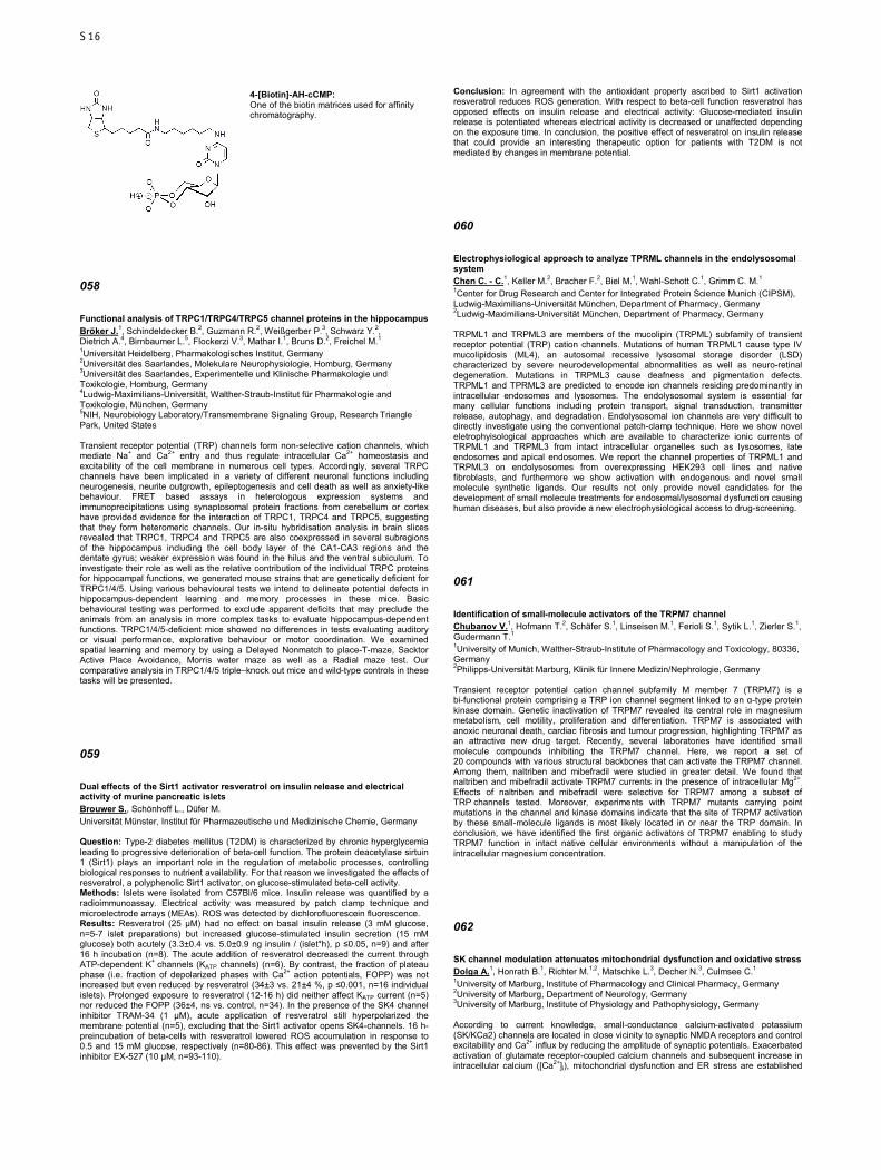
4-[Biotin]-AH-cCMP: One of the biotin matrices used for affinity chromatography.
058
Functional analysis of TRPC1/TRPC4/TRPC5 channel proteins in the hippocampus Bröker J.1, Schindeldecker B.2, Guzmann R.2, Weißgerber P.3, Schwarz Y.2, Dietrich A.4, Birnbaumer L.5, Flockerzi V.3, Mathar I.1, Bruns D.2, Freichel M.1 1Universität Heidelberg, Pharmakologisches Institut, Germany 2Universität des Saarlandes, Molekulare Neurophysiologie, Homburg, Germany 3Universität des Saarlandes, Experimentelle und Klinische Pharmakologie und Toxikologie, Homburg, Germany 4Ludwig-Maximilians-Universität, Walther-Straub-Institut für Pharmakologie and Toxikologie, München, Germany 5NIH, Neurobiology Laboratory/Transmembrane Signaling Group, Research Triangle Park, United States Transient receptor potential (TRP) channels form non-selective cation channels, which mediate Na+ and Ca2+ entry and thus regulate intracellular Ca2+ homeostasis and excitability of the cell membrane in numerous cell types. Accordingly, several TRPC channels have been implicated in a variety of different neuronal functions including neurogenesis, neurite outgrowth, epileptogenesis and cell death as well as anxiety-like behaviour. FRET based assays in heterologous expression systems and immunoprecipitations using synaptosomal protein fractions from cerebellum or cortex have provided evidence for the interaction of TRPC1, TRPC4 and TRPC5, suggesting that they form heteromeric channels. Our in-situ hybridisation analysis in brain slices revealed that TRPC1, TRPC4 and TRPC5 are also coexpressed in several subregions of the hippocampus including the cell body layer of the CA1-CA3 regions and the dentate gyrus; weaker expression was found in the hilus and the ventral subiculum. To investigate their role as well as the relative contribution of the individual TRPC proteins for hippocampal functions, we generated mouse strains that are genetically deficient for TRPC1/4/5. Using various behavioural tests we intend to delineate potential defects in hippocampus-dependent learning and memory processes in these mice. Basic behavioural testing was performed to exclude apparent deficits that may preclude the animals from an analysis in more complex tasks to evaluate hippocampus-dependent functions. TRPC1/4/5-deficient mice showed no differences in tests evaluating auditory or visual performance, explorative behaviour or motor coordination. We examined spatial learning and memory by using a Delayed Nonmatch to place-T-maze, Sacktor Active Place Avoidance, Morris water maze as well as a Radial maze test. Our comparative analysis in TRPC1/4/5 triple–knock out mice and wild-type controls in these tasks will be presented.
059
Dual effects of the Sirt1 activator resveratrol on insulin release and electrical activity of murine pancreatic islets Brouwer S., Schönhoff L., Düfer M. Universität Münster, Institut für Pharmazeutische und Medizinische Chemie, Germany Question: Type-2 diabetes mellitus (T2DM) is characterized by chronic hyperglycemia leading to progressive deterioration of beta-cell function. The protein deacetylase sirtuin 1 (Sirt1) plays an important role in the regulation of metabolic processes, controlling biological responses to nutrient availability. For that reason we investigated the effects of resveratrol, a polyphenolic Sirt1 activator, on glucose-stimulated beta-cell activity. Methods: Islets were isolated from C57Bl/6 mice. Insulin release was quantified by a radioimmunoassay. Electrical activity was measured by patch clamp technique and microelectrode arrays (MEAs). ROS was detected by dichlorofluorescein fluorescence. Results: Resveratrol (25 µM) had no effect on basal insulin release (3 mM glucose, n=5-7 islet preparations) but increased glucose-stimulated insulin secretion (15 mM glucose) both acutely (3.3±0.4 vs. 5.0±0.9 ng insulin / (islet*h), p ≤0.05, n=9) and after 16 h incubation (n=8). The acute addition of resveratrol decreased the current through ATP-dependent K+ channels (KATP channels) (n=6). By contrast, the fraction of plateau phase (i.e. fraction of depolarized phases with Ca2+ action potentials, FOPP) was not increased but even reduced by resveratrol (34±3 vs. 21±4 %, p ≤0.001, n=16 individual islets). Prolonged exposure to resveratrol (12-16 h) did neither affect KATP current (n=5) nor reduced the FOPP (36±4, ns vs. control, n=34). In the presence of the SK4 channel inhibitor TRAM-34 (1 µM), acute application of resveratrol still hyperpolarized the membrane potential (n=5), excluding that the Sirt1 activator opens SK4-channels. 16 h-preincubation of beta-cells with resveratrol lowered ROS accumulation in response to 0.5 and 15 mM glucose, respectively (n=80-86). This effect was prevented by the Sirt1 inhibitor EX-527 (10 µM, n=93-110).
Conclusion: In agreement with the antioxidant property ascribed to Sirt1 activation resveratrol reduces ROS generation. With respect to beta-cell function resveratrol has opposed effects on insulin release and electrical activity: Glucose-mediated insulin release is potentiated whereas electrical activity is decreased or unaffected depending on the exposure time. In conclusion, the positive effect of resveratrol on insulin release that could provide an interesting therapeutic option for patients with T2DM is not mediated by changes in membrane potential.
060
Electrophysiological approach to analyze TPRML channels in the endolysosomal system Chen C. - C.1, Keller M.2, Bracher F.2, Biel M.1, Wahl-Schott C.1, Grimm C. M.1 1Center for Drug Research and Center for Integrated Protein Science Munich (CIPSM), Ludwig-Maximilians-Universität München, Department of Pharmacy, Germany 2Ludwig-Maximilians-Universität München, Department of Pharmacy, Germany TRPML1 and TRPML3 are members of the mucolipin (TRPML) subfamily of transient receptor potential (TRP) cation channels. Mutations of human TRPML1 cause type IV mucolipidosis (ML4), an autosomal recessive lysosomal storage disorder (LSD) characterized by severe neurodevelopmental abnormalities as well as neuro-retinal degeneration. Mutations in TRPML3 cause deafness and pigmentation defects. TRPML1 and TPRML3 are predicted to encode ion channels residing predominantly in intracellular endosomes and lysosomes. The endolysosomal system is essential for many cellular functions including protein transport, signal transduction, transmitter release, autophagy, and degradation. Endolysosomal ion channels are very difficult to directly investigate using the conventional patch-clamp technique. Here we show novel eletrophyisological approaches which are available to characterize ionic currents of TRPML1 and TRPML3 from intact intracellular organelles such as lysosomes, late endosomes and apical endosomes. We report the channel properties of TRPML1 and TRPML3 on endolysosomes from overexpressing HEK293 cell lines and native fibroblasts, and furthermore we show activation with endogenous and novel small molecule synthetic ligands. Our results not only provide novel candidates for the development of small molecule treatments for endosomal/lysosomal dysfunction causing human diseases, but also provide a new electrophysiological access to drug-screening.
061
Identification of small-molecule activators of the TRPM7 channel Chubanov V.1, Hofmann T.2, Schäfer S.1, Linseisen M.1, Ferioli S.1, Sytik L.1, Zierler S.1, Gudermann T.1 1University of Munich, Walther-Straub-Institute of Pharmacology and Toxicology, 80336, Germany 2Philipps-Universität Marburg, Klinik für Innere Medizin/Nephrologie, Germany Transient receptor potential cation channel subfamily M member 7 (TRPM7) is a bi-functional protein comprising a TRP ion channel segment linked to an α-type protein kinase domain. Genetic inactivation of TRPM7 revealed its central role in magnesium metabolism, cell motility, proliferation and differentiation. TRPM7 is associated with anoxic neuronal death, cardiac fibrosis and tumour progression, highlighting TRPM7 as an attractive new drug target. Recently, several laboratories have identified small molecule compounds inhibiting the TRPM7 channel. Here, we report a set of 20 compounds with various structural backbones that can activate the TRPM7 channel. Among them, naltriben and mibefradil were studied in greater detail. We found that naltriben and mibefradil activate TRPM7 currents in the presence of intracellular Mg2+
. Effects of naltriben and mibefradil were selective for TRPM7 among a subset of TRP channels tested. Moreover, experiments with TRPM7 mutants carrying point mutations in the channel and kinase domains indicate that the site of TRPM7 activation by these small-molecule ligands is most likely located in or near the TRP domain. In conclusion, we have identified the first organic activators of TRPM7 enabling to study TRPM7 function in intact native cellular environments without a manipulation of the intracellular magnesium concentration.
062
SK channel modulation attenuates mitochondrial dysfunction and oxidative stress Dolga A.1, Honrath B.1, Richter M.1,2, Matschke L.3, Decher N.3, Culmsee C.1 1University of Marburg, Institute of Pharmacology and Clinical Pharmacy, Germany 2University of Marburg, Department of Neurology, Germany 3University of Marburg, Institute of Physiology and Pathophysiology, Germany According to current knowledge, small-conductance calcium-activated potassium (SK/KCa2) channels are located in close vicinity to synaptic NMDA receptors and control excitability and Ca2+ influx by reducing the amplitude of synaptic potentials. Exacerbated activation of glutamate receptor-coupled calcium channels and subsequent increase in intracellular calcium ([Ca2+]i), mitochondrial dysfunction and ER stress are established
S16

hallmarks of neuronal cell death. Recently we showed that pathological [Ca2+]i deregulation occurring after glutamate receptor stimulation is effectively modulated by SK channels. Besides their plasma membrane localization we have demonstrated that functional SK2 channels are also expressed in the mitochondrial inner membrane and prevent glutamate-induced neuronal oxidative stress and mitochondrial dysfunction. Activation of SK channels inhibits mitochondrial fragmentation, loss of mitochondrial membrane potential, and translocation of apoptosis inducing factor (AIF) to the nucleus. Using a neuronal cell line devoided of NMDA receptors, we demonstrated that the neuroprotective effects were independent of direct interaction of SK channels with NMDA receptors and calcium influx. Furthermore, overexpression of SK channels in mitochondria using mitochondrial-targeted SK channels demonstrated substantial neuroprotective effects in a model of oxidative stress (induced by glutamate and H2O2) and ER stress (induced by brefeldin A). These effects were partially abolished by the dominant-negative SK2 mitochondrial channels. SK channel activation altered the level of unfolded protein response proteins, i.e. further increased CHOP levels and reduced PERK levels compared to brefeldin A-treated cells. Moreover, activation of SK channels resulted in slight ATP depletion and reduced mitochondrial metabolic activity, as assessed by the Seahorse Bioscience XFe cell metabolism analyzer. Positive modulation of SK channels slightly increased mitochondrial ROS production and induced a small mitochondrial depolarization. These results expose a pre-conditioning effect as a mechanism for neuroprotection mediated by SK channel activation. Our findings show a critical role for SK channels in excitotoxic neuronal cell death, mitochondrial dysfunction, oxidative stress and ER stress-associated cell death proposing their activation as potential therapeutic strategy for the treatment of acute and chronic neurodegenerative disorders.
063
Functional interplay of TRPM6 and TRPM7 in trophoblast stem cells Ferioli S., Zierler S., Gudermann T., Chubanov V. University of Munich, Walther-Straub-Institute of Pharmacology and Toxicology, 80336, Germany TRPM6 and TRPM7 are bi-functional proteins comprising a TRP channel segment linked to an α-type protein kinase. In mice, the genetic ablation of any of the kinase-coupled TRPs (TRPM6 and TRPM7) results in embryonic lethality. Loss-of-function mutations in the human TRPM6 gene cause an autosomal recessive disorder, hypomagnesemia 1, intestinal (HOMG1) also called hypomagnesemia with secondary hypocalcemia (HSH). At present, there is no consensus in the scientific community regarding the physiological roles and functional redundancy of TRPM6 and TRPM7. On the cellular level, TRPM6 always co-exists with TRPM7 implying that a lack of TRPM6 may result in alterations of TRPM7 function. Therefore, a key approach of our studies is the characterization of TRPM6 and TRPM7 ion currents in a physiologically relevant model that endogenously expresses both TRPM6 and TRPM7. We found that TRPM6 and TRPM7 are highly expressed in labyrinth trophoblasts of the placenta. To elucidate the functional interactions of TRPM6 and TRPM7, we isolated trophoblast stem (TS) cells from mouse blastocysts derived from Trpm6 gene deficient mice. Patch-clamp experiments revealed that WT TS cells develop divalent cation selective currents induced by depletion of intracellular Mg2+. Trpm6 null cells exhibited reduced current amplitudes. In addition, we found that the currents in Trpm6 null cells are more sensitive to intracellular Mg-ATP as compared to WT cells, suggesting that TRPM6 uncouples cellular Mg2+ entry from energy metabolism specifically in transporting epithelia. Taken together, our findings indicate that TRPM6 modulates key biophysical characteristics of TRPM7 currents.
064
Roles of TRPML and Two-Pore channels in endolysosomal function and disease Grimm C. M., Chen C. - C., Butz E., Biel M., Wahl-Schott C. LMU München, Department of Pharmacy/Pharmacology, Germany Lysosomes are cell organelles involved in the breakdown of proteins, lipids, and other molecules and have been found to be implicated not only in endolysosomal storage disorders (LSDs) such as mucolipidoses or mucopolysaccharidoses but also in metabolic diseases, neurodegenerative disorders such as Alzheimer’s and Parkinson’s disease, pigmentation disorders, or infectious diseases. Highly critical for the proper function of lysosomes, endosomes, and lysosome-related organelles is the tight regulation of various fusion and fission processes and the regulation of proton and other ionic concentrations within the endolysosomal system. Calcium permeable, non-selective cation channels of the TRP (transient receptor potential) superfamily, namely TRPML channels (mucolipins) and TPCs (two-pore channels) have been found to play important roles in these processes. Here we present novel insights into the function and physiology of TRPML and TPC channels, into the molecular basis of pathologies associated with diseases caused by loss or mutation of these channels, and we discuss potential therapeutic approaches. Further, we present newly identified small molecule compounds that specifically gate or block TRPML and TPC channels as demonstrated by whole- endosomal and lysosomal patch-clamp techniques. Grimm, C., Holdt, L.M., Chen, C.-C., Hassan, S., Müller, C., Jörs, S., Cuny, H., Kissing, S., Schröder, B., Butz, E., Northoff, B., Castonguay, J., Luber, C.A., Moser, M., Spahn, S., Lüllmann-Rauch, R., Fendel, C., Klugbauer, N., Griesbeck, O., Haas, A., Mann, M., Bracher, F., Teupser, D., Saftig, P., Biel, M., Wahl-Schott, C. (2014) High susceptibility to fatty liver disease in two-pore channel 2-deficient mice. Nature Commun., 5:4699. doi: 10.1038/ncomms5699; Chen, C.-C.,Keller, M., Hess, M., Schiffmann, R., Urban, N., Wolfgardt, A., Schaefer, M., Bracher, F., Biel, M., Wahl-Schott, C., Grimm, C. (2014) A
small molecule restores function to TRPML1 mutant isoforms responsible for mucolipidosis type IV. Nature Commun., 5:4681. doi: 10.1038/ncomms5681; Dong XP, Shen D, Wang X, Dawson T, Li X, Zhang Q, Cheng X, Zhang Y, Weisman LS, Delling M, Xu H. (2010) PI(3,5)P(2) controls membrane trafficking by direct activation of mucolipin Ca(2+) release channels in the endolysosome. Nat. Commun. 1:38; Shen D, Wang X, Li X, Zhang X, Yao Z, Dibble S, Dong XP, Yu T, Lieberman AP, Showalter HD, Xu H (2012) Lipid storage disorders block lysosomal trafficking by inhibiting a TRP channel and lysosomal calcium release. Nat. Commun. 3:731; Dong XP, Cheng X, Mills E, Delling M, Wang F, Kurz T, Xu H. (2008) The type IV mucolipidosis-associated protein TRPML1 is an endolysosomal iron release channel. Nature 455:992-996; Xu H, Delling M, Li L, Dong X, Clapham DE (2007). Activating mutationin a mucolipin transient receptor potential channel leads to melanocyteloss in varitint-waddler mice. Proc. Natl. Acad. Sci. USA 104:18321-18326; Grimm, C., Cuajungco, M.P., van Aken, A.F.J., Schnee, M., Jörs, S., Kros, C.J., Ricci, A.J., Heller, S. (2007) A helix-breaking mutation in TRPML3 leads to constitutive activity underlying deafness in the varitint-waddler mouse. PNAS, 104:19583-19588
Grimm et al. Fig1: Cholesterol accumulation in TPC2-/- livers visualized with filipin.
065
Contribution of salt-bridge switching within in the ATP binding pocket to the gating of the P2X2 receptor Hausmann R.1, Kuhlmann D.1, Kless A.2, Schmalzing G.1 1RWTH Aachen University, Molecular Pharmacology, Germany 2Grünenthal GmbH, Global Drug Discovery, Aachen, Germany P2X receptors are trimeric ATP-gated cation channels involved in the fast signal transduction in many cell types. A homology model of the closed-state of the rat P2X2 receptor based on the X-ray structure of the apo closed-state of the zebrafish P2X4 receptor shows that the side chains of the residues Glu167/Arg290 and Arg290/Asp82, Glu84 or Glu85 are in close spatial vicinity within the ATP-binding pocket. Functional analysis of mutants resulting in charge reversal, charge swapping and charge neutralization suggests that at least two of the possible four oppositely charged ion pairs are electrostatically coupled, namely Glu84/Arg290 and Glu167/Arg290. A triple mutant cycle analysis of Glu167, Arg290 and Glu84 indicated energetic coupling between Glu167/Arg290 and Arg290/Glu84 and thus cooperative interaction in a larger salt bridge network. A comparison of the closed-state and open-state model of the rat P2X2 receptor revealed that the residues Glu84/Arg290 and Glu167/Arg290 markedly relocate and move apart during the closed-to-open transition into positions that impair the electrostatic interactions (Fig. 1). Indeed, disulfide trapping confirmed that the side chains of the pairs Glu84/Arg290 and Glu167/Arg290 are in close proximity when the channel is in the apo closed state, but in weakly cross-linkable and thus more distant positions in the ATP-bound open state. A driving force for the breaking of the salt bridges Glu84/Arg290 and Glu167/Arg290 seems to be the γ-phosphate oxygen of ATP, which undergoes a strong ionic interaction with Arg290 of the P2X2 receptor (Fig. 1). Our data are in line with the concept that this newly formed electrostatic interaction of Arg290 with ATP competitively releases Glu84 and Glu167 from their strong electrostatic coupling to Arg290 and thus initiates a destabilization of the closed-state that favours channel opening. This work was supported by a Deutsche Forschungsgemeinschaft Grant HA6095/1-1 to RH.
Fig. 1: The transition of the apo closed-state (left) to the ATP-bound open-state (right) of the rP2X2 receptor was visualized by morphing. Residues of adjacent subunits A and B are indicated by superscript A and B.
S17
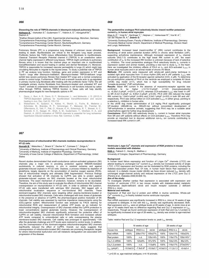
066
Dissecting the role of TRPC6-channels in bleomycin-induced pulmonary fibrosis Hofmann K.1, Vierkotten S.2, Gudermann T.1, Yildirim A. Ö.3, Königshoff M.4, Dietrich A.1 1Walther-Straub-Institut of the LMU, Experimental pharmacology, München, Germany 2Comprehensive Pneumology Center Munich, Germany 3Comprehensive Pneumology Center Munich, Neuherberg/Munich, Germany 4Comprehensive Pneumology Center Munich, Germany Pulmonary fibrosis (PF) is a progressive lung disease of unknown cause ultimately leading to death. Myofibroblasts are critical to the fibrogenic lung repair process. Profibrotic factors like TGFβ induce differentiation from fibroblasts to myofibroblasts. Classical Transient Receptor Potential channel 6 (TRPC6) is an unselective cation channel highly expressed in different lung tissues. TRPC6 might contribute to pulmonary fibrosis since it is known that the channel plays an important role in myofibroblast transdifferentiation and wound healing in cardiac and dermal fibroblasts (1). Moreover, TRPC6 is responsible for increased vascular permeability in lungs (2) which might help circulating fibrocytes to migrate to the injured areas. To study a potential role of TRPC6 in PF we analyze lung function, histology, gene and protein expression in WT and Trpc6-/- lungs after bleomycin-instillation. Bleomycin-treated TRPC6-deficient lungs exhibit less severe pulmonary fibrosis than treated WT lungs and a normal compliance similar to control lungs. Furthermore, TRPC6 and α-smooth muscle actin is up-regulated in primary murine lung fibroblasts after treating with the profibrotic factor TGFβ. We will investigate primary murine fibroblasts from bleomycin-treated and untreated WT and Trpc6-/- animals to understand molecular differences in cell functions induced by cation influx through TRPC6. Defining TRPC6 function in these cells will help identify pharmacological targets for new therapeutic options in PF. 1. Davis, J., Burr, A. R., Davis, G. F., Birnbaumer, L., and Molkentin, J. D. (2012) A
TRPC6-dependent pathway for myofibroblast transdifferentiation and wound healing in vivo. Dev. Cell 23, 705-715.
2. Weissmann, N., Sydykov, A., Kalwa, H., Storch, U., Fuchs, B., Mederos y Schnitzler, M., Brandes, R. P., Grimminger, F., Meissner, M., Freichel, M., Offermanns, S., Veit, F., Pak, O., Krause, K. H., Schermuly, R. T., Brewer, A. C., Schmidt, H. H., Seeger, W., Shah, A. M., Gudermann, T., Ghofrani, H. A., and Dietrich, A. (2012) Activation of TRPC6 channels is essential for lung ischaemia-reperfusion induced oedema in mice. Nat. Commun. 3, 649.
067
Overexpression of mitochondrial SK2 channels mediates neuroprotection in HT-22 cells Honrath B.1, Matschke L.2, Strack S.3, Decher N.2, Culmsee C.1, Dolga A.1 1University of Marburg, Institute of Pharmacology and Clinical Pharmacy, Germany 2University of Marburg, Institute of Vegetative Physiology, Germany 3University of Iowa Carver College of Medicine, Department of Pharmacology, United States Recent studies demonstrated that small-conductance calcium-activated potassium (SK) channels play a major role in providing protection against NMDAR-mediated excitotoxicity in cultured neurons, in vivo in cerebral ischemia and against glutamate-induced oxytosis in HT-22 cells. Oxytosis, a process initiated by depletion of glutathione, largely depends on the accumulation of reactive oxygen species (ROS), loss of mitochondrial integrity and ultimately DNA fragmentation. Previous findings showed that activation of SK channels by CyPPA was able to protect against glutamate-induced oxytosis via SK2 channels located at the inner mitochondrial membrane. The exact mechanism of protection, however, remains to be elucidated. Therefore, the aim of this study was to evaluate the effects of mitochondrial SK2 channel overexpression on neuroprotection in HT-22 cells. In order to address this question, HT-22 cells were transfected with wild-type SK2 channels, SK2 tagged with a mitochondrial localization sequence or non-functional SK2 channels. These dominant-negative SK2 channels were targeting either plasma-membrane-located or mitochondria-located SK2 channels. Cells were challenged with toxic doses of glutamate in the presence or absence of CyPPA, the pharmacological positive modulator of SK channels. Cell viability was assessed by real-time impedance measurements using the xCELLigence system. Mitochondrial function was analyzed by FACS staining for mitochondrial ROS and measurement of cellular ATP levels, as an indicator for mitochondrial integrity. Overexpression of SK2 channels shifted the CyPPA protection to lower concentrations against glutamate toxicity. However, the overexpression of mitochondria-targeted SK2 channels significantly enhanced the protective effect of CyPPA on cell viability, reduced mitochondrial ROS formation and increased cellular ATP levels compared to untransfected cells or cells overexpressing the plasma membrane-located SK2 channels. Cell viability was prolonged to approximately ~10h after the glutamate challenge and ATP levels were restored to control levels. In addition, only the overexpression of the dominant-negative mitochondria-targeted SK2 channels significantly reduced the effect of CyPPA. Overall, our study suggests that overexpression of mitochondria-located SK2 channels are promising therapeutic targets for where mitochondrial dysfunction and cell metabolic deregulation are associated with progression of the disease.
068
The pentamidine analogue PA-6 selectively blocks inward rectifier potassium current IK1 in human atrial myocytes Khan A. P.1, Voigt N.1, Herrlinger Y.1, Heijman J.1, Varkevisser R.2, Vos M. A.2, van der Heyden M. A.2, Dobrev D.1 1University Duisburg-Essen, Faculty of Medicine, Institute of Pharmacology, Germany 2University Medical Center Utrecht, Department of Medical Physiology, Division Heart and Lungs, Netherlands Background: Increased basal inward-rectifier K+ (IRK) current contributes to the shortening of atrial action potential duration (APD) in chronic atrial fibrillation (cAF), supporting arrhythmia maintenance. Although increased expression of IK1 channel subunits (Kir2.1-3) contributes to the high basal IRK current in cAF, the exact contribution of IK1 to the increased IRK function is unknown because of lack of selective IK1 inhibitors. The novel pentamidine analogue PA-6 selectively blocks IK1 currents in expression system and animal models, providing a new tool to study IK1 in the heart. Here we investigated the inhibitory effects of PA-6 on IK1 and ACh-activated IK,ACh in human atrial myocytes and in an animal model of chronic AV-Block. Methods: IRK currents were measured with whole-cell patch-clamp technique in isolated right atrial myocytes from 13 sinus rhythm (SR) and 4 cAF patients. IK,ACh was activated by application of the M-receptor agonist carbachol (CCh, 2 µM). To determine the pro-arrhythmic potential of PA-6 on the ventricle we employed a complete AV block (cAVB) dog model (n=7), which has a high susceptibility for drug induced “torsade-de-pointes” (TdP) arrhythmias. Results: Basal IRK current in the absence of muscarinic-receptor agonists was confirmed to be higher (-12.7±1.9 pA/pF, n=13/4 [myocytes/patients] vs. -6.25±1,0 pA/pF, n=23/13; p<0.01), whereas CCh-activated IK,ACh was lower in cAF vs SR (5.2±1.0 pA/pF vs. 8.65±1.3 pA/pF; p<0.05). PA-6 (200 nM) reduced the basal IRK current by 1.6±0.4 pA/pF (n=8/4) and 3.2±0.1 pA/pF (n=5/2) in both SR and cAF, respectively. PA-6 was without effect on CCh-activated IK,ACh in either group, pointing to a selective IK1 inhibition in human atria. In the cAVB dog model administration of 2.5 mg/kg PA-6 significantly prolonged QT-duration (480±86 ms vs. 390±25 ms) without concomitant development of TdP-arrhythmias in sensitive animals, suggesting that PA-6 may be a suitable atrial antiarrhythmic agent for further drug development. Conclusions: PA-6 (200 nM) selectively blocks IK1 current in human atrial myocytes from SR and cAF patients without effects on CCh-activated IK,ACh. Therefore PA-6 may provide an important tool to discover additional non-IK1 ion currents contributing to increased basal IRK current in cAF.
069
Ventricular L-type Ca2+ channels and expression of RGK proteins in mouse models associated with diabetes Köth J., Fabisch C., Herzig S., Matthes J. University of Cologne, Department of Pharmacology, Germany Background: In human heart failure expression and function of L-type Ca2+ channels (LTCC) are altered leading to unchanged Ca2+-current (ICaL) density but increased activity of single LTCC. LTCC expression and function can be modulated by RGK proteins, including the diabetes-associated protein Rad. Of note, in human heart failure Rad expression is reduced. In a diabetic mouse model (db/db) we have shown reduced ICaL density with unchanged single-channel activity and reduced expression of the LTCC pore Cav1.2 (Pereira et al., Diabetes 2006;55:608-15). Aim of the study: To investigate whether cardiac Rad expression is associated with expression and function of ventricular LTCC in two mouse models with diabetes-related metabolic disturbances (leptin-deficient ob/ob and insulin receptor substrate 2 deficient IRS2-k.o. mice). Material and methods: Expression of Rad and Cav1.2 protein and mRNA in murine ventricles. Whole-cell Ca2+-currents (ICaL) in freshly isolated murine ventricular myocytes. Results: Rad mRNA expression was significantly increased in IRS2-k.o. mice at 16 weeks of age compared to wildtypes. In line with this ICaL density was significantly decreased. Both, Rad expression and ICaL were at wildtype levels at 28 weeks of age. In ob/ob mice at an age of 16 weeks there was no difference regarding Rad and Cav1.2 expression or ICaL, respectively. Though we found ventricular expression of both Rad and Cav1.2 protein to be significantly increased at an age of 28 weeks, ICaL density was similar to age-matched wildtypes. Table: relative Rad and Cav1.2 expression levels vs. peak ICaL density.
16 weeks of age 28 weeks of age
mouse line wildtype IRS2-k.o. ob/ob wildtype IRS2-k.o. ob/ob
Rad mRNA 100% 206±17%* 155±27% 100% 124±11% 150±23%
Rad protein 100% n.d. 135±14% 100% 119±17% 159±14%*
Cav1.2 mRNA 100% 120±9% 97±19% 100% 104±14% 86±12%
Cav1.2 protein 100% n.d. 113±8% 100% 116±17% 273±23%*
ICaL (pA/pF) -10.9±0.9 -7.8±0.8* -10.8±0.7 -8.9±0.5 -11.2±1.3 -8.4±0.4 *: p<0.05 vs. age-matched wildtypes; n=4-16 animals.
S18
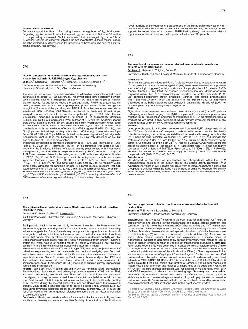
Summary and conclusion: Our data support the idea of Rad being involved in regulation of ICaL in diabetes. Regarding ICaL Rad seems to act either causal (ICaL decrease in IRS2-k.o. at 16 weeks) or compensatory (increased Cav1.2 expression but unchanged ICaL in ob/ob at 28 weeks). Differences observed between the two investigated diabetic mouse models might be explained by differences in the underlying pathomechanisms (lack of IRS2 vs. leptin deficiency, respectively).
070
Allosteric interaction of SUR-tetramers in the regulation of agonist and antagonists action in SUR2B/Kir6.1-type KATP channels Sachs A.1, Schmidt C.1, Rauhaus K.1, Fischer A.2, Braun M.2, Lemoine H.1 1UKD-Universitätsklinik Düsseldorf, Inst. f. Lasermedizin, Germany 2Universität Düsseldorf, Inst. f. Org. Chemie, Germany The vascular type of KATP channels is organized as a tetradimeric complex of Kir6.1 and sulfonylurea receptors 2B (SUR2B/Kir6.1)4. We investigated, how allosterism between SUR-tetramers influences antagonism of agonists (A) and blockers (B) to regulate channel activity. As agonist we chose the cyanoguanidine P1075, as antagonists the cyanoguanidine PNU96293, the sulphonylurea glibenclamide (Glib), the glinide repaglinide (Repa), and the partial agonist NNC414. As a test model we used stably transfected HEK 293(Kir6.1/SUR2B)-cells, cultivated in 96-format 12-well strips, assayed channel activity with a membrane potential dye (R7260, Mol. Probes, 0.125 mg/ml) measured in multichannel, full-kinetic (1 Hz) fluorescence detectors (505/530 nm) built in our laboratories. Prestimulation of KATP with the low-affinity agonist cyclo-pentyl-diazoxide (AF51, 30 µM) caused maximum hyperpolarisation which could be repolarized with all tested B with short half times (t1/2<30 sec). In contrast, after prestimulation with the maximum effective high-affinity agonist P1075 (125 nM), only Glib (2 µM) repolarized exponentially with a short half-time (t1/2~2 min), whereas 2 µM Repa, 30 µM PNU and 60 µM NNC repolarized much slower (t1/2>>6 min) with sigmoidal repolarization kinetics. Thus, the dissociation of P1075 not only depended on koff, but also on the type of B inducing dissociation. Theoretical considerations (compare Dörschner et al., 1999, Mol Pharmacol 55:1060; Russ et al., 2009, Brit J Pharmacol, 156:354) on the tetrameric organization of SUR reveal that the B-induced inhibition of channel acitivity prestimulated with A can occur {setting exp(-koff* t):=EXP} with fast exponential kinetics {1-EXP4, model M1}, if only 1 of 4 SUR~A-complexes have to be antagonized by B, with slow sigmoidal kinetics {(1-EXP)4, M4}, if each SUR~A-complex has to be antagonized, or with intermediate sigmoidal kinetics if two {1 + 3*EXP4 - 4*EXP3, M2} or three complexes {(1-EXP)4 + 4(1 - EXP)3*EXP), M3} have to be antagonized by B for full recovery. Curve fitting clearly attributed repolarization kinetics to different models, revealing that Glib acted according to M1 with fast t1/2 (2.4±0.2 min) and a corresponding koff (0.08 min-1) whereas Repa acted via M2 with t1/2=6.4±0.4 (koff=0.10), PNU via M3 with t1/2=14.3±0.6 (koff=0.07) and NNC via M3 with t1/2=14.3±0.6 (koff=0.07). Concluding, allosteric effects of SUR-tetramers influence antagonist efficiency of blockers in KATP channels.
071
The sodium-activated potassium channel Slack is required for optimal cognitive flexibility in mice Bausch A. E., Dieter R., Ruth P., Lukowski R. Institut für Pharmazie, Pharmakologie, Toxikologie & Klinische Pharmazie, Tübingen, Germany Background: Slack channels are highly expressed throughout the brain where they modulate firing patterns and general excitability of many types of neurons. Increasing evidence suggests that Slack channels may be important for higher brain functions such as cognition and normal intellectual development. In particular, recent findings have shown that human Slack mutations produce very severe intellectual disability and that Slack channels interact directly with the Fragile X mental retardation protein (FMRP), a protein that when missing or mutated results in Fragile X syndrome (FXS), the most common form of inherited intellectual disability and autism in humans. Methods: Slack deficient (Slack KO) and wild type (WT) mice were subjected to a set of behavioral experiments, such as beam walk test, footprint analysis, open field test, Morris water maze test and a variant of the Barnes maze test assess which behavioral aspects depend on Slack. Expression of Slack transcripts was analyzed by qPCR and the cellular distribution of the Slack channel protein was assessed by immunohistochemical/-fluorescence (IHC/IF) stainings of cultured primary hippocampal neurons and brain sections. Results: Using qRT-PCR, Western blot and IHC/IF we confirmed Slack expression in the cerebellum, hippocampus, and primary hippocampal neurons of WT but not Slack KO mice. Importantly, we found that Slack KO mice exhibit several behavioral phenotypes, including decreased overall locomotor activity and habituation deficits in an open field, as well as initial defects during reversal learning tasks. Additionally, tracking of WT animals during the reversal phase of a modified Barnes maze test revealed a primarily visual-spatial orientation strategy to locate the escape hole, whereas Slack KO mice rather favored a serial/non-spatial search strategy. Interestingly, lack of Slack did not affect working memory or general reference memory as well as motor coordination on an elevated beam. Conclusion: Herein, we provide evidence for a role for Slack channels in higher brain functions i.e. learning and memory, cognitive flexibility, locomotoric and habituation to
novel situations and environments. Because some of the behavioral phenotypes of Fmr1 deficient mice were reproduced in the Slack mutant mouse line, our findings further support the recent view of a common FMRP/Slack pathway that underlies distinct cognitive capabilities in mice and that is perturbed in human FXR patients.
072
Composition of the ryanodine receptor channel macromolecular complex in patients with atrial fibrillation Schirmer I., Heijman J., Voigt N., Dobrev D. University of Duisburg-Essen, Faculty of Medicine, Institute of Pharmacology, Germany Introduction: Abnormal sarcoplasmic reticulum (SR) Ca2+-release events due to hyperphosphorylation of the ryanodine receptor channel type-2 (RyR2) have been identified as a potential source of ectopic (triggered) activity in atrial cardiomyocytes from AF patients. RyR2-channel function is regulated by dynamic phosphorylation and dephosphorylation controlled within the RyR2 macromolecular complex via protein kinase-A (PKA), Ca2+/calmodulin-dependent protein kinase-IId (CaMKIId) and protein phosphatases type-1 and type-2A (PP1, PP2A), respectively. In the present study, we discovered differences in the RyR2 macromolecular complex in patients with chronic AF (cAF, > 6 months), potentially contributing to RyR2 dysfunction. Methods: Right atrial tissue samples were collected from sinus rhythm (Ctl) or cAF patients undergoing open heart surgery. The RyR2 complex with its accessory proteins was enriched by SR fractionation and immunoprecipitation (IP). For gel-electrophoreses, a gradient gel was used (4-15% acrylamide), which provided improved separation of the proteins located within the RyR2 complex with immunoblotting. Results: Using phospho-specific antibodies, we observed increased RyR2 phosphorylation at Ser-2808 and Ser-2814 in cAF samples, consistent with previous results. To identify potential underlying mechanisms, we established a novel methodology to isolate the RyR2 macromolecular complex. We found PKA, CaMKIId, PP1, PP2A, and their adaptor proteins spinophilin-1 (for PP1) and PR130 (for PP2A) within the RyR2 macromolecular complex. Calcineurin-Aβ and the SR Ca2+-ATPase type-2a (SERCA2a) were absent and served as negative controls. The amount of PP1 associated with RyR2 was significantly decreased in cAF patients (37.5±3.7%, n=5) compared to Ctl (100±11.5%, n=5, P<0.05), whereas the amount of CaMKIId was strongly increased (271±65.2%, n=5) in cAF compared to Ctl (100±16.3%, n=5, P<0.05). Conclusions: We detected for the first time key kinases and phosphatases within the RyR2 macromolecular complex in the human atrium. The ectopic activity-promoting RyR2 hyperphosphorylation in cAF patients may result from an imbalance between kinase and phosphatase activities within the RyR2 macromolecular complex. Reduced PP1 activity within the RyR2 complex may constitute a novel mechanism for proarrhythmic SR Ca2+-release events.
073
Cardiac L-type calcium channel function in a mouse model of mitochondrial dysfunction Seemann W. K., Annala S., Matthes J., Herzig S. University of Cologne, Department of Pharmacology, Germany Background: The L-type Ca2+ channel is the main route for extracellular Ca2+ entry in cardiomyocytes and essential for the maintenance of accurate cardiac excitation and contraction (1). It is known that alterations in Ca2+ channel activity and Ca2+ homeostasis are associated with cardiomyopathies resulting in cardiac hypertrophy and heart failure (2). Heart failure is a disease of advanced age, mitochondrial dysfunction becomes more prevalent with age (3) and has been associated with heart failure (4). Therefore, we study L-type calcium channel function and expression in a mouse model of mitochondrial dysfunction accompanied by cardiac hypertrophy with increasing age to check if calcium channel function is affected by mitochondrial dysfunction. Methods: Patch-clamp experiments were performed in isolated ventricular cardiomyocytes of mice at the age of 18-20 and 28-30 weeks. We used mtDNA-mutator mouse expressing a proof-reading-deficient version of the mitochondrial DNA (mtDNA) polymerase PolgA leading to premature onset of ageing at 25 weeks of age (5). Furthermore, we quantified cardiac calcium channel expression as well as markers of cardiomyopathy and heart failure (e.g. MHC-β, BNP, CTGF) by qPCR in mice at the age of 18-20, 28-30 and 40-45 weeks. Results: First data indicate that function of cardiac L-type calcium channels of mutant mice do not differ from age-matched wild-type mice at the whole-cell level. At the mRNA level calcium channel expression was not affected in mutant mice, while BNP and CTGF expression is elevated with increasing age. Summary and conclusion: Ventricular L-type calcium currents were similar in mtDNA-mutant mice compared to wild-types while with enhanced age expression of hypertrophy markers increased in mutant ventricles. So far, we cannot exclude that under stimulated conditions (e.g. beta-adrenergic stimulation) calcium channel dysfunction might become evident. (1) Triggle DJ (2006) L-type Calcium Channels, Voltage-Gated Ion Channels as Drug Targets, Volume 29, Wiley-VCH Verlag GmbH & Co. KGaA, Weinheim (2) Benitah JP et al. (2010) L-type Ca2+ current in ventricular cardiomyocytes. J Mol Cell Cardiol; 48: 26-36
S19

(3) Larsson NG (2010) Somatic mitochondrial DNA mutations in mammalian aging. Annu Rev Biochem; 79: 683–706 (4) Finsterer J and Kothari S (2014) Cardiac manifestations of primary mitochondrial disorders. Int J Cardiol; 177: 754-763 (5) Trifunovic A et al. (2004) Premature ageing in mice expressing defective mitochondrial DNA polymerase. Nature; 429: 417-423
074
Analysis of P2X7 protein complexes in a P2X7-EGFP BAC transgenic mouse model Stocklauser R.1, Kaczmarek-Hajek K.2, Saul A.2, Nicke A.1 1Ludwig Maximilian University, Walther Straub Institute for Pharmacology and Toxicology, Munich, Germany 2Max Planck Institute for Experimental Medicine, Göttingen, Germany P2X receptors are ATP-gated cation channels with a high Ca2+ permeability. Unlike other P2X family members, the P2X7 subtype shows particularly low sensitivity to ATP and has a long intracellular C-terminus. Given its functional importance and the ability of this C-terminal domain to interact with other proteins as part of a larger signaling complex, considerable effort has been made to identify interaction partners. However, previous studies were mainly based on over-expression of recombinant tagged proteins in HEK cells and might be biased by artificial protein aggregation. For none of the identified interaction partners interaction domains have been defined and functional consequences of their interaction are poorly described. To identify tissue-specific protein-protein interactions of the P2X7 receptor, we generated a P2X7 BAC transgenic mouse model in which the receptor is fused via a heptahistidyl-linker to an EGFP reporter protein. This model allows P2X7-EGFP expression under its endogenous promotor at near physiological or moderate over-expression levels. Initial biochemical and immunohistochemical experiments show equal expression patterns of P2X7-EGFP and endogenous P2X7 receptors in different tissues and demonstrate its correct transport to the plasma membrane. Using the full-length P2X7-EGFP protein as a bait, endogenous P2X7 subunits were efficiently and specifically co-purified via the His- or EGFP tags. In addition, an association with P2X4 receptors was identified and is further investigated. Experiments to validate other previously identified interaction partners are ongoing and will be presented. In conclusion, we describe a novel BAC transgenic mouse model that allows efficient purification of P2X7-EGFP receptors and provides a valuable tool for proteomic analysis of its interaction partners.
075
Purinergic P2X4 channels are crucial mechanotransducing elements in podocytes causing disorganization of the cytoskeleton Storch U.1, Forst A. - L.1, Olteanu V. S.1, Mollet G.2, Wlodkowski T.3, Schaefer F.3, Dietrich A.1, Reiser J.4, Gudermann T.1, Mederos Y Schnitzler M.1 1Ludwig Maximilians Universität München, Walther-Straub-Institut für Pharmakologie und Toxikologie, Germany 2Imagine Institute (Inserm U1163), Laboratory of Hereditary Kidney Diseases, Paris, France 3Universitätsklinikum Heidelberg, Pädiatrische Nephrologie, Germany 4Rush University Medical Center, Department of Internal Medicine, Chicago, United States Podocytes are highly differentiated visceral epithelial cells lining the glomerular basement membrane thereby fulfilling an important role in kidney filtration. In the glomerulus they are exposed to hypertension and are believed to sense increases in mechanical load which leads to mechanically induced podocyte injury. Until now the molecular identity of the mechanosensors and -transducers in podocytes is largely unknown. The classical transient receptor potential channel 6 (TRPC6) which interacts with the MEC-2 homologue podocin was suggested to form a mechanosensitive ion channel complex in podocytes. Performing calcium imaging and whole-cell measurements we found that podocytes respond to mechanical stress with increased intracellular calcium concentrations and increased inward cation currents. To analyze the role of TRPC6 for mechanosensation we used TRPC6-genedeficient podocytes. These podocytes responded to mechanical stimulation in a way similar to control podocytes suggesting that mechanically induced currents were not mediated by TRPC6. Instead, mechanically induced currents were significantly decreased by the specific P2X4 blocker 5-BDBD indicating an involvement of P2X4 channels. Moreover, mechanical P2X4 channel activation was cholesterol and podocin dependent, but independent of the actin cytoskeleton. Since P2X4 channels are not intrinsically mechanosensitive, we investigated whether mechanical stimulation of podocytes leads to ATP release. Using a fluorometric approach we observed a mechanically induced ATP release from podocytes. To ascertain the physiological role of P2X4 channels as mechanotransducing elements in podocytes we monitored reorganization of the actin cytoskeleton in the presence and absence of 5-BDBD. Interestingly, 5-BDBD could rescue stretch-induced disorganization of the actin cytoskeleton pointing to a significant involvement of P2X4 channels in the adaption of podocytes to mechanical stress. Altogether our findings reveal a new role of P2X4 channels as mechanotransducers in podocytes leading to mechanically-induced podocyte injury that may contribute to hypertensive nephropathy.
076
Lupanine improves glucose homeostasis by influencing KATP-channels of pancreatic beta cells Wiedemann M.1, García-López P.2, Wink M.3, Gurrola-Díaz C.4, Düfer M.1 1Universität Münster, Institut für Pharmazeutische und Medizinische Chemie, Germany 2Universidad de Guadalajara, Departamento de Botánica y Zoología, Centro Universitario de Ciencias Biológicas y Agropecuarias, Mexico 3Universität Heidelberg, Institut für Pharmazie und molekulare Biotechnologie, Germany 4Universidad de Guadalajara, Departamento de Biología Molecular y Genómica, Centro Universitario de Ciencias de la Salud, Mexico Aims: Stimulating insulin release is an important strategy for treatment of type-2-diabetes mellitus (T2DM) although hypoglycaemia is one of the central problems limiting the applicability of insulinotropic drugs. For lupanine (the main alkaloid of many lupin-species) a blood-glucose lowering effect has been described in human and animal studies. To elucidate the underlying mechanism we studied the influence of lupanine on electrical activity and insulin secretion in isolated islets and in an in vivo model of T2DM. Methods: Islets were isolated from C57Bl/6 mice. Insulin secretion was measured by radioimmunoassay. Electrophysiology was performed using the patch-clamp-technique. For determination of the membrane potential (Vm) the perforated patch configuration and for recordings of KATP channel activity the standard whole-cell configuration was used. For glucose tolerance tests (GTT) streptozotocin-diabetic rats were treated with lupanine (20mg/kg BW) or vehicle prior to glucose administration. Results: Diabetic rats treated with lupanine showed an attenuated increase of serum glucose concentration compared to controls in response to a GTT (n=3). In accordance with this observation, lupanine (0.5 mM) increased insulin secretion induced by 15 mM glucose (10.2±1.97 vs. 6.6±0.93 ng insulin / (islet x h), p≤0.01, n=10). By contrast, lupanine had no stimulatory effect on insulin release in the presence of 3 mM glucose in concentrations up to 1 mM. Determination of KATP channel activity revealed that 0.5 mM lupanine reduced the KATP current by ~40% (n=13) whereas 0.05 mM had no effect (n=11). Importantly, 0.5 mM lupanine did not depolarize Vm in the presence of basal or at threshold glucose concentrations (0.5 and 5 to 6 mM glucose, respectively). To test whether lupanine alters electrical activity of beta cells that were already stimulated by glucose the frequency of Ca2+ action potentials was determined. Lupanine reversibly increased the frequency of Ca2+ action potentials in the presence of 15 mM glucose (n=9). Conclusion: Our data show that lupanine improves glucose tolerance in diabetic animals. The underlying mechanism involves direct inhibition of KATP channels. As the insulinotropic effect of lupanine only occurs at elevated glucose concentrations there is potentially no risk of hyperglycaemia.
077
Functional characterization of the genetic polymorphisms in the OCT1 promoter Bokelmann K., Brockmöller J., Tzvetkov M. University Medical Center Göttingen, Clinical Pharmacology, Germany The organic cation transporter OCT1 (SLC22A1) is strongly expressed in the sinusoidal membrane of the human liver. OCT1 plays a role in the hepatic uptake of organic cations from the blood, including drugs like metformin, tropisetron and morphine. More than 100-fold variations of the OCT1 expression were reported in liver samples of different donors. Several genetic and non-genetic factors have been suggested to affect the OCT1 expression, but could not explain the observed high interindividual variability. Here we systematically screened polymorphisms in the OCT1 promoter for effects on OCT1 transcription. We characterized the effects of promoter polymorphisms using electrophoretic mobility shift assays (EMSA) and luciferase reporter gene assays. The analyses were performed in the human hepatocellular carcinoma cell lines HepG2, Hep3B and Huh7. In addition, we analyzed the effects of the functional polymorphisms on OCT1 expression in the human liver, in metformin pharmacokinetics in healthy volunteers, and intropisetron efficacy in patients. Two of the ten analyzed SNPs, the -1690G>A and -97C>G, showed functional effects in vitro. The -1690G>A SNP showed an allele-specific retention signal in EMSA resulting from an allele-specific binding of the transcription factor NF-Y. The -1690A allele showed up to 2.5-fold increased reporter gene activity compared to the -1690G allele in pGL3promoter based constructs. However, no changes in reporter gene activity were present when the native promoter was analyzed and the -1690G>A SNP showed no association with OCT1 expression, metformin pharmacokinetics, or tropisetron efficacy. The -97C>G SNP, which is localized in the close proximity of a known USF1 binding site, showed allele-dependent variations in the strength of USF1 binding in EMSA. In the reporter gene assay of the native OCT1 promoter, the wild type allele conferred 2-fold higher promoter activity than the variant allele. However, the allele frequency of the -97C>G SNP is very low and the polymorphism was completely missing in the clinical samples analyzed. Therefore, the clinical relevance of -97C>G SNP remains unclear. In conclusion, polymorphisms in the OCT1 promoter seem to have a minor effect on OCT1 gene expression. Further factors that could account for the high variability should be analyzed, e.g. genetic polymorphisms in the trans-activating factors.
S20
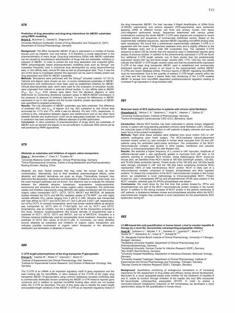
078
Prediction of drug absorption and drug-drug interactions for ABCB1 substrates using PBPK modelling Brück S., Buschner C., Oswald S., Siegmund W. University Medicine Greifswald, Center of Drug Absorption and Transport (C_DAT), Department of Clinical Pharmacology, Germany Background: The efflux transporter ABCB1 (P-gp) is expressed in a number of human tissues such as intestine, liver and kidney. It is therefore a crucial determinant in the pharmacokinetics of many drugs. Drug-drug interactions (DDIs) with ABCB1 substrates can be caused by simultaneous administration of drugs that are substrates, inhibitors or inducers of ABCB1. In order to predict the oral drug absorption and unwanted DDIs, PBPK (physiologically based pharmacokinetic) modelling tools such as Simcyp® are nowadays frequently used in preclinical drug development by using physicochemical data, in vitro affinity data, clinical PK data as well as physiological data. Thus, it was the aim of this study to investigate whether this approach can be used to reliably predict oral drug absorption and DDIs for ABCB1 substrates. Methods: Simulations were performed with the Simcyp® simulator (version 13.1.61.0). Talinolol and digoxin were chosen as non- or poorly metabolized substrates of ABCB1. Erythromycin and clarithromycin have been used as established inhibitors for ABCB1. Clinical pharmacokinetic data, which were used for the evaluation of simulation results, were originated from internal or external clinical studies. In vitro affinity data to ABCB1 (Papp-, KM-, Vmax-, IC50 values) were taken from the literature (digoxin) or were determined by conducting directional transport assay in MDCK-ABCB1-monolayers in the presence or absence of inhibitors (talinolol). To account for differences in ABCB1 expression in our in vitro sytems and the human intestine, protein abundance of ABCB1 was quantified by targeted proteomics. Results: The oral absorption of ABCB1 substrates was fairly predicted. The difference of predicted AUC and Cmax of digoxin (0.5 mg, SD) compared to real clinical data accounted for +17% and +7%, while it was for talinolol -4% and +15%, respectively. In contrast to this, interactions between digoxin and clarithromycin and on the other hand between talinolol and erythromycin could not be adequately predicted. No improvement in prediction has been achieved by different attempts of profile optimization. Conclusion: In silico prediction of pharmacokinetics of drugs which are substrates of ABCB1 such as talinolol and digoxin is not yet sufficient. In particular DDIs cannot yet be well predicted by PBPK approaches.
079
Alkaloids as substrates and inhibitors of organic cation transporters Chen J.1, Brockmöller J.1, Chen X.2, Tzvetkov M.1 1University Medicine Center Göttingen, Clinical Pharmacology, Germany 2China Pharmaceutical University, Centre of Drug Metabolism and Pharmacokinetics Biodrug Division, Nanjing, China Alkaloids are toxic herbal products that may enter the human body as food contamination. Alternatively, due to their beneficial pharmacological effects, some alkaloids and alkaloid derivatives are given as drugs. Transcellular transport may determine the absorption, distribution and elimination of alkaloids in the human body, but the exact transport pathways are not known. Our objective was to evaluate the interplay between the alkaloids atropine, homatropin, scopolamine, butylscopolamine, senecionine and anisodine and the human organic cation transporters. We performed uptake and inhibition experiments using HEK293 cells stably transfected with the human organic cation transporters OCT1, OCT2, OCT3, MATE1 and MATE2K. All alkaloids studied inhibited the OCTs and MATEs mediated uptake of [3H] MPP+, but with highly variable affinities (IC50 values ranging from 3 µM to 2 mM). Atropine was transported with high affinity by OCT1 and OCT2 (Km of 1.3±0.3 µM and 3.0±0.7 µM, respectively), but not by OCT3. In contrast homatropine, which has similar medical effects as atropine, was transported by OCT3 (Km of 71.9±4.3µM), but not by OCT1 and OCT2. Scopolamine, was an inhibitor, but not a substrate for all the transporters screened in this study. However, butylscopolamine (the N-butyl derivate of scopolamine) was a substrate of OCT1, OCT2, OCT3 and MATE1, but not of MATE2-K. Anisodine is a Chinese medicine traditionally used for transmissible shock treatment. Anisodine was a substrate of OCT2 (Km values of 22,2±1,5 µM). In conclusion, our study identifies several alkaloids as substrates and inhibitors of organic cation transporters and indicates possible involvement of organic cation transporter in the absorption, distribution and elimination of alkaloids in human.
080
3’-UTR length polymorphisms of the drug transporter P-glycoprotein Drerup K.1, Kaehler M.1, Röder C.2, Cascorbi I.1, Bruhn O.1 1Institute of Experimental and Clinical Pharmacology, Kiel, Germany 2Institute for Experimental Cancer Research, 2nd Division of Molecular Oncology, Kiel, Germany The 3’-UTR of an mRNA is an important regulatory motif of gene expression and the main binding site for microRNAs. In silico analysis of the 3’-UTR of the major drug transporter ABCB1 (P-glycoprotein) using common databases revealed conflicting data to commercially distributed vectors containing the ABCB1 3’-UTR linked to reporter gene constructs and in addition to reported microRNA binding sites, which are not located within the 3’-UTR as described. The aim of this study was to identify the exact length and possible length variations of the ABCB1 3’-UTR as an important regulatory motive of
the drug transporter ABCB1. For that, two-step 3’-Rapid Amplification of cDNA Ends (3’-RACE) experiments and various standard PCR-experiments were performed applying cDNA of different human cell lines and primary human liver tissue (non-malignant peritumoral tissue). Sequences determined with various primer combinations covering the whole ABCB1 3’-UTR were aligned and compared to recent database entries and sequences of commercially distributed vectors. Based on the experimentally validated sequences, reported ABCB1-regulating microRNA binding sites were reanalyzed. We found that a predominant experimentally validated 3’-UTR is in agreement with the recent 1000genomes database entry and is slightly different to the NCBI database entry and is in total 384 nucleotides long. The validated 3’-UTR sequence is about 230 bp shorter than the sequence used in established reporter gene assays of previous studies. In addition to the predominantly expressed 3’-UTR we found four additional 3’-UTR length variants, one of them shorter than the predominantly expressed variant (247 bp) and three longer variants (902, 1170, 1222 bp). Our results indicate that ABCB1 3’-UTR length variants exists and that the predominantly expressed 3’-UTR of the major drug transporter ABCB1 used in established and commercially distributed reporter gene assays is not equal to the experimentally verified ABCB1 3’-UTRs and recent database entries. Previous reports of ABCB1 regulating microRNAs must be reconsidered. Due to the quantity of existent 3’-UTR length variants within the cell lines and the liver tissue it seems likely that shortening of the 3’-UTR enables ABCB1 to escape from microRNA dependent posttranscriptional regulation leading to increased P-gp expression in drug-resistant cells.
081
Molecular basis of NCX dysfunction in patients with chronic atrial fibrillation Ghezelbash S.1, Molina C. E.1, Badimon L.2, Voigt N.1, Heijman J.1, Dobrev D.1 1University Duisburg-Essen, Institute of Pharmacology, Germany 2Centre d'Investigació Cardiovascular CSIC-ICCC, Barcelona, Spain Introduction: Altered NCX function has been implicated in cellular ectopic (triggered) activity in patient with long-standing persistent (chronic) atrial fibrillation (cAF). However, the molecular basis of NCX dysfunction in cAF patients is largely unknown and was the major focus of the present investigation. Methods: Right atrial tissue samples were obtained from sinus rhythm (Ctl) or cAF patients undergoing open heart surgery. The NCX-mediated calcium-dependent transient inward current (Iti) was recorded in human atrial myocytes from Ctl and cAF patients using the perforated patch-clamp technique. The composition of the NCX1 macromolecular complex was studied in atrial lysates, membrane and cytosolic fractions, or immunoprecipitates (IPs) with Western blot. Results: We detected a higher frequency of Iti current in cAF myocytes. Amplitude of caffeine-induced peak Iti was significantly greater in myocytes from cAF than in Ctl patients, pointing to increased NCX function. Using heterozygous NCX1 knock-out mouse atria, we identified three NCX1 bands at 160 kDa (full-length protein), 120 kDa, and 70 kDa (likely proteolytic bands). Protein levels of the 160 kDa and 120 kDa bands were strongly increased in cAF and the 160 kDa band (underlying functional NCX) represented >90% of total membraneous NCX1 protein. NCX1 is likely part of a macromolecular complex comprised of kinases, phosphatases and other regulator proteins. To dissect the composition of the NCX1 macromolecular complex in the human atrium, we established a novel methodology to immunoprecipitate NCX1. Protein kinase-Cα, type-1 phosphatase (PP1a) and phospholemman (PLM) were present in the NCX1 complex, whereas type-2A (PP2A) phosphatase was absent. Conclusions: We could show for the first time that protein kinase-Cα, PP1a and phospholemman are part of the NCX1 macromolecular protein complex in the human atrium. In addition to the strong increase of NCX1 protein in the plasma membrane of cAF patients, a dysbalance between kinase and phosphatase activities within the NCX1 macromolecular complex may constitute a novel mechanism for the proarrhythmic NCX dysfunction during AF.
082
LC-MS-based bile acid quantification in human blood: a tool to monitor hepatitis B therapy by a novel Na+-taurocholate cotransporting polypeptide inhibitor Haag M.1, Hofmann U.1, Mürdter T. E.1, Heinkele G.1, Leuthold P.1, Blank A.2,3, Haefeli W.2,3, Alexandrov A.4, Urban S.5,3, Schwab M.1,6,7 1Dr. Margarete Fischer-Bosch Institute of Clinical Pharmacology, University of Tübingen, Stuttgart, Germany 2Heidelberg University Hospital, Department of Clinical Pharmacology and Pharmacoepidemiology, Germany 3Heidelberg University, German Center for Infection Research (DZIF), Germany 4Myr GmbH, Bad Homburg, Germany 5University Hospital Heidelberg, Department of Infectious Diseases, Molecular Virology, Germany 6University Hospital Tuebingen, Department of Clinical Pharmacology, Institute of Experimental and Clinical Pharmacology and Toxicology, Tübingen, Germany 7German Centre for Infection Research (DZIF), Tübingen, Germany Background: Quantitative monitoring of endogenous biomarkers is of increasing importance for the assessment of drug safety and efficacy during clinical development. Myrcludex B, a novel lipopeptide-based entry inhibitor for the treatment of hepatitis B and D, exerts its function through inhibition of the hepatic bile acid (BA) transporter Na+-taurocholate cotransporting polypeptide (NTCP). In order to assess a myrcludex-induced metabolomic response on BA homeostasis we developed a mass spectrometric assay for BA quantification in human blood.
S21
Methods: Using quadrupole time-of-flight mass spectrometry 15 BAs were analyzed by accurate mass analysis after separation of isomers by reversed-phase chromatography. Protein precipitation was performed in the presence of deuterium-labeled internal standards which allowed absolute BA quantification in low amounts of serum/plasma. The method was validated according to FDA guidelines and applied to monitor the effect of myrcludex B treatment on plasma BA level. Results: Dynamic quantification was achieved in the range from 7.8 nM to 10000 nM depending on the BA species analyzed. Intraday- and interday accuracy and precision were in the 15% tolerance range for all analytes. Recoveries and matrix effects were between 65-83% and 39-104%, respectively. Mean basal level of BAs ranged from 11 nM tauroursodeoxycholic acid (TUDCA) to 871 nM glycochenodeoxycholic acid (GCDCA) in serum and from 15 nM (TUDCA) to 1321 nM (GCDCA) in plasma. Myrcludex-induced NTCP inhibition resulted in a significant elevation of conjugated BAs (fold-change, > 28), non-conjugated BAs were affected partially. Conclusion: LC-MS-based therapeutic monitoring of endogenous biomarkers has been successfully established and applied to study the effect of myrcludex B treatment on human BA metabolism. The results obtained by our assay demonstrate that a myrcludex-induced NTCP inhibition drastically affects human BA homeostasis. This observation provides valuable insights into the drug´s mode of action and will be indispensable for the assessment of side effects and dose-finding processes during future clinical trials. Bile acids, mass spectrometry, Na+-taurocholate cotransporting polypeptide (NTCP), hepatitis B, hepatitis D
083
Investigation of the molecular conformation of OATP2B1 using Förster resonance energy transfer (FRET) Hagen P.1, Jedlitschky G.1, Schellenberger D.1, Siegmund W.1,2, Grube M.1 1University Medicine Greifswald, Department of Pharmacology, Germany 2University Medicine Greifswald, Department of Clinical Pharmacology, Germany Introduction: Organic anion-transporting polypeptides represent a family of multispecific uptake transporters for a wide range of drugs as well as endogenous compounds like statins and steroid-conjugates. So far, little is known about the molecular structure of these transporters. On that account we applied intramolecular FRET (Förster resonance energy transfer) to investigate the conformational assembly of different intracellular domains of the OATP family member OATP2B1. We therefore modified the OATP2B1 using the fluorescence proteins ECFP and EYFP. The fusion proteins were used to investigate intramolecular FRET in dependence of the labelling position. Methods and Results: In a first step the ECFP was fused to the C-terminus of OATP2B1. Next, the EYFP was inserted into the N-terminal cytoplasmic domain as well as into the third intracellular loop, creating two different fusion proteins. HeLa cells were transiently transfected with those constructs. FRET measurements were performed by confocal laser scanning microscopy using the acceptor photobleaching as well as the sensitized emission methods. Both constructs were inserted at least in part correctly into the plasma membrane and FRET signals were obtained from both fusion proteins. In dependence of the EYFP position, different FRET efficiencies were detected: While the N-terminal EYFP construct demonstrated a FRET efficiency of 7 % (determined by the acceptor photobleaching method), the FRET efficiency of the construct with the EYFP in the intracellular loop was 15 %, indicating a close spatial proximity of this loop to the C-terminal domain. Conclusions: Intramolecular FRET can provide a useful tool to investigate the molecular conformation of organic anion-transporting polypeptides. The work was supported by German Federal Ministry of Education and Research (grant: BMBF InnoProfile Transfer 03IPT612A)
084
Structure Determination of Cell-Free Expressed Transmembrane Transporters Holm-Bertelsen J., Beitz E. University of Kiel, Pharmaceutical Chemistry, Germany Finding new targets for the therapy of infectious diseases is still a topical subject, especially as the resistance of pathogens is an increasing problem. Potential targets might be transmembrane transporters, which take part in the energy metabolism of microorganisms. The knowledge of such protein structures is essential for drug development and can only be acquired by the production of large amounts of protein. An excellent tool to generate these quantities in a short time is the use of the cell-free system. Further advantages are also direct solubilization by detergents and the possibility to optimize to a majority of correctly folded protein. We started with the determination of suitable detergents for expression and purification with the help of green fluorescent protein fusion proteins.[1] A plasmodial lactate-transporter (PfFNT) in varying detergents showed up with differences in the elution profiles of affinity and size exclusion chromatography, however compared to E. coli expressed PfFNT the purification seems to be comparable. Thus indicates that the detergent plays a main role in formation and stabilization of quaternary structure. One important point to verify correct folding is the proof of functionality, which we could demonstrate by stopped flow measurements after reconstitution into liposomes. First crystallization trials were already set up. Next we will advance the buffer conditions for crystallization and to acquire a major variety for screening make some modifications in type of detergent and protein length. [1] Müller-Lucks A., Bock S., Wu B., Beitz E., PLoS One. 2012;7(7):e42186
085
Several adaptor proteins promote intracellular localization of the transporter MRP4/ABCC4 in platelets and hematopoietic cells Schaletzki Y.1, Bröderdorf S.1, Kromrey M. - L.1, Hammer E.2, Grube M.1, Hagen P.1, Greinacher A.3, Rauch B.1, Kroemer H. K.1, Jedlitschky G.1 1University Medicine Greifswald, Dept. of Pharmacology, Germany 2University Medicine Greifswald, Dept. of Functional Genomics, Germany 3University Medicine Greifswald, Dept. of Immunology and Transfusion Medicine, Germany Background: Multidrug resistance protein 4 (MRP4/ABCC4) is a markedly versatile transporter exhibiting a broad substrate specificity comprising several drugs as well as a number of endogenous mediators. It is expressed in several tissues and in blood cells including platelets. In resting platelets, MRP4 was demonstrated to be mainly localized in intracellular storage compartments such as the delta-granules. This localization distinguishes the MRP4 expression in platelets from that in other cell types, where it is mainly localized at the plasma membrane. Accordingly, a diminished localization and function of MRP4 was observed in platelets from patients with delta-storage pool deficiencies, including Hermansky-Pudlak syndrome (HPS).1 It has been recognized that protein-protein interactions are important for transporter localization and function. Methods and Results: We therefore screened for proteins interacting with MRP4 in platelets and hematopoietic cells. Adaptor proteins binding to C-proximal sequences of MRP4 were identified using GST pull-down and co-immunoprecipitation assays followed by immunoblot and mass spectrometry analyses as well as by co-localization studies in platelets. The identified binding proteins comprise the PDZ scaffolds ezrin-binding protein 50 (EBP50/NHERF), postsynaptic density protein 95 (PSD95), and sorting nexin 27 (SNX27), but also the adaptor-related protein complex 3 (AP3) and the heat shock protein 90 (HSP90). Knockdown of SNX27, PSD95, and AP3 subunit beta 1 (AP3B1) by siRNA in megakaryoblastic leukemia M07e cells led to a significant shift of MRP4 from intracellular structures to the plasma membrane, indicating that these proteins are important for the correct localization and function of MRP4 in blood cells. Conclusions: Several PDZ scaffold proteins and the adaptor protein complex AP-3 promote intracellular localization of the transporter MRP4/ABCC4 in platelets and hematopoietic cells. Interestingly, mutations in the AP3 subunit beta 1 (AP3B1) are also known to cause HPS. The work was supported by the Deutsche Forschungsgemeinschaft (DFG) through grant JE 234/4-1 to GJ. 1Jedlitschky G. et al. (2012), Blood, 119:3394
086
Site-specific MIRNA expression influences / Controls transporter protein abundance along the human intestine Martin P.1, Bruckmueller H.1, Cascorbi I.1, Drozdzik M.2, Oswald S.3, Haenisch S.1, Siegmund W.3 1University-Hospital Schleswig-Holstein, UKSH, Campus Kiel, Institute of Experimental and Clinical Pharmacology, Germany 2Pomeranian Medical University, Department of Experimental and Clinical Pharmacology, Szczecin, Poland 3University Medicine of Greifswald, Ernst-Moritz-Arndt-University, Department of Clinical Pharmacology, Germany Background: After oral administration, intestinal drug absorption depends on various factors including intestinal motility, availability of water for dissolution and site-specific pH-value. Moreover, in dependence of the anatomical localization varying expression of efflux- and uptake-transporters make the gut a complex barrier for the drug transfer into the body. In a recent study, we could determine the site-specific protein levels of in total ten transporters along the entire length of the gut. Interestingly, some transporters showed clear differences in their expression between the small intestine and the colon. miRNAs are highly tissue-specific expressed post-transcriptional regulators of gene expression. Hence, in our current study, we aim to investigate the impact of miRNAs on site-specific transporter expression along the human intestine. Methods: Total RNA was isolated from biopsies obtained from six healthy organ donors. Tissue samples were acquired from the duodenum, the upper and lower jejunum, the upper and lower ileum, and the transversal or descending colon. The expression of 754 miRNAs was measured using rt-PCR based low density arrays. Expression of all detected miRNAs was correlated with transporter protein data recently determined by LC-MS/MS-based targeted proteomics. miRNAs and transporter genes showing an inverse Pearson’s correlation between miRNA and protein expression underwent an in-silico search (MicroCosm Targets v.5) for putative miRNA/mRNA interaction. Those predicted interactions are currently under rigorous reporter gene assay-based testing. Results: Out of 754 miRNAs, 248 were detected in all tissue types. Out of ten transporters five showed significant inverse correlations with in total 12 putative targeting miRNAs (e.g. ABCG2 and hsa-miR-212-3p, r=-0.04, p= 0.017; PEPT1 and hsa-miR-27a, r= -0.498 p=0.002). Conclusion: Our data indicate that under physiological conditions miRNAs may be involved in the regulation of site-specific abundance of intestinal drug transporters.
S22

087
Cellular Uptake of Sorafenib into Hepatocellular Carcinoma Cells Is Independent of Human Organic Cation Transporter 1 (OCT1) Neul C.1, Baker S. D.2, Sparreboom A.2, Schaeffeler E.1, Schwab M.1,3, Nies A. T.1 11Dr. Margarete Fischer-Bosch Institute of Clinical Pharmacology, Stuttgart, Germany 2St. Jude Children's Research Hospital, Pharmaceutical Sciences, Memphis, United States 3University Hospital, Institute of Experimental and Clinical Pharmacology and Toxicology, Clinical Pharmacology, Tübingen, Germany Purpose The protein kinase inhibitor sorafenib is an orally active multikinase inhibitor and significantly increases survival of patients with advanced clear-cell renal or hepatocellular carcinomas.1 Sorafenib has a dual mode of action: (i) it inhibits cell division and cell proliferation by inhibiting the RAF/MEK/ERK pathway and (ii) it also targets the VEGF receptor, which causes a reduction of tumor angiogenesis so that the blood supply of the tumor cells is inhibited. After oral administration, sorafenib undergoes either oxidative metabolism, resulting in the primary metabolite sorafenib N-oxide, or glucuronidation. As sorafenib targets intracellular kinases, it is very important to know how sorafenib and the active metabolite sorafenib N-oxide pass the cell membrane of tumor cells. The organic cation transporter OCT1 (SLC22A1) has been suggested by some groups to contribute to sorafenib uptake2,3 whereas other groups could not show any interaction of sorafenib with OCT1.4,5 Because of these conflicting data, we used a combination of different approaches including animal experiments to elucidate comprehensively the impact of OCT1 on cellular sorafenib uptake. Experimental design Transport of sorafenib and sorafenib N-oxide were analyzed using human hepatocellular carcinoma cells (HepG2, Huh7), the mammalian cell line HEK293 stably expressing OCT1, and Oct1-knockout mice. Results The overexpression of functional OCT1 protein in HEK cells did not result in an increased cellular uptake of sorafenib or sorafenib N-oxide. Huh7 cells and HepG2 cells showed a considerable sorafenib uptake although they did not express OCT1 mRNA. Oct1 deficiency in mice had no influence on plasma and hepatic sorafenib and sorafenib N-oxide concentrations. Conclusion Our results clearly show that uptake of sorafenib and sorafenib N-oxide is independent of OCT1 expression and that other, yet unidentified transporters are responsible for cellular sorafenib uptake. Supported by the Robert Bosch Foundation, Stuttgart, Germany. References 1 Wilhelm SM, et al. Mol Cancer Ther 2008; 7: 3129-40. 2 Swift B, et al. Drug Metab Dispos 2013; 41: 1179-86. 3 Herraez E, et al. Hepatology 2013; 58: 1065-73. 4 Hu S, et al. Clin Cancer Res 2009; 15: 6062-69. 5 Johnston RA, et al. Drug Metab Dispos 2014; 42: 1851-57.
088
Characterization of the expression and function of endogenous transporters in widely used cellular models Otter M., Eriksson P. - O., Siegmund W., Oswald S. University Medicine Greifswald, Center of Drug Absorption and Transport (C_DAT), Department of Clinical Pharmacology, Germany Background: Drug transporters are known to be important determinants in the pharmacokinetics and efficacy of many drugs. In order to characterize the affinity or inhibitory properties of drugs to transporter proteins in preclinical drug development, transporter-overexpressing cellular models based on MDCKII and HEK293 cells are widely used and recommended by current guidelines of medical authorities. In this study, we investigated the endogenous (“background”) transporter expression in frequently used HEK293 and MDCKII cells. Furthermore, we examined the functional consequences of endogenous ABCB1 in cellular uptake studies. Methods: The following wild-type and stably transfected cells have been investigated: HEK293 cells: wild type, vector control, OATP1A2, OATP1B1, OATP1B3, OATP2B1; MDCKII cells: wild-type, vector control, ABCB1). The mRNA expression of 5 human ABC and 16 SLC transporters (HEK293 cells) and 16 canine transporters (MDCKII cells) were determined by validated TaqMan® gene expression assays. Protein abundance was quantified by LC-MS/MS-based targeted proteomics. The functional study was performed with talinolol as substrate of OATP1A2 and ABCB1 using wild-type, stably transfected vector control and OATP1A2-overexpressing HEK293 cells. Intracellular accumulation after 5 min and 30 min incubation of [3H]-talinolol was measured by liquid scintillation counting after cell lysis. Results: The observed gene expression and protein abundance data were mostly not correlated. Several transporters could be identified as endogenous transporters in both cell lines. In HEK cells, the endogenous ABCB1 protein content was significantly lower in vector control, OATP1A2, OATP1B1, OATP1B3 and OATP2B1-transfected cells compared to wild-type. On the contrary, markedly higher expression has been observed for endogenous ABCC2 in HEK-OATP1B1 and for endogenous ABCC3 in MDCK-ABCB1. Influx rate per minute of talinolol in wild type, vector control and OATP1A2 was significantly decreased after 30 min in comparison to 5 min incubation. Higher
endogenous ABCB1 expression in wild-type cells resulted in tendency in reduced intracellular accumulation of talinolol compared to vector control and OATP1A2-transfected cells. Conclusion: There are markedly differences in expression of endogenous transporters in the investigated cell lines. These endogenous transporters may affect drug transport and the estimated affinity to the focused transport protein.
089
Characterization of a Plasmodium falciparum lactate/H+ symporter Rambow J., Wu B., Bock S., Holm-Bertelsen J., Wiechert M., Beitz E. University of Kiel, Pharmaceutical Chemistry, Germany Plasmodium falciparum causes the most severe malaria infections in humans and almost entirely relies on glycolysis to fulfill its energetic needs. After invading the erythrocytes the glucose consumption can increase up to 100 fold compared to uninfected red blood cells. This leads to large quantities of lactate that have to be removed in order to keep the cells viable. Until now, the transport proteins through which lactate is exported from plasmodia are unknown. Yet, such proteins must be considered as potential new drug targets. Recently we identified a transporter (PfFNT), i.e. a member of the microbial formate-nitrite transporter family, as a lactate/proton symporter in Plasmodium falciparum. For the biochemical characterization we used a double knock out strain of baker’s yeast Saccharomyces cerevisiae , Δjen1Δady2, that is unable to transport monocarboxylates. Using 14-C-radiolabled substrates we elucidated the transport kinetics, inhibition profile and substrate specificity of PfFNT. Further, in collaboration with Tobias Spielmann of the Bernhard-Nocht-Institut, Hamburg, we localized PfFNT in the plasma membrane of living parasites. All obtained data point toward a central role of PfFNT in the P. falciparum energy metabolism underscoring its potential as a new antimalarial drug target.
090
Aquaporin-mediated drug resistance of trypanosomes Rothert M.1, Song J.1,2, Beitz E.1,1 1University of Kiel, Pharmaceutical chemistry, Germany 2University of Kiel, Physiology, Germany A gene knock-down of the aquaglyceroporin TbAQP2 in Trypanosoma brucei being the pathogen of sleeping sickness leads to pentamidine/melarsoprol cross-resistence but keeps the cells viable[1]. Therefore TbAQP2 is suggested to play an important part in the pentamidine-uptake. Based on our long-standing experience with AQP selectivity filters, we doubt that a large positively charged molecule as pentamidine can pass the AQP channel. The canonical amino acid composition of the AQP selectivity filter is an arginine in an aromatic environment (ar/R). TbAQP2, however, carries a leucine instead of arginine exposing an aspartate to the channel lumen. We found that pentamidine acts as a high affinity binder and inhibitor of TbAQP2 (IC50= 130 nM) with Asp (D265) being the crucial compound for electrostatic interaction with the drug. A milieu of pH 2.5 increases the IC50 tenfold and point mutations of the Asp (D265N, D265A), removing the charge, effects it within the same range. We further replaced Leu by Arg in order to mimic the situation found in classical AQPs. The mutant (L264R) showed the typical glycerol swelling behavior both in absence and after addition of pentamidine prior to the light scattering assay. Furthermore small positively charged molecules such as 3-amino-1,2-propanediol and formamidine were not able to pass the TbAQP2 channel. Based on our data we come to the conclusion that pentamidine cannot pass TbAQP2 due to the large size and the presence of two positive charges. We assume that the uptake occurs by receptor-mediated endocytosis with TbAQP2 serving as the receptor. To visualize the postulated uptake mechanism we are synthesizing microscopy-compatible fluorescence labelled pentamidine. [1] Alsford, S. et al. (2012) Nature 482: 232-236
091
Transport mechanism of Formate-Nitrite Transporters Wiechert M., Beitz E. University of Kiel, Department of Pharmaceutical Chemistry, Germany The steady increase of antimicrobial resistance shows the limitations in our antibiotic treatment options. Since the formate-nitrite transporter (FNT) family is only provided in microorganisms, where they are involved in essential metabolic processes, they represent potential protein targets in the development of antimicrobial substances. FNTs are pentameric channels that transport small monovalent anions. Each protomer in the channel has a pore with a central hydrophobic cavity that is separated from both
S23
sides of the membrane by a narrow constriction [1]. Following from this, the question arises whether the anion substrate is protonated during transport in order to pass the hydrophobic channel regions and which residues are involved in protonation. There is a central histidine which forms hydrogen bonds to a threonine via a fixed water molecule. Moreover, a highly conserved Lysine is located in the extracellular pore entrance. We transformed yeast with the prototypical EcFocA and determined the effect of mutations of residues or chemical modification of the protein by radiolabeled functional assays. Exchange of the central histidine to asparagine, glutamine or alanine yielded a non-functional transporter. Equally, changing the neighboring threonine to valine abolished transport. Yet a serine variant showed attenuated functionality. The replacement of lysine to cysteine yielded a functional transporter. However, contrary to wildtype EcFocA the uptake rates at different pH values remained unchanged. Due to these results we can demonstrate that the central histidine is essential for the transport mechanism of FNTs. The replacement of lysine results in a loss of pH-regulation. Further experiments are planned to uncover the decisive role of this lysine for the transport mechanism. For instance, we will convert the cysteine to a lysine analogon by chemical modification. Furthermore, we will carry out uptake assays with deuterium oxide instead of water. As deuterium oxide has a reduced proton mobility, it should be possible to differentiate between a single protonation step and a protonation cascade. [1] Lü et al. 2012, PNAS, PMID: 22847446
092
Hyaluronan promotes differentiation and modulates function of white and brown adipocytes: implications for obesity and insulin resistance Bayer J. K., Grandoch M., Fender A. C., Fischer J. W. Institut für Pharmakologie und Klinische Pharmakologie, Universitätsklinikum Düsseldorf, Heinrich-Heine-Universität Düsseldorf, Germany Hypertrophy and chronic inflammation of white adipose tissue (WAT) is linked to the development of insulin resistance and type II diabetes, while brown adipose tissue (BAT), due to its capability to conduct thermogenesis, has been described as a potential anti-obesity target. As shown previously, inhibition of hyaluronan (HA) synthesis impairs WAT differentiation, thereby preventing development of severe obesity and insulin resistance. However, underlying mechanisms of HA promoting WAT development as well as HA-mediated effects on BAT are yet unknown. Inhibition of HA synthesis by 4-methylumbelliferone (4-MU) in vitro reduced adipogenesis in 3T3-L1 cells, human mesenchymal stem cells and preadipocytes isolated from murine adipose tissue. This effect was mediated via hyaluronan synthase 2 (HAS2) and the HA receptors CD44 and RHAMM, as evidenced by reduced differentiation of 3T3-L1 cells transfected with Has2, Cd44 and Rhamm siRNA. In vivo, 4-MU treatment for 22 weeks resulted in significantly less weight gain and improved insulin sensitivity in male C57BL/6J mice on diabetogenic diet (DD). Total body fat content and adipocyte size was reduced in 4-MU treated animals, although food intake was not affected. Additionally, H&E staining of liver revealed that mice subjected to 4-MU were protected from hepatic steatosis. Since increased energy expenditure in mice on DD 4-MU suggested changes in thermogenesis, BAT was analyzed. Similar to findings in WAT, brown adipocyte size was reduced and BAT appeared to be denser after 4-MU treatment, consistent with reduced lipid storage of brown adipocytes. Furthermore, both qRT-PCR and immunohistochemical staining of BAT revealed an increase in Ucp-1 expression, indicating higher activation of BAT due to 4-MU treatment. In summary, the present data suggest that inhibition of HAS2-dependent HA synthesis reduces both white and brown adipocyte differentiation and increases BAT activity, which presumably contributes to reduced weight gain and improved insulin sensitivity.
093
Epidermal growth factor induced serine 727 phosphorylation of hypothalamic STAT-3 enhances TRH and SOCS-3 expression independently from tyrosine 705 phosphorylation Breit A., Besik V., Gudermann T. LMU München, Walther-Straub-Institut, Germany Transcriptional activity of signal transducer and activator of transcription-3 (STAT-3) is a key element in the central regulation of appetite and energy homeostasis. Activation of hypothalamic STAT-3 has been attributed to cytokine-promoted phosphorylation at tyrosine-705 (Tyr-705). In non-hypothalamic cells STAT-3 is also phosphorylated at serine-727 (Ser-727), but the functional significance of Ser-727 in the regulation of hypothalamic STAT-3 is not known. We used two hypothalamic cell lines and analysed effects of various hormones on STAT-3-dependent reporter gene activity and observed that interferon-γ (IFN-γ) and epidermal growth factor (EGF) equally induce STAT-3 reporter activation. EGF solely increased Ser-727 and IFN-γ Tyr-705 phosphorylation of STAT-3, indicating that Ser-727 phosphorylation is sufficient to fully activate hypothalamic STAT-3. Next, we analysed effects of IFN-γ and EGF on the expression of the STAT-3-dependent genes TRH and suppressors of cytokine signaling-3 (SOCS-3). EGF- but not IFN-γ enhanced TRH expression via STAT-3. With regard to SOCS-3, we observed prolonged expression induced by IFN-γ and a transient effect of EGF that
required co-activation of the activator protein-1 (AP-1). Thus, EGF-promoted Ser-727 phosphorylation by ERK-1/2 is not only sufficient to fully activate hypothalamic STAT-3, but in terms of targeted genes, kinetics and required co-factors entails distinct modes of STAT-3 actions, when compared to interferon-γ-induced Tyr-705 phosphorylation.
094
Two putative calcineurin interaction domains within mitogen-activated protein kinase kinase kinase DLK Duque Escobar J.1, Lemcke T.2, Hasenpusch D.2, Oetjen E.1 1Universitätsklinikum Hamburg-Eppendorf, Institut für Klinische Pharmakologie und Toxikologie, Germany 2Universität Hamburg, Institut für Pharmazie, Germany Loss of beta-cell mass is the most important factor for the pathogenesis of type 1 and type 2 diabetes. It is unknown which mechanisms induce beta-cell apoptosis though different hypotheses are postulated. Inhibition of the calcium-calmodulin dependent phosphatase calcineurin has been shown to reduce beta-cell function and induce beta-cell apoptosis. Our previous results showed that inhibition of calcineurin by the structurally distinct immunosuppressant drugs cyclosporin A and tacrolimus decreased insulin gene transcription. Furthermore, both drugs stimulated the catalytic activity of the mitogen-activated protein kinase kinase kinase 12 (DLK Dual Leucine zipper kinase) and induced beta-cell apoptosis. In the present study, the regulation of DLK by calcineurin was investigated. In silico analysis revealed two putative calcineurin interaction domains within DLK. The consensus motifs 273-PNMLIT-278, 362-LPVP-365 were mutated to PNRLKT, APAP and a double mutant, respectively, by primerless PCR. The expression vectors for these mutants and DLK wild-type were transiently transfected into the beta-cell line HIT. Immunoblot analysis using an antibody against the COOH-terminus of DLK showed similar expression levels of the single mutants, whereas the double mutant was barely expressed. Both single mutants exhibited no catalytic activity, measured as phosphorylation of DLK on Ser-302 and phosphorylation of JNK. A DLK homology model was generated, using the crystal structures of MLK1 and BRaf as templates. The model confirmed that 273-PNMLIT-278 interfered with the conformation of the catalytic domain. Mutation of L362 to A and V364 to A within the second putative calcineurin interaction domain 362-LPVP-365 did not reduce DLK protein expression. Whereas the L362A DLK mutant showed no catalytic activity in relation to Ser-302 phosphorylation of DLK and JNK phosphorylation, the V364A DLK mutant resulted in 3-fold increased JNK phosphorylation than DLK wild-type. In addition, in reporter gene assays this mutant was more potent to inhibit KCl/Forskolin-induced CRE-dependent gene transcription than DLK wild-type. Our data identified a super-active DLK mutant. Taken together with our previous results, these data suggest that calcineurin directly inhibits DLK actions.
095
Diabetes Mellitus affecting jaw bone architecture influence periodontal ligament and implants survival Mallios N., Seferos N., Tsamouri M., Tesseromatis C. Medical School University of Athens, Pharmacology, Goudi, Greece According to the World Health Organization (WHO) in November 2012, about 347 million people have diabetes worldwide. Excess of fat and increased body mass index, are associated with metabolic (e.g. glucose intolerance, type2 diabetes mellitus, dyslipidemia) and non-metabolic disorders (e.g. neoplasias, polycystic ovary syndrome, non-alcoholic fat liver disease, glomerulopathy, bone fragility increased fracture risk etc). Diabetes mellitus is associated with high advanced glycation end products concentrations in collagen, hypercalciuria followed by glycosuria, reduced renal function, lower insulin-like growth factor-I, microangiopathy, and inflammation. Moreover bone health is particularly important for patients under diabetes with type I, specially children or adolescents since this age accelerated growth and bone deposition with related metabolic occurs. Type2 diabetes and obesity linked to periodontitis, an irreversible destruction of periodontium complicated with microbial flora resulting in edentulism. Microangiopathy at the bone tissue was suggested as a possible cause of diabetic osteopenia expressed as bone tissue deterioration with impaired bone formation and aletarations in osteocalcin levels. This can lead to infection, periodontal ligament destruction and reduction in bone mass and quality around dental implants. Experimental studies in rats demonstrated regeneration of periodontal ligament and new cement formation on the tooth root by the application of enamel matrix derivative (amelogenin). Amelogenin application seems to protect the alveolar bone from resorption in bothcontrol and experimental groups at 7, 14, 28 days.
S24

096
Neonatal isoflavone exposure interferes with reproductive system of female Wistar rats Müller D.1, Basso F.1, Blei T.1, Kurrat A.1, Soukup S.2, Kulling S.2, Diel P.1 1German Sport University, Department of Molecular and Cellular Sports Medicine, Cologne, Germany 2Max-Rubner-Institute, Department of Safety and Quality of Fruit and Vegetables, Karlsruhe, Germany Soy based infant formulas (SBIF) are used in many western countries as one alternative to breast feeding. Recently there is increasing concern about possible adverse effects of such diets. Soy contains high amounts of isoflavones (ISO), which are hormonal active substances. To give SBIF to infants leads to extremely high serum concentrations of ISO in a sensitive phase of child development. Particularly there are concerns that ISO may affect the development and physiological function of reproductive tract later in life. The aim of our study was to investigate effects of ISO on the development of the reproductive tissues in different exposure scenarios with major focus on neonatal exposure in a Wistar rat model. In our study we address and simulate four different exposure scenarios. Exposure to two different diets started with parental animals one week prior mating, continued during in utero period, maintained through adolescence into adulthood. Animals were exposed to: A, a ISO depleted diet (IDD) and B, same diet enriched with an ISO extract (IRD; 508mg ISO/kg). Pups of each group were randomized into subgroups and fed daily by pipette with ISO-suspension [(32 mg ISO/kg BW) ISO+] and placebo from postnatal day (PND) 1 until PND 23. Rats were sacrificed at PND 23 and 80. Body weight and food intake were not affected by ISO+, lifelong IRD diet increased both by tendency. Interestingly, visceral fat mass was significantly reduced in IRD groups. Vaginal opening (VO), a marker for puberty onset, occurred significantly earlier in animals through ISO+ independently whether the animals were kept on IDD (9.4 days earlier) or IRD (5.5 days earlier). At PND70 we observed an irregular estrus cycle in ISO+ rats. Relative uterine weight at PND23 and 80 and rel. ovarian weight at PND80 were not significantly affected by any diet. Ovary weight at PND23 was lowest in IRD groups however ISO+ had no additional influence. A significant increase of vaginal epithelial height was observed in ISO+ on PND23. In summary, our results indicate that ISO intake during weaning period has an estrogenic effect on prepubertal rats indicated by increased vaginal epithelial heights and earlier VO. In addition the exposure of rats during the period of weaning to ISO resulted in estrus cycle irregularities. That indicates that SBIF may result in adverse effects on reproductive tract even later in life. Toxic and molecular mechanisms inducing these effects need to be characterized in future studies.
097
ß-Tanycytes in the regulation of the hypothalamus-pituitary-thyroid-axis Müller-Fielitz H., Stahr M., Bernau M., Schwaninger M. University of Lübeck, Institute of Experimental and Clinical Pharmacology and Toxicology, Germany The hypothalamic network plays an important role in the regulation of different endocrine and metabolic functions. Largely unknown players in this network are the so-called tanycytes. The cell bodies of these specialized ependymal glial cells contact the cerebrospinal fluid in the wall of the 3rd ventricle and send processes into the hypothalamic nuclei and the median eminence. Our recent findings indicated that tanycytes are chemosensors responding to a number of circulating signals such as thyrotropin-releasing hormone (TRH) and the analog taltirelin with intracellular calcium waves only in ß-tanycytes. This suggested a possible role in regulating of the hypothalamus-pituitary-thyroid (HPT) axis. However, the cellular signalling pathway and the physiological functions of tanycytes are unknown. To investigate the role of tanycytes in regulation of the HPT axis we systematically studied the calcium response with pharmacological and genetic tools by measuring intracellular calcium in brain slices of mice. Here we show that the TRH-R1 receptor stimulated the calcium response in ß-tanycytes via a Gq/11 signalling pathway. To clarify the impact of tanycytes on the TRH release in the median eminence we transduced TRH-positive neurons with an adeno-associated virus to express a mutated muscarinergic receptor which was exclusively activated by clozapine-N-oxide. With this technique it became possible to selectively activate TRH-neurons, which led to a temporal increase of energy expenditure and TSH plasma levels. In animals with a tanycytic-specific as well as a glial knockout of Gq/11 the basal levels of TSH were reduced. In summary, ß-tanycytes are active signalling cells within the brain that respond to TRH and modulate the release of hormones of the HPT-axis.
098
Selenium restores endothelial dysfunction and metabolic profile in type 2 diabetic rats Oztürk Z.1, Gurpinar T.2, Vural K.2, Orenay S.3, Korkmaz M.4, Var A.5 1Izmir Atatürk Research Hospital, Clinical Pharmacology and Toxicology, Turkey 2Celal Bayar University Faculty of Medicine, Department of Pharmacology, Manisa, Turkey 3Celal Bayar University Faculty of Medicine, Department of Medical Genetic, Manisa, Turkey 4Celal Bayar University Faculty of Medicine, Department of Medical Biology, Manisa, Turkey 5Celal Bayar University Faculty of Medicine, Deparment of Medical Biochemistry, Manisa, Turkey Aim: Endothelial dysfunction is responsible for diabetic vascular complications and develops as a result of oxidative stress and its pathological process (1). In this study, we investigated the effects of selenium on endothelial dysfunction and oxidative stress in type 2 diabetic rats. Methods: The rats were divided into 5 groups as control rats, untreated diabetic rats and diabetic rats treated with different doses of selenium (180, 300, 500 mcg/kg/day). In order to develop of the type 2 diabetic rat model, the diabetic rats were fed the high-fat diet until the end of study and control rats were fed regular chow. The diabetic rats were injected intraperitoneally with low dose streptozotocin. Endothelium-dependent and - independent relaxations were determined in rat thoracic aorta. Nicotinamide adenine dinucleotide phosphate (NADPH) oxidase and endothelial nitric oxide synthase (eNOS) mRNA expressions were analyzed by RT-PCR. Fasting blood glucose, lipid profile, lipid peroxidation, insulin and nitric oxide were tested in blood samples of rats. Malondialdehyde, superoxide dismutase, catalase and glutathione peroxidase levels were measured in rat liver samples. Results: RT-PCR results showed that selenium reversed the increased NADPH oxidase expression while decreasing the eNOS expression back to control levels, and improved the impairment of endothelium-dependent vasorelaxation in diabetic aorta. Selenium treatment significantly decreased blood glucose, cholesterol, trigliserid levels and enhanced antioxidant status in diabetic rats. Conclusions: The present study confirmed antioxidant and ameliorative effects of selenium in the treatment of type 2 diabetic rats induced by a high- fat diet combined with streptozotocin injection. Because of insulin resistance and insulin deficiency, the combination of high- fat diet and multiple low- dose of STZ could be an experimental animal model for type 2 diabetes and pharmacological screening (2). Our findings indicate that selenium restores metabolic profile and ameliorates vascular responses and endothelial dysfunction in diabetes via regulation of antioxidant enzymes and nitric oxide release. References: 1. Fox CS, Coary S, Sorlie PD et al, Trends in cardiovascular complications of diabetes.JAMA, J.Am.Med.Assoc. 2004; 292:2495-2499. 2. Zhang M, Lv XY, Li J. The characterization of high-fat diet and multiple low-dose streptozotocin induced type 2 diabetes rat model. Exp. Diabetes Res. 2008; 70:40- 45.
099
Influence of bile acids on the viability of pancreatic beta-cells and islet granularity Pajaziti B.1, Drews G.2, Düfer M.1 1Universität Münster, Institut für Pharmazeutische und Medizinische Chemie, Germany 2Universität Tübingen, Institut für Pharmazie, Germany Question: Type-2 diabetes mellitus (T2DM) is not only characterized by hyperglycemia but also by hyperlipidemia. These diabetes-associated alterations also affect the physiological bile acid pool. Bile acids (BAs) are known to influence regulation of pancreatic beta-cells. As excessive substrate supply is one main cause for malfunction and loss of islet cells in T2DM, we investigated whether BAs can conserve beta-cell viability and islet morphology in the presence of elevated glucose and lipid concentrations. Methods: Islets were isolated from C57Bl/6 mice. Cell death was measured by TUNEL assay. For culture, standard (11.1 mM glucose), glucotoxic (25 or 33 mM glucose) or glucolipotoxic (33 mM glucose/10 µM of the LXR-agonist TO-901317) conditions were used. Histological analysis of islet granularity as a measure for insulin content was performed by image digitalization and processing. Results: Ursodeoxycholic and lithocholic acid (UDCA, LCA, 500 nM) had no effect on cell viability of beta-cells cultured in standard medium with the respective BA for 7 d (n=4 independent preparations). Culture in glucolipotoxic milieu increased apoptotic cell death (7 d, 2.3±0.4 vs. 9.7±1.3 %, n=10, p≤0.001). In the presence of UDCA this harmful effect was markedly attenuated by approximately 45 % (n=6, p≤0.05). Importantly, UDCA lost its protective influence in combination with LCA (n=4). To investigate whether BAs and/or nutrient excess affect pancreatic islet histology alterations in islet granularity were determined. Glucotoxicity decreased islet granularity after 1 d of culture (7.7±0.2 vs. 6.3±0.3 px/µm2, n=33-34 islets, p≤0.001). The effect was even more pronounced after 7 days (n=36-39, p≤0.001). UDCA protected against the loss of islet granularity induced by 25 mM glucose (n=30, p≤0.01). In standard medium (11 mM glucose) LCA significantly reduced islet granularity (3 d, 6.7±0.3 vs. 5.0±0.3 px/µm2, n=31, p≤0.001) whereby co-incubation with UDCA prevented this effect (n=34, p≤0.001). Conclusion: BAs influence beta-cell viability and islet morphology differently. The hydrophilic UDCA attenuates the effect of elevated nutrient supply on both parameters. By contrast, the lipophilic LCA lowers islet granularity per se. Of particular interest, UDCA and LCA
S25
influence each other in an antagonistic way. As UDCA protects against the negative effects of LCA and gluco(lipo)toxicity, supplementation of UDCA may represent a promising strategy to prevent beta-cell dysfunction in T2DM.
100
Weight loss after chronic AT1-receptor blockade can at least partially be attributed to an Ang(1-7)-dependent mechanism Schuchard J.1, Winkler M.1, Stölting I.1, Santos R. A.2, Bader M.3,4, Raasch W.1,5 1University of Lübeck, Institute of Experimental and Clinical Pharmacology and Toxicology, Germany 2Federal University of Minas Gerais, Department of Physiology and Biophysics, Institute of Biological Sciences, Belo Horizonte, Brazil 3Max-Delbrück-Center for Molecular Medicine (MDC), Berlin, Germany 4DZHK (German Centre for Cardiovascular Research), partner site Berlin, Germany 5DZHK (German Centre for Cardiovascular Research), partner site Hamburg/Kiel/Lübeck, Lübeck, Germany We and others have repeatedly demonstrated weight loss in rats that had been treated with AT1 blockers (ARB). Weight loss was mostly found after high ARB dosing and was not related to a reduction in blood pressure. The underlying mechanisms still remain a matter of debate due to the paradoxical finding that even AngII lowers body weight. Considering our observations that not only AngII but also Ang(1-7) is increased under AT1 blockade and that development of obesity could be prevented in transgenic rats over-expressing Ang(1-7), we aimed in this study to investigate whether the ARB-induced weight loss can at least partially be attributed to an Ang(1-7)-dependent mechanism. All rats had free access to standard chow and a cafeteria diet (CD; consisting of various chocolate/cookie bars). One group of SD rats was treated with telmisartan (TEL, 8 mg/kg/d) whereas a 2nd and 3rd group received the Mas antagonist A779 (24 or 72 µg/kg/d) in addition to TEL via subcutaneously implanted osmotic minipumps. A 4th group received vehicle instead of TEL. Rats that were treated only with TEL or vehicle received saline instead of A779 also by minipumps. All animals were monitored regarding gain in body weight, energy intake, glycemic control (OGTT, 1 g glucose/kgBW), blood pressure (plethysmography), energy expenditure (indirect calorimetry) and locomotion (infrared sensors). Vehicle+saline-treated controls were hypertensive. Blood pressure was normalized by TEL but not further influenced by A779. Left ventricular weight was diminished according to blood pressure reduction. Gain in body weight was lessened by TEL. The antiobese efficacy of TEL was reduced when rats additionally received 24 (31%, ns) or 72 µg/kg/d A779 (60%, p). Summarizing all these findings, we conclude that the antiobese effect of TEL can at least partially be attributed to an Ang(1-7) mechanism.
101
Weight loss after chronic AT1-receptor blockade can be attributed to a central mechanism Winkler M.1, Schuchard J.1, Stölting I.1, Bader M.2,3, Raasch W.1,4 1University of Lübeck, Institute of Experimental and Clinical Pharmacology and Toxicology, Germany 2Max-Delbrück-Center for Molecular Medicine (MDC), Berlin, Germany 3DZHK (German Centre for Cardiovascular Research), partner site Berlin, Germany 4DZHK (German Centre for Cardiovascular Research), partner site Hamburg/Kiel/Lübeck, Lübeck, Germany AT1-receptor blockers (ARB) are established for treatment of hypertension. In recent studies we demonstrated that ARB diminished food intake, weight gain and fat mass in rats, while energy expenditure was enhanced. Moreover, ARB induced alterations in hypothalamic mRNA levels of (an)orexigenic peptides and restored leptin sensitivity suggesting a central mechanism in regulating food behavior. The aim of this study was to verify whether the central Renin-Angiotensin-System (RAS) has impact on weight regulation and food behavior. As a model of a brain specific impaired RAS we used transgenic rats [TgR(ASrAogen)L680] (TG) with a brain specific deficiency of angiotensinogen (AOGEN).We had already found, that these rats do not develop diet obesity. Age matched Sprague Dawley (SD) rats were used as wild type controls. TG and SD rats were fed with high calorie cafeteria diet (CD, consisting of various chocolate/cookie bars, calorie values 20.3 kJ/g) or standard chow (11.7 kJ/g) for 3 months. Rats were simultaneously treated with telmisartan (TEL, 8 mg/kg/d, by gavage) or vehicle for 3 months. At the end of the feeding-/treatment period various functional tests addressing food behavior and glucose control were performed. TEL induced weight loss as well as normalization of leptin and lipid levels in SD but not TG rats. Energy intake was lower in TG than in SD rats. TEL treatment strain specifically lowered energy intake of SD rats which corresponds to their high POMC levels. TEL treatment improved leptin sensitivity in SD but not in TG rats, since food intake and gain in body weight to exogenous leptin administrations were lower in SD than in TG rats. Energy expenditure (determined by indirect calorimetry) was higher in TG than in SD rats. A lower respiratory ratio in response to TEL was only observed in SD rats. This indicates higher fat burning and corresponded to the lower fat mass of these rats (determined by MRI). Glucose utilization in OGTT (1 g glucose/kgBW) was strain dependently improved by TEL in SD rats since glucose control was impaired only in SD
and not in TG rats by CD feeding. TEL treatment equally reduced blood pressure and enhanced AngII levels in both rat strains. We conclude that weight loss of diet-induced obese rats after chronic TEL treatment is at least partially mediated by a central mechanism since this effect was not observed in brain-specific AOGEN deficient rats.
102
BMP2K plays a crucial role as mediator of inflammatory and angiogenic effects in the human endothelium Bischoff I.1, Dai B.2, Strödke B.3, Bracher F.3, Fürst R.1 1Goethe-University Frankfurt, Institute of Pharmaceutical Biology, Frankfurt am Main, Germany 2Ludwig-Maximilians-University Munich, Pharmaceutical Biology, Center for Drug Research, Germany 3Ludwig-Maximilians-University Munich, Department of Pharmacy - Center for Drug Research, Germany In the healthy organism the process of inflammation is terminated by physiological processes. However, in diseases such as rheumatoid arthritis the tissue is constantly infiltrated with leukocytes, accompanied by an ongoing angiogenic induction leading to severe tissue damage. Thus, there is a demand for compounds that promote the inhibition of inflammation and angiogenesis. The bone morphogenetic protein-2 (BMP-2)-inducible kinase (BMP2K) might be a novel target of such substances. The goal of this study is to clarify the role of BMP2K in the endothelium and in leukocytes during the processes of inflammation and angiogenesis. In this project C81, a potent inhibitor of BMP2K, as well as RNAi-based gene silencing was applied to inhibit BMP2K. Initial experiments demonstrated that C81 concentrations up to 3 µM did not exhibit cytotoxic effect on endothelial cells or THP-1-derived macrophages. The performance of a migration assay revealed that increasing concentrations of C81 or silencing of BMP2K (siRNA) reduced the migratory capacity of a human microvascular endothelial cell line (HMEC-1). Moreover, rising concentrations of C81 reduced the TNFα-triggered expression of cell adhesion molecules (ICAM-1, VCAM-1, E-selectin) on the endothelial cell surface (flow cytometry). Similar results were detected in BMP2K-silenced primary human umbilical vein endothelial cells (HUVEC). Based on these findings a cell adhesion assay using the monocytic cell line THP-1 and HUVECs was performed. THP-1 showed a significantly decreased adhesion to activated HUVECs upon C81 treatment. Also the silencing of BMP2K in HUVECs resulted in a marked decrease of THP-1 adhesion on the TNFα-activated endothelial surface. Analysis of BMP-2-treated HUVECs using quantitative real-time PCR revealed that BMP2K seems not to be regulated by BMP-2, LPS or TNFα. The performance of a tube formation assay using Matrigel demonstrated that the inhibition of the BMP2K by C81 significantly impaired the formation of capillary-like structures in a dose-dependent manner. The results indicate that BMP2K inhibits inflammatory and angiogenic processes of the endothelium and endothelium-leukocyte interactions. Furthermore, these data suggest C81 as a potential anti-inflammatory compound in vitro and as a promising tool to interrupt BMP2K-mediated signalling events. Further studies are needed to elucidate the precise role of BMP2K in inflammatory and angiogenic processes and to clarify the underlying mechanisms.
103
Vitamin E metabolites as potent inhibitors of 5-lipoxygenase Koeberle A.1, Waltenberger B.2, Temml V.3, Schuster D.3, Sautebin L.4, Stuppner H.2, Richomme P.5, Werz O.1 1University Jena, Chair of Pharmaceutical/Medicinal Chemistry, Germany 2University of Innsbruck, Institute of Pharmacognosy and Center for Molecular Biosciences Innsbruck, Austria 3University of Innsbruck, Institute of Pharmaceutical Chemistry and Center for Molecular Biosciences Innsbruck, Austria 4University of Naples Federico II, Department of Pharmacy, Italy 5SONAS, UFR des Sciences Pharmaceutiques et d'Ingénierie de la Santé, Angers, France Functional lipidomics combines comprehensive lipid mediator profiling with mechanistic and cell-biological studies to unravel the molecular mechanism of bioactive agents. We applied this approach to study the role of lipids in mediating the pleiotropic activities of vitamin E (α/β/γ/δ-tocopherol/tocotrienol), which inhibits pro-inflammatory leukotriene formation and protects from leukotriene-related diseases (e.g., asthma, cardiovascular disease and cancer) with controversial clinical evidence. To investigate whether discrepancies between the anti-leukotriene activity and achievable plasma/tissue concentrations of vitamin E might depend on the formation of bioactive metabolites, we synthesized a library of vitamin E derivatives and screened for inhibition of 5-lipoxygenase - the key enzyme of leukotriene biosynthesis. ω-Oxidation of vitamin E led to a variety of long-chain hydroxy- and -carboxy metabolites which potently inhibit human recombinant 5-lipoxygenase (IC50 < 300 nM), apparently by binding to a cavity between the regulatory C2 and catalytic domain as suggested by docking studies and target fishing. Further structural optimization yielded garcinoic acid from Garcinia kola - an α,β-unsaturated 13’-carboxylic acid derivative of δ-tocotrienol - as most potent inhibitor of 5-lipoxygenase within this series (IC50 = 60 nM). Garcinoic acid inhibited leukotriene formation by activated human immune cells with pronounced selectivity within eicosanoid biosynthesis and reduced leukotriene levels during zymosan-induced
S26

peritonitis in mice. Taken together, the multitude of nanomolar inhibitors of 5-lipoxygenase formed from metabolism of vitamin E might contribute to the claimed efficacy of vitamin E in leukotriene-associated disorders.
104
Genome-wide identification and regulation of IL-1-induced enhancers Kracht M.1, Jurida L.1, Soelch J.1, Bartkuhn M.2, Handschick K.3, Weber A.1, Dittrich-Breiholz O.4 1Justus-Liebig-University Giessen, Rudolf-Buchheim-Institute of Pharmacology, Germany 2Justus-Liebig-University Giessen, Institute for Genetics, Germany 3Justus-Liebig-University Giessen, Rudolf-Buchheim-Institute of Pharmacology, Germany 4Medical School Hannover, Institute of Physiological Chemistry, Germany The inflammatory gene response requires activation of the protein kinase TAK1, but it is currently unknown how TAK1-derived signals coordinate transcriptional programs in the genome. We determined the genome-wide binding of the TAK1-controlled NF-κB subunit p65 in relation to active enhancers and promoters of transcribed genes by ChIP-seq experiments. Out of 35,000 active enhancer regions, 410 H3K4me1-positive enhancers show interleukin (IL)-1-induced H3K27ac and p65 binding. Pharmacological inhibition of TAK1 or IKK2 or depletion of p65 blocked inducible enhancer activation and gene expression. As exemplified by the CXC chemokine cluster located on chromosome 4, our data show that TAK1, IKK2 and p65 act as master regulators which prime and control the activation of IL-1-inducible enhancers and promoters by the deposition of H3K27ac marks, the recruitment of the CBP coactivator, p50 NF-κB and a set of AP-1 factors. These events are required for subsequent loading with RNA Pol II and for transcription of inflammatory genes.This study provides a high resolution view of epigenetic changes occurring during the IL-1 response and allows the first genome-wide identification of a novel class of inducible p65 NF-κB-dependent enhancers in epithelial cells. The synchronized and time-dependent IL-1-inducible formation of enhancers can be viewed as an epigenetic amplifying or feedforward mechanism by which cells achieve the particularly strong and rapid gene expression responses which are typically observed in cytokine-stimulated cells.
105
Chloroquine induces IL-23 release from cutaneous dendritic cells via p38 and subsequently promotes IL-17 production by CD4+ T cells Said A., Bock S., Lajqi T., Müller G., Weindl G. Freie Universität Berlin, Institute of Pharmacy (Pharmacology and Toxicology), 14195, Germany Autophagy has been recently implicated in the development of inflammatory diseases by regulating secretion of IL-1 cytokines. The antimalarial drug and autophagy/lysosome inhibitor chloroquine (CQ) is considered as potential trigger of drug-induced or drug-aggravated psoriasis, in which Th17 cells sustain a persistent inflammation. Here, we investigated the effect of CQ on human monocyte-derived Langerhans-like cells (MoLC) and dendritic cells (MoDC) in response to IL-1β. The presence of CQ reduced IL-12p70 release in both subsets, but increased IL-6 production in MoDC and IL-23 in MoLC. Importantly, CQ-treated MoLC promoted IL-17A secretion by CD4+ T cells and elevated RORC mRNA levels, whereas IFN-γ release was reduced. The dysregulation of IL-12 family cytokines in MoLC and MoDC occurred at the transcriptional level. Similar effects were obtained with other inhibitors of late stage autophagy, whereas PI3K inhibitor 3-methyladenine failed to increase IL-23 secretion. The modulated cytokine release was dependent on IL-1 cytokine activation and abrogated by a specific IL-1R antagonist. CQ elevated expression of TRAF6 but not TRAF3, two adaptor proteins involved in IL-1R/TLR signalling. Accordingly, treatment with Pam3CSK4 and CQ enhanced IL-23 release in MoLC and MoDC. CQ inhibited autophagic flux, confirmed by increased LC3-II and p62 expression, and activated ERK, p38, and JNK MAPK, but only inhibition of p38 abolished IL-23 release by MoLC. Collectively, our data indicate that CQ induces IL-23 release in a p38-dependent manner, suggesting an essential role of Langerhans cells and dendritic cells in CQ-provoked psoriasis, possibly by promoting Th17 responses.
106
Inhibition of IL-33 induced IFNgamma - a new aspect of fingolimod's therapeutic profile in multiple sclerosis Ottenlinger F., Dahl B., Schwiebs A., Radeke H. H. pharmazentrum Frankfurt, Institute of Pharmacology and Toxicology, Frankfurt am Main, Germany Multiple sclerosis (MS) is an autoimmune disorder characterized by a destruction of the myelin sheath of neurons by auto-reactive CD4+, CD8+, B and NK cells. Fingolimod (FTY720), the precursor of the partial Sphingosine-1-Phosphate receptor 1 (S1PR1)
agonist FTY720-Phosphate inhibits lymphocyte egress out of lymphoid organs and was successfully introduced for the treatment of MS. In the CNS immune cell sequestration and activation are co-regulated by “alarmins” like IL-33 which activates cells with an ST2 receptor. The IL-33 release and subsequent immune cell activation is also thought to play a role in the relapses of MS. In this work, the influence of FTY720 and the bioactive sphingolipid Sphingosine-1-Phosphate (S1P) in IL-33 dependent ST2/TIR-immune cell activation is analyzed in isolated murine spleenocytes, CD8+ cells and in a newly developed bioassay using ST2 transduced EL4 cells. EL4-ST2 cells express S1PR2, S1PR4 and S1PR5 on mRNA level and co-incubation with FTY720-P, as well as with the dual sphingosine kinase inhibitor SKI II inhibits IL-33 dependent IL-2 secretion dose dependently, whereas S1P induces IL-2 secretion. Moreover, IL-33-ST2/TIR-stimulation doesn´t induce S1PR and Sphk1/2 mRNA expression, as shown for the classical LPS-TLR4/TIR pathway. Primary murine spleenocytes and CD8+ cells which express S1PR1 and S1PR4 produce massive amounts of IFN-gamma but no detectable formation of IL-2 after stimulation with IL-33 and IL-12 which indicates Tc1 effector functions. The IFN-gamma production is only slightly inhibited by FTY720-P and S1P, but the unphosphorylated parent drug FTY720 strongly inhibits IFN-gamma production. Thus, these results describe immune modulating properties of FTY720, independent of its function on S1PR1 in lymphocyte egress and indicate that fingolimod has additional modes of action in MS treatment by inhibiting IL-33 induced immune cell activation and Tc1 responses. Rüger K, Ottenlinger F, Schröder M, Zivković A, Stark H, Pfeilschifter JM, Radeke HH. Modulation of IL-33/ST2-TIR and TLR Signalling Pathway by Fingolimod and Analogues in Immune Cells. Scand J Immunol. (2014) 80(6):398-407.
107
The effector protein ExoY secreted by Pseudomonas aeruginosa augments the inflammatory reaction in the respiratory tract of mice Rothschuh J.1, Munder A.2, Tümmler B.2, Frank D.3, Seifert R.1, Hartwig C.1 1Hannover Medical School, Pharmacology, Germany 2Hannover Medical School, Pediatric Pneumology and Neonatology, Germany 3Medical School of Wisconsin, Microbiology and Molecular Genetics, Milwaukee, United States The gram-negative bacterium Pseudomonas aeruginosa is an important pathogen in nosocomial infections additionally complicated by a wide-ranging antibiotic resistance. The bacterium posseses a type III secretion system (T3SS) as virulence factor, which the bacterium uses to inject effectors into host cells. One of these effectors, ExoY was initially described as an adenylyl cyclase (AC) with structural similarity to edema factor of B. anthracis and CyaA of B. pertussis1. However, ExoY also generates cGMP resulting in gap formation by disrupting the actin cytoskeleton 2-5. Recently we have shown that ExoY is not only a cAMP and cGMP generating enzyme but also an uridylyl and cytidylyl cyclase3 ,6. Nonetheless, the biological function of ExoY remains poorly understood. To address this, we investigate the ExoY-dependent effects of P. aeruginosa infections. Two mutant strains of P. aeruginosa (PA103, no endogenous T3SS factors) were used; one transformed with a plasmid encoding catalytically active (ExoY) and one with inactive (K81M) ExoY. To reflect a more biological situation we also compared environmentally and clinically relevant strains expressing a functional (CHA; PT22), no (B420), or a truncated (F469) ExoY. B6 mice were infected intratracheally and dissected at 0, 2, 4, 8, 12, 24, 48, 72 h post infection. The histological analysis of ExoY infected lungs, in contrast to K81M-infected ones, showed hemorrhage, pulmonary edema and massive infiltration with inflammatory cells, especially neutrophils. This could be caused by the increased concentrations of IL-6, IL-1, MCP-1, TNF-α and KC in serum, BALs (broncho-alveolar lavage) and lung lysates of the ExoY infected group. For most of the cytokines and cNMPs the highest concentrations were observed 8 h post infection. By detecting the serum protein transthyretin in BAL in high concentration as early as 2h after infection, the endothelium-impairing effect of ExoY was confirmed. Furthermore, we performed an anti-ssDNA immunohistochemistry of lung slides to visualize ExoY induced apoptosis; the more time post infection passed, the deeper the apoptosis reached from the respiratory epithelium of the bronchus to the parenchyma. In summary, we demonstrated that ExoY is a highly pathogenic effector protein of P. aeruginosa, which causes severe damage in lung tissue and constitutes a new antibiotic drug target. 1. Yahr TL, Vallis AJ, Hancock MK, Barbieri JT, Frank DW. ExoY, an adenylate cyclase secreted by the pseudomonas aeruginosa type III system. Proc Natl Acad Sci U S A. 1998;95(23):13899-13904. 2. Ochoa CD, Alexeyev M, Pastukh V, Balczon R, Stevens T. Pseudomonas aeruginosa exotoxin Y is a promiscuous cyclase that increases endothelial tau phosphorylation and permeability. J Biol Chem. 2012;287(30):25407-25418. 3. Beckert U, Wolter S, Hartwig C, et al. ExoY from pseudomonas aeruginosa is a nucleotidyl cyclase with preference for cGMP and cUMP formation. Biochem Biophys Res Commun. 2014;450(1):870-874. 4. Stevens TC, Ochoa CD, Morrow KA, et al. The pseudomonas aeruginosa exoenzyme Y impairs endothelial cell proliferation and vascular repair following lung injury. Am J Physiol Lung Cell Mol Physiol. 2014;306(10):L915-24. 5. Balczon R, Prasain N, Ochoa C, et al. Pseudomonas aeruginosa exotoxin Y-mediated tau hyperphosphorylation impairs microtubule assembly in pulmonary microvascular endothelial cells. PLoS One. 2013;8(9):e74343. 6. Seifert R. cCMP and cUMP: Emerging second messengers. Trends Biochem Sci. 2014.
S27
108
The anti-inflammatory compound Oxacyclododecindione reduces the activation of p38 MAPK in in vitro and in vivo models of chronic inflammation Schmidtke L.1, Henke J.1, Weinmann-Menke J.2, Erkel G.3, Kleinert H.1, Pautz A.1 1University Medical Center of the Johannes Gutenberg University, Department of Pharmacology, Mainz, Germany 2University Medical Center of the Johannes Gutenberg University, 1st Department of Medicine, Mainz, Germany 3Technical University Kaiserslautern, Department of Molecular Biotechnology and Systems Biology, Germany The macrocyclic lactone Oxacyclododecindion (Oxa) isolated from the fungus Exserohilum rostratum is a novel anti-inflammatory compound. In in vitro and in vivo models of chronic inflammatory diseases like systemic lupus erythematosus (SLE) we detected an Oxa-mediated down regulation of different pro-inflammatory and pro-fibrotic mediators. In accordance Oxa-treatment of MRL-Faslpr mice, which spontaneously develop an autoimmune disease similar to human SLE, led to a reduced expression of cytokines and chemokines on mRNA and protein level. Deciphering the molecular mechanism of the mode of action of Oxa our results demonstrate that the natural product modulates the mRNA stability of the pro-inflammatory cytokine TNFa. In mouse macrophages (RAW 264.7) as in macrophages isolated from MRL-Faslpr mice, the half-life of TNFa mRNA was remarkably reduced under the influence of Oxa. Amongst others, mRNA stability is modulated by mRNA binding proteins such as the KH-type splicing regulatory protein (KSRP) or tristetraprolin (TTP), which are phosphorylated by p38 MAPK. Since p38 MAPK also regulates transcription of chemokines and cytokines, we analyzed the influence of Oxa on the activation of p38 MAPK. Our study showed that preincubation with Oxa leads to a reduced phosphorylation and thus activation of the p38 MAPK in cytokine-stimulated RAW 264.7 cells. The phosphorylation of JNK and ERK 1/2 was not altered in mouse macrophages under the influence of Oxa. In accordance to these results the p38 MAPK was less phosphorylated in the liver of MRL-Faslpr mice, which were treated with 1 mg/kg Oxa, compared to the control group. Taken together, we provide evidence that the anti-inflammatory compound Oxa has an influence on the activation of p38 MAPK as well as on the mRNA stability of the pro-inflammatory cytokine TNFa in in vitro and in vivo models of chronic inflammation.
109
Resveratrol regulates gene expression of pro-inflammatory mediators by enhancing the RNA-binding activity of KSRP Schrick K.1, Bollmann F.1, Art J.1, Henke J.1, Besche V.2,3, Bros M.2,3, Li H.1, Siuda D.1, Handler N.4, Bauer F.4, Erker T.4, Behnke F.5, Mönch B.6, Härdle L.7, Hoffmann M.8, Chen C. - Y.9, Förstermann U.1, Dirsch V. M.10, Werz O.6, Kleinert H.1, Pautz A.1 1Johannes Gutenberg-University Medical Center, Department of Pharmacology, Mainz, Germany 2Johannes Gutenberg-University Medical Center, Department of Dermatology, Mainz, Germany 3Johannes Gutenberg-University Medical Center, Core Facility Lentiviral Transduction Service, Mainz, Germany 4University of Vienna, Department of Pharmaceutical and Medicinal Chemistry, Austria 5Eberhard-Karls-University Tübingen, Pharmaceutical Institute, Germany 6Friedrich-Schiller-University Jena, Institute of Pharmacy, Germany 7University Hospital Goethe University Frankfurt, pharmazentrum frankfurt/ZAFES, Frankfurt am Mainz, Germany 8Johannes Gutenberg-University Medical Center, Institute of Immunology, Mainz, Germany 9University of Alabama at Birmingham, Department of Biochemistry & Molecular Genetics, United States 10University of Vienna, Department of Pharmacognosy, Austria Resveratrol is a polyphenol, which is found in many plants like grapes and berries. In several diseases such as arteriosclerosis, cancer, arthritis, or autoimmune diseases Resveratrol has been described to perform positive pharmacological effects. Many of these effects can be explained by its anti-oxidative activity. In addition, Resveratrol is known to affect signal transduction pathways like the p38 MAPK pathway or the activity of the pro-inflammatory transcription factor NF-κB redox-independently. We have demonstrated that the RNA-binding protein KSRP is a direct binding partner of Resveratrol. KSRP regulates gene expression of pro-inflammatory mediators like TNFα, IL8 / CXCL1, or iNOS on the post-transcriptional level by binding to AU-rich elements in the 3’UTR of the mRNAs. That causes the decay of these mRNAs. Our study shows that Resveratrol enhances activity of KSRP by prohibiting the inactivating p38 MAPK-dependent phosphorylation at threonine 692. We confirmed this hypothesis using lentiviral transduced DLD-1 cells expressing an EGFP-KSRP(T692A) fusion protein. Modification of the phosphorylation site in this construct diminished the Resveratrol effect on KSRP activity. These results were proved in different cell types as well as in primary peritoneal cells isolated from KSRP+/+ and KSRP-/- mice. Inactivation of the KSRP gene resulted in an increased expression of the TNFα, CXCL1, and iNOS mRNAs and the effect of Resveratrol on the mRNA decay was abrogated. In addition, incubation with Resveratrol facilitated KSRP-binding to the exosome, which is important for KSRP-mediated mRNA degradation. Furthermore, immunoprecipitation-qRT-PCR experiments revealed that Resveratrol enhances binding of KSRP to the 3’UTR of pro-inflammatory mediators. So it is most likely that Resveratrol affects the pro-inflammatory gene expression by activation of RNA-binding activity of the KSRP protein.
110
Differential Effects of Nanoparticle Surface-Functionalization on the Polarization Profiles of M1 and M2 Macrophages Syrovets T.1, Haas K.1, Loos C.1, Musyanovych A.2, Mailänder V.2, Landfester K.2, Simmet T.1 1Ulm University, Institute of Pharmacology of Natural Products & Clinical Pharmacology, Germany 2Max-Planck-Institute for Polymer Research, Mainz, Germany Exposure of macrophages to various cytokines initiates their polarization into either the classical proinflammatory M1 or the alternative anti-inflammatory M2 phenotype, which promotes wound healing and supports cancer growth. Here, we explored the effects of particle surface functionalization on human M1 and M2 macrophages by using carboxyl- (PS-COOH) and amino-functionalized (PS-NH2) polystyrene nanoparticles as a model platform. M1 macrophages exhibit high expression of CD86 and release proinflammatory TNF-α and IL-1β. By contrast, M2 express CD200R, CD206, phagocyte efficiently E. coli, and secrete anti-inflammatory IL-10. PS-COOH led to an increase in protein contents and ATP levels without induction of proliferation, and did not compromise the cell viability of both macrophage subsets or the phagocytosis of E. coli by M2 macrophages. By contrast, PS-NH2 significantly decreased release of IL-10 by M2, reduced the ATP contents, impaired E. coli phagocytosis, and viability of both macrophage subsets. When nanoparticles were added to macrophages together with polarization stimuli, PS-COOH slightly enhanced the release of IL-1β by M1, and significantly inhibited the release of IL-10 by both macrophage subsets. Thus, PS-COOH might impair the M2 macrophage polarization without affecting phagocytosis. Given the importance of macrophage subsets and their products in health and disease, functionalized nanoparticles may provide a useful tool to reprogram their activation state possibly even for therapeutic purposes. Supported by the DFG, SPP1313.
111
Valerian, Melissa, Passion flower and their combination STW 32 in experimental models of anxiety Okpanyi S. N., Kelber O., Abdel-Aziz H., Müller J., Kolb C. Steigerwald Arzneimittelwerk GmbH, Scientific Department, Darmstadt, Germany Several herbal medicinal preparations, such as of Melissae folium, Passiflorae herba and Valerianae radix, are well established in the treatment of tenseness, restlessness and irritability, with difficulty in falling asleep. Being that these phytomedicines do not negatively influence vigilance and reaction time, the primary mode of action is more likely to be anxiolytic and not sedation like in benzodiazepines and barbituric acid derivatives. The anxiolytic actions of the three hydroethanolic extracts and their combination were investigated in elevated plus maze (EPM) and social interaction of the mouse. Influence on exploratory behaviour (vigilance, rearing and locomotory activity) was tested. Diazepam 1.0 mg/kg b.w. was used as a standard anxiolytic agent. Significant results of the EPM test were as follows: Diazepam increased no. of entries from 2+/-0.4 (control) to 9+/-3.7 (p≤0.01), duration of stay 13+/-3.1 to 49+/-14.3 (p≤0.05). Passiflora (P) worked likewise anxiolytic. Valeriana (V) 1040 mg/kg b.w. increased entry 2+/-0.6 (control) to 4+/-1.2 (p≤0.05), duration of stay 20+/-7.6 (control) to 57.7 (p≤0.01). The effect of the combination STW 32 (P 40%, V 20%, M 40% of fluid extract) was very pronounced at a low dose: 60 mg/kg b.w. increased entry from 1.5+/-0.4 to 3.3+/-0.5 (p≤0.05), duration from 11+/-4.8 to 29+/-6.0 (p≤0.05). The higher doses 120mg and 240mg/kg b.w. also significantly increased all effects. The test plant extracts and their combination did significantly increase social interactions as well as vigilance and locomotory activity. A synergistic action was evident in the combination. These results suggest that the calmative and sleep inducing effects of the herbal preparations and their combination, STW 32, are due to an anxiolytic effect.
112
Alterations of striatal interneuron development in an animal model of paroxysmal dystonia: studies of potential targets for novel therapeutics Bode C.1, Richter F.1, Brigadski T.2, Fietz S.3, Richter A.1 1University of Leipzig, Faculty of Veterinary Medicine, Institute of Pharmacology, Pharmacy and Toxicology, Germany 2Otto von Guericke University Magdeburg, Medical Faculty, Institute of Physiology, Germany 3University of Leipzig, Faculty of Veterinary Medicine, Institute of Veterinary Anatomy, Germany Dystonia is a common movement disorder characterized by prolonged or intermittent muscle-contractions leading to abnormal movements and painfully distorted body postures. Investigations in human and animal models indicate an important role of the basal ganglia in pathophysiology of dystonia. The appearance and the early onset of some forms of dystonia are evidence of possible alteration of neuronal development. The insufficient knowledge of pathogenesis and underlying molecular mechanism hamper the development of rational therapy. The dtsz hamster is the best characterized phenotypic animal model for this disorder with an age-related course of disease (onset: 16 days, remission: 90 days). Probably the decreased density of GABAergic
S28
interneurons (IN), e.g. parvalbuminergic, in the striatum, the input structure of the basal ganglia, is one key factor for the pathophysiology of dystonia in this model. Here we thought to investigate if the reduction in GABAergic IN is the result of delayed migration and/or maturation of these neurons to discover new targets for disease modifying therapies. We compared healthy control hamster and dystonic hamster at age of maximum dystonia (P33). We investigated the expression of nkx2.1 (immunhistochemical, qPCR) to quantify striatal IN migrated from the medial ganglionic eminence and mRNA levels of parvalbumin (parv). The protein BDNF, an important factor for striatal IN development, was quantified (immunhistochemistry, ELISA, qPCR). In order to study the maturation of the striatal GABAergic system the mRNA expression of carboanhydrase isotyp VII (CarVII) and calcium-chloride-cotransporter 2 (Slc12a5) was measured (qPCR). The decreased expression of parv mRNA in dtsz hamsters confirms previous reports. Interestingly there was no alteration of nkx 2.1 mRNA expression and no statistical differences of cell density, although they represented the altered IN. This suggests a delayed maturation and not a delayed migration of GABAergic IN at P33. The unaltered CarVII and Slc12a5 mRNA indicate that there is no general delay in maturation of the GABAergic system. Immunohistochemical investigations showed a slight increase of BDNF expression in cortex and striatum of dtsz hamsters which may indicate an ongoing delayed maturation of IN. In summary our data suggests delayed IN maturation at P33 in a model of dystonia. Further investigations in 18 days old hamster and embryos are ongoing.
113
Activation of cerebral peroxisome proliferator-activated receptors γ inhibits the mitochondrial apoptotic pathway after focal cerebral ischaemia in rats Lucht K.1, Zhao Y.2, Lützen U.2, Herdegen T.1, Culman J.1 1University Hospital of Schleswig-Holstein, Campus Kiel, Institute of Experimental and Clinical Pharmacology, Germany 2University Hospital of Schleswig-Holstein, Department of Nuclear Medicine, Molecular Imaging, Diagnostics and Therapy, Kiel, Germany Apoptosis plays a crucial role in the progression of neuronal loss after cerebral ischaemia. Activation of peroxisome proliferator-activated receptor(s) γ (PPARγ) in the brain reduces neuronal cell death in ischaemic tissue. We studied in male Wistar rats the effects of brain PPARγ activation by pioglitazone (PIO) on the Akt/GSK3b (glycogen synthase kinase-3b) signalling cascade and apoptosis in the frontoparietal cortex after occlusion of the middle cerebral artery (MCAO) followed by reperfusion. PIO (3 nmol/h) or vehicle were infused intracerebroventricularly (ICV) over a 5-day period before, during and 2 days after MCAO (90 min).The expression of phosphoinositide 3-kinase (PI3K), PDK1, Akt3, phospho-Akt (pAkt), phospho- GSK3b (pGSK3b) and the apoptosis markers, apoptotic protease activating factor 1 (APAF1), activated caspase 9 and caspase 3 were studied by Western blot in the frontoparietal cortex 24 h and 48 h after MCAO. Activation of brain PPARγ reduced the infarct size at both time points after ischaemic insult. PIO tended to induce PDK1, PI3K, Akt3, pAkt (thr 308), pAkt (ser 473) and pGSK3b already 24 h after MCAO, however all parameters were significantly increased 48 h after ischaemic stroke: PDK1 (+103 %); PI3K (+480 %); Akt3 (+134 %); pAkt (thr 308) (+350 %); pAkt (ser 473) (+ 1000 %); and pGSK3b (+173 %). PIO reduced the levels of apoptosis markers at both time points: [24 h after MCAO: APAF1 (-48 %); activated caspase 9 (-45 %); activated caspase 3 (-25 %)] [48 h after MCAO: APAF1 (-36 %); activated caspase 9 (-28 %); activated caspase 3 (-27 %]. Our results show that it is the central effect of PIO, which accelerates the phosphorylation of Akt3 and GSK3b. Activation of the Akt/GSK3b signalling cascade: i) mediates the inhibition of the intrinsic apoptotic pathway and supports the survival of neurons in the perifocal cortical areas and, ii) is an important mechanism by which PIO effectively suppresses apoptosis in ischaemic brain tissue and limits the progression of ischaemic injury.
114
Thermoregulation in a 5-HT1A-receptor mutant mouse line Dietze S., Klein L., Brosda J., Fink H. Freie Universität Berlin, School of Veterinary Medicine, Institute of Pharmacology and Toxicology, Germany The serotonin 1A (5-HT1A) -receptor is involved in a wide range of physiological functions such as in the regulation of body temperature. Studies have shown that in mice, the hypothermic response induced by the full agonist at the 5-HT1A-receptor 8-OH-DPAT is mediated by presynaptic 5-HT1A-receptors. In contrast, investigations in humans and rats detected an involvement of postsynaptic 5-HT1A-receptors. In our study we used a transgenic mouse line with a permanent overexpression of the 5-HT1A-receptor in the projection areas of serotonergic neurons. We investigated the basal body temperature and the hypothermic effect of different dosages of 8-OH-DPAT (0.1mg/kg – 4 mg/kg ip.) in male transgenic mice in comparison to NMRI wild-type males using radio telemetry, a method that allows non-invasive recordings of body temperature. The basal body temperature of transgenic mice was lower than in NMRI wild-type mice (transgenic mice: 36.0 °C; NMRI wild-type mice: 37.4 °C). In both genotypes, hypothermia by systemic administration of 8-OH-DPAT was induced in a dose dependent manner. However, the temperature decrease was more pronounced in transgenic mice with -2.8 °C compared to -1.5 °C in NMRI wild-types. Dose response
curves of 8-OH-DPAT revealed an ED50 = 0.4 mg/kg in transgenic and ED50 = 0.57 mg/kg in NMRI wild-type mice. Our results suggest that the postsynaptic 5-HT1A-receptor is involved in the regulation of body temperature in mice.
115
Penetration of AT1-receptor antagonists through the blood-brain barrier: Are transporter proteins such as MRP-2 and P-gp involved? Jacob T., Schuster S. O., Cascorbi I., Culman J., Gohlke P. UKSH, Campus Kiel, Institute of Experimental and Clinical Pharmacology, Germany Introduction: The direct blockade of AT1–receptors in the brain exerts neuroprotective effects in ischemic neuronal tissue. However, there are marked differences in the neuroprotective effects of AT1-receptor antagonists (ARBs) following their systemic application, probably due to differences in their ability to cross the blood brain barrier (BBB). Therefore, we investigated whether transporter proteins such as the multi drug resistance protein 2 (MRP-2) and P-glycoprotein (P-gp) can play a determining part in the penetration of ARBs through the BBB. Methods: The penetration of i.v. administered ARBs through the BBB was investigated by their ability to block the effects of intracerebroventricularly (i.c.v.) administered angiotensin II (ANG II) namely the drinking response. ANG II (100 ng) was injected i.c.v. and the water intake was measured. The drinking response was determined before, and 0.5 h and 4 h after i.v. administration of the ARBs candesartan (0.3 mg/kg), telmisartan (1 mg/kg), losartan (10 mg/kg) and irbesartan (30 mg/kg). After a washout period of 2 days, the P-gp inhibitor, verapamil, or the MRP-2 inhibitor, probenecid was injected i.p. and the experiments were repeated. Results: The doses of the ARBs blocked the drinking response to i.c.v. ANG II by about 30 - 50 % after 0.5 h. The inhibition of the drinking response to the peptide was further enhanced by candesartan and losartan after 4 h, but was nearly absent following administration of irbesartan and telmisartan. Pre-treatment with verapamil enhanced the inhibitory effect of candesartan after 0.5.h (32% vs. 60% inhibition) and after 4 h (60 % vs.86 %) but had no effect on the inhibitory action of other ARBs. Pre-treatment with probenecid inhibited the effects of telmisartan after 0.5 h (39.5 % vs 15.7 %). The inhibitory actions of other ARBs were not affected. Conclusion: The results demonstrate marked differences in the ability of different ARBs to inhibit the drinking response to i.c.v. ANG II. The inhibitory effect of candesartan was more effective and of longer duration compared to telmisartan. Blockade of P-gp further increased the effect of candesartan, while blockade of MRP-2 decreased the effect of telmisartan.
116
Search for the function of CB2 cannabinoid receptors in the medial prefrontal cortex Krohmer A. Albert-Ludwigs-Universität, Institut für Pharmakologie und Toxikologie, Freiburg i. Brsg., Germany Introduction. The primary neuronal target of D9-tetrahydrocannabinol, the CB1 cannabinoid receptor, is widely expressed in the nervous system, and mediates presynaptic inhibition of synaptic transmission. It was originally thought that the CB2 receptor is only expressed in peripheral tissues. However, recent anatomical studies indicate that the CB2 receptor is also expressed in neurons (Brusco et al., Ann NY Acad Sci 1139: 450, 2008). The information on the function of neuronal CB2 receptors is limited. Den Boon et al. (Proc Natl Acad Sci USA 109: 3534–3539, 2012) showed that activation of CB2 receptors in pyramidal cells of the prefrontal cortex leads to activation of chloride channels in the cell membrane. Our aim was to clarify the function of CB2 receptors in several brain regions. To begin with, we wanted to replicate the findings of den Boon et al. (2012). Methods. 250 µm-thick coronal slices were prepared from the prefrontal cortex of young NMRI mice and Wistar rats and superfused. Cortical pyramidal neurons were patch-clamped and membrane current and membrane potential were recorded. Results. The CB2-selective agonist JWH-133 (5 x 10⁻⁶ M) did not change the membrane current (recorded in voltage-clamp modus) or the membrane voltage (recorded in current-clamp modus) of cortical pyramidal neurons in mouse brain slices. JWH-133 (5 x 10⁻⁶ M) also did not affect the membrane current and voltage of pyramidal neurons in rat brain slices superfused at 32 °C. An effect also did not appear when JWH-133 was applied in the presence of the sodium channel blocker tetrodotoxin. No membrane effect was observed, when another selective CB2 agonist (HU-308, 2.5 x 10⁻⁵ M) was superfused. Finally, we studied the effects of JWH-133 and HU-308 on spontaneous excitatory postsynaptic potentials (sEPSPs) recorded in pyramidal neurons: the two agonists did not change the frequency and the amplitude of sEPSPs. Conclusion. Although we applied experimental conditions highly similar to those of den Boon et al. (2012), we could not observe CB2 receptor-mediated effects. The reason for the discrepancy between our observations and those of den Boon et al. (2012) is not known. Further experiments are being performed to clarify the discrepancy and to characterize the function of central nervous CB2 receptors.
S29
117
Probing disparate responses of slow and fast sleep spindles to carbamazine and flunarizine Aumann D.1, Hoerschelmann A.1, Paul P.2, Kouchekmanesch A.1, Born J.3, Marshall L.1 1University of Luebeck, Dept. Pharmacology and Toxicology, Germany 2University of Luebeck, Dept. Paediatrics, Germany 3University of Tübingen, Inst. Medical Psychol. and Behav. Neurobiology, Tuebingen, Germany Sleep spindles are a hallmark of non-rapid eye movement (NREM) sleep. They play an important functional role in sleep-dependent memory consolidation, and are grouped by the sleep slow oscillation. Sleep spindles are not a unitary phenomenon but are differentiated by oscillatory frequency, topography, and temporal relation to the slow oscillation. Differential mechanisms underlying their generation are still a matter of debate. Corticothalamic networks are known to be involved in the generation of spindles and the slow oscillation, with Ca2+ and Na+ conductances playing crucial roles. For probing in healthy human subjects mechanisms of corticothalamocortical excitability, we employed the actions of carbamazepine and flunarizine to reduce the efficacy of Na+ and Ca2+ channels, respectively. For each pharmacologic substance a within-design study was conducted on 2 experimental nights in young, healthy adults. Results indicate differential effects for slow frontocortical (approximately 10 Hz) and fast centroparietal (approximately 14 Hz) spindles. Carbamazepine enhanced slow frontal spindle activity conjointly with an increment in slow oscillation power (approximately 0.75 Hz) during slow wave sleep (SWS). In contrast, fast centroparietal spindle activity (approximately 14 Hz) was decreased by carbamazepine. Flunarizine also decreased fast-spindle power in the electroencephalogram, but affected neither slow frontal spindle nor slow oscillation frequency bands. Findings indicate a differential pharmacologic response of slow frontocortical spindle and fast centroparietal sleep spindle EEG power. Pharmacological manipulations affecting signal transmission processes were modulated in a parallel fashion for slow frontocortical spindle activity and the sleep slow oscillation during slow wave sleep. Acknowledge help by A. Hoerschelmann and A. Kouchekmanesch. Supported by the German Research Foundation (DFG, SFB/TR 654 “Plasticity and Sleep” and the Federal Ministry of Education and Research (BMBF) - National Bernstein Network NNCN.
118
TAK1-IKK-NFκB-signaling contributes to blood-brain barrier integrity Mueller K.1, Ridder D. A.1, Wenzel J.1, van Loo G.2,3, Schwaninger M.1 1University of Luebeck, Institute of Experimental and Clinical Pharmacology and Toxicology, Germany 2VIB, Unit of Molecular Signal Transduction in Inflammation, Inflammation Research Center, Ghent, Belgium 3Ghent Univerity, Department of Biomedical Molecular Biology, Belgium The blood-brain barrier (BBB) can be compromised by inflammatory mediators, but it is still not fully understood how cells in the BBB counteract the attack. We try to ascertain how a key component of inflammatory signaling, the TAK1-IKK-NF-κB signaling pathway, is involved in the maintenance of a tight BBB. After deleting members of the TAK1-IKK-NF-κB pathway such as TAK1 and NEMO selectively in brain endothelial cells, mice showed a disrupted BBB. The knockout mice showed an increased permeability of immunoglobulin and fluorescein, in accordance with a disruption of tight junctions (TJ). We found altered occludin protein levels, which were due to augmented proteasomal degradation for which increased ubiquitination of the target protein is a prerequisite. Searching for target genes of NF-κB that are included in the regulation of ubiquitination we found A20, a ubiquitin editing enzyme also known as Tnfaip3, to be significantly reduced at the protein and mRNA levels in Tak1 and Nemo brain endothelial knockout cells. To evaluate the influence of A20 on the BBB we generated a mouse line in which A20 is deleted selectively in brain endothelial cells. In our first experiments these mice showed an increased permeability of IgG and fluorescein but to a lesser extent than mice with a Tak1 and Nemo deletion in brain endothelial cells. Hence, we conclude that the TAK1-IKK-NF-κB pathway is important for maintenance of an intact BBB and this might be partially via A20 signaling.
119
Analysis of circadian regulation of gene expression in CNS microvascular endothelial cells Ohnmacht J., Schwaninger M. University Lübeck, Institute for Experimental and Clinical Pharmacology and Toxicology, Germany Circadian rhythms are defined as 24 hour periodic oscillations in biological processes. They can act as internal clocks to adapt physiology to changes during the day. These periodic changes can also have influence on the interaction of drugs and their targets as well as drug metabolism and transport1,2. Understanding physiological circadian changes can therefore be crucial for proper drug targeting and dosing regimens. Endothelial cells of the brain microvasculature and the integrity of the blood-brain barrier (BBB) have been shown to be critically involved in various diseases and pathological conditions3. Previously published data show changes in the permeability of the BBB in the course of a normal 24-hour interval4. It has been reported for mouse clock gene
knockouts, that endothelial cells display vascular dysfunction and remodelling, are subjected to increased stress5,6, and that some of these changes can be attributed to cell-autonomous effects of clock gene knockout in vessels 7. In the mouse gut, epithelial cells are also displaying circadian regulation of tight junction protein expression resulting in changed permeability8. Together, these data indicate that endothelial cells of the brain vasculature are under circadian regulation, and that changes in BBB permeability could be attributed to changes in clock gene regulated gene expression. We use gene expression analysis of microvascular endothelial cells to identify genes involved in these processes. While gene expression profiling on microvascular endothelial cells have been performed previously, most of these works rely on preprocessing of endothelial cells prior to RNA extraction. Changes introduced by these steps can influence expression levels of genes involved in endothelial barrier function, most importantly cell-cell contacts. By using the enzyme UPRT newly synthesized RNA can be thio-labelled within a short time window after administration of thio-uracil9,10. Specifically expressing the UPRT in endothelial cells using the Slco1c1 promoter allows us to label endothelial transcripts in vivo under physiological conditions for further analysis using RT-qPCR and mircoarrays. Using this approach we aim to identify genes putatively involved in circadian regulation of endothelial cell function. The characterization of the chronophysiology of BBB and brain microvasculature is key to develop improved strategies for the delivery of pharmacological compounds to the central nervous system. 1. Levi, F. & Schibler, U. Circadian rhythms: mechanisms and therapeutic
implications. Annual review of pharmacology and toxicology 47, 593-628 (2007). 2. Zhang, R., Lahens, N. F., Ballance, H. I., Hughes, M. E. & Hogenesch, J. B. A
circadian gene expression atlas in mammals: Implications for biology and medicine. PNAS 111, 16219-16224 (2014).
3. Zlokovic, B. V. The blood-brain barrier in health and chronic neurodegenerative disorders. Neuron 57, 178-201 (2008).
4. Mato, M., Ookawara, S., Tooyama, K. & Ishizaki, T. Chronobiological studies on the blood-brain barrier. Experientia 37, 1013-1015 (1981).
5. Anea, C. B. et al. Increased superoxide and endothelial NO synthase uncoupling in blood vessels of Bmal1-knockout mice. Circulation research 111, 1157-1165 (2012).
6. Anea, C. B. et al. Vascular disease in mice with a dysfunctional circadian clock. Circulation 119, 1510-1517 (2009).
7. Cheng, B. et al. Tissue-intrinsic dysfunction of circadian clock confers transplant arteriosclerosis. PNAS 108, 17147-17152 (2011).
8. Kyoko, O. O. et al. Expressions of tight junction proteins Occludin and Claudin-1 are under the circadian control in the mouse large intestine: implications in intestinal permeability and susceptibility to colitis. PloS one 9, e98016 (2014).
9. Miller, M. R., Robinson, K. J., Cleary, M. D. & Doe, C. Q. TU-tagging: cell type-specific RNA isolation from intact complex tissues. Nature methods 6, 439-441 (2009).
10. Gay, L. et al. Mouse TU tagging: a chemical/genetic intersectional method for purifying cell type-specific nascent RNA. Genes & development 27, 98-115 (2013).
120
MicroRNAs regulating RGS2 and SLC6A4 as novel targets for anxiety disorders Raab A.1, Deckert J.2, Lohse M. J.3, Hommers L.1,2 1Interdisciplinary Center for Clinical Research, University Hospital of Würzburg, Wuerzburg, Germany 2Center of Mental Health, Wuerzburg, Germany 3University of Würzburg, Department of Pharmacology, Wuerzburg, Germany Background Regulator of G Protein Signaling 2 (RGS2) is a protein fine tuning GPCR signaling by increasing the intrinsic GTPase activity of Gα protein subunits, thereby terminating downstream signaling. The serotonin transporter (SLC6A4) terminates serotonin signaling in the synaptic cleft by transporting serotonin back to the presynaptic neuron. Genetic association and translational data suggest down-regulation of RGS2 and SLC6A4 to contribute to anxiety disorders. The aim of this study was to investigate possible RGS2 and SLC6A4 down-regulation mediated by microRNAs, in order to elucidate the influence of microRNAs in anxiety disorders. Methods MicroRNAs predicted to regulate RGS2 and SLC6A4 expression were identified in silico by bioinformatic means and experimentally validated in a luciferase reporter assay using the 3'UTR of RGS2 and SLC6A4 respectively. 276 unrelated German patients suffering from panic disorder with agoraphobia and 554 unrelated healthy controls were tested for genetic association of candidate microRNAs. Results Top six microRNAs indentified to regulate RGS2-3'UTR and SLC6A4-3'UTR reporter genes were investigated further. Hsa-miR-1271-5p, hsa-miR-22-3p, hsa-miR-3591-3p, hsa-miR-377-3p, hsa-miR-4717-5p, hsa-miR-96-5p for RGS2 and hsa-miR-103a-2-5p, hsa-miR-1293, has-miR-1295, hsa-miR-3614-5p, has-miR-3682-3p for SLC6A4. Hsa-miR-4717-5p showed the most robust effect. To confirm specificity of the microRNA regulation their seed sequences in the 3'UTR of RGS2 and SLC6A4 were disrupted and a microRNA concentration dependency of the regulatory effect was shown. Initial data suggest polymorphisms rs150925 and rs161427, located 1kB upstream of MIR4717, to be associated with anxiety-related traits and a trend for association in a case-control sample for panic disorder with agoraphobia. Discussion Data represent a systematic analysis of microRNAs regulating human RGS2 and SLC6A4. Initial evidence suggests MIR4717 gene as a potential novel candidate gene for anxiety disorders. Further analysis of a replication and larger samples as well as molecular investigation of has-miR-4717-5p may enhance functional relevance in future studies. Pharmacological targeting of microRNAs may be a promising option for novel treatments.
S30

121
The effect of macrolide antibiotics on neuronal cells Riffert J., Cordt J., Reinecke K., Haeusgen W., Herdegen T., Waetzig V. University Hospital, Institute of Experimental and Clinical Pharmacology, Kiel, Germany Macrolide antibiotics inhibit bacterial protein synthesis and are indicated for a multitude of infectious diseases. Azithromycin and clarithromycin are important and widely used representatives of this class. For short-term treatment, they have a good safety record apart from a class warning for potential cardiac QT prolongation. However, extended use of azithromycin has been associated with neurological symptoms like paresthesia, while clarithromycin seems to perturb neuronal function less likely. The underlying mechanism of this unwanted effect and the difference between both substances have not been examined so far. Interestingly, in NGF-differentiated PC12 cells, a model system for neuronal cells, azithromycin dose-dependently induced cell death, whereas clarithromycin-treated as well as proliferating cells remained mostly unaffected. By examining survival pathways and autophagic flux, especially differentiated cells showed a reduction of ERK1/2 and Akt activity that was accompanied by an accumulation of autophagosomal proteins. Generally, NGF-treatment resulted in an increased susceptibility to several forms of stress, which also seems to include azithromycin treatment. More specifically, inhibition of autophagy that was not observed under clarithromycin, might be the reason for increased cell death of neuronal cells.
122
Increased proliferation and survival of hippocampal newborn cells in adult mice with an overexpression of the postsynaptic 5-HT1A receptor Sander S. E.1, Noto B.1, Klempin F.2, Alenina N.2, Bader M.2, Fink H.1 1Institute for Pharmacology and Toxicology, Dept. of Veterinary Medicine, FU Berlin, Germany 2Max-Delbrück-Centrum für Molekulare Medizin, Helmholz-Gemeinschaft, Berlin, Germany Depression is among the leading causes of disability and disease burden. Recent studies point to an involvement of altered serotonin1A receptor (5-HT1AR) -mediated adult neurogenesis and gliogenesis in depression. However, the exact underlying mechanisms remain unclear, mainly due to the diverse external and internal influences on neurogenesis and the complexity of the serotonergic system with its various receptors and their locations. Mice with permanent overexpression of postsynaptic 5-HT1ARs (OE mice) represent a unique tool for investigating the involvement of postsynaptic 5-HT1ARs in this context. Previous studies demonstrated correct 5-HT1A receptor coupling and functioning in OE mice. Here, we examined proliferation and survival of newborn cells in the adult dentate gyrus (DG) of OE mice in comparison to wild-type (WT) mice. Ten weeks old OE and WT mice were treated with bromodeoxyuridine (BrdU) to label newly generated cells. (50 mg/kg intraperitoneally on three consecutive days). Animals were killed either one day (proliferation) or 21 days (survival) after the last injection. For labelling of newborn cells, immunohistochemistry for BrdU (1:500) followed by the peroxidase method was performed. Proliferation as well as survival in the adult DG of new born cells in the dentate gyrus of OE mice was significantly increased. According to previously observed sex differences, a further analysis revealed only significant increases in the number of newly generated cells in the female but not in the male subgroup. In line with the hypothesis of a proneurogenic effect of the postsynaptic 5-HT1AR, our studies seem to confirm a leading role of this receptor in adult hippocampal neurogenesis. Future behavioral and immunohistochemical studies are under way to identify the phenotype of newborn cells and the influence of 5-HT1AR stimulation on the found alterations.
123
Acute hyperglycemia is an important modulator of the inflammatory response after ischemic stroke Schultz S., Khan M. A., Schwaninger M. University of Lübeck, Institute of Experimental and Clinical Pharmacology and Toxicology, Germany During ischemic stroke, the disruption of cerebral energy metabolism due to insufficient supply of oxygen and glucose leads to tissue degeneration, inflammation and edema in the affected brain area. Since the most important energy producing pathway is the oxidative metabolism of glucose, the restoration and enhancement of glucose availability seems to be an appealing strategy for the treatment of ischemic stroke. However, it has been shown in several studies that the acute and stress-induced upregulation of the blood glucose concentration after stroke is a predictor of poor outcome and enhanced mortality rate. Although it is well-established, that the mortality following stroke is increased by preexisting diabetes, the mechanistic basis of brain damage following acute hyperglycemia in cerebral ischemia maintains unclear. Here, we investigated the effect of hyperglycemia on the inflammatory response after stroke. We provide evidence that a short-term enhancement of the blood glucose concentration leads to an increased infarct size after permanent middle cerebral artery occlusion (MCAO) in mice, which was associated with a decreased number of tissue protecting CD45hiLy6Clo M2 macrophages in the brain. After ablation of macrophages/monocytes, using CD11b-DTR mice, the effect of hyperglycemia on the infarct volume was reduced. Regarding the importance of macrophages during hyperglycemic stroke, we analyzed the effect of hyperglycemia on
the macrophage polarization in vitro. In the presence of high glucose, the polarization of bone marrow-derived macrophages towards the M1-type was favored, as indicated by enhanced mRNA expression of TNF and IL-1β, while the expression of the M2 markers Fizz1 and YM1 was reduced. Furthermore, the treatment of brain endothelial cells with methylglyoxal, a highly reactive and cytotoxic byproduct of glycolysis, led to a reduction in M-CSF, CCL2, VCAM-1 and IL-6 mRNA expression after oxygen glucose deprivation (OGD), thereby generating a less favorable environment for the immigration of macrophages and their polarization into the tissue regenerative M2-type. In summary, our study has shown the important role of inflammation in aggravating ischemic brain damage under hyperglycemic conditions. Capes, S.E., Hunt, D., Malmberg, K., Pathak, P., and Gerstein, H.C. (2001). Stress hyperglycemia and prognosis of stroke in nondiabetic and diabetic patients: a systematic overview. Stroke; a journal of cerebral circulation 32, 2426-2432. Hamilton, M.G., Tranmer, B.I., and Auer, R.N. (1995). Insulin reduction of cerebral infarction due to transient focal ischemia. Journal of neurosurgery 82, 262-268.
124
Antiinflammatory effect of 7,8-Dihydroxyflavone (7,8-DHF) on microglia Stöckel R.1, Arnold P.1, Wilms H.2, Lucius R.1, Rickert U.1 1Christian-Albrechts-University of Kiel, Institute of Anatomy, Germany 2Texas Tech University Health Sciences Center -3A352, Department of Neurology, Lubbock, United States Microglia mediated neuroinflammation plays a crucial role in Parkinson’s Disease (PD). The degeneration of dopaminergic neurons in the substantia nigra is caused by an inflammatory attack by activated microglia. Therefore, the search for compounds that can inhibit neuroinflammation are greatly needed. In the current study, we examined wether 7,8-Dihydroxyflavone (7,8-DHF), also known as Vitamin P, could prevent LPS-induced microglia activation. 7,8-DHF belongs to the flavenoids class of plant secondary metabolites and is a powerful antioxidant. It interacts with TrkB extracellular domain and intercalates the P13k/AKT-pathway. 7,8-DHF also prevents synaptic loss and memory deficits in Alzheimer disease and dampens the development of a depressive phenotype. Microglia activation was induced by LPS, and the influence of 7,8-DHF on proinflammatory mediators was assessed using qPCR (iNOS,IL-6,IL-1-β and Cox-2), cytotoxity test (MTT), ELISA and western blotting. 7,8-DHF decreased the level of possible proinflammatory metabolites (iNOS, IL-6, IL-1β and Cox-2) as well the protein levels of IL-6 and TNF-α in cell culture supernatants. Furthermore the influence on the principal signalling pathways could be confirmed by western blot analysis. In conclusion, our results reveal that 7,8-DHF has antiinflammatory effects on microglia and thus might be a possible, interesting drug in the therapy of neurodegenerative diseases.
125
Determination of neurotransmitters by HPLC-tandem mass spectrometry: Application to Lesch-Nyhan Syndrome Tschirner S. K.1, Gutzki F.2, Seifert R.1, Kaever V.1,2 1Institute of Pharmacology, Hannover Medical School, Germany 2Research Core Unit Metabolomics, Hannover Medical School, Germany Lesch-Nyhan Syndrome (LNS) is an X-chromosomal monogenic disorder caused by a congenital deficiency of the purine salvage enzyme hypoxanthine-guanine phosphoribosyl transferase (HPRT). Besides an overproduction of uric acid, patients suffer from severe motor handicap, dystonic cerebral palsy, intellectual disability as well as self-injurious behaviour [1]. To date, the mechanisms by which HPRT deficiency leads to such severe neuropsychiatric symptoms are still unknown. To elucidate the pathobiochemistry of LNS in the brain, we developed untargeted as well as targeted metabolomics high performance liquid chromatography (HPLC)-coupled tandem mass spectrometry (MS/MS) methods. Targeted HPLC-MS/MS methods were focused on the quantification of various neurotransmitters in brain tissue. These methods include dopamine, serotonin, norepinephrine, histamine and their metabolites as well as acetylcholine, GABA and glutamate. We quantified neurotransmitters in brains of C57BL/6J HPRT knockout mice, which serve as an animal model for LNS, and compared the amounts to those obtained from wildtype mice. Preliminary data show a significant decrease in dopamine concentrations in the brain of HPRT knockout mice. Our new methods provide an important tool for the general investigation of neuropsychiatric disorders, covering a broad range of substances in one analysis. [1] Torres, R. J., Puig, J. G., Jinnah, H. A., Update on the phenotypic spectrum of Lesch-Nyhan disease and its attenuated variants. Curr Rheumatol Rep 14: 189-194, 2012
S31

126
Pycnogenol® attenuates microglia-mediated neuroinflammation Vollenberg C.1, Wilms H.2, Arnold P.1, Lucius R.1, Rickert U.1 1Christian-Albrechts-University of Kiel, Institute of Anatomy, Germany 2Texas Tech University Health Sciences Center -3A352, Department of Neurology, Lubbock, United States Microglia, the immunocompetent cells in CNS, play an important role in cellular defence but may also lead to neuroinflammation with subsequent neuronal death. According to current studies, chronical neuroinflammation initiates or enhances neurodegenerative diseases like Parkinson’s disease (PD) or Alzheimer’s disease (AD). Therefore there is a considerable demand for new drugs to cure those widespread diseases. Pycnogenol® (PYC) is extracted from the bark of maritime pines (Pinus Pinaster), composed of procyanidins, bioflavonoids and organic acids and has favorable pharmacological properties. Here we evaluated the anti-inflammatory potential of PYC in an in vitro study using LPS activated microglia. The possible influence of PYC on LPS activated microglia (5 ng/ml) was analyzed via mRNA quantification (qPCR), MTT test, ELISA and western blotting. 25 µg/ml PYC significantly decreased the mRNA level of pro-inflammatory enzymes and cytokines (Cox-2, IL-6, IL-1-β and TNF-α) after 3, 6 and 24 hours, whereas iNOS mRNA was only decreased after 6 hours. The protein level of IL-6 in cell culture supernatant was decreased after 6 hours. Western blotting data demonstrated an interaction with MAP kinases. We come to the conclusion that PYC has a great impact on microglia and is able to reduce neuroinflammation considerably. Therefore Pycnogenol® might be a possible drug in the adjuvant therapy of neurodegenerative diseases.
127
NEMO in brain endothelial cells maintains the blood-brain barrier Wenzel J.1, Ridder D. A.1, Müller K.1, Töllner K.2, Assmann J. C.1, Stroobants S.3, Tong X. - K.4, D´Hooge R.3, Balschun D.3, Löscher W.2, Hamel E.4, Schwaninger M.1 1University of Lübeck, Institue of Experimental and Clinical Pharmacology and Toxicology, Germany 2University of Veterinary Medicine, Department of Pharmacology, Toxicology, and Pharmacy, Hannover, Germany 3Catholic University of Leuven, Laboratory of Biological Psychology, Belgium 4McGill University, Montreal Neurological Institute, Canada In many diseases of the central nervous system inflammatory reactions play an important role. One well-known component of pro-inflammatory signaling is the NEMO-IKK2-NF-κB pathway. The aim of this study was to examine the role of NEMO in brain endothelial cells with a special focus on its impact on the structure and function of the blood-brain barrier (BBB). To investigate NEMO in the cerebral vasculature we have created a mouse line that affords an inducible deletion specifically in brain endothelial cells (NemobeKO). Upon deletion of NEMO in brain endothelial cells, mice showed an increased brain weight in combination with extravasated serum proteins and higher water content, which is characteristic for a disruption of the BBB. In addition we found an increase in so-called string vessels, empty basement membrane tubes in NemobeKO mice, which is probably due to endothelial cell death shown by an increased caspase 3-positive vessel length. These effects were accompanied by cerebral hypoperfusion and disturbed vascular reactivity. The alterations in the structure and function of the blood-brain barrier in NemobeKO mice resulted in an infiltration of peripheral blood cells and a change of behavior. NemobeKO mice were less active in the running wheel, lost body weight and were more anxious. Additionally, these mice developed epileptic seizures with generalized as well as focal activities which may account for a decreased survival rate. Our data identify crucial functions of inflammatory NEMO signaling in protecting the BBB and maintaining cerebral perfusion. Disruption of this pathway in brain endothelium explains the so far enigmatic neurological symptoms of incontinentia pigmenti, a hereditary disease caused by inactivating mutations of NEMO.
128
Gene and miRNA expression analyses in patients with inflammatory vs. idiopathic dilated cardiomyopathy: specific transcriptomes in different non-ischemic cardiac diseases Becker S.1, Schaeffeler E.1, Florian A.2, Rösch S.3, Winter S.1, Sechtem U.3, Schwab M.1,4, Yilmaz A.2 1Dr. Margarete Fischer-Bosch Institute of Clinical Pharmacology, Stuttgart, Germany 2University Hospital Münster, Department of Cardiology and Angiology, Germany 3Robert-Bosch-Hospital, Division of Cardiology, Stuttgart, Germany 4University Hospital Tübingen, Department of Clinical Pharmacology, Germany Introduction: Myocarditis or inflammatory cardiomyopathy (iCMP) is a non-ischemic cardiac disease characterised by inflammation and degeneration of the heart muscle, which can lead to dilated cardiomyopathy (DCM). The pathophysiology can be triggered
by many factors such as genetic, epigenetic e.g. miRNAs and environmental factors. We hypothesized that specific genes as well as miRNAs are differently expressed in iCMP as well as DCM and may not only support the diagnosis but also allow a better understanding of the disease pathophysiology. Methods: After exclusion of coronary artery disease by multi-parametric cardiovascular magnetic resonance imaging (CMR) study, left ventricular endomyocardial biopsy (EMB) samples were obtained. Diagnosis of iCMP (N=24) versus DCM (N=17) was based on histological and immunohistochemical criteria. Ventricular samples from transplantation hearts (N=6) were used as control tissue. Gene expression as well as miRNA profiling were performed using Affymetrix gene or miRNA microarrays in EMB samples from patients with iCMP (N=3), with DCM (N=3) as well as in controls. Based on microarray data, candidate genes and miRNAs were selected and validated in the remaining cases of iCMP and DCM using the real-time PCR BioMark system (Fluidigm). In addition, the levels of miRNAs differently expressed in EMB samples were also analysed in blood plasma as well as in plasma exosomes of the same patients. Results: Whole-genome transcriptomic analyses identified several genes differentially expressed between iCMP, DCM and controls. Genes involved in inflammatory pathways were predominantly upregulated in patients with iCMP compared to DCM. Elevated expression of NPPA and NPPB genes encoding diagnostic relevant factors was noted in both cohorts and the expression validated in independent cases. Some candidate genes such as HSPB6 were differently expressed between the two analysed disease groups (p=0.016). Disease-specific miRNAs such as let-7b were observed as well as miRNAs that showed aberrant expression in both disease groups compared to controls such as miR-451. Levels of some of those miRNAs were also altered in plasma and exosomes. Conclusions: Genome-wide transcriptome and miRNA expression profiles may support the histopathological differentiation between iCMP and DCM. To describe the possible role of some promising candidate genes as well as miRNAs in the pathogenesis of both iCMP and DCM further studies are needed.
129
Cardiotoxicity of intravenous haloperidol – an update Böhm R.1, Liebetrau A. - S.2, Weiler N.3, Hedderich J.4, Tag H.5, Göder R.2, Höcker J.3, Herdegen T.1, Hohagen F.2, Aldenhoff J.6, Schulz-Du Bois C.7, Schulz-Du Bois A. C.8 1UKSH Kiel, Institute of Experimental and Clinical Pharmacology, Germany 2UKSH Kiel, Zentrum für Integrative Psychiatrie (ZIP), Germany 3UKSH Kiel, Klinik für Anästhesiologie und Operative Intensivmedizin, Germany 4UKSH Kiel, Institute of Medical Informatics and Statistics, Germany 5UKSH Kiel, Gesellschaft für IT-Services, Germany 6Praxis für Coaching und Psychotherapie, Hamburg, Germany 7Helios Klinikum Schleswig Fachklinik, Gerontopsychiatrie, Psychogeriatrie und Psychotherapie, Germany 8imland Klinik Rendsburg, Psychiatrie und Psychosomatik, Kiel, Germany In 2007, the FDA issued a warning concerning the intravenous use of haloperidol and the occurrence of QT interval prolongations, subsequent “Torsade de Pointes” (TdP)-arrhythmias and cardiac arrest. In 2010, this warning was also added to the Haldol® labeling in Germany. Subsequently, this led to a very emotional debate of the benefit-risk-ratio of intravenous haloperidol which is considered to be one of the few effective standard treatments for delirious intensive care patients. We have previously gathered in vitro and in vivo evidence for and against the plausibility and causality of this adverse drug reaction (Schulz-Du Bois & Böhm 2011, Böhm 2012). To summarize briefly, in vitro findings show a dose-dependent decrease of cardiac potassium repolarisation current but no change in action potential duration in cardiomyocytes due to multi-channel blockade which compensates for hERG blockade. In vivo findings show conflicting results. Most studies agree that haloperidol confers a very low risk for QT interval prolongation compared to a broad range of other neuropsychiatric drugs. However, one study shows a significant increase in QT interval prolongation for intravenous haloperidol (Ozeki 2010). Studies with data on TdP are not available. Here we present (1) an updated pharmacovigilance analysis of the cleansed FDA data (2004Q1 to 2012Q3) using OpenVigil 2 showing an association between haloperidol usage and QT interval prolongation (p<0.01) and ventricular (tachy-) arrhythmia/TdP (p<0.01) and a difference concerning the route of administration (p<0.01 for both adverse events) and (2) an analysis of survival on an intensive care unit related to haloperidol application: patients who received intravenous haloperidol (n = 169) had a 2.9 fold higher survival (95%-CI 1.792 – 4.831) compared to those who were not treated. In conclusion, pharmacovigilance data supports the FDA warning without confirming any causality or plausibility. Other factors like T wave morphology, patient age, sex, comorbidities and electrolyte disturbances which are not recorded in adverse event reports are regarded to be more important risk factors or co-perpetrators of cardiotoxicity. Intravenous haloperidol did not show cardiotoxic adverse events on an intensive care unit but, surprisingly, was strongly associated with higher survival. Schulz-Du Bois C, Boehm R. Haloperidol intravenous – a preliminary risk assessment. Pharmacopsychiatry 2011; 21 - A104 Böhm R, Höcker J, Cascorbi I, Herdegen T. Open Vigil-free eyeballs on AERS pharmacovigilance data. Nat Biotechnol. 2012; 30: 137-138. Ozeki Y1, Fujii K, Kurimoto N, Yamada N, Okawa M, Aoki T, Takahashi J, Ishida N, Horie M, Kunugi H. QTc prolongation and antipsychotic medications in a sample of 1017 patients with schizophrenia. Prog Neuropsychopharmacol Biol Psychiatry. 2010 Mar 17;34(2):401-5.
S32
130
The positive inotropic effect after PKC stimulation is accompanied by a decrease in PP2A activity Brekle C., Boknik P., Müller F. U., Kirchhefer U. Institute of Pharmacology and Toxicology, University of Muenster, Münster, Germany Protein phosphatase 2A (PP2A) is responsible for the dephosphorylation of important myocardial proteins. PP2A represents a heterotrimer that is comprised of catalytic C, scaffolding A, and regulatory B subunits. We have recently demonstrated that cardiac contractility is regulated by B56α-mediated targeting of PP2A to myofibrillar proteins. B56α is the main isoform of the B´ family in the heart. It has been demonstrated by in vitro studies that phosphorylation of B56α at Ser-41 by PKC resulted in the inhibition of PP2A. To test whether this effect is also involved in the regulation of cardiac contraction, we performed physiological and biochemical studies on cardiomyocytes and atrial preparations of mouse hearts. Ventricular cardiomyocytes were enzymatically isolated and loaded with indo-1/AM for measurement of Ca2+ transients ([Ca]i). Cardiomyocytes were stimulated at 0.5 Hz, and the shortening of sarcomere length was measured simultaneously. Maximum stimulation of PKC was achieved by administration of both 10 µmol/L phenylephrine (PE) and 100 nmol/L phorbol 12-myristate 13-acetate (PMA) resulting in a 56% higher shortening of the sarcomere length in isolated cardiomyocytes (n=42, P<0.05 vs. control). This effect was paralleled by a 103% higher peak amplitude of [Ca]i. The time to 50% relaxation was shortened by 18% after application of both PKC activators (n=42, P<0.05 vs. control). Consistently, the decay kinetics of [Ca]i were hastened by 7% in PKC-stimulated cardiomyocytes. Similar data were obtained from measurements on multicellular atrial preparations in perfused organ baths. Force of contraction was increased by 16% after application of PE and PMA (n=13, P<0.05 vs. control). In addition, PP activity was measured in extracts of isolated, PE/PMA-treated cardiomyocytes. PP2A activity was reduced by 17% in PKC-stimulated compared to non-treated control cardiomyocytes (n=7, P<0.05), whereas PP1 activity was unchanged between both groups. At present we are testing by a combination of immunoprecipitation and phosphoprotein detection whether this reduction is linked to an altered phosphorylation of B56α. In conclusion, our data suggest that a PKC-mediated increase of cardiac contraction is partly mediated by a reduction in PP2A activity.
131
Pharmacological Inhibition of HDAC6 Reduces Mal-Adaptive Cardiac Hypertrophy Brix S.1, Grune J.1, Salatzki J.1, Ban Z.1, Benz V.1, Klopfleisch R.2, Kintscher U.1, Foryst-Ludwig A.1 1Charite Mitte- Center for Cardiovascular Research, Pharmakology, Berlin, Germany 2Freie Universität Berlin, Institut für Tierpathologie, Germany HDACs (Histone Deacetylases) have been identified as central regulators of mal-adaptive hypertrophy. HDAC6 enzymatic activity is strongly upregulated in different models of pressure-induced cardiac hypertrophy. Given the importance of HDACs, and in particular HDAC6- in the development of mal-adaptive hypertrophy and the pharmacological effect, a HDAC6 specific inhibitor (HDACi) Tubacin and a general HDACi Trichostatin A (TSA) were investigated in vitro and in vivo. In vitro we tested the pharmacological effects of both inhibitors TSA and Tubacin in murine HL-1 cardiomyocytes to identify the activity of HDAC6 under hypertrophic response. The hypertrophic response of HL-1 cardiomyocytes to Endothelin 1 (ET-1) stimulation led to the induction of mRNA expression of pathological marker atrial natriuretic factor (ANF), brain natriuretic peptide (BNP) und beta-myosin heavy chain (b-MHCH). Treatment with Tubacin and TSA significantly reduced the hypertrophic effect of ET-1 (ANF: ET-1 3.21±0.22; ET-1+TSA: 1.67±0.06; ET-1+Tub 1.69±0.48; P<0.05 vs. Et-1; b-MHCH: ET-1 3.2±0.21; ET-1+TSA 1.32±0.27; ET-1 + Tub 1.074±0.28; P<0.05 vs. ET-1). Furthermore we performed an in vivo experiment with C57Bl/6 male mice to identify the functional importance, the cardiac expression and the pharmacological inhibition of HDAC6 in the development of cardiac hypertrophy by using the HDAC6 specific inhibitor Tubacin, and unspecific HDAC inhibitor TSA in transverse aortic constriction (TAC)-model in mice, with an emphasis on cardiac glucose and fatty acid metabolism. Treatment with both HDAC inhibitors TSA and Tubacin reduced the cardiac hypertrophic response in TAC operated mice. Both diastolic IVS and LVPW values were attenuated in the TSA- and Tubacin–treated mice. Key hypertrophic markers (ANF and BNP) significantly increased in vehicle-treated TAC mice. Treatment with TSA and Tubacin reduced the expression of both markers significantly. In summary, this study demonstrates for the first time that treatment with the selective HDAC6 inhibitor Tubacin significantly reduces cardiac hypertrophic responses in a model of pressure overload in mice. Although the underlying mechanism remains elusive, beneficial anti-hypertrophic effects of Tubacin are most probably not mediated by the metabolic modulation of cardiac tissue homeostasis. HDAC6 specific inhibitors might be promising pharmacological tools to prevent mal-adaptive hypertrophy in the future.
132
The development of cardiac hypertrophy is critically regulated by a Background Ca2+ Entry (BGCE) pathway mediated by TRPC1/TRPC4 Camacho Londoño J. E.1,2, Tian Q.3, Hammer K.3, Camacho Londoño J.4, Reil J.5, Oberhofer M.3, Mannebach S.4, Mathar I.1, Philipp S.4, Tabellion W.3, Schwede F.6, Dietrich A.7, Kästner L.3, Laufs U.5, Birnbaumer L.8, Flockerzi V.4, Lipp P.3, Freichel M.1,2 1Ruprecht-Karls-Universität Heidelberg, Pharmakologisches Institut, Germany 2DZHK (German Centre for Cardiovascular Research), partner site Heidelberg/Mannheim, Germany 3Universität des Saarlandes, Institut für Molekulare Zellbiologie, Homburg, Germany 4Universität des Saarlandes, Experimentelle und Klinische Pharmakologie und Toxikologie, Homburg, Germany 5Universität des Saarlandes, Innere Medizin III, Homburg, Germany 6Universität Regensburg, Institut für Physiologie, Germany 7Ludwig-Maximilians-Universität, Walther-Straub-Institut für Pharmakologie und Toxikologie, München, Germany 8NIEHS, Transmembrane Signaling Group, Research Triangle Park, United States A major predictor for the development of several cardiac diseases is pathological cardiac hypertrophy. It is associated with increased neurohumoral activity and with altered cardiomyocyte Ca2+ signaling required for the gene activation as response to various stressors. TRPC proteins form agonist induced cation channels; however, their role for Ca2+ homeostasis in beating cardiomyocytes and neurohumoral stimulation leading to hypertrophy is unknown. From purified cardiomyocytes we amplified fragments of Trpc1, Trpc2, Trpc3 and Trpc4 transcripts. Fluorescence imaging of electrically paced adult ventricular cardiomyocytes and Mn2+-quench microfluorimetry were used to analyze different TRPC knock-out (KO) mice. We determined that together TRPC1 and TRPC4, but not TRPC3/C6, are essential components of a background Ca2+ entry (BGCE). Reduction of this BGCE in TRPC1/C4-KO cardiomyocytes lowers diastolic and systolic Ca2+ concentrations both, under basal and under neurohumoral stimulation. Such changes in Ca2+ signaling were not produced by differences in L-type Ca2+ currents. In addition, the BGCE was not affected by voltage-gated calcium channel blockage and it was enhanced by neurohormonal stimulation. Neurohumoral-induced cardiac hypertrophy was reduced in TRPC1/C4-KO mice, but not in single TRPC1-KO or TRPC4-KO mice. We ruled out that alterations in (1) cardiac contractility, (2) renal renin secretion or (3) regulation of heart rate or blood pressure are not the main cause of the reduced hypertrophy observed in TRPC1/C4-KO mice. According to our findings regarding the independence of the BGCE in relation to TRPC3 and TRPC6 deletion, TRPC3/C6-KO mice were not protected from hypertrophy development. The TRPC1/C4 dependent BGCE was associated with the regulation of the gene profile involved in this process. The expression of collagen, ANP and BNP in hearts from TRPC1/C4-KO mice was reduced after development of neurohumoral-induced cardiac hypertrophy. In addition, pointing to the mechanism of action of the BGCE through a Ca2+-NFAT-dependent signaling, the expression of RCAN1-4 was also diminished in TRPC1/C4-KO mice. Finally, TRPC1 and TRPC4 proteins together are essential components of the BGCE that fine tunes Ca2+ homeostasis during fast Ca2+ cycling in beating cardiomyocytes. The genetic suppression of this BGCE protects against the development of neurohumoral cardiac hypertrophy with no obvious alteration of cardiac or extracardiac functions.
133
Genetic deletion of hyaluronan synthase 3 is protective in vascular lesion formation Dick L. S., Homann S., Müller J., Rabausch B., Grandoch M., Fischer J. W. Universitätsklinikum Düsseldorf, Heinrich-Heine-Universität Düsseldorf, Institut für Pharmakologie und Klinische Pharmakologie, Germany The glycosaminoglycan hyaluronan (HA) strongly accumulates in vascular lesions and is thought to confer immunomodulatory actions and has the ability to regulate cell proliferation and migration. HA is synthesized by three HA-synthases (HAS1, -2, -3). The present study is the first to investigate specifically the role of HAS3 in vascular pathologies such as restenosis and atherosclerosis. HAS3 deficient mice (HAS3-/-) and wildtype controls (WT) were subjected to carotid artery ligation to induce neointimal hyperplasia. Morphometric analysis of carotid arteries harvested 28 days after ligation revealed a strong inhibition of neointimal hyperplasia in HAS3 deficient mice as indicated by a decrease in intimal area (AUC: HAS3-/-, 1.53 x10⁷ ± 0.23 x107 µm3 vs. WT, 2.59 x 10⁷ ± 0.34 107 µm3, n = 9, p < 0.05) and reduced intima/media ratio (HAS3-/-, 0.94 ± 0.13 vs. WT, 1.39 ± 0.13, n = 9, p < 0.05). Supportive in vitro studies suggested that inhibition of HAS3-dependent HA synthesis attenuates neointimal lesion formation mainly via inhibition of vascular smooth muscle cell (VSMC) migration and to a lesser extent proliferation. Next apolipoprotein E/HAS3 double-deficient (apoE-/-/HAS3-/-) mice were fed a cholesterol-rich Western diet for 15 weeks. A reduced plaque burden compared to apoE-
/- controls was observed in aortic en face preparations stained with Oil Red O (apoE-/-/HAS3-/-, 6.95 ± 2.66, n = 10 vs. apoE-/-, 11.43 ± 4.06, n = 13, p < 0.05). Plasma lipids (total cholesterol, LDL/VLDL, HDL cholesterol) were not affected. The atherosclerotic lesion area measured at the aortic root did not differ (apoE-/-/HAS3-/-, 0.41 ± 0.06, n = 10 vs. control, 0.37 ± 0.05, n = 13). Plaque composition at the aortic root revealed a decrease in collagen content (Picro Sirius Red staining) but no differences with regard to cell density, VSMC content (alpha Actin staining) and macrophage count (Mac2 immunohistochemistry). In contrast, preliminary data point to alterations in circulating blood cell subsets as determined by flow cytometry. Taken together these findings suggest a role for HAS3 in vascular lesion formation. In neointimal hyperplasia HAS3 may support mainly VSMC migration whereas in atherogenesis additional immunomodulatory actions are proposed.
S33
134
Pharmacological activation of mitochondrial Ca2+ uptake suppresses arrhythmogenic events in CPVT cardiomyocytes Drexler M. K.1, Wilting F.1, Dreizehnter L.2, Sedej S.3, Moretti A.2, Priori S.4, Kwon O.5, Gudermann T.1, Schredelseker J.1 1Ludwig Maximilians University Munich, Walther Straub Institute for Pharmacology and Toxicology, München, Germany 2Technical University Munich, Klinikum Rechts der Isar, Cardiology, Germany 3Medical University Graz, Cardiology, Austria 4Salvatore Maugeri Foundation, University of Pavia, Molecular Cardiology, Italy 5University of California Los Angeles, Department of Chemistry, United States The small compound efsevin restores rhythmic cardiac contractions in a zebrafish model for Ca2+ overload induced cardiac fibrillation. By binding to the voltage dependent anion channel VDAC2 in the outer mitochondrial membrane efsevin enhances transfer of Ca2+ from the sarcoplasmic reticulum (SR) into mitochondria in cardiomyocytes. The enhanced SR-mitochondria Ca2+ transfer in the microdomain around the SR Ca2+ release sites restricts the temporal and spatial expansion of single Ca2+ sparks and thereby suppresses propagating Ca2+ waves under conditions of Ca2+ overload. A disturbed Ca2+ homeostasis is associated with many cardiovascular diseases including heart failure and cardiac arrhythmia. To assess the efficacy of efsevin in a mammalian cardiac disease model we used isolated cardiomyocytes from a murine model for catecholaminergic polymorphic ventricular tachycardia (CPVT), an inherited form of arrhythmia caused by mutations in intracellular Ca2+ handling proteins, predominantly the Ryanodine Receptor (RyR). Unlike cardiomyocytes from wild-type littermates cardiomyocytes from RyRR4496C mice displayed pronounced diastolic Ca2+ waves after catecholaminergic stimulation with isoproterenol. Treatment with efsevin completely eliminated diastolic Ca2+ waves without marked effects on electrically evoked intracellular Ca2+ transients. We further used human iPSC-induced cardiomyocytes from a CPVT patient and a healthy control to test efficacy of efsevin in human CPVT cardiomyocytes. Comparable to the results from murine cardiomyocytes we found a significant reduction of diastolic Ca2+ waves in CPVT cells under isoproterenol after treatment with efsevin. Taken together our data indicate that pharmacological activation of SR-mitochondria Ca2+ transfer is a potent target mechanism for the treatment of human cardiac arrhythmia.
135
Lost in transdifferentiation: smooth muscle cells, macrophages, cGMP & atherosclerosis Feil S.1, Fehrenbacher B.2, Lukowski R.3, Essmann F.1, Schulze-Osthoff K.1, Schaller M.2, Feil R.1 1University of Tübingen, Interfakultäres Institut für Biochemie, Germany 2University of Tübingen, Dermatologie, Germany 3University of Tübingen, Pharmakologie, Toxikologie und Klinische Pharmazie, Germany Atherosclerosis and its secondary disorders, heart attack and stroke, are the major causes of death in the western world, but their pathogenesis is poorly understood. One question is how vascular smooth muscle cells (VSMCs) contribute to atherogenesis. In this study, temporally-controlled Cre/lox-mediated cell fate mapping was used to investigate the role of VSMCs in atherosclerotic plaque formation in apolipoprotein E-deficient mice. Prior to development of atherosclerosis, VSMCs were genetically labelled and their fate was followed during disease progression. Our in vivo analyses showed that distinct plaque regions are composed of clonally growing VSMC-derived cells, which drastically changed their phenotype. These phenotypic changes were further evaluated by expression profiling of cultured VSMCs as an in vitro model of atherogenesis. Both the synthetic/dedifferentiated VSMCs in cell culture and the VSMC-derived plaque cells in vivo lost typical smooth muscle markers (e.g., SM alpha-actin) and instead expressed macrophage markers (e.g., Mac-2 and CD68) and proteins associated with cellular growth and stress. Interestingly, we found that cGMP signalling via cGMP-dependent protein kinase I might be involved in the development of the atherogenic VSMC-derived plaque cells. These findings provide strong in vivo evidence for smooth muscle-to-macrophage transdifferentiation and support important roles of cGMP and VSMC plasticity in atherogenesis. Targeting this type of VSMC phenotypic conversion might be a novel strategy for the treatment of atherosclerosis as well as other diseases with a smooth muscle component.
136
Concentration-dependent duality of thrombin action Fender A. C.1, Pavic G.1, Ritchie R. H.2 1Universitätsklinikum der Heinrich-Heine-Universität Düsseldorf, Institut für Pharmakologie und Klinische Pharmakologie, Germany 2Baker IDI Heart Research Institute, Heart Failure Pharmacology, Melbourne, Australia Aim: Protease-activated receptors (PAR) can exhibit dual actions depending on agonist concentration. Thrombin, acting via PAR-1, promotes hypertrophy of neonatal cardiomyocytes when applied at pathologically high concentrations. We therefore examined the effects of low thrombin concentrations on early markers of cellular hypertrophy.
Methods and Results: In adult rat cardiomyocytes, protein synthesis determined by [3H]phenylalanine incorporation was not increased by thrombin at 0.1 U/ml, a concentration 10-fold lower than conventionally used to study PAR function in vitro. Similarly in H9c2 rat cardiomyoblasts, mRNA expression of the hypertrophic marker atrial natriuretic peptide (ANP) was significantly increased only by a high concentration of thrombin (10 U/ml) while lower amounts were ineffective. Surprisingly, low concentrations of thrombin (<0.1 U/ml) suppressed hypertrophic responses in cardiomyocytes or cardiomyoblasts elicited by endothelin-1 (ET-1) or angiotensin II (Ang II). A similar antihypertrophic action was observed with the synthetic PAR-1 activators TFFLRN and SFLLRN at low but not high concentrations. The inhibitory effect of SFLLRN, a dual PAR-1/PAR-2 ligand, was partially reversed by the PAR-1 antagonist RWJ56110. The residual antihypertrophic effect of SFLLRN could be attributed to PAR-2 activation, since the PAR-2 agonists trypsin and SLIGRL also prevented induction of hypertrophic markers in cardiomyocytes and cardiomyoblasts, and like thrombin, elicited pro-hypertrophic effects only when given alone at high concentrations. Conclusion: These findings suggest that physiological concentrations of PAR activators may suppress hypertrophy, in contrast to the pro-hypertrophic effects evident at high concentrations. Thus PAR-1 and PAR-2 may dynamically control cardiomyocyte growth, with the net effect critically dependent upon local agonist concentrations. The precise significance in vivo, where hemodynamic and other regulatory factors may counteract or mask the direct cellular actions described here, remains to be defined.
137
Contribution of thrombin receptor PAR-4 to vascular remodeling in diabetes Fender A. C.1, Pavic G.1, Grandoch M.1, Rauch B.2, Schrör K.1, Fischer J. W.1 1Universitätsklinikum der Heinrich-Heine-Universität Düsseldorf, Institut für Pharmakologie und Klinische Pharmakologie, Germany 2Ernst-Moritz-Arndt-Universität, Institut für Pharmakologie, Greifswald, Germany Aim: Diabetes predisposes to thrombotic and proliferative vascular remodeling, to which thrombin contributes via activation of protease-activated receptor PAR-1. Use of PAR-1 inhibitors to suppress remodeling may however be limited by severe bleeding. We recently reported upregulation of a further thrombin receptor, PAR-4, in human vascular smooth muscle cells (SMC) exposed to high glucose and have now examined PAR-4 as a novel mediator linking hyperglycemia, hypercoagulation and vascular remodeling in diabetes. Methods & Results: PAR-4 expression was increased in carotid atherectomies and saphenous vein specimen from diabetic vs. non-diabetic patients, and in aorta and carotid arteries from streptozotocin-diabetic vs. non-diabetic C57BL/6 mice. Vascular PAR-1 mRNA was not increased in diabetic mice. Ligated carotid arteries from diabetic mice developed more extensive neointimal hyperplasia and showed greater proliferation and macrophage accumulation than arteries from non-diabetic mice. Diabetic mice deficient in PAR-4 were protected from these augmented reponses. Conclusion: These findings demonstrate that (i) vascular PAR-4 is upregulated in diabetic patients and in mice with streptozotocin-induced diabetes, (ii) PAR-4 deficiency protects against the enhanced vascular remodeling response to carotid artery ligation in these mice, and (iii) this appears to be due to blunted proliferation and inflammatory cell accumulation. Collectively these data suggest a critical role of PAR-4 in the vascular complications of diabetes. Development of PAR-4 inhibitors might be beneficial to limit proliferative and inflammatory processes in restenosis-prone diabetic patients, particularly those who do not require anticoagulation or in whom severe bleeding due to selective PAR-1 blockade or complete thrombin inhibition must be avoided.
138
Angiogenesis in the hyperoxic retina Feng Y.1,2, Gross S.1,2, Hammes H. - P.1 1Medical Faculty Mannheim, Heidelberg University, 5th Medical Clinic, Germany 2Medical Faculty Mannheim, Heidelberg University, Institute of Experimental and Clinical Pharmacology and Toxicology, Germany Purpose: Oxygen regulates vessel growth in the retina both in physiological and pathological angiogenesis. Hyperoxia and hypoxia contribute to retinal vasoregression and neovascularisation, respectively. In the model of oxygen-induced retinopathy (OIR), an initial outgrowth of the deep capillary layer at the end of the hyperoxic period can be observed. The aim was to investigate this angiogenic escape effect by using long-term hyperoxia in the mouse retina. Methods: Mice at postnatal day 7 were exposed to the conventional OIR model (c-OIR; 5 days of hyperoxia followed by 5 days of hypoxia) or to a modified OIR model in which hyperoxia lasted until p17 (p-OIR; 10 days of hyperoxia). Outgrowth of deep capillary layers, avascular zone, sprout tips and neovascular tufts were quantified in whole mount retinas stained with vascular markers at p17. PAS staining of paraffin sections were performed to determine the localization of neovascularization in the prolonged hyperoxia. Expressions of VEGF, Epo, Ang1, Ang2 and TNFalpha were determined by real-time PCR. Results: Mice in the p-OIR model did not show any preretinal neovascularization at p17, whereas mice in the c-OIR model had numerous neovascular tufts at p17. Retinas in the p-OIR model showed increased outgrowth of capillaries in the deep layers despite persistent hyperoxia, and a larger avascular zone compared with the c-OIR model. Intraretinal neovascular tufts originating from capillaries of the superficial layer were observed in the p-OIR model at the border of the avascular zone, while numerous neovascular tufts in the c-OIR model were found preretinal and intra-retinal at the border
S34
of avascular zone. The capillaries in the p-OIR model were more regularly formed than those in the c-OIR model. Expression of TNFalpha was significantly upregulated (~4fold) in the p-OIR model at p17 compared with c-OIR model, while the expressions of VEGF, Epo, Ang1 and Ang2 were unchanged Conclusion: Our study demonstrates that retinal vessels undergo angiogenesis despite persistent hyperoxia. The neovascularization is associated with increased TNFalpha. These data disclose TNF-alpha as a possible player in retinal angiogenesis under VEGF reticence/silence.
139
The acetyl-CoA carboxylase (ACC) inhibitor soraphen A blocks the proliferation and migration of primary endothelial cells Glatzel D.1, Bischoff I.1, Müller R.2, Fürst R.1 1Institute of Pharmaceutical Biology, Biocenter, Goethe University Frankfurt, Frankfurt/Main, Germany 2Saarland University, Department of Microbial Natural Products, Helmholtz Institute for Pharmaceutical Research Saarland, Helmholtz Centre for Infection Research and Pharmaceutical Biotechnology, Saarbrücken, Germany Acetyl-CoA carboxylase catalyzes the first step in the biosynthesis of fatty acids in bacterial and eukaryotic cells, i.e. the conversion (carboxylation) of acetyl-CoA into malonyl-CoA. ACC-generated malonyl-CoA functions as a substrate for de novo lipogenesis and acts as an inhibitor of mitochondrial fatty acid β-oxidation. Because of its role in lipid metabolism this enzyme has become an interesting target in drug discovery in the field of metabolic diseases and cancer Despite the high interest in ACC as pharmacological target no attention has as yet been given to the role of ACC in endothelial cells. We aimed to investigate the role of ACC in two functional key aspects of angiogenesis: endothelial cell proliferation and migration. To inhibit the function of ACC, we used the ACC inhibitor soraphen A, a polyketidic natural compound isolated from the myxobacterium Sorangium cellulosum, as well as an RNAi-based approach. Primary human umbilical vein endothelial cells (HUVECs) were used as in vitro model. First, we analyzed the action of soraphen A on cell viability. The compound did not lower the metabolic activity of HUVECs up to a concentration of 100 µM after 24 and 48 h. Neither did soraphen A show any increase in the apoptosis rate after 24, 48, or 72 h up to 100 µM. Interestingly, the compound inhibited the proliferation of endothelial cells with an IC50 value of 34 µM after 72 h. In a wound healing/scratch assay, 30 µM soraphen A lowered the migration of endothelial cells by 65 %. Also gene silencing of ACC1 in HUVECs strongly decreased endothelial migration. Furthermore, Boyden chamber assays revealed that soraphen A can also lower chemotactic migration by 34 %. Surprisingly, soraphen A-treated cells did not exhibit significant alterations in their capacity to form tube-like structures on Matrigel. Microscopic analyses of the F-actin cytoskeleton showed a decreased number of filopodia in migrating cells. An influence of soraphen A on stress fiber formation could not be detected. In summary, we could gather first hints that inhibiting ACC has an immense impact on the proliferation and migration of primary endothelial cells. The mechanistic basis of this phenomenon will be investigated in future studies by analyzing the lipid profile of endothelial cells. Acknowledgement: This work was supported by the German Research Foundation (DFG, FOR 1406, FU 691/9-2).
140
Effects of Finerenone – a novel non-steroidal mineralocorticoid receptor antagonist in a model of pressure overload-induced cardiac hypertrophy Grune J.1, Benz V.1, Brix S.1, Ban Z.1, Janek S.1, Foryst-Ludwig A.1, Klopfleisch R.2, Kolkhof P.3, Kintscher U.1 1Charité - Universitätsmedizin, Insitute of Pharmacology, Berlin, Germany 2Freie Universität Berlin, Insitute for animal pathology, Germany 3Bayer health Care, Cardiology Research, Wuppertal, Germany Activation of mineralocorticoid receptors (MR) by its agonist aldosterone induces several unwanted processes like inflammation, fibrosis, increase of blood pressure and ventricular hypertrophy. Inversely, the blockade of MR is known as a highly efficacious therapy in chronic heart failure and arterial hypertension. Therapy with currently approved MR antagonists is often limited due to side effects. Recently, new highly selective, non-steroidal aldosterone antagonists such as Finerenone have been developed. To investigate the effects of Finerenone on progressive cardiac hypertrophy the transverse aortic constriction (TAC) model was used. C57BL/6 male mice underwent a TAC-operation and were treated daily by oral gavage with Finerenone (Fin; 10 mg/kg/d), Eplerenone (200 mg/kg/d) or Vehicle (Veh). The treatment started one week before the TAC-operation, and was continued 4 weeks postoperative. To examine the efficacy of Finerenone on myocardial wall thickening echocardiography was performed one week before and 4 weeks after TAC and left ventricular mass (LVM) was calculated. Furthermore, gene expression analysis in heart and kidney were carried out to investigate molecular mechanisms. Mice treated with Finerenone showed a significantly lower increase of LVM compared with TAC-operated control animals four weeks after surgery (Fin: 28.44 mg; Veh: 39.33 mg, p <0.05). The diastolic interventricular septum thickness (IVSd) and the LV posterior wall thickness (LVPWd) was significantly reduced in Finerenone-treated
animals compared to TAC-operated control mice. Interestingly, treatment with the already approved MR antagonist Eplerenone failed to significantly reduce the cardiac hypertrophy four weeks after TAC. Additionally, a gene expression analysis by q-RT-PCR-based microarray and qRT-PCRs was performed showing a differential regulation of 23 genes between SHAM and TAC animals. Some of these genes were significantly differentially regulated in Finerenone and Eplerenone-treated animals. In conclusion, these data show for the first time beneficial effects of the new non-steroidal MR-antagonist Finerenone on LVM development in a TAC-model. The distinct actions of Finerenone when compared to Eplerenone may result from different MR-blocking properties and resulting gene expression patterns.
141
Cell-type specific signals that mediate myocardial fibrosis in hypertrophic cardiomyopathy Hackert K., Schmitt J. P. Heinrich-Heine-University Düsseldorf, Institute of Pharmacology and Clinical Pharmacology, Germany Hypertrophic cardiomyopathy (HCM) is the leading cause of sudden cardiac death in young athletes. The disease causing genetic defects are located in proteins of the myocytes and do not compromise cardiac contractility. Nevertheless, a hallmark of HCM is proliferation of fibroblasts leading to myocardial fibrosis and clinical complications. To date, little is known about the cross-talk between myocytes and fibroblasts and the molecular signals that mediate fibrosis in HCM. To approach this question, we have generated a knockin mouse model of severe HCM due to the human mutations V606M and R453C (VM/RC) in the cardiac myosin heavy chain gene. VM/RC mouse hearts produced marked fibrosis and myocardial hypertrophy at adult age limiting life expectancy to 61±9 weeks (compared to 112±8 weeks in wild-types, p<0.01). When 26 weeks old, Sirius Red staining of serial heart sections indicated 9.2±1.6-fold more collagen depositions than in age-, strain- and gender-matched wild-type hearts (p<0.01). Further, left ventricular wall thickness was increased 1.53±0.06-fold as determined by echocardiography (p<0.01). Before the onset of the morphological phenotype, genome wide expression analyses of 7-week-old VM/RC cardiac ventricles revealed 754 differentially expressed genes (p<0.05) with a fold change higher than 2. A majority of dysregulated genes was expressed in the extracellular matrix (enrichment p-value = 1.99x10-36). Real-time PCR analyses ensured good reproducibility of expression patterns (Spearman correlation of 0.76). To analyze the cell-type specific expression profiles of fibroblasts and cardiomyocytes, three independent techniques were employed for separation of cardiac cells. Following lysis of mouse hearts by retrograde perfusion with collagenase and sedimentation of myocytes, fibroblasts were isolated by (i) Percoll gradients, (ii) anti-CD90.2 magnetic beads or (iii) pre-plating of cells. The methods were evaluated for RNA quality, RNA yield and cell-type specific enrichment by chip-based capillary electrophoresis, real-time PCR and fluorescence-activated cell sorting. Taken together, we present a mouse model of HCM that develops extensive myocardial fibrosis due to expression of two missense mutations at physiological levels and regular location within the myocytes of the heart. Cell-type specific gene expression profiles will lead to a better understanding of the cross-talk between myocytes and fibroblasts that triggers myocardial fibrosis in HCM.
142
Annexin A4 decreases cAMP production and negatively influences cardiac performance Husser X.1, Heinick A.1, Himmler K.1, Nunes F.1, Schulte J. S.1, Dedman J. R.2, Kaetzel M. A.2, Schmitz W.1, Müller F. U.1 1Institut für Pharmakologie und Toxikologie, Universitätsklinikum Münster, Germany 2Department of Genome Science, University of Cincinnati Genome Research Institute, United States Annexin A4 (AnxA4), a member of the annexin protein family of calcium-binding proteins, is up-regulated in ventricles of human failing hearts (Matteo & Moravec 2000, Cardiovasc. Res. 45:961–970). We investigated the role of AnxA4 for cardiac function and physiological significance in hearts of AnxA4-deficient (gt) and wild type (wt) mice. In hemodynamic measurements gt mice showed increased cardiac performance after stimulation with isoproterenol (ISO), dP/dtmax gt vs. wt with 25 ng ISO: 13236±627* vs. 9668±920 (mmHg/s, mean±SEM, n=6-8; *p<0.05 vs. wt). Analysis of atria stimulated with 0.1-10 µM ISO revealed significantly increased contraction force of AnxA4-deficient atria compared to contraction force of atria from wt mice (n=14). Though calcium transient amplitude did not differ between the two genotypes, sarcomere shortening of isolated adult ventricular mouse cardiomyocytes was increased in gt mice under basal conditions and upon stimulation with 1 µM ISO. (in µm gt vs. wt, basal: 0.139±0.004* vs. 0.082±0.003, ISO: 0.312±0.006* vs. 0.248±0.009; n=94-109 cells/10 isolations, *p<0.05 vs. wt). Phosphorylation levels of phospholamban (ser16) and troponin I were not different, but phosphorylation of myosin light chain 2 was significantly increased in myocytes of gt mice under basal conditions. We further tested the impact of AnxA4 on β-adrenergic and cAMP-dependent signal transduction. Intracellular cAMP production in living, isolated wt and gt cardiomyocytes was monitored with an EPAC-FRET sensor by measuring the relative FRET-ratio (CFP/YFP), corresponding to intracellular cAMP levels. The increase of FRET-ratio after 5 min of stimulation with forskolin (FSK, 50 µM) in combination with isobutylmethylxanthine (IBMX, 100 µM) was higher in gt compared to wt cardiomyocytes, gt: 85±8.2*, wt: 55±9.1 (% increase of the maximum FRET
S35
response, n=4 isolations, *p<0.05 vs. wt). The disruption of the anxA4 gene resulted in elevated intracellular cAMP levels in isolated adult mouse cardiomyocytes stimulated with ISO (1 µM) or with FSK/IBMX (50 µM FSK/100 µM IBMX). cAMP levels of gt vs. wt in fmol/µg protein in tyrode's solution were with DMSO: 3.1±0.3 vs. 2.5±0.2; with ISO: 6.7±0.6* vs. 5.1±0.3 and with FSK/IBMX: 2796±343* vs. 1891±238 (cAMP enzyme linked immunoassay, n=9-10, *p<0.05 vs. wt). Conclusion: The disruption of AnxA4 increased stimulated cAMP production resulting in higher cardiac performance and contraction force.
143
The role of proteinkinase a in left ventricular hypertrophy Jaekel S., Ludwig A., Herrmann S. Friedrich-Alexander Universität, Erlangen-Nürnberg, Institut für Klinische und Experimentelle Pharmakologie und Toxikologie, Germany Cardiac hypertrophy, mainly a result of pressure or volume overload, increases the risk of cardiac dysfunction, arrhythmia and heart failure. The ß-adrenergic system plays an important role in hypertrophic remodeling as shown by several transgenic mouse models targeting individual components of the β-adrenergic signaling pathway. Proteinkinase A (PKA) is accepted as a key player in this pathway but its functional role in cardiac hypertrophy is neither well understood nor has been directly studied so far. To address this issue, we generated mutant animals with a ventricular specific mutation in the PKA regulatory subunit RIα (PKARIαB). Expression of this mutation was controlled by a heart specific inducible Cre transgene (αMHC-CreERT2) which suppressed PKA activity in a dominant negative manner. DNA and RNA analyses of double transgenic animals confirmed the inducible and tissue selective expression of the mutation. Phospho-specific antibodies revealed that several direct PKA target proteins displayed reduced phosphorylation levels suggesting an insufficient PKA activity in the heart of mutant animals. Furthermore, direct evidence for the successful PKA inhibition in transgenic heart tissue was brought by results of a radioactive PKA assay. To study the role of PKA in cardiac disease we used two well established experimental models of cardiac hypertrophy. On the one hand we implanted osmotic mini pumps, delivering the ß-agonist isoproterenol, which caused a mild hypertrophy. On the other hand we induced a more severe hypertrophic response by constriction of the aortic arch (transverse aortic constriction TAC). Control mice underwent one of these procedures displayed an increase in several hypertrophic markers including elevated heart weight to body weight ratio and myocytes size. However, preliminary results from PKA-inhibited mutants showed a minor increase of these hypertrophic markers in response to either chronic isoproterenol administration or TAC surgery, suggesting a general pro-hypertrophic action of PKA in these cardiac disease states. Hence, our preliminary results suggest that inhibition of PKA may represent a potential target in the therapy of cardiac hypertrophy.
144
Dapagliflozin reduces atherosclerosis in LDLr-deficient mice Klatt C., Grandoch M., Fischer J. W. Institut für Pharmakologie und Klinische Pharmakologie, Universitätsklinikum Düsseldorf, Heinrich-Heine-Universität Düsseldorf, Germany Background: Diabetic patients have an elevated risk of developing cardiovascular diseases. The sodium glucose co-transporter-2 inhibitor (SGLT2i) dapagliflozin is used for the treatment of type 2 diabetes mellitus (T2DM). However, potential cardiovascular effects of dapagliflozin independent of blood glucose lowering are yet unknown. Aim: The present study was designed to elucidate the effects of dapagliflozin on atherogenesis and – progression. Methods and results: Starting at 8 weeks of age male low density lipoprotein receptor (LDLr) deficient mice were fed a diabetogenic diet as a model for T2DM and were treated with or without the addition of 5 mg/kg bodyweight/day dapagliflozin for 8 and 25 weeks, respectively. After 25 weeks of treatment no differences in body weight gain or plasma lipids were observed between the treatment groups. Also analysis of fasting plasma glucose revealed no differences suggesting that the blood glucose lowering effects of dapagliflozin were only moderate. However, total aortic plaque score, determined by en face Oil Red O staining, as well as atherosclerotic plaque burden at the aortic root were significantly reduced in dapagliflozin treated animals compared to the control group. No differences in lesional macrophage content (mac2) were observed. In order to elucidate the underlying pathways leading to atheroprotection early atherosclerosis was analyzed after 8 weeks of treatment. First analyses of blood cell subsets revealed a trend towards decreased granulocyte and monocyte numbers in dapagliflozin treated mice, while absolute lymphocyte numbers were not altered between the treatment groups. Conclusion: Treatment with the SGLT2i dapagliflozin decreases atherogenesis and -progression in diabetic LDLr-deficient mice probably mediated through effects on circulating immune cell subsets in early atherosclerosis. As the effects were observable at a low dose of the SGLT2i without significantly affecting hyperglycemia these results suggest a potential cardioprotective effect of dapagliflozin independent from effects mediated by blood glucose lowering.
145
Crtc1-deficient mice show cardiac hypertrophy and reduced cardiac function Morhenn K.1,2, Schröder S.1, Geertz B.3, Eschenhagen T.2,3, Cardinaux J. - R.4, Lutz S.5,6, Oetjen E.7,2,8 1University Medical Centre Hamburg Eppendorf, Department of Clinical Pharmacology and Toxicology, Germany 2DZHK (German Centre for Cardiovascular Research), Partner Site Hamburg/Kiel/Lübeck, Germany 3University Medical Centre Hamburg Eppendorf, Department of Experimental Pharmacology and Toxicology, Germany 4Centre for Psychiatric Neuroscience, Prilly-Lausanne, Switzerland 5University Medical Centre Göttingen, Department of Pharmacology, Germany 6DZHK (German Centre for Cardiovascular Research), Partner Site Göttingen, Germany 7UKE, Department of Clinical Pharmacology and Toxicology, Hamburg, Germany 8University of Hamburg, Institute of Pharmacy, Germany Cardiac hypertrophy leads to heart failure, one of the common causes for hospitalization. Chronic β-adrenergic signaling contributes to the pathogenesis of cardiac hypertrophy, as evidenced by the therapeutic success of β-adrenoceptor antagonists. The cAMP Regulated Transcriptional Coactivator 1 (CRTC1) is regulated by increases in cAMP and calcium/calcineurin, as elicited by β-adrenergic signaling, both known to participate in the development of cardiac hypertrophy (Bittinger et al., 2004). Our previous data showed that the protein content of CRTC1 is elevated in hearts of mice and humans under conditions of maladaptive hypertrophy. Mice globally deficient in Crtc1 show signs of hypertrophy indicated by a higher ratio of heart weight to tibia length. It is known that the Regulator of G-Protein Signaling 2 (RGS2) reduces hypertrophy via reduction of Gαq-protein induced signaling and that RGS2 gene transcription is induced by CREB (Xie et al., 2011; Zhang and Mende, 2013). Previous data demonstrated stimulation of the transcriptional activity of the RGS2 promoter by CRTC1 in HEK cells and decreased RGS2 protein levels in Crtc1-deficient mice. To study the role of CRTC1 in the pathogenesis of cardiac hypertrophy, in the present study Crtc1-deficient mice were further investigated. Crtc1-deficient mice showed an increased cardiomyocyte size compared to their wild-type littermates. The ejection fraction, the fractional area shortening and the cardiac output were reduced by 53%, 51% and 50%, respectively, in Crtc1-deficient mice compared to their wild-type littermates as seen by echocardiography. As measured by RT-qPCR, mRNA levels of Rgs2 as well as Rgs4 were decreased while mRNA levels of Rgs3, Rgs5 and Rgs6 did not differ in Crtc1-deficient mice. No difference in mRNA levels of ANP, BNP and Myh7 were observed but Acta1 mRNA levels were reduced. Endogenous CRTC1 was recruited to the Rgs2 promoter in cardiac tissue as revealed by chromatin immunoprecipitation. Our data show that Crtc1-deficient mice show cardiac hypertrophy and exhibit reduced cardiac function. In addition, through enhancement of RGS2 and RGS4 expression and thereby attenuation of hypertrophic signaling, CRTC1 might retard the development of hypertrophy. Thus, CRTC1 might provide a novel target for the treatment of cardiac hypertrophy and furthermore heart failure.
146
Tubulin acetylation in cardiac fibroblasts Mügge F., Jatho A., Hartmann S., Lutz S. University Medical Center Göttingen, Pharmacology, Germany BACKGROUND: Tubulin acetylation, which is increased by the a-tubulin acetyltransferase (a-TAT) and decreased by the histone deacetylase 6 (HDAC6), confers the stability of microtubules. In addition, this modification regulates the formation of primary cilia, which functions as routes of secretion and stretch sensors in cells. In the heart an increase in tubulin acetylation by HDAC6 inhibition prevents the impairment of the contractile dysfunction resulting from tachypacing. However, so far the role of tubulin acetylation in the different cardiac cells is not clear. METHODS: Cardiac fibroblasts (CF) were isolated from neonatal rat hearts and adult mouse heart. In addition, commercially available human cardiac fibroblasts from the left ventricle were used. The CFs were either lentivirally infected or treated with 5 µg/mL of the HDAC6 inhibitor tubastatin A (TubA) or 50 mM LiCl in the presence or absence of serum. Cell proliferation was assessed by automated nuclei counting, cell migration by life cell imaging, the localization of cytoskeletal proteins by immunofluorescence analysis and protein expression and modification by immunoblot analysis. RESULTS: In CF the knockdown of the G protein RhoA, as well as the treatment with LiCl, which mobilizes the α-TAT, led to a moderate 1.5-fold increase in tubulin acetylation whereas TubA strongly increased it around 12-fold. All interventions resulted in an inhibition of serum-driven cell proliferation but showed a differential response with respect to cell migration. RhoA downregulation inhibited cell migration whereas TubA did not. This was accompanied by a decrease and increase in actin stress fiber formation, respectively. We further analyzed the impact of the different conditions on the primary cilia formation in CF. We found that primary cilia could be detected in CF as assed by co-staining of g-tubulin and acetylated a-tubulin via IF and confocal microscopy. Moreover, serum starvation increases the number of cells with primary cilia almost 3-fold. HDAC6 inhibition, which led to a random increase in acetylated tubulin, reduced the number of cells with primary cilia. LiCl increased the perinuclear acetylated tubulin and the length of primary cilia. CONCLUSION: The acetylation of tubulin in CF is regulated by RhoA, TubA and LiCl in CF. In the presence of serum an increase in acetylated tubulin generally inhibits cell proliferation, in the absence of serum primary cilia formation is differentially controlled.
S36

147
Lack of Hyaluronan-Synthase 2 leads to reduced cardiac function and impaired cardiac remodeling post Ischemia/Reperfusion in mice Müller J.1, Gorressen S.2, Zimmermann A.1, Löhnes S.1, Fischer J. W.1 1Institut für Pharmakologie und Klinische Pharmakologie, Universitätsklinikum Düsseldorf, Heinrich-Heine-Universität Düsseldorf, Germany 2Klinik für Kardiologie, Pneumologie und Angiologie, Universitätsklinikum Düsseldorf, Heinrich-Heine-Universität Düsseldorf, Germany Hyaluronan (HA) is a non-sulfated glycosaminoglycan which is synthesized by three different HA synthase isoenzymes (HAS 1-3). HA takes part in a variety of physiological and pathophysiological processes like embryogenesis, skin homeostasis, wound healing, lung fibrosis, restenosis and atherosclerosis. In recent studies we showed that inhibiting the formation of a provisional HA-matrix by pharmacologic means in a mouse model of myocardial infarction (AMI) leads to an impaired inflammatory response and a reduced heart function due to adverse remodeling. To identify the involved HAS isoenzymes different HAS knock out mouse models were employed. 18 week old male Has2- and Has3-knock out mice and respective controls underwent 60 min of ischemia followed by up to 3 weeks of reperfusion (I/R, closed chest model). At baseline and during reperfusion mice were analyzed by echocardiography at defined time points (24 h, 4 d, 7 d, 2 and 3 weeks). After harvesting the hearts were fixed in formalin, embedded in paraffin and cut into sections of 5 µm. Sections were subsequently stained for collagen (Gomori), α-smooth-muscle-actin (αSMA) and MAC2. Has3(-/-) mice showed no significant difference compared to wildtyp mice with respect to ejection fraction (EF), endsystolic and enddiastolic volume (ESV, EDV) after 3 weeks of reperfusion. Has2(-/-) mice harvested after 72 h of reperfusion showed a reduced inflammatory response as macrophage infiltration in the ischemic area was reduced. Additionally significantly reduced expression of αSMA-positive cells (αSMA area fraction: con 8.4 % ± 1.1 vs. KO 3.2 % ± 0.4 %, n=3-4) was observed. Furthermore, Has2(-/-) mice developed massive dilation of the left ventricle (LV) and a reduced heart function 3 weeks post I/R (EF: con 37.4 % ± 2.4 % vs. KO 22.8 % ± 2.4 %, ESV: con 57.1 µl ± 5.8 µl vs. KO 134.2 µl ± 10.5 µl, EDV: con 97.8 µl ± 9.8 µl vs. 173.5 µl ± 11.8 µl, n=5-6). In addition infarct size (IS) and collagen content were increased in knock out mice compared to controls (IS/LV: con 12.0 % ± 1.7 % vs. KO 24.7 % ± 4.3 %) 3 weeks post I/R. In summary the deletion of HAS2 leads to an impaired inflammatory response after I/R injury and reduced activation of cardiac fibroblasts which are likely involved in the abnormal remodeling and the deteriorated heart function. Therefore HAS2 mediated HA-matrix might represent a protective provisional matrix which is indispensable for the functional integrity of the heart post I/R injury.
148
Cardiac myocyte de novo DNA methyltransferases 3a/3b are dispensable for cardiac function and remodeling after chronic pressure overload in mice Nührenberg T.1,2, Hammann N.1, Schnick T.1,3, Preißl S.1,4, Witten A.5, Stoll M.5, Gilsbach R.1, Hein L.1 1Universität Freiburg, Institut für Experimentelle und Klinische Pharmakologie und Toxikologie, Germany 2Universitäts-Herzzentrum Freiburg • Bad Krozingen, Klinik für Kardiologie und Angiologie II, Germany 3Universitäts-Herzzentrum Freiburg • Bad Krozingen, Klinik für angeborene Herzfehler und pädiatrische Kardiologie, Germany 4Universität Freiburg, Hermann Staudinger Graduate School, Germany 5Westfälische Wilhelms-Universität Münster, Core Unit Genomics für Hochdurchsatzgenetik und -genomik an der Medizinischen Fakultät Münster, Germany Background. Recent studies reported altered DNA methylation in failing human hearts. This may suggest a role for de novo DNA methylation in the development of heart failure. Here, we tested whether cardiomyocyte-specific loss of de novo DNA methyltransferases Dnmt3a and Dnmt3b altered cardiac function and remodeling after chronic left ventricular pressure overload. Methods. Mice carrying homozygous deletions in their Dnmt3a and Dnmt3b genes were generated by crossing conditional Dnmt3afl and Dnmt3bfl alleles with mice expressing Cre recombinase under control of the atrial myosin light chain gene promoter (MLCCre). The efficacy of combined Dnmt3a/Dnmt3b ablation (Dnmt3abMLCCre, DKO) was characterized on cardiomyocyte-specific, genomic DNA and mRNA levels. Cardiac phenotyping including histology, echocardiography and qPCR for Nppa and Myh7 was carried out without (sham) or with left ventricular pressure overload induced by transverse aortic constriction (TAC). Under similar conditions, cardiac genome-wide transcriptional profiling was performed and DNA methylation levels of promoters of differentially regulated genes were assessed by pyrosequencing. Results. DKO cardiomyocytes showed virtual absence of targeted Dnmt3a and Dnmt3b mRNA transcripts. Cardiac phenotyping revealed no significant differences between DKO and control (CTL) mice under sham and TAC conditions. Transcriptome analyses identified up- respectively down-regulation of 44 and 9 genes between DKO and CTL sham mice. TAC mice showed similar changes with substantial overlap of regulated genes compared to sham. Promoters of upregulated genes were largely unmethylated in DKO compared to CTL mice. Conclusion. The absence of cardiac pathology in the presence of the predicted molecular phenotype suggests that de novo CpG methylation in cardiomyocytes is dispensable for adaptive mechanisms after chronic cardiac pressure overload.
149
G protein activating Pasteurella multocida toxin modulates cardiac remodelling in mice Weise M.1, Vettel C.2, Spiger K.2, Gilsbach R.1, Hein L.1, Lorenz K.3, Wieland T.2, Aktories K.1, Orth J.1 1Universität Freiburg, Institut für Experimentelle und Klinische Pharmakologie und Toxikologie, Germany 2University of Heidelberg, Medical Faculty Mannheim, Institute of Experimental and Clinical Pharmacology and Toxicology, Germany 3University of Würzburg, Institute of Pharmacology and Toxicology, Germany Pasteurella multocida toxin (PMT) is an important virulence factor of P. multocida, which causes infections in animals but also in humans. After receptor-mediated endocytosis, the bacterial protein toxin PMT deamidates α-subunits of heterotrimeric G proteins at a specific glutamine residue leading to a constitutive activation. PMT activates heterotrimeric G proteins of the Gq-, Gi- and G12/13-family. The most prominent effect of PMT is the degradation of bone tissue leading to the syndrome of atrophic rhinitis in animals. Here, we show that systemic administration of PMT to mice leads to deamidation of heterotrimeric G proteins in heart tissue, which was previously not linked to the pathogenesis of PMT. Co-administration of PMT with an α1-adrenoceptor agonist increased normalized ventricle weights and induced fibrosis. The underlying mechanisms of PMT to elicit these effects were studied in isolated neonatal rat cardiomyocytes and neonatal rat cardiac fibroblasts. By activating heterotrimeric G proteins, PMT stimulates small GTPases of the Rho family and trans-activates the mitogen-activated protein kinase pathway. These pathways are implicated in the pathological remodelling of heart tissue like cardiac hypertrophy or fibrosis. Altogether, our results give evidence that PMT affects cardiac remodelling. Moreover, PMT might be an useful tool for studying signalling of heterotrimeric G proteins in cardiac cells.
150
Evaluation the effects of the olive leaf extract on serum lipid profile, some indicators of atherosclerosis and endothelium-dependent relaxations in cholesterol-fed rats Olmez E.1, Vural K.1, Gok S.1, Oztürk Z.2, Kayalar H.3, Ayhan S.4, Var A.5 1Celal Bayar University Faculty of Medicine, Department of Pharmacology, Manisa, Turkey 2Izmir Atatürk Research Hospital, Clinical Pharmacology and Toxicology, Turkey 3Ege University Faculty of Pharmacy, Department of Pharmacognosy, Izmir, Turkey 4Celal Bayar University Faculty of Medicine, Department of Pathology, Manisa, Turkey 5Celal Bayar University Faculty of Medicine, Deparment of Medical Biochemistry, Manisa, Turkey Aim: Coronary heart disease (CHD) due to atherosclerosis is still the most common cause of mortality and serum lipid profile, especially increased levels of LDL-cholesterol is considered one of the most important causes of atherosclerosis. Some nutritions which contain some antioxidant polyphenols and flavonoids and drugs such as statins reducing cholesterol and LDL-cholesterol have been shown to inhibit the progression of atherosclerosis and reduce the incidence of CHD. The aim of this study was to evaluate the effects of the olive leaf extract (OLE) on serum lipid profile, early changes of atherosclerosis and endothelium-dependent relaxations in cholesterol-fed rats. Methods: Male Wistar rats were fed by 2% cholesterol-enriched or standart chow for 8 weeks. Some rats in each group were also fed orally by olive leaf extract at doses of 50 or 100 mg/kg/day. Atorvastatin at dose of 20 mg/kg of body weight daily was also given as positive control. After 8 weeks, lipid profiles of rat serums were analyzed. Antioxidant enzyme activities (SOD, catalase and glutathione peroxidase) and degree of lipid peroxidation (MDA levels) were also measured in the hearts isolated from rats. In addition, endothelium-dependent relaxations of isolated thoracic aortas of rats were evaluated. Expression of adhesion molecules (VCAM, E-selectin and P-selectin) accepted as indicators of early atherosclerosis and of tumor necrosing factor-alpha were investigated in those aortas immunohistochemically. Results: Total cholesterol and LDL-cholesterol levels were found to be increased in cholesterol-fed rats and both doses of olive leaf extract and atorvastatin significantly decreased those levels (p < 0.05). On the other hand, the results of antioxidant enzyme activities, endothelium-dependent relaxations and immunohistochemical studies were not significantly different between any groups. Conclusions: Scientific evidence has shown that OLE have a remarkable impact on blood pressure and heart disease. They also help prevent the oxidation of LDL-cholesterol, which is one of the earliest events in developing atherosclerosis (1-2). This study provided evidence to support the hypocholesterolemic effect of olive leaf extract and the health function of olive leaves against atherosclerosis. References: 1. Jemai H, Bouaziz M, Fki I, El Feki A, Sayadi S. Hypolipidemic and antioxidant activities of oleuropein and its hydrolysis derivative-rich extracts from Chemlali olive leaves. Chem Biol Interact. 2008 Nov 25;176(2-3):88-98. 2. Masella R, Vari R, D’Archivio M, et al. Extra virgin olive oil biophenols inhibit cell-mediated oxidation of LDL by increasing the mRNA transcription of glutathione-related enzymes. J Nutr.2004 Apr;134(4):785-91.
S37

151
Cardiomyocyte-specific overexpression of the transcription factor ER81 results in atrial remodeling and arrhythmia Rommel C.1, Rösner S.1, Mayer S.1, Gilsbach R.1, Kretz O.2, Hein L.1 1University of Freiburg, Institute of Experimental and Clinical Pharmacology and Toxicology, Germany 2University Hospital Freiburg, Department of Internal Medicine IV, Germany Introduction: The transcription factor ER81 is part of the large family of ETS (E26 transformation-specific)-transcription factors which have a variety of functions. They are known to be involved in cellular differentiation and in cancer formation. ER81 acts as a transcriptional activator. Cardiac ER81 mRNA expression is increased in failing human hearts. However mechanical unloading by a left ventricular assist device leads to normalization of ER81 expression. Thus, the aim of the present study was to examine the cardiac function of ER81. Methods and Results: We generated transgenic mice overexpressing ER81 under control of the cardiomyocyte-specific α-myosin heavy chain gene (αMHC) promoter. Electrocardiography (ECG) was assessed in mice at day 5 after birth (P5) and in adult mice (3 months) by ECG telemetry. No differences between the genotypes could be detected at day 5 after birth by ECG analysis. However, we discovered a decreased heart rate, a loss of P-waves and frequent supraventricular extrasystoles in adult ER81aMHC transgenic mice. Isometric contractile force measurements on left atria of ER81αMHC mice showed diminished contractile responses to increasing concentrations of isoprenaline and NKH477 as compared to wildtype atria. In addition atrial morphology was altered. Adult ER81 transgenic mice showed enlarged and dilated cardiac atria. Electron microscopical investigation revealed the loss of atrial myocytes and structural remodeling of ER81aMHC atria at 3 months of age. To gain further insights into early changes in gene expression RNA deep sequencing was performed. Total RNA was isolated from atria one week after birth in order to identify possible cardiac ER81 target genes, which mediate the ER81-induced remodeling. Pathway analysis showed that genes involved in circulatory system processes, cardiovascular system development, blood vessel morphogenesis, system development and muscle structure development played a dominant role. Conclusion: Cardiomyocyte-specific overexpression of the transcription factor ER81 results in atrial remodeling and arrhythmia. Thus, ER81 may play an important role in the pathogenesis of cardiac arrhythmias in chronic heart failure.
152
Inhibition of the miR-29 family in cardiac myocytes prevents cardiac remodeling Sassi Y.1, Avramopoulos P.1, Ramanujam D.1, Werfel S.1, Papadopoulou A.2, Kumarswamy R.3, de Stooper B.2, Thum T.3, Engelhardt S.1,4 1Institut für Pharmakologie und Toxikologie, Technische Universität München, munich, Germany 2VIB Center for the Biology of Disease, Leuven, Belgium 3Institute of Molecular and Translational Therapeutic Strategies (IMTTS), Hannover Medical School, Germany 4DZHK (German Center for Cardiovascular Research), partner site Munich Heart Alliance, Germany MicroRNAs are endogenous regulators of gene expression and many of them are involved in the development of diseases, including those affecting the cardiovascular system. MiR-29 has been shown to target multiple fibrosis-promoting molecules in fibroblasts, leading to the concept that enhancing miR-29 would form a valid anti-fibrotic therapeutic strategy. Whereas previous studies have focused on the antifibrotic role of miR-29 in cultured cardiac fibroblasts and in the healthy heart, the role of miR-29 in cardiac myocytes and during cardiac disease remained unclear. In the present study we investigated the role of miR-29 in the myocardium by chemical and genetic manipulation of miR-29 in vitro and in vivo. We found that miR-29 expression was significantly higher in cardiac myocytes than in cardiac fibroblasts and that it was further transiently increased in mice upon transverse aortic constriction, TAC (a model for chronic pressure overload). Importantly, miR-29 overexpression in primary cardiac myocytes promoted hypertrophic growth. To test whether genetic deletion or inhibition of miR-29 affect myocardial hypertrophy in vivo, we applied the TAC model to mice, which had received antimiR-29 or which were genetically deficient of miR-29. In each setting, these mice showed less cardiac hypertrophy and, importantly, also less fibrosis, than controls. Consistently, these mice exhibited preserved contractile function, marked decrease in cardiac myocyte area and reduced mRNA expression of cardiac hypertrophy and fibrosis markers after TAC. To overexpress miR-29 specifically in cardiac myocytes, an AAV9 construct carrying miR-29a (AAV-miR-29a) was injected intravenously into mice that were subjected to TAC or Sham surgery. AAV-miR-29a significantly increased TAC-induced cardiac hypertrophy, which was paralleled by an increase of the average cardiac myocyte cross-sectional area. Finally, a series of bioinformatic and biochemical analyses revealed that miR-29 directly targets the GSK3b/NFAT pathway in cardiac myocytes and thereby promotes cardiomyocyte hypertrophy. The results of this study indicate that miR-29 family members act as powerful molecules in cardiac myocytes where they promote pathological cardiac remodeling. Our results call for a fundamental reinterpretation of the function of miR-29 in vivo in the myocardium.
153
Strain differences of cardiovascular effects of histamine Schlegel A. M., Gergs U., Neumann J. Institute for Pharmacology and Toxicology, Medical Faculty, Martin-Luther-University Halle-Wittenberg, Halle (Saale), Germany In wild type mice, histamine is widely studied mainly in the field of immunology where histamine is thought to be an important mediator. However, controversy exists in the literature whether mice are an appropriate model system for the cardiovascular effects of histamine in man. Indeed, histamine exerts a positive inotropic effect (PIE) in man via H2 receptors but is unable to affect cardiac function in wild type mouse hearts (WT). Only in transgenic mice (TG), overexpressing the human H2 receptor in the heart a PIE is noted. Hence, it was of interest to reexamine vascular effects in mice. Therefore, we studied WT and TG mice (CD-1 strain) and for comparison C57BL6 mice (BL). In these groups, echocardiography was performed and cardiac as well as vascular effects (flow through the arteria pulmonalis and aorta ascendens) were studied under basal conditions and after application of histamine (100 µl 1 mM and 10 mM) or isoprenaline (100 µl 10 mM; as a positive control). In addition, the contractile effects of histamine in noradrenaline-precontracted aortic strips were studied. Interestingly, we noted biphasic effects, relaxation at 10 µM histamine and contraction at 1 mM histamine, in aortic strips of WT and TG but only contraction in BL (n = 3-10). Moreover, histamine increased peak aortic velocity from 1353 ± 226 to 1650 ± 159 mm/s (n = 5, p < 0.05) in TG but not in WT or BL. In contrast, in arteria pulmonalis peak velocity was reduced in WT (from 706 ± 47 to 624 ± 45 mm/s; n = 7; p < 0.05) but unchanged in TG and BL (10 mM histamine). In all strains, isoprenaline increased the aortic velocity time integral (VTI) but histamine (10 mM) decreased VTI, indicating that isoprenaline dilated the aorta whereas histamine contracted the aorta. In summary, our data support mouse strain differences of histamine effects and extend this assumption to vessels in vitro and in vivo that underscore the difficulties in extrapolating findings from mice to patients.
154
Blood-flow recovery after unilateral hind limb ischemia is influenced by hyaluronan synthesis Schmetter R.1, Driesen T.1, Schuler D.2, Feldmann K.1, Bayer J. K.1, Grandoch M.1, Heiss C.2, Kelm M.2, Fischer J. W.1, Freudenberger T.1 1Institut für Pharmakologie und Klinische Pharmakologie, Universitätsklinikum Düsseldorf, Heinrich-Heine-Universität Düsseldorf, Germany 2Klinik für Kardiologie, Pneumologie und Angiologie, Universitätsklinikum Düsseldorf, Germany Background Angiogenesis describes the development of new vessels from pre-existing ones and their subsequent stabilisation. In this context, extracellular matrix (ECM) has been reported to influence neovessel-formation. Specifically, it is known that exogenous hyaluronan (HA), a carbohydrate component of the ECM, influences angiogenesis depending on its molecular weight. However, the role of endogenous HA synthesis by the HA synthases (HAS) 1-3 in the process of angiogenesis is not understood and was therefore investigated in vitro and under ischemic disease conditions in vivo. Methods In vivo, laser-doppler-perfusion-imaging was applied in male wildtype (WT) and Has3 knockout (KO) mice to investigate recovery of limb perfusion after surgically induced unilateral hind limb ischemia as a measure for angiogenesis. Immunohistochemistry for CD31 was performed to analyse capillary density in calf muscles 35 days after induction of ischemia. Composition of peritoneal immune cell subsets was analysed ex vivo by flow-cytometry using a Live-Dead Cell Stain Kit and antibodies against CD45, CD11b, F4/80 and CD86. In vitro, tube formation of human coronary artery endothelial cells (HCAEC) on matrigel was analysed after lentiviral overexpression and siRNA-mediated knock-down of HAS3 for 24 hours. Results After hind limb ischemia, Has3 KO mice showed significantly impaired perfusion recovery of ischemic hind limbs as compared to WT mice (determined as % perfusion of the non-ischemic hind limbs). Impaired perfusion was associated with a reduced number of CD31 positive events/high power field (manually counted as a measure for capillaries) in sections of ischemic calf muscles of Has3 KO vs. WT mice 35 days after induction of ischemia (WT: 88.94 ± 3.67 vs. Has3 KO: 67.48 ± 3.84, n = 5 – 6, p < 0.05). Flow-cytometric analyses of peritoneal macrophages from Has3 KO vs. WT mice revealed an increased fraction of CD45+ cells and, more specifically, an increased portion of CD11b+ F4/80+ CD86+ cells in Has3 KO mice (n = 4 – 5, p < 0.05) indicative for M1-type macrophages. However, neither lentiviral overexpression of HAS3 nor siRNA-mediated knock-down of HAS3 influenced tube formation of HCAEC on matrigel as evidenced by comparable numbers of closed meshes and branching points per high power field in either of the conditions as compared to control. Conclusion These data suggest that HA synthesis by Has3 influences angiogenesis in vivo possibly by changing macrophage polarisation.
S38
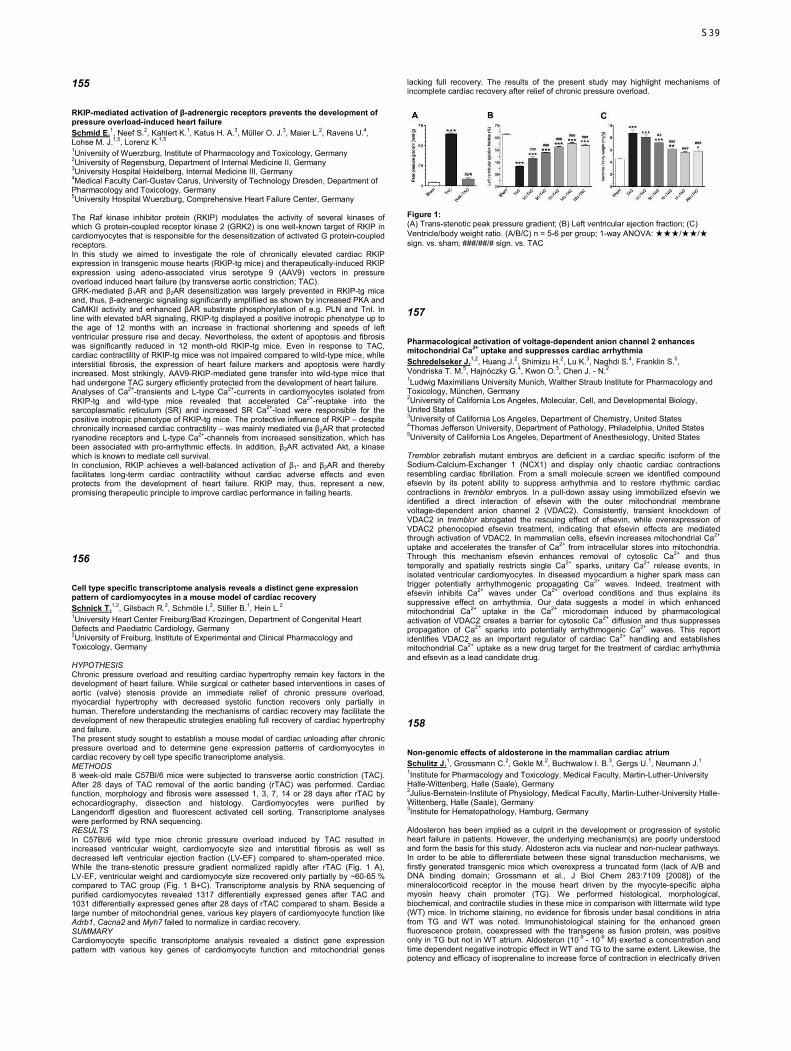
155
RKIP-mediated activation of β-adrenergic receptors prevents the development of pressure overload-induced heart failure Schmid E.1, Neef S.2, Kahlert K.1, Katus H. A.3, Müller O. J.3, Maier L.2, Ravens U.4, Lohse M. J.1,5, Lorenz K.1,5 1University of Wuerzburg, Institute of Pharmacology and Toxicology, Germany 2University of Regensburg, Department of Internal Medicine II, Germany 3University Hospital Heidelberg, Internal Medicine III, Germany 4Medical Faculty Carl-Gustav Carus, University of Technology Dresden, Department of Pharmacology and Toxicology, Germany 5University Hospital Wuerzburg, Comprehensive Heart Failure Center, Germany The Raf kinase inhibitor protein (RKIP) modulates the activity of several kinases of which G protein-coupled receptor kinase 2 (GRK2) is one well-known target of RKIP in cardiomyocytes that is responsible for the desensitization of activated G protein-coupled receptors. In this study we aimed to investigate the role of chronically elevated cardiac RKIP expression in transgenic mouse hearts (RKIP-tg mice) and therapeutically-induced RKIP expression using adeno-associated virus serotype 9 (AAV9) vectors in pressure overload induced heart failure (by transverse aortic constriction; TAC). GRK-mediated β1AR and β2AR desensitization was largely prevented in RKIP-tg mice and, thus, β-adrenergic signaling significantly amplifiied as shown by increased PKA and CaMKII activity and enhanced βAR substrate phosphorylation of e.g. PLN and TnI. In line with elevated bAR signaling, RKIP-tg displayed a positive inotropic phenotype up to the age of 12 months with an increase in fractional shortening and speeds of left ventricular pressure rise and decay. Nevertheless, the extent of apoptosis and fibrosis was significantly reduced in 12 month-old RKIP-tg mice. Even in response to TAC, cardiac contractility of RKIP-tg mice was not impaired compared to wild-type mice, while interstitial fibrosis, the expression of heart failure markers and apoptosis were hardly increased. Most strikingly, AAV9-RKIP-mediated gene transfer into wild-type mice that had undergone TAC surgery efficiently protected from the development of heart failure. Analyses of Ca2+-transients and L-type Ca2+-currents in cardiomyocytes isolated from RKIP-tg and wild-type mice revealed that accelerated Ca2+-reuptake into the sarcoplasmatic reticulum (SR) and increased SR Ca2+-load were responsible for the positive inotropic phenotype of RKIP-tg mice. The protective influence of RKIP – despite chronically increased cardiac contractility – was mainly mediated via β2AR that protected ryanodine receptors and L-type Ca2+-channels from increased sensitization, which has been associated with pro-arrhythmic effects. In addition, β2AR activated Akt, a kinase which is known to mediate cell survival. In conclusion, RKIP achieves a well-balanced activation of β1- and β2AR and thereby facilitates long-term cardiac contractility without cardiac adverse effects and even protects from the development of heart failure. RKIP may, thus, represent a new, promising therapeutic principle to improve cardiac performance in failing hearts.
156
Cell type specific transcriptome analysis reveals a distinct gene expression pattern of cardiomyocytes in a mouse model of cardiac recovery Schnick T.1,2, Gilsbach R.2, Schmöle I.2, Stiller B.1, Hein L.2 1University Heart Center Freiburg/Bad Krozingen, Department of Congenital Heart Defects and Paediatric Cardiology, Germany 2University of Freiburg, Institute of Experimental and Clinical Pharmacology and Toxicology, Germany HYPOTHESIS Chronic pressure overload and resulting cardiac hypertrophy remain key factors in the development of heart failure. While surgical or catheter based interventions in cases of aortic (valve) stenosis provide an immediate relief of chronic pressure overload, myocardial hypertrophy with decreased systolic function recovers only partially in human. Therefore understanding the mechanisms of cardiac recovery may facilitate the development of new therapeutic strategies enabling full recovery of cardiac hypertrophy and failure. The present study sought to establish a mouse model of cardiac unloading after chronic pressure overload and to determine gene expression patterns of cardiomyocytes in cardiac recovery by cell type specific transcriptome analysis. METHODS 8 week-old male C57Bl/6 mice were subjected to transverse aortic constriction (TAC). After 28 days of TAC removal of the aortic banding (rTAC) was performed. Cardiac function, morphology and fibrosis were assessed 1, 3, 7, 14 or 28 days after rTAC by echocardiography, dissection and histology. Cardiomyocytes were purified by Langendorff digestion and fluorescent activated cell sorting. Transcriptome analyses were performed by RNA sequencing. RESULTS In C57Bl/6 wild type mice chronic pressure overload induced by TAC resulted in increased ventricular weight, cardiomyocyte size and interstitial fibrosis as well as decreased left ventricular ejection fraction (LV-EF) compared to sham-operated mice. While the trans-stenotic pressure gradient normalized rapidly after rTAC (Fig. 1 A), LV-EF, ventricular weight and cardiomyocyte size recovered only partially by ~60-65 % compared to TAC group (Fig. 1 B+C). Transcriptome analysis by RNA sequencing of purified cardiomyocytes revealed 1317 differentially expressed genes after TAC and 1031 differentially expressed genes after 28 days of rTAC compared to sham. Beside a large number of mitochondrial genes, various key players of cardiomyocyte function like Adrb1, Cacna2 and Myh7 failed to normalize in cardiac recovery. SUMMARY Cardiomyocyte specific transcriptome analysis revealed a distinct gene expression pattern with various key genes of cardiomyocyte function and mitochondrial genes
lacking full recovery. The results of the present study may highlight mechanisms of incomplete cardiac recovery after relief of chronic pressure overload.
Figure 1: (A) Trans-stenotic peak pressure gradient; (B) Left ventricular ejection fraction; (C) Ventricle/body weight ratio. (A/B/C) n = 5-6 per group; 1-way ANOVA: ★★★/★★/★ sign. vs. sham; ###/##/# sign. vs. TAC
157
Pharmacological activation of voltage-dependent anion channel 2 enhances mitochondrial Ca2+ uptake and suppresses cardiac arrhythmia Schredelseker J.1,2, Huang J.2, Shimizu H.2, Lu K.3, Naghdi S.4, Franklin S.5, Vondriska T. M.5, Hajnóczky G.4, Kwon O.3, Chen J. - N.2 1Ludwig Maximilians University Munich, Walther Straub Institute for Pharmacology and Toxicology, München, Germany 2University of California Los Angeles, Molecular, Cell, and Developmental Biology, United States 3University of California Los Angeles, Department of Chemistry, United States 4Thomas Jefferson University, Department of Pathology, Philadelphia, United States 5University of California Los Angeles, Department of Anesthesiology, United States Tremblor zebrafish mutant embryos are deficient in a cardiac specific isoform of the Sodium-Calcium-Exchanger 1 (NCX1) and display only chaotic cardiac contractions resembling cardiac fibrillation. From a small molecule screen we identified compound efsevin by its potent ability to suppress arrhythmia and to restore rhythmic cardiac contractions in tremblor embryos. In a pull-down assay using immobilized efsevin we identified a direct interaction of efsevin with the outer mitochondrial membrane voltage-dependent anion channel 2 (VDAC2). Consistently, transient knockdown of VDAC2 in tremblor abrogated the rescuing effect of efsevin, while overexpression of VDAC2 phenocopied efsevin treatment, indicating that efsevin effects are mediated through activation of VDAC2. In mammalian cells, efsevin increases mitochondrial Ca2+ uptake and accelerates the transfer of Ca2+ from intracellular stores into mitochondria. Through this mechanism efsevin enhances removal of cytosolic Ca2+ and thus temporally and spatially restricts single Ca2+ sparks, unitary Ca2+ release events, in isolated ventricular cardiomyocytes. In diseased myocardium a higher spark mass can trigger potentially arrhythmogenic propagating Ca2+ waves. Indeed, treatment with efsevin inhibits Ca2+ waves under Ca2+ overload conditions and thus explains its suppressive effect on arrhythmia. Our data suggests a model in which enhanced mitochondrial Ca2+ uptake in the Ca2+ microdomain induced by pharmacological activation of VDAC2 creates a barrier for cytosolic Ca2+ diffusion and thus suppresses propagation of Ca2+ sparks into potentially arrhythmogenic Ca2+ waves. This report identifies VDAC2 as an important regulator of cardiac Ca2+ handling and establishes mitochondrial Ca2+ uptake as a new drug target for the treatment of cardiac arrhythmia and efsevin as a lead candidate drug.
158
Non-genomic effects of aldosterone in the mammalian cardiac atrium Schulitz J.1, Grossmann C.2, Gekle M.2, Buchwalow I. B.3, Gergs U.1, Neumann J.1 1Institute for Pharmacology and Toxicology, Medical Faculty, Martin-Luther-University Halle-Wittenberg, Halle (Saale), Germany 2Julius-Bernstein-Institute of Physiology, Medical Faculty, Martin-Luther-University Halle-Wittenberg, Halle (Saale), Germany 3Institute for Hematopathology, Hamburg, Germany Aldosteron has been implied as a culprit in the development or progression of systolic heart failure in patients. However, the underlying mechanism(s) are poorly understood and form the basis for this study. Aldosteron acts via nuclear and non-nuclear pathways. In order to be able to differentiate between these signal transduction mechanisms, we firstly generated transgenic mice which overexpress a truncated form (lack of A/B and DNA binding domain; Grossmann et al., J Biol Chem 283:7109 [2008]) of the mineralocorticoid receptor in the mouse heart driven by the myocyte-specific alpha myosin heavy chain promoter (TG). We performed histological, morphological, biochemical, and contractile studies in these mice in comparison with littermate wild type (WT) mice. In trichome staining, no evidence for fibrosis under basal conditions in atria from TG and WT was noted. Immunohistological staining for the enhanced green fluorescence protein, coexpressed with the transgene as fusion protein, was positive only in TG but not in WT atrium. Aldosteron (10-9 - 10-6 M) exerted a concentration and time dependent negative inotropic effect in WT and TG to the same extent. Likewise, the potency and efficacy of isoprenaline to increase force of contraction in electrically driven
S39
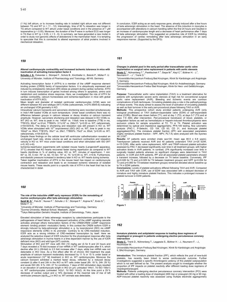
(1 Hz) left atrium, or to increase beating rate in isolated right atrium was not different between TG and WT (n = 11 - 12). Interestingly, time of 90 % relaxation was longer in TG atrium compared to WT atrium under basal conditions and in the presence of 1 µM isoprenaline (p < 0.05). Moreover, the duration of the P-wave in surface ECG was longer in TG than in WT (p < 0.05, n = 5 - 6). In summary, we have generated a new model in order to study non-genomic effects of aldosterone in the mammalian heart. It is tempting to speculate that this is connected to altered expression of atrial proteins involved in mechanical relaxation.
159
Altered cardiomyocyte contractility and increased ischemic tolerance in mice with inactivation of activating transcription factor 4 Schulte J. S., Eskandar J., Stümpel F., Scholz B., Kirchhefer U., Boknik P., Müller F. U. University of Münster, Institute of Pharmacology and Toxicology, 48149, Germany Activating transcription factor 4 (ATF4) is a member of the cAMP response element binding protein (CREB) family of transcription factors. It is ubiquitously expressed and induced by endoplasmic reticulum (ER) stress as present during cardiac ischemia. ATF4 in turn induces transcription of genes involved among others in apoptosis, amino acid metabolism and oxidative stress response. Here, we investigated the role of ATF4 for cardiac contractility, calcium homeostasis and ischemia-reperfusion in mice with global inactivation of ATF4 (KO). Mean length and diameter of isolated ventricular cardiomyocytes (VCM) were not different between KO and wildtype (WT) VCMs (cells/animals, n=275-369/5-6) indicating an unchanged VCM geometry in KO. Intracellular calcium transients and sarcomere shortening were recorded simultaneously in Indo1-AM loaded VCMs at room temperature. Under basal conditions there was no difference between groups in calcium release or decay kinetics or calcium transient amplitude. However, sarcomere shortening and relaxation was delayed in KO VCMs vs. WT (KO vs. WT, time to peak in ms: 146±6* vs. 118±6; time to baseline (TTB) in ms, TTB10%: 80±4* vs 61±4, TTB50%: 311±16* vs. 256±17; *p<0.05 vs. WT; cells/animals, n=54-63/5-6). The delay in shortening and relaxation was still present in KO vs. WT VCMs when stimulated with 10-6 isoproterenol (ISO) (KO vs. WT, time to peak in ms: 104±2* vs. 95±3; TTB10%: 35±1* vs. 29±1, TTB50%: 75±3* vs. 65±4; *p<0.05 vs. WT; cells/animals, n=54-63/5-6). Despite these findings on the cellular level left ventricular cathetherization revealed an unchanged heart rate, stroke volume as well as maximum contraction and relaxation velocity in KO vs. WT mice under basal conditions and when stimulated with ISO (WT n=5, KO n=6). Ischemia-reperfusion experiments with isolated mouse hearts (Langendorff apparatus, 37°C) showed that KO hearts recovered faster during reperfusion as compared to WT hearts (dp/dtmax in % of starting value, KO vs. WT, minutes of reperfusion, 1 min: 83* vs. 44, 5 min: 76* vs. 62, 10 min: 75* vs. 70; *p<0.05 vs. WT, n=5-6). Furthermore, end-diastolic pressure increased in tendency later in KO vs. WT hearts during ischemia. Taken together inactivation of ATF4 in the mouse heart has impact on cardiomyocyte contraction and relaxation and leads to an increased ischemic tolerance of isolated mouse hearts. These results point to a specific role of ATF4 in the heart that has to be characterized in detail.
160
The role of the inducible cAMP early repressor (ICER) for the remodeling of murine cardiomyocytes after beta adrenergic stimulation Seidl M. D.1, Fels B.1, Nunes F.1, Schulte J. S.1, Stümpel F.1, Kojima N.2, Endo S.3, Müller F. U.1 1University of Münster, Institute of Pharmacology and Toxicology, Germany 2Gunma University, Medical School, Maebashi, Japan 3Tokyo Metropolitan Geriatric Hospital, Institute of Gerontology, Tokio, Japan Elevated stimulation of beta adrenergic receptors by catecholamines participate to the pathogenesis of heart failure. The subsequent activation of the cAMP signaling cascade activates amongst others transcription factors of the CREB/CREM (cAMP responsive element- binding protein/ -modulator) family. In this context the CREM isoform ICER is strongly induced by beta-adrenergic stimulation e. g. by isoproterenol (ISO) via cAMP responsive elements (CRE) in its promoter. Contrary to its CRE-mediated induction, ICER acts as a strong inhibitor of CRE-mediated transcription by itself. Here we analyzed the selective impact of ICER induction for the physiological response after beta adrenergic stimulation in murine hearts in a time dependent manner by the use of ICER deficient mice (IKO) and wild type (WT) controls. Stimulation of IKO and WT mice with ISO (10 mg/kg per d) for 6 and 24 hours and 7 days revealed a 75-fold increase of Icer mRNA in WT cardiomyocytes after 6 h, which decline after 24 h (29-fold) to 2.5 fold increase after 7 days, while Icer mRNA was not detectable in IKO mice. As a first effect after 6h of chronic ISO treatment the peak amplitude of calcium transient moderately decreased by 11 % or 15 % under basal or acute isoproterenol (10-6 M) treatment in IKO vs. WT cardiomyocytes. Moreover the calcium transient exhibited a marked faster decay, reflected by a reduced decay constant (t) after 6 and 24h in the IKO vs WT cells under basal (6h: 18 %; 24h: 29 %) and acute ISO (6h: 9 %; 24h: 27 %) conditions. 7 days of ISO stimulation days resulted in an elevation of cardiomyocyte length in IKO (in µm; untreated 144±1, 7d ISO 156±1) vs. WT cardiomyocytes (untreated 142±1, 7d ISO 143±2). At this time point a 29 % decrease of cardiac output and a 16% decrease of the maximal rate of rise of left ventricular pressure (dp/dtmax) in IKO vs. WT animals was detectable.
In conclusion, ICER acting as an early response gene, already induced after a few hours of beta adrenergic stimulation in the heart. The absence of this induction in myocytes is accompanied with alterations in calcium handling within the first 24 hours and resulted in an increase of cardiomyocytes length and a decrease of heart performance after 7 days of beta adrenergic stimulation. This suggested an protective role of ICER by inhibiting the progression of cardiac remodeling after beta adrenergic stimulation in an early responsive manner. (Supported by the DFG)
161
Changes in platelet pool in the early period after transcatheter aortic valve implantation or surgical valve replacement in patients with aortic stenosis Stratz C.1, Nührenberg T.1, Pawlitschek F.2, Siepe M.3, Keyl C.2, Büttner H. - J.1, Neumann F. - J.1, Trenk D.1 1Universitäts-Herzzentrum Freiburg Bad Krozingen, Klinik für Kardiologie und Angiologie II, Germany 2Universitäts-Herzzentrum Freiburg Bad Krozingen, Klnik für Anästhesiologie, Germany 3Universitäts-Herzzentrum Freibur Bad Krozingen, Klinik für Herz- und Gefäßchirurgie, Germany Purpose: Transcatheter aortic valve implantation (TAVI) is a treatment alternative for patients with symptomatic severe aortic stenosis at high risk for conventional surgical aortic valve replacement (AVR). Bleeding and ischemic events are common complications of both techniques. Circulating platelets play a role in the pathophysiology of these events. This study aimed to assess the level of activation of circulating platelets as well as their turnover in the early peri-procedural phase following TAVI or AVR. Methods: This prospective cohort study enrolled patients undergoing AVR (only biological prostheses) or TAVI (balloon-expandable valve (EDW) or self-expandable valve (COR)). Blood was drawn before (T1), and at day 1 (T2), at days 5-7 (T3) and at days 7-9 (M4) after intervention. Peri-procedural transfusion of blood, platelets, or coagulation factors as well as hemodialysis or heparin-induced thrombocytopenia were exclusion criteria for sample acquisition at T2, T3 or T4. Platelet activation was determined by optical and impedance aggregometry, PFA-100 testing, flow cytometric analysis (PAC-1, P-Selectin, CD 154, CD 63, CD 31, and leukocyte platelet aggregates(LPA)). The immature platelet fraction (IPF) and associated parameters (highly immature platelet fraction - hIPF, MPV, PLT-X) were analyzed with the Sysmex XE 2100®. Results: 67 patients were enrolled (male sex=34; mean age 80.5 ± 6.8 years). Twentyseven patients received AVR and 40 patients underwent TAVI (n=25 EDW, n=15 COR). After aortic valve replacement, ADP- and TRAP-induced platelet activation assessed by PAC-1 decreased significantly over time in all treatment groups; with higher activation levels in AVR patients. In contrast, LPA significantly increased over time in surgically treated patients whereas no significant changes were determined in TAVI patients. In COR and AVR patients, IPF and all associated platelet parameters showed a transient increase, followed by a decrease on T4 below baseline. Conversely, IPF (p=0.026 for T3 and p=0.029 for T4 between treatment groups) and hIPF (p=0.054 for T3 and p=0.060 for T4 between treatment groups) remained elevated above baseline up to T4 in EDW patients. Conclusions: Levels of platelet activations differed following TAVI and AVR. Compared to AVR and TAVI with COR, use of EDW was associated with a delayed decrease of immature and highly immature platelet fractions. This indicates a prolonged increase in platelet turnover in EDW patients.
Figure 1
162
Immature platelets and antiplatelet response to loading dose regimens of clopidogrel or prasugrel in patients undergoing elective percutaneous coronary intervention Stratz C., Trenk D., Nührenberg T., Leggewie S., Büttner H. - J., Neumann F. - J., Hochholzer W. Universitäts-Herzzentrum Freiburg Bad Krozingen, Klinik für Kardiologie und Angiologie II, BAd Krozingen, Germany Introduction: The immature platelet fraction (IPF), which reflects the pool of new-build platelets, has recently been linked to worse cardiovascular outcome. Further investigations suggested a specific thrombogenic potency of this platelet subpopulation which is not well defined so far. The present analysis sought to investigate whether the proportion of IPF impacts on platelet reactivity after different loading dose regimens of clopidogrel/prasugrel. Methods: Patients undergoing elective percutaneous coronary intervention (PCI) were treated with either a loading dose of clopidogrel (600 mg) or prasugrel (30 mg or 60 mg). ADP-induced platelet reactivity was assessed using multiple electrode aggregometry
S40

(MEA) on day 1 following coronary intervention. The proportions of IPF were determined with an automated flow-cytometry analyzer (Sysmex XE-2100) before administration of P2Y12 inhibitor and grouped into tertiles. Results: In total, 136 patients were enrolled (mean age: 69 [interquartile range: 58 - 74] years; body mass index: 27.5 [25.3 - 30.1]; women: 21.2 %; diabetes mellitus: 23.5 %). Patients were equally allocated to the three treatment groups (clopidogrel 600 mg: n = 45; prasugrel 30 mg n = 45; prasugrel 60 mg n = 46). Overall, ADP-induced platelet reactivity was significantly higher inpatients with IPF proportions in both upper tertiles (IPF: 2,8 - 4,6 % and > 4,7 %) compared with the lower tertile (< 2,7 % IPF; p < 0.03). We further analyzed the association between IPF and ADP-induced platelet reactivity with regard to the thienopyridine loading dose regimen. While a significant higher platelet reactivity in both upper tertiles was observed after prasugrel 60 mg ( (p < 0.002, Figure), a trend lacking formal statistical significance was observed after clopidogrel 600 mg and prasugrel 30 mg. Conclusion: The heterogeneity of the platelet pool reflected by different proportions of immature platelets seems to interact with antiplatelet response to loading doses of thienopyridines in stable patients undergoing elective PCI. Thus, the immature platelet fraction might be a novel risk marker of high on-treatment platelet reactivity in patients on clopidogrel or prasugrel which can be determined by routine laboratory measurements before start of antiplatelet treatment.
Figure 1
163
Changes in cardiac nitric oxide metabolism in a mouse model of cardiomyocyte-specific thrombospondin 1 over-expression Stümpel F. T.1, Michael A.1, Overbeck N.1,2, Meier M.1, Müller F. U.1 1Münster University, Dept. of Pharmacology and Toxicology, Germany 2Düsseldorf University, Molecular Proteomics Laboratory, Germany Thrombospondin 1 (TSP-1) is an extracellular glycoprotein involved in the inhibition of angiogenesis, de-adhesion, and activation of TGFβ1. In several tissue types it was also shown to interfere with NO signalling by inhibiting NO synthesis and sGC activation via CD47-dependent mechanisms. TSP-1 expression in cardiomyocytes of the adult healthy heart is low, but increases in response to injury or stress. In a knock-out model lacking TSP-1 experimental myocardial ischaemia led to enlarged infarction scars. In our model, over-expressing TSP-1 under control of the cardiomyocyte-specific αMHC promoter, transgenic hearts (TG) showed an ameliorated and accelerated early recovery in an ex-vivo (Langendorff) ischaemia/reperfusion (I/R) setting. To elucidate the role of NO-dependent pathways in this finding, we conducted experiments (i) to assess changes in NO sensitivity in TSP-1 over-expressing hearts and (ii) to determine a possible role of NO in their improved recovery in I/R settings. Isolated intact hearts from TG animals and wild-type (WT) controls were retrogradely perfused according to Langendorff and electrically stimulated at a constant rate of 450 min−1. Left-ventricular developed pressure (devP) and maximal rate of contraction (dP/dtmax) were constantly recorded (in a 1st series) under increasing concentrations (10-10 M to 10-3 M) of sodium nitroprusside (SNP) and (in a 2nd series) during 30 min of ischaemia followed by 30 min of reperfusion under a constant concentration of 10-4 M SNP. (All data are mean±SEM; n=6-9 per group; #: p<0.05 vs. WT; 2-way-RM-ANOVA) SNP (mol/l) 10-10 10-9 10-8 10-7 10-6 10-5 10-4 10-3
devP (mmHg)
WT 64±4 60±4 62±3 80±5 94±5 106±3 108±3 104±3
TG 58±2 62±3 66±3 81±7 91±7 92±6 89±5 84±6 #
dP/dtmax (mmHg/s)
WT 4030±377 3814±380 3675±317 4512±313 5325±250 5686±168 5833±158 5758±154
TG 3423±287 3402±213 3331±177 3989±306 4365±416 4416±308 4263±225 4133±179 #
Under 10-4 M SNP, early recovery (e. g. 7 min) after ischaemia was significantly better in TG hearts (devP 41±7 mmHg, dP/dtmax 1746±350 mmHg/s, n=6) than in WT (devP 12±3 mmHg, dP/dtmax 548±137 mmHg/s, n=6). We conclude that NO efficacy is reduced due to TSP-1 over-expression in cardiomyocytes, and that this might contribute to a better early recovery after ischaemia. This study shows for the first time that cardiomyocyte-produced TSP-1 has effects on NO metabolism in the heart, and that this effect might be beneficial in the early reperfusion phase.
164
Mice with functional inactivation of the cAMP responsive element modulator (CREM) show an altered cardiac remodelling in response to chronic β-adrenergic stimulation Tekook M. A., Schulte J. S., Fehrmann E., Scholz B., Heinick A., Seidl M. D., Müller F. U. University Münster, Institute of Pharmacology and Toxicology, Germany Patients suffering from heart failure (HF) show elevated plasma-catecholamine levels which correlate with mortality rate. As β-adrenergic stimulation raises intracellular cAMP-levels cAMP-dependent transcription factors might play an important role in the remodeling processes in HF that worsen cardiac function and lead to arrhythmia. Short cAMP responsive element modulator (CREM) repressor isoforms (ICER, smICER) are induced by β-adrenergic stimulation. Here, we investigated ventricular cardiomyocytes (VCM) from mice with global inactivation of all CREM-isoforms (KO) for alterations in action potential (AP), calcium transient (CaT) and gene expression induced by a 14 day isoprenaline (ISO) treatment. KO mice and controls (WT) were treated with ISO (30 mg/kg x d; 14d, age 16-20 weeks). Untreated mice (U) were used as controls. Isolated VCMs were electrically stimulated to provoke APs in patch-clamp experiments or CaTs. Samples of these VCMs were used for mRNA expression analysis. AP duration did not differ between groups and were prolonged after 14d ISO to a similar extent. Nevertheless, ISO-KO showed a significantly lower portion of VCMs with early afterdepolarizations (EAD) vs. ISO-WT. In contrast to ISO-WT, ISO-KO cells still showed AP prolongation in response to acute ISO (10-6M) stimulation (APD50 in % basal; ISO-WT:103, ISO-KO:115*; *p<0.05). CaT amplitude was unchanged in KO vs. WT (U), increased in ISO-KO, but in tendency reduced in ISO-WT (in % of U; WT-ISO:97, KO-ISO:110*). Time to peak (50%) was faster in U-KO, but slower in ISO-KO (in % of WT; U-KO:86*, ISO-KO:125*). CaT decay was slower in U-KO, accelerated in both groups by ISO treatment, but not altered in ISO-KO vs. ISO-WT. Analysis of mRNA revealed two groups of transcripts differentially expressed in KO vs. corresponding WT. One group was equally (Adrb1, Adrb2) or higher (Serca, Scn5a, Cacna1c) expressed in U-KO and not repressed in ISO-KO. Another group was equally (Pln, Myh7b, Trd, KChip2) or higher (Kv2.1, Kv4.3, Kv1.5) expressed in U-KO and down-regulated in ISO-KO to a lesser degree. Taken together, inactivation of CREM alters an ISO induced cardiac remodeling with impact on intracellular calcium homeostasis, remaining β-adrenergic responsiveness, a weakened mRNA-repression of ion channels and calcium-handling proteins and less VCMs with EADs despite similar AP duration. This underlines the important role of CREM for cardiac remodeling by catecholamines. Supported by the IZKF Münster
165
Characterization of a peptide that interferes with ERK1/2-mediated pathological cardiac hypertrophy Tomasovic A.1, Hümmert M.1, Ruppert C.1, Lorenz K.1,2 1University of Wuerzburg, Department of Pharmacology, Germany 2University of Wuerzburg, Comprehensive Heart Failure Center, Germany Extracellular signal-regulated kinases 1 and 2 (ERK1/2) are mediators of pathological cardiac hypertrophy. Mechanistically, ERK1/2-mediated cardiomyocyte hypertrophy involves ERK1/2 dimerization and ERKThr188-phosphorylation as triggers for nuclear translocation. However, ERK1/2 activity is a well-known mechanism for cardiomyocyte protection from apoptotic cell death. Here, we investigated the ERK-ERK interface as a potential target in cardiac hypertrophy. To evaluate the impact of monomeric ERK on cardiac function, we overexpressed ERK2Δ174–177, a dimerization-deficient ERK2 mutant, in mouse hearts. Cardiac function of ERK2Δ174–177 transgenic mice was comparable to wild-type mice as shown by echocardiographic evaluation of fractional shortening under basal conditions, in response to chronic pressure overload (transverse aortic constriction; TAC) or running wheel exercise. Interestingly, only in response to TAC, echocardiography revealed a reduced wall thickness in ERK2Δ174–177 transgenic mice compared to wild-type mice. Additionally, mRNA expression of collagen and ANF was attenuated in ERK2Δ174–177 transgenic mice upon TAC while anti-apoptotic signaling was preserved. In line with the in vivo data, overexpression of ERK2Δ174-177 in neonatal rat cardiomyocytes (NRCM) resulted in a reduced hypertrophic response but did not exaggerate apoptosis. Of note, ERK2Δ174–177 did not translocate into the nucleus in response to hypertrophic stimuli and even attenuated phosphorylation of Elk1, a nuclear ERK1/2 target, which is known to mediate cardiac hypertrophy. Next, we generated a peptide consisting of amino acids of the putative ERK-ERK interface to interfere with endogenous ERK dimerization. Cross-linking and co-immunoprecipitation experiments showed that overexpression of the peptide indeed interfered with ERK dimerization. Interestingly, the peptide effectively prevented nuclear translocation of YFP-tagged wild-type ERK2 in response to phenylephrine in NRCM. In line with this effect, the peptide attenuated the phenylephrine-mediated hypertrophic response and Elk1 phosphorylation. Moreover, the peptide did not restrict anti-apoptotic ERK-signaling in NRCM. Taken together, our data suggest that interference with ERK dimerization could be a valuable approach to target hypertrophic ERK1/2 signaling without cardiac adverse effects.
S41

166
Phosphodiesterase 2 regulates resting heart rate and protects against arrhythmias and β-adrenergic overstimulation Vettel C.1,2, Dewenter M.2, Linder M.3, Riedel M.2, Lämmle S.4, Mason F.5, Meinecke S.2, Wieland T.1, Vandecasteele G.3, Geerts A.6, Wunder F.6, Sossalla S.5, Fischmeister R.3, El-Armouche A.4 1Universitätsmedizin Mannheim, Experimentelle Pharmakologie, Germany 2Universitätsmedizin Göttingen, Institut für Pharmakologie, Germany 3INSERM UMR-S 769, LabEx LERMIT, Châtenay-Malabry, France 4Technische Universität Dresden, Institut für Pharmakologie, Germany 5Universitätsmedizin Göttingen, Abteilung für Kardiologie und Pneumologie, Germany 6BAYER Pharma AG, Global Drug Discovery, Wuppertal, Germany Rationale: Phosphodiesterase 2 (PDE2) has the unique property to be stimulated by cGMP, but primarily hydrolyses cAMP, thereby mediating a negative cross-talk between cAMP- and cGMP signaling pathways. PDE2 is upregulated in human and in experimental heart failure (HF) models, where it appears to be part of the desensitization process to β-adrenergic overstimulation. Objective: To investigate PDE2 mediated effects in a cardiac stress models and PDE2 heart-specific transgenic mice (TG). Methods and Results: Chronic β-adrenoceptor (β-AR) stimulation via isoprenaline (ISO) infusions in mice led to an expected desensitization which was partly reversible upon in vivo injections of PDE2 specific inhibitor BAY 60-7550. Inhibition of PDE2 not only normalized β-AR inotropic responsiveness, but also induced significant positive chronotropic effects in both ISO- and NaCl-hearts on top of maximal β-AR stimulation. Conversely, ECG telemetry in PDE2-TG showed a marked reduction in resting and maximal heart rate, however without any impairment of basal contraction force or contractile reserve. The phenotype delivered no indication of any cardiac pathology in ageing PDE2-TG and unexpectedly even promoted a prolonged life span. During arrhythmia provocation induced by double injections of ISO, TG animals were less prone to ventricular tachycardia. Ca2+-spark analysis showed reduced Ca2+-leakage after β-AR stimulation, while immunoblotting revealed lower basal phosphorylation levels of Ca2+-cycling proteins, among those the arrhythmia-associated phosphorylation site p-ryanodine-receptor-2814. Conclusion: PDE2 contributes to heart rate regulation in healthy hearts. Under stress conditions it becomes upregulated and desensitizes against both β-AR mediated chronotropic and inotropic effects. High PDE2 abundance protects against arrhythmia and Ca2+-leakage. Thus, activating myocardial PDE2 may represent a novel intracellular anti-adrenergic and anti-arrhythmic therapeutic strategy in HF.
167
Mineralocorticoid receptor activation increases the systolic Ca2+ transient by increasing SR Ca2+ fractional release rather than SR Ca2+ content in rat cardiomyocytes of endo- and epicardial origin Wagner M.1,2, Wacker C.2, Dams N.2, Volk T.2 1TU Dresden, Institut für Pharmakologie und Toxikologie, Germany 2FAU Erlangen-Nürnberg, Institut für Zelluläre und Molekulare Physiologie, Germany Activation of the cardiac mineralocorticoid receptor (MR) increases the L-type Ca2+ current (ICaL), a key player in Ca2+ homeostasis and hence, regulation of cardiac contractility. Within the left ventricle a gradient of action potential (AP) waveforms is present, with short AP duration (APD) in epicardial (epi) and long APD in endocardial (endo) myocytes. As a consequence, the AP-induced Ca2+ influx is larger in endo than in epi myocytes. We demonstrated that after 24h incubation, ICaL is similar in endo and epi myocytes (n.s.) and that MR activation increases ICaL to a similar extent in both layers (each p<0.001). Since the epi AP was markedly more prolonged by MR activation than the endo AP, AP-induced Ca2+ influx was increased 4-fold (p<0.001) in epi myocytes, while in endo myocytes only a 1.8-fold increase (n.s.) was noted. Here we investigate region-specific effects of these changes in the transmembrane Ca2+ flux on the Ca2+ transient and fractional shortening (FS) in left ventricular rat endo and epi myocytes. Ca2+ transients were assessed simultaneously with sarcomere length using fura2-AM. Despite longer APD and higher AP-induced Ca2+ influx in endo myocytes, the amplitude of the Ca2+ transient, its decay time constant and FS were similar in endo and epi myocytes after 24h under control conditions, as were baseline Ca2+ levels and sarcomere length (each n.s.). After 24h of MR stimulation, Ca2+ transients and FS were affected in a similar way in epi and endo myocytes: in epi myocytes the Ca2+ transient amplitude increased by 43% (p<0.001) and by 70% in endo myocytes (p<0.001). The decay of the Ca2+ transient was significantly accelerated in endo myocytes (p<0.001). Baseline Ca2+ levels were not affected. FS was increased by 48% (p<0.05) in epi and by 74% (p<0.001) in endo myocytes. To examine whether the increase in the Ca2+ transient was due to an increased filling of the SR, its Ca2+ content was assessed using a 10mM caffeine pulse after steady-state pacing. SR filling was similar in endo- and epicardial myocytes under control conditions after 24h (n.s.). MR activation slightly tended to increase SR filling (endo: +5%, n.s., epi: +15%, n.s.). These data indicate that the increase in Ca2+ transients upon MR activation in endo- as well as in epicardial myocytes is predominantly caused by the increase in ICaL density and consequently an increase in fractional Ca2+ release from the SR and to a far lesser extent by an increased filling of the SR.
168
A virus-based approach to analyze calcium channel mutants in cultured adult rat cardiomyocytes Vu E., Göbel P., Engelhardt S., Welling A. Technische Universität München, Pharmakologie und Toxikologie, Germany Several obstacles hamper the analysis of L-type calcium channel (LTCC) function and regulation in the heart. The analysis in heterologous expression systems is complicated because LTCC are multi-subunit proteins with a main pore forming α-subunit and a plurality of modulating auxiliary subunits. In addition, expression systems are often deficient of structural elements or permissive environments for signal pathways e.c. protein kinase modulation. Knock-in mice as an alternative system are costly and time consuming. In the case, that different constructs are necessary it is almost impossible to use knock-in mice. Direct manipulation of the expression of target proteins in adult cardiomyocytes is a wishful alternative, but adult cardiomyocytes are refractory to most transfection methods and normally they can only be maintained in culture for a short time (0-1 d). On the other hand, adult cardiomyocytes can be efficiently infected by viral vectors, but in the case of LTCC a major technical hurdle is that the packaging capacity of commonly used viral vectors is too low. We have adopted a method according to Viero et al. 2008 to culture adult rat cardiomyocytes up to 4 days. Freshly isolated cells were infected late at day 0 with a truncated calcium channel construct via an adenovirus. A dihydropyridine-resistant channel was used to distinguish the over-expressed current from the native one, which was inhibited by nisoldipine. As a test system we analysed a putative protein kinase A (PKA) phosporylation site, which was identified before by mass spectrometry. As proven by a dsRed marker the constructs were expressed successfully and presented a sufficient amount of current in patch-clamp measurements. Unfortunately, there was no difference in ß-adrenergic stimulation between the channels with a wild-type or a mutated PKA-phosphorylation site. Nevertheless, this approach offers a good and quick alternative to manipulate large proteins in adult cardiomyocytes. Supported by the DZHK (German Centre for Cardiovascular Research), partner site MHA (Munich Heart Alliance). Viero C, Kraushaar U, Ruppenthal S, Kaestner L, Lipp P. A primary culture system for sustained expression of a calcium sensor in preserved adult rat ventricular myocytes. Cell Calcium. 2008 Jan;43(1):59-71
169
Flow cytometric analysis of cell cycle activity in postnatal cardiomyocytes Wullkopf L., Nührenberg T., Preißl S., Gilsbach R., Hein L. UNIVERSITY OF FREIBURG, EXPERIMENTAL AND CLINICAL PHARMACOLOGY AND TOXICOLOGY, Freiburg, Germany Introduction: Mammalian cardiomyocytes were traditionally classified as a post-mitotic cell type, withdrawing permanently from the cell cycle shortly after birth. Currently, there is increasing evidence for continuous cardiomyocyte proliferation in the adult murine heart. Yet, reported estimations of cardiomyocyte renewal are highly variable. We therefore aimed to establish a reliable method determining cardiomyocyte-specific cell cycle activity. Methods: Hearts of C57Bl6/N mice were harvested at 1.5, 3, 5, 7, 11, 15 and 28 days after birth (n=4 replicates for each time point). After isolation of nuclei, multi-colour flow cytometry with antibodies against PCM-1 and phosphorylated histone H3 (at serine 10; PHH3), a specific marker for the mitotic phase was performed. Counterstaining of nuclei with Draq-7 allowed for simultaneous ploidy analysis. Results: Cell cycle activity of murine cardiomyocytes decreased rapidly within the first fortnight after birth. At 28 days after birth, a low level of continuous cell cycle activity was observed in cardiomyocytes (Figure). Ploidy analyses demonstrated that about 87 % of PHH3-positive nuclei were tetraploid. Conclusion: This novel, flow-cytometric method establishes cardiomyocyte cell cycle analysis on a large and highly reproducible scale. In congruence with previous data, a rapid decrease of cardiomyocyte cell cycle activity was detected after birth. Thus, this method provides a solid base to scrutinize regenerative therapeutic approaches that aim to enhance cardiomyocyte renewal in heart failure.
Figure 1: Cell cycle activity decreased rapidly in postnatal murine cardiomyoctes. Flow cytometric analysis of phospho histone H3 showed a decline from about 0.3% mitotic active cardiomyoctes during the first days after birth to 0.05% at postnatal day 28.
S42
170
ADAM10 as therapeutic target in acute lung inflammation Dreymueller D., Schumacher J., Koenen A., Pruessmeyer J., Groth E., Martin C., Hess F. - M., Donners M., Uhlig S., Ludwig A. Uniklinik RWTH Aachen, Pharmacology and Toxicology, Germany Acute lung inflammation involves changes in permeability, leukocyte recruitment, mediator secretion, and remodeling of lung tissue. A disintegrin and metalloproteinase (ADAM) 10 releases a huge variety of inflammatory molecules by the proteolytic cleavage near to the cell surface, a process called shedding. These substrates regulate junction formation and permeability (e.g. cadherins), adhesion (e.g VCAM-1), chemotaxis (e.g. CX3CL1), transmigration (e.g. JAM-A), and regeneration (e.g. HB-EGF), making ADAM10 a suitable therapeutic target in acute lung inflammation. The focus of our investigations was the cell-specific and time-dependent role of ADAM10 in acute lung inflammation. For in vivo experiments we used tissue-specific knockout mice lacking either ADAM10 in endothelial cells (EC), smooth muscle cells (SMC) or leukocytes. Those animals were compared to inhibitor treated animals in endotoxin-induced acute lung inflammation. In vivo-studies were combined with in vitro research with isolated primary murine and human cells to further study the regulatory influence of ADAM10 on permeability, leukocyte recruitment and mediator release in acute lung inflammation. The protease was specifically addressed by lentiviral knockdown or inhibitors. We found that leukocyte ADAM10 acts proinflammatory by promoting migration of neutrophils and monocytes in vitro and their recruitment at early (24h) and late stages (72h) of acute lung inflammation. While ADAM10 knockout in SMC had no effect, EC knockout of ADAM10 led to a protection of animals due to reduced cell recruitment and edema formation at early time points. Treatment of animals with the preferential ADAM10 inhibitor GI254023 also protected against vascular permeability, edema formation, and leukocyte recruitment, pointing towards a general pro-inflammatory role of ADAM10 in the first phase of acute lung inflammation. However, at later stages, we observed a delayed inflammatory response in mice lacking EC ADAM10 resulting in persistent increase in neutrophil recruitment and edema formation. These recent data indicate a switch from pro- to antiinflammatory functions of EC ADAM10 when inflammation turns into a post acute phase. Our studies present novel conceptual insights into the cell-specific and time-dependent function of ADAM10 in the development of acute lung inflammation, and highlight that possible interventional strategies targeting ADAM10 will be most effective in the early acute phase of the disease.
171
Identification of HMGB1 as mediator of hyperinflammation in the acid-injured murine lung Reiss L. K., Schlepütz M., Martin C., Uhlig S. Medical Faculty of RWTH Aachen University, Institute of Pharmacology and Toxicology, Germany Rationale: Pulmonary inflammation caused by exogenous pathogens is a frequent event, which mostly ceases with the removal of the pathogen. Nevertheless, in some cases inflammation exacerbates into destructive hyperinflammation and causes life-threatening organ dysfunction. We hypothesize that hyperinflammation is based on the liberation of a well-defined set of mediators not present during salutary inflammation. After having identified TNF and CXCL10 as mediators of hyperinflammation previously, we designed the present study to examine the potency of High-Mobility-Group-Protein B1 (HMGB1) to exacerbate inflammation in the lungs. Methods: C57/BL6 mice were tracheotomised and instilled with 50µL HCl pH=2.0 or 0.9% NaCl, followed by mechanical ventilation for 1h at VT=16mL/kg, f=90min-1, PEEP=2cmH2O and FiO2=0.3. Ventilation was shortly interrupted to aerosolize HMGB1 (5µg in 50µL PBS) into the lungs and then continued for further 6h during which lung mechanics, oxygen saturation and cardiovascular parameters were monitored. Lungs were analyzed for inflammation and edema formation. In addition, precision-cut lung slices (PCLS) were prepared from mice as well as resected human lungs to examine acid-induced tissue injury by MTT-viability test and quantification of released damage associated molecular patterns (DAMPs). Results: Mild inflammation was found in all lungs instilled with saline or acid pH=2.0. Additional application of HMGB1 augmented inflammation and resulted in organ dysfunction in those lungs pre-treated with HCl pH=2.0, but not on those initially instilled with saline. Hyperinflammation was further characterized by neutrophil recruitment, edema formation and increased pro-inflammatory mediators. Cell-death and tissue-injury, indicated by decreased half-lives and the release of DAMPs such as HMGB1, was evident in PCLS treated with acid pH=2.0. In contrast, saline had no damaging effect. Conclusions: We conclude that tissue injury – induced by acid pH=2.0 in this study – is necessary to make the lungs susceptible to hyperinflammation, which can be triggered by HMGB1.
172
Exotoxin Y of Pseudomonas aeruginosa inhibits actin polymerization Schirmer B.1, Rajendraprasad G.2, Tsiavaliaris G.2, Rehmann H.3, Seifert R.1 1Hannover Medical School, Institute of Pharmacology, Germany 2Hannover Medical School, Institute of Biophysical Chemistry, Germany 3UMC Utrecht, Department of Molecular Cancer Research, Netherlands Exotoxin Y (ExoY) of Pseudomonas aeruginosa is delivered into host cells via the bacterial type III secretion system. Once arrived in the host cell nucleotidyl cyclase activity of ExoY is activated by a yet unknown cofactor and thus has a profound effect on concentrations of cyclic nucleotides. A marked increase of cytosolic cyclic GMP (cGMP) and -much more prominently- cUMP1,2 leads to an impairment of microtubule assembly and cell death3,4. Furthermore, ExoY disrupts the actin cytoskeleton of infected cells via poorly understood mechanisms. Since even a catalytically inactive ExoY mutant promotes disruption of actin cytoskeleton5 we now hypothesize that in addition to its cyclase activity, ExoY directly interacts with actin modulating its polymerization dynamics. For measurements of actin polymerization, pyrene labelled rabbit skeletal muscle G-actin was mixed with recombinant ExoY and polymerization was induced. Enhancement of pyrene fluorescence intensity was measured as a marker for filament association. For binding experiments ExoY was incubated with F-actin and co-sedimented by ultracentrifugation. The pellet was analyzed by densitometry of dyed gels. ExoY binds to, and co-sediments with, F-actin in our in vitro binding assays. Assuming binding equilibrium we calculated a dissociation constant of 500 nM. Several parameters of actin polymerization were consistently altered in the presence of ExoY: The initial slopes as well as the maximum intensities of fluorescence enhancement curves are decreased in an ExoY-dependent fashion. Additionally, there is an ExoY-dependent loss of fluorescence signal after reaching the maximum indicating that the exotoxin indeed has a filament-destabilizing effect. Analysis of ExoY-dependency of maximum fluorescence intensity yielded an apparent equilibrium dissociation constant of 700 nM. We could show for the first time that ExoY affects actin polymerization kinetics in vitro. Together with its effects on microtubule dynamics, ExoY thus has the potential to disrupt both the actin and microtubule cytoskeleton leading to a breakdown of cellular structure and tension. The resulting loss of tissue integrity could allow the pathogen to penetrate into deeper tissue areas and thus could be an important pathogenic principle of how P. aeruginosa spreads in infected hosts. In future studies we will investigate how ExoY interferes with actin polymerization by total internal reflection fluorescence (TIRF) and confocal microscopy. 1. Beckert U, Wolter S, Hartwig C, et al. ExoY from pseudomonas aeruginosa is a nucleotidyl cyclase with preference for cGMP and cUMP formation. Biochem Biophys Res Commun. 2014;450(1):870-874. 2. Seifert R. cCMP and cUMP: Emerging second messengers. Trends Biochem Sci. 2014. 3. Balczon R, Prasain N, Ochoa C, et al. Pseudomonas aeruginosa exotoxin Y-mediated tau hyperphosphorylation impairs microtubule assembly in pulmonary microvascular endothelial cells. PLoS One. 2013;8(9):e74343. 4. Ochoa CD, Alexeyev M, Pastukh V, Balczon R, Stevens T. Pseudomonas aeruginosa exotoxin Y is a promiscuous cyclase that increases endothelial tau phosphorylation and permeability. J Biol Chem. 2012;287(30):25407-25418. 5. Cowell BA, Evans DJ, Fleiszig SM. Actin cytoskeleton disruption by ExoY and its effects on pseudomonas aeruginosa invasion. FEMS Microbiol Lett. 2005;250(1):71-76.
173
Acid-induced injury of precision-cut lung slices leads to airway hyperresponsiveness and discharge of damage-associated molecular patterns Schlepütz M., Klassen S., Schneider X., Reiss L. K., Uhlig S., Martin C. Uniklinik RWTH Aachen, Institute of Pharmacology and Toxicology, Germany Rationale: Gastroesophageal reflux disease (GERD) is often accompanied by airway hyperresponsiveness (AHR), which is based on chronic inflammation, acid exposure or modulated nervous reflex mechanisms. However, the immediate effect of tissue damage on AHR is almost unknown. Therefore, we aimed to study the effect of a single acid exposure of airways in in vitro models, which omit chronic and reflex mechanisms. Methods: Precision-cut lung slices (PCLS) or isolated airway smooth muscle cells (ASM) were prepared from Wistar rats. PCLS or ASM were treated with HCl (pH=1.5-pH=3 in 0.3 % NaCl) or NaCl (0.9 %) for 1-10 min until the solution was replaced with fresh medium. Intracellular pH (pHi) response to acid treatments was followed by fluorescence-analysis with ageladine A. Viability of PCLS was measured by the MTT-assay. After recovery (10min) from acid-treatments (5min), concentration-response curves with methacholine (10-8-10-4M) or electric field stimulation (EFS) were conducted on the PCLS. In medium transfer experiments, the medium from the recovery phase was applied to naive PCLS and concentration-response curves with methacholine and EFS were conducted. Damage-associated molecular patterns (DAMPs), i.e. HSP70, S100A9, IL1α and ATP, were determined in the supernatants of PCLS. Results: In PCLS, pH=1.5 was immediately toxic, whereas pH=2 set a defined damage (t1/2=4.1±0.6min). pHs>2 did not affect viability significantly within 10min. pHi of PCLS and ASM strongly decreased within 5min of pH=2 treatment. In contrast, pHs>2 did not affect pHi within 5min. Regeneration of pHi during the recovery phase after acid treatment largely depended on the pH-value. In PCLS, only pH=2-treatment induced AHR by methacholine and EFS compared to saline treatment. In medium transfer experiments, AHR was also found in naive PCLS exposed to medium-supernatants from the recovery phase of pH=2-treated PCLS versus NaCl-treated PCLS. DAMPs were increased in supernatants of pH=2-treated PCLS. HSP70, S100A9 and ATP were
S43
released into the acid solution. In the recovery medium, HSP70, S100A9 and IL1α were found. Conclusion: Lung tissue is quite robust against acid challenge, but exceeding a definite threshold (pH=2) ends in a profound tissue-damage immediately paralleled by AHR. This AHR relies on liberated mediators as the medium transfer experiment proved. In this regard, DAMPs are of special interest.
174
Identification of Histamine H4-receptor expression on non-haematopoietic cells in murine colon Bösche D., Schirmer B., Seifert R., Neumann D. Hannover Medical School, Institute of Pharmacology, Germany Histamine is a well-characterized soluble mediator molecule, which is produced in high amounts in mast cells and basophile granulocytes. Its effects are mediated by binding to G-protein coupled receptors, of which four subtypes (H1R - H4R) have been identified yet. The H4R was originally identified on haematopoietic cells such as dendritic cells, mast cells and T-cells and participates in inflammatory processes and immune response. Furthermore, recent studies point towards a role of the H4R in inflammatory bowel disease (IBD) like Crohn’s Disease and Ulcerative Colitis. Although the H4R has been detected in colon of rodents or in simian longitudinal colon muscle, the exact localization and the H4R expressing cell-types remain unclear. For detailed information about H4R expression in colon of Balb/c mice, tissue of the whole colon or even of single colon layers, generated by Laser Captured Microdissection, were processed by RNA-isolation followed by cDNA synthesis. A nested quantitative PCR was performed for detection and quantification of H4R transcripts. H4R-deficient BALB/c mice served as negative control. Our findings were confirmed by an independent method, i.e. RNA-Hybridisation. H4R expression in mouse colon tissue including all layers was proved. The RT-qPCR method was validated for the use of strongly reduced tissue amounts. The suspected involvement of the H4R in IBD made us assume a local expression near the lumen. For that reason, we started isolating colonic epithelial cells. Our results confirm our conjectures that detection of the H4R in the epithelium of the colon is possible. In conclusion, our findings indicate the expression of the H4R on non-haematopoietic cells of the murine colon.
175
Intestinal adaptation processes were observed on mRNA and microRNA level after Roux-en-Y gastric bypass surgery Bruckmueller H.1, Haenisch S.1, Oswald S.2, Grube M.2, Häsler R.3, Modess C.2, Werk A.1, Ludwig K.4, Cascorbi I.1, Siegmund W.2 1University Hospital Schleswig-Holstein, Campus Kiel, Institute of Experimental and Clinical Pharmacology, Germany 2University of Greifswald, Department of Clinical Pharmacology, Germany 3University Hospital Schleswig-Holstein, Campus Kiel, Institute of Clinical Molecular Biology, Germany 4Clinic Südstadt Rostock, Department of Surgery, Germany In morbid obese patients (BMI ≥ 40 kg/m2) bariatric surgery, in particular the most commonly used Roux-en-Y gastric bypass (RYGB), is often the only chance to achieve long-term weight loss and substantial improvement or remission of obesity related comorbidity. The mechanisms underlying the metabolic benefit of RYGB are only poorly understood. Several studies gave evidence that intestinal adaptation processes might be involved. The aim of our study was to elucidate the molecular background of mucosal adaptation and it’s possible regulation more generally. Biopsies from twelve obese patients were obtained during RYGB (duodenum, jejunum) and one year later (Roux limb, former jejunum). Genome-wide mRNA and microRNA analysis was done using Affymetrix Gene 1.0 ST chip and TaqMan microRNA Arrays Pool A+B. Overrepresented pathways were identified using gene ontology analysis, mRNA-microRNA pairs were identify through correlation analysis, subjected to in-silico target prediction and were confirmed using luciferase assays and western blot analysis. The impact of bile acids was verified using cell culture based experiments. Duodenum and jejunum showed physiologically (before RYGB) only slight differences in gene expression and 114 differentially expressed microRNAs. When comparing jejunum before RYGB with Roux limb one year after RYGB, both the same tissue at different time points, differentially expressed genes were in particular associated with cholesterol and vitamin metabolism. Additionally, 124 microRNAs were differentially expressed between both. The comparison of duodenum before RYGB with Roux limb one year after RYGB showed as well differentially expressed genes associated with cholesterol and vitamin metabolism but interestingly no differences on microRNA level. As example for the posttranscriptional regulation during intestinal adaptation, β-carotene 15,15'-monooxygenase 1 (BCMO1) was identified as target of miR-301a. Additionally, the regulatory effect of bile acids in adaptation was confirmed by 16% down-regulation of BCMO1 protein level after treatment with cholic acid (p=0.014). The RYGB leads to strong changes of the mucosal gene expression in Roux limb during one year possibly due to altered external influences while microRNA expression level adapted to former duodenal level. The impact of microRNAs during adaptation was analysed for the pair BCMO1/miR-301a and bile acid was identified as potential regulatory factor for gene expressionin adaptation.
176
Aggravation of inflammatory bowel disease by hyaluronan synthase 3 Grandoch M., Heinisch N., von Gliniski A., Simone J., Müller J., Fischer J. W. Universitätsklinikum Düsseldorf, Institut für Pharmakologie u. Klinische Pharmakologie, Germany Background: Hyaluronan (HA), synthesized by the HA synthase (HAS) isoforms 1, 2 and 3, accumulates during inflammatory bowel disease (IBD). The role of increased HA amounts in colonic tissue is controversially discussed. In addition, the cell types involved in HA synthesis as well as the responsible HAS isoform are currently unknown. Aim of the present study was to elucidate the effects of (i) general HAS inhibition by 4-methylumbelliferone (4-MU) versus (ii) a HAS3-specific knockout (HAS3-KO) in a murine model of dextran sodium sulfate (DSS)-induced colitis. In order to identify the responsible cell type, additionally, the effects of epithelial-, smooth muscle (SMA)- and endothelial cell-specific HAS3-KO on IBD were analyzed. Results: HA was significantly reduced in the colon of male C57BL/6J mice treated with 4-MU for two weeks prior to induction of colitis. Assessment of colitis severity by a histological scoring system revealed an aggravation of colonic destruction by 4-MU treatment. However, no alterations in infiltrating immune cell subsets such as leukocytes (CD45+) and macrophages (F4/80+) could be observed by immunohistochemical staining. Also pro-inflammatory tumor necrosis factor (TNF)-alpha was not significantly altered in plasma and supernatants of colonic tissue from 4-MU-treated mice compared to vehicle-treated mice. As HAS3 mRNA expression was increased in colonic tissue of DSS treated animals a crucial role of this isoform in IBD was assumed. Ubiquitous HAS3-KO showed a significantly decreased colitis severity after DSS-treatment compared to their littermate controls. This was accompanied by decreased influx of leukocytes (CD45) and macrophages (F4/80) into the colon as well as decreased amounts of TNF-alpha in plasma and supernatants of colonic tissue of DSS-treated HAS3-KO animals. Importantly, this protection seemed to be mediated by endothelial HAS3 since the effect could be mimicked particularly in endothelial-specific HAS3-deficient mice but not in epithelial- and smooth muscle cell-specific knock out mice. Conclusion: The results of the present study suggest that general, non isotype-specific inhibition of HA synthesis is detrimental (4-MU treatment) while endothelial-specific HAS3-knockout is protective in IBD, thereby pointing to HAS3 as a potential new therapeutical target.
177
STW5 normalizes gastric emptying and related parameters in a stress model of functional dyspepsia Khayyal M. T.1, El-Abhar H.1, Wadie W.1, Farouk M.1, Kelber O.2, Abdel-Aziz H.2 1Faculty of Pharmacy, Cairo University, Department of Pharmacology, Egypt 2Steigerwald Arzneimittelwerk GmbH, Darmstadt, Germany Clinical evidence suggests that stress in early life followed by stress in adulthood could lead to FD1,2, involving derangement in gastric function. We developed a sequential stress model for functional dyspepsia (FD) to try and simulate the clinical situation. Weanling rats were subjected to neonatal maternal stress (NMS) where they were removed from the mother cage and isolated separately for 3h/day from post-natal day 2-21. They were then weaned and allowed to reach adulthood. They were then subjected to restraint stress (RS) where they were restrained in tightly fitting hollow tubes for 90 min/day for 1 week. Gastric emptying was measured in one group of animals using phenol red 24 h after the last restraining session3. In another group of similarly treated animals, blood was withdrawn upon sacrifice to measure ghrelin, corticosterone and corticotropin releasing factor (CRF). Since the stomach fundus is known to affect gastric accommodation, fundus strips were isolated to test sensitivity changes ex-vivo towards carbachol, serotonin, adrenaline and potassium chloride. STW5 (Iberogast®, Steigerwald, Germany), a multicomponent herbal preparation used effectively in FD4-7, was given orally daily to the adult rats (0.5 – 2 ml/kg) during their exposure to RS, after having been subjected earlier to NMS. Subjecting animals to the sequential model markedly reduced gastric emptying and raised plasma level of active ghrelin, corticosterone and CRF. The delay in gastric emptying as well as the hormonal changes tended to be normalized after treatment with STW5. Similarly, responses of the fundus to the mentioned agents were suppressed by the model but tended towards normalization after giving STW5. The results lend further evidence to the beneficial use of STW5 in FD showing that it can effectively counteract the adverse effects of stress on gastric function and improve gastric accommodation. 1Kim HI et al. J Korean Med Sci 2013;28:431-7; 2Monnikes H et al. Dig Dis 2001;19:201-11; 3Scarpignato C et al. Arch Int Pharmacodyn.Ther; 1980,246: 286-94 4Rösch W et al. Phytomedicine 2006;13:114-121; 5Madisch A et al. Digestion 2004;69:45-52; 6Gundermann K et al. Adv.Therapy 2003;20:43-49; 7von Arnim U et al. Am.J.Gastroenterol. 2007;102:1268-1275.
S44
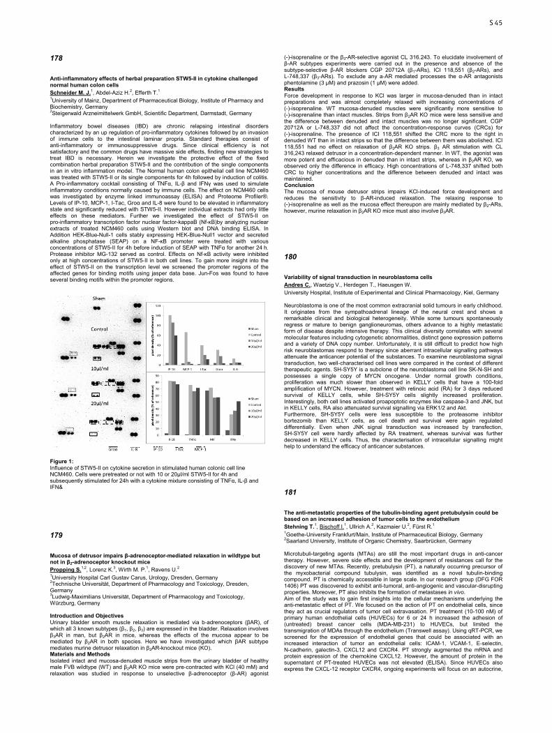
178
Anti-inflammatory effects of herbal preparation STW5-II in cytokine challenged normal human colon cells Schneider M. J.1, Abdel-Aziz H.2, Efferth T.1 1University of Mainz, Department of Pharmaceutical Biology, Institute of Pharmacy and Biochemistry, Germany 2Steigerwald Arzneimittelwerk GmbH, Scientific Department, Darmstadt, Germany Inflammatory bowel diseases (IBD) are chronic relapsing intestinal disorders characterized by an up regulation of pro-inflammatory cytokines followed by an invasion of immune cells to the intestinal laminar propria. Standard therapies consist of anti-inflammatory or immunosuppressive drugs. Since clinical efficiency is not satisfactory and the common drugs have massive side effects, finding new strategies to treat IBD is necessary. Herein we investigate the protective effect of the fixed combination herbal preparation STW5-II and the contribution of the single components in an in vitro inflammation model. The Normal human colon epithelial cell line NCM460 was treated with STW5-II or its single components for 4h followed by induction of colitis. A Pro-inflammatory cocktail consisting of TNFα, IL-β and IFNγ was used to simulate inflammatory conditions normally caused by immune cells. The effect on NCM460 cells was investigated by enzyme linked immunoassay (ELISA) and Proteome Profiler®. Levels of IP-10, MCP-1, I-Tac, Groα and IL-8 were found to be elevated in inflammatory state and significantly reduced with STW5-II. However individual extracts had only little effects on these mediators. Further we investigated the effect of STW5-II on pro-inflammatory transcription factor nuclear factor-kappaB (Nf-κB)by analyzing nuclear extracts of treated NCM460 cells using Western blot and DNA binding ELISA. In Addition HEK-Blue-Null-1 cells stably expressing HEK-Blue-Null1 vector and secreted alkaline phosphatase (SEAP) on a NF-κB promoter were treated with various concentrations of STW5-II for 4h before induction of SEAP with TNFα for another 24 h. Protease inhibitor MG-132 served as control. Effects on Nf-κB activity were inhibited only at high concentrations of STW5-II in both cell lines. To gain more insight into the effect of STW5-II on the transcription level we screened the promoter regions of the affected genes for binding motifs using jasper data base. Jun-Fos was found to have several binding motifs within the promoter regions.
Figure 1: Influence of STW5-II on cytokine secretion in stimulated human colonic cell line NCM460. Cells were pretreated or not with 10 or 20µl/ml STW5-II for 4h and subsequently stimulated for 24h with a cytokine mixture consisting of TNFα, IL-β and IFN&
179
Mucosa of detrusor impairs β-adrenoceptor-mediated relaxation in wildtype but not in β2-adrenoceptor knockout mice Propping S.1,2, Lorenz K.3, Wirth M. P.1, Ravens U.2 1University Hospital Carl Gustav Carus, Urology, Dresden, Germany 2Technische Universität, Department of Pharmacology and Toxicology, Dresden, Germany 3Ludwig-Maximilians Universität, Department of Pharmacology and Toxicology, Würzburg, Germany Introduction and Objectives Urinary bladder smooth muscle relaxation is mediated via b-adrenoceptors (βAR), of which all 3 known subtypes (β1, β2, β3) are expressed in the bladder. Relaxation involves β3AR in man, but β2AR in mice, whereas the effects of the mucosa appear to be mediated by β2AR in both species. Here we have investigated which βAR subtype mediates murine detrusor relaxation in β2AR-knockout mice (KO). Materials and Methods Isolated intact and mucosa-denuded muscle strips from the urinary bladder of healthy male FVB wildtype (WT) and β2AR KO mice were pre-contracted with KCl (40 mM) and relaxation was studied in response to unselective β-adrenoceptor (β-AR) agonist
(-)-isoprenaline or the β3-AR-selective agonist CL 316.243. To elucidate involvement of β-AR subtypes experiments were carried out in the presence and absence of the subtype-selective β-AR blockers CGP 20712A (β1-ARs), ICI 118,551 (β2-ARs), and L-748,337 (β3-ARs). To exclude any a-AR mediated processes the α-AR antagonists phentolamine (3 µM) and prazosin (1 µM) were added. Results Force development in response to KCl was larger in mucosa-denuded than in intact preparations and was almost completely relaxed with increasing concentrations of (-)-isoprenaline. WT mucosa-denuded muscles were significantly more sensitive to (-)-isoprenaline than intact muscles. Strips from β2AR KO mice were less sensitive and the difference between denuded and intact muscles was no longer significant. CGP 20712A or L-748,337 did not affect the concentration-response curves (CRCs) for (-)-isoprenaline. The presence of ICI 118,551 shifted the CRC more to the right in denuded WT than in intact strips so that the difference between them was abolished. ICI 118,551 had no effect on relaxation of β2AR KO strips. β3 AR stimulation with CL 316,243 relaxed detrusor in a concentration-dependent manner. In WT, the agonist was more potent and efficacious in denuded than in intact strips, whereas in β2AR KO, we observed only the difference in efficacy. High concentrations of L-748,337 shifted both CRC to higher concentrations and the difference between denuded and intact was maintained. Conclusion The mucosa of mouse detrusor strips impairs KCl-induced force development and reduces the sensitivity to β-AR-induced relaxation. The relaxing response to (-)-isoprenaline as well as the mucosa effect thereupon are mainly mediated by β2-ARs, however, murine relaxation in β2AR KO mice must also involve β3AR.
180
Variability of signal transduction in neuroblastoma cells Andres C., Waetzig V., Herdegen T., Haeusgen W. University Hospital, Institute of Experimental and Clinical Pharmacology, Kiel, Germany Neuroblastoma is one of the most common extracranial solid tumours in early childhood. It originates from the sympathoadrenal lineage of the neural crest and shows a remarkable clinical and biological heterogeneity. While some tumours spontaneously regress or mature to benign ganglioneuromas, others advance to a highly metastatic form of disease despite intensive therapy. This clinical diversity correlates with several molecular features including cytogenetic abnormalities, distinct gene expression patterns and a variety of DNA copy number. Unfortunately, it is still difficult to predict how high risk neuroblastomas respond to therapy since aberrant intracellular signalling pathways attenuate the anticancer potential of the substances. To examine neuroblastoma signal transduction, two well-characterised cell lines were compared in the context of different therapeutic agents. SH-SY5Y is a subclone of the neuroblastoma cell line SK-N-SH and possesses a single copy of MYCN oncogene. Under normal growth conditions, proliferation was much slower than observed in KELLY cells that have a 100-fold amplification of MYCN. However, treatment with retinoic acid (RA) for 3 days reduced survival of KELLY cells, while SH-SY5Y cells slightly increased proliferation. Interestingly, both cell lines activated proapoptotic enzymes like caspase-3 and JNK, but in KELLY cells, RA also attenuated survival signalling via ERK1/2 and Akt. Furthermore, SH-SY5Y cells were less susceptible to the proteasome inhibitor bortezomib than KELLY cells, as cell death and survival were again regulated differentially. Even when JNK signal transduction was increased by transfection, SH-SY5Y cell were hardly affected by RA treatment, whereas survival was further decreased in KELLY cells. Thus, the characterisation of intracellular signalling might help to understand the efficacy of anticancer substances.
181
The anti-metastatic properties of the tubulin-binding agent pretubulysin could be based on an increased adhesion of tumor cells to the endothelium Stehning T.1, Bischoff I.1, Ullrich A.2, Kazmaier U.2, Fürst R.1 1Goethe-University Frankfurt/Main, Institute of Pharmaceutical Biology, Germany 2Saarland University, Institute of Organic Chemistry, Saarbrücken, Germany Microtubuli-targeting agents (MTAs) are still the most important drugs in anti-cancer therapy. However, severe side effects and the development of resistances call for the discovery of new MTAs. Recently, pretubulysin (PT), a naturally occurring precursor of the myxobacterial compound tubulysin, was identified as a novel tubulin-binding compound. PT is chemically accessible in large scale. In our research group (DFG FOR 1406) PT was discovered to exhibit anti-tumoral, anti-angiogenic and vascular-disrupting properties. Moreover, PT also inhibits the formation of metastases in vivo. Aim of the study was to gain first insights into the cellular mechanisms underlying the anti-metastatic effect of PT. We focused on the action of PT on endothelial cells, since they act as crucial regulators of tumor cell extravasation. PT treatment (10-100 nM) of primary human endothelial cells (HUVECs) for 6 or 24 h increased the adhesion of (untreated) breast cancer cells (MDA-MB-231) to HUVECs, but limited the transmigration of MDAs through the endothelium (Transwell assay). Using qRT-PCR, we screened for the expression of endothelial genes that could be associated with an increased interaction of tumor an endothelial cells: ICAM-1, VCAM-1, E-selectin, N-cadherin, galectin-3, CXCL12 and CXCR4. PT strongly augmented the mRNA and protein expression of the chemokine CXCL12. However, the amount of protein in the supernatant of PT-treated HUVECs was not elevated (ELISA). Since HUVECs also express the CXCL-12 receptor CXCR4, ongoing experiments will focus on an autocrine,
S45
intracellular action of CXCL12. Furthermore, we found that the mRNA of both N-cadherin and E-selectin are upregulated by PT after 12 and 24 h in HUVECs. The upregulation of N-cadherin could be confirmed on the protein level. Using microscopy, we found that the distribution pattern of N-cadherin was not altered by PT. The hypothesis of N-cadherin as crucial adhesive protein responsible for the functional effects triggered by PT will be further analyzed in the future. In summary, the anti-metastatic action of pretubulysin might be based on the enhanced adhesion (i.e. trapping) of tumor cells onto the endothelium by an increased expression of endothelial cell adhesion molecules. This work was supported by the German Research Foundation (DFG, FOR 1406, FU 691/9-2).
182
Tamoxifen efficacy in metastatic breast cancer – impact of potential candidate genes in addition to CYP2D6 Bolbrinker J.1, Karle J.2, Vogl S.1, Eucker J.2, Kreutz R.1, Wischnewsky M.3, Regierer A. C.2 1Charité - Universitätsmedizin Berlin, Institut für Klinische Pharmakologie und Toxikologie, Germany 2Charité - Universitätsmedizin Berlin, Med. Klinik Onkologie, Germany 3Universität Bremen, FB Mathematik u. Informatik, Germany Introduction: In addition to tumor-specific parameters, e.g. hormone-receptor status, localization and amount of metastases, pharmacogenetic factors with impact on the chosen drug regimen might contribute to the well-known inhomogeneous clinical courses in metastatic breast cancer patients. We have previously shown a significant effect of CYP2D6 genotype on progression free survival (PFS) and overall survival (OS) in a cohort of advanced breast cancer patients treated with tamoxifen. Thus, our objective was to analyze the potential influence of further postulated candidate genes, e.g. related to drug transport and metabolism, on PFS, objective response rate, and OS in metastatic breast cancer patients. Here we present preliminary results of our study on CYP2C19. Methods: Genomic DNA was extracted from whole blood (n=47) or formalin-fixed, paraffin-embedded (FFPE) non-tumor tissues (n=42). All patients had prior or ongoing tamoxifen treatment (20mg/d). Samples were subsequently analyzed for single nucleotide polymorphisms (SNPs) in CYP2C19 (rs12248560, rs4244285, and rs4986893) using predesigned TaqMan® Genotyping Assays and/or by direct sequencing. Wild-type allele was presumed when testing for the variant alleles was negative. Patient’s medical records were retrospectively evaluated according to PFS, response rate, and OS. Results: DNA damage as a result of the formalin fixation process in FFPE tissues resulted in an overall low call rate when genotyping was performed with TaqMan® Assays. Thus, genotyping results for the tissue specimen were mainly obtained by direct sequencing. For one tissue sample no genotyping result could be obtained at all. Clinical data analysis revealed that PFS was significantly longer in *17-carriers compared to *1, *2, and *3-carriers (HR=2.34, 95% CI (1.27–4.30), p=0.006). This effect remained when restricting the evaluation to CYP2D6-EM patients (n=75, HR=2.13, 95% CI (1.15–3.93), p=0.016), suggesting an additive effect. Conclusions: Our preliminary analysis on the influence of postulated candidate genes on the efficacy of tamoxifen in advanced breast cancer reveals a significant effect of CYP2C19*17 on PFS. This seems to be additive to the previously reported effect of CYP2D6. Comprehensive pharmacogenetic profiling might thus help to identify metastatic breast cancer patients who will benefit most from treatment with tamoxifen.
183
Drug targeting in photodynamic therapy with albumin-folate-conjugates Butzbach K.1, Rasse Suriani F. A.2, Gonzales M. M.2, Cabrerizo F. M.2, Epe B.1 1Johannes Gutenberg-University, Institute for Pharmacy and Biochemistry, Mainz, Germany 2IIB-INTECH-UNSAM-CONICET, Photochemistry, Chascomús, Argentina In photodynamic therapy (PDT), cancer cells are killed by irradiation with visible light in the presence of a photosensitizer, which produces cytotoxic reactive oxygen species (ROS). The therapeutic concept would be more powerful if a specific uptake and thereby accumulation of the photosensitizer in malignant cells can be achieved. An attractive transporter for that purpose is the folic acid receptor α (FRα), which is over-expressed on the surface of many tumor cells to ensure the supply of folic acid (FA). FRα is an endocytotic receptor with a high FA affinity and a fast turnover rate. To find out whether albumin-folate-conjugates can serve as vehicles to transport photosensitizers into FRα overexpressing cancer cells, we used the highly fluorescent β-carboline derivate N2-carboxyethyl-norharmane, which we coupled covalently to albumin. Afterwards, we coupled FA to this albumin-β-carboline-conjugate (AβC) to produce an albumin-β-carboline-FA-conjugate (AβCF). AβC and AβCF were characterized with respect to size, conjugation pattern and photophysical properties. The fluorescence quantum yield of the fluorophor was not diminished significantly by albumin; therefore this conjugate is suitable to monitor the uptake of AβCF under the microscope. For our studies, we used KB cells (human nasopharyngal carcinoma), which are FRα overexpressing. We could show that AβCF accumulates in the lysosomes of the cells with increasing incubation time while under the same conditions the corresponding conjugate without folate AβC is not taken up by the cells. Therefor the uptake of AβCF is FA mediated. By using a β-carboline derivate with a higher triplet quantum yield and a
comparable coupling behavior, N2-carboxyethyl-harmine, we could demonstrate a FA dependent phototoxicity in the FRα overexpressing cells. In conclusion our results suggest that albumin-folate-conjugates are promising vehicles for a tumor cell targeted PDT.
184
Pioglitazone inhibits the MEK/ERK signalling pathway, reduces proliferation and induces apoptosis in human uterine leiomyosarcoma cells Zhao Y.1, Lucht K.2, Zuhayra M.1, Cascorbi I.2, Lützen U.1, Culman J.2 1University Hospital of Schleswig-Holstein, Campus Kiel, Department of Nuclear Medicine, Molecular Imaging, Diagnostics and Therapy, Germany 2University Hospital of Schleswig-Holstein, Campus Kiel, Institute of Experimental and Clinical Pharmacology, Germany The human uterine leiomyosarcoma is a malignant tumour with poor prognosis. Previous findings have demonstrated that SK-UT-1 cells, a human uterine leiomyosarcoma cell line, and human uterine smooth muscle cells (HutSMC) express the peroxisome proliferator-activated receptor g (PPARγ). In the present study, we investigated the effects of the PPARγ agonist, pioglitazone (PIO), on the proliferation and induction of apoptosis in SK-UT-1 cells and HutSMC. Cell proliferation of SK-UT-1 cells exposed to pioglitazone (10 or 25 µM) in the presence or absence of the selective PPARγ antagonist, GW9226, (1 µM) was evaluated by WST-1 assay and cresyl violet staining. Western blot analysis was used to quantify the cleaved caspase-3, Bax and Bad proteins and to assess the effects of PIO on the levels of activated MEK and ERK. Cellular toxicity of pioglitazone was assessed by lactate-dehydrogenase (LDH) release. PIO increased the expression of the PPARγ in SK-UT-1 cells and in HutSMC by a PPARγ – dependent mechanisms. PIO reduced concentration- and time-dependently the cell proliferation of quiescent as well as proliferating SK-UT-1 cells. These effects were partially mediated by the PPARγ. PIO at 25 µM concentration induced cleaved (activated) caspase-3, Bad, Bax and p53 proteins in SK-UT-1 cells. Quiescent SK-UT-1 cells have higher protein levels of the activated MEK and ERK, p-MEK and p-ERK, respectively, than quiescent HutSMC. PIO time-dependently reduced p-ERK and p-MEK in SK-UT-1 cells by a PPARγ – independent mechanism, but did not alter p-MEK/MEK and p-ERK/ERK levels in HutSMC. These findings point to the important role of MEK/ERK signalling pathway in the pioglitazone – induced inhibition of proliferation in SK-UT-1 cells. Pioglitazone did not exert any cytotoxic effects in SK-UT-1 or HutSMC. We demonstrate that PIO reduces proliferation and promotes apoptosis in quiescent and proliferating leiomyosarcoma cells. Our data address an important issue about the therapeutic relevance of PIO in the treatment of the uterine leiomyosarcoma.
185
MicroRNA expression profiling of responding and non-responding CML-patients to imatinib therapy Diewock T.1, Kaehler M.1, Pott C.2, Kneba M.2, Haenisch S.1, Cascorbi I.1, Bruhn O.1 1Institute of Experimental and Clinical Pharmacology, Kiel, Germany 2Clinic for Internal Medicine II, Hematology and Oncology, Kiel, Germany Although chronic myeloid leukemia (CML) can be effectively treated with the small BCR/ABL inhibitor imatinib, resistance in a significant proportion of patients remains a severe clinical problem. In order to identify potential biomarkers that predict the response of a patient to imatinib treatment, we have analyzed microRNA expression profiles of 45 newly diagnosed non-mutated, treatment-naive CML-patients who later responded (n=26) or were insensitive (n=19) to imatinib therapy. Response was defined as reaching molecular remission within the first 18 months after therapy start. For that, expression of 384 microRNAs was analyzed using a TaqMan Low-Density Array system. Relative fold change of microRNAs between responders and non-responders was calculated according to the 2-∆∆Ct-method. MicroRNAs with a fold change >2 and a p-value <0.01 were considered significant. MicroRNAs with minor expression (Ct-value >35) were excluded. Bioinformatic target gene prediction of deregulated microRNAs was performed and targets predicted by at least two databases independently were further analyzed using the bioinformatics database "DAVID". We show, that six microRNAs (miR-1, miR-193a-3p, miR-331-5p, miR-365, miR-449 and miR-636) are significantly deregulated (fold change -2 to 5, p-value 0.005 to 0.01) in non-responders. In-depth-analysis of the predicted targets reveals predominantly phosphoproteins involved in transcription regulation and membrane associated transport proteins. Pathway analysis shows that several genes are involved in cancer pathways including MAPK-signaling and PI3K/Akt-signaling, both mediated by BCR/ABL. In addition, twelve targeted genes are directly involved in the chronic myeloid leukemia pathway. Moreover, down-regulated miR-331-5p was earlier reported as a potent P-gp regulator and is associated with chemotherapy resistance and relapse in leukemia. Our results indicate, that the microRNA pattern is different between responding and non-responding CML-patients prior to imatinib therapy. Targets of significantly deregulated microRNAs are primarily transcription regulators and transport proteins of the OCT- and ABC-transporter families indicating that pharmacoresistance is mainly based on imatinib transmembrane transport and deregulated secondary driver pathways, particularly MAPK- and PI3K/Akt-signaling.
S46
186
The Chick Chorioallantoic Membrane (CAM) as a Cancer Xenograft Model for the Development of Therapeutics and Diagnostics Based on Engineered Nanomaterials Hafner S.1, Wu Y.2, Weil T.2, Syrovets T.1, Simmet T.1 1Ulm University, Institute of Pharmacology of Natural Products & Clinical Pharmacology, Germany 2Ulm University, Department of Organic Chemistry III/Macromolecular Chemistry, Germany Nanomaterials are gaining increasing attention for their potential applications in biomedicine. Particularly intriguing are nanomaterials designed to harbor both a therapeutic and diagnostic functionality. For such material the term ‘theranostic’ has recently been coined. Generally, colloidal instability of nanomaterials poses a considerable problem, which is often compromising their in vivo use. Here we used human albumin polethylene glycol (PEG) copolymer and chemically inert and stably fluorescent nanodiamonds (ND) as theranostic platform. Albumin PEG copolymer coating of ND warranted excellent plasma stability evenhandedly offering the option of covalent linkage of therapeutics such as doxorubicin and/or diagnostics such as gadolinium. As cancer model, we used the treatment-resistant human breast cancer cell line MDA-MB-231 that stably expresses luciferase thereby enabling in vivo imaging. MDA-MB-231 cells were xenotransplanted into silicone rings placed onto the chorioallantoic membrane of fertilized chick eggs. This model enabled continuous in vivo recording of the tumor growth and development. The albumin PEG copolymer coated ND carrying doxorubicin in an acid-labile covalent linkage, were efficiently delivered to the tumor tissue, where they exhibited, in comparison to conventional doxorubicin, increased antiproliferative effects on the breast cancer tumors. This was evident by imaging of luciferase activity as well as at the end of the experiment by the immunohistochemical analysis of the tumor tissues for the Ki-67 proliferation antigen. Besides, after intravenous injection of functionalized albumin PEG nanomaterial into CAM vessels, the nanomaterial-bound doxorubicin exhibits a better biocompatibility as indicated by significantly higher survival rates of chick embryos in comparison to those treated with equivalent injections of conventional doxorubicin. Thus, inclusion of doxorubicin into this particular nanomaterial allows tumor treatment at higher doxorubicin doses, which is enhancing the therapeutic antitumor efficacy. As the toxicity of cancer chemotherapeutics is a major factor that limits treatment efficacy, this approach warrants further investigation. We also developed MRI imaging in this model and preliminary data indicate that inclusion of gadolinium into the albumin PEG copolymer nanomaterial allows excellent imaging resolution of the tumors further expanding the applicability of the CAM model. Supported by the VolkswagenStiftung.
187
Pharmacokinetic interactions of selected anticancer drugs with doxorubicin reducing enzymes Hofman J.1,2, Skarka A.1, Havrankova J.1, Maser E.2, Wsol V.1 1Charles University in Prague, Faculty of Pharmacy in Hradec Kralove, Department of Biochemical Sciences, Czech Republic 2University Medical School Schleswig-Holstein, Campus Kiel, Institute of Toxicology and Pharmacology for Natural Scientists, Germany Doxorubicin (DOX) is classified as efficient cytostatic drug that is used for the treatment of several cancers. To date, several studies have demonstrated that the elevated enzymatic reduction of DOX to its less potent metabolite, DOXol, constitutes one of the mechanisms causing pharmacokinetic DOX resistance in tumors. 5-fluorouracil (5-FU), paclitaxel (PTX), docetaxel (DTX), cyclophosphamide (CYC) or tamoxifen (TMX) are combined with DOX in the first-line chemotherapy regimens indicated in breast cancer patients. Although effectiveness of these drugs in combination treatments have been proved in clinical practice, their possible interference with DOX metabolism have not been described in detail to date. In this study, we utilized appropriate experimental approaches to investigate the possible interactions of carbonyl reducing enzymes with 5-FU, PTX, DTX, TMX, CYC and its preactivated form, 4-hydroperoxycyclophosphamide (4-HC). First, we explored the ability of selected cytosolic carbonyl reducing enzymes to metabolize doxorubicin to doxorubicinol. Regarding their activity, the tested enzymes can be ranked as follows: AKR1C3 >> CBR1 ≈ AKR1A1. Other tested enzymes (CBR3, AKR1B1, AKR1B10, AKR1C1, AKR1C2, AKR1C4) exhibited only negligible activity toward doxorubicin and, therefore, were not further evaluated in subsequent study that investigated possible inhibition of AKR1C3, CBR1 and AKR1A1 by selected anticancer agents. AKR1C3 was shown to be significantly inhibited by PTX, CYC, 4-HC and TMX. CBR1 inhibition was induced by the presence of PTX, 4-HC and TMX. The last tested enzyme, AKR1A1, was inhibited by PTX and 4-HC. In the follow-up cellular studies, we tracked the changes in AKR1C3, CBR1 and AKR1A1 expression after exposure to tested cytostatics in MCF7 and HepG2 cells. As a result, no significant changes in the expression of tested enzymes were detected at mRNA and protein levels in both cell lines. In view of these findings, it is feasible to presume, that inhibition rather than induction plays a role in the interactions of tested anticancer agents with DOX reducing enzymes. In conclusion, our results describe important molecular events emerging during the combination breast cancer therapy which might modulate the pharmacokinetic doxorubicin resistance and/or behavior. This work is co-financed by the European Social Fund and the state budget of the Czech Republic. Projects no. CZ.1.07/2.3.00/30.0061 and CZ.1.07/2.3.00/20.0235.
188
Influence of miR-212 on imatinib-sensitivity of leukemic K562 cells Kaehler M.1, Turrini E.2, Bruckmueller H.1, Haenisch S.1, Bruhn O.1, Cascorbi I.1 1University Hospital Schleswig-Holstein, Campus Kiel, Institute of Experimental and Clinical Pharmacology, Germany 2Alma Mater Studiorum-University of Bologna, Department for Life Quality Studies, Rimini, Italy The hematopoetic disorder chronic myeloid leukemia (CML) is one of the most extensively studied neoplasms. It is caused by translocation between chromosomes 9 and 22 leading to the formation of the Philadelphia chromosome and the BCR-ABL fusiongene. First-line therapy is still the tyrosine-kinase inhibitor imatinib (IM), which led to tremendous success in treatment. However, the amount of therapeutic resistances is increasing, caused either by BCR-ABL-dependent mechanisms (e.g. BCR-ABL amplification/overexpression, point mutations) or BCR-ABL-independent mechanism, which can be linked to altered expression of drug transporters or particularly, microRNA-expression levels. In our previous study, we analyzed the alterations of microRNA expression profiles during the development of IM-resistances in the leukemic cell line K562. We found a relation of miR-212 to IM-resistance, indicated by an inversely correlation of miR-212 expression and protein levels of the efflux transporter ABCG2 (ATP-binding cassette transporter G2) in cells resistant to different IM-concentrations. In this study, we investigated whether miR-212 has a direct effect on IM-sensitivity. Therefore, we transfected K562 cells either with miR-mimic pre-miR-212 or inhibitory anti-miR-212, challenged them with IM and analyzed effects on cell viability, activation of apoptosis and cell death using WST-1-, Caspase Glo 9-assay and cell counting. We found that under IM-treatment, inhibition of endogenous miR-212 using anti-miR-212 significantly promotes cell survival apparent on the level of respiratory chain function (p<0.01) and cell membrane integrity and reduces capase-9 activity (p<0.05). A concentration row using increasing miR-212 concentrations (pre-miR: 25 nM, 50 nM; anti-miR: 75 nM, 150 nM) showed that the effects on the cells could be potentiated, leading to two-fold increased cell death in cells after pre-miR-transfection and vice versa using the anti-miR (10 to 30% of negative control). Overall, these experiments indicate that miR-212 does not only affect the ABCG2-expression, but does also influence cell sensitivity to IM in a more direct manner. Further analysis could now reveal, which mRNAs are targeted by miR-212, which pathways are influenced by this and how cell sensitivity to IM is altered. These findings could be relevant in CML-therapy, overcoming IM-resistances with a better understanding of miRNA-alteration in CML.
189
Hyperthermia enhances cisplatin and doxorubicin cytotoxicity by inhibition of PARP Schaaf L.1, Schmid J.1, Mürdter T. E.1, Steurer W.2, Aulitzky W.3, Schwab M.1, Ulmer C.2, van der Kuip H.1 1Dr Margarete Fischer-Bosch Institute and University of Tuebingen, Clinical Pharmacology, Stuttgart, Germany 2Robert Bosch Hospital, Surgery, Stuttgart, Germany 3Robert Bosch Hospital, Oncology, Stuttgart, Germany Hyperthermic intraperitoneal chemotherapy (HIPEC) in concert with cytoreductive surgery improves survival of patients with peritoneal carcinomatosis (PC) from colorectal and ovarian cancer. The molecular mechanisms behind the potentiating properties of elevated temperature on chemotherapy efficacy are fairly unknown. We here investigated the role of Poly(ADP-Ribose)-Polymerase (PARP) for the efficacy of HIPEC. This enzyme binds to single strand breaks (SSBs) or other abnormal DNA structures and Poly(ADP-Ribosy)lates (PARylates) histones and repair proteins surrounding the side of DNA damage. Ovarian and colon cancer cell lines were treated for 1 hour at 37 °C or 42 °C with clinically relevant dosages of cisplatin or doxorubicin. We observed a synergistic effect of hyperthermia together with cisplatin and doxorubicin. Combination treatment led to a significantly reduced long term survival. Time course experiments using COMET assay revealed a significant delay in DNA repair at 42 °C. DNA breaks were almost completely repaired after 8 hours at 37 °C whereas we observed hardly any repair at 42 °C. Importantly, this significantly diminished DNA repair capacity was paralleled by a reduction of PARylation at 42 °C immediately after treatment. This result indicates that chemotherapy induced PARP enzymatic activity is attenuated at the higher temperature. This is also supported by the finding that pharmacological inhibition of PARP mimicked the effects of elevated temperature on survival and DNA repair upon chemotherapy. Both hyperthermia and PARP inhibitors combined with either cisplatin or doxorubicin led to a significant increase of γH2AX and P-53BP1 foci formation indicating an accumulation of lethal DSBs as consequence of the compromised PARP dependent DNA repair. Our results indicate that hyperthermia attenuates chemotherapy induced PARP activation and thereby reduces DNA repair capacity. This might be central to the observed synergistic effect of hyperthermia or PARP inhibitor treatment together with clinically relevant cytotoxic compounds. This might be of clinical importance since in contrast to elevated temperature PARP inhibitors are generally well tolerated and could therefore serve as an alternative for hyperthermia.
S47
190
Celecoxib increases lung cancer cell lysis by lymphokine-activated killer cells via upregulation of ICAM-1 Schellhorn M.1, Haustein M.1, Linnebacher M.2, Frank M.3, Hinz B.1 1University of Rostock, Institute of Toxicology and Pharmacology, Germany 2University of Rostock, Department of General Surgery, Germany 3University of Rostock, Electron Microscopy Center, Germany The antitumorigenic mechanism of the selective cyclooxygenase-2 (COX-2) inhibitor celecoxib is still a matter of debate. Using lung cancer cell lines (A549, H460) and metastatic cells derived from a lung cancer patient, the present study investigates the impact of celecoxib on the expression of intercellular adhesion molecule 1 (ICAM-1) and cancer cell lysis by lymphokine-activated killer (LAK) cells. Celecoxib but not other structurally related selective COX-2 inhibitors (i.e., rofecoxib, etoricoxib, valdecoxib) was found to cause a substantial upregulation of ICAM-1 protein levels. Likewise, ICAM-1 mRNA expression was increased by celecoxib. Celecoxib enhanced the susceptibility of cancer cells to be lysed by LAK cells with the respective effect being reversed by a neutralizing ICAM-1 antibody. In addition, enhanced killing of celecoxib-treated cancer cells was reversed by preincubation of LAK cells with an antibody to lymphocyte function associated antigen-1 (LFA-1), suggesting intercellular ICAM-1/LFA-1 crosslink as crucial event within this process. Finally, celecoxib elicited no significant increase of LAK cell-mediated lysis of non-tumor bronchial epithelial cells, BEAS-2B, associated with a far less ICAM-1 induction as compared to cancer cells. Altogether, our data demonstrate celecoxib-induced upregulation of ICAM-1 on lung cancer cells to be responsible for intercellular ICAM-1/LFA-1 crosslink that confers increased cancer cell lysis by LAK cells. These findings provide proof for a novel antitumorigenic mechanism of celecoxib.
191
Lovastatin lactone elicits human lung cancer cell apoptosis via a COX-2/PPARγ-dependent pathway Walther U., Emmrich K., Ramer R., Mittag N., Hinz B. University of Rostock, Institute of Toxicology and Pharmacology, Germany Statins (3-hydroxy-3-methylglutaryl coenzyme A [HMG-CoA] reductase inhibitors) are well-established agents to treat hyperlipidemic states. In addition, experimental and epidemiological evidence implies an anticancer effect of these substances. This study investigates the mechanism underlying human lung cancer cell death by lovastatin and the role of the prostaglandin (PG)-synthesizing enzyme cyclooxygenase-2 (COX-2) in this process. In A549 and H358 lung carcinoma cells the lipophilic prodrug lovastatin lactone led to a concentration-dependent induction of DNA fragmentation and cell death, whereas its HMG-CoA-inhibitory, ring-open acid form was inactive in this respect. Analyses of lysates from lovastatin lactone-treated cells revealed profound concentrations of lovastatin lactone following a 4-h incubation period, thus proving a substantial uptake of the lipophilic form. Induction of cell death by lovastatin was accompanied by upregulation of COX-2 mRNA and protein as well as by increased formation of PPARγ-activating PGD2 and 15-deoxy-Δ12,14-PGJ2. Cells were significantly less sensitive to lovastatin-induced apoptotic cell death when the expression or activity of COX-2 was suppressed by siRNA or by the COX-2 inhibitor NS-398. In addition, apoptosis by lovastatin was reversed by the peroxisome proliferator-activated receptor γ (PPARγ) antagonist, GW-9662. Further experiments revealed a lovastatin-induced cytosol-to-nucleus translocation of PPARγ that was inhibited by NS-398. Collectively, this study demonstrates COX-2 induction and subsequent COX-2-dependent activation of PPARγ as a mechanism by which lovastatin lactone induces human lung cancer cell death. Moreover, this data challenge the prevalent view of the acid form being the sole active entity of statins.
192
Sphingosine-1-phosphate induces COX-1 and COX-2 expression and stimulates cell migration but not proliferation in colorectal cancer cells Wegner S.1, Mahajan-Thakur S.1, Böhm A.1, Polster S.1, Zopf D.2, Schrör K.3, Rauch B.1 1Universitätsmedizin Greifswald, Institut für Pharmakologie, Germany 2Bayer Pharma AG, Berlin, Germany 3Universitätsklinikum Düsseldorf, Institut für Pharmakologie und Klinische Pharmakologie, Germany Colorectal cancer (CRC) is one of the most common malignancies worldwide. Epidemiological evidence is cumulating that acetylsalicylic acid (ASA) effectively attenuates CRC risk after regular long-term intake. This effect is observed at low doses selective for inhibition of cyclooxygenase (COX)-1. Sphingosine-1-phosphate (S1P) is an immunomodulatory signaling lipid released from platelets and involved in proliferation, migration and cell invasion. Recently, it was shown that ASA can inhibit platelet-derived S1P release. Here, we determine possible effects of S1P on COX expression in various CRC cell lines and its impact on cell vitality and migration in vitro. Expression of COX-1 and COX-2 at the level of mRNA and total protein was determined by quantitative real-time PCR and Western blotting, respectively, in the CRC cell lines Caco-2, HT29 and SW620. Cell vitality was determined by crystal violet assay, cell migration in Boyden chamber assays. Incubation of CRC cells with S1P (1 µM) for 1, 3, 6, 16 and 24 h resulted in a differential regulation of COX-1 and COX-2 mRNA and protein levels. In Caco-2 and HT29 cells, COX-1 mRNA was significantly elevated after 3 to 16 h and after 16 to 24 h,
respectively. In both cell lines, COX-1 and COX-2 protein levels were upregulated within 6 to 16 h (n=4-6). In Caco2 cells, COX-2 mRNA expression was also enhanced after 6 and 16 h. However, despite an increased COX-2 protein expression in HT29 cells, COX-2 mRNA was not elevated in these cells, which may point to a described posttranslational mechanism of COX-2 regulation. In SW620 cells, S1P also significantly enhanced COX-1 mRNA within 16 to 24 h (n=6), while COX-2 mRNA was not detectable in these cells. In Boyden chamber migration assay, S1P significantly stimulated chemotaxis of Caco-2 and HT29 cells (n=5). In comparison, S1P (0.01 to 1 µM) and ASA (1 to 300 µM) did not significantly affect cell vitality over 72 h (n=3-5). In addition, expression of the known S1P receptors S1P1-5 was determined by real-time PCR. Caco-2 and HT-29 cells expressed all five receptor subtypes, while in SW620 cells the S1P3 receptor was missing. In summary, our data indicate that the immunomodulatory lipid S1P can regulate COX-1 and COX-2 protein in different CRC cell line to a variable extend. Enhanced COX expression in response to locally elevated S1P levels, i.e. released from platelets, may contribute to varying sensitivity of CRC to long-term ASA treatment.
193
Distribution and adduct formation of a fluorescent cisplatin analogue in the cytosol Zabel R.1, Kullmann M.2, Weber G.1 1Leibniz-Institut für Analytische Wissenschaften, Systemanalyse, Dortmund, Germany 2Pharmazeutisches Institut Universität Bonn, Klinische Pharmazie, Germany Platin-based anti-cancer drugs, such as cisplatin, are effective chemotherapeutic agents and widely used in the therapy of various types of cancer and are known to undergo several (bio-) chemical transformation steps after application. Hydrolysis and adduct formation with small nucleophils and bigger proteins are thought to be the most relevant reactions on their way to the final reaction site (DNA), but there are still many open questions regarding the identity and pharmacological relevance of different proposed adducts and intermediates. The aim of the project is the identification of binding partners inside tumor cells in vitro. CFDA-Platin, a fluorescent cisplatin analogue with related pharmacodynamic and -kinetic properties should help to identify binding partners. Due to the high separation power and sensitivity, capillary electrophoresis with laser induced fluorescence (CE-LIF) is the method of choice. Capillary electrophoresis with mass spectrometry (CE-MS) is used for the identification of unknown compounds. For the in vitro experiments we compared the cytosolic fraction of a human ovarian cancer cell line and its corresponding cisplatin-resistant variant. In addition, we performed model experiments with CFDA-Platin and potential binding partners, e.g. glutathione. CE-LIF and CE-MS are appropriate analytical methods to detect and identify CFDA-Platin and different complexes up to the ppb/ppm range. First results show a complex formation of CFDA-Platin with biological molecules of interest. A comparison of human ovarian cancer cells and its cisplatin-resistant variant indicates differences in the intracellular accumulation of CFDA-Platin.
194
Lipid-lowering drugs in the prevention of antitumor therapy-induced oral mucositis Ziegler V., Albers A., Henninger C., Fritz G. Heinrich Heine University Düsseldorf, Institute of Toxicology, Germany Oral mucositis is one of the most debilitating side-effects of antitumor therapy and frequently occurs in cancer patients receiving systemic chemotherapy or local irradiation of head and neck cancers. It is characterized by ulceration and inflammation of the oral mucosal tissue leading to severe pain and increased infection risk, which often impair the efficacy of anticancer treatment. Due to the fact that the pathogenesis of oral mucositis is only sparsely known and the supportive interventions are limited, we set out to identify novel pharmacologic approaches for the prevention and treatment of chemotherapy/radiation-induced mucositis. Since HMG-CoA reductase inhibitors (statins) that are clinically used for lipid-lowering purpose are attributed to protect from genotoxin-induced toxicity, we hypothesized that this might also hold true for keratinocytes, which are of particular relevance for the pathogenesis of mucositis. As in vitro model for ionizing radiation (IR) and doxorubicin (DOX)-induced oral mucositis we used human keratinocyte cells (HaCaT) and evaluated whether pretreatment with lovastatin protects them from IR- and DOX-induced cyto- and genotoxicity. Pretreatment of HaCaT cells with lovastatin resulted in an increased cell viability both in the Alamar Blue Assay and in an impedance-based cell analysis. In line with this, flow cytometric analysis showed a reduced subG1 fraction. This result was reassured by flow cytometric analysis of Annexin V positive cells. Western blot analysis confirmed a lovastatin-mediated reduction of the activity of caspases and PARP-1. In order to determine whether the aforementioned cytoprotective effects were based on initial DNA damage reduction by lovastatin, we measured Ser139 phosphorylated histon 2AX (gH2AX) and 53BP1 nuclear foci as surrogate markers for DNA double-strand breaks. Together with the Comet Assay our results strongly indicate that this is indeed the case for DOX, but not for IR treatment. To assess differences in the DNA damage response, we analyzed the expression and the activity status of key proteins involved in downstream DNA damage signaling pathways. In summary, our data demonstrate a protection of human keratinocytes from IR- and DOX-induced cell death by the lipid-lowering drug lovastatin. Hence, future in vivo studies are preferable to evaluate the relevance of these data under clinically more relevant settings.
S48
195
In Vitro Assessment of Direct and Time-Dependent Inhibitory Effects on Major Human Cytochrome P450 Enzymes by Spasmolytics Dahlinger D., Frechen S., Aslan S., Fuhr U. University Hospital of Cologne, Department of Pharmacology, Germany Objective: Approximately one in six adults in the United States and Europe suffer from overactive bladder syndrome (OAB) 1,2. In the latest guideline3 issued by the German Society of Obstetricians and Gynaecologists, anticholinergic agents are stated as the pharmacotherapy of choice with seven drugs being approved for the German market. Yet, only limited information on the drug-drug interaction potential of these agents exists. Therefore, we examined inhibition of the seven major cytochrome P450 (CYP) enzymes by darifenacin, fesoterodin, oxybutinin, propiverin, solifenacin, tolterodin, trospium chloride. Methods: To assess direct inhibition, an in vitro cocktail of seven specific, clinically relevant model substrates was incubated with pooled human liver microsomes for 10 minutes. For all experiments, the major metabolites of the probe substrates were simultaneously analysed using a validated liquid chromatography - tandem mass spectrometry method and enzyme kinetics were estimated by determining IC50 values. These IC50 values were then converted to an inhibition constant Ki using the Cheng-Prusoff equation4. Subsequently to assess time-dependent inhibition , a single concentration equivalent to the IC25, determined in the previous direct inhibition experiments, was used to examine TDI of the test compounds. After a 30 minute preincubation with NADPH, a TDI was identified by a decrease in enzyme activity. Results: In this study 49 IC50 experiments were conducted. In 18 out of these 49 cases, point estimates for the IC50 values were smaller than 100µM. In these cases CYP2D6 and CYP3A4 were most frequently inhibited (5 cases for CYP2D6; 5 cases for CYP3A4). The strongest inhibition was observed for darifenacin, propiverin and tolterodin on CYP2D6 with the calculated Ki in the lower micromolar range (associated 95% confidence intervals): darifenacin 0.0053 µM (0.0032 µM-0.0075 µM); propiverin 1.9µM (1.4µM-2.3µM) and tolterodin 2.7µM (1.7µM-3.6µM). The assessment of the time-depentdent inhibitory potential of the spasmolytics is currently ongoing. Conclusion: These screening experiments suggest that in particular darifenacin, propiverin and tolterodin may have clinically relevant inhibitory effects on especially CYP2D6 in humans. To characterize the potential clinical impact of these interactions and recommend dosage modifications, further in vitro data needs to be gathered, allowing for physiologically based pharmacokinetic modelling. 1Stewart, WF; Van Rooyen, JB; Cundiff, GW; Abrams, P; Herzog, AR; Corey, R; Hunt, TL; Wein, AJ (2003). "Prevalence and burden of overactive bladder in the United States". World Journal of Urology 20 (6): 327–336. doi:10.1007/s00345-002-0301-4. (20) 2 Milsom, I; Abrams, P; Cardozo, L; Roberts, RG; Thuroff, J; Wein, AJ (2001). "How widespread are the symptoms of an overactive bladder and how are they managed? A population-based prevalence study". BJU Int. 87 (9): 760–6. doi:10.1046/j.1464-410x.2001.02228.x. 3 Die überaktive Blase (ÜAB), AWMF, Register Nr. 015/007 Klasse: S2k, Stand Juni 2010. http://www.awmf.org/uploads/tx_szleitlinien/015-007l_S2k_Ueberaktive_Blase_Add_2014-07.pdf (assessed August 2014) 4 Cheng, Y;. Prusoff, W.H(1973). „Relationship between the inhibition constant (Ki) and the concentration of an inhibitor which causes 50 per cent inhibition (IC50) of an enzymatic reaction“ Biochem Pharmacol, 22, pp. 3099–3108
196
-Targeting of the thrombospondin-α2δ-1 interaction — Development of analgesics against neuropathic pain El-Awaad E.1, Fried C.1, Pryymachuk G.2, Matthes J.1, Hucho T.3, Neiss W. F.2, Herzig S.1, Zaucke F.4, Pietsch M.1 1University of Cologne, Department of Pharmacology, Germany 2University of Cologne, Department of Anatomy I, Germany 3University of Cologne, Experimental Anesthesiology and Pain Research, Department of Anesthesiology and Intensive Care Medicine, Germany 4University of Cologne, Center for Biochemistry, Germany The calcium channel subunit α2δ-1 has been identified as a neuronal thrombospondin (TSP) receptor. This interaction, thought to be mediated by the von Willebrand factor A (VWF-A) domain of α2δ-1, is crucial for excitatory synapse formation in the CNS (Eroglu et al., 2009). The α2δ-1 subunit is also the molecular target for the analgesics gabapentin and pregabalin (Field et al., 2006). Binding of these compounds to α2δ-1 was shown to inhibit its interaction with TSPs and therefore synaptogenesis (Eroglu et al., 2009). Here, we propose to develop a novel method to identify inhibitors of the α2δ‐1–TSP interaction in order to find new analgesics against neuropathic pain. Preliminary data from immunofluorescent staining indicated co-localization of TSP-4 and α2δ-1 in various murine brain regions, thus, we developed biochemical binding assays to investigate the direct interaction of the two proteins in vitro. For this purpose, soluble variants of human α2δ-1 and rat TSP-4 were expressed in HEK-293/EBNA cells and purified by affinity chromatography. Using ELISA, we were able to show and quantify for the first time the direct binding of TSP-4 to α2δ‐1 (KD = 252 nM). In addition, Alexa Fluor 488-labeled TSP-4 was generated and tested for its binding to α2δ‐1 (KD = 26.2 nM) in solution using a Microscale Thermophoresis (MST) approach. The difference in the obtained KD values is thought to be due to the immobilization-free nature of the measurements done with the MST system.
Using biochemical assays together with cell-based and histochemical analysis, we plan to analyze the ability of small molecules to interfere with the binding of TSPs to α2δ-1. Compounds inhibiting the α2δ‐1–TSP interaction will be selected for analgesia testing in wild type mice and for validation of their target specificity in α2δ‐1 and TSP knockout mice. Eroglu, C.; Allen, N. J.; Susman, M. W. et al. Cell 2009, 139, 380-92. Field, M. J.; Cox, P. J.; Stott, E. et al. Proc. Natl. Acad. Sci. U.S.A. 2006, 103, 17537-42.
197
Dictyostelium discoideum as a model for the investigation of Tetraspanin protein interactions Albers T., Beitz E., von Bülow J. University of Kiel, Pharmaceutical Chemistry, Germany Tetraspanins (Tsps) are evolutionary highly conserved proteins with four transmembrane spans. There is evidence, that Tsps are involved in diseases, such as cancer spreading and defects in blood coagulation [1]. However, little is known about their precise structure or protein-protein interaction sites, i.e. requirements to evaluate the druggability. The complexity of 33 human Tsp isoforms and the diversity of interactions in the tetraspanin-enriched microdomains prompted us to employ Dictyostelium discoideum as a simple model for the production of recombinant human Tsps and for in vivo testing of protein interactions. In order to preclude interferences with the recombinant human Tsps, we characterized and localized the endogenous Tsps of D. discoideum. Therefore, in silico, we identified five Tsps in the D. discoideum genome (TspA-E). Carrying out cDNA synthesis with oligo(dT)primers from D. discoideum total RNA and PCR-amplification with specific primers we found tspA, C, and D to be expressed in the amoeba stage. For intracellular localization of Tsps, we generated fusion proteins with an N- or C-terminal GFP and obtained signals in the living cell mainly at cytosolic organellar structures. Immunofluorescence and colocalization with the proton pump of D. discoideum confirmed the occurance in the contractile vacuole network. Currently, we are expressing human Tsps in D. discoideum for indepth biochemical characterization. [1] Hemler, Martin (2008):Targeting of tetraspanin proteins--potential benefits and strategies, Nature Reviews 7:707-758
198
The role of the hydroxycarboxylic acid receptor 2 in mice on a ketogenic diet in experimental autoimmune encephalomyelitis Assmann J. C.1, Chen H.1, Okun J. G.2, Wettschureck N.3, Schwaninger M.1 1University of Lübeck, Institute of Experimental and Clinical Pharmacology and Toxicology, Germany 2University Children's hospital Metabolic Centre Heidelberg, Metabolic Laboratory, Germany 3Max-Planck-Institute for Heart and Lung Research, Department of Pharmacology, Bad Nauheim, Germany The recently licensed drug dimethyl fumarate (DMF) has been shown to improve functional outcomes for patients with multiple sclerosis (MS). Previous work identified the hydroxycarboxylic acid receptor 2 (HCA2), a G protein-coupled membrane receptor, as a mediator of the therapeutic effect of DMF in experimental autoimmune encephalomyelitis (EAE), a mouse model of MS. Oral administration improved neurological symptoms and reduced neutrophil infiltration into the CNS, possibly by interfering with their adhesion to brain endothelial cells as seen in bEnd.3 cells in vitro. To further clarify the role of HCA2 activation during EAE we hypothesized that β-hydroxybutyrate (BHB), the endogenous ligand of HCA2, might also have beneficial effects similar to DMF. As BHB is a major ketone body, levels in the blood can be raised by feeding mice a ketogenic diet. Previous work in a permanent stroke model already showed neuroprotective effects under a ketogenic diet that were lost in Hca2-/- mice. We fed both Hca2-/- and wild-type mice with a ketogenic diet containing 80 % fat, 8 % protein, and 1.2 % carbohydrates (ssniff #E15149-30) starting 14 days prior to immunization. On the diet production of ketone bodies was elevated. The diet significantly dampened the disease severity compared to mice fed with matched control food (4.2 % fat, 20.8 % protein, 57.6 % carbohydrates; ssniff #E15000-00). However, the protective effect was not dependent on HCA2 with this specific ketogenic diet. It is unclear, whether a HCA2-dependent effect is masked by other mechanisms or components of the ketogenic diet that already maximally reduced disease severity. This question could be addressed by administration of BHB alone to rule out diet-mediated effects. Together, these data indicate that the ketogenic diet improves outcome, but the dependency on HCA2 needs further analysis. Future work will show, if other agonist of HCA2 are able to improve outcome in EAE as seen for DMF.
S49
199
Influence of the frankincense resin extract Casperome® on the lipopolysaccharide (LPS) induced systemic inflammation and consecutive liver dysfunction in mice Löser K.1, Seemann S.1, Lenhardt I.1, Werz O.2, Lupp A.1 1Jena University Hospital, Institute of Pharmacology and Toxicology, Germany 2Friedrich Schiller University Jena, Department of Pharmacy, Germany Despite recent advances in critical care, sepsis, septic shock and subsequent multi-organ failure remain an important cause of morbidity and mortality in intensive care units. With about 20-25% of patients displaying a severe inflammatory reaction impaired liver function can be observed, which has been shown to be of major consequence for overall patient outcome. Since still no specific therapy exists so far for the treatment of sepsis and sepsis-associated liver dysfunction, the aim of the present study was to evaluate if frankincense resin extract used in traditional medicine for the treatment of infections and chronic inflammatory disorders can serve as a new therapeutic option in this respect. For this purpose, the orally bioavailable frankincense extract Casperome® was tested in an animal model of sepsis, the intraperitoneal administration of lipopolysaccharides (LPS) in mice. Male 60-day-old mice were assigned to six treatment groups: (1) controls, (2) LPS treatment, (3) treatment with Casperome®, (4) treatment with Epikurone® (phosphatidylcholine used for dissolving the frankincense extract), (5) treatment with LPS plus Casperome®, (6) treatment with LPS plus Epikurone®. LPS was administered at a dosage of 5 mg/kg body weight and Casperome® or Epikurone® were given three hours before LPS treatment at a dosage of 240 mg/kg or 80 mg/kg body weight, respectively. 24 hours after LPS administration animals were sacrificed and organ weights and different physiological parameters (body temperature, blood glucose levels) as well as various parameters representing liver damage or liver function or indicating oxidative stress in liver but also in different other tissues were assessed. LPS caused a distinct decrease in body temperature and blood glucose levels as well as in liver glycogen content and biotransformation capacity together with an increase in oxidative stress in different organs. After pretreatment of the animals with Casperome® a distinct protective effect was seen on many of the parameters tested not only in liver, but also in other organs such as brain, lung, kidneys, spleen and adrenals. However, it also turned out that Casperome® is a strong inducer of various cytochrome P450 isoforms. Altogether, these results point to a protective effect of the frankincense extract Casperome® in systemic inflammation which was not restricted to liver tissue only, thus suggesting that frankincense extracts may possibly serve as a new treatment option in sepsis.
200
Target gene evaluation of a MIRNA differently expressed in hippocampal brain tissue in a rat post-status epilepticus model Wahmkow H.1, Michler C.2, Russmann V.2, Rettenbeck M. L.2, von Rüden E. - L.2, Cascorbi I.1, Potschka H.2, Haenisch S.1 1University-Hospital Schleswig-Holstein, UKSH, Campus Kiel, Institute of Experimental and Clinical Pharmacology, Germany 2Ludwig-Maximilians-University, Institute of Pharmacology, Toxicology & Pharmacy, Munich, Germany Background: Epilepsy is a complex nervous system disorder and the underlying mechanisms are multifactorial. Mesial temporal lobe epilepsy (MTLE) is a common type of epilepsy. The disease is discussed to be associated with signs of hippocampal neurogenesis in the subgranular zone as well as gliosis and neuronal cell loss in the cornu ammonis area. Epigenetic mechanisms (DNA methylation, chromatin modifications as well as miRNAs) are key players in controlling neuronal cell fate during the differentiation of neural stem cells into mature neurons, astrocytes or oligodendrocytes. In our study we hypothesized that seizure- and concomitant neuroinflammation-induced alterations in miRNA expression may cause changes in the expression of target genes leading to neuronal reorganization and hyperexcitable neuronal networks. Methods: Self-sustained status epilepticus (SSSE) was electrically induced in 12 of 20 electrode implanted female Sprague Dawley rats. Electrodes were implanted into the right anterior basolateral nucleus of the amygdala (BLA). After 17 weeks non-stimulated control (n=8) and SE rats, having developed spontaneous seizures, were sacrificed. Hippocampal brain tissue of SE and control animals was obtained. After total RNA isolation the expression of 354 miRNAs was determined using rt-PCR based TaqMan® arrays. Comparison of miRNA expression between both tissue types by Mann-Whitney-U test was performed in R with the HTqPCR package. Target gene search was performed using different data bases (miRDB, TargetScan 6.2., microRNA.org, MicroCosm Targets v.5). Reporter gene assays are currently conducted to confirm predicted miRNA/mRNA interactions. Results: Out of 354 miRNAs, 176 were detected in all examined tissues. One miRNA showed a 3.5 fold lower (p<0.001) expression in the epileptogenic hippocampus relative to the hippocampus of control animals. In-silico target gene search has identified five genes involved in neuronal inflammation, differentiation and signal transmission (e.g. TNF, NEUROG2, KCNK10) as potential targets for this differentially regulated miRNA. Conclusion: Seizure- and neuroinflammation-induced differential regulation of miRNAs could contribute to an altered function of several genes resulting in neuronal reorganization and hyperexcitability in the hippocampus.
201
Induced loss of ADAM10 in murine epidermis leads to multifaceted disease processes Weber S.1, Saftig P.2, El-Armouche A.1 1TU Dresden, Medical Faculty Carl Gustav Carus, Pharmacology and Toxicology, Germany 2CAU University of Kiel, Biochemistry, Germany Background: Deregulated expression of the zinc-dependent A disintegrin and metalloprotease 10 (Adam10) has been shown in different skin diseases including basal cell carcinoma, malignant melanoma [1], Psoriasis [2, 3] and Eczematous Dermatitis [4]. We therefore hypothesized that genetic modulation of Adam10 can be the underlying course of the observed skin phenotypes and generated inducible epidermal Adam10 knock out mice due to the fact that germline or constitutive epidermal Adam10 deletion led to pre- and perinatal lethality and have therefore prevented analysis of Adam10 function during skin disease [5, 6]. Results:The Keratin5-driven postnatal deletion of Adam10 in the epidermis (Adam10epi-induc) resulted in multifaceted disease phenotypes either in directly targeted Keratin5-Cre expressing tissues including e.g. the epidermis, thymic epithelium, tooth epithelium and corneal epithelium as well as bidirectional responses towards stromal tissues like the dermis or even systemic phenotypes in bone marrow and cell of peripheral blood circulation. Approximately 18 days after the induction of deletion first hair loss became apparent in Adam10epi-induc mice that gradually became worse with almost complete baldness after 40 days and no re-growth of hair. Additionally postnatal deletion of ADAM10 resulted in epidermal thickening characterized by an increased number of proliferating cells in the basal layer, and loss of K10 and increased K14 in the suprabasal layers, indicating hyperproliferation and disturbed differentiation. In addition, hair follicles were destroyed accompanied by epidermal cyst formation, which were positively stained for loricirin. Giemsa staining of back skin samples already revealed an increase of leucocyte infiltration in inducibly deleted skin. Finally Adam10 deficient epidermis showed decreased recovery after barrier disruption by tape stripping assays, while incisional wound healing was unaltered between Adam10 deficient and wild type mice. Conclusion: The observed phenotypes resemble many aspects of atopic dermatitis-like diseases like psoriasis and could therefore prove our hypothesis that Adam10 deregulation can directly induce skin diseases. These results have been strengthened by recent GWAS studies showing skin pigmentation defects in patients with Adam10 point mutations [7, 8]. 1. Singh B, Schneider M, Knyazev P, Ullrich A. Uv-induced egfr signal transactivation is dependent on proligand shedding by activated metalloproteases in skin cancer cell lines. International journal of cancer. Journal international du cancer. 2009;124:531-5392. Oh ST, Schramme A, Stark A, Tilgen W, Gutwein P, Reichrath J. Overexpression of adam 10 and adam 12 in lesional psoriatic skin. The British journal of dermatology. 2008;158:1371-13733. Kawaguchi M, Mitsuhashi Y, Kondo S. Overexpression of tumour necrosis factor-alpha-converting enzyme in psoriasis. The British journal of dermatology. 2005;152:915-9194. Maretzky T, Scholz F, Koten B, Proksch E, Saftig P, Reiss K. Adam10-mediated e-cadherin release is regulated by proinflammatory cytokines and modulates keratinocyte cohesion in eczematous dermatitis. The Journal of investigative dermatology. 2008;128:1737-17465. Weber S, Niessen MT, Prox J, Lullmann-Rauch R, Schmitz A, Schwanbeck R, Blobel CP, Jorissen E, de Strooper B, Niessen CM, Saftig P. The disintegrin/metalloproteinase adam10 is essential for epidermal integrity and notch-mediated signaling. Development. 2011;138:495-5056. Weber S, Saftig P. Ectodomain shedding and adams in development. Development. 2012;139:3693-37097. Kono M, Sugiura K, Suganuma M, Hayashi M, Takama H, Suzuki T, Matsunaga K, Tomita Y, Akiyama M. Whole-exome sequencing identifies adam10 mutations as a cause of reticulate acropigmentation of kitamura, a clinical entity distinct from dowling-degos disease. Human molecular genetics. 2013;22:3524-35338. Tharmarajah G, Faas L, Reiss K, Saftig P, Young A, Van Raamsdonk CD. Adam10 haploinsufficiency causes freckle-like macules in hairless mice. Pigment cell & melanoma research. 2012;25:555-565
202
Establishment of a human in vitro model of pathological cardiac hypertrophy Werner T.1,2, Hirt M. N.1,2, Stenzig J.1,2, Vollert I.1,2, Breckwoldt K.1,2, Letuffe-Brenière D.1,2, Neuber C.1,2, Benzin A. E.1,2, Hansen A.1,2, Eschenhagen T.1,2 1University Medical Center Hamburg-Eppendorf, Department of Experimental Pharmacology, Germany 2DZHK (German Centre for Cardiovascular Research), partner site Hamburg/Kiel/Lübeck, Germany Background Pathological cardiac hypertrophy is known to be one of the major risk factors for heart failure. In the past years we have established an in vitro hypertrophy model based on rat engineered heart tissue (rEHT) in which an afterload enhancement (AE) was mimicked by mechanical reinforcement of the silicone posts to which the EHTs are attached. One week after the afterload enhancement these EHTs show reduced contractile forces, prolonged relaxation times, cardiomyocyte enlargement, increased fibrosis and an activation of the fetal (hypertrophic) gene program. The aim of this study was to transfer the model from rat to human EHTs using human induced pluripotent stem cell derived cardiomyocytes (hiPSC-CMs) to better model the situation in patients. Methods The differentiation of the hiPSC was performed in a two-step protocol. The induction of mesodermal differentiation via BMP4, activin A and bFGF in an embryoid body format preceded the inhibition of wnt-signaling to provoke cardiac differentiation yielding up to 90 % α-actinin-positive cells. The cardiomyocytes were then used to generate
S50
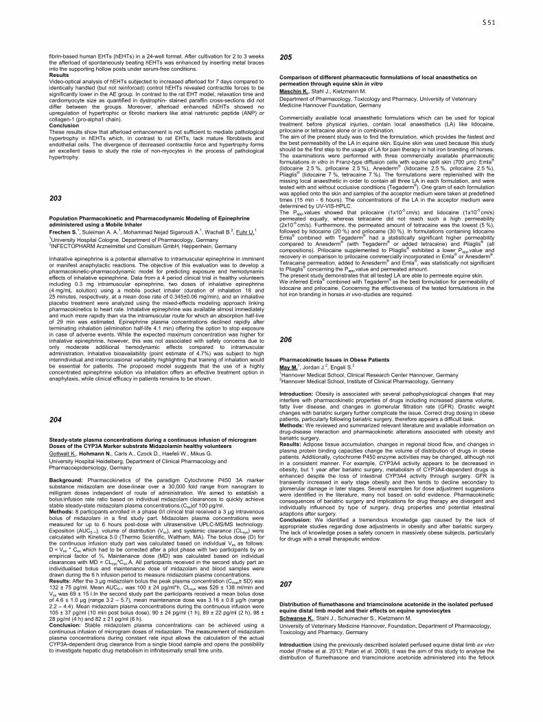
fibrin-based human EHTs (hEHTs) in a 24-well format. After cultivation for 2 to 3 weeks the afterload of spontaneously beating hEHTs was enhanced by inserting metal braces into the supporting hollow posts under serum-free conditions. Results Video-optical analysis of hEHTs subjected to increased afterload for 7 days compared to identically handled (but not reinforced) control hEHTs revealed contractile forces to be significantly lower in the AE group. In contrast to the rat EHT model, relaxation time and cardiomyocyte size as quantified in dystrophin- stained paraffin cross-sections did not differ between the groups. Moreover, afterload enhanced hEHTs showed no upregulation of hypertrophic or fibrotic markers like atrial natriuretic peptide (ANP) or collagen-1 (pro-alpha1 chain). Conclusion These results show that afterload enhancement is not sufficient to mediate pathological hypertrophy in hEHTs which, in contrast to rat EHTs, lack mature fibroblasts and endothelial cells. The divergence of decreased contractile force and hypertrophy forms an excellent basis to study the role of non-myocytes in the process of pathological hypertrophy.
203
Population Pharmacokinetic and Pharmacodynamic Modeling of Epinephrine administered using a Mobile Inhaler Frechen S.1, Suleiman A. A.1, Mohammad Nejad Sigaroudi A.1, Wachall B.2, Fuhr U.1 1University Hospital Cologne, Department of Pharmacology, Germany 2INFECTOPHARM Arzneimittel und Consilium GmbH, Heppenhein, Germany Inhalative epinephrine is a potential alternative to intramuscular epinephrine in imminent or manifest anaphylactic reactions. The objective of this evaluation was to develop a pharmacokinetic-pharmacodynamic model for predicting exposure and hemodynamic effects of inhalative epinephrine. Data from a 4 period clinical trial in healthy volunteers including 0.3 mg intramuscular epinephrine, two doses of inhalative epinephrine (4 mg/mL solution) using a mobile pocket inhaler (duration of inhalation 18 and 25 minutes, respectively, at a mean dose rate of 0.345±0.06 mg/min), and an inhalative placebo treatment were analyzed using the mixed-effects modeling approach linking pharmacokinetics to heart rate. Inhalative epinephrine was available almost immediately and much more rapidly than via the intramuscular route for which an absorption half-live of 29 min was estimated. Epinephrine plasma concentrations declined rapidly after terminating inhalation (elimination half-life 4.1 min) offering the option to stop exposure in case of adverse events. While the expected maximum concentration was higher for inhalative epinephrine, however, this was not associated with safety concerns due to only moderate additional hemodynamic effects compared to intramuscular administration. Inhalative bioavailability (point estimate of 4.7%) was subject to high interindividual and interoccasional variability highlighting that training of inhalation would be essential for patients. The proposed model suggests that the use of a highly concentrated epinephrine solution via inhalation offers an effective treatment option in anaphylaxis, while clinical efficacy in patients remains to be shown.
204
Steady-state plasma concentrations during a continuous infusion of microgram Doses of the CYP3A Marker substrate Midazolamin healthy volunteers Gottwalt K., Hohmann N., Carls A., Czock D., Haefeli W., Mikus G. University Hospital Heidelberg, Department of Clinical Pharmacology and Pharmacoepidemiology, Germany Background: Pharmacokinetics of the paradigm Cytochrome P450 3A marker substance midazolam are dose-linear over a 30,000 fold range from nanogram to milligram doses independent of route of administration. We aimed to establish a bolus:infusion rate ratio based on individual midazolam clearances to quickly achieve stable steady-state midazolam plasma concentrations (Css)of 100 pg/ml. Methods: 8 participants enrolled in a phase 0/I clinical trial received a 3 µg intravenous bolus of midazolam in a first study part. Midazolam plasma concentrations were measured for up to 6 hours post-dose with ultrasensitive UPLC-MS/MS technology. Exposition (AUC0-∞), volume of distribution (Vss), and systemic clearance (CLsys) were calculated with Kinetica 5.0 (Thermo Scientific, Waltham, MA). The bolus dose (D) for the continuous infusion study part was calculated based on individual Vss as follows: D = Vss * Css which had to be corrected after a pilot phase with two participants by an empirical factor of ⅔. Maintenance dose (MD) was calculated based on individual clearances with MD = CLsys*Css.A. All participants received in the second study part an individualised bolus and maintenance dose of midazolam and blood samples were drawn during the 6 h infusion period to measure midazolam plasma concentrations. Results: After the 3 µg midazolam bolus the peak plasma concentration (Cmax± SD) was 132 ± 75 pg/ml. Mean AUC0-∞ was 100 ± 24 pg/ml*h, CLsys was 526 ± 138 ml/min and Vss was 69 ± 15 l.In the second study part the participants received a mean bolus dose of 4.6 ± 1.0 µg (range 3.2 – 5.7), mean maintenance dose was 3.16 ± 0.8 µg/h (range 2.2 – 4.4). Mean midazolam plasma concentrations during the continuous infusion were 105 ± 37 pg/ml (10 min post bolus dose), 90 ± 24 pg/ml (1 h), 89 ± 22 pg/ml (2 h), 98 ± 28 pg/ml (4 h) and 82 ± 21 pg/ml (6 h). Conclusion: Stable midazolam plasma concentrations can be achieved using a continuous infusion of microgram doses of midazolam. The measurement of midazolam plasma concentrations during constant rate input allows the calculation of the actual CYP3A-dependent drug clearance from a single blood sample and opens the possibility to investigate hepatic drug metabolism in infinitesimally small time units.
205
Comparison of different pharmaceutic formulations of local anaesthetics on permeation through equine skin in vitro Maschin K., Stahl J., Kietzmann M. Department of Pharmacology, Toxicology and Pharmacy, University of Veterinary Medicine Hannover Foundation, Germany Commercially available local anaesthetic formulations which can be used for topical treatment before physical injuries, contain local anaesthetics (LA) like lidocaine, prilocaine or tetracaine alone or in combination. The aim of the present study was to find the formulation, which provides the fastest and the best permeability of the LA in equine skin. Equine skin was used because this study should be the first step to the usage of LA for pain therapy in hot iron branding of horses. The examinations were performed with three commercially available pharmaceutic formulations in vitro in Franz-type diffusion cells with equine split skin (700 µm): Emla® (lidocaine 2.5 %, prilocaine 2.5 %), Anesderm® (lidocaine 2.5 %, prilocaine 2.5 %), Pliaglis® (lidocaine 7 %, tetracaine 7 %). The formulations were replenished with the missing local anaesthetic in order to contain all three LA in each formulation, and were tested with and without occlusive conditions (Tegaderm®). One gram of each formulation was applied onto the skin and samples of the acceptor medium were taken at predefined times (15 min - 6 hours). The concentrations of the LA in the acceptor medium were determined by UV-VIS-HPLC. The Papp-values showed that prilocaine (1x10⁻⁵ cm/s) and lidocaine (1x10⁻⁵ cm/s) permeated equally, whereas tetracaine did not reach such a high permeability (2x10⁻⁶ cm/s). Furthermore, the permeated amount of tetracaine was the lowest (5 %), followed by lidocaine (20 %) and prilocaine (30 %). In formulations containing lidocaine Emla® combined with Tegaderm® had a statistically significant higher permeability compared to Anesderm® (with Tegaderm® or added tetracaine) and Pliaglis® (all compositions). Prilocaine supplemented to Pliaglis® exhibited a lower Papp-value and recovery in comparison to prilocaine commercially incorporated in Emla® or Anesderm®. Tetracaine permeation, added to Anesderm® and Emla®, was statistically not significant to Pliaglis® concerning the Papp-value and permeated amount. The present study demonstrates that all tested LA are able to permeate equine skin. We inferred Emla® combined with Tegaderm® as the best formulation for permeability of lidocaine and prilocaine. Concerning the effectiveness of the tested formulations in the hot iron branding in horses in vivo-studies are required.
206
Pharmacokinetic Issues in Obese Patients May M.1, Jordan J.2, Engeli S.2 1Hannover Medical School, Clinical Research Center Hannover, Germany 2Hannover Medical School, Institute of Clinical Pharmacology, Germany Introduction: Obesity is associated with several pathophysiological changes that may interfere with pharmacokinetic properties of drugs including increased plasma volume, fatty liver disease, and changes in glomerular filtration rate (GFR). Drastic weight changes with bariatric surgery further complicate the issue. Correct drug dosing in obese patients, particularly following bariatric surgery, therefore appears a difficult task. Methods: We reviewed and summarized relevant literature and available information on drug-disease interaction and pharmacokinetic alterations associated with obesity and bariatric surgery. Results: Adipose tissue accumulation, changes in regional blood flow, and changes in plasma protein binding capacities change the volume of distribution of drugs in obese patients. Additionally, cytochrome P450 enzyme activities may be changed, although not in a consistent manner. For example, CYP3A4 activity appears to be decreased in obesity, but 1 year after bariatric surgery, metabolism of CYP3A4-dependent drugs is enhanced despite the loss of intestinal CYP3A4 activity through surgery. GFR is transiently increased in early stage obesity and then tends to decline secondary to glomerular damage in later stages. Several examples for dose adjustment suggestions were identified in the literature, many not based on solid evidence. Pharmacokinetic consequences of bariatric surgery and implications for drug therapy are divergent and individually influenced by type of surgery, drug properties and potential intestinal adaptions after surgery. Conclusion: We identified a tremendous knowledge gap caused by the lack of appropriate studies regarding dose adjustments in obesity and after bariatric surgery. The lack of knowledge poses a safety concern in massively obese subjects, particularly for drugs with a small therapeutic window.
207
Distribution of flumethasone and triamcinolone acetonide in the isolated perfused equine distal limb model and their effects on equine synoviocytes Schwanse K., Stahl J., Schumacher S., Kietzmann M. University of Veterinary Medicine Hannover, Foundation, Department of Pharmacology, Toxicology and Pharmacy, Germany Introduction Using the previously described isolated perfused equine distal limb ex vivo model (Friebe et al. 2013; Patan et al. 2009), it was the aim of this study to analyse the distribution of flumethasone and triamcinolone acetonide administered into the fetlock
S51

joint. Furthermore, equine synoviocytes were used to determine efficacious antiinflammatory concentrations of the glucocorticoids. Materials and Methods 2 mg flumethasone aqueous solution or 10 mg triamcinolone acetonide aqueous suspension were administered into the fetlock joints of distal forelimbs from slaughtered horses exarticulated in the mediocarpal joint. The limbs were perfused with oxygenated tyrode solution via the A. mediana for at least 7.5 h. Venous perfusate from the V. radialis was collected to determine the glucocorticoid concentration by means of high performance liquid chromatography. Additionally, equine synoviocytes from different horses were cultivated and pretreated for 4 h with the glucocorticoids (10-13-10-8 mol/l). Thereafter cells were stimulated with lipopolysaccharide for 24 h. In the supernatans PGE2 concentration was measured. Results The mean concentration of flumethasone reached a maximum of 0.12 µg/ml in the venous perfusate 3.5 h after intraarticular administration. The mean maximum concentration of triamcinolone acetonide was similar to that of flumethasone but it was already reached after 30 minutes. With regard to the inhibition of lipopolysaccharide induced PGE2 production triamcinolone acetonide is slightly more effective at nanomolar concentrations than flumethasone. Conclusion The isolated perfused equine distal limb is a suitable model to study the distribution of intraarticularly administered glucocorticoids over a period of up to 8.5 h. The acquired data allow for estimations of the residence time of the compounds and may be useful for the estimation of withdrawal periods after intraarticular treatment. Furthermore, flumethasone and triamcinolone acetonide have potent antiinflammatory effects on the inner cell lining of the joint capsule. References Friebe, M., Stahl, J., Kietzmann, M. (2013). The isolated perfused equine distal limb as an ex vivo model for pharmacokinetic studies. J vet Pharmacol Therap, 36, 292–297 Patan, B., Budras, K. (2009). Effects of long-term extracorporeal blood perfusion of the distal portion of isolated equine forelimbs on metabolic variables and morphology of laminar tissue. Am J Vet Res, 70, 669–677
208
Pharmacokinetics of the experimental non-nucleosidic DNA methyl transferase inhibitor N-phthalyl-L-tryptophan (RG108) in rats Stenzig J.1,2, Schneeberger Y.3,1,2, Hübner F.4, Reichenspurner H.3,2, Eschenhagen T.1,2 1University Medical Center Hamburg-Eppendorf, Department of Experimental Pharmacology and Toxicology, Germany 2DZHK (German Centre for Cardiovascular Research), partner site Hamburg/Kiel/Lübeck, Germany, Germany 3University Heart Center Hamburg, Department of Cardiovascular Surgery, Germany 4University of Münster, Institute of Food Chemistry, Germany Background: Inhibition of DNA methyl transferases DNMT1, 3a and 3b represents a therapeutic strategy to re-establish the expression of tumour suppressor genes in malignant diseases, but might also be useful in other diseases. Inhibitors in current clinical use are nucleosidic cytotoxic agents that need to be integrated into the DNA of dividing cells to be active. In this study we aimed at establishing the in vivo kinetics of a non-nucleosidic inhibitor that is potentially free of cyctotoxic effects and does not require cell division. Methods and Results: Per application 2 mg of the nonspecific DNMT inhibitor N-phthalyl-L-tryptophan (RG 108) were dissolved in a mixture of 33 µl dimethyl sulfoxide (DMSO), 15 µl ethanol and 352 µl corn oil and injected subcutaneously in male rats. Blood was drawn at 0, 0.5, 1, 2, 4, 6 and 8 h post injection, and RG 108 was extracted from plasma or snap frozen tissue by acetonitrile extraction. For tissue level analysis, rats were sacrificed 0.5 h after RG 108 injection. RG 108 was measured by high performance liquid chromatography coupled to mass spectrometry. The following conditions were compared: Single dose application in the absence (group 1, n=2) or presence (group 2, n=2) of the cytochrome inhibitor ketoconazole (keto) in chow (0.03 %, started 3 days before RG 108 application) and multiple dose application of RG 108 (once daily for 3 days) in the absence (group 3, n=2) or presence (group 4, n=4) of keto in the chow. RG 108 treatment had no apparent effects on the animals. RG 108 levels were neither influenced by cytochrome inhibition nor did it differ between single or multiple dose regimens. Time to maximal plasma concentration (tmax) was 37.5 ± 15 min, plasma half life 1.4 h (95 % CI: 1 – 2.8 h), maximal plasma concentration (cmax) 6.8 ± 0.8 mg/l (20.5 ± 2.5 µM) and area under the curve was 22.3 ± 6.1 mg*h/l (67 ± 18 µM*h/l). Cmax was 1.0 mg/kg tissue (3.0 µM) in liver, 0.17 mg/kg tissue (0.52 µM) in skeletal and 0.47 mg/kg tissue (1.42 µM) in heart muscle. Conclusion: This study describes for the first time pharmacokinetics of the experimental non-nucleosidic DNMT inhibitor RG 108 after s.c. application. Despite its high lipophilicity it can be used for in vivo experiments, appears safe and yields plasma and tissue levels in the range of the described 50% inhibitory concentration (IC50) of around 1 µM. RG 108 can therefore be a useful tool for applications in which in vivo DNMT inhibition is to be studied.
209
The FNTB -609G>C polymorphism as a possible predictive factor for efficacy of lonafarnib-treatment? – exploratory analysis of a randomized phase II clinical trial in stage IIB-IV ovarian cancer, treated with first line platinum-based chemotherapy ± lonafarnib Bachmann H. S.1, Meier W.2, du Bois A.3, Kimmig R.4, Kuhlmann J. D.4,5, Siffert W.1, Sehouli J.6, Wollschläger K.7, Huober J.8, Hillemanns P.9, Burges A.10, Schmalfeldt B.11, Aminossadati B.12, Wimberger P.4,5 1University Hospital Essen, Institute of Pharmacogenetics, Germany 2Evangelisches Krankenhaus Düsseldorf, Department of Gynecology and Obstetrics, Germany 3Klinikum Essen Mitte, Department of Gynecology and Gynecologic Oncology, Germany 4University Hospital Essen, Department of Gynecology and Obstetrics, Germany 5University Hospital Carl Gustav Carus, Department of Gynecology and Obstetrics, Dresden, Germany 6Charité Medical University of Berlin, Department of Gynecology, Germany 7University Hospital Magdeburg, Department of Gynecology and Obstetrics, Germany 8University Hospital Ulm, Department of Gynecology and Obstetrics, Germany 9Medical University of Hannover, Department of Gynecology and Obstetrics, Germany 10Klinikum Großhadern, Department of Gynecology and Obstetrics, München, Germany 11Klinikum rechts der Isar, Department of Gynecology and Obstetrics, München, Germany 12Philipps University of Marburg, Coordinating Centre for Clinical Trials, Germany Background: Despite promising preclinical findings regarding clinical utility of farnesyltransferase inhibitors (FTI), such as lonafarnib, success of clinical studies is limited. In some studies, response to FTI was observed in 20-30% of patients, whereas other studies failed to resolve a benefit of lonafarnib, or even showed a negative effect of lonafarnib on the outcome of suboptimally debulked ovarian cancer patients (AGO-OVAR 15 phase II). In this regard, aim of the present study was to investigate clinical utility of a -609G>C polymorphism, located within the promoter core region of the farnesyltransferase ß-subunit gene (FNTB), in terms of a molecular biomarker for lonafarnib response. Methods: Influence of -609G>C on FNTB promoter activity was investigated in vitro by electrophoretic mobility shift assay, luciferase reporter assay and RT-qPCR. Subsequently, 57 out of 105 patients from the prospective, randomized AGO-OVAR 15 phase II trial, treated with carboplatin and paclitaxel ± lonafarnib, were genotyped for -609G>C by pyrosequencing. Progression-free survival (PFS) and overall survival (OS) was analyzed by Kaplan-Meier analysis with regard to the underlying genotype. Results: The presence of the -609G allele was associated with increased FNTB promoter activity in vitro, compared to the -609C allele. However, adverse effect of lonafarnib treatment towards PFS and OS was exclusively limited to patients, bearing a -609GG genotype (HRPFS 4.9, p<0.001; HROS 9.0, p=0.001). Median PFS for patients with -609GG genotype in the FTI treated arm was 10 months, whereas median PFS for placebo treated patients was 40 months. Median OS in the FTI treated group was 19 months, whereas median OS was not reached in the non-treated group. Conclusions: Observed discrepancies between preclinical and clinical results are putatively related to higher inter-individual variability of the FNTB locus in patients and effects of the FNTB -609G>C polymorphism. Therefore, our results encourage to re-evaluate FNTB polymorphisms in other FTI studies, especially those including patients with FTI response.
210
Relation between genotype, phenotype and therapeutic drug concentrations of nortriptyline or venlafaxine users in old age psychiatry Berm E. J. J.1, Kok R. M.2, Hak E.3, Wilffert B.1,4 1University of Groningen, Pharmacotherapy & Pharmaceutical Care, Netherlands 2Parnassia Psychiatric Institute, Old Age Psychiatry, The Hague, Netherlands 3University of Groningen, Pharmacoepidemiology & Pharmacoeconomics, Netherlands 4University Medical Centre Groningen, Clinical Pharmacy and Pharmacology, Netherlands Background The relationship between phenotype and genotype of the polymorphic cytochrome P450 2D6 enzyme (CYP2D6) has been intensively studied, however few studies are conducted among older persons. In a study among 900 relatively young venlafaxine users (mean age 45 years), 83% were genotyped as an extensive metabolizer (EM), but 21% were observed to have a poor metabolizer (PM) phenotype (1). This so-called ‘phenoconversion’ is one of the reasons for clinicians to mainly rely on therapeutic drug monitoring (TDM) as a sufficient indicator for genotype and to adjust the drug dosage if indicated. We determined associations between blood drug levels, genotype and phenotype in an old age sample of patients treated for depression with nortriptyline (NTP) or venlafaxine (VFX). Methods We analyzed post-hoc data from a clinical trial among older starters of NTP or VFX (2). The study population was monitored by TDM for twelve weeks. The drug levels of NTP and VFX as well as the main CYP2D6 metabolites, OH-nortriptyline and O-desmethylvenlafaxine, were measured. In addition, the genotypes for the CYP2D6 *3 and *4 alleles were available. We sub-grouped the data into phenotypes according to the metabolite/mother compound ratio. Next, we compared the phenotype with the genotype results and translated the blood levels into clinical outcomes being below, above, or within drug-specific therapeutic windows according to guidelines and tested for significant (p<0.05) differences between the phenotypes. Results Data from 75 patients (40 NTP;35 VFX) were available. The majority of patients had three blood samples taken. No phenoconversion from the PM to the EM genotype, or vice versa, was observed for both NTP and VFX. Phenoconversion from PM to IM, IM to EM, or vice versa was found in 28% for NTP users and in 44% for VFX users. For NTP, patients with a slow metabolizer phenotype more frequently had blood drug levels
S52

above the therapeutic window after three weeks (p<0.001) and after five weeks (p=0.067). For VFX such differences were not found. Conclusion In this old age patient population, a considerable part phenoconversed from PM to IM, or IM to EM or vice versa, but we did not observe phenoconversion from PM to EM, or vice versa. Furthermore, for NTP, it was found that even when intensive TDM was applied, there is room for improvement and genotyping could serve as a tool to prevent excessive levels of NTP among PM. 1. Preskorn SH, Kane CP, Lobello K, Nichols AI, Fayyad R, Buckley G, et al. Cytochrome P450 2D6 phenoconversion is common in patients being treated for depression: implications for personalized medicine. J Clin Psychiatry 2013 Mar 13. 2. Kok RM, Nolen WA, Heeren TJ. Venlafaxine versus nortriptyline in the treatment of elderly depressed inpatients: a randomised, double-blind, controlled trial. Int J Geriatr Psychiatry 2007 Dec;22(12):1247-1254.
211
Human sterol regulatory element-binding protein 1a contributes significantly to hepatic lipogenic gene expression Bitter A.1, Nüssler A. K.2, Thasler W. E.3, Klein K.1, Zanger U. M.1, Schwab M.1,4, Burk O.1 1Dr. Margarete Fischer-Bosch-Institute of Clinical Pharmacology, Stuttgart, Germany 2University of Tübingen, Department of Traumatology, Germany 3Ludwig-Maximilians-University Munich, Department of Surgery, Germany 4University Hospital Tübingen, Institute of Experimental and Clinical Pharmacology and Toxicology, Germany Background/Aims: Sterol regulatory element-binding protein (SREBP) 1, the master regulator of lipogenesis, was shown to be associated with non-alcoholic fatty liver disease (NAFLD), which is attributed to its major isoform SREBP1c. Based on studies in mice, the minor isoform SREBP1a is regarded as negligible for hepatic lipogenesis. This study aims to elucidate the expression and functional role of SREBP1a in human liver. Methods: mRNA expression of both isoforms was quantified in cohorts of human liver samples histologically classified for NAFLD and primary human hepatocytes. Hepatocytes were treated with PF-429242 to inhibit the proteolytic activation of SREBP precursor protein. SREBP1a-specific and pan-SREBP1 knock-down were performed by transfection of respective siRNAs. Lipogenic SREBP-target gene expression was analyzed by real-time RT-PCR. Results: In human liver, SREBP1a accounts for up to half of the total SREBP1 pool and its expression increases with NAFLD severity. Treatment with PF-429242 indicated SREBP-dependent auto-regulation of SREBP1a, which however was much weaker than of SREBP1c. SREBP1a-specific knock-down also reduced significantly the expression of SREBP1c and of SREBP-target genes. Regarding most SREBP-target genes, simultaneous knock-down of both isoforms resulted in effects of only similar extent as SREBP1a-specific knock-down. Conclusion: We have clearly shown here that human SREBP1a contributes considerably stronger to the total hepatic SREBP1 pool and has a much stronger impact on hepatic lipogenic gene expression, both quantitatively and qualitatively, than mouse SREBP1a. Given the crucial role of lipogenesis in NAFLD etiology and progression, our data support a pathophysiological role of SREBP1a in human hepatic lipogenesis and emphasize importance of its interindividual expression variability.
212
GenoChip CYP: Easy, fast, cheap and high reproducible way to detect Cyp P450 mutation for research and clinical use Freese P., Fleischer M., Lorenz M., Weise A., Eidens M. Pharmgenomics, Mainz, Germany Detecting mutations in Cytochrome P450 (CYP) enzymes has a high impact for a wide range of treatment decisions. Most common used method is sequencing gene areas of interest. That method takes a lot of time, is cost expensive and also needs an expert for data interpretation. Therefore we had the aim to develop a method, which is easy to use, fast and high-throughput able. METHODS GenoChip based on a modified reaction tube. The tube contains a glass bottom with an array of surface-immobilized probes. The different spots contain sequence specific probes against in total 34 genetic variations of nine different CYP genes and probes for the unmutated regions as well as different kind of controls. PCR is performed as a multiplex. Samples are analyzed with a small single tube reader. Data interpretation is performed with a self-developed software. RESULTS GeneChip Cyp were done in parallel to the Roche AmpliChip®. Within 200 samples a concordance of 99% were found. Only 2 samples showed a different outcome. Subsequent sequencing demonstrated that the GeneChip Cyp gave a true result whereas the AmpliChip result was not in correspondence with the results of sequencing. DISCUSSION GeneChip Cyp is an easy, fast and reliable assay with a short hands-on-time. It can be used for research topics, within clinical trials or as routine diagnostic to identify Cytochrome P450 mutations of interest.
213
Relationship between the polymorphism of the n-th acetyltransferase (NAT2) and the development of erysipelas Kadkina V.1, Rizvanov A.1, Kravchenko I.2, Ibadova G.3 1Kazan Federal University, Department of Genetics, Russia 2Kazan State Medical University, Department of Infectious Diseases, Russia 3Professor Agafonov Republican Clinical Hospital of Infectious Diseases M3, Kazan, Russia Despite using etiotropic therapy for the treatment of different forms of erysipelas, frequent recurrence of the disease and general complications are observed. Genetic peculiarities of the metabolic processes have impact on the pharmacological effect during treatment. In this respect, it is important to study peculiarities of the genetic systems involved in detoxification of xenobiotics in patients with erysipelas and to determine their relationship with the clinical progression of the disease. A group of 67 patients with various forms of erysipelas (46.6 % of patients with erythematous form, 29.3 % with erythematous-hemorrhagic, 18.9 % with bullous-hemorrhagic, and 5.2 % with erythematous-bullous) in the age of 55 to 59 years (67.2 % women and 32.8 % men) was examined. Primary erysipelas was diagnosed in 30 (51.7 %) and recurrent erysipelas in 26 (44.8 %) patients. The control group consisted of 40 healthy persons in the age from 50 to 60 years. The frequencies of the NAT2 Lys268Arg (803A/G) genotypes in the group of patients with erysipelas was 16 % for AA , 76 % for AG, and 7 % for GG, which significantly differs from the frequencies in the control group (6 % for AA, 50 % for AG, and 44 % for GG). A significant difference in the genotype of patients with the recurrent (25 % for AA, 45 % for AG, and 100 % for GG) and primary erysipelas (75 % AA and 56 % for AG), as well as with severe (25 % for AA, 19 % AG, and 33 % for GG) and moderate (75 % for AA, 82 % for AG, and 67 % for GG) forms of the disease (p < 0.05 χ2 = 34.36 > 5.99) was observed. The correlation between the polymorphism of the NAT2 Lys268Arg gene in the genotype carriers (GG, GA, AA) with the clinical progression of erysipelas is established, that suggests the relationship between this mutation and the incidence of erysipelas. Thus, the polymorphism (GG, AA, AG) of the NAT2 gene, which is involved in the II phase of detoxification, was analyzed for the first time. The erysipelas disease occurs by 1.5 times more frequent in the suffered patients than in the control healthy group. Differences in the frequency distribution of the GG genotype in patients with erysipelas depending on the recurrence of the disease are established, with the verified larger frequency for the recurrent form in comparison with primary erysipelas (100 % and 0 %, respectively), which allows one to associate the GG genotype with the probability of the development of recurrent erysipelas.
214
Targeted exome resequencing of ADME genes in human liver: assessment of SNP- and CNV frequencies and possible functional relevance in case of cytochrome P450’s Klein K.1, Fehr S.2, Tremmel R.1, Schaeffeler E.1, Winter S.1, Schwab M.1,3, Biskup S.2, Zanger U. M.1 1Dr. Margarete Fischer-Bosch Institut für Klinische Pharmakologie, Stuttgart, Germany 2CeGaT GmbH, Tuebingen, Germany 3University Hospital Tuebingen, Department of Clinical Pharmacology, Germany High-throughput approaches like Next-Generation Sequencing (NGS) offers the opportunity to analyze a large number of genes and detect known and novel single nucleotide variants (SNVs) and copy number variations (CNVs). We have developed a panel based targeted NGS pipeline for comprehensive sequence analysis of 340 genes involved in absorption, distribution, metabolism and excretion (ADME) of xenobiotics and endogenous substances. Cytochrome P450s are involved in the metabolism many drugs and endogenous substances and display high expression variability. We here focus on SNVs and CNVs within 11 genes of human CYP families 1, 2 and 3 and their potential functional implications. DNA enrichment was specifically designed for optimal capturing of all exonic, exon/intron boundaries, 5’ and 3’UTRs of the selected target genes with the Sureselect enrichment kit (Agilent). NGS was performed at a very high depth with 2x100 bp paired-end reads (Hiseq2500, Illumina). A set of 150 extensively characterized Caucasian liver samples was analyzed. Variants were compared to available SNV data in databases (dbSNP, DGV). CNVs were estimated using the sequence coverage compared to the mean of reads of all samples at a certain position. SNV validation was performed using Illumina HumanHAP300 SNP arrays and other genotyping methods. The impact of SNVs on CYP protein expression, quantified by western blot, and specific activity, quantified using specific probe substrates, was assessed. SNV data were in good agreement with previously determined genotypes. CNVs within 2A6, 2D6 and 2E1 could be detected. A total of 226 variants were found of which 40 (18%) were not yet listed in dbSNP. Novel SNVs were distributed over all CYP genes (1A1: n=5; 1A2: n=4; 2A6: n=1; 2B6: n=11; 2C8: n=3; 2C9: n=2; 2C19: n=3; 2D6: n=4; 2E1: n=2; 3A4: n=3; 3A5: n=2), of which most had low frequency. Of ten missense mutations found as single observations functional prediction (SIFT/PolyPhen) revealed deleterious effect for CYP1A1 (R98W, H60L), CYP2C8 (R124W, L268Q), CYP2C9 (S115R, Q324*), and CYP2E1 (I383M). Association analysis of SNVs, CNVs and haplotypes with expression and enzyme function data will be presented. Our targeted NGS approach on an ADME gene panel with its custom bioinformatics pipeline is a highly efficient and sensitive method for comprehensive pharmacogenomic studies. It provides high coverage to detect rare known and novel variations as shown for CYP-variome in Caucasian livers. The study was supported by the German BMBF (VLN grant 0315755) and by the Robert Bosch Foundation, Stuttgart, Germany
S53
215
Breast cancer susceptibility loci as potential predictors of outcome in ER-positive postmenopausal early breast cancer patients treated with tamoxifen Lo W. Y.1,2, Obazee O.1,2, Schroth W.1,2, Hoppe R.1,2, Fritz P.1, Ihring J.1,2, Hamann U.3, Schmidt M.4, Fasching P.5,6, Brauch H.1,2,7 1Dr. Margarete Fischer-Bosch - Institute of Clinical Pharmacology, Stuttgart, Germany 2University of Tuebingen, Germany 3German Cancer Research Center (DKFZ), Molecular Genetics of Breast Cancer, Heidelberg, Germany 4University of Mainz, Department of Gynecology and Obstetrics, Germany 5University Hospital Erlangen, Department of Gynecology and Obstetrics, Germany 6University of California, Department of Medicine, David Geffen School of Medicine, Los Angeles, United States 7German Cancer Consortium (DKTK), German Cancer Research Center (DKFZ), Heidelberg, Germany Breast cancer is the most frequent cancer in women. Numerous genetic factors contribute to its occurrence including the highly penetrant BRCA1/2 mutations and over 90 low penetrant susceptibility loci. Currently it is unknown whether the common genetic variants can also affect drug treatment outcome. The selective estrogen receptor modulator, tamoxifen, is a common breast cancer treatment principle because it blocks the binding of estrogen to the estrogen receptor (ER). Estrogen is critical for tumor growth and its carcinogenic effects are mediated through the ER which is expressed in approximately 75% of all breast tumors. Our group has a long standing interest in potential tamoxifen outcome predictors1,2 as up to 33% of treated patients relapse or die at 15 years of follow up, therefore it is important to determine which patient will benefit from tamoxifen or not. Here, we investigated whether some of the reported susceptibility loci may influence the outcome of ER-positive breast cancer patients treated with tamoxifen. Our study included 915 postmenopausal women with European ancestry, ER-positive, invasive and non-metastatic disease, available follow-up data as well as planned 5-year adjuvant mono-tamoxifen treatment. Their constitutional DNA has been genotyped at the 30 strongest breast cancer susceptibility loci3 and association studies including Cox regression and Kaplan-Meier analyses were carried out with recurrence-free and disease-free survival as the endpoint. Three polymorphisms showed significant associations: the strongest effect was observed for rs11249433 located in the 1p11.2 chromosome region with NOTCH2, EMBP1, FCGR1B being the nearest plausible genes. Patients with the variant G allele had a 2.2-fold increased risk for recurrence (HR 2.18, 95% CI 1.32-4.54). A protective gene-dose effect was observed at the FGFR2_rs35054928 locus where carriers of the C variant allele had a 24% reduction in the risk to develop recurrence (HR 0.76, 95% CI 0.58-0.99). Patients with the ancestral T allele at the TERT_rs7734992 locus also had a reduction in the risk to experience recurrence or death (Log-rank P = 0.03). Altogether, we provide the first exploratory evidence for the putative role of common polymorphisms in the tamoxifen outcome prediction in ER-positive postmenopausal early breast cancer. This suggests that not only can they potentially influence breast cancer risk, but it also has the potential to become useful pharmacogenetic markers. 1. Schroth W et al. Association between CYP2D6 polymorphisms and outcomes among women with early stage breast cancer treated with tamoxifen. JAMA. 2009;302(13):1429-36 2. Saladores P et al. Tamoxifen metabolism predicts drug concentrations and outcome in premenopausal patients with early breast cancer. Pharmacogenomics J. 2014;doi:10.1038/tpj.2014.34 3. Michailidou K et al. Large-scale genotyping identifies 41 new loci associated with breast cancer risk. Nat Genet. 2013;45(4):353-61
216
Does the apparently predictive haplotype Met408-Del420 really exist and how strong it affects OCT1 activity? Seitz T.1, Bokelmann K.1, Pereira J. D. S.1, Müller T.2, Brockmöller J.1, Koepsell H.2, Tzvetkov M.1 1University Medicine Center Göttingen, Clinical Pharmacology, Germany 2Universität Würzburg, Julius-von-Sachs-Institut für Biowissenschaften, Germany The human organic cation transporter OCT1 is highly genetically variable. The most common polymorphism causing a substrate specific loss of OCT1 activity is the deletion of methionine420 (Met420del). It has been suggested that another common polymorphism, the Met408Val substitution, strongly modulates the effects of the Met420del. However, the data is highly controversial. Here we genotyped 1319 individuals worldwide for the Met420del and Met408Val polymorphisms and analyzed the available genotype data for another 1092 individuals from the 1000 genomes project. Haplotype inferring using PHASE software showed that the deletion of Met420 was present only on Val408, but not on the Met408 background. Furthermore, using semiconductor-based massive parallel sequencing we proved that the double heterozygous carriers do not carry the 420 deletion and Met408 alleles on the same chromosome. Due to contradictory in vitro findings about the influence of the Met408Val substitution on the effects of Met420del, we analyzed morphologically and functionally the Met408 – Del420 and Val408 – Del420 haplotypes. We generated HEK293 cells stably transfected with these two theoretically possible haplotypes and used them to analyze the effects of the variants on subcellular localization and activity of OCT1. To analyze the subcellular localization we used confocal microscopy after co-staining OCT1 with the cytoplasmic membrane marker Na+/K+ -ATPase or the ER-marker calnexin. OCT1 activity was measured using model substrates (MPP+, TEA+, ASP+), drugs (morphine, metformin, debrisoquine) and the biogenic amine tyramine. Neither Met420del, nor Met408Val affected the membrane localization of OCT1. We observed substrate-specific loss of
OCT1 activity caused by the Met420del. However, Met408Val substitution did not affect the uptake of the model substrates, tyramine, or the drugs metformin or debrisoquine. A small increase of morphine uptake was observed with the Met408Val substitution, but the increase was present both in the presence and absence of the Met420 deletion. In conclusion, in humans the 420 deletion allele exists only together with the Val408 allele and the Met408Val polymorphism does not strongly modulate the effects of Met420del on OCT1 activity. Therewith, the mechanism of the substrate-specific loss of OCT1 activity caused by Met420del remains unclear and needs further investigations.
217
Characterization of the promoter region of a variant of the ß-subunit of the human farnesyltransferase Stelmach P., Manthey I., Siffert W., Bachmann H. S. University Hospital Essen, Institute of Pharmacogenetics, Germany The enzyme farnesyltransferase consists of two subunits: An α-subunit (48kDa) and a β-subunit (46kDa). The enzyme transfers a farnesyl group to the C-terminal end of proteins. By this posttranslational modification proteins are able to anchor to cellular membranes by the hydrophobic farnesyl residue. Thus, they can carry out their physiological function. Preliminary studies of our research group showed that there are at least 4 other variants (F2, F3, CF1 and CF2) of the ß-subunit of the enzyme farnesyltransferase in addition to the already known variant F1. Here, we characterize the promoter region of the F2 variant of the ß-subunit and the impact of different transcription factors on F2 expression. First, we performed a 5’RACE assay in order to experimentally determine the transcription start of the F2 variant. The transcription start was 506 base pairs upstream of the translation start point (ATG). According to this result the promoter region was cloned and deletion constructs were generated by restriction enzymes. After that a luciferase reporter assay was performed in HEK 293, A2780 and HepG2 cell lines in order to determine the basal promoter activity. In HEK 293 cells, the basal activity was twice as high as the control. In HepG2 cells the basal activity was similar to that of the control and in A2780 cells the basal activity was reduced by half. In an in silico analysis of the promoter region 12 putative binding sites for the activating transcription factor HNF1α and 7 putative binding sites for the inhibiting transcription factor HBP1 were found. In an over-expression experiment with the transcription factor HNF1α and in knock-down experiments with the transcription factor HBP1 we were able to demonstrate that these two transcription factors have a significant influence on the regulation of the promoter activity. In summary, a region of 1961 base pairs 5`upstream of the translation start point (ATG) has been identified as the promoter region of the F2 variant of the ß-subunit. By comparison with the basal promoter activity and in electrophoretic mobility shift assays it was shown that the transcription factors HNF1α and HBP1 influence the promoter activity significantly.
218
OCT1 is the clinically relevant hepatic transporter of sumatriptan: in vitro and in vivo evidences Kuron D., Matthaei J., Brockmöller J., Tzvetkov M. University Medicine Göttingen, Clinical Pharmacology, Germany Sumatriptan is by far the most prescribed anti-migrane drug in Germany. However, sumatriptan treatment does not lead to sufficient pain relief in approximately 20 % of the patients. Another 1 to 10 % of the patients develop adverse effects including nausea, reversible hyprethony and dizziness. Sumatriptan is bio-inactivated by mono amine oxidase and patients with decreased liver function show strongly increased plasma concentrations of sumatriptan. Sumatriptan is a hydrophilic organic base that may need transport to enter the liver. More than 90 % of the sumatriptan molecules are positively charged under physiological conditions. Here, we analyzed whether the liver organic cation transporter OCT1 mediates sumatriptan uptake and whether genetic polymorphisms in OCT1 affect sumatriptan pharmacokinetics in humans. In vitro, we analyzed sumatriptan transport using primary human hepatocytes and HEK293 cells overexpressing wild-type and polymorphic OCT1. In vivo, we analyzed sumatriptan pharmacokinetics in healthy volunteers with preselected OCT1 genotypes. A single oral dose of 50 mg sumatriptan was administrated, the plasma concentrations and surrogate markers for adverse effects were monitored. Sumatriptan was transported by OCT1 with a very high capacity (VMAX of above 2000 pmol/mg protein/ml) and sumatriptan uptake in primary human hepatocytes was strongly inhibited by the OCT inhibitor MPP+. Compared to OCT1, OCT3 (the other organic cation transporter expressed in the human liver) transported sumatriptan with 10-fold lower capacity. OCT1-mediated transport was strongly reduced in the polymorphic isoforms OCT1*3 and OCT1*4 and was completely abolished in OCT1*5 and OCT1*6. The OCT1-mediated transport was also inhibited by metoclopramid, propranolol, and amitriptyline; drugs commonly administrated with sumatriptan in treatment of migraine. In humans, the plasma concentration of sumatriptan was significantly increased in homozygous carriers of reduced function OCT1 alleles. The plasma concentrations were not changed in the heterozygous carriers and in the carrier of the OCT1*2 allele. No genotype dependent differences were observed in TMAX and VD. In conclusion, sumatriptan is a high capacity substrate of OCT1 and common genetic polymorphisms strongly effect sumatriptan pharmacokinetics. The ability of OCT1 to transport other triptanes was also analyzed in vitro and potential clinical effects were discussed.
S54

219
XD-14, a new small molecule inhibitor of BRD4, limits cardiac hypertrophy Barg M.1, Gilsbach R.1, Lucas X.2, Günther S.2, Hein L.1 1Universität Freiburg, Institut für Pharmakologie und Toxikologie, Germany 2Universität Freiburg, Institut für Pharmazeutische Wissenschaften, Pharmazeutische Bioinformatik, Germany Background: Chronic heart failure is accompanied by gene expression alterations. Several studies suggest that pathophysiologically relevant gene expression changes involve epigenetic mechanisms. One important principle is the acetylation of histone tails and recognition of these modification by dedicated reader proteins like BET bromodomain proteins. One member of this family is BRD4. Recently it was shown (Anand et al., Cell, 2013) that inhibition of BRD4 by the small molecule JQ1 blocks induction of gene expression programs that are crucial for cardiac hypertrophy. Treatment with JQ1 suppresses cardiac hypertrophy and remodeling in vitro and in vivo. In silico studies identified another BET-inhibitor XD-14. This compound is structurally different from JQ1 and has different binding properties (Lucas et al., Angew. Chem. Int. Ed., 2013). Therefore we aimed to compare the antihypertrophic effects of XD-14 and JQ1 in an in vitro hypertrophy model. Methods: Neonatal rat cardiomyocytes (NRCM) were isolated and cultured in serum-free medium for 48 hours. Subsequently, the cells were pre-treated with JQ1 (250 or 500 nM) or XD-14 (5 µM) for 6 hours and then stimulated with phenylephrine for further 24 hours. The cell-size was analyzed by immunofluorescence. Further the expression levels of the hypertrophy marker Nppb were examined by qPCR. Results: Stimulation of NRCMs with phenylephrine (100 µM, 48 h) led to hypertrophy of the cells and to increased expression of Nppb (p < 0.05, n = 3) compared to untreated control cells. Pre-treatment with JQ1 reduced the development of cellular hypertrophy (JQ1, 250 nM) in response to phenylephrine. This effect on cardiomyocyte hypertrophy was accompanied by decreased Nppb gene expression. Treatment with XD-14 led also to a significant reduction of phenylephrine-induced hypertrophy and Nppb gene expression. Conclusions: Our results indicate that the novel BRD4 inhibitor XD-14 reduces phenylephrine-induced hypertrophy of cardiomyocytes in an in vitro model comparable to JQ1.
220
Expression of the Monocarboxylate Transporter 4 (MCT4/SLC16A3) and its epigenetic regulation in primary clear cell renal cell carcinoma (ccRCC) and metastasis Fisel P.1, Kruck S.2, Winter S.1, Bedke J.2, Hennenlotter J.2, Nies A. T.1, Scharpf M.3, Schwab M.1,4, Schaeffeler E.1 1Dr. Margarete Fischer-Bosch - Institute of Clinical Pharmacology, University of Tuebingen, Stuttgart, Germany 2Department of Urology, University Hospital Tuebingen, Germany 3Institute of Pathology and Neuropathology, University Hospital Tuebingen, Germany 4Department of Clinical Pharmacology, University Hospital Tuebingen, Germany Background: Clear cell renal cell carcinoma (ccRCC) is the most common subtype of renal cell carcinoma. It is characterized by a metabolic shift towards enhanced aerobic glycolysis and hence, increased lactate production. Due to its role as mediator of H+-coupled lactate export, the monocarboxylate transporter 4 (MCT4) plays a pivotal role for the maintenance of glycolytic metabolism and the prevention of intracellular acidification in ccRCC cells. Moreover, the acidic extracellular environment resulting from export of lactate and H+, promotes anti-apoptotic effects and metastasis. Since metastatic ccRCC remains a major challenge for treatment, MCT4 may represent a promising target for therapeutic intervention also for metastatic disease. Methods: MCT4 protein expression was assessed in a cohort of 207 primary ccRCC and corresponding non-tumor tissues by immunohistochemical staining of tissue microarrays. A second cohort of 64 primary ccRCC and matching non-tumor tissues was used to validate findings and to additionally investigate MCT4 mRNA expression and DNA methylation in the regulatory promoter region of SLC16A3. MCT4 expression data and DNA methylation at specific CpG sites in the SLC16A3 promoter were correlated with clinicopathological parameters and outcome data. Furthermore, MCT4 protein and mRNA expression as well as SLC16A3 promoter DNA methylation were examined in 18 distant metastases of primary ccRCC. Results: MCT4 protein expression was upregulated in primary ccRCC compared to non-tumor kidney tissue in both cohorts (P<0.0001) and showed significant association with clinicopathological features (e.g. occurrence of metastasis) and cancer-related death. MCT4 expression was increased on mRNA level as well. Moreover, DNA methylation at single CpG sites in the SLC16A3 promoter correlated inversely with MCT4 protein as well as mRNA expression and was associated significantly with patient survival. ccRCC metastases showed similar protein and mRNA expression levels as well as DNA methylation patterns as the primary tumor tissue. Conclusions: Our results suggest that MCT4 expression underlies epigenetic regulation by DNA methylation in the SLC16A3 promoter not only in primary ccRCC, but also in metastasis. DNA methylation at specific CpG sites in the 5’-regulatory region of MCT4 may therefore not only serve as a predictor for patient outcome, but has also potential as novel target for therapeutic intervention, especially for metastatic disease.
221
Genome wide epigenetic profiling of purified cardiomyocytes enables deep insights into gene expression control Gilsbach R.1, Preißl S.1, Grüning B.2, Köbele C.1, Schnick T.1, Hein L.1 1University of Freiburg, Institute of experimental and clinical Pharmacology and Toxicology, Germany 2University of Freiburg, Bioinformatics Group, Department of Computer Science, Germany Background: DNA-methylation as well as histone modifications are important epigenetic mechanisms to maintain transcriptional control. In the present study we analysed the interplay of these mechanisms with gene expression in cardiomyocyte development and disease. Methods: We purified cardiomyocyte nuclei from mouse hearts by FACS. These nuclei were used to generate genome-wide maps of DNA methylation and histone marks (H3K27me3, H3K27ac) at base-pair resolution. The datasets were accompanied with gene expression profiles of FACS-sorted cardiomyocytes. Results: Bioinformatic analysis identified large genomic regions which were demethylated in cardiomyocytes. These large domains affected gene-bodies of 687 genes. More than 80 % of these genes were abundantly expressed in cardiomyocytes. Furthermore, these genes were decorated with active histone marks, including H3K27ac. These genes included highly cardiomyocyte specific genes like Myh6 or Myh7. Analysis of different developmental stages of cardiomyocytes revealed a decline of DNA methylation that was initiated during early development and extended well into postnatal maturation. The loss of DNA methylation was tightly linked to increasing gene expression. Remarkably genic regions of developmental genes, with no evident postnatal gene expression, were substantially demethylated in adult cardiomyocytes, too. These genes were marked by the repressive histone mark H3K27me3. In addition, our analysis identified 127 genes with increased postnatal methylation. This gain of DNA methylation was correlated with suppression of gene expression (p<0.0001). The affected genes included genes of the cardiac contractile apparatus like Tnni1 or Myl7. Ablation of DNA methyltransferases 3a/b in cardiomyocytes prevented de-novo methylation and lead to partial reactivation of gene expression. In contrast, differentially methylated regions acquired during pressure overload were located outside of intragenic. Interestingly, the methylation status of these regions partially reflected the neonatal methylation patterns. Conclusion: The presented data describes for the first time the dynamic DNA methylation landscape of cardiomyocytes during postnatal growth and adaptation to pathological stress at base-pair resolution. Integrative analysis of these data and cardiomyocyte-specific histone signatures as well as gene expression profiles enables deep insights into epigenetic mechanisms of gene regulation in cardiomyocytes.
222
Dynamics of epigenetic modifications in cardiomyocyte-specific cis-regulatory regions Preißl S.1, Gilsbach R.1, Grüning B.2, Köbele C.1, Hein L.1 1Institut für Experimentelle und Klinische Pharmakologie und Toxikologie, Abteilung 2, Universität Freiburg, Germany 2Bioinformatik, Institut für Informatik, Universität Freiburg, Germany Background: Information for individual protein components of the cell is stored in the coding regions of the genome. In addition, the genome contains regulatory regions for precise activation and inactivation of gene expression. These regions are marked by epigenetic modifications including DNA methylation or histone modifications. DNA methylation occurs predominantly at the 5`-position of cytosine residues within CpG dinucleotides. Methods: Whole genome bisulfite sequencing (methylC-seq) was performed from purified cardiomyocyte nuclei from neonatal and adult mouse hearts. In addition, chromatin immunoprecipitation experiments (ChIP-seq) were conducted to generate cardiomyocyte-specific maps of the active histone modification H3K27ac. Cardiomyocyte-specific transcriptomes were obtained by RNA sequencing (RNA-seq). Results: Cardiomyocyte DNA methylomes revealed more than 60,000 non-coding genomic regions with low levels of CpG methylation. Binding motifs for central cardiac transcription factors, e.g. GATA1-4, MEF2A/C and NKX2.5, were enriched at these regions (p < 10-50). 25% of these loci contained GATA1-4 consensus sites. In addition, 35 % of these regions were active regulatory sites as indicated by the presence of the active histone mark H3K27ac. A substantial number of these sites showed dynamic CpG methylation and histone modification patterns during cardiomyocyte development and postnatal maturation. Remarkably, alterations in CpG methylation at enhancer regions correlated with CpG methylation changes in genic regions and inversely correlated with gene expression. For example, the fetal cardiac troponin I isoform was not expressed in adult cardiomyocytes and inactivation of this gene was accompanied by a postnatal loss of the active histone modification H3K27ac and a postnatal gain of CpG methylation in the enhancer region as well as in the intragenic region.
S55
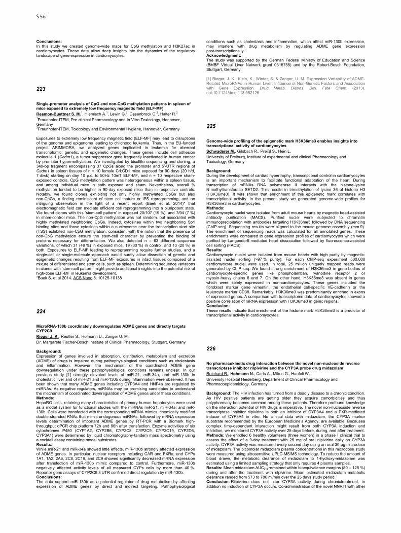
Conclusions: In this study we created genome-wide maps for CpG methylation and H3K27ac in cardiomyocytes. These data allow deep insights into the dynamics of the regulatory landscape of gene expression in cardiomyocytes.
223
Single-promoter analysis of CpG and non-CpG methylation patterns in spleen of mice exposed to extremely low frequency magnetic field (ELF-MF) Reamon-Buettner S. M.1, Hiemisch A.1, Lewin G.2, Dasenbrock C.2, Halter R.2 1Fraunhofer-ITEM, Pre-clinical Pharmacology and In Vitro Toxicology, Hannover, Germany 2Fraunhofer-ITEM, Toxicology and Environmental Hygiene, Hannover, Germany Exposures to extremely low frequency magnetic field (ELF-MF) may lead to disruptions of the genome and epigenome leading to childhood leukemia. Thus, in the EU-funded project ARIMMORA, we analyzed genes implicated in leukemia for aberrant transcriptomic, genetic, and epigenetic changes. These genes include cell adhesion molecule 1 (Cadm1), a tumor suppressor gene frequently inactivated in human cancer by promoter hypermethylation. We investigated by bisulfite sequencing and cloning, a 349-bp fragment encompassing 37 CpGs along the promoter and 5’-UTR regions of Cadm1 in spleen tissues of n = 10 female Crl:CD1 mice exposed for 90-days (20 h/d, 7 d/wk) starting on day 10 p.c. to 50Hz 10mT ELF-MF, and n = 10 respective sham-exposed controls. CpG methylation pattern was heterogeneous within a spleen tissue, and among individual mice in both exposed and sham. Nevertheless, overall % methylation tended to be higher in 90-day exposed mice than in respective controls. Notably, we found clones exhibiting not only highly methylated CpGs but also non-CpGs, a finding reminiscent of stem cell nature or iPS reprogramming, and an intriguing observation in the light of a recent report (Baek et al. 2014)* that electromagnetic field can mediate efficient cell reprogramming into a pluripotent state. We found clones with this ‘stem-cell pattern’ in exposed 20/107 (19 %), and 7/94 (7 %) in sham-control mice. The non-CpG methylation was not random, but associated with highly methylated neighboring CpGs. Indeed, cytosines within two neighboring Sp1 binding sites and those cytosines within a nucleosome near the transcription start site (TSS) exhibited non-CpG methylation, consistent with the notion that the presence of non-CpG methylation ensure the stem-cell character by preventing the binding of proteins necessary for differentiation. We also detected n = 63 different sequence variations, of which 31 (49 %) in exposed mice, 19 (30 %) in control, and 13 (20 %) in both. Exposures to ELF-MF leading to reprogramming require further studies, and a single-cell or single-molecule approach would surely allow dissection of genetic and epigenetic changes resulting from ELF-MF exposures in intact tissues composed of a mixure of differentiated and stem cells, such as spleen. Determining sequence variations in clones with ‘stem cell pattern’ might provide additional insights into the potential risk of high-dose ELF-MF in leukemia development. *Baek S. et al 2014. ACS Nano 8: 10125-10138
224
MicroRNA-130b coordinately downregulates ADME genes and directly targets CYP2C9 Rieger J. K., Reutter S., Hofmann U., Zanger U. M. Dr. Margarete Fischer-Bosch Institute of Clinical Pharmacology, Stuttgart, Germany Background: Expression of genes involved in absorption, distribution, metabolism and excretion (ADME) of drugs is impaired during pathophysiological conditions such as cholestasis and inflammation. However, the mechanism of the coordinated ADME gene downregulation under these pathophysiological conditions remains unclear. In our previous study [1] strongly elevated levels of miR-21, miR-34a, and miR-130b in cholestatic liver and of miR-21 and miR-130b during inflammation were observed. It has been shown that many ADME genes including CYP3A4 and HNF4a are regulated by miRNAs. As negative regulators, miRNAs may be promising candidates to understand the mechanism of coordinated downregulation of ADME genes under these conditions. Methods: HepaRG cells, retaining many characteristics of primary human hepatocytes were used as a model system for functional studies with the miRNAs miR-21, miR-34a, and miR-130b. Cells were transfected with the corresponding miRNA mimics, chemically modified double-stranded RNAs that mimic endogenous miRNAs, followed by mRNA expression levels determination of important ADME genes by RT-PCR with a Biomark high-throughput qPCR chip platform 72h and 96h after transfection. Enzyme activities of six cytochromes P450 (CYP1A2, CYP2B6, CYP2C8, CYP2C9, CYP2C19, CYP2D6, CYP3A4) were determined by liquid chromatography-tandem mass spectrometry using a cocktail assay containing model substrates. Results: While miR-21 and miR-34a showed little effects, miR-130b strongly affected expression of ADME genes. In particular, nuclear receptors including CAR and FXRa, and CYPs 1A1, 1A2, 2A6, 2C8, 2C19, and 2C9 showed significantly decreased mRNA expression after transfection of miR-130b mimic compared to control. Furthermore, miR-130b negatively affected activity levels of all measured CYPs cells by more than 40 %. Reporter gene assays of CYP2C9 3’UTR confirmed direct regulation by miR-130b. Conclusions: The data support miR-130b as a potential regulator of drug metabolism by affecting expression of ADME genes by direct and indirect targeting. Pathophysiological
conditions such as cholestasis and inflammation, which affect miR-130b expression, may interfere with drug metabolism by regulating ADME gene expression post-transcriptionally. Acknowledgment: The study was supported by the German Federal Ministry of Education and Science (BMBF Virtual Liver Network grant 0315755) and by the Robert-Bosch Foundation, Stuttgart, Germany. [1] Rieger, J. K., Klein, K., Winter, S. & Zanger, U. M. Expression Variability of ADME-Related MicroRNAs in Human Liver: Influence of Non-Genetic Factors and Association with Gene Expression. Drug Metab. Dispos. Biol. Fate Chem. (2013). doi:10.1124/dmd.113.052126
225
Genome-wide profiling of the epigenetic mark H3K36me3 enables insights into transcriptional activity of cardiomyocytes Schwaderer M., Gilsbach R., Preißl S., Hein L. University of Freiburg, Institute of experimental and clinical Pharmacology and Toxicology, Germany Background: During the development of cardiac hypertrophy, transcriptional control in cardiomyocytes is an important mechanism to facilitate functional adaptation of the heart. During transcription of mRNAs RNA polymerase II interacts with the histone-lysine N-methyltransferase SETD2. This results in trimethylation of lysine 36 of histone H3 (H3K36me3). It was shown that enrichment of this epigenetic mark correlates with transcriptional activity. In the present study we generated genome-wide profiles for H3K36me3 in cardiomyocytes. Methods: Cardiomyocyte nuclei were isolated from adult mouse hearts by magnetic bead-assisted antibody purification (MACS). Purified nuclei were subjected to chromatin immunoprecipitation with antibodies targeting H3K36me3 followed by DNA sequencing (ChIP-seq). Sequencing results were aligned to the mouse genome assembly (mm 9). The enrichment of sequencing reads was calculated for all annotated genes. These enrichments were compared to gene expression profiles of cardiomyocytes which were purified by Langendorff-mediated heart dissociation followed by fluorescence-assisted cell sorting (FACS). Results: Cardiomyocyte nuclei were isolated from mouse hearts with high purity by magnetic-assisted nuclei sorting (>97 % purity). For each ChIP-seq experiment 500,000 cardiomyocyte nuclei were used. In total, 25 million uniquely mapped reads were generated by ChIP-seq. We found strong enrichment of H3K36me3 in gene-bodies of cardiomyocyte-specific genes like phospholamban, ryanodine receptor 2 or myosin-heavy chains 6 and 7. On the other hand, H3K36me3 was absent in genes which were solely expressed in non-cardiomyocytes. These genes included the fibroblast marker gene vimentin, the endothelial cell-specific VE-cadherin or the leukocyte marker CD38. Remarkably, H3K36me3 was predominantly enriched in exons of expressed genes. A comparison with transcriptome data of cardiomyocytes showed a positive correlation of mRNA expression with H3K36me3 in genic regions. Conclusion: These results indicate that enrichment of the histone mark H3K36me3 is a predictor of transcriptional activity in cardiomyocytes.
226
No pharmacokinetic drug interaction between the novel non-nucleoside reverse transcriptase inhibitor rilpivirine and the CYP3A probe drug midazolam Reinhard R., Hohmann N., Carls A., Mikus G., Haefeli W. University Hospital Heidelberg, Department of Clinical Pharmacology and Pharmacoepidemiology, Germany Background: The HIV infection has turned from a deadly disease to a chronic condition. As HIV positive patients are getting older they acquire comorbidities and thus polypharmacy becomes common among these patients. Therefore profound knowledge on the interaction potential of HIV drugs is imperative. The novel non-nucleoside reverse transcriptase inhibitor rilpivirine is both an inhibitor of CYP3A4 and a PXR-mediated inducer of CYP3A4 in vitro. No clinical data with midazolam, the CYP3A marker substrate recommended by the European Medicine’s Agency, are available. Becausea complex time-dependent interaction might result from both CYP3A induction and inhibition, we monitored CYP3A activity over 25 days before, during, and after treatment. Methods: We enrolled 6 healthy volunteers (three women) in a phase I clinical trial to assess the effect of a 9-day treatment with 25 mg of oral rilpivirine daily on CYP3A activity. CYP3A activity was measured every second day using an oral 30 µg microdose of midazolam. The picomolar midazolam plasma concentrations in this microdose study were measured using ultrasensitive UPLC-MS/MS technology. To reduce the amount of blood drawn, the metabolic clearance of midazolam to 1-hydroxy-midazolam was estimated using a limited sampling strategy that only requires 4 plasma samples. Results: Mean midazolam AUC2-4 remained within bioequivalence margins (80 – 125 %) during and after the treatment with rilpivirine. Mean estimated midazolam metabolic clearance ranged from 573 to 786 ml/min over the 25 days study period. Conclusion: Rilpivirine does not alter CYP3A activity during chronictreatment, in addition no induction of CYP3A occurs. Co-administration of the novel NNRTI with other
S56
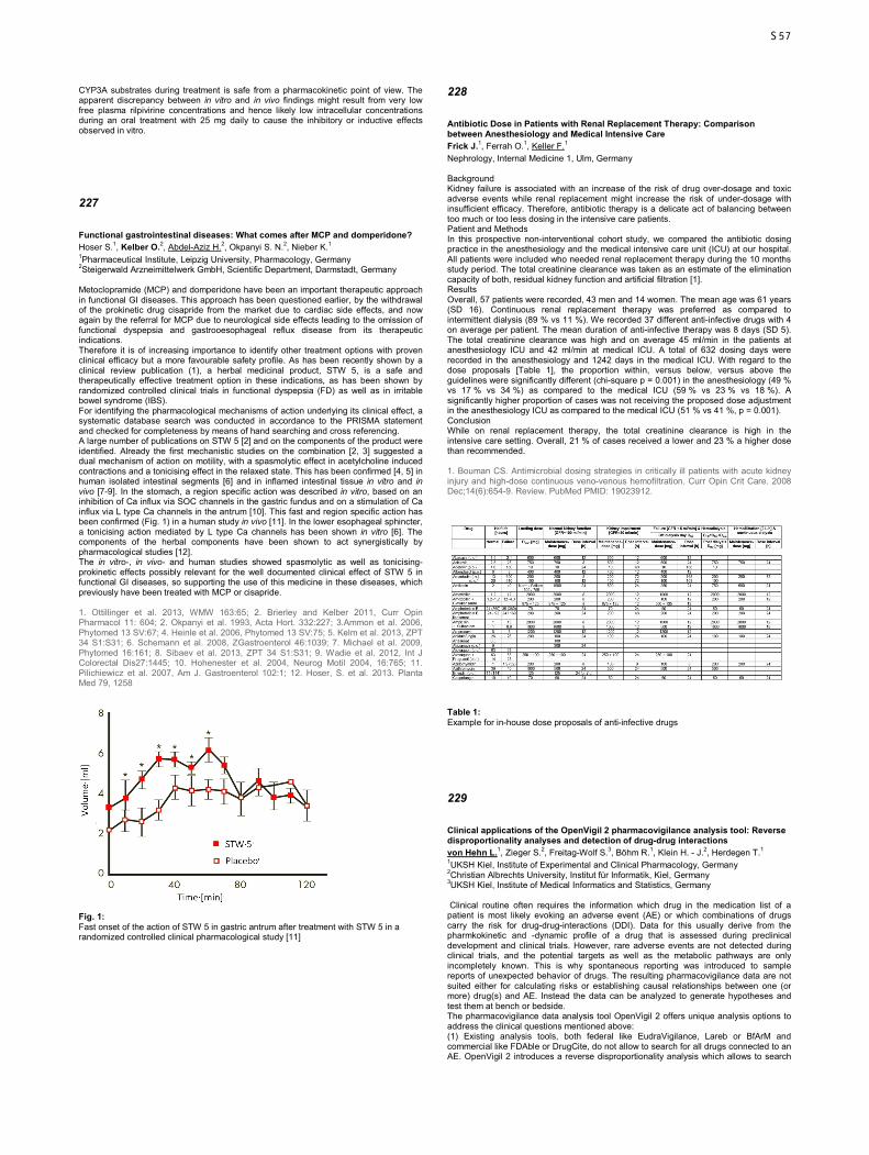
CYP3A substrates during treatment is safe from a pharmacokinetic point of view. The apparent discrepancy between in vitro and in vivo findings might result from very low free plasma rilpivirine concentrations and hence likely low intracellular concentrations during an oral treatment with 25 mg daily to cause the inhibitory or inductive effects observed in vitro.
227
Functional gastrointestinal diseases: What comes after MCP and domperidone? Hoser S.1, Kelber O.2, Abdel-Aziz H.2, Okpanyi S. N.2, Nieber K.1 1Pharmaceutical Institute, Leipzig University, Pharmacology, Germany 2Steigerwald Arzneimittelwerk GmbH, Scientific Department, Darmstadt, Germany Metoclopramide (MCP) and domperidone have been an important therapeutic approach in functional GI diseases. This approach has been questioned earlier, by the withdrawal of the prokinetic drug cisapride from the market due to cardiac side effects, and now again by the referral for MCP due to neurological side effects leading to the omission of functional dyspepsia and gastrooesophageal reflux disease from its therapeutic indications. Therefore it is of increasing importance to identify other treatment options with proven clinical efficacy but a more favourable safety profile. As has been recently shown by a clinical review publication (1), a herbal medicinal product, STW 5, is a safe and therapeutically effective treatment option in these indications, as has been shown by randomized controlled clinical trials in functional dyspepsia (FD) as well as in irritable bowel syndrome (IBS). For identifying the pharmacological mechanisms of action underlying its clinical effect, a systematic database search was conducted in accordance to the PRISMA statement and checked for completeness by means of hand searching and cross referencing. A large number of publications on STW 5 [2] and on the components of the product were identified. Already the first mechanistic studies on the combination [2, 3] suggested a dual mechanism of action on motility, with a spasmolytic effect in acetylcholine induced contractions and a tonicising effect in the relaxed state. This has been confirmed [4, 5] in human isolated intestinal segments [6] and in inflamed intestinal tissue in vitro and in vivo [7-9]. In the stomach, a region specific action was described in vitro, based on an inhibition of Ca influx via SOC channels in the gastric fundus and on a stimulation of Ca influx via L type Ca channels in the antrum [10]. This fast and region specific action has been confirmed (Fig. 1) in a human study in vivo [11]. In the lower esophageal sphincter, a tonicising action mediated by L type Ca channels has been shown in vitro [6]. The components of the herbal components have been shown to act synergistically by pharmacological studies [12]. The in vitro-, in vivo- and human studies showed spasmolytic as well as tonicising-prokinetic effects possibly relevant for the well documented clinical effect of STW 5 in functional GI diseases, so supporting the use of this medicine in these diseases, which previously have been treated with MCP or cisapride. 1. Ottillinger et al. 2013, WMW 163:65; 2. Brierley and Kelber 2011, Curr Opin Pharmacol 11: 604; 2. Okpanyi et al. 1993, Acta Hort. 332:227; 3.Ammon et al. 2006, Phytomed 13 SV:67; 4. Heinle et al. 2006, Phytomed 13 SV:75; 5. Kelm et al. 2013, ZPT 34 S1:S31; 6. Schemann et al. 2008, ZGastroenterol 46:1039; 7. Michael et al. 2009, Phytomed 16:161; 8. Sibaev et al. 2013, ZPT 34 S1:S31; 9. Wadie et al. 2012, Int J Colorectal Dis27:1445; 10. Hohenester et al. 2004, Neurog Motil 2004, 16:765; 11. Pilichiewicz et al. 2007, Am J. Gastroenterol 102:1; 12. Hoser, S. et al. 2013. Planta Med 79, 1258
Fig. 1: Fast onset of the action of STW 5 in gastric antrum after treatment with STW 5 in a randomized controlled clinical pharmacological study [11]
228
Antibiotic Dose in Patients with Renal Replacement Therapy: Comparison between Anesthesiology and Medical Intensive Care Frick J.1, Ferrah O.1, Keller F.1 Nephrology, Internal Medicine 1, Ulm, Germany Background Kidney failure is associated with an increase of the risk of drug over-dosage and toxic adverse events while renal replacement might increase the risk of under-dosage with insufficient efficacy. Therefore, antibiotic therapy is a delicate act of balancing between too much or too less dosing in the intensive care patients. Patient and Methods In this prospective non-interventional cohort study, we compared the antibiotic dosing practice in the anesthesiology and the medical intensive care unit (ICU) at our hospital. All patients were included who needed renal replacement therapy during the 10 months study period. The total creatinine clearance was taken as an estimate of the elimination capacity of both, residual kidney function and artificial filtration [1]. Results Overall, 57 patients were recorded, 43 men and 14 women. The mean age was 61 years (SD 16). Continuous renal replacement therapy was preferred as compared to intermittent dialysis (89 % vs 11 %). We recorded 37 different anti-infective drugs with 4 on average per patient. The mean duration of anti-infective therapy was 8 days (SD 5). The total creatinine clearance was high and on average 45 ml/min in the patients at anesthesiology ICU and 42 ml/min at medical ICU. A total of 632 dosing days were recorded in the anesthesiology and 1242 days in the medical ICU. With regard to the dose proposals [Table 1], the proportion within, versus below, versus above the guidelines were significantly different (chi-square p = 0.001) in the anesthesiology (49 % vs 17 % vs 34 %) as compared to the medical ICU (59 % vs 23 % vs 18 %). A significantly higher proportion of cases was not receiving the proposed dose adjustment in the anesthesiology ICU as compared to the medical ICU (51 % vs 41 %, p = 0.001). Conclusion While on renal replacement therapy, the total creatinine clearance is high in the intensive care setting. Overall, 21 % of cases received a lower and 23 % a higher dose than recommended. 1. Bouman CS. Antimicrobial dosing strategies in critically ill patients with acute kidney injury and high-dose continuous veno-venous hemofiltration. Curr Opin Crit Care. 2008 Dec;14(6):654-9. Review. PubMed PMID: 19023912.
Table 1: Example for in-house dose proposals of anti-infective drugs
229
Clinical applications of the OpenVigil 2 pharmacovigilance analysis tool: Reverse disproportionality analyses and detection of drug-drug interactions von Hehn L.1, Zieger S.2, Freitag-Wolf S.3, Böhm R.1, Klein H. - J.2, Herdegen T.1 1UKSH Kiel, Institute of Experimental and Clinical Pharmacology, Germany 2Christian Albrechts University, Institut für Informatik, Kiel, Germany 3UKSH Kiel, Institute of Medical Informatics and Statistics, Germany Clinical routine often requires the information which drug in the medication list of a patient is most likely evoking an adverse event (AE) or which combinations of drugs carry the risk for drug-drug-interactions (DDI). Data for this usually derive from the pharmkokinetic and -dynamic profile of a drug that is assessed during preclinical development and clinical trials. However, rare adverse events are not detected during clinical trials, and the potential targets as well as the metabolic pathways are only incompletely known. This is why spontaneous reporting was introduced to sample reports of unexpected behavior of drugs. The resulting pharmacovigilance data are not suited either for calculating risks or establishing causal relationships between one (or more) drug(s) and AE. Instead the data can be analyzed to generate hypotheses and test them at bench or bedside. The pharmacovigilance data analysis tool OpenVigil 2 offers unique analysis options to address the clinical questions mentioned above: (1) Existing analysis tools, both federal like EudraVigilance, Lareb or BfArM and commercial like FDAble or DrugCite, do not allow to search for all drugs connected to an AE. OpenVigil 2 introduces a reverse disproportionality analysis which allows to search
S57
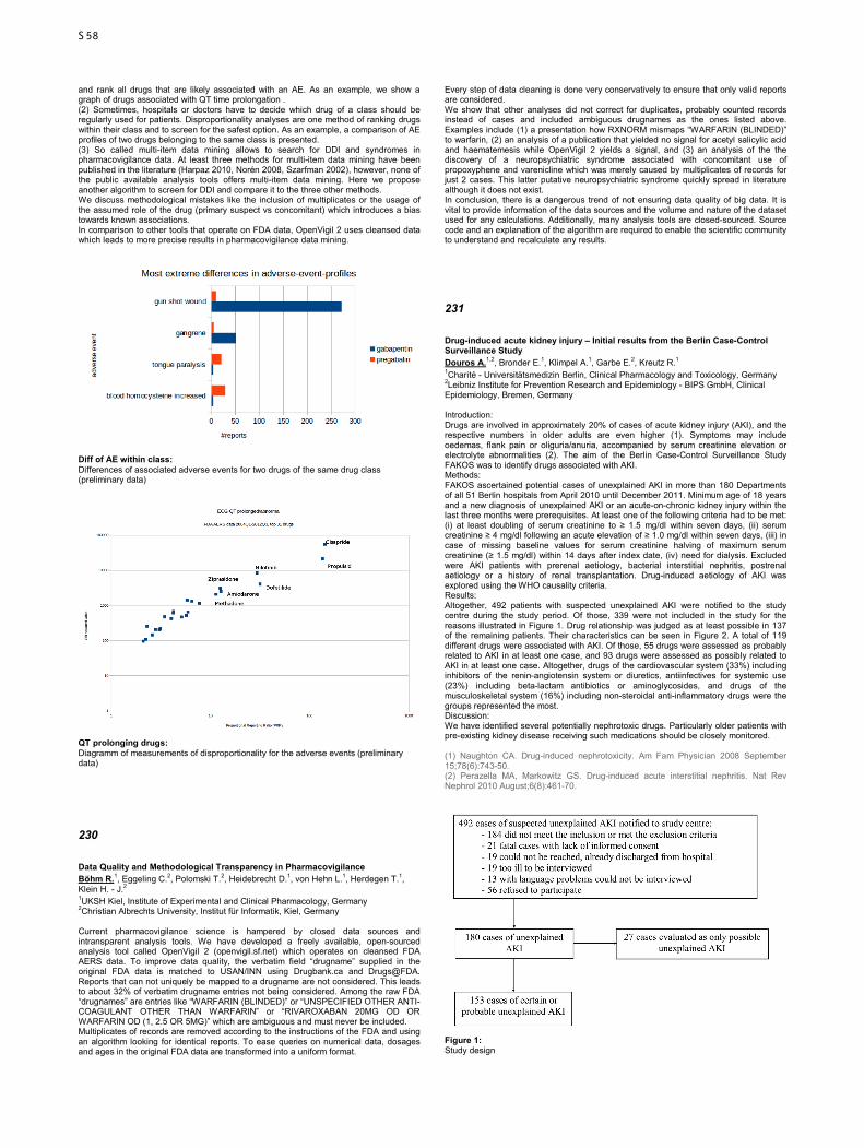
and rank all drugs that are likely associated with an AE. As an example, we show a graph of drugs associated with QT time prolongation . (2) Sometimes, hospitals or doctors have to decide which drug of a class should be regularly used for patients. Disproportionality analyses are one method of ranking drugs within their class and to screen for the safest option. As an example, a comparison of AE profiles of two drugs belonging to the same class is presented. (3) So called multi-item data mining allows to search for DDI and syndromes in pharmacovigilance data. At least three methods for multi-item data mining have been published in the literature (Harpaz 2010, Norén 2008, Szarfman 2002), however, none of the public available analysis tools offers multi-item data mining. Here we propose another algorithm to screen for DDI and compare it to the three other methods. We discuss methodological mistakes like the inclusion of multiplicates or the usage of the assumed role of the drug (primary suspect vs concomitant) which introduces a bias towards known associations. In comparison to other tools that operate on FDA data, OpenVigil 2 uses cleansed data which leads to more precise results in pharmacovigilance data mining.
Diff of AE within class: Differences of associated adverse events for two drugs of the same drug class (preliminary data)
QT prolonging drugs: Diagramm of measurements of disproportionality for the adverse events (preliminary data)
230
Data Quality and Methodological Transparency in Pharmacovigilance Böhm R.1, Eggeling C.2, Polomski T.2, Heidebrecht D.1, von Hehn L.1, Herdegen T.1, Klein H. - J.2 1UKSH Kiel, Institute of Experimental and Clinical Pharmacology, Germany 2Christian Albrechts University, Institut für Informatik, Kiel, Germany Current pharmacovigilance science is hampered by closed data sources and intransparent analysis tools. We have developed a freely available, open-sourced analysis tool called OpenVigil 2 (openvigil.sf.net) which operates on cleansed FDA AERS data. To improve data quality, the verbatim field “drugname” supplied in the original FDA data is matched to USAN/INN using Drugbank.ca and Drugs@FDA. Reports that can not uniquely be mapped to a drugname are not considered. This leads to about 32% of verbatim drugname entries not being considered. Among the raw FDA “drugnames” are entries like “WARFARIN (BLINDED)” or “UNSPECIFIED OTHER ANTI-COAGULANT OTHER THAN WARFARIN” or “RIVAROXABAN 20MG OD OR WARFARIN OD (1, 2.5 OR 5MG)” which are ambiguous and must never be included. Multiplicates of records are removed according to the instructions of the FDA and using an algorithm looking for identical reports. To ease queries on numerical data, dosages and ages in the original FDA data are transformed into a uniform format.
Every step of data cleaning is done very conservatively to ensure that only valid reports are considered. We show that other analyses did not correct for duplicates, probably counted records instead of cases and included ambiguous drugnames as the ones listed above. Examples include (1) a presentation how RXNORM mismaps “WARFARIN (BLINDED)” to warfarin, (2) an analysis of a publication that yielded no signal for acetyl salicylic acid and haematemesis while OpenVigil 2 yields a signal, and (3) an analysis of the the discovery of a neuropsychiatric syndrome associated with concomitant use of propoxyphene and varenicline which was merely caused by multiplicates of records for just 2 cases. This latter putative neuropsychiatric syndrome quickly spread in literature although it does not exist. In conclusion, there is a dangerous trend of not ensuring data quality of big data. It is vital to provide information of the data sources and the volume and nature of the dataset used for any calculations. Additionally, many analysis tools are closed-sourced. Source code and an explanation of the algorithm are required to enable the scientific community to understand and recalculate any results.
231
Drug-induced acute kidney injury – Initial results from the Berlin Case-Control Surveillance Study Douros A.1,2, Bronder E.1, Klimpel A.1, Garbe E.2, Kreutz R.1 1Charité - Universitätsmedizin Berlin, Clinical Pharmacology and Toxicology, Germany 2Leibniz Institute for Prevention Research and Epidemiology - BIPS GmbH, Clinical Epidemiology, Bremen, Germany Introduction: Drugs are involved in approximately 20% of cases of acute kidney injury (AKI), and the respective numbers in older adults are even higher (1). Symptoms may include oedemas, flank pain or oliguria/anuria, accompanied by serum creatinine elevation or electrolyte abnormalities (2). The aim of the Berlin Case-Control Surveillance Study FAKOS was to identify drugs associated with AKI. Methods: FAKOS ascertained potential cases of unexplained AKI in more than 180 Departments of all 51 Berlin hospitals from April 2010 until December 2011. Minimum age of 18 years and a new diagnosis of unexplained AKI or an acute-on-chronic kidney injury within the last three months were prerequisites. At least one of the following criteria had to be met: (i) at least doubling of serum creatinine to ≥ 1.5 mg/dl within seven days, (ii) serum creatinine ≥ 4 mg/dl following an acute elevation of ≥ 1.0 mg/dl within seven days, (iii) in case of missing baseline values for serum creatinine halving of maximum serum creatinine (≥ 1.5 mg/dl) within 14 days after index date, (iv) need for dialysis. Excluded were AKI patients with prerenal aetiology, bacterial interstitial nephritis, postrenal aetiology or a history of renal transplantation. Drug-induced aetiology of AKI was explored using the WHO causality criteria. Results: Altogether, 492 patients with suspected unexplained AKI were notified to the study centre during the study period. Of those, 339 were not included in the study for the reasons illustrated in Figure 1. Drug relationship was judged as at least possible in 137 of the remaining patients. Their characteristics can be seen in Figure 2. A total of 119 different drugs were associated with AKI. Of those, 55 drugs were assessed as probably related to AKI in at least one case, and 93 drugs were assessed as possibly related to AKI in at least one case. Altogether, drugs of the cardiovascular system (33%) including inhibitors of the renin-angiotensin system or diuretics, antiinfectives for systemic use (23%) including beta-lactam antibiotics or aminoglycosides, and drugs of the musculoskeletal system (16%) including non-steroidal anti-inflammatory drugs were the groups represented the most. Discussion: We have identified several potentially nephrotoxic drugs. Particularly older patients with pre-existing kidney disease receiving such medications should be closely monitored. (1) Naughton CA. Drug-induced nephrotoxicity. Am Fam Physician 2008 September 15;78(6):743-50. (2) Perazella MA, Markowitz GS. Drug-induced acute interstitial nephritis. Nat Rev Nephrol 2010 August;6(8):461-70.
Figure 1: Study design
S58

Figure 2: Clinical and laboratory characteristics of 137 patients with drug-induced acute kidney injury
232
Examination and risk assessment of critical prescriptions in patients on polypharmacy in ambulatory care Laidig F.1, May M.1, Brinkmann J.1, Herbarth L.2, Jordan J.1, Stichtenoth D. O.1 1Medizinische Hochschule Hannover, Institut für Klinische Pharmakologie, Germany 2Kaufmännische Krankenkasse, Hannover, Germany Background Adverse Drug Events (ADEs) are estimated to cause 5 % of all hospital admissions. Patients with polypharmacy are more likely to experience an ADE, thus structured analyses of polymedications for detecting potential prescribing errors can be essential to discover potential ADEs. Risk assessment can be used to grade severity of identified critical prescriptions. Methods Within a program of a local health insurance company to increase medication safety, structured medication analyses were carried out in 400 patients (mean age 70 years) on at least 5 drugs and an increased risk for hospital admission who consented to take part in the program. Critical prescriptions were identified and categorized into 8 main categories and 33 subcategories. Severity was rated in a Delphi method by an expert team consisting of clinical pharmacologists and internists. A risk score adapted from Bertsche et al. 2008 was used to combine severity of critical prescriptions and risk of drugs involved for each category and subcategory. Results Polymedications consisted of 13.4 drugs in average. Of the identified 3005 critical prescriptions most frequencies were found in the category unclear indication (28.7 %), in interactions (28.0 %), in risk or PRISCUS medication (13.0 %) and in dosage (13.0 %). Severity was rated highest for the categories contraindication and contraindicated interactions while lowest severity was seen for minor and not clinical relevant interactions, wrong application time and possible reduction of the number of tablets. Risk score category was rated highest for contraindication and PRISCUS medication while the lowest risk score was found for minor and debatable interaction and no interaction found. Conclusion Critical prescriptions are of very high frequency in polymedications of older adults with a high risk to suffer ADEs. However, polypharmacy needs to be reviewed constantly to ensure medication safety. Appropriate programs should be evolved and made realizable. Medication reviews need to be implemented into daily practice of ambulant settings of medication processes of patients receiving polypharmacy.
233
Increased occurence of St. John's Wort induced phototoxic neuropathy in women Maus A., Hohmann N., Carls A., Blank A., Haefeli W., Mikus G. University Hospital Heidelberg, Department of Clinical Pharmacology and Pharmacoepidemiology, Germany Background: St. John’s Wort (SJW) is frequently used as a mild antidepressant, especially among women. SJW is available as over-the-counter medication although adverse drug reactions such as phototoxicity and pharmacokinetic interactions are well known. Information on SJW-induced neuropathy is scarce. Its frequency is unknown. Methods: We analyzed adverse events in a clinical phase I trial to asses dose-dependent metabolic effects of SJW co-administered with rifampicin in 12 healthy volunteers (aged between 20 and 31 years, six females). Participants were treated for 2 weeks with 300 mg oral SJW QD, followed by 2 weeks of 300 mg SJW TID, and 2 weeks of 600 mg SJW TID for 2 more weeks. Thenthe highest dose was co-administered with 600 mg p.o. rifampicin QD for 1 week. Results: Within 3 to 6 days of increasing the SJW dose to 600 mg TID, 5 of 6 female participants developed ambient temperature-dependent allodynia and paresthesia in sun-exposed areas (face and hands) along with phototoxic erythrodermia. Combination with rifampicin did not influence the neuropathy. None of the males experienced any SJW-related adverse reactions. The study was terminated early in one case and ended as planned in 4 of the 5 cases where the symptoms were mild to moderate and only required active surveillance. The symptoms resolved completely within 12 to 16 days afterstopping the study medication without further treatment. Conclusion: The observed symptoms closely resembled the findings described in a single case report from 1998 but the high frequency in our study was surprising. Also the apparently gender-specific occurrence of phototoxic neuropathy is noteworthy. The etiology of neuropathy is unknown but SJW contains hypericin, pseudohypericin, and hyperforin, all substances with known photoirritant potential. Excitation of hypericin by ultraviolet (UV)-A light leads to oxidative species that activates the Ca2+-permeable Transient receptor potential cation channel (TRP) A1 expressed on nociceptive peripheral neurons. Gender differences in the dermal reaction to UV-light might provide an explanation. Male mice express less of the pro-inflammatory cytokine IL-6 and a higher amount of immunosuppressive IL-10 than female mice in UV-light exposed skin. Hence inflammatory edema is less pronounced. IL-10 also attenuates neuropathic pain. In conclusion we hypothesize that males experience less neuropathic pain because of higher IL-10 levels after UV-light exposure.
234
Drug risk assessment in pregnancy: 5 years’ experience in clinical pharmacology Oztürk Z.1, Olmez E.2, Gurpinar T.2, Vural K.2 1Izmir Atatürk Research Hospital, Clinical Pharmacology and Toxicology, Turkey 2Celal Bayar University Faculty of Medicine, Department of Pharmacology, Manisa, Turkey Aim: Drug use in pregnancy poses a potential risk to both the mother and the fetus. The purpose of this study was to determine which medications are most commonly used by pregnant women who referred to our department for teratology consultations. Methods: A retrospective study was performed among pregnant women who referred to our department between 2007 and 2012. Drug data were evaluated using anatomical therapeutic classification (1) and pregnancy risk categorization established by United States Food and Drug Adminstration (FDA). Data were obtained from the medical records of patients. SPSS 16.0 statistical software was used for data analysis. Results: A total of 483 pregnancies with drug exposure during the period 2007 through 2012 were registered. The median age of these women was 29 years (min: 17, max: 44). Based on anatomical therapeutic classification, drugs that affect the brain and nervous system were observed in the highest proportion of women (23.5 %, except nonsteroidal anti-inflammatory drugs, NSAIDs). Many of them were psychotropic medications such as antidepressants, antipsychotics, anxiolytics and mood stabilizers (14.1 %). Other commonly used drugs are anti-infectives for systemic use (16.2 %), gastrointestinal and metabolism drugs (15.2 %), and NSAIDs (14.6 %). Using the FDA categorization, we determined that 1.2 % of the drugs taken by women were in category A, 39.2 % in category B, 42.2 % in category C, 9.8 % in category D, 5.2 % in category X and 2.4 % unknown. 136 women (28.1 %) received only a single drug, and 178 women (36.8 %) received 3 or more drugs. Many of the pregnant women received these drugs during the first trimester. Conclusions: Based on our findings, among the pregnant women exposed to drugs, the number of psychotropic drugs and nonsteroidal anti-inflammatory drugs were higher than expected. Unplanned pregnancies in women result in exposure to multiple medications during the first trimester. Patients exposed to drugs and known teratogens during pregnancy should be informed of the potential side effects (2). It is important to contact pharmacology consulting services for more information relating to chemical exposure and medication. Drug utilization studies in pregnant women and additional research focussing on possible associations between medications and birth defects are also required to provide information about safety (or otherwise) of drugs already used in pregnancy. References: 1. World Health Organization Collaboration Center for Drug Statistics Methodology 2014. Guidelines for ATC classification and DDD assignment, 17th edition World Health Organization, Oslo, Norway. 2. Drugs During Pregnancy and Lactation. Christof Schaefer, Paul Peters, Richard K. Miller. 2nd Edition. 2007.
S59
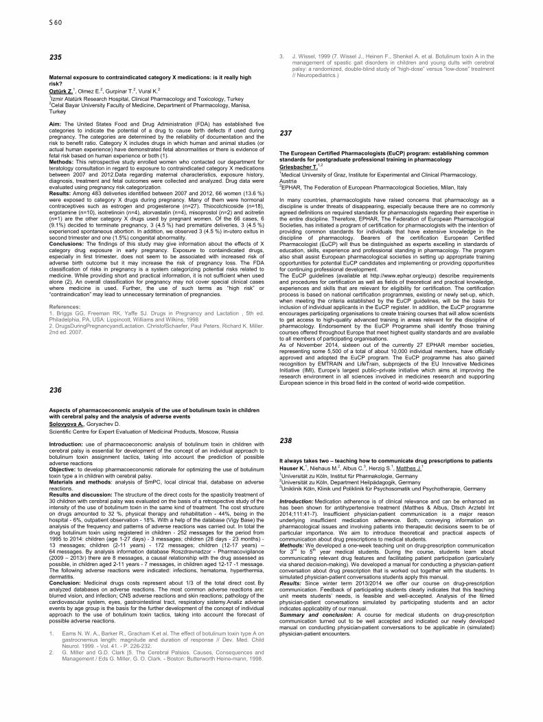
235
Maternal exposure to contraindicated category X medications: is it really high risk? Oztürk Z.1, Olmez E.2, Gurpinar T.2, Vural K.2 1Izmir Atatürk Research Hospital, Clinical Pharmacology and Toxicology, Turkey 2Celal Bayar University Faculty of Medicine, Department of Pharmacology, Manisa, Turkey Aim: The United States Food and Drug Administration (FDA) has established five categories to indicate the potential of a drug to cause birth defects if used during pregnancy. The categories are determined by the reliability of documentation and the risk to benefit ratio. Category X includes drugs in which human and animal studies (or actual human experience) have demonstrated fetal abnormalities or there is evidence of fetal risk based on human experience or both (1). Methods: This retrospective study enrolled women who contacted our department for teratology consultation in regard to exposure to contraindicated category X medications between 2007 and 2012.Data regarding maternal characteristics, exposure history, diagnosis, treatment and fetal outcomes were collected and analyzed. Drug data were evaluated using pregnancy risk categorization. Results: Among 483 deliveries identified between 2007 and 2012, 66 women (13.6 %) were exposed to category X drugs during pregnancy. Many of them were hormonal contraceptives such as estrogen and progesterone (n=27). Thiocolchicoside (n=18), ergotamine (n=10), isotretinoin (n=4), atorvastatin (n=4), misoprostol (n=2) and acitretin (n=1) are the other category X drugs used by pregnant women. Of the 66 cases, 6 (9.1%) decided to terminate pregnancy, 3 (4.5 %) had prematüre deliveries, 3 (4.5 %) experienced spontaneous abortion. In addition, we observed 3 (4.5 %) in-utero exitus in second trimester and one (1.5%) congenital abnormality. Conclusions: The findings of this study may give information about the effects of X category drug exposure in early pregnancy. Exposure to containdicated drugs, especially in first trimester, does not seem to be associated with increased risk of adverse birth outcome but it may increase the risk of pregnancy loss. The FDA classification of risks in pregnancy is a system categorizing potential risks related to medicine. While providing short and practical information, it is not sufficient when used alone (2). An overall classification for pregnancy may not cover special clinical cases where medicine is used. Further, the use of such terms as “high risk” or “contraindication” may lead to unnecessary termination of pregnancies. References: 1. Briggs GG, Freeman RK, Yaffe SJ. Drugs in Pregnancy and Lactation , 5th ed. Philadelphia, PA, USA: Lippincott, Williams and Wilkins, 1998 2. DrugsDuringPregnancyandLactation. ChristofSchaefer, Paul Peters, Richard K. Miller. 2nd ed. 2007.
236
Aspects of pharmacoeconomic analysis of the use of botulinum toxin in children with cerebral palsy and the analysis of adverse events Solovyova A., Goryachev D. Scientific Centre for Expert Evaluation of Medicinal Products, Moscow, Russia Introduction: use of pharmacoeconomic analysis of botulinum toxin in children with cerebral palsy is essential for development of the concept of an individual approach to botulinum toxin assignment tactics, taking into account the prediction of possible adverse reactions Objective: to develop pharmacoeconomic rationale for optimizing the use of botulinum toxin type a in children with cerebral palsy. Materials and methods: analysis of SmPC, local clinical trial, database on adverse reactions. Results and discussion: The structure of the direct costs for the spasticity treatment of 30 children with cerebral palsy was evaluated on the basis of a retrospective study of the intensity of the use of botulinum toxin in the same kind of treatment. The cost structure on drugs amounted to 32 %, physical therapy and rehabilitation - 44%, being in the hospital - 6%, outpatient observation - 18%. With a help of the database (Vigy Base) the analysis of the frequency and patterns of adverse reactions was carried out. In total the drug botulinum toxin using registered in children - 252 messages for the period from 1995 to 2014: children (age 1-27 days) - 3 messages; children (28 days - 23 months) - 13 messages; children (2-11 years) - 172 messages; children (12-17 years) – 64 messages. By analysis information database Roszdravnadzor - Pharmacovigilance (2009 – 2013г) there are 8 messages, a causal relationship with the drug assessed as possible, in children aged 2-11 years - 7 messages, in children aged 12-17 -1 message. The following adverse reactions were indicated: infections, hematoma, hyperthermia, dermatitis. Conclusion: Medicinal drugs costs represent about 1/3 of the total direct cost. By analyzed databases on adverse reactions. The most common adverse reactions are: blurred vision, and infection; CNS adverse reactions and skin reactions; pathology of the cardiovascular system, eyes, gastrointestinal tract, respiratory sistemy.Analiz adverse events by age group is the basis for the further development of the concept of individual approach to the use of botulinum toxin tactics, taking into account the forecast of possible adverse reactions. 1. Eams N. W. А., Barker R., Gracham К.еt al. The effect of botulinum toxin type А оn
gastrocnemius length: magnitude and duration of response // Dev. Med. Child Neuгol. 1999. - Vol. 41. - Р. 226-232.
2. G. Miller and G.D. Clark [5. The Сеrеbral Palsies. Causes, Consequences and Management / Eds G. Miller, G. О. Clark. - Boston: Butterworth Heine-mann, 1998.
3. J. Wissel, 1999 (7. Wissel J., Heinen F., Shenkel А. еt al. Botulinum toxin А in the management of spastic gait disorders in children and young dults with сеrebral palsy: а randomized, double-blind study of “high-dose” versus “low-dose” treatment // Neuropediatrics.)
237
The European Certified Pharmacologists (EuCP) program: establishing common standards for postgraduate professional training in pharmacology Griesbacher T.1,2 1Medical University of Graz, Institute for Experimental and Clinical Pharmacology, Austria 2EPHAR, The Federation of European Pharmacological Societies, Milan, Italy In many countries, pharmacologists have raised concerns that pharmacology as a discipline is under threats of disappearing, especially because there are no commonly agreed definitions on required standards for pharmacologists regarding their expertise in the entire discipline. Therefore, EPHAR, The Federation of European Pharmacological Societies, has initiated a program of certification for pharmacologists with the intention of providing common standards for individuals that have extensive knowledge in the discipline of pharmacology. Bearers of the certification European Certified Pharmacologist (EuCP) will thus be distinguished as experts excelling in standards of education, skills, experience and professional standing in pharmacology. The program also shall assist European pharmacological societies in setting up appropriate training opportunities for potential EuCP candidates and implementing or providing opportunities for continuing professional development. The EuCP guidelines (available at http://www.ephar.org/eucp) describe requirements and procedures for certification as well as fields of theoretical and practical knowledge, experiences and skills that are relevant for eligibility for certification. The certification process is based on national certification programmes, existing or newly set-up, which, when meeting the criteria established by the EuCP guidelines, will be the basis for inclusion of individual applicants in the EuCP register. In addition, the EuCP programme encourages participating organisations to create training courses that will allow scientists to get access to high-quality advanced training in areas relevant for the discipline of pharmacology. Endorsement by the EuCP Programme shall identify those training courses offered throughout Europe that meet highest quality standards and are available to all members of participating organisations. As of November 2014, sixteen out of the currently 27 EPHAR member societies, representing some 5,500 of a total of about 10,000 individual members, have officially approved and adopted the EuCP program. The EuCP programme has also gained recognition by EMTRAIN and LifeTrain, subprojects of the EU Innovative Medicines Initiative (IMI), Europe’s largest public–private initiative which aims at improving the research environment in all sciences involved in medicines research and supporting European science in this broad field in the context of world-wide competition.
238
It always takes two – teaching how to communicate drug prescriptions to patients Hauser K.1, Niehaus M.2, Albus C.3, Herzig S.1, Matthes J.1 1Universität zu Köln, Institut für Pharmakologie, Germany 2Universität zu Köln, Department Heilpädagogik, Germany 3Uniklinik Köln, Klinik und Poliklinik für Psychosomatik und Psychotherapie, Germany Introduction: Medication adherence is of clinical relevance and can be enhanced as has been shown for antihypertensive treatment (Matthes & Albus, Dtsch Arztebl Int 2014;111:41-7). Insufficient physician-patient communication is a major reason underlying insufficient medication adherence. Both, conveying information on pharmacological issues and involving patients into therapeutic decisions seem to be of particular importance. We aim to introduce theoretical and practical aspects of communication about drug prescriptions to medical students. Methods: We developed a one-week teaching unit on drug-prescription communication for 3rd to 5th year medical students. During the course, students learn about communicating relevant drug features and facilitating patient participation (particularly via shared decision-making). We developed a manual for conducting a physician-patient conversation about drug prescription that is worked out together with the students. In simulated physician-patient conversations students apply this manual. Results: Since winter term 2013/2014 we offer our course on drug-prescription communication. Feedback of participating students clearly indicates that this teaching unit meets students’ needs, is feasible and well-accepted. Analysis of the filmed physician-patient conversations simulated by participating students and an actor indicates applicability of our manual. Summary and conclusion: A course for medical students on drug-prescription communication turned out to be well accepted and indicated our newly developed manual on conducting physician-patient conversations to be applicable in (simulated) physician-patient encounters.
S60
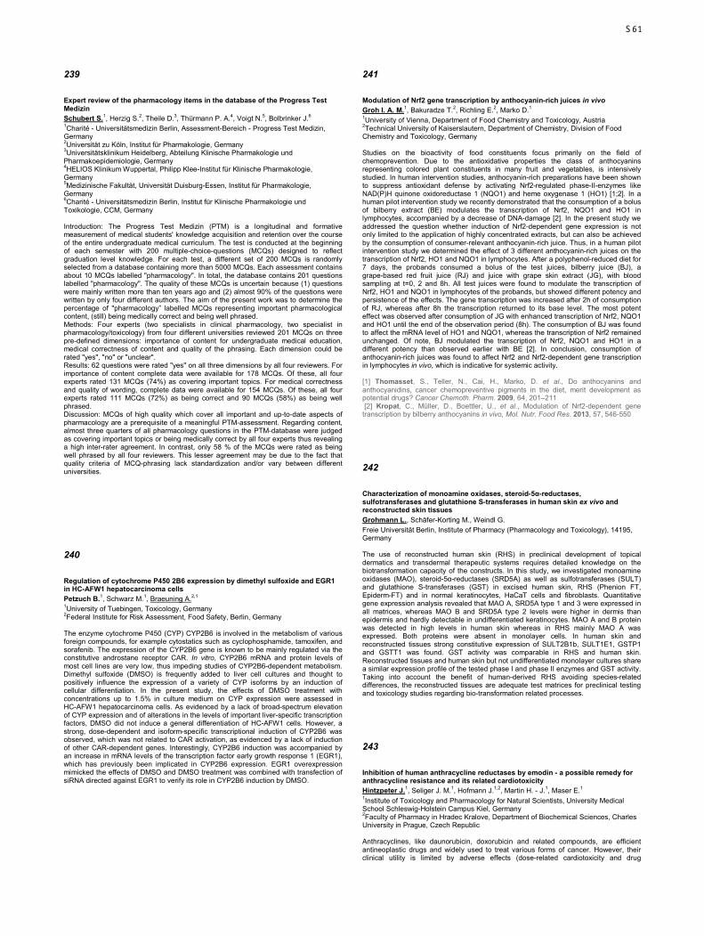
239
Expert review of the pharmacology items in the database of the Progress Test Medizin Schubert S.1, Herzig S.2, Theile D.3, Thürmann P. A.4, Voigt N.5, Bolbrinker J.6 1Charité - Universitätsmedizin Berlin, Assessment-Bereich - Progress Test Medizin, Germany 2Universität zu Köln, Institut für Pharmakologie, Germany 3Universitätsklinikum Heidelberg, Abteilung Klinische Pharmakologie und Pharmakoepidemiologie, Germany 4HELIOS Klinikum Wuppertal, Philipp Klee-Institut für Klinische Pharmakologie, Germany 5Medizinische Fakultät, Universität Duisburg-Essen, Institut für Pharmakologie, Germany 6Charité - Universitätsmedizin Berlin, Institut für Klinische Pharmakologie und Toxikologie, CCM, Germany Introduction: The Progress Test Medizin (PTM) is a longitudinal and formative measurement of medical students' knowledge acquisition and retention over the course of the entire undergraduate medical curriculum. The test is conducted at the beginning of each semester with 200 multiple-choice-questions (MCQs) designed to reflect graduation level knowledge. For each test, a different set of 200 MCQs is randomly selected from a database containing more than 5000 MCQs. Each assessment contains about 10 MCQs labelled "pharmacology". In total, the database contains 201 questions labelled "pharmacology". The quality of these MCQs is uncertain because (1) questions were mainly written more than ten years ago and (2) almost 90% of the questions were written by only four different authors. The aim of the present work was to determine the percentage of "pharmacology” labelled MCQs representing important pharmacological content, (still) being medically correct and being well phrased. Methods: Four experts (two specialists in clinical pharmacology, two specialist in pharmacology/toxicology) from four different universities reviewed 201 MCQs on three pre-defined dimensions: importance of content for undergraduate medical education, medical correctness of content and quality of the phrasing. Each dimension could be rated "yes", "no" or "unclear". Results: 62 questions were rated "yes" on all three dimensions by all four reviewers. For importance of content complete data were available for 178 MCQs. Of these, all four experts rated 131 MCQs (74%) as covering important topics. For medical correctness and quality of wording, complete data were available for 154 MCQs. Of these, all four experts rated 111 MCQs (72%) as being correct and 90 MCQs (58%) as being well phrased. Discussion: MCQs of high quality which cover all important and up-to-date aspects of pharmacology are a prerequisite of a meaningful PTM-assessment. Regarding content, almost three quarters of all pharmacology questions in the PTM-database were judged as covering important topics or being medically correct by all four experts thus revealing a high inter-rater agreement. In contrast, only 58 % of the MCQs were rated as being well phrased by all four reviewers. This lesser agreement may be due to the fact that quality criteria of MCQ-phrasing lack standardization and/or vary between different universities.
240
Regulation of cytochrome P450 2B6 expression by dimethyl sulfoxide and EGR1 in HC-AFW1 hepatocarcinoma cells Petzuch B.1, Schwarz M.1, Braeuning A.2,1 1University of Tuebingen, Toxicology, Germany 2Federal Institute for Risk Assessment, Food Safety, Berlin, Germany The enzyme cytochrome P450 (CYP) CYP2B6 is involved in the metabolism of various foreign compounds, for example cytostatics such as cyclophosphamide, tamoxifen, and sorafenib. The expression of the CYP2B6 gene is known to be mainly regulated via the constitutive androstane receptor CAR. In vitro, CYP2B6 mRNA and protein levels of most cell lines are very low, thus impeding studies of CYP2B6-dependent metabolism. Dimethyl sulfoxide (DMSO) is frequently added to liver cell cultures and thought to positively influence the expression of a variety of CYP isoforms by an induction of cellular differentiation. In the present study, the effects of DMSO treatment with concentrations up to 1.5% in culture medium on CYP expression were assessed in HC-AFW1 hepatocarcinoma cells. As evidenced by a lack of broad-spectrum elevation of CYP expression and of alterations in the levels of important liver-specific transcription factors, DMSO did not induce a general differentiation of HC-AFW1 cells. However, a strong, dose-dependent and isoform-specific transcriptional induction of CYP2B6 was observed, which was not related to CAR activation, as evidenced by a lack of induction of other CAR-dependent genes. Interestingly, CYP2B6 induction was accompanied by an increase in mRNA levels of the transcription factor early growth response 1 (EGR1), which has previously been implicated in CYP2B6 expression. EGR1 overexpression mimicked the effects of DMSO and DMSO treatment was combined with transfection of siRNA directed against EGR1 to verify its role in CYP2B6 induction by DMSO.
241
Modulation of Nrf2 gene transcription by anthocyanin-rich juices in vivo Groh I. A. M.1, Bakuradze T.2, Richling E.2, Marko D.1 1University of Vienna, Department of Food Chemistry and Toxicology, Austria 2Technical University of Kaiserslautern, Department of Chemistry, Division of Food Chemistry and Toxicology, Germany Studies on the bioactivity of food constituents focus primarily on the field of chemoprevention. Due to the antioxidative properties the class of anthocyanins representing colored plant constituents in many fruit and vegetables, is intensively studied. In human intervention studies, anthocyanin-rich preparations have been shown to suppress antioxidant defense by activating Nrf2-regulated phase-II-enzymes like NAD(P)H quinone oxidoreductase 1 (NQO1) and heme oxygenase 1 (HO1) [1;2]. In a human pilot intervention study we recently demonstrated that the consumption of a bolus of bilberry extract (BE) modulates the transcription of Nrf2, NQO1 and HO1 in lymphocytes, accompanied by a decrease of DNA-damage [2]. In the present study we addressed the question whether induction of Nrf2-dependent gene expression is not only limited to the application of highly concentrated extracts, but can also be achieved by the consumption of consumer-relevant anthocyanin-rich juice. Thus, in a human pilot intervention study we determined the effect of 3 different anthocyanin-rich juices on the transcription of Nrf2, HO1 and NQO1 in lymphocytes. After a polyphenol-reduced diet for 7 days, the probands consumed a bolus of the test juices, bilberry juice (BJ), a grape-based red fruit juice (RJ) and juice with grape skin extract (JG), with blood sampling at t=0, 2 and 8h. All test juices were found to modulate the transcription of Nrf2, HO1 and NQO1 in lymphocytes of the probands, but showed different potency and persistence of the effects. The gene transcription was increased after 2h of consumption of RJ, whereas after 8h the transcription returned to its base level. The most potent effect was observed after consumption of JG with enhanced transcription of Nrf2, NQO1 and HO1 until the end of the observation period (8h). The consumption of BJ was found to affect the mRNA level of HO1 and NQO1, whereas the transcription of Nrf2 remained unchanged. Of note, BJ modulated the transcription of Nrf2, NQO1 and HO1 in a different potency than observed earlier with BE [2]. In conclusion, consumption of anthocyanin-rich juices was found to affect Nrf2 and Nrf2-dependent gene transcription in lymphocytes in vivo, which is indicative for systemic activity. [1] Thomasset, S., Teller, N., Cai, H., Marko, D. et al., Do anthocyanins and anthocyanidins, cancer chemopreventive pigments in the diet, merit development as potential drugs? Cancer Chemoth. Pharm. 2009, 64, 201–211 [2] Kropat, C., Müller, D., Boettler, U., et al., Modulation of Nrf2-dependent gene transcription by bilberry anthocyanins in vivo, Mol. Nutr. Food Res. 2013, 57, 546-550
242
Characterization of monoamine oxidases, steroid-5α-reductases, sulfotransferases and glutathione S-transferases in human skin ex vivo and reconstructed skin tissues Grohmann L., Schäfer-Korting M., Weindl G. Freie Universität Berlin, Institute of Pharmacy (Pharmacology and Toxicology), 14195, Germany The use of reconstructed human skin (RHS) in preclinical development of topical dermatics and transdermal therapeutic systems requires detailed knowledge on the biotransformation capacity of the constructs. In this study, we investigated monoamine oxidases (MAO), steroid-5α-reductases (SRD5A) as well as sulfotransferases (SULT) and glutathione S-transferases (GST) in excised human skin, RHS (Phenion FT, Epiderm-FT) and in normal keratinocytes, HaCaT cells and fibroblasts. Quantitative gene expression analysis revealed that MAO A, SRD5A type 1 and 3 were expressed in all matrices, whereas MAO B and SRD5A type 2 levels were higher in dermis than epidermis and hardly detectable in undifferentiated keratinocytes. MAO A and B protein was detected in high levels in human skin whereas in RHS mainly MAO A was expressed. Both proteins were absent in monolayer cells. In human skin and reconstructed tissues strong constitutive expression of SULT2B1b, SULT1E1, GSTP1 and GSTT1 was found. GST activity was comparable in RHS and human skin. Reconstructed tissues and human skin but not undifferentiated monolayer cultures share a similar expression profile of the tested phase I and phase II enzymes and GST activity. Taking into account the benefit of human-derived RHS avoiding species-related differences, the reconstructed tissues are adequate test matrices for preclinical testing and toxicology studies regarding bio-transformation related processes.
243
Inhibition of human anthracycline reductases by emodin - a possible remedy for anthracycline resistance and its related cardiotoxicity Hintzpeter J.1, Seliger J. M.1, Hofmann J.1,2, Martin H. - J.1, Maser E.1 1Institute of Toxicology and Pharmacology for Natural Scientists, University Medical School Schleswig-Holstein Campus Kiel, Germany 2Faculty of Pharmacy in Hradec Kralove, Department of Biochemical Sciences, Charles University in Prague, Czech Republic Anthracyclines, like daunorubicin, doxorubicin and related compounds, are efficient antineoplastic drugs and widely used to treat various forms of cancer. However, their clinical utility is limited by adverse effects (dose-related cardiotoxicity and drug
S61

resistance). Anthracycline related cardiotoxicity has been linked to reductive metabolism of the parent drug to their metabolites. The enzymes catalyzing the two electron reduction of the C-13 side chain carbonyl moiety generate the secondary alcohol metabolites daunorubicinol (DAUNOL) and doxorubicinol (DOXOL), respectively. These secondary alcohol metabolites have significantly reduced anti-tumor activity and show increased cardiotoxicity, thereby limiting the clinical use of anthracyclines. Members of two enzyme superfamilies have been connected with this conversion: CBR1, belonging to the short-chain dehydrogenase/reductase superfamily (SDR) and AKR1B1, AKR1B10, AKR1C3 and AKR7A2 as members of the aldo-keto reductase superfamily (AKR). Thus, inhibition of these anthracycline reductases may increase their efficacy in cancer tissues and simultaneously decrease their cardiotoxicity. Here, we report the finding of a natural phenolic compound, emodin, which is a potent inhibitor for almost all anthracycline reductases. Intensive in vitro enzyme inhibition studies show that emodin has a high potency in inhibiting the specific anthracycline reductases. Cell lysates of the multi-resistant lung cancer cell line (A549), which contain all above mentioned reductases, have been used to show the effect of emodin on the reduction of DAUN to DAUNOL. Additionally, emodin has a good cytotoxicity profile, when administered to A549 cells and HepG2 cells from human liver. Furthermore, a significant shift of the dose-response curves in MTT-tests using A549 and HepG2 cell lines for the co-administration of emodin and DAUN was observed. In summary, emodin may yield the potential to enhance the therapeutic effectiveness of DAUN by preventing heart tissue damage through the inhibition of the anthracycline reductases mediated reduction of DAUN to DAUNOL.
244
Gene expression and protein abundance of clinically relevant phase I & II enzymes in the human intestine Busch D.1, Drozdzik M.2, Siegmund W.1, Oswald S.1 1University Medicine Greifswald, Clinical Pharmacology, Germany 2Pomeranian Medical University, Department of Experimental and Clinical Pharmacology, Szczecin, Poland Background: The pharmacokinetics of many drugs is markedly influenced by intestinal metabolism mediated by biotransformation enzymes such as cytochrome P450 (CYP450) enzymes and UDP-glucuronosyltransferases (UGT). In order to predict their impact on oral drug absorption as well as on drug-drug interactions, data on their intestinal expression are required. Therefore, it was the aim of this study to quantify the gene expression and the absolute protein abundance of clinically relevant CYP and UGT enzymes in the human intestine. Methods: Gene expression of CYP2B6, CYP2C8, CYP2C9, CYP2C19, CYP2D6, CYP3A4/3A5, UGT1A family, UGT2B7 and UGT2B15 was performed by using validated real-time RT-PCR assays (TaqMan® Low Density Arrays). A validated LC-MS/MS-based targeted proteomics method was used for the simultaneous quantification of CYP1A2, CYP2B6, CYP2C8, CYP2C9, CYP2C19, CYP2D6, CYP2E1, CYP3A4, CYP3A5, UGT1A1, UGT1A3, UGT2B7 and UGT2B15. Proteins were quantified by measuring proteospecific tryptic peptides using stable isotope-labelled internal standards. These assays were applied to quantify gene expression and protein abundance in pooled human intestinal microsomes (HIM) from 8 donors and pooled human liver microsomes (HLM) from 25 donors. In parallel, the intestinal expression was analysed along the length of the human intestine using healthy intestinal tissue (in each case, 9 samples from duodenum, jejunum, ileum and colon) from 8 organ donors (age: 24-56). Results: With exception of CYP2C8 and UGT2B15, all investigated genes of drug metabolizing enzymes could be detected in the human intestine. Expression (mRNA) of all studied genes was found to be highest in duodenum or jejunum and significantly decreased from the upper intestine to colon. Intestinal protein abundance could only be verified for six of these enzymes (CYP3A4, CYP2C9, CYP2C19, UGT1A1, UGT1A3 and UGT2B7). The following relative percentage contribution of metabolizing enzymes have been observed in HIM: CYP3A4, 47.1%; UGT1A1, 19.7%; CYP2C9, 10.8%; UGT1A3, 8.3%; UGT2B7, 7.1% and CYP2C19, 7%. Conclusion: The human intestine is equipped with several clinically relevant metabolizing phase I / II enzymes. The expression of these enzymes is not homogeneous along the entire length of the intestine which may be an explanation for site-depended differences in drug absorption. These expression data may allow more precise prediction of drug disposition using PBPK modelling.
245
Metabolic activation of food carcinogens 5-hydroxymethylfurfural and furfuryl alcohol Sachse B.1, Meinl W.2, Glatt H.2, Seidel A.3, Monien B. H.1 1German Institute of Human Nutrition (DIfE), Genotoxic Food Contaminants, Nuthetal, Germany 2German Institute of Human Nutrition (DIfE), Nutritional Toxicology, Nuthetal, Germany 3Biochemical Institute for Environmental Carcinogens, Prof. Dr. Gernot Grimmer-Foundation, Grosshansdorf, Germany 5-Hydroxymethylfurfural (HMF) and furfuryl alcohol (FFA) are moderately potent rodent carcinogens present in many heat-processed foodstuffs. They are activated by sulfo conjugation of the hydroxyl groups yielding mutagenic sulfuric acid esters. The margins between the doses that induce tumors in animal experiments and the estimated daily human intake are small. Moreover, due to considerable differences in sulfotransferase
(SULT) expression and substrate specificity of SULTs between species, humans may be more sensitive to HMF- and FFA-mediated genotoxic effects than rodents. We investigated the sulfo conjugation of HMF by 30 individual SULT forms from human, mouse and rat in vitro by direct quantification of 5-sulfoxymethylfurfural via UPLC-MS/MS. The catalytic efficiencies of the orthologous SULT1A1 forms of all species were at least 3-fold higher compared to those of the other SULT forms. Due to the reactivity of furfuryl sulfate (t1/2 = 20 s at 37°C) FFA sulfo conjugation was monitored by measuring the stable reaction product with adenosine, N6-((furan-2-yl)methyl)-adenosine. As shown for HMF sulfo conjugation, the orthologous SULT1A1 forms from all species turned out to play a predominant role for the bioactivation of FFA. To better understand the contribution of different SULT forms to the genotoxic effect of FFA in vivo, we determined levels of the DNA adduct N2-((furan-2-yl)methyl)-2′-deoxyguanosine using isotope-dilution UPLC-MS/MS in different tissues of four mouse lines with varying human (h) and mouse (m) SULT status (wild-type, ko mSult1a1, ko mSult1d1 and hSULT1A1/1A2 x ko mSult1a1 x ko mSult1d1) following a single intraperitoneal dose of FFA. The absence of mSult1a1 caused a decrease of about 70 % of DNA adduct levels in most tissues whereas the knockout of mSult1d1 had no significant effect. The mice expressing hSULT1A1/1A2 showed 2- to 6-fold higher adduct levels compared to the wild-type. We further showed that the co-treatment with ethanol or the drug 4-methylpyrazole led to an up to 5-fold increase in DNA adduct levels in different tissues. In summary, the capacity of hSULT1A1-catalyzed HMF and FFA sulfo conjugation is comparable to that of mSult1a1 in mice, the species in which carcinogenic effects of the furan derivatives were detected. The animal experiments support the hypotheses that FFA may also be converted to the genotoxic furfuryl sulfate in humans and that consumption of alcoholic beverages has a co-carcinogenic effect.
246
Hormonal influences on human SDR and AKR enzymes catalysing carbonyl reduction of the tobacco specific carcinogen NNK Stapelfeld C., Maser E. Institute of Toxicology and Pharmacology for Natural Scientists, Kiel, Germany Regarding all kinds of cancers, lung cancer is one of the most common and lethal cancers of men and women in North America, Europe, and East Asia. Every year more than one million people die of the consequences of this disease worldwide. It is estimated that 90% of this death toll is caused by cigarette smoking. For this reason tobacco use is the most important risk factor for the development of lung cancer. One of the most potent carcinogens in tobacco smoke is the nitrosamine 4-(methylnitrosamino)-1-(3-pyridyl)-1-butanone (NNK), which causes lung cancer specifically. The human body is able to initiate NNK detoxification through carbonyl reduction. Meanwhile, seven different reductases of the short-chain dehydrogenase/ reductase (SDR) and the aldo-keto reductase (AKR) superfamilies are known to participate in NNK elimination. The physiologic substrates of these enzymes are mainly steroid hormones. Accordingly, it is very likely that steroid hormones interfere with NNK detoxification to shift the metabolic balance and cause an increased cancer risk. The question arises whether sex hormones influence NNK reduction differently in men and women, thereby increasing or decreasing the lung cancer-risk, respectively. Five different sex hormones were tested for their inhibitory effect on NNK carbonyl reduction by different reductases. Two of them were synthetic hormones used as hormonal contraceptives and the other three were physiological sex hormones. NNK carbonyl reduction by carbonyl reductase 1 (CBR1), which belongs to the SDR superfamily, was not found to be significantly inhibited by any of the tested hormones. However, the enzymes of the AKR superfamily were significantly inhibited by the tested sex hormones. It turned out that gestagens have the strongest effect on NNK carbonyl reduction in comparison to estrogens or androgens. This may be an indication that women have an increased lung cancer risk when smoking.
247
Is the urinary metabolic ratio of flurbiprofen (probe drug for CYP2C9) affected by simultaneous phenotyping with dextromethorphan (CYP2D6) or omeprazole (CYP2C19)? Vogl S.1,2, Schönfelder G.2,3, Lutz W. K.1 1University of Würzburg, Institute of Pharmacology and Toxicology, Germany 2Charité-Universitätsmedizin Berlin, Department of Clinical Pharmacology and Toxicology, Germany 3Federal Institute for Risk Assessment (BfR), Berlin, Germany Purpose Several cytochrome P450 (CYP) monooxygenases are highly polymorphic and/or prone to induction or inhibition, which cause marked differences in individual enzyme activity and affects drug response. Assessment of metabolic activity of a patient before drug treatment (phenotyping) might minimize the occurrence of adverse drug reactions and treatment failures. CYP phenotyping procedures can be simplified by combining several probe drugs into a so-called probe drug cocktail. We investigated whether the urinary metabolic ratio of flurbiprofen as a parameter for CYP2C9 activity is affected by concomitant administration of the probe drugs dextromethorphan (for CYP2D6) or omeprazole (for CYP2C19). Method The cohort of 23 subjects consisted of 19 females and 4 males. The volunteers ingested 8.75 mg flurbiprofen on day 1 of the study. On day 8, volunteers were given
S62
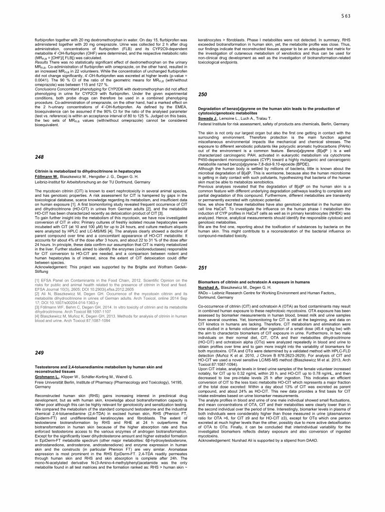
flurbiprofen together with 20 mg dextromethorphan in water. On day 15, flurbiprofen was administered together with 20 mg omeprazole. Urine was collected for 2 h after drug administration, concentrations of flurbiprofen (FLB) and its CYP2C9-dependent metabolite 4’-OH-flurbiprofen (OHF) were determined, and the respective metabolic ratio (MRFLB = [OHF]/[ FLB]) was calculated. Results There was no statistically significant effect of dextromethorphan on the urinary MRFLB. Co-administration of flurbiprofen with omeprazole, on the other hand, resulted in an increased MRFLB in 22 volunteers. While the concentration of unchanged flurbiprofen did not change significantly, 4’-OH-flurbiprofen was excreted at higher levels (p-value = 0.0041). The 90 % CI of the ratio of the geometric means for MRFLB (with/without omeprazole) was between 115 and 127 %. Conclusions Concomitant phenotyping for CYP2D6 with dextromethorphan did not affect phenotyping in urine for CYP2C9 with flurbiprofen. Under the given experimental conditions, both probe drugs can therefore be used in a combined phenotyping procedure. Co-administration of omeprazole, on the other hand, had a marked effect on the 2 h-urinary concentrations of 4’-OH-flurbiprofen. As defined by the EMEA, bioequivalence can be assumed if the 90% CI for the ratio of the analysed parameter (test vs. reference) is within an acceptance interval of 80 to 125 %. Judged on this basis, the two sets of MRFLB values (with/without omeprazole) cannot be considered bioequivalent.
248
Citrinin is metabolized to dihydrocitrinone in hepatocytes Föllmann W., Blaszkewicz M., Hengstler J. G., Degen G. H. Leibniz-Institut für Arbeitsforschung an der TU Dortmund, Germany The mycotoxin citrinin (CIT) is known to exert nephrotoxicity in several animal species, and has genotoxic properties. A risk assessment for CIT is hampered by gaps in the toxicological database, scarce knowledge regarding its metabolism, and insufficient data on human exposure [1]. A first biomonitoring study revealed frequent occurrence of CIT and dihydrocitrinone (HO-CIT) in urines from German adults [2], and the metabolite HO-CIT has been characterized recently as detoxication product of CIT [3]. To gain further insight into the metabolism of this mycotoxin, we have now investigated conversion of CIT in vitro: Primary cultures of freshly isolated mouse hepatocytes were incubated with CIT (at 10 and 100 µM) for up to 24 hours, and culture medium aliquots were analyzed by HPLC and LC-MS/MS [4]. The analysis clearly showed a decline of parent compound over time and a concomitant appearance of HO-CIT metabolite: It accounts for about 4% of the dose after 3 hours, and about 22 to 31 % of the dose after 24 hours. In principle, these data confirm our assumption that CIT is mainly metabolized in the liver. Further studies aimed to identify the enzymes (oxidoreductases) responsible for CIT conversion to HO-CIT are needed, and a comparison between rodent and human hepatocytes is of interest, since the extent of CIT detoxication could differ between species. Acknowledgement: This project was supported by the Brigitte and Wolfram Gedek-Stiftung [1] EFSA Panel on Contaminants in the Food Chain, 2012. Scientific Opinion on the risks for public and animal health related to the presence of citrinin in food and feed. EFSA Journal 10(3), 2605; DOI 10.2903/j.efsa.2012.2605 [2] Ali N, Blaszkewicz M, Degen GH. Occurrence of the mycotoxin citrinin and its metabolite dihydrocitrinone in urines of German adults. Arch Toxicol, online 2014 Sep 17; DOI 10.1007/s00204-014-1363-y [3] Föllmann WF, Behm C, Degen GH, 2014. In vitro toxicity of citrinin and its metabolite dihydrocitrinone. Arch Toxicol 88:1097-1107 [4] Blaszkewicz M, Muñoz K, Degen GH, 2013. Methods for analysis of citrinin in human blood and urine. Arch Toxicol 87:1087-1094
249
Testosterone and 2,4-toluenediamine metabolism by human skin and reconstructed tissues Grohmann L., Klipper W., Schäfer-Korting M., Weindl G. Freie Universität Berlin, Institute of Pharmacy (Pharmacology and Toxicology), 14195, Germany Reconstructed human skin (RHS) gains increasing interest in preclinical drug development, but as with human skin, knowledge about biotransformation capacity is rather poor although this can be highly relevant for genotoxicity and sensitization testing. We compared the metabolism of the standard compound testosterone and the industrial chemical 2,4-toluenediamine (2,4-TDA) in excised human skin, RHS (Phenion FT, Epiderm-FT) and undifferentiated keratinocytes and fibroblasts. The extent of testosterone biotransformation by RHS and RHE at 24 h outperforms the biotransformation in human skin because of the higher absorption rate and thus enforced testosterone access to the various enzymes of androgen biotransformation. Except for the significantly lower dihydrotesterone amount and higher estradiol formation in EpiDerm-FT metabolite spectrum (other major metabolites: 6β-hydroxytestosterone, androstanedione, androsterone, androstenedione) and enzyme expression in human skin and the constructs (in particular Phenion FT) are very similar. Aromatase expression is most prominent in the RHS EpiDerm-FT. 2,4-TDA readily permeates through human skin and RHS and skin absorption is complete after 24h. The mono-N-acetylated derivative N-(3-Amino-4-methylphenyl)acetamide was the only metabolite found in all test matrices and the formation ranked as: RHS > human skin ~
keratinocytes > fibroblasts. Phase I metabolites were not detected. In summary, RHS exceeded biotransformation in human skin, yet, the metabolite profile was close. Thus, our findings indicate that reconstructed tissues appear to be an adequate test matrix for the investigation of cutaneous metabolism of xenobiotics and thus can be used for non-clinical drug development as well as the investigation of biotransformation-related toxicological endpoints.
250
Degradation of benzo[a]pyrene on the human skin leads to the production of cytotoxic/genotoxic metabolites Sowada J., Lemoine L., Luch A., Tralau T. Federal Institute for risk assessment, safety of products ans chemicals, Berlin, Germany The skin is not only our largest organ but also the first one getting in contact with the surrounding environment. Therefore protection is the main function against miscellaneous environmental impacts like mechanical and chemical stresses. The exposure to different xenobiotic pollutants like polycyclic aromatic hydrocarbons (PAHs) out of the environment is a common feature. Benzo[a]pyrene (B[a]P ) is a well characterized carcinogenic PAH, activated in eukaryotic metabolism via cytochrome P450-dependent monooxygenases (CYP) toward a highly mutagenic and cancerogenic metabolite named benzo[a]pyrene-7,8-diol-9,10-epoxide (BPDE). Although the human body is settled by millions of bacteria, little is known about the microbial degradation of B[a]P. This is worrisome, because also the human microbiome is getting in daily contact with such pollutants, hypothesizing that bacteria of the human skin must be able to metabolize xenobiotics. Previous analyses revealed that the degradation of B[a]P on the human skin is a common feature with different underlying degradation pathways leading to complete and partial degradation of this compound. Furthermore, different metabolites are transiently or permanently excreted with cytotoxic potential. Now, we show that these metabolites have also genotoxic potential in the human skin cell line HaCaT. To investigate the influence on the human phase I metabolism the induction of CYP profiles in HaCaT cells as well as in primary keratinocytes (NHEK) was analyzed. Hence, analytical measurements should identify the responsible cytotoxic and genotoxic metabolites. We are the first one, reporting about the toxification of substances by bacteria on the human skin. This might contribute to a reconsideration of the bacterial influence on compound-mediated toxicity.
251
Biomarkers of citrinin and ochratoxin A exposure in humans Nurshad A., Blaszkewicz M., Degen G. H. IfADo – Leibniz Research Centre for Working Environment and Human Factors,, Dortmund, Germany Co-occurrence of citrinin (CIT) and ochratoxin A (OTA) as food contaminants may result in combined human exposure to these nephrotoxic mycotoxins. OTA exposure has been assessed by biomarker measurements in human blood, breast milk and urine samples from several countries. Yet, biomonitoring for CIT is still at the beginning, and data on CIT kinetics in humans are lacking. Therefore, CIT metabolism and elimination were now studied in a female volunteer after ingestion of a small dose (45.4 ng/kg bw) with the aim to characterize biomarkers of CIT exposure in urine. Furthermore, in two male individuals on their normal diet, CIT, OTA and their metabolites dihydrocitrinone (HO-CIT) and ochratoxin alpha (OTα) were analyzed repeatedly in blood and urine to obtain profiles over time and to gain more insight into the variability of biomarkers for both mycotoxins. OTA and OTα were determined by a validated method with HPLC-FLD detection (Muñoz K et al. 2010, J Chrom B 878:2623-2629). For analysis of CIT and HO-CIT we used a novel sensitive LC/MS-MS method (Blaszkewicz M et al. 2013, Arch Toxicol 87:1087-1094). Upon CIT intake, analyte levels in timed urine samples of the female volunteer increased notably, for CIT up to 0.32 ng/mL within 20 h, and HO-CIT up to 0.78 ng/mL, and then decreased to low pre-intake levels 25 h after ingestion. This indicates an efficient conversion of CIT to the less toxic metabolite HO-CIT which represents a major fraction of the total dose excreted: Within a day about 13% of CIT was excreted as parent compound, and about 24% as HO-CIT. This new data provides a first basis for CIT intake estimates based on urine biomarker measurements. The analyte profiles in blood and urine of one male individual showed small fluctuations, and mean concentrations of OTA, CIT and their metabolites were clearly lower than in the second individual over the period of time. Interestingly, biomarker levels in plasma of both individuals were considerably higher than those measured in urine (plasma/urine ratio for OTA >6, for CIT ≥9 and for HO-CIT ≥3), except for OTα which one person excreted at much higher levels than the other, possibly due to more active detoxification of OTA to OTα. Finally, it can be concluded that interindividual variability for the investigated biomarkers reflects dietary exposure and also conversion of ingested mycotoxins. Acknowledgement: Nurshad Ali is supported by a stipend from DAAD.
S63
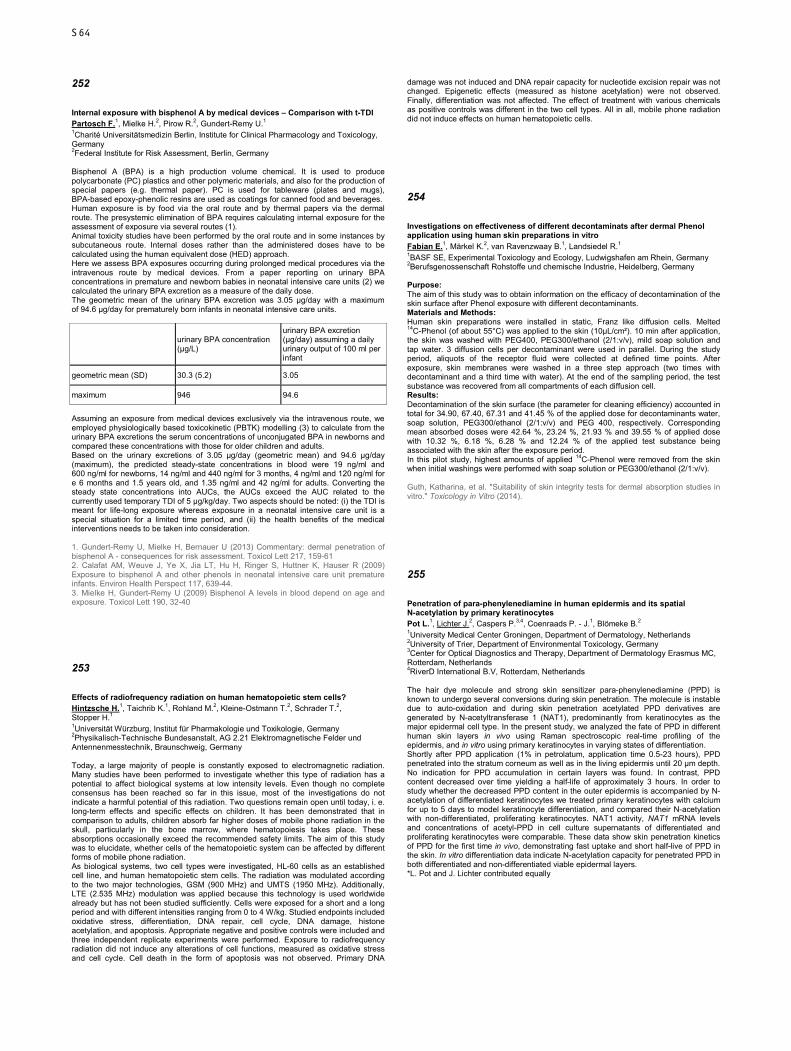
252
Internal exposure with bisphenol A by medical devices – Comparison with t-TDI Partosch F.1, Mielke H.2, Pirow R.2, Gundert-Remy U.1 1Charité Universitätsmedizin Berlin, Institute for Clinical Pharmacology and Toxicology, Germany 2Federal Institute for Risk Assessment, Berlin, Germany Bisphenol A (BPA) is a high production volume chemical. It is used to produce polycarbonate (PC) plastics and other polymeric materials, and also for the production of special papers (e.g. thermal paper). PC is used for tableware (plates and mugs), BPA-based epoxy-phenolic resins are used as coatings for canned food and beverages. Human exposure is by food via the oral route and by thermal papers via the dermal route. The presystemic elimination of BPA requires calculating internal exposure for the assessment of exposure via several routes (1). Animal toxicity studies have been performed by the oral route and in some instances by subcutaneous route. Internal doses rather than the administered doses have to be calculated using the human equivalent dose (HED) approach. Here we assess BPA exposures occurring during prolonged medical procedures via the intravenous route by medical devices. From a paper reporting on urinary BPA concentrations in premature and newborn babies in neonatal intensive care units (2) we calculated the urinary BPA excretion as a measure of the daily dose. The geometric mean of the urinary BPA excretion was 3.05 µg/day with a maximum of 94.6 µg/day for prematurely born infants in neonatal intensive care units.
urinary BPA concentration (µg/L)
urinary BPA excretion (µg/day) assuming a daily urinary output of 100 ml per infant
geometric mean (SD) 30.3 (5.2) 3.05
maximum 946 94.6
Assuming an exposure from medical devices exclusively via the intravenous route, we employed physiologically based toxicokinetic (PBTK) modelling (3) to calculate from the urinary BPA excretions the serum concentrations of unconjugated BPA in newborns and compared these concentrations with those for older children and adults. Based on the urinary excretions of 3.05 µg/day (geometric mean) and 94.6 µg/day (maximum), the predicted steady-state concentrations in blood were 19 ng/ml and 600 ng/ml for newborns, 14 ng/ml and 440 ng/ml for 3 months, 4 ng/ml and 120 ng/ml for e 6 months and 1.5 years old, and 1.35 ng/ml and 42 ng/ml for adults. Converting the steady state concentrations into AUCs, the AUCs exceed the AUC related to the currently used temporary TDI of 5 µg/kg/day. Two aspects should be noted: (i) the TDI is meant for life-long exposure whereas exposure in a neonatal intensive care unit is a special situation for a limited time period, and (ii) the health benefits of the medical interventions needs to be taken into consideration. 1. Gundert-Remy U, Mielke H, Bernauer U (2013) Commentary: dermal penetration of bisphenol A - consequences for risk assessment. Toxicol Lett 217, 159-61 2. Calafat AM, Weuve J, Ye X, Jia LT, Hu H, Ringer S, Huttner K, Hauser R (2009) Exposure to bisphenol A and other phenols in neonatal intensive care unit premature infants. Environ Health Perspect 117, 639-44. 3. Mielke H, Gundert-Remy U (2009) Bisphenol A levels in blood depend on age and exposure. Toxicol Lett 190, 32-40
253
Effects of radiofrequency radiation on human hematopoietic stem cells? Hintzsche H.1, Taichrib K.1, Rohland M.2, Kleine-Ostmann T.2, Schrader T.2, Stopper H.1 1Universität Würzburg, Institut für Pharmakologie und Toxikologie, Germany 2Physikalisch-Technische Bundesanstalt, AG 2.21 Elektromagnetische Felder und Antennenmesstechnik, Braunschweig, Germany Today, a large majority of people is constantly exposed to electromagnetic radiation. Many studies have been performed to investigate whether this type of radiation has a potential to affect biological systems at low intensity levels. Even though no complete consensus has been reached so far in this issue, most of the investigations do not indicate a harmful potential of this radiation. Two questions remain open until today, i. e. long-term effects and specific effects on children. It has been demonstrated that in comparison to adults, children absorb far higher doses of mobile phone radiation in the skull, particularly in the bone marrow, where hematopoiesis takes place. These absorptions occasionally exceed the recommended safety limits. The aim of this study was to elucidate, whether cells of the hematopoietic system can be affected by different forms of mobile phone radiation. As biological systems, two cell types were investigated, HL-60 cells as an established cell line, and human hematopoietic stem cells. The radiation was modulated according to the two major technologies, GSM (900 MHz) and UMTS (1950 MHz). Additionally, LTE (2.535 MHz) modulation was applied because this technology is used worldwide already but has not been studied sufficiently. Cells were exposed for a short and a long period and with different intensities ranging from 0 to 4 W/kg. Studied endpoints included oxidative stress, differentiation, DNA repair, cell cycle, DNA damage, histone acetylation, and apoptosis. Appropriate negative and positive controls were included and three independent replicate experiments were performed. Exposure to radiofrequency radiation did not induce any alterations of cell functions, measured as oxidative stress and cell cycle. Cell death in the form of apoptosis was not observed. Primary DNA
damage was not induced and DNA repair capacity for nucleotide excision repair was not changed. Epigenetic effects (measured as histone acetylation) were not observed. Finally, differentiation was not affected. The effect of treatment with various chemicals as positive controls was different in the two cell types. All in all, mobile phone radiation did not induce effects on human hematopoietic cells.
254
Investigations on effectiveness of different decontaminats after dermal Phenol application using human skin preparations in vitro Fabian E.1, Märkel K.2, van Ravenzwaay B.1, Landsiedel R.1 1BASF SE, Experimental Toxicology and Ecology, Ludwigshafen am Rhein, Germany 2Berufsgenossenschaft Rohstoffe und chemische Industrie, Heidelberg, Germany Purpose: The aim of this study was to obtain information on the efficacy of decontamination of the skin surface after Phenol exposure with different decontaminants. Materials and Methods: Human skin preparations were installed in static, Franz like diffusion cells. Melted 14C-Phenol (of about 55°C) was applied to the skin (10µL/cm²). 10 min after application, the skin was washed with PEG400, PEG300/ethanol (2/1:v/v), mild soap solution and tap water. 3 diffusion cells per decontaminant were used in parallel. During the study period, aliquots of the receptor fluid were collected at defined time points. After exposure, skin membranes were washed in a three step approach (two times with decontaminant and a third time with water). At the end of the sampling period, the test substance was recovered from all compartments of each diffusion cell. Results: Decontamination of the skin surface (the parameter for cleaning efficiency) accounted in total for 34.90, 67.40, 67.31 and 41.45 % of the applied dose for decontaminants water, soap solution, PEG300/ethanol (2/1:v/v) and PEG 400, respectively. Corresponding mean absorbed doses were 42.64 %, 23.24 %, 21.93 % and 39.55 % of applied dose with 10.32 %, 6.18 %, 6.28 % and 12.24 % of the applied test substance being associated with the skin after the exposure period. In this pilot study, highest amounts of applied 14C-Phenol were removed from the skin when initial washings were performed with soap solution or PEG300/ethanol (2/1:v/v). Guth, Katharina, et al. "Suitability of skin integrity tests for dermal absorption studies in vitro." Toxicology in Vitro (2014).
255
Penetration of para-phenylenediamine in human epidermis and its spatial N-acetylation by primary keratinocytes Pot L.1, Lichter J.2, Caspers P.3,4, Coenraads P. - J.1, Blömeke B.2 1University Medical Center Groningen, Department of Dermatology, Netherlands 2University of Trier, Department of Environmental Toxicology, Germany 3Center for Optical Diagnostics and Therapy, Department of Dermatology Erasmus MC, Rotterdam, Netherlands 4RiverD International B.V, Rotterdam, Netherlands The hair dye molecule and strong skin sensitizer para-phenylenediamine (PPD) is known to undergo several conversions during skin penetration. The molecule is instable due to auto-oxidation and during skin penetration acetylated PPD derivatives are generated by N-acetyltransferase 1 (NAT1), predominantly from keratinocytes as the major epidermal cell type. In the present study, we analyzed the fate of PPD in different human skin layers in vivo using Raman spectroscopic real-time profiling of the epidermis, and in vitro using primary keratinocytes in varying states of differentiation. Shortly after PPD application (1% in petrolatum, application time 0.5-23 hours), PPD penetrated into the stratum corneum as well as in the living epidermis until 20 µm depth. No indication for PPD accumulation in certain layers was found. In contrast, PPD content decreased over time yielding a half-life of approximately 3 hours. In order to study whether the decreased PPD content in the outer epidermis is accompanied by N-acetylation of differentiated keratinocytes we treated primary keratinocytes with calcium for up to 5 days to model keratinocyte differentiation, and compared their N-acetylation with non-differentiated, proliferating keratinocytes. NAT1 activity, NAT1 mRNA levels and concentrations of acetyl-PPD in cell culture supernatants of differentiated and proliferating keratinocytes were comparable. These data show skin penetration kinetics of PPD for the first time in vivo, demonstrating fast uptake and short half-live of PPD in the skin. In vitro differentiation data indicate N-acetylation capacity for penetrated PPD in both differentiated and non-differentiated viable epidermal layers. *L. Pot and J. Lichter contributed equally
S64
256
Advanced Prediction of Sensitization Potency of Chemicals: THP-1 in Coculture with HaCaT Keratinocytes Hennen J., Blömeke B. University Trier, Department of Environmental Toxicology, Germany Although several in vitro approaches address key steps of chemical-induced skin sensitization, there is uncertainty how factors such as metabolism and coinciding skin inflammation impact on chemicals’ potential and potency of dendritic cell activation. We integrated this in an in vitro model by coculturing THP-1 cells with HaCaT keratinocytes (1). In this study, we found high levels of interleukin (IL)-6 and 4.7-fold enhanced IL-8 in coculture supernatants, while no IL-6 was found for THP-1 cells alone. We exposed the cells to 14 sensitizers (including 7 prohaptens) and 10 non-sensitizers for 24h. Compared to THP-1 cells alone, coculturing resulted in up to 3.1-fold enhanced maximal CD86 and/or CD54 upregulation on THP-1 cells for 10 of 14 sensitizers. All sensitizers reached positivity for CD86 (Δmean fluorescence intensity (MFI) ≥ 10 compared to control) and/or CD54 (ΔMFI ≥ 50). No non-sensitizer gave a positive result regarding CD86, and only 1 non-sensitizer exceeded the threshold for CD54, resulting in an overall accuracy of 96%. Evaluating the concentrations needed to reach positivity, i.e. the potency for CD86 and/or CD54 upregulation, revealed that higher concentrations were needed for 4 of 13 sensitizers, mainly haptens, and lower concentrations for 6 of 13, among them 5 prohaptens, in coculture compared to THP-1 cells alone. Correlating this potency with human and animal data on sensitization potency revealed a clearly improved correlation with in vivo data compared to THP-1 cells alone. These data indicate that this coculture model has the potential to fill the gap regarding the prediction of sensitization potency. (1) Hennen J, Aeby P, Goebel C, Schettgen T, Oberli A, Kalmes M, et al. Cross Talk between Keratinocytes and Dendritic Cells: Impact on the Prediction of Sensitization. Toxicol Sci. 2011;123(2):501-10.
257
Development of a co-culture model for the prediction of idiosyncratic drug-induced liver injury Granitzny A.1, Hansen T.1, Knebel J. W.1, Dasenbrock C.1, Steinberg P.2 1Fraunhofer Institute for Toxicology and Experimental Medicine, In vitro and Mechanistic Toxicology, Hannover, Germany 2University of Veterinary Medicine Hannover, Institute for Food Toxicology and Analytical Chemistry, Germany Hepatotoxicity is one of the leading causes for withdrawals of drugs during late stages of the drug development process but also after being launched onto the market. A particularly severe and rare form of drug-induced liver injury (DILI) is the so-called idiosyncratic DILI (iDILI). IDILI is characterized by the absence of a clear dose dependency and the underlying mechanisms are as yet not fully understood. Furthermore, the prediction of iDILI from currently available pre-clinical animal studies remains difficult due to the lack of specific biomarkers. Although many drug-related mechanisms can directly induce cell death, there is growing evidence for the hypothesis that the recruitment of immune cells may play a critical role in the development of iDILI. In order to evaluate this hypothesis, which suggests that modest inflammatory stress can lower the threshold for hepatotoxicity, we established an in vitro co-culture system combining human monocytic (THP-1) with human hepatoma (HepG2) cells. With this model we aimed to identify whether the introduction of inflammatory immune cells, like monocytes, could increase the sensitivity of liver cells towards iDILI compounds. Troglitazone (TGZ), a drug that had already been withdrawn from the markets due to severe iDILI reactions, and rosiglitazone (RGZ), a compound from the same substance class, which has no potential to induce iDILI, were chosen as reference compounds. Using the WST-assay we observed a significant increase in cytotoxicity for TGZ in the co-culture model as compared to the single cell cultures while this effect was not observed with RGZ. In conclusion, for the model compounds used in this study, the co-culture model was able to differentiate between non-DILI and iDILI compounds. Our results suggest that this co-culture model could provide a useful tool for the prediction of inflammation-associated idiosyncratic drug-induced hepatotoxicity.
258
Disturbance of gene expression in primary human hepatocytes by hepatotoxic pyrrolizidine alkaloids: a whole genome transcriptome analysis Luckert C.1, Lenze D.2, Lampen A.1, Hessel S.1 1Federal Institute for Risk Assessment, Food Safety, Berlin, Germany 2Charité-Universitätsmedizin Berlin, Institute of Pathology, Germany 1,2-unsaturated pyrrolizidine alkaloids (PA) are widely distributed plant metabolites that predominantly occur in the plant families Asteraceae, Boraginaceae and Fabaceae. Acute and chronic PA poisoning due to contaminated food or feed causes severe hepatotoxicity. Carcinogenic effects have been demonstrated in animal studies. So far, the molecular mechanisms of PA toxicity are not well understood. For the analysis of the mode of action of PA, primary human hepatocytes were exposed to 100 µM of four structurally different PA: echimidine, heliotrine, senecionine and senkirkine. Changes in mRNA expression were analyzed by a whole genome microarray. Employing cut-off
values with a fold change of -2/2 and a q-value of 0.01, data analysis revealed numerous changes in gene expression patterns after PA exposure. 4556, 1806, 3406 and 8623 genes were regulated by echimidine, heliotrine, senecione and senkirkine, respectively. A total of 1304 genes were identified as commonly regulated by all four PA, among them numerous xenobiotic-metabolizing enzymes and transport proteins. Data were analyzed using a pathway analysis tool (Ingenuity® Pathway Analysis). PA affected pathways related to cell cycle regulation, cell death and development of cancer. The transcription factors p53, MYC, NFκB and NUPR1 were activated upon PA treatment. In addition, gene expression data showed a considerable interference of PA with lipid metabolism and bile acid flow. The associated transcription factors FXR, LXR, SREBF1/2, as well as PPARα, γ and δ were predicted to be inhibited by PA. Moreover, data propose a molecular interaction of PA with numerous nuclear receptors. In conclusion, although structurally different, all four PA significantly regulated a great number of genes in common. This proposes a similar mode of action for all PA, albeit to different extents, reflected by potential hepatotoxicity and individual PA structure.
259
Hepatotoxic effects of cyproconazole and prochloraz in wildtype and hCAR/hPXR mice Marx-Stoelting P.1, Ganzenberg K.2, Schmidt F.1, Rieke S.1, Braeuning A.1,2 1Federal Institute for Risk Assessment, Pesticides Safety, Berlin, Germany 2University of Tübingen, Toxicology, Germany Background: Cyproconazole and prochloraz are hepatotoxic agricultural fungicides that show a tumorigenic potential in long term rodent bioassays. A CAR/PXR-dependent mechanism of hepatotoxicity has been postulated for both substances and human relevance of these findings has been questioned. Methods: A short term (28-days) feeding study with both substances in humanised mice (hCAR/hPXR) and respective wildtype mice was conducted at two dose levels and using Phenobarbital (PB) as a reference substance. After sacrifice, livers were isolated, weighed and analysed histopathologically (H&E staining, specific antibody stainings for proliferation and protein expression) and for gene expression changes by quantitative real time PCR. Results: Our results show that all compounds caused a treatment-related increase in absolute and relative liver weights with some differences between the two genotypes. Accordingly, changes in gene expression related to classic CAR target genes such as Cyp2b10 were caused by both substances in both genotypes and were more pronounced at the higher dose levels. Wildtype mice were generally much more affected as compared to hCAR/hPXR mice. Pronounced differences between the genotypes were observed, when proliferation of cyproconazole-treated mice was evaluated: here, wildtype mice showed a strong increase in their proliferative response to cyproconazole, whereas hCAR/hPXR mice did not. Gene expression related to adverse outcome pathways potentially involved in the induction of proliferative changes (CDKN1a, GADD45) or in other adverse effects like cyproconazole-induced steatosis (FASN, FAT) was also much more affected at the higher dose level in wildtype mice than in humanized mice.
260
Platinum based neurotoxicity – the role of mitochondrial damage and establishment of C. elegans as an in vivo model system Bormann S., Alpert G., Honnen S., Wellenberg A., Fritz G. Heinrich-Heine-University, Institute of Toxicology, Düsseldorf, Germany Platinating agents such as oxaliplatin, cisplatin and carboplatin are chemotherapeutic drugs frequently used for the treatment of a wide variety of cancers. After entering the cell, the drugs exert their function by forming DNA monoadducts, which are subsequently processed into DNA intra- and interstrand crosslinks. Such adducts interfere with transcription and replication of the DNA which will eventually lead to cell death. However, as these cytotoxic effects are not cancer cell specific, all aforementioned platinum compounds cause different adverse affects through normal tissue damage. One such adverse effect is peripheral neuropathy (CIPN), which manifests in over 90% of patients treated with oxaliplatin and over two thirds of patients treated with cisplatin, while it only rarely occurs after carboplatin treatment. As neuronal cells require high energy amounts, it is believed that mitochondrial damage caused by platinating agents might play a role in neurodegeneration. In order to further examine the role of mitochondria in platinum based neurotoxicity, the murine neuronal cell line Neuro2a, which shows a dose dependent reduction of viability when treated with cis-, oxali- or carboplatin, was used as an in vitro model system. After treatment with platinum compounds, mitochondrial mass and membrane potential as well as the expression of genes involved in processes such as mitochondrial fission and fusion were examined. 48 h after treatment cis- and carboplatin had the strongest reducing effect on mitochondrial mass, while oxaliplatin caused the most distinct mitochondrial membrane deopolarisation. The expression of a set of genes involved in mitochondrial maintenance was, however, not greatly changed after a 24 h platinum treatment. Results on the expression levels of additional genes will be presented and discussed. To gain insights into the neurotoxic effects of platinating agents in vivo and to develop novel neuroprotective strategies, the nematode C. elegans is used as model organism. Initial experiments showed that the pharyngeal pumping rate is strongly reduced when the worms were fed with increasing concentrations of cisplatin. Elucidation of the effects of other platinum compounds as well as the establishment of additional assays is currently on-going.
S65
261
Characterization of the stress response of normal rat kidney cells to platinating anticancer drugs and modulation by lipid lowering drugs Krüger K.1, Thomale J.2, Fritz G.1 1Heinrich Heine University, Institute of Toxicology, Düsseldorf, Germany 2University Duisburg Essen, Institute of Cell Biology, Germany Background: Platinating agents are frequently used for the treatment of various types of cancer. Cisplatin and carboplatin are extensively used in the therapy of urogenital cancers, whereas oxaliplatin is particular useful for colorectal cancer treatment. Most relevant for the anticancer efficacy of these platinating agents is the formation of bivalent DNA intrastrand crosslinks. These adducts impair DNA replication, transcription and eventually trigger cell death. The clinically most relevant adverse effect associated with cisplatin treatment is nephrotoxicity. Here, we characterized the stress responses of rat renal proximal tubular (NRK-52E) and rat glomerular endothelial (RGE) cells stimulated by the aforementioned platinum compounds. Furthermore we started to analyze whether these responses can be modulated by the lipid lowering drug Lovastatin. Methods: Cell viability was determined by using the MTT assay, as well as by electrical impedance measurements via the iCELLigence system. The amount of DNA double-strand breaks (DSBs) was quantified by measuring the level of S139 phosphorylated H2AX (γH2AX) via immunocytochemistry and Western blot. Mechanisms of the DNA damage response (DDR) were analyzed by Western blot as well as by quantitative RT-PCR. In addition, formation and repair of Pt-(GpG) intrastrand crosslinks was determined via Southwestern blot analysis. Results: Based on the IC50 and IC80 values, RGE cells were more sensitive to cisplatin, oxaliplatin and carboplatin as compared to NRK-52E cells. Cisplatin provoked the highest cytotoxicity in both cell lines. In line with this, cisplatin induced the highest level of γH2AX and triggered the most intense DDR. Cytotoxicity resulting from carboplatin treatment occured with a delay, what might be related to a retarded formation of Pt-(GpG) intrastrand crosslinks. A pretreatment of NRK-52E cells with Lovastatin reduced the cytotoxicity evoked by cisplatin. Currently ongoing experiments aim to identify the mechanism underlying this protection. Conclusion: Apart from tubular epithelial cells, glomerular endothelial cells might also be a relevant target for nephrotoxicity provoked by cisplatin-based anticancer therapy. The mechanisms involved in platinum-induced cytotoxicity are exceptionally agent- and cell type-specific. Lovastatin might be useful to prevent cisplatin-induced cytotoxicity in renal cells.
262
NADPH oxidase subunit 4 (Nox4) is not the only responsible isoform for generation of ROS and DNA damage in the kidney during Ang II-treatment in vivo Zimnol A.1,2, Schupp N.1,2 1University of Düsseldorf, Institute of Toxicology, Germany 2University of Würzburg, Institute of Pharmacology and Toxicology, Germany In higher concentrations, the blood pressure regulating hormone Angiotensin II (Ang II), leads to vasoconstriction, hypertension and oxidative stress by activation of the renin angiotensin system (RAS). A stimulated RAS can lead to oxidative stress by activating NADPH oxidases which are the major enzymatic source of reactive oxygen species (ROS). The predominant and constitutively active form in the kidney is Nox4. From in vitro studies, it is known, that Nox4 is participating in the formation of oxidative DNA damage. To examine the contribution of Nox4 to oxidative DNA damage in vivo, male wildtype (WT) C57BL/6 and Nox4-deficient mice were equipped with osmotic minipumps, delivering either vehicle (PBS) or Ang II in a concentration of 600 ng/kg × min during 28 days. In WT and Nox4-deficent mice, Ang II induced hypertension, elevated urinary albumin levels and formation of ROS in the kidney. Furthermore, genomic damage, in form of single-strand breaks quantified by the comet assay, and double strand breaks detected immunohistochemically, was augmented in both groups. Also in the absence of Ang II, Nox4-deficient mice exhibited a higher background of single- and double strand breaks. Additionally we could show that Nox2 mRNA is upregulated in Nox4-deficient mice treated with Ang II. Due to these results we conclude that Nox4 is not the only responsible isoform for generation of ROS in the kidney under situation of Ang II-treatment and that there must be an involvement of other Nox isoforms, as well. Surprisingly we obtained evidence that Nox4 might have a protective role in renal diseases because genomic damage in the kidney of Nox4 deficient mice in the absence of Ang II was higher as compared to WT mice.
263
The SILICOAT project: Approaches for an effective quartz surface coating in the traditional ceramics industry to increase workers' safety - toxicological investigations Ziemann C.1, Rahmer H.1, Escrig Vidal A.2, Bonvicini G.3, Ibánez García M. J.2, Monfort Gimeno E.2, Creutzenberg O.1 1Fraunhofer Institute for Toxicology and Experimental Medicine ITEM, Hannover, Germany 2University Jaume I, Instituto de Tecnología Cerámica UJI-AICE, Castellón, Spain 3Centro Ceramico di Bologna, Italy Respirable crystalline silica (RCS) in the form of quartz or cristobalite has been classified as human lung carcinogen (category 1) by the International Agency for Research on Cancer (1997), however, acknowledging differences in hazardous potential depending on source, as well as chemical, thermal and mechanical history. As quartz-containing raw materials are indispensable for manufacturing processes in traditional ceramics industry (wall tiles, tableware, sanitary ware), many workers are potentially at risk to develop respiratory diseases, e.g. lung inflammation, silicosis, or even lung tumors. The SILICOAT project thus aimed at increasing workers’ safety by developing and implementing cost-effective RCS coating technologies based on stable, covalent saturation of reactive surface silanol groups. During technical development, in vitro lactate dehydrogenase (LDH) release and alkaline comet assays with primary rat alveolar macrophages (4 h of incubation; 75 µg/cm2; particulate negative control: Al2O3; positive control: quartz DQ12) were used for concomitant efficiency testing of promising organosilane coatings, i.e. propyltrimethoxysilane (PTMO) and SIVO160. The industrial quartz F1 served as model quartz for coating development. To judge for quartz-specificity assays were done ± aluminum lactate (quencher of quartz-specific effects). In the final in vitro assays, F1 clearly increased (~5-fold) mean LDH release and tail intensity, compared to Al2O3. Activity of F1 was markedly quenched when coated with 0.5% PTMO or 0.2% SIVO160 (w/w of quartz), leading to 99% inhibition of LDH release, and 81 and 83% inhibition of DNA damage. For the technically most feasible SIVO160-coating an intratracheal instillation study was performed with Wistar rats (2 x 0.5 mg quartz/rat; 5 rats/group; bronchoalveolar lavage [BAL] after 28 or 90 days; vehicle control: 0.9% NaCl; positive control: DQ12). After 28 days, SIVO160 almost completely quenched the F1-mediated changes in BAL parameters. After 90 days, the SIVO160-coating still clearly reduced the marked lung effects of F1 by 76% (total leukocytes), 68% (polymorphonuclear cells), 87% (lymphocytes), 56% (LDH), and 80% (ß-glucuronidase), indicating a persisting coating effect. In conclusion, covalent organosilane-coating of quartz might represent a promising strategy to increase workers’ safety in the ceramics industry. The project was funded by the EU’s 7th Framework Programme (FP7/2007-2013) under grant agreement n° 285787.
264
Determination of toxicological equivalency of racemates and their enantiomers using metabolomics Kamp H.1, Ramirez T.1, Montoya G.1, Frericks M.1, Strauss V.1, Fabian E.1, Herold M.2, Krennrich G.1, Looser R.2, Strigun A.2, Mellert W.1, Spitzer M.2, Peter E.2, Walk T.2, van Ravenzwaay B.1 1BASF SE, Ludwigshafen, Germany 2metanomics GmbH, Berlin, Germany GV/T and metanomics have developed the MetaMap®Tox database containing toxicity- and metabolome profiles of > 700 reference compounds from 28 day rat studies. More than 110 toxicity-specific metabolome patterns were identified. This methodology is now routinely used to predict the toxicity of novel compounds at an early stage of development. Recently we have shown that toxicity-specific metabolic changes can also be detected on cell-based metabolome data gained from an in-vitro approach. Chiral properties of molecules may result in different biological effects. In such cases it is often desirable to only produce the biologically active enantiomer. To prove the toxicological equivalency between racemates and their corresponding pure enantiomers extensive toxicological “bridging studies” are required. The purpose of the present research was to investigate if metabolomics could be used to demonstrate equivalency of toxicity. The rat plasma metabolome of two agrochemical compounds (racemic and pure enantiomer) after 28 day treatment was determined. The rat plasma metabolome of the two forms was investigated in terms of number and extent of metabolite changes, comparison of the metabolite changes against the patterns in the MetaMap®Tox data base as well as statistical correlation analyses of the rat plasma metabolome profiles. No relevant differences in the rat plasma metabolome profiles between the two forms of the respective active ingredients were observed. Using total profile comparison, the best correlation for the metabolite profile of the racemic mixtures was always their active isomer showing. As the toxicity of the tested racemates and optically active forms were known to be equivalent, this study is a proof of concept for equivalency testing. Potential resource reductions are up to hundreds of animals. The corresponding in-vitro metabolomics approach, based on metabolome data gained from dosed HepG2 cells additionally confirm the results derived from the rat-plasma metabolome.
S66
265
Quantification of transporters and cytochrome P450 in fungicide treated rats by ms- based immunoassays Hammer H.1, Schmidt F.2, Kneuer K.2, Dabrowski A.2, Weiss F.1, Marx-Stoelting P.2, Poetz O.1 1NMI, Protein Analytics, Reutlingen, Germany 2Bundesinstitut für Risikobewertung, Berlin, Germany Transporters and Cytochrome P450 enzymes are essential components of the xenobiotics metabolism. Their expression levels and activity influence the velocity of the metabolism, the efficiency of the first pass effect and barrier maintenance. Since xenobiotics can influence activity and expression levels of these protein classes strongly, absolute quantification can help to clarify mechanism of activity alteration, for example discrimination between activation and induction. Moreover, identification and quantification of potential combinatorial effects of substances affecting a similar adverse outcome pathway is also an interesting scientific question. However, transporters and cytochrome P450 enzymes cannot be quantified by conventional bioanalytical methods such as sandwich immunoassays because it is very difficult to generate antibodies against these conserved hydrophobic proteins. For this reason, we developed a test system to quantify the expression level of transporters and cytochrome P450 enzymes via mass spectrometry-based immunoassays. Hydrophobic proteins are measurable by this method because samples are digested with trypsin and subsequently analyzed on peptide level. From each protein, one peptide which can be assigned unambiguously, is identified via tandem MS and quantified by means of an isotope labeled reference. Prior to MS-read-out the peptides are enriched by antibodies which recognize a very short c-terminal epitope. These epitopes are selected in such way that they are common in peptides derived from several transporters and therefore allow the analysis of transporter groups with few antibodies. This method was employed to analyze the effect of single fungicides and combinations thereof on the expression level of transporters and cytochrome P450 enzymes in rat in comparison to respective mRNA levels as analysed by qRT-PCR. Animals were fed with a diet containing Cyproconazole, Prochloraz, Epoxiconazole or a combination of these fungicides. Liver tissue was analyzed after 4 weeks treatment. Fungicide-dependent induction was observed for ABCB1a/b, ABCC2, ABCC3, CYP2B1/CYP2B3 with a good correlation at protein and mRNA level. No additional effects of the combinatorial treatments were measured in comparison to single treatments.
266
Monotoring 11β-hydroxysteroid dehydrogenase type 1 levels in mouse liver during caloric restriction Loerz C., Maser E. Institute of Toxicology and Pharmacology for Natural Scientists, Kiel, Germany The microsomal NADP(H)-dependent enzyme 11β-hydroxysteroid dehydrogenase type 1 (11β-HSD1) is a key regulatory enzyme in glucocorticoid metabolism. It predominantly functions as a reductase by catalyzing the interconversion of inactive cortisone to receptor-active cortisol in vivo (11-dehydrocorticosterone and corticosterone in rodents, respectively). It is known that high levels of glucocorticoids are associated with insulin resistance, hyperglycemia, visceral obesity and dyslipidemia, all symptoms of the Metabolic Syndrome. Also, 11β-HSD1 expression and activity in the liver is either maintained or modestly lowered. On the other hand, in obesity elevated levels are found in adipose tissue, notably in visceral fat. Furthermore, in a mouse model selective overexpression of 11β-HSD1 in adipose tissue is associated with obesity and other symptoms of the Metabolic Syndrome. For this reason extensive research has been performed on the role of 11β-HSD1 in obesity and its metabolic complications. Thus, it has received considerable attention as being a potential therapeutic target to develop therapies against obesity-related diseases. There are indications that expression and activity of 11β-HSD1 is elevated in the liver and reduced in visceral adipose tissue during caloric restriction. To assess whether caloric restriction affects 11β-HSD1 activity in glucocorticoid target tissues, we used a HPLC-based assay to determine corticosterone reduction activity in mouse liver samples during the course of caloric restriction. Mice were divided into two groups: the ad libitum fed group (AL) and the caloric restriction group (CR). CR mice were dietarily restricted for six months with 75% of the amount consumed by the AL control group. Subsequently the CR animals were refed by having ad libitum access for another six months. Our results indicate that 11β-HSD1 enzyme activity in mouse liver samples is significantly increased in the CR group. Furthermore, after two and six weeks of re-feeding 11β-HSD1 hepatic enzyme activity was still higher than in the AL group. Hence, upregulation of enzymatic activity through caloric restriction persists after short- and long-term re-feeding. Therefore, caloric restriction seems to have a beneficial effect on 11β-HSD1 enzyme activity in liver. But further investigations are required to test whether caloric restriction goes along with downregulation of 11β-HSD1 enzyme activity in adipose tissue.
267
Phase I metabolism of beta-asarone Cartus A., Stegmüller S., Schrenk D. TU Kaiserslautern, Food Chemistry and Toxicology, Germany Phenylpropenes (PP) are natural occurring compounds present in many foodstuffs. Some PP e.g. methyleugenol (ME), safrole or estragole are well known genotoxic carcinogens in rodents. The activation of those allylic compounds occurs via a cytochrome P450 (CYP)-catalyzed oxidation leading to an 1’-alcohol which is afterwards sulfonated by sulfotransferases (SULT). Upon loss of sulfate, a carbocation is formed, which can react with DNA. Otherwise, the majority of PP with a propenylic side chain e.g. anethole, methylisoeugenol or isosafrole are not genotoxic or carcinogenic in rodents. An exception is beta-asarone (bA; cis-2,4,5-trimethoxy-1-propenylbenzene) which caused liver tumors in male mice. bA was mutagenic in the Ames test only with metabolic activation and inhibition of SULT had no effect on the incidences of hepatomas in mice. Therefore a bioactivation of bA via SULT seems unlikely and the molecular mechanism(s) responsible for its carcinogenicity is still not understood. We investigated the phase I metabolism of bA using liver microsomes of different species (rat, porcine, bovine, human), and SupersomesTM (expressing human CYP1A2, 2A6, 2C19, 2D6, 2E1 or 3A4). The analytical methods were based on HPLC-DAD and LC-MS/MS. However, due to the identical retention times and isobaric properties of some formed metabolites, we expanded our analytical methods to 1H-NMR-spectroscopy, and synthesized the majority of metabolites to use them as reference compounds. This allowed us to distinguish the formed metabolites. We identified several metabolites, e.g. 1’-hydroxy-, (E)- and (Z)-3’-hydroxy-asarone, there oxidation products, O-monodemethylated metabolites as well as a side-chain-epoxide and the corresponding diols (HPLC chromatograms and metabolic pathways are depicted in figure 1 and 2). The derived (apparent) kinetic data indicate that over a broad range of substrate concentrations, side-chain epoxidation and concomitant formation of diols was the predominant metabolic pathway of bA in all liver microsomes tested, whereas side-chain hydroxylation, oxidation and O-monodemethylation took place to a far lower extent. All six investigated human CYPs were capable to generate the epoxide (and diols) with CYP1A2, 2C19 and 3A4 being the most active enzymes for this reaction(s). Our results imply that bA-epoxide is the ultimate carcinogen of bA.
Figure 1: HPLC-DAD chromatograms of the incubation supernatant of rat liver microsomes (1 mg/ml) with beta-asarone (400 µM, 1h) at three different wavelenghts (bottom: 280; middle: 261, top 340 nm)
Figure 2: Metabolic pathways of beta-asarone in liver microsomes
S67

268
The Eucalyptus oil ingredient 1,8-cineol induces oxidative DNA damage Dörsam B.1, Wu C. - F.2, Efferth T.2, Kaina B.1, Fahrer J.1 1University Medical Center Mainz, Department of Toxicology, Germany 2Johannes Gutenberg University Mainz, Department of Pharmaceutical Biology, Germany The epoxy-monoterpene 1,8-cineol, also known as eucalyptol, is a major constituent of eucalyptus oil. It is widely used as flavor and fragrance in consumer goods as well as in medical therapies. Owing to its anti-inflammatory properties 1,8-cineol is also applied to treat upper and lower airway diseases. Although 1,8-cineol is used for many purposes, only little is known about its potential genotoxicity in mammalian cells. Aim of this study was to investigate the genotoxicity and cytotoxicity of 1,8-cineol in human and hamster epithelial cells. First, we were able to detect a significant and concentration-dependent increase of oxidative DNA lesions in human colon cancer cells using the Formamidopyrimidine-DNA glycosylase (Fpg)-modified alkaline comet assay. Pre-treatment of cells with the antioxidant N-acetylcysteine prevented the generation of Fpg-sensitive sites following 1,8-cineol treatment, supporting the notion that 1,8-cineol induces oxidative DNA damage. In the dose range of DNA damage induction, 1,8-cineol did neither reduce the viability of colon cancer cells nor influence their cell cycle distribution, suggesting that cells tolerate the induced oxidative DNA damage due to ongoing DNA repair. To verify this hypothesis, hamster cell lines with defects in essential proteins of homologous recombination (HR)-mediated repair, BRCA2 and Rad51, were treated with 1,8-cineol. The monoterpene induced oxidative DNA damage and subsequent DNA double-strand breaks in the analyzed hamster cell lines. Strikingly, we detected a significant concentration-dependent decrease in viability of the HR-defective cells, while the corresponding HR-proficient parental cell lines were not affected. In summary, we conclude that 1,8-cineol displays weak genotoxic potential, by inducing primarily oxidative DNA lesions, which are very likely tolerated in DNA repair proficient cells without resulting in cell cycle arrest or cell death induction. In contrast, cells with defective HR were compromised after 1,8-cineol treatment, suggesting a protective role of HR in response to high doses of 1,8-cineol [1]. [1] Dörsam, B. et al. (2014) The eucalyptus oil ingredient 1,8-cineol induces oxidative DNA damage, Archives of Toxicology, epub ahead of print
269
Lipoic acid induces p53-independent cell death in colorectal cancer cells and enhances the cytotoxicity of 5-fluorouracil Dörsam B., Göder A., Seiwert N., Kaina B., Fahrer J. University Medical Center Mainz, Department of Toxicology, Germany The antioxidant alpha-lipoic acid (LA) is an endogenous dithiol compound, which plays an important role in mitochondrial energy metabolism. Recent studies revealed its potential to trigger cell death in tumor cells, whereas it hardly affects primary non-transformed cells. This study analyzed the cytotoxicity of LA on colorectal cancer (CRC) cells with different p53 status and investigated a putative synergistic effect with the anticancer drug 5-fluorouracil (5-FU). First, we demonstrated a dose-dependent decrease of cell viability after treatment with LA, which was independent of p53 as shown in isogenic p53-proficient and p53-deficient tumor cell lines. The reduction in viability was largely attributable to cell death induction as revealed by Annexin-V/PI staining. LA-treated HCT116 cells underwent caspase-dependent and -independent cell demise, which was suppressed by the pan-caspase inhibitor zVAD and the RIP kinase inhibitor necrostatin-1. In CaCO-2 and HT29, LA triggered caspase-dependent cell death via activation of caspase-9, -3 and -7 with subsequent PARP-1 cleavage as demonstrated by cells immunoblot analysis, activity assays and pan-caspase inhibition. Intriguingly, LA did neither activate p53 nor induced genotoxic effects, which was confirmed by lack of DNA strand breaks and phosphorylation of histone 2AX (γ-H2AX). Furthermore, we provided evidence that LA increases the cytotoxic effects of the anticancer drug 5-FU as attested by significantly enhanced cell death rates in HCT116 and CaCO-2 cells. Taken together, these results demonstrate that LA induces cell death in CRC cells in a p53-independent manner status potentiates 5-FU-mediated cytotoxicity without causing DNA damage on its own [1]. [1] Dörsam, B. et al. (2014) Lipoic acid induces p53-independent cell death in colorectal cancer cells and potentiates 5-fluorouracil-dependent cytotoxicity, Archives of Toxicology, in press
270
Cytotoxicity and Embryotoxicity of a Chinese Herbal Medicine Extraction Used for Atopic Dermatitis Li L.1,2,3, Haase A.2, Cheng L.3, Lau C. B. S.3, Tang L. Y.3, Liang B.3, Wang C. C.3, Leung P. C.3, Luch A.2, Spielmann H.1, Schäfer-Korting M.1 1Freie Universität, Institut für Pharmazie, Berlin, Germany 2German Federal Institute for Risk Assessment (BfR), Department of Product Safety, Berlin, Germany 3The Chinese University of Hong Kong, Prince of Wales Hospital, Institute of Chinese Medicine, Hong Kong (China) Background: Atopic dermatitis (AD) There is considerable interest in traditional Chinese herbal medicines (CHM) as an alternative treatment for AD. A palatable and well
tolerated Chinese medicine formula (PHF), composed of five herbal extracts, appears effective in reducing the need for glucocorticoids in moderate-to-severe AD[1]. No obvious adverse effects were recorded in the clinical studies[2]. Yet, further evidence on the safety is still necessary, in particular for the use in pregnancy. Objectives: The OECD approved Embryonic Stem Cell Test (EST)[3] is employed to determine the embryotoxicity potentials of the PHF formula and the individual components. Methods & Results: Analytical technologies (TLC and/or HPLC) are conducted for chemical authentications to ensure the quality of each test herbal extracts. The commercially available permanent mouse embryonic stem cell line D3 and mouse fibroblast cell line 3T3 were applied to the cytotoxicity and differentiation experiments. The embryotoxic potential was derived from the biological and molecular endpoints (ID50D3, IC503T3 and IC50D3). According to the linear discriminant functions, both the formula PHF and its individual herbal extracts are classified as non-embryotoxic. Conclusions: Our study provides evidence on the safety of the formula PHF, which could be used as an adjunctive treatment for AD during pregnancy. Acknowledgement: Deutsche Akademische Austausch Dienst (DAAD 57076385), Hong Kong General Research Fund (GRF 476210) and National Natural Science Foundation of China (NSFC 81303302). [1] Hon KL, et al. Therapeutic effect and safety of a traditional Chinese medicine for atopic dermatitis in children: a randomised, double-blind, placebo-controlled study. Hong Kong Med J. 2011;17:S38-40. [2] Hon KL, et al. A Pentaherbs Capsule as a Treatment Option for Atopic Dermatitis in Children: An Open-Labeled Case Series. Am J Chin Med. 2004;32:941–950. [3] Seiler AEM and Spielmann H. The validated embryonic stem cell test to predict embryotoxicity in vitro. Nat Protocols 2011;6(7):961-978.
271
Pak Choi Fed to Mice: Formation of DNA Adducts and Influence on Xenobiotic-Metabolizing Enzymes Wiesner M.1,2, Barknowitz G.2, Florian S.2, Haack M.3, Lehmann C.3, Lippmann D.3, Mewis I.1, Schumacher F.2,4, Brigelius-Flohé R.3, Schreiner M.1, Glatt H.2 1Leibniz Institute of Vegetable and Ornamental Crops Großbeeren/Erfurt e.V., Quality Research, Germany 2German Institute of Human Nutrition Potsdam-Rehbrücke, Department of Nutritional Toxicology, Nuthetal, Germany 3German Institute of Human Nutrition Potsdam-Rehbrücke, Department Biochemistry of Micronutrients, Nuthetal, Germany 4University of Potsdam, Institute of Nutritional Science, Department of Nutritional Toxicology, Nuthetal, Germany 1-Methoxy-indol-3-ylmethyl glucosinolate (1MOI3M), a secondary plant metabolite in Brassica species, and its degradation product N-methoxy-indole-3-carbinol are mutagenic in S. typhimurium strains after activation by myrosinase and human sulphotransferase (SULT) 1A1, respectively. Both compounds react with DNA and proteins after administration as individual substances to mice. The aim of the present study was to investigate the effects of 1MOI3M as part of a plant matrix in order to compare them with previous experiments using isolated compounds. Male FVB/N mice were fed a diet containing 1.2% lyophilized pak choi sprouts and an extract of intact glucosinolates prepared from methyl jasmonate-treated pak choi sprouts for a maximum of 8 days. After day 1, 2, 4, and 8 subgroups of animals (n=4) were sacrificed by cervical dislocation. To evaluate the persistence of DNA adducts, two subgroups were fed a semisynthetic diet for further 8 or 16 days (+8 and +16 follow up) and then sacrificed. Blood and organs (liver, lung, kidney, and intestinal tissues) were taken and frozen immediately in liquid nitrogen. DNA adducts were determined by UPLC-ESI-MS/MS using stable isotopic-labeled internal adduct standards. Additionally, the influence of this diet on the activity of selected xenobiotic-metabolizing enzymes was investigated. We observed a continuous accumulation of 1MOI3M DNA adducts formed in the intestine, liver, kidney and lung during 8 days of treatment. After cessation of the pak choi diet the DNA adducts decreased rapidly in jejunum, caecum, and colon, but persisted in liver, lung and kidney. The activity of several xenobiotic-metabolizing enzymes was weak induced by the diet. After 4 days of continuous feeding with pak choi diet, 7-ethoxy- and 7-methoxyresorufin-O-dealkylase activities as well as NAD(P)H:quinone oxidoreductase 1 and thioredoxin reductase activity were enhanced slightly (p < 0.05) compared to the pooled control groups. After cessation of the diet, the activity levels decreased to reach the levels of untreated groups. On the contrary, the activity of the toxifying enzyme, Sult1a1, was unaffected by the treatment. Our results and recent published literature demonstrate that 1MOI3M forms DNA adducts in mice. DNA adducts are able to trigger mutations and therefore indicate a possible cancer risk. Further studies to investigate the mechanistic effects of 1MOI3M glucosinolate and its degradation products are needed.
S68
272
Artificial digestion including food components do not prevent cytotoxicity of silver nanoparticles on human intestinal cells Lichtenstein D.1, Ebmeyer J.1, Knappe P.2, Juling S.1, Böhmert L.1, Selve S.3, Niemann B.1, Thünemann A.2, Lampen A.1 1Federal Institute for Risk Assessment, Food Safety, Berlin, Germany 2Federal Institute for Materials Research and Testing, Polymers in Life Science and Nanotechnology, Berlin, Germany 3Technical University Berlin, ZELMI, Germany Due to the increasing use of silver nanoparticles in food-associated consumer products, the uptake of silver nanoparticles by the oral route has become a serious scenario. Two barriers must be considered regarding the question of NP fate: first, the strong shift in chemical conditions during digestion and second, the intestinal barrier which is mainly composed by enterocytes. Therefore, this study aimed to analyze changes in cytotoxicity and uptake of silver nanoparticles by using an in vitro digestion model. Carbohydrates, proteins and fatty acids were implemented as food components in the digestion process to simulate realistic conditions. Silver nanoparticles coated with poly (acrylic acid) were digested in absence or presence of food components. All particle suspensions were characterized by small-angel X-ray scattering (SAXS) to estimate particle core size, size distribution and stability in cell culture medium. For transport studies, the Transwell system was used. Particle uptake was monitored by high pressure chemical digestion and AAS. The CTB assay was applied for cytotoxicity testing in the Caco-2 cell system, an established model for enterocytes in vitro. Particles proved to be stable under cell culture conditions and showed radii from 4 to 16 nm. Cytotoxicity testing revealed that neither the digestion process nor the presence of food components affected the cytotoxicity of the nanoparticles. Additionally, undigested as well as particles digested in the presence of food components were comparably taken up by Caco-2 cells, whereas the uptake of particles digested without food components was decreased by 60 %. Our findings suggest that ingested nanoparticles may reach the intestine in a nanoscaled form. Apparently, it seems to play a crucial role if the particles are embedded in a food matrix during ingestion or not. The absence of food components in NP digestion protocols could lead to misinterpretation of uptake results. Therefore, digestion models and especially the presence of food components should be considered in further investigations.
273
Comparison of two different application routes of nanosilver in rats — Evidence for nanoparticles after silver ion treatment Juling S.1, Lichtenstein D.1, Selve S.2, Oberemm A.1, Creutzenberg O.3, Braeuning A.1, Lampen A.1 1Federal Institute for Risk Assessment, Effect-based Analytics and Toxicogenomics, Berlin, Germany 2Zentraleinrichtung Elektronenmikroskopie (ZELMI), Transmissionselektronenmikroskopie, Berlin, Germany 3Fraunhofer ITEM, Inhalationstoxikologie, Hannover, Germany The unique properties of silver nanoparticles have a great potential to advance food safety and revolutionize food packaging. Despite these promising applications, there are concerns about the impact of silver nanoparticles on human health. Therefore, the tissue distribution and excretion of coated silver nanoparticles was investigated in rats after single oral administration including a 7-days post treatment observation time. The animals received silver nanoparticles at a dose of 20 mg/kg body weight and an equimolar dose of silver ions by gavage. The silver content was measured in different organs after days 1, 2, 3 and 7. To assess the effect of the intestinal passage on systemic bioavailability, an additional group of rats was intravenously administered with 4 mg/kg body weight silver nanoparticles and another group was treated with an equimolar dose of silver ions. The fate of the silver was compared after 24 h single intravenous administration. An accumulation of silver was observed in different organs. As expected, most of the nanosilver was excreted via feces and minor amounts were detected in intestine and colon. Orally administered ionic silver was detected in feces and additionally in the urine. Remarkably, also after intravenous injection of nanosilver, silver was found in feces, which suggests biliary excretion of silver nanoparticles. In addition to this, the formation of small Ag2S nanoparticles was observed after ionic administration. It is concluded that the oral resorption of nanosilver results in an accumulation of silver in different organs and feces. Results obtained after intravenous application suggest a biliary excretion. Formation of small Ag2S nanoparticles can be detected after ionic silver application.
274
A method for the identification of iron oxide nanoparticle protein corona in a complex HepG2 cell lysate Juling S., Lichtenstein D., Lalkowski G., David J., Rozycki C., Niemann B., Braeuning A., Lampen A. Federal Institute for Risk Assessment, Effect-based Analytics and Toxicogenomics, Berlin, Germany There is a widespread use of different materials in the nanometer range. In medicine, iron oxide nanoparticles are used as contrast media to improve the visibility of internal body structures in magnetic resonance imaging (MRI). The food industry use iron oxide and hydroxide as food additives (E172). The protein corona, the interface between a nanoparticle and the environment has an influence on absorption, distribution and cellular downstream effects. Therefore, knowledge about the corona is of pivotal importance. In this study, a method was established for the isolation of the protein corona of iron oxide nanoparticles from a complex HepG2 cell lysate. Iron oxide nanoparticles were incubated with whole cell lysate from HepG2 human liver hepatocellular carcinoma cells or with individual subcellular fractions from HepG2 cells, in order to reduce the complexity of the proteome in the lysate. Then the nanoparticles along with their corona were isolated via magnetization. After four washing steps for the removal of unspecifically bound proteins, the protein corona was separated from the nanoparticles via SDS-PAGE. Corona-forming proteins were identified by 1D-LC QTOF MS/MS. It was observed that the iron oxide nanoparticles were taken up by HepG2 cells. After duplicate analysis, 19 proteins were identified as a part of the corona in the complex HepG2 whole cell lysate, whereas between 5 and 20 corona-forming proteins were identified when cellular fractions were used. In conclusion, the presented method is suited for the identification of the protein corona of iron oxide nanoparticles. Reducing the complexity of the proteome by sub-cellular fractionation allows for the identification of less abundant proteins of the protein corona.
275
Inflammatory effect of cerium oxide nanoparticles on human lung A549 cells Burchardt K.1, Papavlassopoulos H.1, Niemeier D.2, Lüllmann R.2, Röhl C.1 1Christiana Albertina University of Kiel, Institute of Toxicology and Pharmacology for Natural Scientists, Germany 2Christiana Albertina University of Kiel, Department of Anatomy, Germany Cerium oxide (CeO2) nanoparticles (NP) are widely used, e.g. as fuel additive and for industrial and biomedical applications. Yet the exposure to those small particles is a public concern and contradictory publications about their cytotoxicity and inflammatory properties exist. In this work, four different sizes of CeO2 NP with average diameters of 10 nm, 15-30 nm, 33 nm and 50-105 nm as well as CeO2 bulk particles are compared for cellular uptake by, cytotoxicity for and inflammatory effects on human lung adenocarcinoma cells (A549). Fluorescence-activated cell sorting showed that A549 cells internalize CeO2 NP in a concentration and time dependent manner, while bulk particles are taken up by a lesser extent. Cytotoxicity was analyzed via the MTT assay for CeO2 concentrations between 5 and 5000 µg/ml. Most NP show a concentration dependent cytotoxicity with an EC50 value of about 200 µg/ml. Only the 15-30 nm sized and the bulk particles are less toxic also at higher concentrations with and EC50 value of about 5000 µg/ml. To investigate the proinflammatory properties of CeO2 on A549 cells the reactive oxygen species (ROS) levels and the Interleukin-8 (IL-8) secretion was measured. In lower concentrations intracellular ROS-levels were measured below the level of the untreated control, but at 500 µg/ml cells exposed to 15-30 nm and 50-105 nm NP show higher ROS-levels around 230% of the control. The IL-8 secretion increased up to ~730 pg/cm² following treatment with the 15-30 nm NP at concentrations between 50-1000 µg/ml (control: ~140 pg/cm²). This could not be shown for the other CeO2 particles. Our results show, that CeO2 nano- and bulk particles can be taken up by human lung A549 cells, but that only certain particles induce proinflammatory effects in this model system. Further investigations will analyze which particle properties may be of importance in this respect.
276
Method for Identification of Low Soluble, Biopersistent Dusts (GBS) at Workplaces Creutzenberg O., Hansen T., Schuchardt S., Tillmann T. Fraunhofer ITEM, Inhalation Toxicology, Hannover, Germany Fine fractions of dusts occurring in occupational settings are of high relevance for the safety at workplaces and therefore are strictly regulated by authorities. Appropriate threshold values should guarantee that lung diseases are not induced in workers exposed up to long periods. Respirable biopersistent granular dusts (GBS) are defined as respirable fine dusts with a negligible solubility in physiological lung fluid that do not exhibit a specific chemistry-related toxicity at volumetric non-overload conditions. In 2012, the MAK Commission derived a new threshold value of 0.3 mg/m3 for GBS, recognizing that in concentrations exceeding the physiological lung clearance capacity GBS can cause chronic inflammation and increase the lung cancer risk in animal experiments. – The objectives were to investigate, whether a general value for the term
S69
‘low soluble’ can be derived (e.g. a solubility in lung fluid of approx. 1 mg/l). In addition, measuring the inflammatory response in lung lavage fluid, it was investigated whether nanoscaled dusts could possibly fulfill the criteria to be included in the GBS class. - Six micro- and nanoscaled dusts (some of them of commercial relevance) were compared analysing the solubility in the lung fluid (day 3, 28 and 90) and the lung toxicity after intratracheal instillation in rats (day 3 and 28): TiO2 (rutile, micro), TiO2 (anatase, nano), Eu2O3 (micro-nano mixed), BaSO4 (micro), ZrO2 (micro) and amorphous SiO2 (nano). Two doses of 0.5 and 1.5 µl per rat were administered to Wistar rats; these volume doses resulted in a non-overload and moderate overload of lungs, resp. – The differential cell count showed only slight inflammatory cell levels after treatment with TiO2 (rutile) and BaSO4 (PMN < 5% after 3 days in the low dose group; < 15% in the high dose group). In contrast, the TiO2 (anatase) showed a stronger response (PMN > 30% after 3 days). The rare earth Eu2O3 (micro-nano) dust showed the strongest effect (approx. 40% PMN) including a red-coloured lung lavage fluid (BAL after 28 days and solubility analysis are still underway). - New dusts to be included in the GBS group have to fulfill the criteria of low solubility and a non-significant inflammatory response at doses below the overload range. For the most nanoscaled dusts an individual toxicological characterization seems to be adequate. The project was funded by the Federal Institute for Occupational Safety and Health (BAuA), Dortmund – F 2336.
277
Biokinetics and inhalation toxicity of nano-BaSO4 after 1, 4, 13 and 52 weeks of exposure Keller J.1, Ma-Hock L.1, Küttler K.1, Strauss V.1, Groeters S.1, Wiench K.2, Wohlleben W.3, van Ravenzwaay B.1, Landsiedel R.1 1BASF SE, Experimental Toxicology and Ecology, Ludwigshafen am Rhein, Germany 2BASF SE, Product Safety, Ludwigshafen am Rhein, Germany 3BASF SE, Polymer Physics, Ludwigshafen am Rhein, Germany Little is known about long-term effects of airborne, poorly soluble nanoparticles (PSP); this question is currently addressed by a long-term inhalation study with CeO2 and BaSO4 according to OECD 453. Nanomaterials were selected to represent a range of different biokinetic and biodynamic properties among PSP. Results of 1 and 4 week inhalation studies (OECD 412) with BaSO4 (NM-220) were used to design the long-term study; interim results of this study after 13 and 52 weeks are presented. Female Wistar rats were whole-body exposed to aerosol concentrations of 50 mg/m3 BaSO4 for 6 h/day, 5 days/week. The Ba burdens of the lungs, lung-associated lymph nodes and extrapulmonary organs (inter alia bones) were analysed by ICP-MS at the end of the exposure and at different time points post-exposure (short-term studies). Lung burdens after 1 and 4 weeks of exposure were 1.0 and 0.84 mg/lung, respectively, and decreased during post-exposure with a half-time of about a week. The rapid lung clearance was confirmed by IT-instillation, and the translocation was further investigated. 13 and 52 weeks of exposure resulted in lung burdens of 1.7 and 10 mg/lung, respectively. Animals were examined by bronchoalveolar lavage and histopathology. No morphological changes or significant increases in BALF were detected after short-term exposure. After 13 weeks, a slight increase of neutrophils (13-fold compared to control) was observed. After 52 weeks, 260-fold increased neutrophils and further changes in BALF (enzymes, mediators) indicated pulmonary inflammation. Compared to other PSP, such as CeO2 and TiO2, BaSO4 is remarkably rapidly cleared from the lung. Compared to similar lung burdens of other poorly soluble nanoparticles, only slight effects on the lung tissue were observed. After 52 weeks of exposure, however, a decrease in lung clearance and an increase in lung inflammation were observed. Landsiedel, Robert, et al. "Testing Metal‐Oxide Nanomaterials for Human Safety." Advanced Materials 22.24 (2010): 2601-2627. Klein, Christoph L., et al. "Hazard identification of inhaled nanomaterials: making use of short-term inhalation studies." Archives of toxicology 86.7 (2012): 1137-1151.
278
Nanotoxicological assessment of shape-engineered titanium dioxide nanoparticles Kolling J.1, Albrecht C.1, Pellegrino F.2, Deiana C.2, Schins R.1 1IUF - Leibniz Research Institute for Environmental Medicine, Particle, inflammation and genome integrity, Düsseldorf, Germany 2University of Turin, Department of Chemistry, Torino, Italy Nanoparticles have gained tremendous importance in various technological and medical applications but these developments have also raised nanosafety concerns. The European seventh framework project SETNanoMetro explores the highly-defined development and production of TiO2 nanoparticles in terms of homogeneous bulk structure, size, shape and surface structure. The outcomes of the project are anticipated to benefit hallmark applications in the specific areas of environment (i.e. photocatalytic treatment of polluted air and water), energy (i.e. dye sensitized solar cells) and health (i.e. nanostructured coatings in prostheses). The nanosafety aspects in SetNanoMetro are being addressed using in vitro toxicological testing of the highly-defined TiO2 nanoparticles in respiratory and gastrointestinal tract cell lines, as inhalation and ingestion are considered to represent the most relevant uptake routes of nanoparticles.
Metabolic activity analysis, evaluated by WST-1 assay in A549 and Caco-2 cells, revealed marked time-, concentration- and cell type-dependent differences in effects among the various TiO2 powders. So far however, all tested TiO2 samples were clearly less toxic than SiO2 nanoparticles that were used as reference material. For both epithelial cell lines, also differences in the production of the pro-inflammatory cytokine interleukin-8 were observed. Taken together our findings provide further support on the importance of the physico-chemical characteristics in the toxic and pro-inflammatory effects of TiO2 nanoparticles. The inclusion of currently developed and highly-characterised TiO2 nanoparticles in upcoming investigations will allow for a more precise evaluation of the role of subtle shape- and surface structure changes for this type of nanomaterial. Acknowledgements: The SetNanoMetro is supported by funding under the Seventh Research Framework Programme of the European Union. Specific types of TiO2 particles were provided by Solaronix (Switzerland) and the University of Torino (Italy). Commercial TiO2 nanoparticles were kindly provided by Evonik and Cristal.
279
Screening of the toxicity, oxidant generating and inflammogenic properties of sixteen types of nanoparticles in human lung cell lines Neumeyer-Sickinger A.1, Albrecht C.1, Werner J.1, Wedekind R.1, Hellack B.2, Kuhlbusch T.2, Schins R.1 1IUF – Leibniz Research Institute for Environmental Medicine, Particles, inflammation and genome integrity, Düsseldorf, Germany 2Institute of Energy and Environmental Technology e.V. (IUTA), Air Quality & Sustainable Nanotechnology, Duisburg, Germany Evaluation of the toxicity of nanomaterials requires a comprehensive knowledge and understanding of their physicochemical properties and behaviour in biological systems. Various studies strongly indicate that the surface area and the potential to form reactive oxidants are highly promising metrics to predict the toxic potency of nanoparticles (NP). In the framework of the nanOxiMet project, sixteen different types of NP were investigated including metals, metal oxides and carbonaceous NP. As inhalation is considered as their most relevant exposure route, cellular effects were studied in the human alveolar epithelial cell line A549 and in the rat alveolar macrophage cell line NR8383. Cellular viability was assessed by the WST-1 assay. The oxidant generation capacity of the NP was investigated via electron spin resonance (ESR) and 2',7'-dichlorofluorescein (DCF) fluorescence assay. The mRNA expression of antioxidant and inflammatory genes was evaluated by qRT-PCR. After 24 h treatment a strong decrease in the metabolic viability was observed for ZnO and CuO treated cells, whereas other NP showed a less pronounced but cell-type specific toxicity. The ESR results illustrated that CuO and NiO NP caused reactive oxygen species (ROS) formation in NR8383 cells. The DCF-DA assay confirmed the NiO-induced increase in ROS and demonstrated an increase in ROS after exposure to Ag and carbon black nanoparticles as well. First findings from mRNA expression analysis indicated a NP-type specific up-regulation of antioxidant and inflammatory genes under involvement of the Nrf2 and NF-κB signalling pathways. The findings from these and ongoing investigations will be collated to evaluate whether the intrinsic ROS generating capacity and surface reactivity of NP can be used as alternative metrics for hazard assessment and grouping of nanomaterials. Acknowledgement: The nanOxiMet project (www.nanoximet.eu) is funded by the German Federal Ministry of Education (BMBF) and the French National Research Agency (ANR) under the European 7th framework ERA-NET activity “Safe Implementation of Innovative Nanoscience and Nanotechnology” (SIINN).
280
Cellular uptake of different nano and bulk metal oxide particles by cells derived from epidermal, intestinal and leukocyte cell lines Papavlassopoulos H.1, Burchardt K.1, Niemeier D.2, Lüllmann-Rauch R.2, Röhl C.1 1Christiana Albertina University of Kiel, Institute of Toxicology and Pharmacology for Natural Scientists, Germany 2Christiana Albertina University of Kiel, Institute of Anatomy, Germany Nanoscaled metal oxide particles are used in a wide range of practical applications, as well as in consumer products or in the area of biology and medicine, leading to an increased human exposure. Thus, impact assessments on cellular level and analysis of the underlying mechanisms are increasingly gaining importance in order to avoid harmful consequences for human health. In this study, we analyzed the uptake of different metal oxide nanoparticles (ZnO, TiO2 and CeO2) in human cell lines: epithelial colorectal adenocarcinoma cells (Caco-2), monocytic cells (U937) and keratinocyte (HaCaT) by means of flow cytometry and electron microscopy. To distinguish adsorbed from uptaken nanoparticles we used energy depletion by sodium azide and 4°C incubation, respectively. Our findings show that uptake of nanoparticles occurs in all cell lines, but to a varying extent, in a time and concentration dependent manner. Furthermore, the electron microscopy analysis revealed that TiO2 and CeO2 were localized in the cytoplasm and partially in lysosomes of Caco-2 cells; however, intracellular ZnO particles could not be detected by neither flow cytometry nor electron microscopy. Inhibition of energy-dependent or passive
S70
mechanisms resulted in a significantly reduced uptake measured via flow cytometry indicating that nanoparticles enter cells by mainly energy-dependent mechanisms. Moreover, our aim is to examine the impact of nanoparticle-internalization on cellular response. Therefore, uptake of CeO2 nanoparticles (15-30 nm) (CONP), which induce the expression of the pro-inflammatory cytokine interleukine 8 (Il-8) in Caco-2 cells, served as model system. We analyzed the uptake of CONP into Caco-2 cells in more detail by use of various endocytosis inhibitors and achieved a reduction of intracellular CONP partially up to 95 %. Subsequently, we assessed the effect of perturbed internalization on CeO2 induced Il-8 expression after 2 h exposure by qRT-PCR using a TaqMan gene expression assay and received a significant decrease in Il-8 expression. This study demonstrates the high potential of nanoparticles to penetrate cellular barriers and suggests that proinflammatory effects are causally connected with the internalization of nanoparticles.
281
Cellular uptake of different nano and bulk metal oxide particles by cells derived from epidermal, intestinal and leukocyte cell lines Papavlassopoulos H.1, Burchardt K.1, Niemeier D.2, Lüllmann-Rauch R.2, Röhl C.1 1Christiana Albertina University of Kiel, Institute of Toxicology and Pharmacology for Natural Scientists, Germany 2Christiana Albertina University of Kiel, Department of Anatomy, Germany Nanoscaled metal oxide particles are used in a wide range of practical applications, as well as in consumer products or in the area of biology and medicine, leading to an increased human exposure. Thus, impact assessments on cellular level and analysis of the underlying mechanisms are increasingly gaining importance in order to avoid harmful consequences for human health. In this study, we analyzed the uptake of different metal oxide nanoparticles (ZnO, TiO2 and CeO2) in human cell lines: epithelial colorectal adenocarcinoma cells (Caco-2), monocytic cells (U937) and keratinocyte (HaCaT) by means of flow cytometry and electron microscopy. To distinguish adsorbed from uptaken nanoparticles (NP) we used energy depletion by sodium azide and 4°C incubation, respectively. Our findings show that uptake of nanoparticles occurs in all cell lines, but to a varying extent, in a time and concentration dependent manner. Furthermore, the electron microscopy analysis revealed that TiO2 and CeO2 were localized in the cytoplasm and partially in lysosomes of Caco-2 cells; however, intracellular ZnO particles could not be detected by neither flow cytometry nor electron microscopy. Inhibition of energy-dependent or passive mechanisms resulted in a significantly reduced uptake measured via flow cytometry indicating that nanoparticles enter cells by mainly energy-dependent mechanisms. Moreover, our aim is to examine the impact of nanoparticle-internalization on cellular response. Therefore, uptake of CeO2 NP (15-30 nm), which induce the expression of the pro-inflammatory cytokine interleukin 8 (IL-8) in Caco-2 cells, served as model system. We analyzed the uptake of CeO2 NP into Caco-2 cells in more detail by use of various endocytosis inhibitors and achieved a reduction of intracellular CeO2 NP partially up to 95%. Subsequently, we assessed the effect of perturbed internalization on CeO2 induced IL-8 expression after 2 h exposure by qRT-PCR using a TaqMan gene expression assay and received a significant decrease in IL-8 expression. This study demonstrates the high potential of nanoparticles to penetrate cellular barriers and suggests that in our model system proinflammatory effects are causally connected with the internalization of nanoparticles.
282
In vitro toxicity testing of two Multi-walled Carbon Nanotubes determined for the purpose of targeted drug delivery Requardt H.1, Hansen T.1, Eisenbarth E.2, Hampel S.3, Dasenbrock C.1 1Fraunhofer Institute for Toxicology and Experimental Medicine, Department for Mechanistic and In-vitro Toxicology, Hannover, Germany 2South Westphalia University of Applied Science, Iserlohn, Germany 3Leibniz Institute for Solid State and Materials Research, Dresden, Germany Carbon Nanotubes (CNTs) are novel, carbon based nanomaterials that gain more and more interest in fields like mechanical engineering or pharmacy and medicine. This study was an initial determination of the cytotoxic potential of two multi-walled Carbon Nanotubes that were determined for the development of a multifunctional drug delivery carrier. A549 lung epithelial cells and HepG2 cells were employed for the experiments. The cells were exposed with a dose of 1, 10 and 25µg/cm² growth area (24 and 48h) of the two CNT types. The two CNT types were produced using the identical chemical vapour deposition (CVD) synthesis for both types (IFW; Dresden, Germany). The first type was used as produced, still containing an iron catalyst that was added during synthesis (Fe-CNT). The second type was heat treated after synthesis for 1h at 2600°C to eliminate all remaining iron from the CNT structure (nonFe-CNT). Both CNT types showed an identical morphology (16.5µm (± 8) length, 48nm (±12) diameter) and appeared curved/flexible in REM investigation. The heat treated CNT type (nonFe) showed a significantly reduced number of surface defects, indicated by a reduction in d-peak intensity during Raman spectroscopy. The WST-8 assay was applied to evaluate cell viability and the LDH cytotoxicity assay was used to detect potential membrane damage. To investigate the production of reactive oxygen species (ROS) the DCFH-DA assay was applied. Additionally a cell cycle analysis was performed to assess cell proliferation.
The results showed a time and dose dependent decrease in cell viability for both cell lines. In A549 cells the severe leakage of lactate dehydrogenases (LDH) after 48h of exposure resulted in a total cytotoxicity of 25% (Fe-CNT) and 45% (nonFe-CNT). The DCFH-DA assay showed an increase in ROS production, primarily under the influence of the nonFe-CNTs. The cell cycle analysis demonstrated in heat treated CNTs an increased number of cells in the G2/M-phase while the number in S-phase was reduced, indicating a G2-phase arrest. Summarizing, most cytotoxic effects were more prominent for the heat treated (nonFe) CNTs. This indicates that the number of surface defects plays an important role in CNT-related cytotoxicity. Additionally we conclude that the CNTs can’t be used as drug delivery carriers without achieving biocompatibility via further modification.
283
An overview of the InhalT-90 project – a 90-day inhalation toxicity study with CeO2 nanoparticles Schwotzer D., Hansen T., Niehof M., Creutzenberg O. Fraunhofer Institute for Toxicology and Experimental Medicine, Inhalation Toxicology, Hannover, Germany The application of nanomaterial and the use of its advantageous characteristics for many different products is strongly expanding during the last decades. Cerium dioxide nanoparticles are commonly used as additive in diesel fuel, due to its catalytic activity. As a consequence they are released into the environment via fuel combustion, and organisms might be exposed via inhalation. This example shows that the investigation of nanoparticle toxicity is important for adequate regulation and risk assessment. However, little is known about the adverse effects of CeO2 nanoparticles after inhalation especially regarding the long term exposure of humans and the environment. The BMBF-funded project InhalT-90 (contract number: 03X0149) was initiated to investigate the effects of CeO2 nanoparticles following a 90-day nose-only inhalation in the rat according to OECD 413. In order to compare the results to a combined chronic inhalation toxicity and carcinogenicity study (BASF, Ludwigshafen, Germany), early indicators of genotoxic and carcinogenic effects should be determined. This will particularly be facilitated by gene expression analyses. Groups of rats will be exposed to the test substance CeO2 in concentrations of 0, 0.1, 0.3, 1 and 3 mg/cm³ or to one high concentration of barium sulfate (50 mg/m³) for a time period of 90 days (6 h/day, 5 days/week) in a nose-only exposure system. After one and 28 days of exposure, as well as after one, 28 and 90 days recovery period, animals will be sacrificed for different investigations including hematology, histopathology, immunohistochemistry, retention analytics, bronchoalveolar lavage and gene expression analyses. The latter will be performed in isolated pneumocytes type II using pathway arrays for genotoxicity, oxidative stress, inflammation, apoptosis and lung cancer related genes. Subsequently, genes will be selected as marker, whose mRNA expression is markedly modified by nanoparticle exposure. Based on this an in vitro screening system for nanomaterials should be developed: a lung cell line will be exposed to the nanoparticles in an air-liquid exposure system. The in vivo selected marker genes will be evaluated in this in vitro system, checking if the gene expression is consistent and if it is comparable to the in vivo situation. All in all, these investigations should contribute to the improvement of nanomaterial risk assessment by the development of screening methods and the supply of toxicity data.
284
“NanoEmission”: The project and first results of the toxicological assessment of investigated particles Thomas S., Schumann B., Wiese J., Glahn F., Foth H. Martin-Luther-University, Institute of Environmental Toxicology, Halle (Saale), Germany In the “NanoEmission”-Project the entire route of the nanoparticles (NPs) from the waste via incineration and filtration of the exhaust gas to a possible release to the environment and toxicological evaluation of effects on humans and environment, is considered. The objective of the project is to describe the influence of thermal waste treatment on the toxicological profile of NPs, contained in the waste. First of all stable suspensions of commercially available pure NPs e.g. Barium sulfate, with a reproducible size distribution in several media have to be established. In this process, different preparation methods (stirring, ultrasonic bath, ultrasound probe) and compositions of media lead to different results in agglomeration state and size distribution. The addition of a tenside improved the size distribution in suspension. The characterization of the pure NP was performed by SEM/EDX whereas the characterization of the suspension occurs by DLS. In the present study the short-term (24h) and long-term (72h) effects of BaSO4 (0,001 mg/ml - 1 mg/ml) on primary cell cultures of normal human bronchus epithelial cells (NHBEC) and peripherial lung cells (PLC) were investigated. Therefore we analyzed cytotoxicity, oxidative stress markers, as glutathione (GSH) and ROS (Reactive oxygen species) formation, gene expression of CYP1A1 and furthermore induction of apoptosis. Overall viability assays (MTT, Resazurin and LDH assay) showed different sensitivities to detect effects of BaSO4. Treatment with 1 mg/ml BaSO4 for 24h decreased viability below 80% (MTT) whereas viability was not affected in the Resazurin assay. Long-term exposure (1 mg/ml BaSO4) resulted in lower viability levels (below 60% for MTT and Resazurin). After 24h incubation PLC showed no LDH release, whereas NHBEC showed a 3-fold (1 mg/ml, 24h) increase.
S71

After 72h incubation (1 mg/ml) both cell culture models showed only a 2-fold increase of LDH release. Due to the decreased number of remaining cells after 72h the effect of membrane damage seems to be lower. Investigations in questions of oxidative balance where made, too. In short time ROS formation determined by DCF assay (2´,7´-dichlorofluorescin) BaSO4 showed no increase. As a marker of oxidative stress the level of intracellular glutathione was determined by HPLC. In both cell models the GSH-level was decreased significantly after 72h exposure to 1 mg/ml BaSO4 (80% in NHBEZ and 60% in PLC). However, the short time exposure had no significant effect.
285
Evaluation of the toxicity and data gaps of e-cigarette aerosol May M.1, Schwarz K.2, Koch W.2, Bitsch A.1 1Fraunhofer Institute of Toxicology and Experimental Medicine (ITEM), Chemical Risk Assessment, Hannover, Germany 2Fraunhofer Institute of Toxicology and Experimental Medicine (ITEM), Aerosol Research, Hannover, Germany The electronic cigarette (e-cigarette) is a device for inhaling vaporized liquid that is mostly consumed by smokers and has emerged in recent years as a supposedly less harmful alternative to conventional cigarettes. As no combustion is associated with the "smoking" of an e-cigarette it is often tolerated in confined areas. A harmonized regulation does not exist currently. At the moment e-cigarettes and refills are covered by the EU Directive 2014/40 / EU if it is not declared as a medical product. The increase in the consumption of e-cigarettes in recent years suggests that consumption of e-cigarettes has already attained a general social significance. In commercial products excipients typical used are propylene glycol (PG) and glycerin that are classified as GRAS (GeneRally Assumed to be Safe). However, this classification relates exclusively to oral ingestion. In addition to the uncertainty of possible long-term effects of excipients due to inhalation exposure, uncertainties arise regarding the uniformity of nicotine release, and on the exact chemical composition of the aerosol phase compared to the original liquid. It is intended that the EU Commission reports potential risks from the use of refillable electronic cigarettes until May 20, 2016. In this point uniform quality standards and test methods are required. The aim of this presentation is to address data gaps for the toxicological evaluation of e-cigarettes to identify further study requirements, and the need for standardization of product testing and assessment.
286
Consumer products made of polymers: An unfailing source for dermal uptake of potentially harmful substances Mielke N., Hutzler C., Kappenstein O., Vieth B., Luch A. Federal Institute for Risk Assessment, Safety of Consumer Products, Berlin, Germany Polymeric materials became a part of industrial usage and especially of commodity fabrication well established. Due to individual additivation of the materials, the final good meets requirements in design, durability and functionality issues. Apart from a broad variety of additives with a defined function such as plasticizers, stabilizers and vulcanization accelerators, contaminants as well as decomposition products or non-intentionally added substances (NIAS) can be part of the material. For those polymeric materials that are used in consumer products with intensive skin contact, dermal exposure to the above-mentioned classes of substances may thus be expected. Because some of the compounds are known to cause adverse health effects in humans, it is important to estimate the substances potential to increase internal exposure of consumers as a consequence of skin contact to contaminated products. We therefore investigated consumer products which are designated for skin contacts as handles of tools or toys with respect to individual additive composition, potentially harmful substances and their distribution in skin models in vitro. To this end we used static diffusion cells, Franz cells, with varying skin models including porcine and human epidermis, synthetic membrane or artificial skin and simulated dermal exposure to polymeric material of commodities. Due to its toxicological relevance, in subsequent studies we focused on polycyclic aromatic hydrocarbons (PAH) and on substitutes for the plasticizers diethylhexylphthalate (DEHP) trioctyltrimellitate (TOTM). We assessed the penetration of these additives by applying FTIR microscopy. Thereby we demonstrated that TOTM is capable of overcoming the skin barrier in vitro. On the other hand, PAH accumulation in human skin after contact with contaminated consumer products has been visualized by means of fluorescence microscopy. Since some toxicologically non-relevant additives might form decomposition products of considerable toxicological concern, we also focused on such degradation products applying several ageing scenarios. With this we verified cresol derivatives being degradation products of several less harmful polymer additives. Our results clearly demonstrate that dermal exposure to both migrating components of commodities and its decomposition products is of toxicological relevance as it can contribute to the individual body burden.
Plasticizers: Structures of plasticizer DEHP (diethylhexylphthalate) and substitute additive TOTM (trioctyltrimellitate).
TOTM Penetration: IR-microscopy. Cryo section of porcine skin, treated with 1 µg TOTM per cm² for 24 h. Heat map: Red areas indicate high TOTM amounts after mapping of transmission at TOTM-specific band 1110 cm-1. s.c.: stratum corneum; hf: hair follicle
287
Laser-induced Decomposition of Light-Fast Organic Tattoo Pigments Schreiver I.1, Gebhardt M.1, Hutzler C.1, Laux P.1, Berlien H. - P.2, Luch A.1 1Federal Institute for Risk Assessment, Chemicals and Product Safety, Berlin, Germany 2Evengelical Elisabeth Hospital, Department of Laser Medicine, Berlin, Germany In recent times, tattoos have become mainstream across the population (prevalence of 9.1 % in Germany). Yet, hardly anything is known about the toxicological risks associated with the ingredients currently used. In previous investigations, cleavage patterns of organic pigments found in tattoo inks were identified using online-coupled pyrolysis to gas chromatography with mass spectrometric detection (Py-GC/MS). The fragments found include a wide range of toxic and carcinogenic compounds. For example, benzene can potentially be cleaved from any organic pigment due to their aromatic structure. Cleavage of pigments at weak bonds does not only occur upon pyrolysis but also due to thermal energy and photochemical reactions as induced by laser light. Thus, potential cleavage of pigments may occur during laser removal of tattoos, which is known from literature for a few azo pigments. In the past, such concerns led to a switch from azo compounds towards non-azo pigments by some tattoo ink manufacturers despite the lack of any data on whether or not these would be indeed safer than their progenitors. This gap of knowledge has now been addressed in our study. Here, we demonstrate for the first time that the widely applied pigments Phthalocyanine Blue 15:3, Pyrrolo-pyrrole Red 254 and Quinophthalone Yellow 138 are cleaved by medical laser light. Aqueous pigment dispersions were irradiated with medical ruby (694 nm) and neodymium-doped yttrium aluminium garnet (Nd:YAG, 532 and 1064 nm) lasers and subsequently analyzed for decomposition products using GC/MS. The main decomposition products after laser irradiation are equal to those detected with Py-GC/MS. For Blue 15:3, Red 254 and Yellow 138 main cleavage products were 1,2-benzene dicarbonitrile, 4-chlorobenzonitrile and 2-(8-aminoquinolin-2-yl)-4,5,6,7-tetrachloro-3-hydroxy-1H-inden-1-one, respectively. However, for pigment Quinacridone Violet 19 no cleavage products were detected under given laser irradiation parameters and it is therefore considered to be stable under these conditions. As expected, pigment decomposition was highly dependent on laser light absorption at the respective wavelengths. Our results show that the risk of very light-fast pigments to decompose under laser irradiation conditions have likely been underestimated in the past. Thus, appropriate stability testing for photo-induced cleavage of tattoo pigments becomes mandatory.
S72

Pigment structures: Chemical structures of four highly light-fast organic pigments. Main cleavage sites for irradiation with medical lasers (Ruby and Nd:YAG) indicated with dashed arrows.
288
DNA damage response and its impact on alkylating agent induced toxicity Eich M., Roos W. P., Nikolova T., Kaina B. University Medical Center Mainz, Institute of Toxicology, Germany DNA strand-breaks are detected by different sensor proteins. Among these are Nibrin (NBN) and Ataxia telangiectasia mutated (ATM), which are crucial for the detection of DNA double-strand breaks (DSBs), and ATM- and RAD3-related (ATR) that sensors single-stranded DNA regions occurring at stalled replication forks. Activation of these sensors leads to initiation of DNA repair, blockage of cell cycle progression and induction of apoptosis, but also survival signaling via phosphorylation of various downstream targets like the checkpoint kinases 1 and 2 (CHK1; CHK2). Since Temozolomide (TMZ), which is being used as therapeutic drug against glioma and malignant melanoma, induces DSBs as secondary DNA lesions, it is essential to understand how cells respond to TMZ, if these proteins are nonfunctional. To address this question, lymphoblastoid cells and fibroblasts harboring mutations in NBN, ATM and ATR were treated with TMZ. The toxicity was analyzed with the AnnexinV/PI assay and compared to corresponding wild-type cells. All mutated cells showed an increased sensitivity, which was due to the O6-methylguanine lesion as the cells in presence of the repair enzyme O6-methylguanine-DNA methyltransferase (MGMT) were completely resistant to TMZ. Additionally, glioma and melanoma cells, in which NBN, ATM and ATR were down regulated by RNAi, were also hypersensitive to TMZ. Interestingly, the ATR knockdown led to greater sensitization compared to the ATM knockdown, showing that ATR is the more important kinase. Consistent with this, western blot analysis revealed that ATR, but not ATM is essential for the phosphorylation of the downstream targets CHK1 and CHK2. Finally, inhibition of CHK1 and CHK2 by specific inhibitors also caused a sensitization of glioma and melanoma cells to TMZ as investigated with the AnnexinV/PI assay. In conclusion, these data show that NBN, ATM, ATR, CHK1 and CHK2 are involved in the defense against alkylating agent induced toxicity. We therefore suggest these proteins as therapeutic targets for glioma and metastatic melanoma therapy using alkylating agents including TMZ. Work is supported by DFG KA724. Eich et al. (2013), Contribution of ATM and ATR to the resistance of glioblastoma and malignant melanoma cells to the methylating anticancer drug temozolomide. Mol Cancer Ther 12(11): 2529-2540 Eich et al. (2010), Nijmegen breakage syndrome protein (NBN) causes resistance to methylating anticancer drugs such as temozolomide. Mol Pharmacol 78(5): 943-951
289
Characterization of apoptosis induction by the mycotoxin phomoxanthone A Stuhldreier F.1, Böhler P.1, Schlütermann D.1, Niemann H.2, Frank M.2, Proksch P.2, Wesselborg S.1, Stork B.1 1University Hospital Düsseldorf, Institute for Molecular Medicine I, Germany 2Heinrich-Heine University Düsseldorf, Institute for Pharmaceutical Biology and Biotechnology, Germany Phomoxanthone A (PXA), a mycotoxin derived from the endophytic fungus Phomopsis longicolla, is a small molecule with antibiotic activity against organisms as diverse as bacteria, algae, fungi, and animals. A recent study from our groups showed for the first time that PXA induces apoptosis in human cancer cell lines, including the highly apoptosis resistant B lymphoma cell line DG75, with an IC50 of 0.1 – 0.5 μM and as soon as 4 h after treatment. In the same study we showed that PXA has immune-stimulatory effects in human leukocytes.a Furthermore we could show that PXA precipitates Ca2+ out of solution, indicating that it binds and possibly chelates Ca2+. Investigations in caspase-9 deficient and reconstituted cells as well as in caspase-8 knock-out and wild-type cells revealed that PXA induces intrinsic but not extrinsic apoptosis. Subsequently we could show that BCL-2 overexpression inhibits PXA-related cleavage of the caspase-3 substrate poly (ADP-ribose) polymerase (PARP). However, no study so far has shed light on the mechanism of action of PXA. Here, we report for the first time that PXA induces Ca2+ flux in human lymphoma cell lines including Jurkat J16 (T cell lymphoma) and Ramos (Burkitt lymphoma). Ca2+ is mobilised from both extracellular and intracellular stores. Pre-treatment with the cell-permeable Ca2+ chelator BAPTA-AM completely blocks PXA-induced Ca2+ mobilization and partially inhibits caspase-3 activation and PARP-cleavage, indicating Ca2+ dependency of PXA-induced apoptosis. Considering that disruption of mitochondrial Ca2+ homeostasis is a known trigger of cell death, we used the mitochondrium-targeted fluorescent Ca2+ sensor mito-Pericam in order to investigate the effect of PXA on mitochondrial Ca2+ levels. Interestingly
treatment with PXA does not lead to mitochondrial Ca2+ overload like treatment with many well-known intrinsic apoptosis stimuli, but to depletion of mitochondrial Ca2+. Further studies using the fluorescent probe tetramethylrhodamine ethyl ester (TMRE) could show that treatment with PXA causes a breakdown of the mitochondrial membrane potential (∆ψm) within seconds, similar to the protonophore CCCP. Upcoming experiments should determine cause-and-effect chain of Ca2+ flux, ∆ψm-breakdown and apoptosis induction, potential protonophoric attributes of PXA and to which extent effects on the mitochondrial permeability transition pore are involved in the observed processes. a Rönsberg D, Debbab A, Mándi A, Vasylyeva V, Böhler P, Stork B, Engelke L, Hamacher A, Sawadogo R, Diederich M, Wray V, Lin W, Kassack MU, Janiak C, Scheu S, Wesselborg S, Kurtán T, Aly AH, Proksch P. Pro-apoptotic and immunostimulatory tetrahydroxanthone dimers from the endophytic fungus Phomopsis longicolla. J Org Chem 2013; 78:12409-25
290
Reduction of Oxidative Stress and Genomic Damage, After Weight Loss either by Gastric bypass or Caloric Restriction Alone in a Rodent Model of Obesity Bankoglu E. E.1, Seyfried F.2, Rotzinger L.2, Nordbeck A.2, Corteville C.2, Jurowich C.2, Germer C. T.2, Otto C.2, Stopper H.1 1University of Wuerzburg, Institute of Pharmacology and Toxicology, Germany 2University Hospital of Wuerzburg, Department of General-, Visceral-, Vascular- and Paediatric Surgery, Germany Morbid Obesity is an independent risk factor for cardiovascular disease, Type 2 Diabetes mellitus and certain types of cancer. Bariatric surgery - with the Roux-en-Y gastric bypass (RYGB) being the gold standard -has become the therapeutic option of choice as a sustained weight loss and improvement of associated morbidity is achieved in the majority of patients. There is, however, a lack of evidence focusing on bariatric surgery induced sustained weight loss and its possible impact on cancer risk. We aimed to investigate the association between obesity, oxidative stress and genomic damage in a rodent model of obesity (male Zucker fa/fa rats) after weight loss either induced by caloric restriction alone or by RYGB. Therefore male Zuc-Lrpfa (aged 12 weeks) rats were randomly divided into four groups: Lean control (fa/+), RYGB (Zuc-Lrfpa, gastric bypass surgery group) and sham (Zuc-Lrfpa, sham operated group) and sham BWM (body weight matched to RYGB). Body weight and food intake were measured before and after 4 weeks after intervention. Urine was collected at baseline, 1, 2 and 3 weeks after intervention. After an oral glucose tolerance test (OGTT) was performed measuring blood levels of insulin, glucose and glucagon like peptide 1 rats were sacrificed and kidney, liver and colon were collected for further analysis. The sham rats showed elevated insulin levels and insulin resistance response. The RYGB and BWM rats showed elevated insulin levels, but decreased insulin resistance. Evaluation of DNA damage was performed with freshly isolated primary cells by comet assay and gamma-H2AX staining of paraffin-embedded tissue sections. DHE staining was performed in order to detect ROS production and oxidative stress in kidney, liver and colon. Additionally the heat shock proteins (HSPs) HSP70 and HO-1 were measured by western blot in order to evaluate cellular stress response. To quantify the oxidation of DNA and RNA urinary 8-oxodG, 8-oxoGuo and 8-oxoGua measurements were performed by LC/MS/MS. The obese rats showed elevated oxidative stress and genomic damage in comparison to lean rats. After bodyweight loss, a decrease in oxidative stress level and genomic damage was observed. The response of the gastric bypass surgery group and the caloric restriction group were slightly different in their pattern. This might be due to a difference in metabolism.
291
DNA double-strand breaks induced by cytolethal distending toxin (CDT) trigger autophagy in a p53-dependent manner Seiwert N.1, Jaurich H.1, Hülsenbeck J.1, Dörsam B.1, Frisan T.2, Kaina B.1, Fritz G.3, Fahrer J.1 1University Medical Center Mainz, Department of Toxicology, Germany 2Karolinska Institute, Department of Cell and Molecular Biology, Stockholm, Sweden 3Heinrich Heine University Düsseldorf, Institute of Toxicology, Germany Cytolethal distending toxin (CDT) is a unique tripartite genotoxin produced by several pathogenic bacteria. CDT is internalized into mammalian cells by dynamin-mediated endocytosis followed by its nuclear translocation, where it induces DNA double-strand breaks (DSBs) due to its DNase I-like activity. We have recently shown that CDT from Haemophilus ducreyi is a radiomimetic agent and triggers a DNA damage response (DDR) similar to that of ionizing radiation [1]. Here, we analyzed whether CDT-induced DSBs engage autophagy, a cellular mechanism that contributes to genomic integrity and cell survival. First, recombinant His-tagged CDT was purified to homogeneity by Ni-NTA affinity chromatography with great yields. Recombinant CDT was highly active in GM637 human fibroblasts and HCT116 human colon epithelial cells, as demonstrated by induction of the well-known DSB marker phosphorylated histone 2AX (γ-H2AX). The dose-dependent increase of γ-H2AX correlated well with a reduction of cell viability. At the dose range of DSB induction CDT promoted the accumulation of autophagosomes in HCT116 cells, which occurred in a time- and dose-dependent manner as shown by CytoID® staining. CDT-mediated autophagosome induction was dependent of p53 as demonstrated in isogenic p53-proficient and -deficient HCT116 cells. Furthermore, a
S73

time- and dose-dependent accumulation of the autophagy marker LC3B was revealed by confocal microscopy in CDT-treated cells. In line with this finding, CDT-induced DSB formation resulted in the degradation of the autophagy substrate p62 as demonstrated by western blot analysis. Interestingly, we also detected a substantial increase of Lysotracker®-positive vesicles in response to CDT treatment, containing micronucleated DNA. Taken together, our findings revealed, for the first time, that CDT-induced DSBs trigger autophagy in a process dependent of p53. Our data further indicate that autophagy may promote genomic integrity in response to DSBs, which will be detailed in ongoing studies. [1] Fahrer et al. (2014) Cytolethal distending toxin (CDT) is a radiomimetic agent and induces persistent levels of DNA double-strand breaks in human fibroblasts, DNA repair, 18:31-43
292
Response of monocytes and macrophages to temozolomide in mice Berte N.1, Eich M.1, Koks C.2, van Gool S.3, Kaina B.1 1University Medical Center, Department of Toxicology, Mainz, Germany 2University Hospital, Laboratory of Pediatric Immunology, Leuven, Belgium 3University Hospital, Pediatric Neuro-Oncology, Leuven, Belgium Acute hematotoxicity and subsequent immunodeficiency is a severe and therapy limiting side effect of DNA damaging anticancer therapy. In previous studies we analyzed human monocytes and compared them with macrophages, which were derived from them, as to DNA repair and sensitivity to the anticancer drug temozolomide (TMZ) as well as ionizing radiation (IR). We observed that monocytes were more sensitive than macrophages to the killing effect of TMZ and IR. We also showed that the expression of the base excision repair (BER) proteins XRCC1, Lig3, PARP-1 and DNA-PK is lacking in monocytes, but increased during their differentiation into macrophages [1, 2]. Here, we translated this study to mouse where we investigated monocytes in vivo. We compared mouse monocytes isolated from bone marrow (BM) of C57BL/6 mice with T lymphocytes collected from spleen by Western blot analysis. As shown previously for human cells, mouse monocytes lack the expression of XRCC1, LigIII, PARP-1 and DNA-PK on protein level. To verify this, we investigated the mRNA expression in monocytes, macrophages and T lymphocytes by qPCR. We isolated these populations stained with CD115 and GR-1 (monocytes), F4/80 (macrophages) and CD3 (T lymphocytes) by FACS. As expected, we observed less XRCC1 expression in monocytes compared to macrophages and T lymphocytes. Furthermore, we assessed the sensitivity of mouse monocytes compared to macrophages after TMZ treatment or radiation with 2 Gy. Following TMZ injection in C57BL/6 mice or whole body irradiation, apoptosis of monocytes, isolated from BM, and macrophages, which were collected from the peritoneum, was measured by annexin V staining and flow cytometry. The data revealed that monocytes were more sensitive than macrophages to TMZ and IR. Additionally, peripheral blood was analyzed after TMZ treatment of C57BL/6 mice and a depletion of different cell populations, including monocytes was observed. In summary, we show that murine monocytes are more sensitive than macrophages to TMZ and IR, which is likely the result of downregulation of base excision and DNA double-strand repair. Work is supported by DFG-Ka724 and MAIFOR. 1) Bauer et al., PNAS, 108:21105-21110 (2011) 2) Bauer et al., PloS One 7(6):e39956 (2012)
293
PARP-1 deficiency protects against colitis-associated colorectal cancer Dörsam B.1, Nagel G.1, Kraus A.1, Reißig S.2, Dantzer F.3, Kaina B.1, Fahrer J.1 1University Medical Center Mainz, Department of Toxicology, Germany 2University Medical Center Mainz, Department of Molecular Medicine, Germany 3Université de Strasbourg, Institut de recherche de l’Ecole de biotechnologie de Strasbourg, Illkirch, France Poly(ADP-ribose) polymerase-1 (PARP-1) is implicated in a variety of cellular processes such as DNA repair and chromatin remodeling. PARP-1 is well-known to promote base excision repair, a conserved pathway that removes DNA base modifications, including alkylated DNA bases. These lesions are induced by N-nitroso compounds (NOC) that are tightly linked to the etiology of colorectal cancer (CRC) [1]. PARP-1 deficient mice display an enhanced sensitivity towards alkylating agents in terms of toxicity and genomic instability. Here, we set out to determine the impact of PARP-1 on colitis-associated CRC in response to NOC. PARP-1-proficient (WT) and PARP-1–deficient (PARP-1-/-) mice were challenged with the azoxymethane (AOM)/dextran sodium sulfate (DSS) protocol of colorectal carcinogenesis and tumor formation was monitored by non-invasive mini-endoscopy. WT mice displayed 2.5 tumors per animal at a dose of 10 mg AOM/kg body weight, which was further increased to about 4 tumors per animal at 15 mg AOM. In contrast, PARP-1 deficient mice displayed a strongly reduced tumor number (0.8 and 1 tumor per mouse after 10 and 15 mg AOM, respectively). To dissect the role of PARP-1 in the different stages of CRC formation, we first analyzed NOC-induced DNA damage induction and response. PARP-1-/- and WT mice displayed a comparable level of O6-methylguanine (O6-MeG), which is the critical lesion driving NOC-induced CRC. In line with this finding, O6-MeG repair activity was depleted similarly in both mouse strains as measured in liver and colon. Furthermore, we did not detect differences in cell proliferation and AOM-induced cell death induction as shown by PCNA and TUNEL staining in colorectal tissue of
PARP-1 proficient and –deficient mice. As PARP-1 is also a known co-regulator of the pro-inflammatory transcription factor NF-κB, the acute DSS-induced inflammation was assessed by mini-endoscopy and revealed comparable levels of gut inflammation. However, IHC staining of infiltrated leukocytes indicated a reduced inflammatory response in PARP-1-/- animals. Collectively, we demonstrate that PARP-1 deficiency confers resistance to colitis-associated CRC, which may be attributable to an attenuated inflammatory response in the colorectum of PARP-1-/- mice. This aspect, which will be dissected in ongoing studies, could have significant implications for the etiology and therapy of colitis-associated CRC. [1] Fahrer and Kaina (2013) O6-methylguanine-DNA methyltransferase in the defense against N-nitroso compounds and colorectal cancer, Carcinogenesis, 34 (11):2435-2442
294
Evidence for nucleotide excision repair of abasic site in human cells Kitsera N., Khobta A. Johannes Gutenberg University of Mainz, Institute of Pharmacy and Biochemistry, Germany Apurinic/apyrimidinic (AP-) sites are generated in the course of base excision repair (BER) as an intermediate product generated by monofunctional DNA glycosylases and by a number of non-enzymatic depurination reactions. In eukaryotes AP-sites are efficiently recognised and incised by dedicated enzymes called AP-endonucleases, which thereby initiate the BER. In addition to the APE1-initiated BER, the role of transcription-coupled (TC) nucleotide excision repair (NER) as a backup pathway for removal of AP-sites from actively transcribed DNA has been suggested based on genetic studies in yeast [1]. However, to date there was no evidence of an analogous mechanism in mammalian cells. A necessary condition for recognition of AP-site as a TC NER substrate in cells would be its capacity to efficiently block elongating RNA polymerase II. To detect such transcription-blocking capacity, we have introduced a chemically stable AP-site analogue tetrahydofuran (THF) into transcribed DNA strand of an appropriately designed expression vector. In order to render the AP-site resistant to the APE1 (which is essential and highly active in mammalian cells), we substituted a single phosphordiester bond 5′ to the THF with a phosphorothioate. By quantitative comparison of expression of constructs containing the APE1-sensitive and -resistant AP-sites, we demonstrated that transcription in NER-proficient human cells is strongly inhibited by the incised but not by the intact AP-site. However, the phosphorothioate flanked AP-site caused a significant decrease of transcription in the NER-deficient XP-A cell line (which could be largely corrected by XPA expression), clearly indicating that unprocessed AP-sites are transcription blocking and can be repaired by NER. We hereby suggest that this alternative repair mechanism can be of particular importance for repair in tissues with low APE1 activity as well as for repair of poorly accessible or chemically modified AP-sites which are resistant to APE1. Reference: [1] Kim and Jinks-Robertson, Mol Cell Biol 30 (2010) 3206-3215
295
Circadian differences in the repair of ionizing-radiation-induced DNA damage in mouse splenocytes Palombo P., Moreno-Villanueva M., Mangerich A. University of Konstanz, Biology, Germany In mammals, biological rhythms synchronize physiological and behavioral processes to the 24-hour light-dark (LD) cycle. At the molecular level, self-sustaining processes, such as oscillations of transcription-translation feedback loops, control the circadian clock, which in turn regulates a wide variety of cellular processes, including gene expression and cell cycle progression. Furthermore, previous studies reported circadian oscillations in the repair capacity of DNA lesions specifically repaired by nucleotide excision repair (NER). However, so far it is poorly understood if DNA repair pathways other than NER are under circadian control, in particular base excision and DNA strand break repair. In the present study, we analyzed potential day and night variations in the repair of DNA lesions induced by ionizing radiation (i.e., mainly oxidative damage and DNA strand breaks) in living mouse splenocytes using a modified protocol of the automated FADU assay. Our results reveal that splenocytes isolated from mice during the light phase (ZT06) displayed higher DNA repair activity than those of the dark phase (ZT18). As analyzed by highly sensitive and accurate qPCR arrays, these alterations were accompanied by significant differences in expression profiles of genes involved in the circadian clock and DNA repair. Notably, the majority of the DNA repair genes were expressed at higher levels during the light phase (ZT06). This included genes of all major DNA repair pathways with the strongest differences observed for genes of base excision and DNA double strand break repair. In conclusion, here we provide novel evidence that mouse splenocytes exhibit significant differences in the repair of IR-induced DNA damage during the LD cycle, both on a functional and on a gene expression level. These results could have important implications in terms of risk assessment of day-time-dependent genotoxic exposures as well as in terms of disease development due to disrupted circadian rhythms (e.g., higher cancer risk in shift workers). Furthermore, it will be interesting to test if these findings could be exploited for therapeutic purposes, e.g. time-of-the-day-specific application of DNA-damaging treatments used against blood malignancies.
S74

296
Deficiency in the DNA repair glycosylase OGG1 in mice results in decreased expression of TNFalpha Seifermann M.1, Ulges A.2, Klingler P.1, Bopp T.2, Epe B.1 1Johannes Gutenberg University Mainz, Institute of Pharmacy and Biochemistry, Germany 2Universitätsmedizin Mainz, Institute of Immunology, Germany 8-Oxoguanine-glycosylase (OGG1) is a DNA repair glycosylase which initiates the base excision repair of 7,8-dihydro-8-oxoguanine (8-oxoGua), one of the most common DNA lesions resulting from reactive oxygen species. Mice with a deficiency for OGG1 have a normal lifespan and do not show increased cancer incidence. Surprisingly, several groups observed that Ogg1-/- mice showed a reduced response in various inflammation models. Here, we show that the basal levels of TNFalpha mRNA in splenocytes of Ogg1-/- mice are significantly lower than in wild-type mice. Furthermore, the characteristic increase of both mRNA and protein levels of TNFalpha following stimulation with lipopolysaccharide (LPS) or ionomycin/PMA is strongly attenuated in the repair-deficient mice. Flow cytometric analysis indicates a normal distribution of cell types (T lymphocytes, B lymphocytes, macrophages, monocytes) in the spleen of Ogg1-/- mice and that the induction of TNFalpha is reduced in all cell types except B lymphocytes. Since recently a novel function of OGG1 in the epigenetic gene regulation involving the histone demethylase LSD1 was described (B. Perillo et al., Science 2008), we pre-treated the splenocytes with the LSD1-specific inhibitor OG-L002 prior to incubation with LPS. The results obtained so far indicate that the TNFalpha expression is reduced in consequence of the LSD1 inhibition in wild-type, but not in Ogg1-/- splenocytes. The data suggests an unexpected role of OGG1 (and/or 8-oxoGua) in the transcriptional regulation of immune response genes.
297
Anti-proliferative effect of heterologously expressed nuclear receptor NR0B2 in renal carcinoma cells Prestin K.1, Olbert M.2, Hussner J.1, Isenegger T.1, Gliesche D.1, Böttcher K.2, Zimmermann U.3, Meyer zu Schwabedissen H. E.1 1University of Basel, Biopharmacy, Switzerland 2University Medicine Greifswald, 2C_DAT, Center of Drug Absorption and Transport, Institute of Pharmacology, Germany 3University Medicine Greifswald, Department of Urology, Germany Introduction: Mammalian nuclear receptors are transcription factors regulating expression of target genes which play an important role in drug metabolism, transport and cellular signaling pathways. One unique member of this protein superfamily is the orphan nuclear receptor NR0B2 which lacks the typical DNA binding domain of this transcription factor family. Current findings show that NR0B2 is down-regulated in human hepatocellular carcinoma and suggest that NR0B2 functions as a tumor suppressor via inhibition of cellular growth and activation of apoptosis [1]. Aims: The aim of our study was to test whether NR0B2 also plays a role in other tumor entities such as renal cell carcinoma. Methods and Results: Performing quantitative real-time RTPCR and Western Blot analysis revealed that NR0B2 expression was significantly lower in tumor samples of human renal cell carcinoma comparing non-malignant transformed tissue, suggesting that mechanisms of reduced NR0B2 expression might also play a role in this tumor entity. To find a strategy to modulate NR0B2 expression in human kidney cells we performed adenoviral-transfer of NR0B2 in a renal carcinoma cell line (RCC-EW) and performed protein expression analysis of infected cells, showing that increasing amount of adenoviral load induced an increasing expression of NR0B2. After validation of adenoviral-transfer of NR0B2 the impact of heterologous expression of NR0B2 on cell cycle progression and proliferation was analyzed. Cell viability assays were performed by monitoring fluorescence intensity of resazurin turnover, showing no significant differences in metabolic activity after viral transfer of NR0B2. However, there was a significant decrease of cellular proliferation in cells overexpressing NR0B2. Flow cytometry analysis showed that heterologous overexpression of NR0B2 significantly reduced the amount of cells passing G1 phase while on the other hand more cells were detected performing S/G2 phase. Conclusion: Taken together, our findings reveal that NR0B2 is down-regulated in renal cell carcinoma. Our data also show that heterologous NR0B2 expression diminishes cellular proliferation of kidney tumor cells in vitro, supporting its regulatory role in renal cell cancer progression. Future studies have to specify the mechanism responsible for diminished proliferation of renal carcinoma cells and the shift of cell cycle phases after overexpression of NR0B2. [1] Park, YY, Choi, HS and Lee, JS. Mol Cells 2010 ; 30(5) : 485-91.
298
Sphingosine-1-phosphate regulates migration of glioblastoma multiforme cells and S1P receptor expression levels are associated with patient prognosis Bien-Möller S.1,2, Lange S.1, Holm T.1, Böhm A.1, Herzog S.1, Küpper J.1, Havemann C.3, Weitmann K.3, Vogelgesang S.4, Hoffmann W.3, Schroeder H.2, Rauch B.1 1University Medicine Greifswald, Institute of Pharmacology, Germany 2University Medicine Greifswald, Clinic of Neurosurgery, Germany 3University Medicine Greifswald, Institute of Community Medicine, Germany 4University Medicine Greifswald, Institute of Neuropathology, Germany The glioblastoma multiforme (GBM) is the most common primary brain tumor in adults with a median survival time of only 12 to 15 months. So far, no groundbreaking improvement in the therapeutic management of GBM has been achieved. A signaling molecule which is involved in proliferation, migration and invasion of healthy and malignant cells is the sphingosine-1-phosphate (S1P). In GBM the S1P receptors S1P1, S1P2, S1P3 and S1P5 are expressed and S1P levels are strongly elevated compared to non-malignant brain. Therefore, we undertook a comprehensive analysis of the expression of S1P receptors and enzymes involved in S1P metabolism in human GBM samples (n=116) in comparison to healthy brain specimens (n=10) and evaluated their possible role for patient´s survival. Furthermore, the effects of S1P receptor inhibition and siRNA-mediated silencing on proliferation and migration were studied in LN18 GBM cells. Compared to control brain the mRNA levels of S1P1, S1P2 and S1P3 receptor and of the S1P generating enzyme sphingosine kinase-1 (SphK1) were significantly elevated in human GBM specimens. Kaplan Meier survival curves demonstrated a marked association between S1P2, S1P3 and S1P lyase mRNA content and patient´s survival rates, whereby higher expression levels were linked to a worse prognosis. In vitro experiments with LN18 cells showed a concentration-dependent inhibition of cell viability by the SphK inhibitor SKI (1-25 µM) and by the S1P2 inhibitor JTE-013 (0.01-10 µM) after 48 h. S1P itself had no effect on cell viability but significantly stimulated LN18 cell migration (1.7-fold at 1 µM) in a concentration-dependent manner (0.1-5 µM), which was completely blocked by inhibition of S1P1 (W146, 10 µM) and S1P2 (JTE-013, 10 µM). The involvement of S1P1 and S1P2 in LN18 cell migration was further supported by siRNA-mediated silencing of these receptors. Treatment of LN18 cells with the SphK inhibitor SKI (5 µM) also decreased cell migration to the 0.72-fold of control cells. Additional immunoblot analyses and inhibition experiments suggest an involvement of the PI3 kinase/AKT1 pathway in the chemotactic effects of S1P in LN18 cells. In summary, our data argue for a complex participation of the S1P signaling system in proliferation and migration of GBM cells which might be a reason for the aggressive behavior of GBM. Perspectively, a combined inhibition of S1P receptors could represent a therapeutic approach for GBM patients and requires further evaluation.
299
Modulating Rho GTPase activity in a 3D breast epithelial system Lang S.1, Brummer T.2, Aktories K.1, Schmidt G.1 1Institute for Experimental and Clinical Pharmacology and Toxicology, Albert-Ludwigs-University of Freiburg, Germany 2Institute of Molecular Medicine and Cell Research, Albert-Ludwigs-University of Freiburg, Germany Rho GTPase family members are key players for numerous cellular functions [1]. Their deregulation was shown to substantially contribute to tumor progression by affecting adhesive and motile properties of cells [2]. In particular, an essential role in the process of metastasis was assigned to RhoC [3] whereas RhoA has been reported to provoke either anti- or prometastatic behaviour depending on the cellular context and the type of study. In order to obtain deeper insights into the effect of transient Rho GTPase overexpression on benign epithelial cells, we generated a doxycycline-inducible derivative of the mammary epithelial cell line MCF10A that overexpresses either wildtype RhoA or C simultaneously with GFP. Furthermore, we take advantage of diverse bacterial toxins acting specifically on the activity status of certain Rho GTPases in order to further manipulate their background signaling network. Benign epithelial cells such as MCF10A cultured in an ECM-like three-dimensional environment recapitulate numerous features of the glandular epithelium in vivo and form polarized hollow acini. This phenotype changes dramatically upon Rho GTPase manipulation, as multiple cells now acquire the ability to disrupt the normal acinus architecture. Our current investigation indicates that this represents an invasive process. Hence, our model system will serve as a valuable tool to understand Rho signaling in the context of cancer progression and might on top improve our knowledge about some aspects of Rho-targeting bacterial toxins. 1. Jaffe, A. B., and Hall, A., Rho GTPases: Biochemistry and Biology. Annual Review of Cell and Developmental Biology, 2005, 21:247-269 2. Ellenbroek, S. I., and Collard, J. G., Rho GTPases: functions and association with cancer. Clin Exp Metastasis 2007, 24:657-672 3. Hakem, A., et al., RhoC is dispensable for embryogenesis and tumor initiation but essential for metastasis. Genes and Development, 2005, 19:1974-1979 4. Bellovin, DI., et al., Reciprocal regulation of RhoA and RhoC characterizes the EMT and identifies RhoC as a prognostic marker of colon carcinoma. Oncogene, 2006, 25: 6959-6967
S75
300
A transmembrane c-terminal fragment of syndecan-1 is generated by proteolytic shedding and promotes lung epithelial tumor cell migration and lung metastasis formation Pasqualon T., Pruessmeyer J., Weidenfeld S., Babendreyer A., Groth E., Schumacher J., Dreymueller D., Ludwig A. University Hospital RWTH Aachen, Institute of Pharmacology and Toxicology, Germany Syndecan-1 is a critical player in cell migration, invasion, tumor growth and spread. Syndecan-1 functions as binding and presenting molecule of growth factors and chemokines via its extracellular heraran sulphate chains but also contributes to intracellular signal transduction pathways via its cytoplasmic tail. We have recently reported that syndecan-1 is inducibly shed by a disintegrin and metalloproteinase (ADAM) 17 in vitro and in vivo leading to the release of a soluble ectodomain. While several reports exist on the biological activities of soluble syndecan-1 ectodomain, the function of the cell associated fragment generated by shedding is not yet understood. We provide pharmacologic and genetic evidence that ADAM17 generates a transmembrane C-terminal syndecan fragment (tCTFs) that undergoes further proteolysis by the gamma secretase complex. The production of this fragment is enhanced during the wound closure response of lung epithelial tumor A549 cells and this associates with enhanced release of soluble syndecan-1. We questioned whether enhanced generation of syndecan-1 tCTF plays a regulatory role in cell migration, tumor growth and metastasis formation. We show that syndecan-depletion critically downregulates signaling pathways, cell proliferation, migration and invasion during wound closure in vitro as well as metastasis formation in vivo. Importantly, syndecan-1 tCTF reconstitution in syndecan-depleted cells is sufficient to fulfil the critical syndecan- 1 related functions in lung metastasis formation in vivo and in vitro. Our data reveal a novel promigratory function of the transmembrane syndecan-1 cleavage fragment generated by ADAM17. We conclude that loss of syndecan expression as a result of enhanced shedding by ADAM17 would not directly lead to inhibition of cell migration because of the generated promigratory tCTF. We therefore propose that therapeutic strategies aiming at blocking cancer cell migration by inhibition of syndecan-1 should target both extracellular and intracellular activities of the molecule. Pruessmeyer J, Martin C, Hess FM, Schwarz N, Schmidt S, Kogel T, Hoettecke N, Schmidt B, Sechi A, Uhlig S and Ludwig A (2010) A disintegrin and metalloproteinase 17 (ADAM17) mediates inflammation-induced shedding of syndecan-1 and -4 by lung epithelial cells. J Biol Chem 285: 555-564
301
Expression profiling of novel MKL target genes involved in HCC growth identifies Myoferlin as the mediator of the MKL-associated senescence response Hermanns C.1, Hampl V.1, Aigner A.2, Martin D.3, Maier K.3, Gudermann T.1, Muehlich S.1 1Ludwig-Maximilians-University, Walther Straub Institute of Pharmacology and Toxicology, Munich, Germany 2University of Leipzig, Rudolf Boehm Institute of Pharmacology and Toxicology, Germany 3Ludwig-Maximilians-University, Department of Chemistry and Pharmacy, Munich, Germany Megakaryoblastic Leukemia 1 and 2 (MKL1/2) are coactivators of the transcription factor Serum Response Factor (SRF) with an essential role for hepatocellular carcinoma (HCC) growth. We recently demonstrated that depletion of MKL1 and 2 abolished hepatocellular carcinoma xenograft growth by inducing oncogene-induced senescence. In order to identify MKL target genes mediating the effect on HCC growth and the senescence response, we performed microarray experiments in HuH7 hepatocellular carcinoma cells depleted of MKL1 and 2 and verified the novel target genes in vivo in HCC-xenografts. This approach revealed Myoferlin as a novel MKL- and SRF-dependent target gene that is required for HCC cell proliferation and anchorage-independent cell growth. Myoferlin plays a critical role in controlling degradation of phosphorylated EGFR. Herein, we show that depletion of MKL1 and 2 induces phosphorylation of the EGFR via downregulating Myoferlin expression. Myoferlin depletion activated downstream MAPK-, p16-/Rb pathways, culminating in oncogene-induced senescence. These findings identify Myoferlin as the long sought for link between MKL1/2 depletion and oncogene-induced senescence.
302
Differential induction of Cyp2b10 in Ha-ras and B-raf mutated mouse liver tumors by phenobarbital Kollotzek F.1, Heubach Y.2, Templin M.3, Braeuning A.4,1, Schwarz M.1 1University of Tuebingen, Toxicology, Germany 2University of Tübingen, Department of Pharmaceutical Biotechnology, Germany 3Natural and Medical Sciences Institute at the University of Tübingen (NMI), Reutlingen, Germany 4Federal Institute for Risk Assessment, Food Safety, Berlin, Germany Mutations in Ha-ras or B-raf are the most frequently occurring driver mutations linked to activation of the mitogen-activated protein kinase (MAPK) signaling pathway in chemically induced mouse liver tumors. Aberrant characteristics of these tumors can be
explained by constitutive activation of MAPK signaling. Former comparative transcriptome and proteome analyses of Ha-ras and B-raf mutated mouse liver tumors revealed that both tumor phenotypes share almost 100% identity with respect to their mRNA and protein expression patterns. Here, we have analyzed the response of Ha-ras and B-raf mutated mouse liver tumors to treatment of tumor-bearing mice with phenobarbital (PB). Tumors were characterized for Ha-ras and B-raf mutations by restriction fragment length polymorphism analyses. Quantitative analyses of mRNA and protein expression were performed by real-time RT-PCR, immunohistochemical staining and Western blotting. We have identified a significant correlation between B-raf mutations and PB-induced high expression of constitutive androstane receptor (CAR) target genes, whereas Ha-ras mutated tumors showed no or poor expression of CAR target genes after exposure to PB. This correlated well with the phosphorylation of ERK, a downstream kinase in the MAPK pathway, which was high in Ha-ras mutated tumors, but low in B-raf mutated tumors. In conclusion, mouse liver tumors with Ha-ras or B-raf mutations possess almost identical gene expression profiles but can be distinguished upon stimulation of CAR-dependent signaling by an exogenous activator. The differential phosphorylation of ERK, a known inhibitory player in CAR signaling, offers an explanation for the lack of responsiveness of tumors with Ha-ras mutations.
303
Influence of 2-naphthylamine on the bioactivation and genotoxic effects of benzo[a]pyrene in RT4 cells Bastian L., Plöttner S., Welge P., Käfferlein H., Brüning T. Institut für Prävention und Arbeitsmedizin der Deutschen Gesetzlichen Unfallversicherung, Institut der Ruhr-Universität Bochum (IPA), Germany Exposure to aromatic amines and polycyclic aromatic hydrocarbons (PAHs) are risk factors for bladder cancer development in humans. At the workplace both substance classes often occur concurrently, thus making risk estimations difficult. Individual substances within mixtures can influence the bioactivation of pre-carcinogens and in turn their genotoxic properties. In the present study we investigated the effects of benzo[a]pyrene (B[a]P; 0.1-3 µM) and 2-naphthylamine (2-NA; 0.1-100 µM) in comparison to those of binary mixtures of 1 µM B[a]P + 2-NA (0.1-100 µM) in RT4 cells, a human urinary bladder papilloma cell line. We assessed cytotoxic effects, induction of cytochrome P450 1A1 (CYP1A1) on the protein and activity level as well as the formation of anti-B[a]P-7,8-diol-9,10-epoxide (anti-BPDE) DNA-adducts. A concentration-dependent increase of anti-BPDE DNA-adduct rates was observed after 24-h incubation with 0.1-3 µM B[a]P. This was accompanied by a strong concentration-dependent induction of CYP1A1 on both, protein and activity level. Expectedly no anti-BPDE DNA-adducts were observed after incubation with 2-NA alone, while a significant induction of CYP1A1 protein and activity was observed. Induction by 2-NA was in its magnitude even higher than that by B[a]P alone, though at much higher still sub-cytotoxic concentrations. When co-incubated with 1 µM B[a]P + 2-NA, an increase of anti-BPDE DNA-adduct rates could be observed in cells treated with 1 µM B[a]P + 30 or 100 µM 2-NA, while at lower 2-NA-concentrations specific DNA adduct rates were within the range of those resulting from incubation with 1 µM B[a]P alone. These effects were again accompanied by similar changes in CYP1A1 induction. In summary, our results show that genotoxicity of B[a]P was modulated by 2-NA in terms of increasing anti-BPDE DNA-adduct rates and that these genotoxic effects were closely related to the bioactivation of B[a]P. Though CYP1A1 may not to be main phase I enzyme responsible for the bioactivation of aromatic amines, it is one of the most important extrahepatic enzymes responsible for the bioactivation of PAHs. Thus, aromatic amines appear to be an important factor in modulating genotoxic effects by B[a]P.
304
Investigations on the cancerogenic mechanism of N-Vinyl-2-pyrrolidone (NVP) Vogel D.1, Fabian E.1, Schulz M.1, Berger F. I.2, Oesch F.1, van Ravenzwaay B.1, Landsiedel R.1 1BASF SE, Experimental Toxicology and Ecology, Ludwigshafen am Rhein, Germany 2BASF SE, Product Safety, Ludwigshafen am Rhein, Germany NVP is widely used e. g. as reactive diluent for radiation (UV) curing adhesives, inks, and coatings and as a monomer for polyvinylpyrrolidones (PVP) with applications in food technology or cosmetics. NVP is a known hepatocarcinogen in rats after inhalative exposure to 5, 10, and 20 ppm for 2 years, but it is negative in the complete genotoxicity testing battery (e.g. Ames, HPRT, mouse lymphoma, UDS, chromosome aberration, cell transformation assay, oral micronucleus test (MNT) in mice bone marrow) (1). Nevertheless, NVP induces cell proliferation in liver (LOAEC: 0.5 ppm) after whole body exposure to vapor (2). To confirm the absence of genotoxicity and to prove the hypothesis of a non-genotoxic mode of action a five day whole body inhalation study to NVP vapor with concentrations of 0, 5, 10, 20 ppm was conducted in Wistar rats (six animals per gender and group, ethyl methanesulfonate 200 mg/kg bw p.o. as positive control). The MNT was carried out in bone marrow and the Comet assay was performed in lung and liver with and without formamidopyrimidine-glycosylase (FPG) treatment. To screen for other possible mechanisms of carcinogenesis, lung and liver microsomes were investigated for potential enzyme induction (EROD, PROD, BROD) and by measuring markers for oxidative stress (GSH status).
S76
At carcinogenic inhalative doses the results indicate no genotoxicity in lung, liver and bone marrow as neither the tail intensity in the Comet assay nor the number of micronuclei in the MNT was relevantly increased compared to the controls. There was also no detectable influence on CYP-enzyme activity in lung and liver. Based on these data direct genotoxicity is not a likely mechanism of NVP-induced hepatocarcinogenicity. In vitro investigations concerning the metabolism for further understanding of possibly causative mechanisms are planned for the future. 1 Klimisch, H. J.; Deckardt, K.; Gembardt, C.; Hildebrand, B.; Kuettler, K.; Roe, F. J. C. Long-term inhalation toxicity of N-vinylpyrrolidone-2 vapours. Studies in rats. Food and Chemical Toxicology 1997, 35, 1041-1060 2 BASF SE report. N-Vinyl-2-pyrrolidone, Subacute 28-day inhalation study in Wistar rats vapor exposure, unpublished data 2011
305
Intra- and Interlaboratory validation of the LuSens assay – an ARE-Nrf2 luciferase based reporter gene assay for the detection of sensitizers Ramirez T.1, Aumann A.1, Stein N.1, Wöhrle T.2, Fehr M.2, Edwards A.3, Burleson F.3, Ryan C.4, Foertsch L.4, Wang X.4, Gerberick F.4, Norman K. G.5, Mehling A.6, van Ravenzwaay B.1, Landsiedel R.1 1BASF SE, Experimental Toxicology and Ecology, Ludwigshafen am Rhein, Germany 2DSM Nutritional Products, Human Nutrition & Health, Product Safety, Duebendorf, Switzerland 3BRT-Burleson Research Technologies, Morrisville, United States 4The Procter and Gamble Company, Mason, United States 5Institute for In Vitro Sciences, Gaitersburg, United States 6Personal Care and Nutrition GmbH, Düsseldorf, Germany Several in vitro methods address the steps leading to skin sensitization as defined by the adverse outcome pathway (AOP) described by the OECD. KeratinoSensTM has been validated in the EU to address the cellular event of keratinocyte activation – key event 2 of the AOP. Herein, we report on the me-too validation of the LuSens assay, a simple bioassay that uses a human keratinocyte cell line harboring a reporter gene construct composed of the rat antioxidant response element (ARE) of the gene of the NADPH:quinone oxidoreductase 1 and the luciferase gene. In-house validation with 74 substances showed predictivity of 82% in comparison to human data. LuSens is, however, intended to be used in a battery of in vitro methods which results in even higher predictivities. To meet European validation criteria, a study was conducted with 5 partners from US, Germany and Switzerland. The study was divided into two phases, to assess 1) transferability of the method and 2) reproducibility and reliability. Phase I showed a good transferability to naïve labs and a good within laboratory reproducibility with an 80 % accuracy. Phase II was performed with 20 coded test substances (current performance standards defined in OECD Draft TG 442D). Preliminary data show a remarkable reproducibility within the testing labs and a good concordance of the data towards the human in vivo data. The study demonstrates the transferability and reliability of LuSens for detecting skin sensitizers. Mehling, Annette, et al. "Non-animal test methods for predicting skin sensitization potentials." Archives of toxicology 86.8 (2012): 1273-1295. Basketter, David, et al. "Skin sensitisation–Moving forward with non-animal testing strategies for regulatory purposes in the EU." Regulatory Toxicology and Pharmacology 67.3 (2013): 531-535. Bauch, Caroline, et al. "Putting the parts together: Combining< i> in vitro methods to test for skin sensitizing potentials." Regulatory toxicology and pharmacology 63.3 (2012): 489-504. Patlewicz, Grace, et al. "Towards AOP application–Implementation of an integrated approach to testing and assessment (IATA) into a pipeline tool for skin sensitization." Regulatory Toxicology and Pharmacology 69.3 (2014): 529-545. Rovida, C., et al. "Integrated Testing Strategies (ITS) for safety assessment." ALTEX (2014). Ramirez, Tzutzuy, et al. "LuSens: A keratinocyte based ARE reporter gene assay for use in integrated testing strategies for skin sensitization hazard identification." Toxicology in Vitro 28.8 (2014): 1482-1497.
306
Assessing skin sensitization hazard in mice and men using non-animal test methods Urbisch D.1, Honarvar N.1, Kolle S.1, Guth K.1, Mehling A.1, Natsch A.2, Jaworska J.3, Miyazawa M.4, Ashikaga T.5, Landsiedel R.1 1BASF SE, Experimental Toxicology and Ecology, Ludwigshafen am Rhein, Germany 2Givaudan Schweiz AG, Duebendorf, Switzerland 3The Procter & gamble Company, Bever, Belgium 4Kao Corporation, Safety Science Research Laboratories, Haga, Japan 5Shiseido Co., Ltd., Shiseido Research Center, Yokohama-shi, Japan Skin sensitization is a key parameter in both hazard and risk assessments. The pathways involved have recently been formally described in the OECD adverse outcome pathway (AOP). One single non-animal test method will not be sufficient to fully address
this AOP and in many cases the use of a battery of tests will be necessary. Test methods addressing AOP key events include the direct peptide reactivity assay (DPRA) (molecular initiating event: ‘peptide reactivity’), the LuSens and KeratinoSensTM (cellular response: ‘keratinocyte activation’) and the human cell-line activation test (h-CLAT) as well as the (modified) myeloid U937 skin sensitization test ((m)MUSST; 2nd cellular response: ‘dendritic cell activation’). To assess the overall skin sensitizing potentials of the substances, a simple ‘2 out of 3’ integrated testing strategy (ITS) was applied (Urbisch et al, submitted). In order to facilitate regulatory acceptance of these methods, test results of 213 substances were compared to both local lymph node assay (LLNA) and human data. The individual test methods provide predictivities between 73 and 76% or 78 and 84%, when compared to LLNA or human data, respectively. The ‘2 out of 3’ ITS shows accuracies of 79 or 90%, when compared to LLNA or human data, respectively. These results demonstrate that the non-animal test methods correlate better with human data rather than with LLNA data and confirm their utility for reliably discriminating skin sensitizers from non-sensitizers. In addition, different mechanistic domains were identified by probable protein-binding mechanisms of the 213 substances. This approach shows that all assays predict Michael acceptors with the highest accuracies of at least 80%. In the domain of acylating agents, the keratinocyte based assays show decreased predictivities of £ 58%. Taking mechanistic domains into account offers a more accurate estimation of the predictive performance of the individual non-animal test methods as well as the overall ITS prediction. Mehling, Annette, et al. "Non-animal test methods for predicting skin sensitization potentials." Archives of toxicology 86.8 (2012): 1273-1295. Bauch, Caroline, et al. "Intralaboratory validation of four< i> in vitro assays for the prediction of the skin sensitizing potential of chemicals." Toxicology in Vitro 25.6 (2011): 1162-1168. Basketter, David, et al. "Skin sensitisation–Moving forward with non-animal testing strategies for regulatory purposes in the EU." Regulatory Toxicology and Pharmacology 67.3 (2013): 531-535.
307
Towards the Assessment of Skin Sensitization Potency: Quantitative Estimations by the Direct Peptide Reactivity Assay Wareing B., Kolle S., van Ravenzwaay B., Landsiedel R. BASF SE, Experimental Toxicology and Ecology, Ludwigshafen am Rhein, Germany Evaluation of the skin sensitization potential of a chemical is one of the principal endpoints in both hazard and risk assessments. Three non-animal test methods addressing key events in the sensitization process have passed formal validation and OECD draft test guidelines are available. A simple prediction model using the results of the three assays to estimate the skin sensitization potential has been proposed (Bauch et al. 2012). One of these methods is the in chemico Direct Peptide Reactivity Assay (DPRA) assessing the ability of a chemical to bind to proteins to form a complete antigen (Gerberick et al., 2004 and 2007). The test is used to obtain a yes/no answer on whether the substance has a protein-binding potential. For a risk assessment such as done in the evaluation of cosmetic ingredients, however, an estimation of a chemical’s potency is also needed. In this study we examined whether a quantitative readout of the DPRA can be used for potency assessment of skin sensitizers. The standard protocol of the DPRA was amended by testing three concentrations (i.e. 1, 10, and 100 mM) instead of only one. 20 Reference substances with available potency information from the local lymph node assay were tested. Potency classes were assigned using the interpolated concentration of a test substance that is needed to cause a peptide depletion of 6.38% (the EC6.38% value) For the prediction of the LLNA potency classes (non-sensitizer, weak, moderate, strong/extreme) an accuracy of 65% was achieved. Based on the same data set classes of the Globally Harmonized System (non-sensitizer, GHS Category 1B and GHS Category 1B) yielded an overall accuracies of 80%. In summary, the DPRA EC6.38% value is providing useful data to aid the assessment of the skin sensitizing potency of a substance. A more robust prediction model needs, however, be developed. Mehling, Annette, et al. "Non-animal test methods for predicting skin sensitization potentials." Archives of toxicology 86.8 (2012): 1273-1295. Gerberick, G.F., Vassallo, J.D., Bailey, R.E., Chaney, J.G., Morrall, S.W., Lepoittevin, J.P., 2004. Development of a peptide reactivity assay for screening contact allergens. Toxicol. Sci. 81 (2), 332–343. Gerberick, G.F., Vassallo, J.D., Foertsch, L.M., Price, B.B., Chaney, J.G., Lepoittevin, J.P., 2007. Quantification of chemical peptide reactivity for screening contact allergens: a classification tree model approach. Toxicol. Sci. 97 (2), 417–427. Bauch, Caroline, et al. "Putting the parts together: Combining in vitro methods to test for skin sensitizing potentials." Regulatory toxicology and pharmacology 63.3 (2012): 489-504. Bauch, Caroline, et al. "Intralaboratory validation of four< i> in vitro assays for the prediction of the skin sensitizing potential of chemicals." Toxicology in Vitro 25.6 (2011): 1162-1168.
S77
308
Generation of a novel In vitro-cell culture model to study active carrier-mediated transport of chemicals in the rabbit placenta Halwachs S.1, Kneuer C.2, Gohlsch K.2, Müller M.1, Ritz V.2, Honscha W.1 1Universität Leipzig, Institute of Pharmacology, Germany 2Federal Institute for Risk Assessment (BfR), Department of Pesticides Safety, Berlin, Germany In human placenta, the ATP-binding cassette efflux transporter ABCG2 is expressed in the apical membrane of syncytiotrophoblast cells and mediates cellular excretion of various drugs and toxins. Hence, ABCG2 substantially contributes to the fetoprotective barrier function of the placenta. During testing of chemicals, developmental toxicity studies are performed in rabbit. However, despite its toxicological relevance, there is no data so far available on ABCG2 expression in this species. Besides, due to interspecies differences, data derived in animal studies are difficult to translate directly to human. Hence, we aimed to investigate functional expression of ABCG2 in the rabbit placenta and its substrate specificity compared to that of the human ABCG2 (hABCG2) transporter. ABCG2 was cloned from placenta tissues of a chinchilla rabbit and the rabbit ABCG2 clone (rbABCG2) was stably transduced in MDCKII cells. Sequencing showed 84-86 % sequence identity at the amino acid level to the orthologs from man, rat and mouse. Confocal microscopy localized rbABCG2 protein to the apical membrane of polarized MDCKII cells. The rbABCG2 efflux activity was shown with the Hoechst H33342 assay using the specific ABCG2 inhibitor Ko143. Then, we examined the effect of established human ABCG2 drug substrates including the antibiotic danofloxacin or the histamine H2-receptor antagonist cimitidin on Hoe33342 accumulation in MDCKII-rbABCG2 or -hABCG2 cells. Human therapeutic plasma concentrations of all tested drugs caused a comparable competitive inhibition of Hoe33342 excretion in both ABCG2 clones. Thus, our data suggest that the selected drugs are substrates of rbABCG2 as well as hABCG2. In subsequent transport studies including transepithelial flux studies in polarized MDCKII-rbABCG2 monolayers, we further identified several pesticides like the insecticide chlorpyrifos as potential rbABCG2 substrates. Altogether, we first showed functional expression of the ABCG2 transporter in rabbit placenta. Moreover, the obtained data indicate a comparable drug substrate specificity of rbABCG2 and hABCG2. Hence, our results overall corroborate the rabbit as an adequate reproductive model for testing the developmental toxicity of chemicals. In addition, the generated MDCKII-rbABCG2 cells may serve as a suitable In vitro-tool to add to the mandatory In vivo developmental toxicity studies in rabbit and thereby may help to reduce the number of animals needed for these mechanistic studies.
309
Binding to and uptake of C3 exoenzyme into glycosylation mutant CHO cells Berndt S., Kleinmanns K., Just I., Rohrbeck A. Hannover Medical School, Institute of Toxicology, Germany C3 from Clostridium botulinum (C3) specifically modifies Rho GTPases RhoA, RhoB, and RhoC by ADP-ribosylation. Although C3 does not possess cell-binding/-translocation domains, C3 is able to efficiently enter intact cells including neuronal, macrophage-like and chinese hamster ovary (CHO) cells. Recently, we have shown that the glycosylation state of outer cell membranes is critical for C3 binding and uptake (Rohrbeck et al., 2014). To find out which sugar residues are important for binding and internalization of C3, CHO C6 (wild type), as well as CHO mutant cells (Lec1, Lec2 and Lec8) were used. Lec cells possess mutations inhibiting glycosylation and transport of glycoproteins. Thus, the Lec cells harbour different carbohydrate structures at their extracellular space. Removal of sugar residues with glycosidase F resulted in decreased binding of C3 strongly supporting the notion that carbohydrates are involved in binding of C3. Lec cells (with different extracellular sugar structures lacking sialic acid, galactose or N-acetylglucosamine) showed a decreased binding of C3 compared to wild type CHO cells. Although ADP-ribosylation of RhoA was detected within 10 minutes in CHO wild type cells, indicative of C3bot cell entry, morphological changes were not detected until 48 h of treatment with C3. Thus, CHO cells, which have been classified as poorly sensitive towards C3bot, are in fact sensitive but morphological changes/cytoskeletal rearrangements are not the appropriate read out system for C3 cell entry. Vimentin mediates uptake of C3 exoenzyme. Rohrbeck A, Schröder A, Hagemann S, Pich A, Höltje M, Ahnert-Hilger G, Just I. PLoS One. 2014 Jun 26;9(6):e101071. doi: 10.1371/journal.pone.0101071. eCollection 2014.
310
Toxin B of Clostridium difficile activates neutrophil granulocytes via FPR1 Goy S.1, Olling A.1, Neumann D.2, Pich A.1, Just I.1, Gerhard R.1 1Hannover Medical School, Toxicology, Germany 2Hannover Medical School, Pharmacology, Germany Clostridium difficile is known to be the most common cause of antibiotic associated diarrhea and pseudomembranous colitis for decades. Two large toxins (TcdA/TcdB) produced by this bacterium are the main pathogenicity factors. They are known to act as
glucosyltransferases, functionally inactivating their substrate proteins of the Rho GTPase-family. As C.difficile infections are associated with massive neutrophil infiltration, we investigated the effect of TcdA and TcdB on human peripheral granulocytes. Neutrophils were stimulated with TcdA and TcdB as well as various recombinant toxin fragments (rTcdA/rTcdB). Neutrophil activation was determined primarily by monitoring of intracellular free calcium. While recombinant TcdA had no effect, recombinant TcdB induced a marked increase in intracellular free calcium as well as neutrophil degranulation and led to production of reactive oxygen species. By applying different toxin fragments the N-terminal glucosyltransferase domain (GTD) of TcdB was determined to be sufficient to stimulate neutrophils. In contrast to rTcdB, native TcdB, isolated from culture supernatant of C.difficile, exhibited reduced neutrophil activating properties. Incubation of native TcdB of at 37°C for 4 hours or limited proteolytical digestion using trypsin led to a partial regain of stimulatory potency. This finding indicates that the stimulatory epitope is only accessible in certain conformations and highlights that expression system or purification and storage conditions can induce conformational changes. Due to the close resemblance of TcdB-effects to fMLF-effects transfection experiments were performed. HEK-293 cells transfected with the FPR1-receptor isoform hFPR-26 were responsive towards stimulation with both fMLF and TcdB. Additionally the selective FPR1 inhibitor Cyclosporin H was able to block TcdB effects on intracellular calcium levels in neutrophils. Competition assays indicate different binding sites for fMLF and TcdB on the FPR. Taken together, the glucosyltransferase domain of TcdB is sufficient to activate neutrophils in a fMLF-like manner via activation of FPR1. This effect is of particular interest since it shows that pro-inflammatory effects can be triggerted by TcdB fragments that are generally considered as inactive.
311
Interplay between the Clostridium difficile binary toxin CDT and its host cell receptor LSR Hemmasi S., Czulkies B., Schorch B., Aktories K., Papatheodorou P. Albert-Ludwigs-Universität Freiburg, Institut für Experimentelle und Klinische Pharmakologie und Toxikologie, Germany CDT (Clostridium difficile transferase) is a toxin produced by hypervirulent strains of the human enteric pathogen Clostridium difficile, the most serious cause of hospital-acquired antibiotic-associated diarrhea and pseudomembranous colitis. CDT modifies actin monomers by ADP-ribosylation and thereby causes cell death of host cells due to the depolymerization of the actin cytoskeleton. At low doses of the toxin, destruction of the cortical actin results in the formation of microtubule-based protrusions on the surface of epithelial cells that increase the adherence and colonization of Clostridia. We recently identified the lipolysis-stimulated lipoprotein receptor (LSR) as the host cell receptor for CDT. LSR is a type I, single-pass, transmembrane protein and a hepatic receptor for triglyceride-rich lipoproteins. In addition, LSR is involved in the recruitment of tricellulin to tricellular contacts that are important for the integrity of epithelial barriers. Our aim was to gain a deeper insight into the interplay between CDT and LSR and to study the binding kinetics of both proteins. Furthermore, we generated an LSR knockout cell line and ectopically expressed truncated LSR proteins to clarify whether the deleted parts of LSR are required for the endocytic uptake of CDT. Finally, we generated a series of N- and C-terminal truncations of the receptor-binding domain of CDT and applied transposon-based, random mutagenesis to narrow down the LSR-interacting region of the toxin.
312
Effect of the menthol content of cigarettes on the cold-menthol receptor TRPM8 Paschke M., Hutzler C., Tkachenko A., Henkler F., Luch A. German Federal Institute for Risk Assessment (BfR), Department of Product Safety, Berlin, Germany For cigarettes, more than 600 ingredients are used in manufacturing processes. Menthol is a widely used additive in cigarettes to modify the flavour, but it has also physiological properties that might promote the inhalation of tobacco smoke. Menthol is a well-known agonist of the cold-menthol receptor “transient receptor potential melastatin 8” (TRPM8) resulting in a cooling sensation in the respiratory tract during smoking of mentholated cigarettes. Thus, mentholated cigarettes are suspected to increase the depth of inhalation and concurrent nicotine uptake. With the revision of the European Tobacco Products Directive (2014/40/EU) mentholated cigarettes will be prohibited. However, the TRPM8 can also be activated by other agonists, e.g. by the frequently used flavoring agent linalool. Product control will therefore require analytical methods to define and subsequently detect agonists of TRPM8. According to this background a bioassay was developed to characterize the effect of menthol and other agonists on TRPM8 in HEK293 cells using fluo-4 AM as calcium indicator. Human TRPM8 was inserted into pcDNA3 and controlled digestion of the generated plasmid was performed thereby confirming the expected sequence. The expression of TRPM8 in HEK293 cells was shown by immunohistochemistry. The increase of fluorescence in transfected HEK293 cells triggered by menthol-mediated calcium influx, was visualized via confocal fluorescence microscopy and quantified for single cells. An increase in fluorescence through activation of TRPM8 was achieved with menthol at concentrations of 0.25 µM and higher.
S78

To quantify the menthol content in cigarettes a headspace-solid phase microextraction (HS-SPME) technique was developed and coupled to subsequent gas chromatography-mass spectrometry (GC/MS). A standard addition method using isomenthol as internal standard was applied.
313
HepaRG cells: a human cell model to investigate hepatotoxic effects of azole fungicides Schmidt F.1, Steinberg P.2, Rieke S.1, Niemann L.1, Heise T.1, Pfeil R.1, Marx-Stoelting P.1 1Federal Institute for Risk Assessment, Pesticides Safety, Berlin, Germany 2TiHo, Food Toxicology and Analytical Chemistry, Hannover, Germany Background: Azole fungicides are used in agriculture against several fungal diseases such as Septoria leaf blotch or Fusarium head blight. Consequently, consumers are continuously exposed to azole residues via the consumption of conventionally cultivated fruits, vegetables and crops. Toxicity studies in rodents showed that oral exposure to azoles at higher doses leads to pathological alterations in liver. Further investigations in liver tissue at the mRNA level indicate that the hepatotoxicity is mediated by activation of distinct xenobiotic sensing nuclear receptors, in particular by CAR and PXR, and subsequent changes in expression of distinct genes. Unlike many other cell lines, HepaRGTM cells express functionally intact nuclear receptors like CAR and PXR. Thus, HepaRGTM cells may be used to study the modulation of various important signal transduction pathways by fungicides. Methods: HepaRGTM cells were exposed to cyproconazole, epoxiconazole, and prochloraz individually and to a binary and ternary mixture for 24 and 48 hours at non-cytotoxic doses, which were determined beforehand in a cell viability assay (WST-1). Gene expression analyses were conducted using the Molecular Toxicology PathwayFinder PCR Array (Qiagen) and qRT PCR. Results: Our results show that azole fungicides affect gene expression in HepaRGTM cells in a concentration- and time-dependent manner. Besides changes in gene expression of various xenobiotic metabolizing enzymes (CYP1A1, CYP2B6, CYP3A4), the expression of genes involved in fatty acid and lipid metabolism (ACAD9, ACAT2) or in hepatic lipid storage disorders such as steatosis (FASN), cholestasis (SCL10A1) and phospholipidosis (ASNS, S100A8, SC4MOL) were found to be altered. In addition, data analysis indicates that combined azole exposure can additively enhance differential expression of various genes. Conclusion: HepaRGTM cells are a suitable human in vitro model to study the mechanisms underlying the hepatotoxic effects of azole fungicides.
314
Xenobiotic agonists are not required for nuclear translocation of the arylhydrocarbon receptor (AhR) Tkachenko A., Kern C., Brinkmann J., Luch A., Henkler F. German Federal Institute for Risk Assessment (BfR), Department of Chemicals and Product Safety, Berlin, Germany The arylhydrocarbon receptor (AhR) is a ligand dependent transcription factor, which is located in the cytosol and integrated in a protein complex that consists of several co-chaperones and associated factors. According to the current model, the AhR dissociates from the complex upon binding to one of its ligands and then translocates into the cell nucleus. In this way, the AhR acts as a transcriptional enhancer thereby triggering the expression of metabolizing enzymes and other target genes. In the present study, we have analyzed the nuclear translocation in detail, using online fluorescence microscopy in transfected cells. The human AhR gene was inserted into pEYFP-C1 to express a fusion protein that was then characterized in transfected HEK293 cells. The YFP-AhR construct was found at the expected molecular weight. Analysis of target gene induction (CYP1A1, CYP1B1) showed a strongly increased basal expression, as well as higher induction levels. We have then compared the kinetics of the nuclear import after treatment with an exogenous (ß-naphthoflavone) and an endogenous AhR ligand (kynurenine), respectively. To address the basal import, control cells were treated with leptomycin B (LMB), an inhibitor of crm1-related export processes. Interestingly, the AhR import rate was similar in all three cases, suggesting that kinetics of the ligand induced and the basal AhR import appear to be similar. This was also confirmed for endogenous AhR by immunofluorescence staining, since ß-naphthoflavone and nuclear export inhibition showed comparable effects on nuclear compartmentalization. However, a substantial induction of target genes was only induced by AhR agonists, but not by LMB mediated accumulation in the nucleus. Our findings suggest that xenobiotic ligands do not affect nuclear translocation of the AhR, but play an essential role in the induction of target genes.
315
Inhibition of Wnt/ β-Catenin Signaling and Calpain activity by Phenobarbital Groll N.1, Pötz O.1, Kollotzek F.2, Schwarz M.2, Braeuning A.3 1Natural and Medical Sciences Institute at the University of Tübingen, Biochemistry, Reutlingen, Germany 2Institute of Experimental and Clinical Pharmacology and Toxicology, University of Tübingen, Germany 3Federal Institute for Risk Assessment, Food Safety, Berlin, Germany The antiepileptic drug phenobarbital (PB) is frequently used in rodent carcinogenicity assays where it exerts tumor-promoting or anti-promoting effects in the liver dependent on treatment regimen and genetic background of the animals. Previous studies indicated a repression of β-catenin signal transduction mediated in the presence of PB. The inhibitory effect of the barbiturate was independent of the constitutive androstane receptor, the known target of PB, and also not connected to the canonical degradation pathway of β-catenin via glycogen synthase kinase 3β and the proteasome. The cytosolic calcium-dependent papain-like protease calpain is an alternative pathway for β-Catenin processing. Calpain is capable of modulating β-catenin signaling by cutting off the N-terminal regulatory region of β-catenin. The truncated ß-catenin accumulates in the cytosol and is transcriptionally active. We show that the activity of calpain is decreased after PB treatment of hepatoma cell cultures. The decrease correlates with calpain mRNA and protein levels. However, treatment with calpain inhibitiors did not mimic the effects of PB on β-catenin activity in our cell system. In conclusion, the inhibition of β-catenin signaling by PB is independent of the calpain pathway. Inhibition of calpain activity was identified as a novel molecular effect of PB.
316
Intracellular Plasma Membrane Guidance of Photorhabdus asymbiotica Toxin is Crucial for Cell Toxicity Jank T., Trillhaase C., Brozda N., Steinemann M., Schwan C., Aktories K. University of Freiburg, Department of Experimental and Clinical Pharmacology and Toxicology, Germany The plasma membrane serves as a specific interaction platform for intracellular signaling proteins, including active Rho GTPases and heterotrimeric Gα proteins. The bacterial toxin PaTox from Photorhabdus asymbiotica modifies Rho proteins by unique tyrosine GlcNAcylation and heterotrimeric Gα proteins by deamidation. Here, we analyzed the subcellular distribution of PaTox and show that the glycosyltransferase domain of PaTox associates with the plasma membrane. Membrane interaction is mediated by a patch of positively charged lysine and arginine residues of the N-terminal helix1 in the glycosyltransferase domain and is essential for cytotoxicity of PaTox. Using a helix1 deletion and site-directed mutants of PaToxG we show that plasma membrane localization is essential for cytotoxicity and proved this by substitution of helix1 by an N-terminal myristoylation signal peptide which restored plasma membrane localization and cytotoxicity. Furthermore, we show that also the intracellular deamidase activity of PaTox depends on the presence of the membrane localization domain. While the spatial arrangement of the membrane binding domain of PaTox is very similar to the recently identified 4-helix-bundle membrane binding domain of Pasteurella multocida toxin PMT, Vibrio cholerae MARTX or clostridial glucosylating toxins, the different topology of the PaTox domain suggests different evolutionary pathways to efficiently target Rho proteins.
317
Aldosterone activates the potentially pro-survival transcription factor STAT3 via the ERK pathway in vitro and in vivo Queisser N.1,2, Schwarz E.1, Oteiza P.3, Schupp N.1,2 1University of Würzburg, Institute of Toxicology, Germany 2University of Düsseldorf, Institute of Toxicology, Germany 3University of California Davis, Department of Nutrition and Department of Environmental Toxicology, United States Background: Epidemiological studies found an increased risk for kidney cancer in hypertensive patients. In a subgroup of these patients, the blood pressure regulating hormone aldosterone (Ald) is increased. We recently showed that Ald produces oxidative stress and is genotoxic in kidney tubular cells and in rats with mineralocorticoid-mediated hypertension. The present work investigates in vitro and in vivo the Ald-induced activation of the MAP kinase pathway leading to the activation of the potentially pro-survival transcription factor STAT3, which is often constitutively active in cancer. Results: Ald induced cRaf, MEK and ERK phosphorylation rapidly. Ald-induced ERK phoshorylation led to the activation of nuclear proteins like pMSK1 and p90RSK and finally to the subsequent activation of the transcription factor STAT3 in LLC-PK1 cells and in rat kidneys. As a consequence, proliferation was significantly enhanced in Ald-treated cells and in rat kidneys, while apoptosis was decreased. To reassure that ERK is responsible for the activation of STAT3, the effect of the MEK inhibitor U0126 was investigated in vitro. U0126 inhibited the Ald-induced ERK activation and the subsequent phosphorylation of STAT3. The activation of ERK and STAT3 by Ald was dependent on the mineralocorticoid receptor in vitro and in vivo. The activation
S79

furthermore required cellular oxidants, since the antioxidant tempol significantly reduced their phosphorylation. The observed increase in reactive oxidant species caused by Ald was due to the activation of the enzymes NADPH oxidase and NO synthase (NOS), given that inhibitors of both enzymes reduced ROS and RNS formation as well as ERK and STAT3 activation. Conclusion: An aberrant or long-term activation of STAT3 via ERK by persistently high Ald levels could unfold the pro-survival activity of this transcription factor in Ald-exposed kidney cells and might therefore contribute to the survival of cells with damaged DNA, probably facilitating carcinogenesis.
318
Strategies to inactivate oncogenic Ras proteins in cancer cells via the Clostridium perfringens TpeL toxin Schorch B.1,2, Heni H.1, Sundermann L.1, Papatheodorou P.1, Aktories K.1 1Albert-Ludwigs-Universität Freiburg, Institut für Experimentelle und Klinische Pharmakologie und Toxikologie, Germany 2Albert-Ludwigs-Universität Freiburg, Spemann Graduate School of Biology and Medicine (SGBM), Germany The small GTPase Ras is an essential component of signaling networks that control cell proliferation and survival. Deregulation of Ras-dependent signaling leads to tumorigenesis. Therefore, oncogenic Ras mutations that retain the GTPase in a constitutively active state are frequently found in many tumors. Consequently, there is an urgent need of specific inhibitors for therapeutic targeting of oncogenic, hyperactive Ras proteins. Recently, we and others showed that the Clostridium perfringens TpeL toxin inactivates Ras via mono-O-GlcNAcylation. The covalent attachment of a GlcNAc moiety to threonine-35 of the Ras protein prevents effector protein interaction and downstream signaling. Here, we evaluated whether TpeL is capable of inactivating oncogenic Ras mutants in vitro and in cultured cancer cell lines. We further aimed to develop strategies for the targeted delivery of the enzymatic portion of TpeL into cultured cancer cell lines, which might be of future therapeutic interest to abolish the oncogenic potential in Ras-driven cancers.
319
Comparable recovery of soman-blocked respiratory muscle function by different tert-butyl-substituted bispyridinium compounds Seeger T., Bierwisch A., Niessen K. V., Worek F., Thiermann H. Bundeswehr Institute of Pharmacology and Toxicology, Experimental Pharmacology, Munich, Germany Oximes are rather inefficient in restoring muscle function blocked by different organophosphorus compounds (OP), e.g. soman. The 4-tert-butyl-substituted bispyridinium compound MB327 showed therapeutic efficacy in soman and tabun poisoned guinea pigs in vivo. Therefore this study investigated the 2-, 3-, and 4-tert-butyl-substituted bispyridinium compounds (BP) with linker length of C3 on the restoration effect of soman blocked diaphragm muscle function. Since muscle type nicotinic receptor is not available for patch-clamp recordings, the binding properties of the substances are tested on the human α7 subtype (h-α7nAChR). Force generation of rat diaphragm hemispheres stimulated by an indirect electrical field (20, 50, 100 Hz) was determined before and after soman exposure and subsequent application of different tert-butyl-substituted BP (1-1000 µM) after wash-out of soman. Muscle force was analyzed as time–force integral and expressed as percentage of the control. Whole-cell patch clamping under voltage-clamping conditions (-70 mV) was performed with planar electrodes in automatic system (Nanion Technologies). Cholinergic currents of human α7nAChR expressed in stably transfected CHO cells were activated by 100 µM nicotine. The dose-response relationship of the tert-butyl-BP was measured at concentrations between 0.01-100 µM. Muscle force production was completely blocked by 3 µM soman. After wash-out of the OP no recovery of muscle force could be observed. The partial restoration of muscle strength was comparable with all testet tert-butyl-BP. Hereby, the recovery of the muscle force was most pronounced at 20 Hz and 300 µM (2-tert-butyl: 40±16%, 3-tert-butyl 44±12%, and 4-tert-butyl substituted BP: 31±11%). At high stimulation frequencies, the restoration of the muscle generation was low to non-existent. Cholinergic induced inward sodium currents by the h-α7nAChR could be triggered concentration-dependently by nicotine (1-100 µM). The 4-tert-butyl-BP MB327 enhanced nicotinic inward current of the α7nAChR concentration-dependent within 10 µM. This study showed that all tert-butyl bispyridinium compounds with a linker length of C3 were able to restore in part soman-induced neuromuscular block in rat diaphragms. The whole cell measurements may explain the improvement of the nerve-muscle transmission by the tert-butyl BP through a positive modulation of the h-α7nAChR. Therapeutic consequences in cases of poisoning by OP has yet to be investigated.
320
A toxin effector from Yersinia ruckeri impairs development of zebrafish embryos by inactivation of RhoA Steinemann M.1, Jank T.1, Eckerle S.2, Trillhaase C.1, Driever W.2, Aktories K.1 1Albert-Ludwigs-University Freiburg, Institute of Experimental and Clinical Pharmacology and Toxicology, Germany 2Albert-Ludwigs-University Freiburg, Developmental Biology, Institute Biology I, Faculty of Biology, Germany Yersinia species cause zoonotic infections, including enterocolitis and plague. We studied a type VI secretion effector from Yersinia ruckeri, which is the causative agent of enteric redmouth disease in salmonid fish species. Microinjection of the C-terminal domain of the effector into zebrafish embryos blocks cytokinesis, actin-dependent motility and eventually, abrogates gastrulation. We found that in zebrafish (ZF4) cells, the effector depolymerizes actin stress fibers and that this is caused by mono-O-GlcNAcylation of RhoA. Modification of RhoA blocks the activation by guanine nucleotide exchange factors, and blocks RhoA, but not Rac and Cdc42 downstream signaling. Unraveling of the molecular mechanism of the toxin component as glycosyltransferase opens new perspectives in studies of these protein translocation systems, which are preserved from archaea to human pathogenic prokaryotes.
321
C3 from C. botulinum is a modulator of MAPK signalling in cell proliferation von Elsner L., Hagemann S., Freund L., Just I., Rohrbeck A. Medical School Hanover, Institute of Toxicology, Hannover, Germany Mitogen activated protein kinases (MAPK) such as ERK and p38 are involved in the regulation of various cellular processes such as cell growth, cell proliferation, apoptosis, and differentiation. So far, the ADP-ribosyltransferase C3-exoenzyme from Clostridium botulinum is known to selectively inactivate the Rho-GTPases RhoA, B and C and is, therefore used as a tool to study the cellular role of Rho-GTPases. Interestingly, prolonged incubation of hippocampal HT22 cells with C3 or enzyme-deficient C3-E174Q caused inhibition of cell proliferation independently of enzymatically activity of C3. To further study the mechanism of C3-mediated anti-proliferative effects the influence of C3 on the MAP kinases ERK and p38 were analysed by phosphorylation-specific antibodies in Western blot analyses. Treatment of cells with C3 for 48 and 72 h reduced the phosphorylation of the kinases in a moderate manner. Application of specific inhibitors in combination with C3 didn’t enhance or reverse the inhibitions of relevant kinases. However, an additional incubation with C3 and ERK-inhibitor PD98059 or p38-inhibitor Skepinone-L enhanced the anti-proliferative effect of C3. Results of p38-deficient mouse embryonal fibroblasts supported a p38-dependence of C3-mediated anti-proliferative effect. Our results suggest that C3 is able to modulate the activity of the analysed kinases in a moderate manner. P38 kinase seems to play a major role in C3-mediated anti-proliferative effect.
322
Dose-dependent induction of signaling pathways by the flavonoid quercetin in human primary hepatocytes: a transcriptomic study Waizenegger J.1,2, Lenze D.3, Luckert C.1, Seidel A.2, Lampen A.1, Hessel S.1 1Federal Institute for Risk Assessment, Food Safety, Berlin, Germany 2Biochemical Institute for Environmental Carcinogens, Grosshansdorf, Germany 3Charité-Universitätsmedizin Berlin, Institute of Pathology, Germany Quercetin is widespread in plant kingdom and consumed regularly with human diet (16 mg/day). Due to reported positive effects on health, quercetin supplements with recommended doses up to 2 g/day are offered. However, molecular effects of such high doses on human liver have not been assessed yet. Therefore, molecular effects of quercetin on human hepatocytes were analyzed to help assessing the risk of quercetin supplementation. Molecular effects of three different concentrations of quercetin on gene expression in human hepatocytes were investigated by µ-array analysis. Possible new signaling pathways were verified using reporter gene assays. Quercetin concentrations representing the normal intake showed only weak effects on mRNA expression in liver cells. In contrast, supplemental doses affect immune response and p53 signaling and might be associated with cancer. Additionally, quercetin showed inhibition of transcriptional activation and mRNA expression of HNF4α and its target genes. Inhibitory effects were also found for FXR, LXRα and PXR. Normal intake of quercetin seems to affect gene expression in hepatocytes only to a minor degree, whereas supplement doses may have great effects, while supplement doses may have great effects on gene expression in hepatocytes. However, since it is not clarified whether such high doses of quercetin exert positive or negative effects, the consumption of quercetin supplements should be avoided.
S80

323
Proteomics of Caco-2 cells after silver nanoparticle treatment Juling S.1, Lichtenstein D.1, Böhmert L.1, Selve S.2, Schümann M.3, Niedzwiecka A.1, Thünemann A.4, Braeuning A.1, Krause E.3, Lampen A.1 1Federal Institute for Risk Assessment, Effect-based Analytics and Toxicogenomics, Berlin, Germany 2Zentraleinrichtung Elektronenmikroskopie (ZELMI), Transmissionselektronenmikroskopie, Berlin, Germany 3Leibniz-Institut für Molekulare Pharmakologie im Forschungsverbund Berlin e.V. (FMP), Mass Spectrometry, Germany 4Federal Institute for Materials Research and Testing, Polymers in Life Science and Nanotechnology, Berlin, Germany Due to the increasing use of silver nanoparticles in food-associated consumer products the uptake of silver nanoparticles by the oral route has become a serious scenario. Therefore, knowledge about silver nanoparticle-induced effects should be studied. In order to achieve this, a comparative proteomic analysis of Caco-2 cells which were exposed to nanosilver and ionic silver was performed. The study design provides the possibility to differentiate between a ionic release of the particles as well as the influence of the nanoparticle coating (stabilizer). The characterization of the nanoparticles was performed via DLS and SAXS measurements. Differentiated Caco-2 cells were incubated for 24 h with silver nanoparticles, silver ions and the nanoparticle stabilizer. Cellular proteins were isolated and 1D LC ESI-Orbitrap MS/MS analysis with 18O stabile isotope quantification was performed to identify the deregulated proteins. It was observed that the silver nanoparticles, silver ions and stabilizer itself showed no reduction of cell viability in the concentration used. After expression analysis, 50 - 100 deregulated proteins for each nanoparticle, ion and matrix/stabilizer treatment were identified. Mass spectrometry results show that the deregulated proteins may have a relation to lipid metabolism and are associated with tight junctions. We can conclude that different proteins were significantly deregulated for nanoparticles, ions and the stabilizer treatment. The hypothesis that the combination of ions and matrix/ stabilizer effects to proteins may result in the same effect that is observed for nanoparticles, cannot be asserted on the basis of the experimental results. Depending on the results, a nanoparticle-specific effect cannot be ruled out and should be taken into account for the risks posed by nanoparticles.
324
Cyclophilin 40 is a novel interaction partner of bacterial ADP-ribosylating toxins providing a starting point for new therapeutic strategies Ernst K.1, Schiene-Fischer C.2, Barth H.1 1University of Ulm Medical Center, Institute of Pharmacology and Toxicology, Germany 2Martin Luther University Halle-Wittenberg, Institute for Biochemistry and Biotechnology, Halle (Saale), Germany Bacterial AB-type protein toxins are the causative agents for severe human and animal diseases e.g. diphtheria or pertussis and consist of functionally different domains. A binding/translocation (B-) domain facilitates the receptor-mediated endocytosis and the subsequent translocation of the enzymatic active (A-) domain across the membrane of acidified endosomes into the cytosol of mammalian target cells. We discovered that the membrane translocation of the binary actin ADP-ribosylating Clostridium (C.) botulinum C2 toxin, C. perfringens iota toxin and C. difficile CDT toxin requires the host cell factors Hsp90 and peptidyl prolyl cis/trans isomerases (PPIases) of the cyclophilin (Cyp) and FK506-binding protein (FKBP) families [1,2]. In contrast, the cellular uptake of bacterial toxins, which are not ADP-ribosyltransferases (ADPRTs) did not depend on these host cell factors. However, recombinant fusion toxins containing an artificial ADPRT domain require Hsp90, Cyps and FKBPs for their transport across endosomal membranes [3], as well as the isolated recombinant ADPRT domain, which was recently demonstrated for the ADPRT domain of the ADP-ribosylating PTC3 toxin of Photorhabdus luminescens [4]. Thus, the requirement of Hsp90/PPIase for intracellular membrane transport is likely a common characteristic for ADP-ribosylating toxins where the ADPRT domain commands the interaction with Hsp90/PPIases in mammalian cells. We identified Cyp40, a known Hsp90 co-chaperone in the steroid hormone receptor complexes in cells, as a novel interacting partner of the enzyme components of binary clostridial ADP-ribosylating toxins [5]. Cyp40 co-precipitated with the enzyme component C2I of the C2 toxin from toxin-treated cells and bound directly to C2I in vitro with a calculated affinity of 101 nM. The results suggest that Cyp40 might act as a component of an Hsp90-multi-chaperone complex to facilitate membrane translocation of ADP-ribosylating toxins during their uptake into mammalian cells. Moreover, they might be a starting point for development of new pharmacological strategies based on tailored cyclosporine-derivatives [5] to inhibit the cytotoxic action and clinical symptoms caused by ADP-ribosylating toxins from pathogenic bacteria. [1] Barth H (2011). Naunyn-Schmiedebrg's Arch. Pharmacol. 383 [2] Kaiser et al. (2012) Cell. Microbiol. 14 [3] Dmochewitz et al. (2011) Cell. Microbiol. 13 [4] Lang, Ernst, Lee et al. (2014) Cell. Microbiol. 16 [5] Ernst et al. (2014) J. Mol. Biol, DOI: 10.1016/j.jmb.2014.07.013 (epub ahead of print)
325
Structure-specific transport of the hepatotoxic pyrrolizidine alkaloids heliotrine and echimidine in human Caco-2 cells Hessel S.1, Luckert C.1, Schumann D.1, These A.2, Preiss-Weigert A.1,2, Lampen A.1 1Federal Institute for Risk Assessment, Food Safety, Berlin, Germany 2Federal Institute for Risk Assessment, Department Safety in the Food Chain, Berlin, Germany 1,2-Unsaturated pyrrolizidine alkaloids (PA) are found in plants such as Asteraceae and Boraginaceae. Chronic intake of PA-contaminated food like honey and tea or feed can cause severe damage to the liver depending on species-specific oral bioavailability. For assessing PA bioavailability, their passage across the intestinal barrier was investigated using human intestinal Caco-2 cells. Differentiated Caco-2 cells were exposed in transport chambers to the PA heliotrine (Hn), echimidine (Em), senecionine (Sc), and senkirkine (Sk). Cell supernatants were analyzed by LC-MS/MS. PA pass the Caco-2 monolayer from the apical into the basolateral compartment depending on their chemical structure. Compared to the cyclic diesters Sc and Sk with a passage rate of 47% and 40%, respectively, the transferred amount of the monoester Hn (32%) and open-chained diester Em (13%) was substantially lower. This suggested an active luminal transport of Hn and Em. Using Madin–Darby canine kidney II/P-glycoprotein (MDCKII/ABCB1)-overexpressing cells, the active excretion of Hn and Em by ABCB1 from the gastrointestinal epithelium into the gut lumen could be demonstrated. Additionally, the molecular interaction between transport and ABCB1 expression was investigated using a human pregnane X receptor (hPXR)-dependent reporter gene assay as marker for the induction of ABCB1 expression. Here, a strong structure-specific interaction of Em with PXR was also detected. In conclusion, PA cross the intestinal barrier in a structure-dependent manner. The passage of the noncyclic PA Hn and Em is reduced by an ABCB1-driven efflux into the gastrointestinal lumen resulting in a decreased oral bioavailability.
326
Vimentin-mediated uptake of C3 exoenzyme involves dynamin Rohrbeck A., von Elsner L., Hagemann S., Just I. MHH, Institute of Toxicology, 30625, Germany Clostridium botulinum C3 exoenzyme selectively inactivates low molecular weight GTPases RhoA, B, C by N-glycosidic ADP-ribosylation resulting in change of cellular functions such as motility, endocytosis, proliferation and apoptosis. Although C3 is a mere enzyme devoid of binding or translocation domains, C3 is able to effectively enter cells and to intracellularly inactivate Rho GTPases. C3 uptake was thought to be due to pinocytosis in the presence of high concentrations of C3. However, recent data indicates that C3 is specifically endocytosed by involvement of the intermediate filament vimentin. In this study, we show that binding of C3 to cell surfaces and the subsequent internalization involved extracellular vimentin. Knock down of vimentin, extracellular addition of vimentin and application of the selective vimentin disruptor acrylamide support this notion. So far classical inhibitors of endocytosis such as bafilomycin A, methyl-beta-cyclodextrin, nocodazol or latrunculin B were without effect. However, dynasore a selective inhibitor of dynamin strongly inhibited internalization of C3. Thus, at least two essential players of the uptake of C3 have been identified.
327
Role of thioredoxin reductase during intoxication of human cells with diphtheria toxin Schnell L.1, Dmochewitz-Kück L.1, Feigl P.1, Montecucco C.2, Barth H.1 1University of Ulm Medical Center, Institute of Pharmacology and Toxicology, Germany 2University of Padova, Department of Biomedical Sciences, Italy The causative agent of diphtheria, the diphtheria toxin (DT), belongs to the single-chain AB-type protein toxins. Binding of the B-domain to the cell receptor triggers receptor-mediated endocytosis of the toxin and its internalization into early endosomal vesicles. Endosomal acidification induces membrane insertion and pore formation of a translocation (T-) domain and translocation of the (partially) unfolded enzyme domain (DTA) into the cytosol. There, DTA catalyzes the ADP-ribosylation of elongation factor 2 which inhibits protein synthesis and causes cell death [1]. In HeLa cells, these events are associated with cell-rounding which can serve as a specific endpoint to monitor DTA uptake into the host cell cytosol. During membrane translocation, reduction of the interchain disulfide bond between the DTA and DTB moieties is necessary for productive intoxication and was identified as the rate-limiting step of the entire intoxication process [2]. By using a recombinant DTA-containing fusion toxin, the TrxR was identified as component of the cytosolic translocation factor (CTF) complex which binds to the translocating DTA at the cytosolic side of endosomes in vitro [3]. However, the functional role of TrxR for translocation of wild-type DT in living cells remains to be determined. The specific TrxR inhibitor auranofin was shown to inhibit the intoxication of cells by clostridial neurotoxins BoNTs and TeNT which also require disulfide bond reduction for efficient delivery of their A-domains into the cytosol [4-6]. Here, we demonstrated that auranofin delayed the intoxication of HeLa cells with DT. Moreover, less EF-2 was ADP-ribosylated in DT-treated cells in the presence of auranofin. Auranofin had no effect on the delivery of fusion toxin LFNDTA, which translocates from endosomes into the cytosol
S81

via the anthrax transport protein PA-63 and does not require disulfide bond reduction, indicating that auranofin does not inhibit the enzyme activity of DTA in the cytosol. These results indicate an essential role of TrxR in the uptake of DTA of native DT into the host cell cytosol and indicate that TrxR mediating reduction of the interchain disulfide bond of DT is an essential step of the cell intoxication process. The molecular mechanism underlying the inhibitory effect of auranofin on DT uptake will be investigated. [1] Murphy (2011) Toxins 3, 294-308. [2] Papini et al. (1993) J. Biol. Chem. 268, 1567-1574. [3] Ratts et al. (2003) J. Cell Biol. 160, 1139-1150. [4] Gromer et al. (1998) J. Biol. Chem. 273, 20096-20101. [5] Pirazzini et al. (2013) FEBS Letters 587, 150-155. [6] Pirazzini et al. (2014) Cell Reports 8, 1870-1878.
328
Dual role of Rac1 in C. difficile TcdB-induced generation of reactive oxygen species Beer L. - A., Goy S., Tatge H., Just I., Gerhard R. Medizinische Hochschule Hannover, Institut für Toxikologie, Germany Clostridium difficile toxins A (TcdA) and B (TcdB) are glucosyltransferases that inhibit small GTPases predominantly of the Rho family. By glucosylation of Rho GTPases both toxins induce cell rounding and eventually induce apoptosis due to abrogation of all Rho signaling. In contrast to TcdA, TcdB does also induce early cell death characterized by excessive production of reactive oxygen species by the NADPH oxidase. ROS generation induces chromatin condensation and blistering of the nuclear envelope which are hallmarks of pyknotic cell death. The mechanism by which TcdB induces early cell death is not known. Although TcdB induces complete cell rounding and Rac1 glucosylation at concentration of 3-30 pM in HEp-2 cells, pyknosis is induced at 100fold higher concentrations (3 nM). Activation of the NADPH oxidases (NOX1-3) depends on Rac1. We investigated the role of Rac1 in TcdB-induced ROS production and found that TcdB (from cdi VPI10463) but not variant TcdB (= TcdBF; from cdi 1470 serotype F) was able to induce pyknosis with associated loss of cell viability. Both toxins differ in their substrate specificity. TcdB glucosylates Rho, Rac, and Cdc42, and TcdBF glucosylates Ras and Rac GTPases as primary substrates. Sequential application of both toxins showed that specific inactivation of Rac1 by TcdBF prevented subsequent TcdB-induced ROS production and pyknotic cell death. Specific Rho glucosylation verified, that uptake of TcdB was not affected by previous TcdBF-treatment of cells. Involvement of Rac1 was additionally verified by using Rac-/- fibroblasts which were full susceptible to TcdB as shown by cell rounding, albeit these cells did not show pyknotic cell death. Thus, TcdB induced ROS production and subsequent pyknosis requires active Rac1. Interestingly, ROS production occurs approximately 1 hour after toxin addition, when maximum Rac1 glucosylation was already detectable. We further investigated activation of Rac1 by specific pull down assay using the PAK-CRIB domain and found that cells treated with 3 nM TcdB have more active Rac1 than cells treated with only 0.03 nM TcdB. We conclude that in cells where the NADPH oxidase has been activated a pool of Rac1 remains constantly active within the NADPH oxidase complex that is not accessible for toxin-catalyzed glucosylation. As reason for differences in TcdB and TcdBF regarding induction of pyknosis different property to stabilize the Rac1/NADPH oxidase complex is discussed.
329
Cloning and biochemical characterization of carbonyl reductases and sniffer from the cladoceran model organisms Daphnia pulex and Daphnia magna Kisiela M.1, Ebert B.1, Weißbach J.1, Kühl J.1, Ebert D.2, Maser E.1 1Institute of Toxicology and Pharmacology for Natural Scientists, Kiel, Germany 2Basel University, Zoological Institute, Switzerland Reactive oxygen species (ROS) are continuously produced by normal metabolic activity such as cellular respiration. Via lipid peroxidation, ROS can generate aggressive carbonyl compounds that contribute to ageing processes and several degenerative diseases. However, cellular organisms have evolved defense mechanisms that effectively protect against effects of oxidative stress. One mechanism is the reductive metabolism performed by enzymes from the short-chain dehydrogenase/reductase (SDR) superfamily. The importance of carbonyl reduction in the pathogenesis of different diseases could be strikingly demonstrated before by employing model organisms: studies on Drosophila melanogaster show that the carbonyl reductase “sniffer” protects against age-dependent neurodegeneration and also affects the lifespan of those animals. This project investigated possible functional homologs of “sniffer” and the human carbonyl reductase 1 in Daphnia pulex and Daphnia magna, classical organisms for aquatic toxicity testing. Our bioinformatics pre-analyses showed that both D. pulex and D. magna have only one “sniffer” but multiple copies of carbonyl reductases, most likely as a result of gene duplications. A total of 12 carbonyl reducing SDRs were successfully cloned from a cDNA synthesized from D. magna (clone Xinb3) and D. pulex (strain arenata). Recombinant proteins were produced in E. coli and purified by nickel-affinity chromatography. The
biochemical characterization of selected enzymes included co-factor preference, pH-optimum, buffer-conditions, freezing stability, effects of reducing agents and salts. Protein Thermal Shift Assays were performed to determine the most stabilizing buffer-salt combinations for selected proteins. Our initial catalytic characterization of two carbonyl reductases and one “sniffer” from each Daphnia species showed a preference for long-chain aliphatic diketones over short-chain aliphatic diketones and other small molecules such as precursors for advanced glycation end products (AGEs). This might suggest that one physiological role of these enzymes could be the metabolism of degradation-products of long-chain fatty acids, which are abundant in micro algae, the main feed of Daphnia. This is the first investigation of carbonyl reducing enzymes from the SDR superfamily in D. magna and D. pulex on a functional level.
330
Nrf2 activators protect cells from oxidative damage inflicted by angiotensin II or aldosterone Ellenberger P.1, Lotz A.1, Queisser N.1,2, Schupp N.1,2 1University of Würzburg, Toxicology, Germany 2University of Düsseldorf, Toxicology, Germany The blood pressure regulating hormones angiotensin II (AngII) and aldosterone (Ald) cause oxidative stress and oxidative DNA damage in kidney cells in vitro and in vivo. Strategies to prevent oxidative damage with antioxidants were ineffective in humans. Fortifying the intrinsic cellular antioxidative defense by pharmacological activation of the transcription factor Nrf2, which is the key regulator of anti-oxidative genes, is a new approach to protect cells from oxidative attack. In human kidney cells the effect of the four different Nrf2 activators curcumin, oltipraz, methysticin and sulforaphane (Sulf) on Nrf2 activation was studied. Further, their impact on Ald-induced DNA damage was investigated. In pig kidney cells, sulforaphane was tested for its potential to prevent AngII-induced DNA damage and NF-κB activation. All Nrf2 activators were able to protect from Ald-induced DNA damage. Sulf and curcumin led to a significant activation of Nrf2, detected as translocation to the nucleus. These two substances were also able to increase the expression of Nrf2 in a positive feedback manner. Sulf further protected from damage inflicted by AngII. Besides increasing the expression of the antioxidative protein HO-1, Sulf also prevented activation of the pro-inflammatory transcription factor NF-κB. In conclusion, Nrf2 activators efficiently protect from Ald- and AngII-induced oxidative DNA damage. Not for all of these substances a clear Nrf2 activation could be shown in the cell systems used. Therefore Nrf2-activation might be not the only mode of genoprotective action of especially oltipraz and methysticin.
331
Investigation of molecular mechanisms involved in the resistance against ROS-induced regulated necrosis in confluent mammalian cells Wenz C., Faust D., Turmann C., Dietrich C. Universitätsmedizin Mainz, Toxikologie, Germany Oxidative stress is a prominent inducer of cellular damage and death leading to the on-set of pathological processes such as inflammation, neurodegeneration and cancer. However, mechanistic studies have been performed almost exclusively in non-confluent cells despite the fact that in vivo most of the cells have established cell-cell contacts, which regulate proliferation and differentiation. We previously demonstrated that confluent murine fibroblasts (NIH3T3) and human keratinocytes (HaCaT) are resistant against necrotic cell death triggered by the peroxide t-BOOH. To ascertain that the resistance of confluent cells against t-BOOH-induced cytotoxicity is mediated by cell-cell contacts and not by cell cycle arrest, we treated early confluent cultures prior to G0/G1 arrest and confirmed resistance against t-BOOH-mediated cell death. Furthermore, semi-confluent serum-starved, and U0126 (MEK-inhibitor)-treated cultures, which exhibited a comparable cell cycle distribution to early confluent cultures (~70% G0/G1), died by necrosis after t-BOOH-treatment supporting the hypothesis of cell-cell contact-mediated survival. In accordance, we observed a time-dependent increase in DNA double-strand breaks in all semi-confluent, but not in confluent cultures. Since our data strongly argued for a necrotic, but not apoptotic cell death in semi-confluent cultures, and the signalling cascade of ROS-mediated necrotic cell death is largely unknown, we investigated potential players in t-BOOH-induced necrosis. Inhibition of cell death by Necrostatin-1 indicated the involvement of RIPK1 (receptor interacting protein kinase 1), a key player for the induction of regulated necrosis. Interestingly, cell death was independent of PARP-1. We further revealed activation of ATR/Chk1 and ATM/Chk2, as well as activation of the stress-activated protein kinases (SAPK) p38MAPK and JNK. However, pharmacological inhibition of these proteins did not decrease, but increased t-BOOH-induced cell death indicating pro-survival functions of these proteins in regulated necrosis. A possible involvement of p53 is currently under investigation. Finally our data display a new type of a survival mechanism under conditions of oxidative stress and may be of interest to overcome resistance of solid tumours against apoptosis induced by the classical ROS-based chemo- or irradiation therapy. The work is currently supported by the Hoffmann-Klose-Stiftung and the Promotionsförderung Rheinland-Pfalz.
S82

332
Characterization of xenobiotic metabolism and its regulation by enzyme inducers in HC-AFW1 hepatocarcinoma cells Braeuning A.1,2, Thomas M.3, Petzuch B.2, Zeller E.2, Schwarz M.2 1Federal Institute for Risk Assessment, Food Safety, Berlin, Germany 2University of Tuebingen, Toxicology, Germany 3Margarethe-Fischer-Bosch-Institute for Clinical Pharmacology, Stuttgart, Germany The metabolism of many exogenous substances as well as the manifestation of their toxic effects takes place in the liver. In vitro analyses of hepatic xenobiotic metabolism or hepatotoxicity are complicated by the fact that most permanent cell lines lack major enzymes involved in the toxification or detoxification of exogenous chemicals and are defective in important signaling pathways regulating the activity of these enzymes, whereas high inter-donor variability and limited availability are drawbacks for the use of primary human hepatocytes. HC-AFW1 is a new cell line isolated from a pediatric hepatocellular carcinoma which has previously been used for studies on tumorigenicity and chemotherapy resistance. In the present work, HC-AFW1 cells were characterized with respect to their ability to metabolize foreign compounds and analyzed for the functionality of important regulators of xenobiotic metabolism. Results obtained in HC-AFW1 were compared to primary human hepatocyte cultures and the commercially available cell line HepaRG. Data show that the nuclear xenobiotic-sensing receptors aryl hydrocarbon receptor (AHR), constitutive androstane receptor (CAR), pregnane-X-receptor (PXR), Nuclear factor (erythroid-derived 2)-like 2 (NRF2), and peroxisome proliferator-activated receptor α (PPARα) are functional in HC-AFW1 cells, leading to results which are comparable to the other systems tested. The cells possess considerable cytochrome P450 (CYP) activities, which, however, are generally lower than in primary hepatocytes or HepaRG cells for most CYP isoforms. Because of their easy handling, HC-AFW1 cells constitute a promising new tool for the study of the mechanisms of the regulation of xenobiotic-metabolizing enzymes in human liver cells in vitro.
333
Bile formation and release in 3D sandwich cultured human hepatocytes in dependence of extracelluar matrix composition Deharde D.1, Schneider C.2, Hiller T.2, Kegel V.1, Lübberstedt M.2, Freyer N.2, Seehofer D.1, Zeilinger K.2, Damm G.1 1Charité Universitätsmedizin Berlin, Department of General-, Visceral- and Transplantation Surgery, Germany 2Charité Universitätsmedizin Berlin, Berlin-Brandenburg Center for Regenerative Therapies (BCRT), Germany Two-dimensionally cultured primary human hepatocytes (PHH) rapidly loose their differentiation and consequently specific hepatic functions. In contrast three-dimensionally (3D) sandwich cultured hepatocytes (SCH) are able to maintain hepatic functions for a prolonged time. Additionally the cultivation between two layers of extracellular matrix (ECM) leads to formation of physiological cell-cell-interactions, repolarization and formation of bile canaliculi. It has been shown that the ECM composition appears to influence the cell shape and cell-cell-contacts between hepatocytes as well as their functionality. Aim of the present study was the investigation of bile canaliculi formation and canalicular transport in dependence on the ECM composition in SCH. PHH were isolated from human liver resectates using a two-step collagenase perfusion technique. The cells were cultured in different sandwich configurations consisting of collagen and/or Matrigel® for 6 days. The cell viability was measured using the XTT-assay and a fluorescent dead or alive assay. Membrane integrity was determined by analysis of transaminase activities in the supernatant. Hepatic transporters were characterized by MRP2 and TJP1 immunostaining. Functional characterization includes measurement of the uptake, metabolism and efflux of the fluorescent dye dichlorofluorescein (CDF) via MRP2 into the bile canaliculi by fluorescence measurement and live cell imaging. The investigation of different combinations of matrices revealed differences in morphology and functionality of cells and bile canaliculi as observed by MRP2 and TJP1 staining. Viability measured by XTT showed less dependence of ECM composition. In contrast the dead or alive assay showed double positive staining for collagen-containing sandwich cultures while the pure Matrigel® sandwich culture showed a clear distinction between alive and dead cells. Morphological and functional characterization revealed ECM dependant numbers of bile canaliculi. The latter decreased time dependently and the release of CDF in the supernatant showed an oscillating course of concentration. The bile efflux curves as well as oscillating patterns of CDF concentrations suggest an ECM depending spontaneous bile release probably by leakage. The double positive staining in the dead or alive assay point to a cellular leakage. Intracellular bile release could lead to cholestasis and inflammation which could be a reason for loss of hepatic functions in SCH.
334
Lovastatin induces a longevity phenotype in C. elegans Honnen S., Jahn A., Hertel M., Fritz G. Heinrich-Heine-University, Toxicology, 40225, Germany C. elegans is a well-established model organism to study the aging process as well as effects of various substances in vivo. Its lifespan is regulated by multiple signaling pathways (e.g. Insulin or mTOR signaling), which are well conserved up to humans. Human pathologies related to the accumulation of DNA damage lead to accelerated aging. This can be measured by biomarkers such as age pigments, showing increasing abundance with age in a wide variety of species like C. elegans and also humans. Inhibition of Rho GTPases by HMG-CoA reductase inhibitors, which are frequently used as cholesterol-lowering agents in the clinic, has been shown to attenuate genotoxic stress response in vitro and in vivo. Notably, prenylated-, membrane-bound small Ras-homologous (Rho) GTP-binding proteins are also important for the regulation of the afore mentioned age-related signaling pathways. Recently, a cohort study showed that a decreased mortality rate in humans between age 78 – 90 correlates with statin treatment, which is independent of total cholesterol levels. As C. elegans harbors the mevalonate pathway, but the branch leading to cholesterol synthesis is missing, it is a well-suited model to study non-cholesterol effects of statins with regard to their impact on aging-associated phenotypes and the underlying molecular mechanisms. Here, we show that exposure of C. elegans to statins substantially decelerated the accumulation of age pigments. While the level of age pigments roughly doubled in control animals, there was only a slight increase in the lovastatin group. This could be partly phenocopied using an inhibitor for the small GTPase Rac1. The reduced age pigment level was prognostic for an elevated mean lifespan (about 20%). Furthermore, we could show mild reduction of fertility and a developmental delay as well as a marked increase in acute thermal stress resistance mediated by lovastatin. In line with an increased nuclear localization of DAF-16(hFOXO3a), which is associated to aging in different species. In summary, statin exposure induces a longevity phenotype in C. elegans implying that Rho GTPases influence aging processes.
335
Cytotoxicity of unloaded and chemotherapeutics loaded nanoparticles in a multicellular 3D tumor spheroid model Janko C., Hornung A., Pöttler M., Friedrich R. P., Zaloga J., Lyer S., Cicha I., Alexiou C. University Hospital Erlangen, Department of Otorhinolaryngology, Head and Neck Surgery, Section for Experimental Oncology and Nanomedicine (SEON), Germany Background: Major problems of cancer treatment using systemic chemotherapy are severe side effects. Magnetic Drug Targeting (MDT) employing superparamagnetic iron oxide nanoparticles (SPION) loaded with chemotherapeutic agents (e.g. mitoxantrone, MTO) may overcome this dilemma [1,2]. For technology transfer from bench to bedside, nanoparticle mediated effects have to be studied carefully. Although comprehensive in vitro experiments on drug loaded nanoparticles have been performed in two-dimensional cell monolayers previously [3,4,5], data have limited predictive value for the in vivo situation and more relevant cell culture models are urgently needed. In this study, a multicellular HT-29 colon carcinoma spheroid model was established to investigate the effect of pure MTO, SPION-loaded MTO (SPIONMTO) and unloaded SPION in a three-dimensional cellular system. Materials and Methods: Tumor spheroids were generated by seeding dispersed HT-29 cells on agarose coated cell culture wells and spheroids were grown for 72 hours. Then, fluid MTO, SPIONMTO and unloaded SPION were added to the spheroids in various concentrations. Growth of the spheroids was analyzed by transmission microscopy. Cell death and cell count analyses of single cell suspensions prepared from tumor spheroids were performed in flow cytometry. Results: Transmission microscopy and flow cytometry revealed that unloaded SPION have minimal influence on proliferation and viability of the tumor spheroids, while both fluid MTO and SPIONMTO inhibited cell proliferation and spheroid growth in a dose dependent manner. AnnexinV-Fitc/Propidium iodide staining indicates apoptosis and necrosis induction by both MTO and SPIONMTO. Conclusion: Fluid MTO as well as SPIONMTO effectively inhibited spheroid growth and induced cell death, indicating that the chemotherapeutic drug was able to penetrate into three-dimensional tumor-like structures. In further studies it has to be investigated, if MTO infiltrates in its nanoparticle bound form or if it has to be released from the nanoparticles before. Acknowledgements: Bavarian State Ministry of the Environment and Consumer Protection. [1] Tietze R, Lyer S, Dürr S, Struffert T, Engelhorn T, Schwarz M, Eckert E, Göen T, Vasylyev S, Peukert W, Wiekhorst F, Trahms L, Dörfler A, Alexiou C. Efficient drug-delivery using magnetic nanoparticles-biodistribution and therapeutic effects in tumour bearing rabbits. Nanomedicine. 2013 Oct;9(7):961-71. [2] Janko C, Dürr S, Munoz LE, Lyer S, Chaurio R, Tietze R, von Löhneysen S, Schorn C, Herrmann M, Alexiou C. Magnetic Drug Targeting Reduces the Chemotherapeutic Burden on Circulating Leukocytes. Int J Mol Sci. 2013 Apr 2;14(4):7341-55. [3] Dürr S, Lyer S, Mann J, Janko C, Tietze R, Schreiber E, Herrmann M, Alexiou C.: Real-time Cell Analysis of Human Cancer Cell Lines after Chemotherapy with Functionalized Magnetic Nanoparticles, Anticancer Res. 2012 May;32(5):1983-9. [4] Unterweger H, Tietze R, Janko C, Zaloga J, Dürr S, Taccardi N, Goudouri M, Hoppe A, Boccaccini AR, Eberbeck D, Schubert DW, Alexiou C; Development and characterization of magnetic iron oxide nanoparticles with cisplatin-bearing polymer coating for targeted drug delivery; Int J Nanomedicine. 2014 Aug 5;9:3659-76 [5] Zaloga J, Janko C, Nowak J, Matuszak J, Knaup S, Eberbeck D, Tietze R, Unterweger H, Friedrich RP, Heimke-Brinck R, Reuter E, Cicha I, Dörje F, Odenbach S, Lyer S, Lee G, Alexiou C. Development of a lauric acid/albumin hybrid coated iron oxide nanoparticle system with improved biocompatibility; Int J Nanomedicine 2014:9 4847–4866
S83
336
The influence of glucose and insulin supplementation during the isolation of primary human hepatocytes Kießig M.1, Zeilinger K.2, Seehofer D.1, Damm G.1 1Charité Universitätsmedizin Berlin, Department of General-, Visceral- and Transplantation Surgery, Germany 2Charité Universitätsmedizin Berlin, Berlin-Brandenburg Center for Regenerative Therapies (BCRT), Germany The use of primary human hepatocytes (PHH) is still considered as the gold standard in in vitro testing of drug metabolism and hepatotoxicity. In contrast to other human liver models like liver slices or subcellular fractions hepatocytes represent a well-balanced liver model regarding complexity and relevance. The use of PHH reflect human relevance, but suffer from less reproducibility due to a broad intra-species variety. However their main disadvantage is their limited accessibility due to tissue scarcity. Therefore the improvement of isolation procedures and minimizing loss during cold storage is perquisite. Especially PHH with less energy resources of glycogen or lipids are more sensitive to loss in viability and yield. Aim of the present study was the improvement of the PHH isolation procedure to stabilize the energy resources and in consequence improve viability and regenerative capacities of PHH. PHH were isolated using a two-step-collagenase-perfusion technique. For stabilization of energy resources, we simultaneously isolated PHH from the same donor with or without glucose and insulin supplementation (GIS). We quantified yield and viability after isolation and cold storage. Additionally PHH were cultured for three days and cell regeneration was investigated by measurement of protein synthesis (albumin and fibrinogen ELISA), ammonia detoxification (urea synthesis) and xenobiotic metabolism (fluorescent based cytochrome P 450 (CYP) and phase II (PII) enzyme assays). In case of a low or normal donor Body Mass Index (BMI) the viability of freshly isolated cells profited from GIS. In contrast hepatic functions (Albumin, Fibrinogen, Urea, CYP-and PII activity) were improved only in some donors by energy supplementation. In case of a high donor BMI our data indicated no significant effect on viability and hepatic functions. In general a loss of viability during cold storage could not be reduced by GIS. Our results suggest that the effect of GIS depends on intracellular lipid content correlating partly with donor BMI. In case of a low intracellular glycogen content before, during and after cell isolation the only alternative available energy source are stored lipids. Therefore we assume GIS potentially stabilizes energy resources in cells with little or no lipid availability.
337
Cell-free Synthesis and characterization of difficult-to-express proteins based on eukaryotic cell extracts Mascher C.1, Aktories K.2, Wüstenhagen D.1, Kubick S.1 1Fraunhofer Institute for Cell Therapy and Immunology, Cell-free Bioproduktion, Potsdam, Germany 2Institute for Experimental and Clinical Pharmacology and Toxicology, Department 1, Freiburg i. Brsg., Germany Difficult to express or even cytotoxic proteins represent an increasing amount of pharmaceutical relevant proteins. Expression of proteins in living cells is often tedious and time consuming, resulting in low amounts of insoluble and missfolded target protein. In contrast in vitro expression of proteins in translationally active cell extracts (called cell-free protein synthesis) of different eukaryotic cell types provides an excellent tool to obtain sufficient amounts of functional protein. Cell-free protein synthesis is of increasing interest for rapid and hight-throughput production of many proteins, including toxins, transmembrane receptors and recombinant antibody fragments for diagnostics. For instance, the characterization of toxins is important for the development of tests for food monitoring. To demonstrate the applicability of cell-free eukaryotic systhems, the cytotoxic protein Pierisin was synthesized in translationally active lysates derived from cultured Spodoptera frugiperda (Sf21) cells. A new procedure for automated cell-free protein synthesis was developed - called TRITT (Transcription-RNA-Immobilization-Transfer-Translation) platform. All processes required for automated production are involved. PCR products as well as plasmids were provided as DNA templates, and RNA templates encoding various proteins (eYFP, Luciferase, Pierisin) were synthesized, immobilized and transferred. The synthesis in a cell mimicking environment can be adjusted and manipulated by addition of chemicals or biologically active molecules, such as radioactive or fluorescent labeled amino acids. Cell-free synthesis is not affected by toxic effects of the synthesized proteins, as long as they are not directed against the translational machinery itself. Eukaryotic cell-free systems in particular enable a wide-range of post-translational modifications (PTMs) thereby promoting the expression of functionally active proteins. These PTMs include signal peptide cleavage, glycosylation, disulfide bonding and lipid modifications such as palmitoylation. Cell-free protein synthesis based on eukaryotic cell extracts has become a valuable tool for structural and functional proteomics.
338
In vitro/ex vivo release and antibacterial effect of enrofloxacin-containing bone cement Mielke-Kuschow S., Schumacher S., Stahl J., Kietzmann M. University of Veterinary Medicine Hannover Foundation, Department of Pharmacology, Toxicology and Pharmacy, Germany Introduction: Polymethylmethacrylate (PMMA) bone cement is combined with antibiotics like vancomycin or gentamicin to protect from infections after orthopedic implantations. The aim of this study was to analyze the release of antibacterials from PMMA in vitro and ex vivo as well as the antibacterial activity. Enrofloxacin was used as a test compound. For ex vivo experiments the isolated perfused bovine udder model was used. Besides, the influence of PMMA on two murine keratinocytes (MSC) and fibroblasts (L929) cell lines regarding to proliferation and viability was examined. Material and Methods: PMMA cylinders loaded with enrofloxacin (25 mg/g or 50 mg/g PMMA) were incubated in phosphate buffered solution (PBS, incubation medium) up to 40 weeks. Incubation medium was analyzed by measuring the antibiotic concentration at different time points by high-performance liquid chromatography. The antibacterial effect was analyzed by a brilliant-black reduction test with Geobacillus stearothermophilus var. calidolactis C95. Therefore, incubation media were taken after 3 and 24 hours as well as 20 weeks. Furthermore, the isolated bovine udder was used to examine the enrofloxacin-release from PMMA. Udders from healthy cows were obtained from the slaughterhouse and perfused via the left and right external pudendal arteries with tyrode solution. A PMMA-cylinder was positioned in the gland tissue. Microdialysis technique was used to determine the diffusion of enrofloxacin. The biocompatibility was analyzed by BrdU- and MTS-assay, both of which are capable to determine the proliferation-rate and viability from MSC and L929. Results: In vitro, PMMA releases the highest amount of enrofloxacin during the first 24 hours. Afterwards, the enrofloxacin-release decreases. These results were confirmed by the study performed in the isolated bovine udder. The brilliant-black reduction test demonstrates that the incubation media are antibacterially effective. This activity decreases also within days. The proliferation and viability of MSC or L929 are not affected by PMMA. Conclusion: During the first 24 hours, the amount of released enrofloxacin seems to be sufficient to ensure antibacterial activity. Afterwards, the amount of diffused enrofloxacin and the antimicrobial activity decrease. It has to be questioned if the low antibiotic-concentration could result in resistance development. Thus, further experiments have to be performed with regard to resistance development.
339
Particle-induced cell migration is accompanied by the upregulation of pro-inflammatory cytokines and chemokines Schremmer I., Bryk O., Rosenkranz N., Weber D., Johnen G., Brüning T., Bünger J., Westphal G. Ruhr-University Bochum, Institute for Prevention and Occupational Medicine of the German Social Accident Insurance - Institute of the Ruhr-University Bochum (IPA), Germany Objectives: Accumulation of macrophages and neutrophils in the lung is a key feature of inflammatory reactions towards particle exposures. This study investigates the migration of inflammatory cells in response to particle challenge and the corresponding cell signaling in vitro. Methods: NR8383 rat alveolar macrophages were challenged with coarse quartz, barium sulfate (inert control), nanosized SiO2, and coarse and nanosized titanium dioxide (rutile and anatase) in concentrations ranging from 32 to 96 µg/cm2. The cell supernatants were then used to induce cell migration of NR8383 rat macrophages and dHL-60 cells. Chemokines and cytokines that play an important role in the inflammatory process inside the lung were determined by quantitative real-time PCR (qPCR). Results: The cell supernatants induced significant and dose-dependent cell migration of both NR8383 and dHL-60 cells (nanosized SiO2 > nanosized rutile > coarse rutile > coarse SiO2 ≈ coarse anatase ≈ nanosized anatase > barium sulfate). CCL3, CCL4, CXCL1, CXCL3, and TNF-α were up-regulated in response to the various particles to an extent correlating with the chemotactic effects. Conclusions: Challenge with particles caused NR8383 cells to secrete compounds that induced the migration of dHL-60 cells and alveolar macrophages. Because barium sulfate and anatase did not induce considerable cell migration, the test appears to differentiate between inert and inflammatory particles. Parallel up-regulation of pro-inflammatory cytokines and chemokines served as functional validation of the test.
340
Translational models for acute respiratory injury and inflammation using precision cut lung slices Sewald K. Fraunhofer ITEM, Pre-clinical Pharmacology and In Vitro Toxicology, Hannover, Germany Research from basic science has remarkably changed the general perception of 3D organotypic tissue models. Precision cut lung slices (PCLS) display such a suitable ex vivo translational tissue model that maintains microanatomy and functionality of the
S84
respiratory tract. The model allows the investigation of effects of compounds and drugs directly on cytokine release and functional responses such as bronchoconstriction under similar experimental conditions in different species. The tissue can be stimulated with e.g. chemicals, lipopolysaccharides, bronchoconstricting agents and disease-related proteins. By this, different features of diseases such as asthma, COPD, and lung injury can be investigated. Depending on the underlying immunology, lipopolysaccharides and proteins such as IL-13 induce an acute increase of pro-inflammatory cytokines and/or airway hyperresponsiveness. Effects of chemicals were shown to correlate with in vivo inhalation toxicity studies. We found that the tissue response is highly comparable with the in vivo response. In summary, PCLS can be used as model to study several features of lung injury, COPD and asthma ex vivo. The different tissue responses can be used for the prediction of toxicological endpoints and adverse health outcomes such as organ injury, respiratory sensitization and inflammation. The presentation will give an overview about the current use of lung tissue in inhalation toxicology but also state their use for drug research.
341
Strong Allergens Induce Formation of CD44+ T Cells and Tc17 Proliferation in an Extended Setup of the Loose-fit Coculture Based Sensitization Assay (LCSA) Sonnenburg A.1, Frombach J.2, Schreiner M.3, Stahlmann R.1 1Charité Universitätsmedizin Berlin, Institute for Clinical Pharmacology and Toxicology, Germany 2Charité Universitätsmedizin Berlin, Department of Dermatology, Venerology and Allergology, Germany 3Bundeswehr Hospital Berlin, Department of Internal Medicine, Germany The standard setup of the LCSA consists of a monolayer of primary human keratinocytes cocultured with dendritic cell-related cells (DC-rc) derived from peripheral blood mononuclear cells (PBMC).1 Activation of DC-rc is measured by flow cytometric determination of CD86, a dendritic cell maturation marker, after 5 days of culture. In the extended setup T cells, which are present in the coculture as part of freshly isolated PBMC, are cultured separately for further 7 days and specific surface markers and cytokine secretion are measured by flow cytometry and ELISA, respectively. We tested a panel of known sensitizing and non-sensitizing substances in this extended setup in order to investigate, if a specific T cell activation takes place. In addition to the routine analysis, we examined the expression of the T cell surface markers CD44, CD4 and CD8 as well as the secretion of T cell cytokines IL-17, IFN-γ and IL-4. We found that potent sensitizers, like 2,4-dinitrochlorobenzene and cinnamic aldehyde, caused a shift in the CD4/CD8 ratio towards CD8, an increased expression of CD44, an increased secretion of IL-17 and IL-4 and a decreased secretion of IFN-γ compared to controls. We hypothesize that these substances induce the formation of memory CD44+ T cells and the clonal expansion of so called Tc17 cells. The latter being in vitro correlates for Th17 cells. However, weak sensitizers, e.g. resorcinol and coumarin, caused no significant alteration of T cell surface markers, but an increase in IFN-γ secretion and a decreased secretion of IL-4 and did therefore lead to a Th1 cell polarization. This correlates with the pathophysiological events observed during an allergic contact dermatitis. Addition of non-sensitizers (salicylic acid and glycerol) did not lead to an altered expression of surface markers, secretion of cytokines was decreased or left unaltered. These data – although limited - show that in the expanded setup of the LCSA non-sensitizers are still identified correctly. In summary, it can be stated that the expanded setup of the LCSA provides a method to detect T cell activation by sensitizing substances in vitro. Nevertheless, none of the chosen parameters is suitable as solitary endpoint to determine T cell activation and polarization. In fact, specific immunomodulatory properties and the concentration of a hapten are crucial for the route of T cell polarization. This work was supported by BMBF as part of the project 31P5924. 1: Wanner R. and Schreiner, M. An in vitro assay to screen for the sensitizing potential of xenobiotics. ALTEX. 2008;25(2):115-20.
342
A Saline Lavage Model in the Isolated Perfused Rat Lung Walter D.1, Fischer M.1, Steinberg P.2, Dasenbrock C.1 1Fraunhofer Institute for Toxicology and Experimental Medicine ITEM, Toxicology and Environmental Hygiene, Hannover, Germany 2University of Veterinary Medicine, Food Toxicology and Chemical Analytics, Hannover, Germany The rat lung lavage (RLL) model is often used to test the effectiveness of new lung surfactant formulations. In this secondary surfactant deficiency model, repetitive lung lavages are performed in anesthetized, tracheostomized and pressure-controlled ventilated animals. This procedure leads to dramatically reduced gas exchange with similarities to acute respiratory distress syndrome (ARDS). After the administration of aerosolized surfactant, the restoration of lung function is indicated by the recovery of oxygenation. For the refinement and reduction of the RLL animal tests, the establishment of an ex vivo model such as the isolated perfused rat lung (IPL) is desirable. We tested the effect of saline lavage procedures using IPL. Sprague-Dawley rats were ventilated under negative conditions (end-inspiratory pressure (Pinsp) / end-expiratory pressure of -15 / -3 cm H2O), an inspiration : expiration ratio of 1 : 1 and 100% oxygen at a respiratory rate of 80 breaths / min. Every 5 min one deep breath with a Pinsp of 20 cm
H2O was performed to reduce atelectasis formation. Up to 6 lavages with 0,9 % NaCl-solution caused a decreasing O2 level of at least 200 mm Hg. LDH and total protein were measured in single lavages from IPL and RLL trials. Both biochemical parameters did not differ significantly between lavages. Furthermore, LDH and total protein in lavages from IPL and RLL experiments were in equal ranges. Our data show that a high Pinsp of 15 cm H2O, combined with repeated saline lavages, does not severely damage lung tissue in the IPL model. With this procedure we are able to imitate the oxygenation status of moderate ARDS (100 mm Hg < PaO2 / FiO2 ≤ 200 mm Hg) in the IPL. In addition, lavaged lung tissue in the IPL model is equally affected as lungs in RLL trials. In the future, the development of a new test system for surfactant formulations shall replace the in vivo surfactant formulation testing.
343
Development of a non-vertebrate alternative method for cumulative toxicity testing of pesticides using the model organism C. elegans Wittkowski P.1, Stein B.1, Schönfelder G.1,2, Vogl S.2 1Federal Institute for Risk Assessment (BfR), Berlin, Germany 2Charité- Universitätsmedizin Berlin, Department of Clinical Pharmacology and Toxicology, Germany The toxicological evaluation of pesticides is traditionally done on the basis of single chemicals. However, in reality humans are exposed to a mixture of pesticides, due to agricultural practice. Therefore, the investigation of cumulative toxicity of pesticides is essential for human risk assessment and required by the EU legislation. Testing in vertebrate animals is currently the only way to represent the interactions of pesticides with the human organism. An invertebrate in vivo screening system with the model organism Caenorhabditis elegans (C. elegans) is tested in order to determine the cumulative effects of chemicals to reduce vertebrate testing. It is known that toxicity rankings (LD50) of different classes of chemicals obtained from rat experiments can be reproduced in C. elegans. We focus on developmental toxicology because of the large number of potential endocrine disrupting pesticides in relevant concentrations. Using three important azole fungicides (epoxiconazole, cyproconazole and prochloraz), a 96 h test for developmental toxicity on microtitre plates was established. The growth inhibition of C. elegans was employed as valid (SD ± 3-5 %) endpoint. The effects of pesticides (single and combined) can be studied by this model system and ultimately compared with the results of vertebrate animal tests.
344
Characterization of stably transfected HEK-293 cells expressing OATPs to study drug uptake Jia J.1, Keiser M.2, Siegmund W.2, Runge D.1 1PRIMACY Cell Culture Technology, Schwerin, Germany 2Universität Greifswald, Klinische Pharmakologie, Germany Introduction: Membrane transporters are major variables for disposition, efficacy and safety of many drugs. Transport proteins can be divided into uptake and ATP-dependent efflux transporters. Organic anion transporting polypeptides (OATPs, gene family: SLCO) belong to the uptake transporters and mediate the uptake of a broad range of substrates. OATPs are expressed in several tissues (like brain, liver, intestine and kidney) and are important for the absorption, distribution, and excretion of drugs. Therefore we developed and characterized a cell platform using stable transfected cells to predict the affinity of drugs to pharmacologic relevant OATPs. Methods: Stably transfected HEK-293 cells expressing OATP1A2, 1B1, 1B3 and 2B1 were generated by using a retroviral expression vector. Expression and localization of transport proteins were analysed using immunofluorescence microscopy and western-blot analysis. Cells were characterized with suitable reference substrates and inhibitors. Results: Western-blot and immunofluorescence microscopy showed the expression and localization of all transporter proteins in the plasma membrane. OATP1A2 tansfected cells showed an uptake of taurocholic acid (Km = 80.5 µmol/l, Vmax = 20.5 pmol/mg × min) and estrone-3-sulfate (E1S, Km=23.1 µmol/l, Vmax = 87.8 pmol/mg × min). The uptake of E1S was efficient inhibited by fexofenadine (IC50 = 77.9 µmol/l) and naringin (IC50 = 21.3 µmol/l), respectively. HEK-OATP1B1 showed an uptake of bromosulfophthalein (BSP; Km = 4.2 µmol/l, Vmax = 52.9 pmol/mg × min) and E1S (Km = 1.0 µmol/l, Vmax = 2.2 pmol/mg × min), which was inhibited by rifampicin (IC50 of 14.2 µmol/l) and cremophor (IC50 = 0.2%). BSP showed an OATP1B3-mediated uptake (Km = 10.3 µmol/l, Vmax = 24.2 pmol/mg × min), which could be inhibited by rifampicin (IC50 = 2.5 µmol/l) and cremophor (IC50 = 0.005%). Uptake of BSP (Km = 11.1 µmol/l, Vmax = 22.4 pmol/mg × min) and E1S (Km = 21.9 µmol/l, Vmax of 55.7 pmol/mg × min) in OATP2B1 transfected cells were inhibited by atorvastatin and rifampicin with IC50-values of 0.4 and 3.8 µmol/l, respectively. Conclusion: Our cell platform can be used as an in vitro model to characterize the affinity of drugs to pharmacological relevant OATPs.
S85
345
Glutamine synthetase reporter mouse is a tool for detecting Wnt/ β-catenin active cells Zeller E., Schwarz M. Eberhard Karls University, Department of Toxicology, Tübingen, Germany Glutamine synthetase (GS) plays an important role in nitrogen metabolism by catalyzing the condensation of glutamate and ammonia. GS has been shown to be a target of β-catenin signaling (Cadoret et al. 2002). Mutations and overexpression of β-catenin are often associated with cancer, including liver cancer. In the present study, an engineered GS reporter mouse was used to determine GS expression and localization. This mouse expresses three reporter genes (β-galactosidase, LacZ; thymidine kinase 1, Tk-1; and Firefly luciferase, FLuc) under the control of a GS promoter specifically activated in cells with active β-catenin signaling and therefore also in Ctnnb1 (encoding β-catenin) mutated cells. The three reporters were used to determine GS expression via X-Gal staining, positron emission tomography (PET) and luciferase assay. All three reporters were shown to be functional. Since GS is a direct target of Wnt/ β-catenin signaling, the GS reporter mouse was used to generate a glutamine synthetase gene expression “atlas”, which indirectly predicts in which organs and cells Wnt/ β-catenin signaling is active. Most GS promoter activity and therefore active Wnt signaling was found in the brain, in pericentral hepatocytes, in testis, in the stomach, in the kidney and in the heart. Most parts of the digestive tract, however, displayed less GS promoter activity. In a currently ongoing experiment, formation of Ctnnb1 mutated liver tumors should be traced with non-invasive imaging technologies. In order to generate Ctnnb1 mutated liver tumors, mice were given a single intraperitoneal DEN dose followed by chronic treatment with PB as previously described (Moennikes et al. 2000). Growth of Ctnnb1 mutated liver tumors should then be visualized non-invasively using magnetic resonance imaging (MRI) and PET. In summary, the GS reporter mouse is a useful tool to study GS expression and therefore predicts in which organs and cells Wnt/ β-catenin signaling is active in the whole mouse body.
346
Urinary bisphenol a levels in preschool children from 23 day-care centers in North Rhine-Westphalia, Germany Kroesen S.1, Sievering S.1, Rudzok S.1, Koch H.2, Kraft M.1, Chovolou Y.1 1North Rhine-Westphalia State Agency for Nature, Environment and Consumer Protection, Department 33: Environmental Medicine, Toxicology, Epidemiology, NIS, Essen, Germany 2Institute for Prevention and Occupational Medicine of the German Social Accident Insurance, Institute of the Ruhr-Universität Bochum (IPA), Germany Bisphenol A (BPA) is a high production volume chemicals used in the manufacture of polycarbonate plastics and epoxy resins. BPA containing poylcarbonates are used in food contact plastics such as reusable bottles and storage containers, whereas BPA containing epoxy resins are used in protective coatings for food and beverage cans. As a result of the presence of BPA in many consumer products, human exposure is widespread. BPA is an endocrine disrupting chemical which may have adeverse effects on development and function of the reproductive organs as well as on neurolgical and behavioral development. Due to their rapid development, their increased foof intake per kg body-weight and child specific exposure routes like mouthing young children are considered to be more susceptible to BPA exposure than the general population. Therefore we aimed to inversitgate exposure to BPA in preschool children. We collected urine specimens from children aged from 20 to 80 months from 23 different German day-care centers (North Rhine-Westphalia). In total, 253 preschool children were recruited with a mean age of 54 months. Total BPA was analyzed after enzymatic hydrolysis with high-performance liquid chromatography coupled to tandem mass spectrometry and quantified via isotope dilution. The limit of quantification (LOQ) was 0.5 µg/l. Overall, 93 % of the urine samples had BPA concentrations equal to or above the LOQ. The geometric mean (GM) and the arithmetic mean (AM) concentration of urinary BPA level were 2.31 µg/l and 4.29 µg/l respectively, while the creatinine-adjusted urinary BPA concentration were 5.35 µg/g (GM) and 11,44 µg/g (AM). The maximum BPA level found was 72.4 µg/l and the 95%-percentile 14.64 µg/l. There are only few literature data available that describe BPA exposure of preschool children. The measured urinary BPA concentrations in this study population were comparable to those reported in a recent study from six European member states (GM 1.97 µg/l) and slightly lower compared to older studies from Germany (GM 3.55 µg/l) and Spain (median 4,2 µg/l) which were used to estimate the average daily exposure to BPA for children aged 3-5 years by EFSA. Additional data like social status, food consumption habits and use of personal care products were obtained via questionnaire. These data will be used to investigate associations between the urinary levels of BPA and potential exposure factors.
347
Biological monitoring of exposure to polycyclic aromatic hydrocarbons in non-smoking Polish pregnant women Seidel A.1, Polanska K.2, Hanke W.2, Dettbarn G.1, Sobala W.2, Gromadzinska J.3, Magnus P.4 1Biochemical Institute for Environmental Carcinogens, Prof. Dr. Gernot Grimmer-Foundation, Grosshansdorf, Germany 2Nofer Institute of Occupational Medicine, Department of Environmental Epidemiology, Lodz, Poland 3Nofer Institute of Occupational Medicine, Department of Toxicology and Carcinogenesis, Lodz, Poland 4Norwegian Institute of Public Health, Division of Epidemiology, Oslo, Norway Polycyclic aromatic hydrocarbon (PAH) exposure of pregnant women can result in impaired child neurodevelopment such as decreased cognitive and motor functions, reduced IQ and increased risk of behavioral problems. The aim of this study was to characterize the PAH exposure level among non-smoking Polish pregnant women based on a variety of different urinary metabolites used as biomarkers and to identify the minimal separating set of metabolites that specifically reflect environmental PAH exposure. The study population consisted of 210 pregnant women from both urban and rural areas and was part of the prospective Polish Mother and Child Cohort Study. The non-smoking status of each study participant was verified by cotinine determination in saliva. Additional individual lifestyle information was collected from the study participants by questionnaires. Urine samples were taken between 20th and 24th week of gestation and analyzed by stable isotope dilution GC-MS for the following PAH metabolites: 1-hydroxypyrene, 1,6+1,8-dihydroxypyrene, 1-, 2-, 3-, 4-, and 9-hydroxyphenanthrene (1-, 2-, 3-, 4-, 9-OH-PHE), phenanthrene trans-1,2-dihydrodiol and phenanthrene trans-9,10-dihydrodiol (PHE-9,10-diol). The mean PAH metabolite concentrations were in the range of 0.15 (±0.2) µg/g creatinine for 9-OH-PHE to 5.9 (±10.6) µg/g creatinine for PHE-9,10-diol. The present study further corroborates that residential coal heating, living in the city center with high housing density as well as exposure to environmental tobacco smoke are significant predictors of environmental PAH exposure. Four out of the nine determined urinary PAH biomarkers, namely 2-OH-PHE, 3-OH-PHE, 9-OH-PHE, and PHE-9,10-diol, are sufficient as predictors of environmental PAH exposure. The urinary levels of PAH metabolites found in the non-smoking pregnant Polish women indicate that they suffer from a PAH exposure level higher than those found in other western countries. This higher PAH exposure level probably poses a significant health risk for the newborns and young children and will require further attention in the future. This work was supported by the project ‘Prenatal and postnatal exposure to tobacco smoke, PAH and heavy metals and the risk of respiratory diseases, allergy and poor mental and physical development’ (grant PNRF-218-AI-1/07) from Norway by the Norwegian Financial Mechanism within the Polish-Norwegian Research Fund and the project financed with a grant for statutory activity IMP 10.6/2013.
348
Polygenic risk score and relapse-free survival in bladder cancer patients Bürger H.1,2, Golka K.1, Blaszkewicz M.1, Hengstler J. G.1, Selinski S.1 1Leibniz-Institut für Arbeitsforschung an der TU Dortmund (IfADo), Germany 2TU Dortmund, Fakultät Statistik, Germany Bladder cancer (BC) is a smoking and occupational related disease showing a substantial genetic component. Though the prognosis is generally good, a major problem is the frequent relapses affecting about half of the patients. Meanwhile a panel of susceptibility SNPs for BC development has been discovered and validated by GWAS (1). We aim to investigate the common impact of these BC SNPs on relapse-free survival using a weighted polygenic risk score (PRS). We used follow-ups of three case-control studies from Lutherstadt Wittenberg (n=205), Dortmund (n=167) and Neuss (n=258) without missing values for age, gender, smoking habits and 12 polymorphisms (GSTM1 deletion, rs1014971, rs1058396, rs11892031, rs1495741, rs17674580, rs2294008, rs2978974, rs710521, rs798766, rs8102137, rs9642880). Adjusted (age, gender, smoking habits, study group) Cox proportional hazards models predicting relapse-free survival were fitted for each polymorphism assuming an additive mode of inheritance. A PRS was calculated as the weighted sum of risk alleles across all polymorphisms. Weights for the individual polymorphisms were their adjusted log HRs. Association of the PRS (as linear predictor) with relapse-free survival was tested adjusted for age, gender, smoking habits and study group. Quartiles of the PRS were used for Kaplan-Meier curves. For 349 (55%) of the cases at least one relapse was confirmed. Higher PRS was associated significantly with shorter recurrence-free time (adjusted HR=2.093, p=0.000098). Kaplan-Meier curves of PRS quartiles showed shortest relapse-free survival times for the upper quartile, especially in the first 5 years after first diagnosis, whereas the lowest 75% PRS showed quite similar survival times. Weighted PRS that are common in case-control studies (1) are also useful to investigate common effects of polymorphism panels in survival analysis. (1) Garcia-Closas et al. (2013). Common genetic polymorphisms modify the effect of smoking on absolute risk of bladder cancer. Cancer Res 73:2211-20.
S86

349
Protein reactivity associated with skin sensitization - A comparison of the direct peptide reactivity with computational programs Urbisch D.1, Mehling A.2, Honarvar N.1, Kolle S.1, Teubner W.3, Guth K.1, van Ravenzwaay B.1, Landsiedel R.1 1BASF SE, Experimental Toxicology and Ecology, Ludwigshafen am Rhein, Germany 2Personal Care and Nutrition GmbH, Düsseldorf, Germany 3BASF Schweiz AG, Basel, Switzerland Skin sensitization can be induced after initial contact to haptens. Typically, haptens are small-sized electrophilic molecules that must bind to proteins in order to mount an immunogenic response. This step is the molecular initiating event of a skin sensitization. A non-animal test method to assess the protein-binding property of a test chemical is the in chemico direct peptide reactivity assay (DPRA), which measures the depletion of two model peptides with different nucleophilic sites following exposure to the hapten. An alternative approach is to analyze structural characteristics associated with the chemical binding properties of the substance. The in silico tools ‘QSAR Toolbox’ and ‘TIMES SS’ provide profilers to assess these properties (Teubner et al. 2013, PMID: 24090701; www.qsartoolbox.org; http://oasis-lmc.org/products/software/times.aspx). Protein-binding properties for 213 and 199 molecules were assessed by in silico and in chemico assays, respectively, and the results were compared to results from both the in vivo local lymph node assay (LLNA) and human data (Urbisch et al., submitted). The overall in silico accuracies for the parent compounds was 67 – 71% or 67 - 69%, when compared to LLNA or human data, respectively. When additionally considering alerts for probable pre- and pro-haptens, accuracies were increased to 83 – 89% (LLNA data as reference) or 79 – 83% (human data as reference). The DPRA showed an overall accuracy of 75 or 84%, when compared to LLNA or human data, respectively. Within this dataset, 17 sensitizing chemicals lacking of intrinsic electrophilicity were unexpectedly positive in the DPRA. Subsequent LC-MS analyses confirmed the pre-hapten nature of these chemicals and indicated the DPRA combined with LC-MS to be amenable to the detection of pre-haptens. In addition, the DPRA yielded a higher accuracy when comparing to the human data than the LLNA data, indicating these approaches to have a higher relevance for the species of interest, namely humans. Rovida, C., et al. "Integrated Testing Strategies (ITS) for safety assessment." ALTEX (2014). Bauch, Caroline, et al. "Intralaboratory validation of four< i> in vitro assays for the prediction of the skin sensitizing potential of chemicals." Toxicology in Vitro 25.6 (2011): 1162-1168. Jäckh, Christine, et al. "Relevance of xenobiotic enzymes in human skin in vitro models to activate pro-sensitizers." Journal of immunotoxicology 9.4 (2012): 426-438. Fabian, E., et al. "Xenobiotic metabolizing enzyme activities in cells used for testing skin sensitization in vitro." Archives of toxicology 87.9 (2013): 1683-1696. Oesch, F., et al. "Xenobiotic-metabolizing enzymes in the skin of rat, mouse, pig, guinea pig, man, and in human skin models." Archives of toxicology (2014): 1-56. Gerberick, G. Frank, et al. "Development of a peptide reactivity assay for screening contact allergens." Toxicological Sciences 81.2 (2004): 332-343. Teubner, Wera, et al. "Computer models versus reality: How well do< i> in silico models currently predict the sensitization potential of a substance." Regulatory Toxicology and Pharmacology 67.3 (2013): 468-485.
350
The use of computer models for the in silico prediction of the mutagenic and carcinogenic potential of heat-induced food contaminants Frenzel F., Buhrke T., Lampen A. Federal Institute for Risk Assessment, Food Safety, Berlin, Germany Numerous chemical compounds are formed in the course of the thermal treatment of foods. Some of these heat-induced food contaminants such as acrylamide or furan are toxicologically well-characterised, however, there are hardly any toxicological data available for most of these substances. There are at least more than 800 reaction products of the Maillard reaction and lipid oxidation products known to be present in foods such as heated cereals, roasted meat, refined oils, coffee, juices, and many others. Due to the lack of experimental toxicological data for most of these compounds, an in silico approach was conducted in order to predict the hazard potential of these substances. Freely available software tools for the prediction of toxicological endpoints such as mutagenicity and carcinogenicity on the basis of the structure of the respective compound were employed to examine (quantitative) structure-activity relationships (QSAR) for these more than 800 compounds. The T.E.S.T. software package (version 4.1; US EPA) was used to predict the mutagenic potential. In addition, three models of the VEGA platform (version 1.0.6; Mario Negri Institute, Milan) were used for the prediction of mutagenicity (CEASAR, Benigni-Bossa, and SarPy) and two models to predict the carcinogenic potential (CEASAR and Benigni-Bossa) of the compounds. Finally, the results of the four different mutagenicity models as well as the results of the two carcinogenicity models were combined in order to rank the 800 compounds for their predicted mutagenic and carcinogenic potential, respectively. Based on this in silico analysis, the class of vinylic aldehydes as well as furan derivatives and chloropropanols were predicted to be the most harmful compounds and are therefore highly recommended to be characterised toxicologically in detail in the future in order to generate the needed experimental data for a prospective risk assessment of these heat-induced food contaminants.
351
Determination of the mutagenicity of α-, β- and γ- asarone Berg K.1, Esselen M.2, Schrenk D.1 1University of Kaiserslautern, Food Chemistry and Toxicology, Germany 2WWU Münster, Food Chemistry, Germany Asarones are secondary plant constituents, which can be found in the family of Aristolochiaceae (Asarum) and Acoraceae (Acorus). Depending on the variety of the plant the contents vary significantly. The tetraploid Indian form of Acorus calamus, especially its volatile oil, contains about 95 % ß-asarone which is mainly used as a flavoring agent in alcoholic beverages, herbal medicine and in cosmetics as fragrance substance. Previous studies have shown a carcinogenic potential of α- and β-asarone in rodents; the mechanism of action, however, is unclear. Studies on mutagenicity are inconsistent and data on carcinogenicity and genotoxicity of the (allylic) γ-asarone are lacking completely. Thus the present study analyzed the mutagenicity of the three asarone isomers using the Ames fluctuation test with and without exogenous metabolic activation (S9 mix) in different Salmonella typhimurium-strains (TA97a, TA98, TA100, TA102). A concentration dependent increase in mutagenicity could be verified for α- and β-asarone only, i.e. in strain TA100 (basepair-substitution) with exogenous metabolic activation. These data demonstrate the importance of metabolic activation of α- and β-asarone. Evidence exists that epoxidation may play a major role in the observed mutagenicity. Therefore, we further analyzed the three asarone epoxides in the Ames fluctuation assay. The assumption that the formed α- and β-asarone epoxides cause the mutagenicity of the parent compounds after metabolic activation could be confirmed in TA100. The negative result obtained for γ-asarone epoxide may be based on a higher reactivity outside the cell preventing intracellular mutagenic effects. In addition to the bacterial mutation assay the HPRT mutagenicity assay in V79 cells was used. None of the test compounds resulted in a significant increase of mutation rate. This result is probably due to a lack of metabolic activation of the parent compounds, and the high extracellular reactivity of the epoxides.
352
Impact of an acrylamide-niacin adduct on the cellular redox status of human colon carcinoma cells Haben M.1, Eisenbrand G.1, Esselen M.2 1University of Kaiserslautern, Food Chemistry and Toxicology, Germany 2University of Münster, Institute of Food Chemistry, Germany Acrylamide (AA) is formed during thermal processing in the Maillard reaction. AA has been observed to be carcinogenic in rodents and has been classified as a probable human carcinogen, requiring to minimize human exposure to AA in foods. It has been reported that the B-vitamin niacin reduces the level of AA by forming a 1-propanamide-3-carboxy pyridinium (AA-niacin adduct, ANA) [1]. We proposed the questions whether ANA affects cell viability and the cellular redox status and whether ANA exhibits DNA-strand breaking properties in human colon carcinoma HT29 cells as well as in glutathione (GSH)-depleted HT29 cells. GSH is responsible for the detoxification of many xenobiotics in phase II metabolism. AA and niacin were included in our test systems. In the Alamar Blue Assay, ANA and niacin (1 h, 24 h of incubation) as well as AA (1 h of incubation) did not reduce cell viability up to 1 mM in HT29 cells with and without GSH-depletion. However, AA significantly reduces cell viability at concentrations ≥ 500 µM after 24 h of incubation. GSH-depletion leads to an increase of the cytotoxic properties of AA (≥ 100 µM). ANA and niacin did not enhance the intracellular level of reactive oxygen species (ROS) in the dichlorofluorescein (DCF) assay in the concentration range from 10 µM up to 1 mM, whereas AA was found to enhance the ROS level. Also in GSH-depleted cells, only AA significantly induced oxidative stress. In the comet assay, ANA up to 1 mM did not exhibit DNA-strand breaking properties. In line with the results of the DCF assay, ANA did not increase the amount of formamidopyrimidine-DNA-glycosylase (FPG)-sensitive sites, indicative for oxidative DNA-damage. In GSH-depleted HT29 cells, ANA increased the number of DNA-strand breaks at concentrations ≥ 500 µM and enhanced the amounts of FPG-sensitive sites. Niacin did not show DNA-damaging properties up to 1 mM in both cell systems. AA induced DNA-strand breaks in HT29 cells at concentrations ≥ 500 µM, depletion of cellular GSH did not enhance the DNA-damaging properties of AA. Significantly increased amounts of FPG-sensitive sites were observed at concentrations ≥ 500 µM. In summary, we showed that the binding of acrylamide to the B-vitamin niacin leads to an adduct (ANA) with decreased toxicity in comparison to AA. In further experiments we will investigate the reactivity towards GSH of the test compounds and the potential consequence for the cellular GSH status. [1] Zeng et al., Chem. Res. Toxicol. 2010, 23, 802–807.
S87
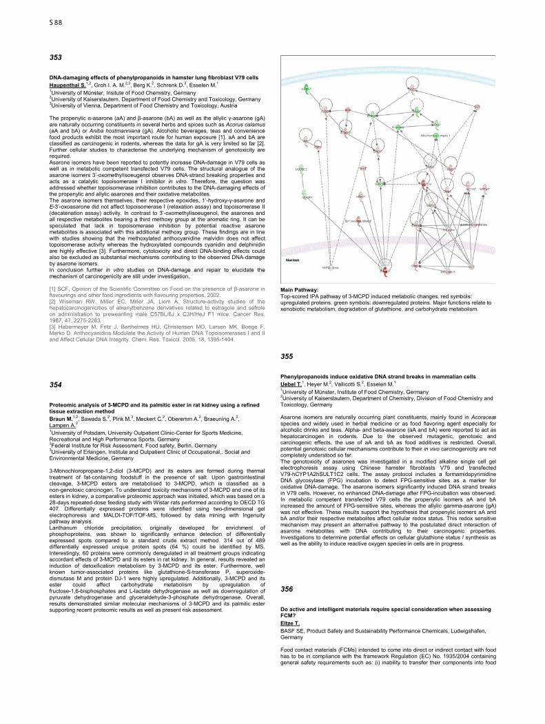
353
DNA-damaging effects of phenylpropanoids in hamster lung fibroblast V79 cells Haupenthal S.1,2, Groh I. A. M.2,3, Berg K.2, Schrenk D.2, Esselen M.1 1University of Münster, Insitute of Food Chemistry, Germany 2University of Kaiserslautern, Department of Food Chemistry and Toxicology, Germany 3University of Vienna, Department of Food Chemistry and Toxicology, Austria The propenylic α-asarone (aA) and β-asarone (bA) as well as the allylic γ-asarone (gA) are naturally occurring constituents in several herbs and spices such as Acorus calamus (aA and bA) or Aniba hostmanniana (gA). Alcoholic beverages, teas and convenience food products exhibit the most important route for human exposure [1]. aA and bA are classified as carcinogenic in rodents, whereas the data for gA is very limited so far [2]. Further cellular studies to characterise the underlying mechanism of genotoxicity are required. Asarone isomers have been reported to potently increase DNA-damage in V79 cells as well as in metabolic competent transfected V79 cells. The structural analogue of the asarone isomers 3’-oxomethylisoeugenol observes DNA-strand breaking properties and acts as a catalytic topoisomerase I inhibitor in vitro. Therefore, the question was addressed whether topoisomerase inhibition contributes to the DNA-damaging effects of the propenylic and allylic asarones and their oxidative metabolites. The asarone isomers themselves, their respective epoxides, 1’-hydroxy-γ-asarone and E-3’-oxoasarone did not affect topoisomerase I (relaxation assay) and topoisomerase II (decatenation assay) activity. In contrast to 3’-oxomethylisoeugenol, the asarones and all respective metabolites bearing a third methoxy group at the aromatic ring. It can be speculated that lack in topoisomerase inhibition by potential reactive asarone metabolites is associated with this additional methoxy group. These findings are in line with studies showing that the methoxylated anthocyanidine malvidin does not affect topoisomerase activity whereas the hydroxylated compounds cyanidin and delphinidin are highly effective [3]. Furthermore, cytotoxicity and direct DNA-binding effects could also be excluded as substantial mechanisms contributing to the observed DNA-damage by asarone isomers. In conclusion further in vitro studies on DNA-damage and repair to elucidate the mechanism of carcinogenicity are still under investigation. [1] SCF, Opinion of the Scientific Committee on Food on the presence of β-asarone in flavourings and other food ingredients with flavouring properties, 2002. [2] Wiseman RW, Miller EC, Miller JA, Liem A. Structure-activity studies of the hepatocarcinogenicities of alkenylbenzene derivatives related to estragole and safrole on administration to preweanling male C57BL/6J x C3H/HeJ F1 mice. Cancer Res. 1987, 47, 2275-2283. [3] Habermeyer M, Fritz J, Barthelmes HU, Christensen MO, Larsen MK, Boege F, Marko D. Anthocyanidins Modulate the Activity of Human DNA Topoisomerases I and II and Affect Cellular DNA Integrity. Chem. Res. Toxicol. 2005, 18, 1395-1404.
354
Proteomic analysis of 3-MCPD and its palmitic ester in rat kidney using a refined tissue extraction method Braun M.1,2, Sawada S.2, Pink M.3, Meckert C.2, Oberemm A.2, Braeuning A.2, Lampen A.2 1University of Potsdam, University Outpatient Clinic-Center for Sports Medicine, Recreational and High Performance Sports, Germany 2Federal Institute for Risk Assessment, Food safety, Berlin, Germany 3University of Erlangen, Institute and Outpatient Clinic of Occupational,. Social and Environmental Medicine, Germany 3-Monochloropropane-1,2-diol (3-MCPD) and its esters are formed during thermal treatment of fat-containing foodstuff in the presence of salt. Upon gastrointestinal cleavage, 3-MCPD esters are metabolised to 3-MCPD, which is classified as a non-genotoxic carcinogen. To understand toxicity mechanisms of 3-MCPD and one of its esters in kidney, a comparative proteomic approach was initiated, which was based on a 28-days repeated-dose feeding study with Wistar rats performed according to OECD TG 407. Differentially expressed proteins were identified using two-dimensional gel electrophoresis and MALDI-TOF/TOF-MS, followed by data mining with Ingenuity pathway analysis. Lanthanum chloride precipitation, originally developed for enrichment of phosphoproteins, was shown to significantly enhance detection of differentially expressed spots compared to a standard crude extract method. 314 out of 489 differentially expressed unique protein spots (64 %) could be identified by MS. Interestingly, 60 proteins were commonly deregulated in all treatment groups indicating accordant effects of 3-MCPD and its esters in rat kidney. In general, results revealed an induction of detoxification metabolism by 3-MCPD and its ester. Furthermore, well known tumor-associated proteins like glutathione-S-transferase P, superoxide-dismutase M and protein DJ-1 were highly upregulated. Additionally, 3-MCPD and its ester could affect carbohydrate metabolism by upregulation of fructose-1,6-bisphosphates and L-lactate dehydrogenase as well as downregulation of pyruvate dehydrogenase and glyceraldehyde-3-phosphate dehydrogenase. Overall, results demonstrated similar molecular mechanisms of 3-MCPD and its palmitic ester supporting recent proteomic results as well as present risk assessment.
Main Pathway: Top-scored IPA pathway of 3-MCPD induced metabolic changes. red symbols: upregulated proteins. green symbols: downregulated proteins. Major functions relate to xenobiotic metabolism, degradation of glutathione, and carbohydrate metabolism.
355
Phenylpropanoids induce oxidative DNA strand breaks in mammalian cells Uebel T.1, Heyer M.2, Vallicotti S.2, Esselen M.1 1University of Münster, Institute of Food Chemistry, Germany 2University of Kaiserslautern, Department of Chemistry, Division of Food Chemistry and Toxicology, Germany Asarone isomers are naturally occurring plant constituents, mainly found in Acoraceæ species and widely used in herbal medicine or as food flavoring agent especially for alcoholic drinks and teas. Alpha- and beta-asarone (aA and bA) were reported to act as hepatocarcinogen in rodents. Due to the observed mutagenic, genotoxic and carcinogenic effects, the use of aA and bA as food additives is restricted. Overall, potential genotoxic cellular mechanisms contribute to their in vivo carcinogenicity are not completely understood so far. The genotoxicity of asarones was investigated in a modified alkaline single cell gel electrophoresis assay using Chinese hamster fibroblasts V79 and transfected V79-hCYP1A2hSULT1C2 cells. The assay protocol includes a formamidopyrimidine DNA glycosylase (FPG) incubation to detect FPG-sensitive sites as a marker for oxidative DNA-damage. The asarone isomers significantly induced DNA strand breaks in V79 cells. However, no enhanced DNA-damage after FPG-incubation was observed. In metabolic competent transfected V79 cells the propenylic isomers aA and bA increased the amount of FPG-sensitive sites, whereas the allylic gamma-asarone (gA) was not effective. These results support the hypothesis that propenylic isomers aA and bA and/or their respective metabolites affect cellular redox status. This redox sensitive mechanism may present an alternative pathway to the postulated direct interaction of asarone metabolites with DNA contributing to their carcinogenic properties. Investigations to determine potential effects on cellular glutathione status / synthesis as well as the ability to induce reactive oxygen species in cells are in progress.
356
Do active and intelligent materials require special consideration when assessing FCM? Eltze T. BASF SE, Product Safety and Sustainability Performance Chemicals, Ludwigshafen, Germany Food contact materials (FCMs) intended to come into direct or indirect contact with food has to be in compliance with the framework Regulation (EC) No. 1935/2004 containing general safety requirements such as: (i) inability to transfer their components into food
S88
that could endanger human health, (ii) change composition of food in an unacceptable way or, (iii) deteriorate its taste or odour. Additionally, Regulation (EC) No. 450/2009 lays down specific rules for active and intelligent materials and articles intended to be used in contact with foodstuffs. Active materials and articles are intended to e.g. prolong the shelf-life by releasing or absorbing substances into or from packed food, while intelligent materials and articles monitor the condition of packed food. These materials interact with food and may evoke changes in the composition or organoleptic characteristics of food which need a special emphasis ensuring that they are without health risk. For their safe use, active and intelligent substances are only authorized when the European Food Safety Authority (EFSA) has performed a risk assessment and issued an opinion on each substance. The presentation will focus on the general toxicological assessment of food contact materials, the kind of active and intelligent materials existing and the special considerations in order to guarantee, that food in contact with active and intelligent materials and articles remains safe, healthy and eatable.
357
Structure-dependent cytotoxicity and nuclear receptor activation of different short-chain perfluorinated acids Brettschneider J.1, Oberemm A.1, Buhrke T.1, Braeuning A.1, Hengstler J. G.2, Lampen A.1 1Federal Institute for Risk Assessment, Effect-based Analytics and Toxicogenomics, Berlin, Germany 2Leibniz Research Centre for Working Environment and Human Factors (IFADO), Systems toxicology, Dortmund, Germany Background: Perfluorooctanesulfonic acid (PFOS) and perfluorooctanoic acid (PFOA) are fully fluorinated, organic compounds (PFCs). They were commonly used in a variety of industrial and commercial products, e. g. fast food containers, raincoats, nonstick cookware, electronics and fire extinguisher foam. These PFCs are extremely persistent and have half-lives in humans ranging from 2 to 9 years. Rodent studies raised high concerns about adverse health effects in humans which led to a voluntary phase-out production of PFOS in 2002 and a stewardship program toward eliminating PFOA from emission and production by 2015 for the primary manufacturers. Due to their shorter half-lives in the human body, shorter PFCs are considered less harmful and are used as PFOS and PFOA substitutes, together with other structurally related compounds. The aim of this study was to reveal toxicity levels of PFOS/PFOA alternatives and to identify structure activity relationships. Methods: Test substances were grouped into sulfonic and carbonic acids: the sulfonic acids PFOS, perfluorohexanesulfonic acid (PFHxS) and perfluorobutanesulfonic acid (PFBS); and the carbonic acids PFOA, perfluorohexanoic acid (PFHxA), perfluorobutanoic acid (PFBA) and two additional, structurally related fluorinated carbonic acids. Substance cytotoxicity was assessed in different established hepatoma cell lines. A HEK-293T cell-based reporter assay was used to investigate the potential to activate nuclear receptors. Results: Cytotoxicity studies revealed that toxicity decreases with decreasing chain length and that sulfonic acids are more cytotoxic than carbonic acids. The two not fully fluorinated compounds showed similar behavior as the other short carbonic acids. No activation was observed for the following nuclear receptors: hFXR, hPXR, hLXRα, hRARα, hRXRα and hPPARδ. All substances, except PFBS, showed high peroxisome proliferator-activated receptor-alpha (PPARα) activation. For the two fluorinated compounds, activation was strongest. These substances, as well as PFOS and PFHxS, acted as transactivators for PPARγ. These findings confirm the assumption that shorter fluorinated compounds are less cytotoxic but show similar modes of action. Thus, additional studies for an appropriate risk assessment for these substances are required.
358
Meta-analytical derivation of an inhalation TTC for the workplace Keller D., Hoffmann-Doerr S. Henkel AG & Co. KGaA, HSA – Corporate Scientific Services, Toxicology, Düsseldorf, Germany The threshold of toxicological concern (TTC) concept defines a generic tolerable exposure for chemicals of unknown toxicity below which the risk of adverse health effects is considered very small. In analogy, a cut-off criterion for occupational exposure could be applied to avoid unnecessary animal testing in those cases where workers are exposed at very low levels. However, so far such a generally accepted threshold level has not been established. This is remarkable, since in other areas the TTC concept has gained wide acceptance for exposure based waiving in risk assessment (food, pharmaceuticals, cosmetics, & chemicals). As an example, in 1995 the U.S. FDA introduced a Threshold of Regulation of 1.5 µg per person and day for trace amounts of food contact substances. A limiting factor of the TTC concept is that the underlying probabilistic approach requires a comprehensive database to cover the chemical diversity. In the meantime, several thousand long-term inhalation DNELs (= Derived No Effect Levels) for workers have been derived within the scope of the European REACH legislation. By statistical evaluation of the DNEL distribution and the resulting percentile values, an inhalation TTC can be defined. Our initial dataset is based on an already existing compilation conducted by the German social accident insurance (DGUV) and contains ca. 3250 DNEL values from REACH registrations. The occurrence of the substances in the database is triggered by their respective tonnage band and thus represents an unbiased selection of relevant chemicals. Certain limitations of the dataset
as well as the influence of specific assumptions regarding the analysis are discussed. These include exclusion criteria for certain substance categories and the impact of local versus systemic effects. U.S. FDA (1995) Food Additives: Threshold of Regulation for Substances Used in Food-Contact Articles; Final Rule. Fed Reg., 60: 36582-36596. July 17 Hauge-Nilsen K, Keller D (2014) Feasibility study: refinement of the TTC concept by additional rules based on in silico and experimental data. Arch Toxicol. 2014 May 14 [Epub ahead of print]
359
Guidance documents for assessment of dermal absorption of pesticides and biocides: Data-based evaluation of current recommendations Kneuer C., Wend K., Lütte S., Herzler M., Martin S., Niemann L., Solecki R. Bundesinstitut für Risikobewertung, Pesticide Safety, Berlin, Germany The extent of dermal absorption represents a major factor determining the outcome of risk assessments for plant protection, biocidal and other products. However, there may be differences in the way the information on dermal absorption is used, possibly causing deviating assessments in regulatory decision making. Although Guidance Documents have been published by OECD and EFSA and other bodies such as SCCS, recommendations therein are not always identical and, in some cases, leave room for interpretation. Finally, refinement of some default assumptions was suggested recently. Therefore, we developed and evaluated a database describing dermal absorption studies for pesticides with regard to identity and physico-chemical properties of the active substance, its concentration, details of exposure, experimental set-up and key outcomes. Currently, the database contains information from 316 reports on 728 items (based on 343 formulations) examined in 679 in vivo, 384 rat in vitro and 490 human in vitro experiments. The high level of recovery of 95-105% recommended by EFSA was met in only 55/60 % (in vitro/in vivo) of datasets, while it was mostly in the range of 90-110% required by OECD. When a relative standard deviation of ≥ 25% as recommend by EFSA and OECD is applied as criterion to identify high variability that requires compensation through use of mean + SD for risk assessment, two thirds of all datasets were judged to contain highly variable data. Comparison of data for serial dilutions confirmed that the approach to bridge dermal absorption data using the dilution factor for correction would be sufficiently conservative. Finally, the default assumptions on dermal absorption for untested formulations of 25/75% depending on concentration of the active substance could essentially be confirmed. Further analyses will be presented.
360
Carbamazepine residues in the aquatic environment – an in-silico approach Mückter H.1, Meyer J.1, Durmaz V.2, Weber M.2, Gudermann T.1 1Walther-Straub-Institut, Toxicology, München, Germany 2Zuse-Institut, Berlin-Dahlem, Germany Introduction: A variety of drug residues have been detected in sewage plant run-offs, rivers and lakes, but also in groundwater and tap water samples. Studies have yet to identify a risk for human health from these contaminants, but adverse health effects have been reported for various species, including fish and birds. It has recently been suggested that for a comprehensive risk assessment toxicologists should also consider transformation products (TPs) of such water contaminants that may arise from abiotic and biotic (metabolic) reactions. With carbamazepine (CBZ), a well-known anticon-vulsant and mood modifier, as the parent drug we tried an in-silico approach to identify TPs that would be of interest due to some mutagenic or carcinogenic toxicophores. Methods: From a literature and database search we picked up 50+ CBZ-TPs. Acute toxicities and mutagenic / carcinogenic properties for these TPs were derived from an expert system analysis using the LAZAR portal (http://lazar.in-silico.ch/) as front-end. Results: The in-silico analysis revealed 4 TPs with suspected mutagenic / carcinogenic properties. Remarkably, CBZ itself was identified by the system (based on animal data) although numerous in-vitro studies have shown that CBZ alone is devoid of mutagenic properties. A possible way out of this dilemma is the detection of 9-acridine-carboxy-aldehyde (CAS RegNr 885-23-4), a recently identified (intermediate) TP of CBZ with known mutagenic properties. Other flagged TPs include the CBZ-o-quinone and acridone. A computer-based simulation of the respective affinities to human CYP isozymes suggested that the predicted mutagenicities may not be relevant for humans because these TPs may be further metabolized to highly water-soluble and virtually non-toxic degradation products. Conclusions: Our in-silico analyses point to acridine derivatives as the underlying cause for the predicted mutagenicity / carcinogenicity of CBZ. The results emphasize that caution is necessary when connecting pattern similarity results to empirical data from poorly defined in-vivo systems. On the other hand, if discordant biological activity results are obtained from in-vivo and in-vitro studies, the missing link is likely one (or more) yet unidentified transformation product that is responsible for the observed adverse effects in vivo. Sinués B et al. (2005) Six mutagenicity assays in exposure biomonitoring of patients receiving carbamazepine for epilepsy or trigeminal neuralgia. Mutation Research 334: 259-265
S89
361
Refinement and enhancement of a database for dermal absorption data Schmeinck S.1, Bitsch A.1, Genth H.2 1Fraunhofer Institute for Toxicology and Experimental Medicine ITEM, Chemical Risk Assessment, Hannover, Germany 2Medical School Hannover, Institute of Toxicology, Germany Dermal absorption is an important factor in regulatory science regarding the registration of chemicals, agrochemicals and cosmetics. The issue has gained importance since it has been discovered that the skin is not completely impenetrable for chemical substances.[1] Different ways to assess dermal absorption range from QSAR models to complex in vivo studies including a complete toxicokinetic examination. The choice of method depends on the question that has to be answered as different systems give different results: absorption as % of applied dose in in vivo studies or permeability coefficient and lag time in infinite dose in vitro studies [1]. Since the OECD has adopted a guideline for assessing dermal penetration in vitro in 2004 the number of in vitro studies is rising continuously. Depending on the chosen method results may vary in reliability and in acceptance by regulatory authorities. Based on data shown by Scholz et al. in 2014 [2] about 4200 data sets from the eChemPortal [3] extended with data from the EDETOX database [4] has been analyzed in depth. In a first approach the references have been reviewed for scientific reliability and categorized in one out of four reliability categories ranging from A as the highest to C as the lowest reliability score. The fourth category D is used for special studies that cannot be compared directly. These are for example repeated dose studies including data on absorption. The data can be used for multiple scenarios: i.e. data for a certain substance or substance class could support read across for registration purposes but also statistic evaluations concerning species or methods used are possible. The first results taken from the data will be presented showing that up to now 62% of the references are in vitro studies and the number of QSAR studies is rising during the past ten years. Nearly one third of all references are studies using rat skin (30%); about 35% used human skin. Regarding the reliability criteria about one third are marked with the highest category A while only 4% were categorized C for unreliable. In summary a large dataset with consistent and reliable data from various exposure scenarios can help to provide a reasonable prediction of dermal absorption which can be achieved by our database. [1]WHO (2006) EHC 235 Dermal absorption. [2]Scholz et al (2014) Naunyn-Schmiedeberg´s Arch Pharmacol 3 (Suppl 1):S86 [3] www.echemportal.org [4] http://edetox.ncl.ac.uk
362
Permanent Senate Commission for the Investigation of Health Hazards of Chemical Compounds in the Work Area - An example about the procedure in threshold derivation and classification of chemical substances Volz N.1, Greim H.2, Hartwig A.1 1Senate Commission on the Investigation of Health Hazards of Chemical Compounds in the Work Area, Karlsruhe, Germany 2Senate Commission on the Investigation of Health Hazards of Chemical Compounds in the Work Area, Freising-Weihenstephan, Germany The Permanent Senate Commission for the Investigation of Health Hazards of Chemical Compounds in the Work Area (MAK Commission) proposes maximum workplace concentrations (MAK values) for volatile chemicals and dusts, biological tolerance values (BAT values), biologische Leitwerte (BLW), biological reference values for workplace substances (BAR) and analytical methods for substances in the air and biological material. Substances which are carcinogenic, germ cell mutagenic, sensitising or absorbed percutaneously or which pose a risk during pregnancy are classified accordingly. The task of the MAK Commission is to provide comprehensive and authoritative information for health and safety professionals and researchers and to give scientific policy advice. On 1 July of every year the proposals for maximum workplace concentrations (MAK values), biological tolerance values (BAT values) and classifications are published in the annual List of MAK and BAT Values and sent to the German Federal Minister of Labour and Social Affairs. The Ministry's Committee on Hazardous Substances (AGS) reviews the proposals and usually recommends that they be adopted in the Hazardous Substances Ordinance. Since 2005 the MAK and BAT data, proposed classifications and associated documentation have been opened for public discussion six months before publication. Each year detailed documentation is published for all proposed MAK values, BAT values and classifications. Since January 2012 the MAK Collection has been available online in German and English. It includes the documentation for the MAK and BAT values and the analytical methods for air and biological material. All publications that have appeared since 1972 are therefore available free of charge in electronic format: www.dfg.de/en/mak http://onlinelibrary.wiley.com/book/10.1002/3527600418/topics(Begründungen/Methoden) http://onlinelibrary.wiley.com/book/10.1002/9783527682010 (MAK- und BAT-Werte-Liste) To gain an insight into the procedure of the MAK Commission the present poster provides an example for the derivation of a MAK value and the allocation in the classification categories.
363
Support for the use of computer-based predictions like quantitative structure-activity relationships (QSAR methods) to avoid animal testing under REACH Escher S.1, Batke M.1, Arning J.2, Brandt M.2, Kupper J.3, Eifert H.3, Wibbertmann A.1 1Fraunhofer ITEM, Registration and Risk assessment, Hannover, Germany 2Umweltbundesamt, IV 2.3 Chemicals, Dessau-Roßlau, Germany 3EurA Consult, Niederlassung Hamburg, Germany The use of alternative methods in risk assessment is requested in different regulatory areas, e.g. REACH Annex XI, in order to minimise animal testing and optimise the use of existing knowledge. The experiences of the regulatory processes reveal that QSAR models are used only rarely for selected endpoints. Alternatives like read across or weight of evidence are thus used more often. The application of alternative approaches in dossiers does not necessarily imply their acceptance by regulatory bodies. Within this project especially small and medium size enterprises (SME) shall be encouraged to use QSAR methods within their registration dossiers e.g. for REACH. The aim is to achieve a broader use and better documentation of QSAR resulting in an increased acceptance by regulatory bodies. Therefore two international workshops will be held (summer 2015 and spring 2016), inviting users, developers and representatives of regulatory agencies. These workshops will address the use of QSAR models within REACH to predict (eco)toxicity properties of chemicals. As it has been shown that the availability of models is not the bottle neck, within the workshops strategies for a professional use of QSAR models will be developed to facilitate: • Evaluation of quality, relevance and reliability of a model by the user • Criteria for a scientific reasoning on the modelled data • Reasonable, transparent and efficient documentation of models and respective predictions The outcome of this project will be summarised in a pragmatic guidance document for small and medium enterprises. By doing this the quality of the QSAR use within registration dossiers will enhance and consequently the acceptance of predictions by regulatory bodies will increase. In preparation of the workshops the status quo for the use of alternative methods is assessed by summarising published data and opinions as well as by collecting experiences from different users (experts, practitioners, experienced beginners) by questionnaires. These results as well as details on the workshop program will be presented to invite further participants. UBA Projekt: FKZ 3714674130
364
REACH Registration Dossiers for High Tonnage Chemicals: A Journey to its Insides Springer A., Sittner D., Herrmann H., Herbst U., Schulte A. Bundesinstitut für Risikobewertung, Chemikalien- und Produktsicherheit, Berlin, Germany The chemical legislation REACH was adopted to improve the protection of human health and the environment by increasing the knowledge about chemicals that are produced, marketed and used in the European Union. The obligation to register their chemicals at the European Chemicals Agency (ECHA) lies with the companies, aiming at providing sufficient information for hazard and risk assessment of chemicals as well as their safety management. The information requirements and possible adaptations for chemicals produced with equal and above 1000 tons per year are set out in the REACH Annexes VII to XI and depend basically on their production volume. Registrants are obliged to consider all existent data to fulfill the requirements. If information gaps are identified, new data are to be generated. For the registration of chemicals produced above 1000 tonnes per year the full set of information, in particular, on developmental and reproductive toxicity according to the OECD test guidelines has to be submitted. The examination of the data availability for these chemicals seems, therefore, of high priority to identify areas for further action, e.g. to fill the data gaps. Within the scope of a project, the Federal Institute for Risk Assessment (BfR) and the Environmental Agency (UBA) developed a systematic web-based scheme in order to assess the availability of the required data. In this regard, the data on complex environmental and human health endpoints of about 1800 lead and individual dossiers of high tonnage chemicals were checked in a standardized manner for compliance with the appropriate REACH annexes. The reviewed endpoints comprised repeated dose toxicity, developmental and reproductive toxicity and genetic toxicity regarding health protection as well as degradation, accumulation, ecotoxicity and exposure into the environment. As a result, the dossiers have been categorized into three domains, indicating incompleteness of decision making (“complex”), “compliance” or “non-compliance” according to the developed checking scheme. Furthermore, exemplary dossiers of incomplete decision making were individually evaluated. The project revealed that the majority of dossiers contain incomplete or unreliable data in at least one of the regarded endpoints. The poster will present the overall results and first results in relation to human health endpoints.
S90
Author Index
A
Abdel-Aziz H. ........................................................................................ 227, 178, 177, 111 Agouri S. ....................................................................................................................... 033 Ahles A. ........................................................................................................................ 001 Aigner A. ....................................................................................................................... 301 Akpınar E. ..................................................................................................................... 031 Aktories K. ............................................ 337, 320, 318, 316, 311, 299, 149, 039, 035, 034 Albers A. ....................................................................................................................... 194 Albers T. ....................................................................................................................... 197 Albrecht C. ............................................................................................................ 279, 278 Albus C. ........................................................................................................................ 238 Aldenhoff J. ................................................................................................................... 129 Alenina N. ..................................................................................................................... 122 Alexandrov A. ............................................................................................................... 082 Alexiou C. ..................................................................................................................... 335 Alonso Cañizal M. C. .................................................................................................... 002 Alpert G. ....................................................................................................................... 260 Al-Sabah S. .................................................................................................................. 030 Aminossadati B. ............................................................................................................ 209 Ammer H. ..................................................................................................................... 024 Andres C. ...................................................................................................................... 180 Annala S. ...................................................................................................................... 073 Arning J. ....................................................................................................................... 363 Arnold P. ............................................................................................................... 126, 124 Art J. ............................................................................................................................. 109 Ashikaga T. ................................................................................................................... 306 Aslan S. ........................................................................................................................ 195 Assmann J. C. ...................................................................................................... 198, 127 Atmaca H. T. ................................................................................................................. 031 Augspach A. ................................................................................................................. 034 Aulitzky W. .................................................................................................................... 189 Aumann A. .................................................................................................................... 305 Aumann D. .................................................................................................................... 117 Avramopoulos P. .......................................................................................................... 152 Ayhan S. ....................................................................................................................... 150
B
Babendreyer A. ............................................................................................................. 300 Bachmann H. S. ................................................................................................... 217, 209 Bader M. ....................................................................................................... 122, 101, 100 Badimon L. ................................................................................................................... 081 Bähre H. ....................................................................................................................... 053 Baker S. D. ................................................................................................................... 087 Bakuradze T. ................................................................................................................ 241 Balkow A. ...................................................................................................................... 003 Balschun D. .................................................................................................................. 127 Balszuweit F. ................................................................................................................ 027 Baltus D. ....................................................................................................................... 041 Ban Z. ................................................................................................................... 140, 131 Bankoglu E. E. .............................................................................................................. 290 Barg M. ......................................................................................................................... 219 Barknowitz G. ............................................................................................................... 271 Barth H. ................................................................................................................ 327, 324 Bartkuhn M. .................................................................................................................. 104 Bas M. .......................................................................................................................... 033 Basso F. ....................................................................................................................... 096 Bastian L. ...................................................................................................................... 303 Batke M. ....................................................................................................................... 363 Bauer F. ........................................................................................................................ 109 Bausch A. E. ................................................................................................................. 071 Bayer J. K. ............................................................................................................ 154, 092 Bayraktutan Z. ...................................................................................................... 031, 031 Becker S. ...................................................................................................................... 128 Bedke J......................................................................................................................... 220 Beer L. - A. ................................................................................................................... 328 Behnke F. ..................................................................................................................... 109 Beitz E. ......................................................................................... 197, 091, 090, 089, 084 Benz V. ................................................................................................................. 140, 131 Benzin A. E. .................................................................................................................. 202 Berg K................................................................................................................... 353, 351 Berger F. I. .................................................................................................................... 304 Berlien H. - P. ............................................................................................................... 287 Berm E. J. J. ................................................................................................................. 210 Bernau M. ..................................................................................................................... 097 Berndt S. ....................................................................................................................... 309 Berrisch S. ............................................................................................................ 053, 042 Berte N. ........................................................................................................................ 292 Besche V. ..................................................................................................................... 109 Besik V. ........................................................................................................................ 093 Biedermann T. .............................................................................................................. 046 Biel M.................................................................................................................... 064, 060 Bien-Möller S. ............................................................................................................... 298 Bierwisch A. .................................................................................................................. 319 Birnbaumer L. ....................................................................................................... 132, 058 Bischoff I. ...................................................................................................... 181, 139, 102 Bisha M......................................................................................................................... 033 Biskup S. ...................................................................................................................... 214 Bitsch A. ............................................................................................................... 361, 285 Bitter A. ......................................................................................................................... 211 Blank A. ................................................................................................................ 233, 082 Blaszkewicz M. ............................................................................................. 348, 251, 248
Blei T.............................................................................................................................096 Blömeke B. ...........................................................................................................256, 255 Bock A. ......................................................................................................... 043, 010, 004 Bock S. .................................................................................................................105, 089 Bode C. .........................................................................................................................112 Bodmann E. - L. ............................................................................................................017 Boege F. .......................................................................................................................020 Böhler P. .......................................................................................................................289 Böhm A. ................................................................................................................298, 192 Böhm R. ........................................................................................................ 230, 229, 129 Böhmert L. ............................................................................................................323, 272 Bokelmann K. .......................................................................................................216, 077 Boknik P. ...............................................................................................................159, 130 Bolbrinker J. ..........................................................................................................239, 182 Bollmann F. ...................................................................................................................109 Bonvicini G. ...................................................................................................................263 Bopp T. .........................................................................................................................296 Bormann S. ...................................................................................................................260 Born J. ..........................................................................................................................117 Bornholz B. ...................................................................................................................020 Bösche D. .....................................................................................................................174 Böser E. ........................................................................................................................033 Böttcher K. ....................................................................................................................297 Bracher F. .............................................................................................................102, 060 Braeuning A. ......................................... 357, 354, 332, 323, 315, 302, 274, 273, 259, 240 Brandt M. ......................................................................................................................363 Brauch H. ......................................................................................................................215 Braun M. ...............................................................................................................354, 070 Breckwoldt K. ................................................................................................................202 Brede M. .......................................................................................................................026 Breit A. ..................................................................................................................093, 027 Brekle C. .......................................................................................................................130 Brenowitz M. .................................................................................................................040 Brettschneider J. ...........................................................................................................357 Brigadski T. ...................................................................................................................112 Brigelius-Flohé R. .........................................................................................................271 Brinkmann J. .........................................................................................................314, 232 Brix S. ...................................................................................................................140, 131 Brockmöller J. ....................................................................................... 218, 216, 079, 077 Bröderdorf S. ................................................................................................................085 Bröker J. .......................................................................................................................058 Bronder E. .....................................................................................................................231 Bros M. .........................................................................................................................109 Brosda J. .......................................................................................................................114 Brouwer S. ....................................................................................................................059 Brozda N. ......................................................................................................................316 Brück S. ........................................................................................................................078 Bruckmueller H. ............................................................................................ 188, 175, 086 Bruhn O. ....................................................................................................... 188, 185, 080 Brummer T. ...................................................................................................................299 Brüning T. .............................................................................................................339, 303 Bruns D. ........................................................................................................................058 Bryk O. ..........................................................................................................................339 Büch T. .........................................................................................................................027 Buchwalow I. B. ............................................................................................................158 Buhrke T. ..............................................................................................................357, 350 Bünemann M. ....................................................................................... 030, 017, 016, 001 Bünger J. ......................................................................................................................339 Burchardt K. .................................................................................................. 281, 280, 275 Bürger H. ......................................................................................................................348 Burges A. ......................................................................................................................209 Burk O. ..........................................................................................................................211 Burleson F. ...................................................................................................................305 Busch D. .......................................................................................................................244 Buschner C. ..................................................................................................................078 Butcher A. .....................................................................................................................030 Büttner H. - J. ........................................................................................................162, 161 Butz E. ..........................................................................................................................064 Butzbach K. ..................................................................................................................183
C
Cabrerizo F. M. .............................................................................................................183 Çadırcı E. ......................................................................................................................031 Camacho Londoño J. ....................................................................................................132 Camacho Londoño J. E. ...............................................................................................132 Cardinaux J. - R. ...........................................................................................................145 Carls A. ......................................................................................................... 233, 226, 204 Cartus A. .......................................................................................................................267 Cascorbi I. ............................................................. 200, 188, 185, 184, 175, 115, 086, 080 Caspers P. ....................................................................................................................255 Çayır Y. .........................................................................................................................031 Chen C. - C. ..........................................................................................................064, 060 Chen C. - Y. ..................................................................................................................109 Chen H. .........................................................................................................................198 Chen J. .........................................................................................................................079 Chen J. - N. ...................................................................................................................157 Chen X. .................................................................................................................079, 010 Cheng L. .......................................................................................................................270 Chirinda B. ....................................................................................................................004 Chovolou Y. ..................................................................................................................346 Chubanov V. .........................................................................................................063, 061 Cicha I...........................................................................................................................335 Coenraads P. - J. ..........................................................................................................255
S91
Cordt J. ......................................................................................................................... 121 Corteville C. .................................................................................................................. 290 Creutzenberg O. ................................................................................... 283, 276, 273, 263 Culman J. ..................................................................................................... 184, 115, 113 Culmsee C. ........................................................................................................... 067, 062 Czock D. ....................................................................................................................... 204 Czulkies B. .................................................................................................................... 311 Czupalla C. ................................................................................................................... 040
D
D´Hooge R. ................................................................................................................... 127 Dabrowski A. ................................................................................................................ 265 Dahl B. .......................................................................................................................... 106 Dahlinger D. .................................................................................................................. 195 Dai B. ............................................................................................................................ 102 Dallanoce C. ................................................................................................................. 005 Damm G. .............................................................................................................. 336, 333 Dams N. ........................................................................................................................ 167 Dantzer F. ..................................................................................................................... 293 Dao T. ........................................................................................................................... 033 Dasenbrock C. ...................................................................................... 342, 282, 257, 223 David J.......................................................................................................................... 274 de Stooper B. ................................................................................................................ 152 de Wit C. ....................................................................................................................... 055 Decher N. ............................................................................................................. 067, 062 Decker M. ..................................................................................................................... 010 Deckert J. ..................................................................................................................... 120 Dedman J. R. ................................................................................................................ 142 Degen G. H. .......................................................................................................... 251, 248 Deharde D. ................................................................................................................... 333 Deiana C. ...................................................................................................................... 278 Demin A. ....................................................................................................................... 005 Demir &. ........................................................................................................................ 031 Demir R. ....................................................................................................................... 031 Dettbarn G. ................................................................................................................... 347 Dewenter M. ................................................................................................................. 166 Dhayade S. ................................................................................................................... 046 Dick L. S. ...................................................................................................................... 133 Diel P. ........................................................................................................................... 096 Dieter R. ....................................................................................................................... 071 Dietrich A. ............................................................................................. 132, 075, 066, 058 Dietrich C. ..................................................................................................................... 331 Dietze S. ....................................................................................................................... 114 Diewock T. .................................................................................................................... 185 Dirsch V. M. .................................................................................................................. 109 Dittmar F. ...................................................................................................................... 044 Dittrich-Breiholz O. ....................................................................................................... 104 Dmochewitz-Kück L. ..................................................................................................... 327 Dobrev D. ..................................................................................................... 081, 072, 068 Dobrowinski H. ............................................................................................................. 045 Dolga A. ................................................................................................................ 067, 062 Donners M. ................................................................................................................... 170 Dörsam B. ............................................................................................. 293, 291, 269, 268 Douros A. ...................................................................................................................... 231 Dreizehnter L. ............................................................................................................... 134 Drerup K. ...................................................................................................................... 080 Drews G. ............................................................................................................... 099, 032 Drexler M. K. ................................................................................................................. 134 Dreymueller D. .............................................................................................. 300, 170, 015 Driesen T. ..................................................................................................................... 154 Driever W. ..................................................................................................................... 320 Drozdzik M. ........................................................................................................... 244, 086 du Bois A. ..................................................................................................................... 209 Düfer M. ................................................................................................ 099, 076, 059, 032 Duque Escobar J. ......................................................................................................... 094 Durmaz V. ..................................................................................................................... 360
E
Ebert B.......................................................................................................................... 329 Ebert D. ........................................................................................................................ 329 Ebmeyer J. ................................................................................................................... 272 Eckerle S. ..................................................................................................................... 320 Edalat A. ....................................................................................................................... 032 Edwards A. ................................................................................................................... 305 Efferth T. ............................................................................................................... 268, 178 Eggeling C. ................................................................................................................... 230 Ehrenmann J. ............................................................................................................... 013 Eich M................................................................................................................... 292, 288 Eidens M. ...................................................................................................................... 212 Eifert H.......................................................................................................................... 363 Eisenbarth E. ................................................................................................................ 282 Eisenbrand G. ............................................................................................................... 352 El-Abhar H. ................................................................................................................... 177 El-Armouche A. .................................................................................................... 201, 166 El-Awaad E. .................................................................................................................. 196 Ellenberger P. ............................................................................................................... 330 Eltze T. ......................................................................................................................... 356 Emmrich K. ................................................................................................................... 191 Endo S. ......................................................................................................................... 160 Engelhardt S. ................................................................................................ 168, 152, 001 Engeli S. ............................................................................................................... 206, 054 Epe B. ................................................................................................................... 296, 183 Eriksson P. - O. ............................................................................................................ 088 Erkel G.......................................................................................................................... 108 Erker T. ......................................................................................................................... 109
Ernst K. .........................................................................................................................324 Eschenhagen T. ............................................................................................ 208, 202, 145 Escher S. ......................................................................................................................363 Escrig Vidal A. ..............................................................................................................263 Eskandar J. ...................................................................................................................159 Esselen M. ............................................................................................ 355, 353, 352, 351 Essmann F. ...................................................................................................................135 Etzrodt J. .......................................................................................................................050 Eucker J. .......................................................................................................................182
F
Fabian E. ...................................................................................................... 304, 264, 254 Fabisch C. .....................................................................................................................069 Faerber L. .....................................................................................................................056 Fahrer J. ............................................................................................... 293, 291, 269, 268 Farouk M. ......................................................................................................................177 Fasching P. ...................................................................................................................215 Faust D. ........................................................................................................................331 Fehr M. .........................................................................................................................305 Fehr S. ..........................................................................................................................214 Fehrenbacher B. ...........................................................................................................135 Fehrmann E. .................................................................................................................164 Feigl P...........................................................................................................................327 Feil R. ........................................................................................... 135, 055, 051, 046, 045 Feil S............................................................................................................. 135, 055, 046 Feldmann K. .................................................................................................................154 Fels B............................................................................................................................160 Fender A. C. ................................................................................................. 137, 136, 092 Feng Y. .................................................................................................................138, 006 Ferioli S. ................................................................................................................063, 061 Ferrah O. ......................................................................................................................228 Fietz S. ..........................................................................................................................112 Fink H. ..................................................................................................................122, 114 Fischer A. ......................................................................................................................070 Fischer A. W. C. ............................................................................................................050 Fischer J. W. ................................................................. 176, 154, 147, 144, 137, 133, 092 Fischer M. .............................................................................................................342, 049 Fischmeister R. .............................................................................................................166 Fisel P. ..........................................................................................................................220 Fleischer M. ..................................................................................................................212 Flockerzi V. ...........................................................................................................132, 058 Florian A. ......................................................................................................................128 Florian S. ......................................................................................................................271 Foertsch L. ....................................................................................................................305 Föllmann W. ..................................................................................................................248 Forst A. - L. ...................................................................................................................075 Förstermann U. .............................................................................................................109 Foryst-Ludwig A. ...................................................................................................140, 131 Foth H. ..........................................................................................................................284 Frank D. ........................................................................................................................107 Frank M. ................................................................................................................289, 190 Franklin S. .....................................................................................................................157 Frechen S. ............................................................................................................203, 195 Freese P. ......................................................................................................................212 Freichel M. ............................................................................................................132, 058 Freitag-Wolf S. ..............................................................................................................229 Frenzel F. ......................................................................................................................350 Frericks M. ....................................................................................................................264 Freudenberger T. ..........................................................................................................154 Freund L. ......................................................................................................................321 Freyer N. .......................................................................................................................333 Frick J. ..........................................................................................................................228 Fried C. .........................................................................................................................196 Friedrich R. P. ...............................................................................................................335 Frisan T. ........................................................................................................................291 Fritz G. .................................................................................. 334, 291, 261, 260, 194, 036 Fritz P. ..........................................................................................................................215 Frombach J. ..................................................................................................................341 Fuhr U. ..................................................................................................................203, 195 Fürst R. ......................................................................................................... 181, 139, 102
G
Ganzenberg K. ..............................................................................................................259 Garbe E. .......................................................................................................................231 García-López P. ............................................................................................................076 Garg J. ..........................................................................................................................006 Gawaz M. ......................................................................................................................055 Gebhardt M. ..................................................................................................................287 Geerts A. .......................................................................................................................166 Geertz B. .......................................................................................................................145 Gekle M. .......................................................................................................................158 Genth H. .......................................................................................................................361 Gerberick F. ..................................................................................................................305 Gergs U. ....................................................................................... 158, 153, 028, 018, 011 Gerhard R. ............................................................................................................328, 310 Gerke V. ........................................................................................................................048 Germer C. T. .................................................................................................................290 Ghezelbash S. ..............................................................................................................081 Ghigo A. ........................................................................................................................040 Giesen J. .......................................................................................................................047 Gilsbach R. ................................................... 225, 222, 221, 219, 169, 156, 151, 149, 148 Glahn F. ........................................................................................................................284 Glatt H. ..................................................................................................................271, 245 Glatzel D. ......................................................................................................................139 Gliesche D. ...................................................................................................................297
S92
Gnad T.......................................................................................................................... 007 Göbel P......................................................................................................................... 168 Göder A. ....................................................................................................................... 269 Göder R. ....................................................................................................................... 129 Goedhart J. ................................................................................................................... 026 Gohlke P. ...................................................................................................................... 115 Gohlsch K. .................................................................................................................... 308 Gok S............................................................................................................................ 150 Golka K. ........................................................................................................................ 348 Gonzales M. M. ............................................................................................................ 183 Gorressen S. ................................................................................................................ 147 Goryachev D. ................................................................................................................ 236 Gottwalt K. .................................................................................................................... 204 Goy S.................................................................................................................... 328, 310 Graemer M. .................................................................................................................. 008 Grandoch M. ......................................................................... 176, 154, 144, 137, 133, 092 Granitzny A. .................................................................................................................. 257 Greim H. ....................................................................................................................... 362 Greinacher A. ............................................................................................................... 085 Griesbacher T. .............................................................................................................. 237 Grimm C. M. ......................................................................................................... 064, 060 Groeters S. ................................................................................................................... 277 Groh I. A. M. ......................................................................................................... 353, 241 Grohmann L. ......................................................................................................... 249, 242 Groll N. ......................................................................................................................... 315 Gromadzinska J. ........................................................................................................... 347 Gross S. ........................................................................................................................ 138 Grossmann C. .............................................................................................................. 158 Groth E. ................................................................................................................ 300, 170 Grube M. ....................................................................................................... 175, 085, 083 Grune J. ................................................................................................................ 140, 131 Grüning B. ............................................................................................................ 222, 221 Gudermann T. .............................................. 360, 301, 134, 093, 075, 066, 063, 061, 027 Gundert-Remy U. ......................................................................................................... 252 Günther S. .................................................................................................................... 219 Günther T. .................................................................................................................... 009 Gurpinar T. ................................................................................................... 235, 234, 098 Gurrola-Díaz C. ............................................................................................................ 076 Guth K. ................................................................................................................. 349, 306 Gutzki F. ....................................................................................................................... 125 Guzmann R. ................................................................................................................. 058
H
Haack M. ...................................................................................................................... 271 Haag M. ........................................................................................................................ 082 Haas K. ......................................................................................................................... 110 Haase A. ....................................................................................................................... 270 Haben M. ...................................................................................................................... 352 Hackert K. ..................................................................................................................... 141 Haefeli W. ............................................................................................. 233, 226, 204, 082 Haenisch S. .................................................................................. 200, 188, 185, 175, 086 Haeusgen W. ........................................................................................................ 180, 121 Hafner S. ...................................................................................................................... 186 Hagedorn T. .................................................................................................................. 057 Hagemann S. ........................................................................................................ 326, 321 Hagen P. ............................................................................................................... 085, 083 Hajnóczky G. ................................................................................................................ 157 Hak E. ........................................................................................................................... 210 Halter R. ....................................................................................................................... 223 Halwachs S. .................................................................................................................. 308 Hamann U. ................................................................................................................... 215 Hamel E. ....................................................................................................................... 127 Hammann N. ................................................................................................................ 148 Hammer E. ................................................................................................................... 085 Hammer H. ................................................................................................................... 265 Hammer K. ................................................................................................................... 132 Hammes H. - P. ............................................................................................................ 138 Hampel S. ..................................................................................................................... 282 Hampl V. ....................................................................................................................... 301 Handler N. .................................................................................................................... 109 Handschick K. ............................................................................................................... 104 Hanke W. ...................................................................................................................... 347 Hansen A. ..................................................................................................................... 202 Hansen F. K. ................................................................................................................. 033 Hansen T. ............................................................................................. 283, 282, 276, 257 Härdle L. ....................................................................................................................... 109 Häring H. - U. ................................................................................................................ 037 Harteneck C. ................................................................................................................. 040 Hartmann S. ................................................................................................................. 146 Hartwig A. ..................................................................................................................... 362 Hartwig C. ............................................................................................................. 107, 044 Hasenpusch D. ............................................................................................................. 094 Häsler R. ....................................................................................................................... 175 Haupenthal S. ............................................................................................................... 353 Hauser K. ...................................................................................................................... 238 Hausmann R. ................................................................................................................ 065 Haustein M. .................................................................................................................. 190 Havemann C. ................................................................................................................ 298 Havrankova J. ............................................................................................................... 187 Hedderich J. ................................................................................................................. 129 Heeren J. ...................................................................................................................... 050 Heidebrecht D. .............................................................................................................. 230 Heijman J. ..................................................................................................... 081, 072, 068 Hein L. .......................................................... 225, 222, 221, 219, 169, 156, 151, 149, 148 Heinick A. ..................................................................................................... 164, 142, 048 Heinisch N. ................................................................................................................... 176
Heinkele G. ...................................................................................................................082 Heise T. ........................................................................................................................313 Heiss C. ........................................................................................................................154 Hellack B. ......................................................................................................................279 Hemmasi S. ..................................................................................................................311 Hengstler J. G. .............................................................................................. 357, 348, 248 Heni H. ..................................................................................................................318, 035 Henke J. ................................................................................................................109, 108 Henkler F. .............................................................................................................314, 312 Hennen J. .....................................................................................................................256 Hennenlotter J. .............................................................................................................220 Henninger C. .........................................................................................................194, 036 Herbarth L. ....................................................................................................................232 Herbst F. .......................................................................................................................033 Herbst U. .......................................................................................................................364 Herdegen T. .......................................................................... 230, 229, 180, 129, 121, 113 Hermanns C. .................................................................................................................301 Herold M. ......................................................................................................................264 Herrlinger Y. ..................................................................................................................068 Herrmann H. .................................................................................................................364 Herrmann S. .........................................................................................................143, 049 Hertel M. .......................................................................................................................334 Herzig S. ....................................................................................... 239, 238, 196, 073, 069 Herzler M. .....................................................................................................................359 Herzog S. ......................................................................................................................298 Hess F. - M. ..................................................................................................................170 Hessel S. ...................................................................................................... 325, 322, 258 Heubach Y. ...................................................................................................................302 Heyer M. .......................................................................................................................355 Hiemisch A. ...................................................................................................................223 Hilfiker-Kleiner D. ..........................................................................................................042 Hillemanns P. ................................................................................................................209 Hiller T. .........................................................................................................................333 Himmler K. ....................................................................................................................142 Hintzpeter J. ..................................................................................................................243 Hintzsche H. .................................................................................................................253 Hinz B. ..................................................................................................................191, 190 Hinz L............................................................................................................................001 Hirsch E. .......................................................................................................................040 Hirt M. N. .......................................................................................................................202 Hochholzer W. ..............................................................................................................162 Höcker J. .......................................................................................................................129 Hoerschelmann A. ........................................................................................................117 Hoffmann C. ..........................................................................................................026, 002 Hoffmann L. S. ..............................................................................................................050 Hoffmann M. .................................................................................................................109 Hoffmann W. .................................................................................................................298 Hoffmann-Doerr S. ........................................................................................................358 Hofman J. .....................................................................................................................187 Hofmann J. ...................................................................................................................243 Hofmann K. ...................................................................................................................066 Hofmann T. ...................................................................................................................061 Hofmann U. ...........................................................................................................224, 082 Hohagen F. ...................................................................................................................129 Hohmann N. .................................................................................................. 233, 226, 204 Holm T. .........................................................................................................................298 Holm-Bertelsen J. .................................................................................................089, 084 Holze J. .........................................................................................................................010 Holzgrabe U. .........................................................................................................010, 005 Homann S. ....................................................................................................................133 Hommers L. ..................................................................................................................120 Honarvar N. ..........................................................................................................349, 306 Honnen S. .............................................................................................................334, 260 Honrath B. .............................................................................................................067, 062 Honscha W. ..................................................................................................................308 Hoppe R. .......................................................................................................................215 Hornung A. ....................................................................................................................335 Hoser S. ........................................................................................................................227 Huang J. .......................................................................................................................157 Hübner F. ......................................................................................................................208 Hucho T. .......................................................................................................................196 Hülsenbeck J. ...............................................................................................................291 Hümmert M. ..................................................................................................................165 Huober J. ......................................................................................................................209 Husser X. ..............................................................................................................142, 048 Hussner J. .....................................................................................................................297 Hutzler C. ...................................................................................................... 312, 287, 286
I
Ibadova G. ....................................................................................................................213 Ibánez García M. J........................................................................................................263 Ihring J. .........................................................................................................................215 Isenegger T. ..................................................................................................................297
J
Jacob T. ........................................................................................................................115 Jaekel S. .......................................................................................................................143 Jagow J. ........................................................................................................................003 Jahn A...........................................................................................................................334 Janek S. ........................................................................................................................140 Jank T. ..................................................................................................................320, 316 Janko C. ........................................................................................................................335 Jatho A. .................................................................................................................146, 041 Jaurich H. ......................................................................................................................291 Jaworska J. ...................................................................................................................306
S93
Jedlitschky G. ....................................................................................................... 085, 083 Jia J. ............................................................................................................................. 344 Johnen G. ..................................................................................................................... 339 Jordan J. ....................................................................................................... 232, 206, 054 Juling S. ................................................................................................ 323, 274, 273, 272 Jung F........................................................................................................................... 011 Jurida L. ........................................................................................................................ 104 Jurowich C. ................................................................................................................... 290 Just I. ............................................................................................ 328, 326, 321, 310, 309
K
Kaczmarek-Hajek K. ..................................................................................................... 074 Kadkina V. .................................................................................................................... 213 Kaehler M. .................................................................................................... 188, 185, 080 Kaesler S. ..................................................................................................................... 046 Kaetzel M. A. ................................................................................................................ 142 Kaever V. ...................................................................................................... 125, 053, 042 Käfferlein H. .................................................................................................................. 303 Kahlert K. ...................................................................................................................... 155 Kaina B. ................................................................................ 293, 292, 291, 288, 269, 268 Kamp H......................................................................................................................... 264 Kappenstein O. ............................................................................................................. 286 Karle J. ......................................................................................................................... 182 Kästner L. ..................................................................................................................... 132 Katus H. A. ................................................................................................................... 155 Kayalar H. ..................................................................................................................... 150 Kazmaier U. .................................................................................................................. 181 Kegel V. ........................................................................................................................ 333 Keiser M. ...................................................................................................................... 344 Kelber O. ...................................................................................................... 227, 177, 111 Keller D. ........................................................................................................................ 358 Keller F. ........................................................................................................................ 228 Keller J.......................................................................................................................... 277 Keller M. ....................................................................................................................... 060 Kelm M. ........................................................................................................................ 154 Kenet S. ........................................................................................................................ 051 Kern C. ......................................................................................................................... 314 Keyl C. .......................................................................................................................... 161 Khan A. P. .................................................................................................................... 068 Khan M. A. .................................................................................................................... 123 Khayyal M. T. ................................................................................................................ 177 Khobta A. ...................................................................................................................... 294 Khosravani F. ....................................................................................................... 033, 012 Kießig M. ...................................................................................................................... 336 Kietzmann M. ................................................................................................ 338, 207, 205 Kilic A............................................................................................................................ 003 Kimmig R. ..................................................................................................................... 209 Kintscher U. .......................................................................................................... 140, 131 Kirchhefer U. ................................................................................................. 159, 130, 048 Kisiela M. ...................................................................................................................... 329 Kitsera N. ...................................................................................................................... 294 Klassen S. .................................................................................................................... 173 Klatt C. .......................................................................................................................... 144 Klein H. - J. ........................................................................................................... 230, 229 Klein K. ................................................................................................................. 214, 211 Klein L........................................................................................................................... 114 Kleine-Ostmann T. ........................................................................................................ 253 Kleinert H. ............................................................................................................. 109, 108 Kleinmanns K. .............................................................................................................. 309 Klempin F. .................................................................................................................... 122 Klenk C. ........................................................................................................................ 013 Kless A. ........................................................................................................................ 065 Kliewer A. ..................................................................................................................... 014 Klimpel A. ..................................................................................................................... 231 Klingler P. ..................................................................................................................... 296 Klipper W. ..................................................................................................................... 249 Klöckner J. .................................................................................................................... 010 Klopfleisch R. ........................................................................................................ 140, 131 Knappe P. ..................................................................................................................... 272 Kneba M. ...................................................................................................................... 185 Knebel J. W. ................................................................................................................. 257 Kneuer C. ............................................................................................................. 359, 308 Kneuer K. ...................................................................................................................... 265 Köbele C. .............................................................................................................. 222, 221 Koch H. ......................................................................................................................... 346 Koch W. ........................................................................................................................ 285 Koeberle A. ................................................................................................................... 103 Koenen A. ............................................................................................................. 170, 015 Koepsell H. ................................................................................................................... 216 Koesling D. ................................................................................................................... 047 Kojda G................................................................................................................. 033, 012 Kojima N. ...................................................................................................................... 160 Kok R. M. ...................................................................................................................... 210 Koks C. ......................................................................................................................... 292 Kolb C. .......................................................................................................................... 111 Kolkhof P. ..................................................................................................................... 140 Kolle S. ......................................................................................................... 349, 307, 306 Kolling J. ....................................................................................................................... 278 Kollotzek F. ........................................................................................................... 315, 302 Königshoff M. ................................................................................................................ 066 Korkmaz M. .................................................................................................................. 098 Köth J. .......................................................................................................................... 069 Kouchekmanesch A. ..................................................................................................... 117 Kracht M. ...................................................................................................................... 104 Kraft M. ......................................................................................................................... 346 Krämer M. ..................................................................................................................... 045
Krasel C. ...............................................................................................................030, 016 Kraus A. ........................................................................................................................293 Krause E. ..............................................................................................................323, 040 Kravchenko I. ................................................................................................................213 Krennrich G. ..................................................................................................................264 Krett A. - L. ....................................................................................................................017 Kretz O. .........................................................................................................................151 Kreutz R. ...............................................................................................................231, 182 Krippeit-Drews P. ..........................................................................................................032 Kroemer H. K. ...............................................................................................................085 Kroesen S. ....................................................................................................................346 Krohmer A. ....................................................................................................................116 Kromrey M. - L. .............................................................................................................085 Kruck S. ........................................................................................................................220 Krüger K. .......................................................................................................................261 Kubick S. .......................................................................................................................337 Kühl J. ...........................................................................................................................329 Kuhlbusch T. .................................................................................................................279 Kuhlmann D. .................................................................................................................065 Kuhlmann J. D. .............................................................................................................209 Kulling S. .......................................................................................................................096 Kullmann M. ..................................................................................................................193 Kumarswamy R. ...........................................................................................................152 Kunak C. S. ...................................................................................................................031 Künstler B. ....................................................................................................................018 Kupper J. ......................................................................................................................363 Küpper J. ......................................................................................................................298 Kuron D. ........................................................................................................................218 Kurrat A. ........................................................................................................................096 Kurz T. ..........................................................................................................................033 Küttler K. .......................................................................................................................277 Kwon O. ................................................................................................................157, 134
L
Laidig F. ........................................................................................................................232 Lajqi T. ..........................................................................................................................105 Lalkowski G. .................................................................................................................274 Lämmle S. .....................................................................................................................166 Lampen A. ............................................ 357, 354, 350, 325, 323, 322, 274, 273, 272, 258 Landfester K. ................................................................................................................110 Landsiedel R. ................................................................ 349, 307, 306, 305, 304, 277, 254 Lang S. .........................................................................................................................299 Lange S. .......................................................................................................................298 Langer H. ......................................................................................................................055 Lanzerstorfer P. ............................................................................................................030 Lassmann S. .................................................................................................................034 Lau C. B. S. ..................................................................................................................270 Laufs U. ........................................................................................................................132 Laux P...........................................................................................................................287 Leggewie S. ..................................................................................................................162 Lehmann A. ..................................................................................................................019 Lehmann C. ..................................................................................................................271 Lehners M. ....................................................................................................................045 Leiss V. .........................................................................................................................037 Lemcke T. .....................................................................................................................094 Lemoine H. ...........................................................................................................070, 020 Lemoine L. ....................................................................................................................250 Lenhardt I. .....................................................................................................................199 Lenze D. ...............................................................................................................322, 258 Letuffe-Brenière D.........................................................................................................202 Leung P. C. ...................................................................................................................270 Leuthold P. ....................................................................................................................082 Lewin G. ........................................................................................................................223 Li H. ..............................................................................................................................109 Li L. ...............................................................................................................................270 Liang B. .........................................................................................................................270 Lichtenstein D. ...................................................................................... 323, 274, 273, 272 Lichter J. .......................................................................................................................255 Liebetrau A. - S. ............................................................................................................129 Linder M. .......................................................................................................................166 Linnebacher M. .............................................................................................................190 Linseisen M. ..................................................................................................................061 Lipp P............................................................................................................................132 Lippmann D. .................................................................................................................271 Lo W. Y. ........................................................................................................................215 Loerz C. ........................................................................................................................266 Löhnes S. ......................................................................................................................147 Lohse M. J. ................................................................................................... 155, 120, 043 Loos C. .........................................................................................................................110 Looser R. ......................................................................................................................264 Lopiccolo J. ...................................................................................................................040 Lorenz K. .............................................................................................. 179, 165, 155, 149 Lorenz M. ......................................................................................................................212 Löscher W. ....................................................................................................................127 Löser K. ........................................................................................................................199 Lotz A............................................................................................................................330 Lu K. .............................................................................................................................157 Lübberstedt M. ..............................................................................................................333 Lucas X. ........................................................................................................................219 Luch A................................................................................... 314, 312, 287, 286, 270, 250 Lucht K. .................................................................................................................184, 113 Lucius R. ...............................................................................................................126, 124 Luckert C. ..................................................................................................... 325, 322, 258 Ludwig A. ...................................................................................... 300, 170, 143, 049, 015 Ludwig K. ......................................................................................................................175 Lukowski R. ..........................................................................................................135, 071
S94
Lüllmann R. .................................................................................................................. 275 Lüllmann-Rauch R. ............................................................................................... 281, 280 Lupp A. ......................................................................................................................... 199 Lütte S. ......................................................................................................................... 359 Lutz S. .................................................................................................. 146, 145, 041, 038 Lutz W. K. ..................................................................................................................... 247 Lützen U. .............................................................................................................. 184, 113 Lyer S. .......................................................................................................................... 335
M
Machann J. ................................................................................................................... 037 Magnus P. .................................................................................................................... 347 Mahajan-Thakur S. ....................................................................................................... 192 Ma-Hock L. ................................................................................................................... 277 Maiellaro I. .................................................................................................................... 043 Maier K. ........................................................................................................................ 301 Maier L.......................................................................................................................... 155 Mailänder V. ................................................................................................................. 110 Mallios N. ...................................................................................................................... 095 Mancinella I. ................................................................................................................. 036 Mangerich A. ................................................................................................................ 295 Mann A. ........................................................................................................................ 021 Mannebach S. .............................................................................................................. 132 Manthey I. ..................................................................................................................... 217 Märkel K. ...................................................................................................................... 254 Marko D. ....................................................................................................................... 241 Marshall L. .................................................................................................................... 117 Martin C. ....................................................................................................... 173, 171, 170 Martin D. ....................................................................................................................... 301 Martin H. - J. ................................................................................................................. 243 Martin P. ....................................................................................................................... 086 Martin S. ....................................................................................................................... 359 Marx-Stoelting P. .......................................................................................... 313, 265, 259 Mascher C. ................................................................................................................... 337 Maschin K. .................................................................................................................... 205 Maser E. ....................................................................................... 329, 266, 246, 243, 187 Mason F. ....................................................................................................................... 166 Matera C. ...................................................................................................................... 005 Mathar I. ............................................................................................................... 132, 058 Matschke L. .......................................................................................................... 067, 062 Matthaei J. .................................................................................................................... 218 Matthes J. ............................................................................................. 238, 196, 073, 069 Maus A. ........................................................................................................................ 233 May M. .......................................................................................................... 285, 232, 206 Mayer S. ............................................................................................................... 151, 026 Meckert C. .................................................................................................................... 354 Mederos Y Schnitzler M................................................................................................ 075 Mehling A. ..................................................................................................... 349, 306, 305 Meier M. ........................................................................................................................ 163 Meier W. ....................................................................................................................... 209 Meinecke S. .................................................................................................................. 166 Meinl W. ........................................................................................................................ 245 Meléndez Mayorga A. V. .............................................................................................. 039 Mellert W. ..................................................................................................................... 264 Merx M.......................................................................................................................... 036 Messerer R. .................................................................................................................. 005 Mewis I. ........................................................................................................................ 271 Meyer J. ........................................................................................................................ 360 Meyer zu Schwabedissen H. E. .................................................................................... 297 Michael A. ..................................................................................................................... 163 Michler C. ..................................................................................................................... 200 Mielke H. ....................................................................................................................... 252 Mielke N. ....................................................................................................................... 286 Mielke-Kuschow S. ....................................................................................................... 338 Miess E. ........................................................................................................................ 022 Mikus G. ....................................................................................................... 233, 226, 204 Mittag N. ....................................................................................................................... 191 Miyazawa M. ................................................................................................................. 306 Modess C. .................................................................................................................... 175 Mohammad Nejad Sigaroudi A. .................................................................................... 203 Mohr K. ......................................................................................................... 010, 005, 004 Molina C. E. .................................................................................................................. 081 Mollet G. ....................................................................................................................... 075 Mönch B. ...................................................................................................................... 109 Monfort Gimeno E. ....................................................................................................... 263 Monien B. H. ................................................................................................................. 245 Montecucco C. .............................................................................................................. 327 Montoya G. ................................................................................................................... 264 Monzel M. ..................................................................................................................... 053 Moreno-Villanueva M. ................................................................................................... 295 Moretti A. ...................................................................................................................... 134 Morhenn K. ................................................................................................................... 145 Mückter H. ............................................................................................................ 360, 027 Muehlich S. ................................................................................................................... 301 Mueller K. ..................................................................................................................... 118 Mügge F. ...................................................................................................................... 146 Müller D. ....................................................................................................................... 096 Müller F. U. ................................................................... 164, 163, 160, 159, 142, 130, 048 Müller G. ....................................................................................................................... 105 Müller J. ................................................................................................ 176, 147, 133, 111 Müller K. ....................................................................................................................... 127 Müller M. ....................................................................................................................... 308 Müller O. J. ................................................................................................................... 155 Müller R. ....................................................................................................................... 139 Müller T. ........................................................................................................................ 216 Müller-Fielitz H. ............................................................................................................. 097
Munder A. .....................................................................................................................107 Mürdter T. E. .........................................................................................................189, 082 Musyanovych A. ...........................................................................................................110
N
Nagel G. ........................................................................................................................293 Naghdi S. ......................................................................................................................157 Natsch A. ......................................................................................................................306 Naumann U. ..................................................................................................................046 Neef S. ..........................................................................................................................155 Neiss W. F. ...................................................................................................................196 Neuber C. .....................................................................................................................202 Neul C. ..........................................................................................................................087 Neumann D. .......................................................................................... 310, 174, 029, 023 Neumann F. - J. ....................................................................................................162, 161 Neumann J. .................................................................................. 158, 153, 028, 018, 011 Neumann M. .................................................................................................................057 Neumeyer-Sickinger A. .................................................................................................279 Nicke A. ........................................................................................................................074 Nieber K. .......................................................................................................................227 Niedzwiecka A. .............................................................................................................323 Niehaus M. ....................................................................................................................238 Niehof M. ......................................................................................................................283 Niemann B. ...........................................................................................................274, 272 Niemann H. ...................................................................................................................289 Niemann L. ...........................................................................................................359, 313 Niemeier D. ................................................................................................... 281, 280, 275 Nies A. T. ..............................................................................................................220, 087 Niessen K. V. ................................................................................................................319 Nikolaev V. ....................................................................................................................020 Nikolova T. ....................................................................................................................288 Nölke T. ........................................................................................................................039 Nordbeck A. ..................................................................................................................290 Norman K. G. ................................................................................................................305 Noto B. ..........................................................................................................................122 Nührenberg T. ....................................................................................... 169, 162, 161, 148 Nunes F. ....................................................................................................... 160, 142, 048 Nürnberg B. ..........................................................................................................040, 037 Nurshad A. ....................................................................................................................251 Nüssler A. K. .................................................................................................................211
O
Obazee O. ....................................................................................................................215 Oberemm A. ................................................................................................. 357, 354, 273 Oberhofer M. .................................................................................................................132 Oesch F. .......................................................................................................................304 Oetjen E. ...............................................................................................................145, 094 Ohlig J...........................................................................................................................036 Ohnmacht J. .................................................................................................................119 Okpanyi S. N. ........................................................................................................227, 111 Okun J. G. .....................................................................................................................198 Olbert M. .......................................................................................................................297 Olbrich M. .....................................................................................................................055 Olling A. ........................................................................................................................310 Olmez E. ....................................................................................................... 235, 234, 150 Olteanu V. S. ................................................................................................................075 Ongherth A. ..................................................................................................................038 Orenay S. ......................................................................................................................098 Orth J. ...................................................................................................................149, 035 Oswald S. ..................................................................................... 244, 175, 088, 086, 078 Oteiza P. .......................................................................................................................317 Ottenlinger F. ................................................................................................................106 Otter M. .........................................................................................................................088 Otto C. ..........................................................................................................................290 Overbeck N. ..................................................................................................................163 Ozawa T. ......................................................................................................................026 Oztürk Z. ............................................................................................... 235, 234, 150, 098
P
Pajaziti B. ......................................................................................................................099 Palombo P. ...................................................................................................................295 Papadopoulou A. ..........................................................................................................152 Papatheodorou P. .................................................................................................318, 311 Papavlassopoulos H. .................................................................................... 281, 280, 275 Partosch F. ...................................................................................................................252 Pasch S. .......................................................................................................................038 Paschke M. ...................................................................................................................312 Pasqualon T. .................................................................................................................300 Paul P. ..........................................................................................................................117 Pautz A. ................................................................................................................109, 108 Pavic G. ................................................................................................................137, 136 Pawlitschek F. ...............................................................................................................161 Pellegrino F. ..................................................................................................................278 Pereira J. D. S. .............................................................................................................216 Peter E. .........................................................................................................................264 Peters S. .......................................................................................................................051 Petrich A. ......................................................................................................................014 Petzuch B. ............................................................................................................332, 240 Pfeifer A. ....................................................................................... 052, 050, 025, 007, 003 Pfeifer T. .......................................................................................................................020 Pfeil R. ..........................................................................................................................313 Philipp S. .......................................................................................................................132 Pich A. ..........................................................................................................................310 Pietsch M. .....................................................................................................................196 Pink M. ..........................................................................................................................354
S95
Pirow R. ........................................................................................................................ 252 Plöttner S. ..................................................................................................................... 303 Plückthun A. ................................................................................................................. 013 Poetz O......................................................................................................................... 265 Polanska K. .................................................................................................................. 347 Pöll F. ........................................................................................................................... 014 Polomski T. ................................................................................................................... 230 Polster S. ...................................................................................................................... 192 Pot L. ............................................................................................................................ 255 Potschka H. .................................................................................................................. 200 Pott C............................................................................................................................ 185 Pöttler M. ...................................................................................................................... 335 Pötz O........................................................................................................................... 315 Prajwal .......................................................................................................................... 040 Preißl S. ........................................................................................ 225, 222, 221, 169, 148 Preiss-Weigert A. .......................................................................................................... 325 Prestin K. ...................................................................................................................... 297 Priori S. ......................................................................................................................... 134 Prokopets O. ................................................................................................................. 016 Proksch P. .................................................................................................................... 289 Propping S. ................................................................................................................... 179 Pruessmeyer J. ..................................................................................................... 300, 170 Pryymachuk G. ............................................................................................................. 196
Q
Queisser N. ........................................................................................................... 330, 317 Quitterer U. ................................................................................................................... 008
R
Raab A.......................................................................................................................... 120 Raasch W. ............................................................................................................ 101, 100 Rabausch B. ................................................................................................................. 133 Radeke H. H. ................................................................................................................ 106 Rahmer H. .................................................................................................................... 263 Rajab H......................................................................................................................... 049 Rajendraprasad G. ....................................................................................................... 172 Ramanujam D. .............................................................................................................. 152 Rambow J. .................................................................................................................... 089 Ramer R. ...................................................................................................................... 191 Ramirez T. ............................................................................................................ 305, 264 Rasse Suriani F. A. ....................................................................................................... 183 Rauch B. ............................................................................................... 298, 192, 137, 085 Rauhaus K. ................................................................................................................... 070 Ravens U. ............................................................................................................. 179, 155 Reamon-Buettner S. M. ................................................................................................ 223 Reeh P.......................................................................................................................... 049 Regierer A. C. ............................................................................................................... 182 Rehmann H. ................................................................................................................. 172 Reichenspurner H. ........................................................................................................ 208 Reil J............................................................................................................................. 132 Reinecke K. .................................................................................................................. 121 Reinhard R. .................................................................................................................. 226 Reiser J. ....................................................................................................................... 075 Reiss L. K. ............................................................................................................ 173, 171 Reißig S. ....................................................................................................................... 293 Reizlein J. A. ................................................................................................................. 024 Requardt H. .................................................................................................................. 282 Rettenbeck M. L. .......................................................................................................... 200 Reutter S. ..................................................................................................................... 224 Reverte Salisa L. .......................................................................................................... 052 Rezniczek T. ................................................................................................................. 023 Richling E. .................................................................................................................... 241 Richomme P. ................................................................................................................ 103 Richter A. ...................................................................................................................... 112 Richter F. ...................................................................................................................... 112 Richter M. ..................................................................................................................... 062 Rickert U. .............................................................................................................. 126, 124 Ridder D. A. .......................................................................................................... 127, 118 Riedel M. ...................................................................................................................... 166 Rieger J. K. ................................................................................................................... 224 Rieke S. ................................................................................................................ 313, 259 Riffert J. ........................................................................................................................ 121 Ritchie R. H. ................................................................................................................. 136 Ritz V. ........................................................................................................................... 308 Rizvanov A. .................................................................................................................. 213 Röder C. ....................................................................................................................... 080 Rodewald F. ................................................................................................................. 001 Röhl C........................................................................................................... 281, 280, 275 Rohland M. ................................................................................................................... 253 Rohrbeck A. .................................................................................................. 326, 321, 309 Rommel C. .................................................................................................................... 151 Roos W. P. ................................................................................................................... 288 Rösch S. ....................................................................................................................... 128 Rosenkranz N. .............................................................................................................. 339 Rösner S. ...................................................................................................................... 151 Rothert M. ..................................................................................................................... 090 Rothschuh J. ................................................................................................................. 107 Rotzinger L. .................................................................................................................. 290 Rozycki C. .................................................................................................................... 274 Rudzok S. ..................................................................................................................... 346 Runge D. ...................................................................................................................... 344 Ruppert C. .................................................................................................................... 165 Russmann V. ................................................................................................................ 200 Russwurm M. ................................................................................................................ 047 Ruth P........................................................................................................................... 071
Ryan C. .........................................................................................................................305
S
Sachs A. .......................................................................................................................070 Sachse B. .....................................................................................................................245 Saftig P. ........................................................................................................................201 Said A. ..........................................................................................................................105 Salatzki J. .....................................................................................................................131 Sander S. E. .................................................................................................................122 Santos R. A. ..................................................................................................................100 Sanyal A. ......................................................................................................................050 Sartorius T. ...................................................................................................................037 Sassi Y. .........................................................................................................................152 Saul A. ..........................................................................................................................074 Sautebin L. ....................................................................................................................103 Sawada S. ....................................................................................................................354 Schaaf L. .......................................................................................................................189 Schaefer F. ...................................................................................................................075 Schaeffeler E. ....................................................................................... 220, 214, 128, 087 Schäfer S. .....................................................................................................................061 Schäfer-Korting M. ........................................................................................ 270, 249, 242 Schaletzki Y. .................................................................................................................085 Schaller M. ....................................................................................................................135 Scharpf M. ....................................................................................................................220 Scheibler S. ..................................................................................................................025 Scheja L. .......................................................................................................................050 Schellenberger D. .........................................................................................................083 Schellhorn M. ................................................................................................................190 Schiene-Fischer C. .......................................................................................................324 Schindeldecker B. .........................................................................................................058 Schinner E. ...................................................................................................................056 Schins R. ..............................................................................................................279, 278 Schirdewahn C. ............................................................................................................049 Schirmer B. ................................................................................................... 174, 172, 023 Schirmer I. ....................................................................................................................072 Schittek B. .....................................................................................................................046 Schlegel A. M. ...............................................................................................................153 Schlepütz M. .........................................................................................................173, 171 Schlossmann J. ............................................................................................................056 Schlütermann D. ...........................................................................................................289 Schmalfeldt B. ...............................................................................................................209 Schmalzing G. ..............................................................................................................065 Schmeinck S. ................................................................................................................361 Schmetter R. .................................................................................................................154 Schmid B. .....................................................................................................................026 Schmid E. .....................................................................................................................155 Schmid J. ......................................................................................................................189 Schmidt A. ....................................................................................................................027 Schmidt C. ............................................................................................................070, 020 Schmidt F. ..................................................................................................... 313, 265, 259 Schmidt G. ............................................................................................................299, 034 Schmidt H. ....................................................................................................................051 Schmidt K. ....................................................................................................................055 Schmidt M. ............................................................................................................215, 006 Schmidtke L. .................................................................................................................108 Schmitt J. P. ..................................................................................................................141 Schmitz W. ............................................................................................................142, 048 Schmöle I. .....................................................................................................................156 Schneeberger Y. ...........................................................................................................208 Schneider C. .................................................................................................................333 Schneider E. H. .....................................................................................................053, 042 Schneider M. J. .............................................................................................................178 Schneider X. .................................................................................................................173 Schnell L. ......................................................................................................................327 Schnick T. ..................................................................................................... 221, 156, 148 Scholz B. ...............................................................................................................164, 159 Schönfelder G. ......................................................................................................343, 247 Schönhoff L. ..................................................................................................................059 Schönsiegel A. ..............................................................................................................037 Schorch B. ............................................................................................................318, 311 Schrader T. ...................................................................................................................253 Schrage R. ....................................................................................................................010 Schredelseker J. ...................................................................................................157, 134 Schreiner M. .........................................................................................................341, 271 Schreiver I. ....................................................................................................................287 Schremmer I. ................................................................................................................339 Schrenk D. .................................................................................................... 353, 351, 267 Schrick K. ......................................................................................................................109 Schröder S. ...................................................................................................................145 Schroeder H. .................................................................................................................298 Schrör K. ...............................................................................................................192, 137 Schroth W. ....................................................................................................................215 Schubert S. ...................................................................................................................239 Schuchard J. .........................................................................................................101, 100 Schuchardt S. ...............................................................................................................276 Schuler D. .....................................................................................................................154 Schulitz J. .....................................................................................................................158 Schulte A. .....................................................................................................................364 Schulte J. S. .................................................................................. 164, 160, 159, 142, 048 Schultheis J. .................................................................................................................032 Schultz S. ......................................................................................................................123 Schulz M. ......................................................................................................................304 Schulz S. ....................................................................................... 022, 021, 019, 014, 009 Schulz-Du Bois A. C. ....................................................................................................129 Schulz-Du Bois C. .........................................................................................................129 Schulze-Osthoff K. ........................................................................................................135
S96

Schumacher F. ............................................................................................................. 271 Schumacher J. ...................................................................................................... 300, 170 Schumacher S. ..................................................................................................... 338, 207 Schumann B. ................................................................................................................ 284 Schumann D. ................................................................................................................ 325 Schümann M. ............................................................................................................... 323 Schupp N. ..................................................................................................... 330, 317, 262 Schuster D. ................................................................................................................... 103 Schuster S. O. .............................................................................................................. 115 Schütz M. ...................................................................................................................... 013 Schwab M. .................................................................... 220, 214, 211, 189, 128, 087, 082 Schwaderer M. ............................................................................................................. 225 Schwan C. ............................................................................................................ 316, 039 Schwaninger M. .................................................................... 198, 127, 123, 119, 118, 097 Schwanse K. ................................................................................................................. 207 Schwarz E. ................................................................................................................... 317 Schwarz K. ................................................................................................................... 285 Schwarz M. ................................................................................... 345, 332, 315, 302, 240 Schwarz Y. ................................................................................................................... 058 Schwede F. ................................................................................................... 132, 057, 044 Schwiebs A. .................................................................................................................. 106 Schwotzer D. ................................................................................................................ 283 Sechtem U. ................................................................................................................... 128 Sedej S. ........................................................................................................................ 134 Seeger T. ...................................................................................................................... 319 Seehofer D. .......................................................................................................... 336, 333 Seemann S. .................................................................................................................. 199 Seemann W. K. ............................................................................................................ 073 Seferos N. ..................................................................................................................... 095 Sehouli J. ...................................................................................................................... 209 Seidel A. ....................................................................................................... 347, 322, 245 Seidl M. D. .................................................................................................... 164, 160, 048 Seifermann M. .............................................................................................................. 296 Seifert R. ............................................... 174, 172, 125, 107, 057, 053, 044, 042, 029, 023 Seitz T. ......................................................................................................................... 216 Seiwert N. ............................................................................................................. 291, 269 Seliger J. M. .................................................................................................................. 243 Selinski S. ..................................................................................................................... 348 Selve S. ........................................................................................................ 323, 273, 272 Sewald K. ..................................................................................................................... 340 Seyfried F. .................................................................................................................... 290 Shimizu H. .................................................................................................................... 157 Shymanets A. ............................................................................................................... 040 Siegmund W. ................................................................ 344, 244, 175, 088, 086, 083, 078 Siepe M. ....................................................................................................................... 161 Sievering S. .................................................................................................................. 346 Siffert W. ............................................................................................................... 217, 209 Simmet T. ............................................................................................................. 186, 110 Simone J. ...................................................................................................................... 176 Sinnberg T. ................................................................................................................... 046 Sittner D. ....................................................................................................................... 364 Siuda D. ........................................................................................................................ 109 Skarka A. ...................................................................................................................... 187 Sobala W. ..................................................................................................................... 347 Soelch J. ....................................................................................................................... 104 Solecki R. ..................................................................................................................... 359 Solovyova A. ................................................................................................................. 236 Song J. ......................................................................................................................... 090 Sonnenburg A. .............................................................................................................. 341 Sossalla S. .................................................................................................................... 166 Soukup S. ..................................................................................................................... 096 Sowada J. ..................................................................................................................... 250 Sparreboom A. ............................................................................................................. 087 Spielmann H. ................................................................................................................ 270 Spiger K. ....................................................................................................................... 149 Spitzer M. ..................................................................................................................... 264 Springer A. .................................................................................................................... 364 Stahl J........................................................................................................... 338, 207, 205 Stahlmann R. ................................................................................................................ 341 Stahr M. ........................................................................................................................ 097 Stapelfeld C. ................................................................................................................. 246 Stasch J. - P. ................................................................................................................ 050 Stegmüller S. ................................................................................................................ 267 Stehning T. ................................................................................................................... 181 Stein B. ......................................................................................................................... 343 Stein N. ......................................................................................................................... 305 Steinberg P. .................................................................................................. 342, 313, 257 Steinemann M. ..................................................................................................... 320, 316 Steinritz D. .................................................................................................................... 027 Stelmach P. .................................................................................................................. 217 Stenger B. ..................................................................................................................... 027 Stenzig J. .............................................................................................................. 208, 202 Steurer W. .................................................................................................................... 189 Stichtenoth D. O. .......................................................................................................... 232 Stieber J. ...................................................................................................................... 049 Stiller B. ........................................................................................................................ 156 Stöckel R. ..................................................................................................................... 124 Stocklauser R. .............................................................................................................. 074 Stoll M........................................................................................................................... 148 Stölting I. ............................................................................................................... 101, 100 Stopper H. ............................................................................................................ 290, 253 Storch U. ....................................................................................................................... 075 Stork B. ......................................................................................................................... 289 Strack S. ....................................................................................................................... 067 Stratz C. ................................................................................................................ 162, 161 Strauss V. ............................................................................................................. 277, 264 Strigun A. ...................................................................................................................... 264
Strödke B. .....................................................................................................................102 Stroobants S. ................................................................................................................127 Stuhldreier F. ................................................................................................................289 Stümpel F. ............................................................................................................160, 159 Stümpel F. T. ................................................................................................................163 Stuppner H. ...................................................................................................................103 Suleiman A. A. ..............................................................................................................203 Sundermann L. .............................................................................................................318 Syrovets T. ............................................................................................................186, 110 Sytik L. ..........................................................................................................................061
T
Tabellion W. ..................................................................................................................132 Tag H. ...........................................................................................................................129 Taichrib K. .....................................................................................................................253 Tang L. Y. .....................................................................................................................270 Tatge H. ........................................................................................................................328 Tekook M. A. .................................................................................................................164 Temml V. ......................................................................................................................103 Templin M. ....................................................................................................................302 Tesseromatis C. ............................................................................................................095 Teubner W. ...................................................................................................................349 Thasler W. E. ................................................................................................................211 Theile D. .......................................................................................................................239 These A. .......................................................................................................................325 Thiermann H. ........................................................................................................319, 027 Thomale J. ....................................................................................................................261 Thomas M. ....................................................................................................................332 Thomas S. ....................................................................................................................284 Thum T. ........................................................................................................................152 Thünemann A. ......................................................................................................323, 272 Thunemann M. .............................................................................................. 055, 046, 045 Thürmann P. A. .............................................................................................................239 Tian Q. ..........................................................................................................................132 Tillmann T. ....................................................................................................................276 Tkachenko A. ........................................................................................................314, 312 Tobin A. ........................................................................................................................030 Töllner K. ......................................................................................................................127 Tomasovic A. ................................................................................................................165 Tong X. - K. ...................................................................................................................127 Topalidis W. ..................................................................................................................054 Tralau T. .......................................................................................................................250 Tränkle C. .....................................................................................................................010 Tremmel R. ...................................................................................................................214 Trenk D. ................................................................................................................162, 161 Trillhaase C. ..........................................................................................................320, 316 Trinks A. ........................................................................................................................049 Tsamouri M. ..................................................................................................................095 Tschirner S. K. ..............................................................................................................125 Tsiavaliaris G. ...............................................................................................................172 Tümmler B. ...................................................................................................................107 Turmann C. ...................................................................................................................331 Turrini E. .......................................................................................................................188 Tzvetkov M. .......................................................................................... 218, 216, 079, 077
U
Uebel T. ........................................................................................................................355 Uhlig S. ......................................................................................................... 173, 171, 170 Ulges A. ........................................................................................................................296 Ullrich A. .......................................................................................................................181 Ulmer C. ........................................................................................................................189 Ün H..............................................................................................................................031 Urban S. ........................................................................................................................082 Urbisch D. .............................................................................................................349, 306
V
Vadas O. .......................................................................................................................040 Vallicotti S. ....................................................................................................................355 van der Heyden M. A. ...................................................................................................068 van der Kuip H. .............................................................................................................189 van Gool S. ...................................................................................................................292 van Loo G. ....................................................................................................................118 van Ravenzwaay B. ...................................................... 349, 307, 305, 304, 277, 264, 254 van Unen J. ...................................................................................................................026 Vandecasteele G. .........................................................................................................166 Var A. ....................................................................................................................150, 098 Varkevisser R. ..............................................................................................................068 Vettel C. ................................................................................................................166, 149 Vierkotten S. .................................................................................................................066 Vieth B. .........................................................................................................................286 Vogel D. ........................................................................................................................304 Vogelgesang S. ............................................................................................................298 Vogl S. .......................................................................................................... 343, 247, 182 Voigt N. ................................................................................................. 239, 081, 072, 068 Volk T............................................................................................................................167 Vollenberg C. ................................................................................................................126 Vollert I. .........................................................................................................................202 Volz N. ..........................................................................................................................362 von Bülow J. .................................................................................................................197 von Elsner L. .........................................................................................................326, 321 von Gliniski A. ...............................................................................................................176 von Hehn L. ..........................................................................................................230, 229 von Kügelgen I. .............................................................................................................025 von Rüden E. - L. ..........................................................................................................200 Vondriska T. M. .............................................................................................................157
S97
Vos M. A. ...................................................................................................................... 068 Vu E. ............................................................................................................................. 168 Vural K. ................................................................................................. 235, 234, 150, 098
W
Wachall B. .................................................................................................................... 203 Wacker C. ..................................................................................................................... 167 Wadie W. ...................................................................................................................... 177 Waetzig V. ............................................................................................................ 180, 121 Wagner E. ..................................................................................................................... 038 Wagner M. .................................................................................................................... 167 Wahl-Schott C. ..................................................................................................... 064, 060 Wahmkow H. ................................................................................................................ 200 Waizenegger J. ............................................................................................................. 322 Walk T. ......................................................................................................................... 264 Waltenberger B. ............................................................................................................ 103 Walter D. ....................................................................................................................... 342 Walther U. ..................................................................................................................... 191 Wang C. C. ................................................................................................................... 270 Wang X. ........................................................................................................................ 305 Wareing B. .................................................................................................................... 307 Weber A. ....................................................................................................................... 104 Weber D. ...................................................................................................................... 339 Weber G. ...................................................................................................................... 193 Weber M. ...................................................................................................................... 360 Weber P. ....................................................................................................................... 041 Weber S. ............................................................................................................... 201, 033 Wedekind R. ................................................................................................................. 279 Weghuber J. ................................................................................................................. 030 Wegner S. ..................................................................................................................... 192 Weidenfeld S. ............................................................................................................... 300 Weil T. .......................................................................................................................... 186 Weiler N. ....................................................................................................................... 129 Weindl G. ...................................................................................................... 249, 242, 105 Weinmann-Menke J. ..................................................................................................... 108 Weise A. ....................................................................................................................... 212 Weise M. ....................................................................................................................... 149 Weisgut J. ..................................................................................................................... 028 Weiss F. ........................................................................................................................ 265 Weißbach J. .................................................................................................................. 329 Weißgerber P. .............................................................................................................. 058 Weitmann K. ................................................................................................................. 298 Welge P. ....................................................................................................................... 303 Wellenberg A. ............................................................................................................... 260 Welling A. ..................................................................................................................... 168 Wen L. .................................................................................................................. 055, 051 Wend K. ........................................................................................................................ 359 Wenz C. ........................................................................................................................ 331 Wenzel J. .............................................................................................................. 127, 118 Werfel S. ....................................................................................................................... 152 Werk A. ......................................................................................................................... 175 Werner J. ...................................................................................................................... 279 Werner K. ..................................................................................................................... 029 Werner T. ...................................................................................................................... 202 Werz O. ........................................................................................................ 199, 109, 103 Wesselborg S. .............................................................................................................. 289 Westphal G. .................................................................................................................. 339 Wettschureck N. ........................................................................................................... 198 Wetzker R. .................................................................................................................... 040 Wetzl V. ........................................................................................................................ 056 Wibbertmann A. ............................................................................................................ 363
Wiechert M. ...........................................................................................................091, 089 Wiedemann M. ..............................................................................................................076 Wieland T. ............................................................................................. 166, 149, 041, 006 Wiench K. .....................................................................................................................277 Wiese J. ........................................................................................................................284 Wiesner M. ....................................................................................................................271 Wilffert B. ......................................................................................................................210 Williams R. ....................................................................................................................040 Wilms H. ...............................................................................................................126, 124 Wilting F. .......................................................................................................................134 Wimberger P. ................................................................................................................209 Wink M. .........................................................................................................................076 Winkler C. .....................................................................................................................002 Winkler M. .............................................................................................................101, 100 Winter S. ....................................................................................................... 220, 214, 128 Wirth M. P. ....................................................................................................................179 Wischnewsky M. ...........................................................................................................182 Witten A. .......................................................................................................................148 Wittkowski P. ................................................................................................................343 Wlodkowski T. ...............................................................................................................075 Wohlleben W. ...............................................................................................................277 Wöhrle T. ......................................................................................................................305 Wollschläger K. .............................................................................................................209 Wolter S. ...............................................................................................................057, 044 Wolters V. .....................................................................................................................016 Worek F. .......................................................................................................................319 Wsol V. .........................................................................................................................187 Wu B. ............................................................................................................................089 Wu C. - F. .....................................................................................................................268 Wu Y. ............................................................................................................................186 Wullkopf L. ....................................................................................................................169 Wunder F. .....................................................................................................................166 Wüstenhagen D. ...........................................................................................................337
Y
Yildirim A. Ö. .................................................................................................................066 Yilmaz A. .......................................................................................................................128
Z
Zabel R. ........................................................................................................................193 Zaloga J. .......................................................................................................................335 Zanger U. M. ................................................................................................. 224, 214, 211 Zaucke F. ......................................................................................................................196 Zehfuß F. ......................................................................................................................027 Zeilinger K. ............................................................................................................336, 333 Zelarayan-Behrend L. ...................................................................................................041 Zeller E. ................................................................................................................345, 332 Zhao Y. .................................................................................................................184, 113 Zieger S. .......................................................................................................................229 Ziegler N. ......................................................................................................................002 Ziegler V. ......................................................................................................................194 Ziemann C. ...................................................................................................................263 Zierler S. ...............................................................................................................063, 061 Zimmermann A. ............................................................................................................147 Zimmermann U. ............................................................................................................297 Zimnol A. .......................................................................................................................262 Zindel D. ...............................................................................................................030, 016 Znamirowski K. .............................................................................................................033 Zopf D. ..........................................................................................................................192 Zuhayra M. ....................................................................................................................184
S98
Related Documents