electronic reprint Acta Crystallographica Section B Structural Science ISSN 0108-7681 Editor: Carolyn P. Brock Determination of the crystal structure of nifedipine form C by synchrotron powder diffraction Mauro Bortolotti, Ivan Lonardelli and Giancarlo Pepponi Acta Cryst. (2011). B67, 357–364 Copyright c International Union of Crystallography Author(s) of this paper may load this reprint on their own web site or institutional repository provided that this cover page is retained. Republication of this article or its storage in electronic databases other than as specified above is not permitted without prior permission in writing from the IUCr. For further information see http://journals.iucr.org/services/authorrights.html Acta Crystallographica Section B: Structural Science publishes papers in structural chem- istry and solid-state physics in which structure is the primary focus of the work reported. The central themes are the acquisition of structural knowledge from novel experimental observations or from existing data, the correlation of structural knowledge with physico- chemical and other properties, and the application of this knowledge to solve problems in the structural domain. The journal covers metals and alloys, inorganics and minerals, metal-organics and purely organic compounds. Crystallography Journals Online is available from journals.iucr.org Acta Cryst. (2011). B67, 357–364 Mauro Bortolotti et al. · Determination of nifedipine form C

Welcome message from author
This document is posted to help you gain knowledge. Please leave a comment to let me know what you think about it! Share it to your friends and learn new things together.
Transcript
electronic reprintActa Crystallographica Section B
StructuralScience
ISSN 0108-7681
Editor: Carolyn P. Brock
Determination of the crystal structure of nifedipine form C bysynchrotron powder diffraction
Mauro Bortolotti, Ivan Lonardelli and Giancarlo Pepponi
Acta Cryst. (2011). B67, 357–364
Copyright c© International Union of Crystallography
Author(s) of this paper may load this reprint on their own web site or institutional repository provided thatthis cover page is retained. Republication of this article or its storage in electronic databases other than asspecified above is not permitted without prior permission in writing from the IUCr.
For further information see http://journals.iucr.org/services/authorrights.html
Acta Crystallographica Section B: Structural Science publishes papers in structural chem-istry and solid-state physics in which structure is the primary focus of the work reported.The central themes are the acquisition of structural knowledge from novel experimentalobservations or from existing data, the correlation of structural knowledge with physico-chemical and other properties, and the application of this knowledge to solve problemsin the structural domain. The journal covers metals and alloys, inorganics and minerals,metal-organics and purely organic compounds.
Crystallography Journals Online is available from journals.iucr.org
Acta Cryst. (2011). B67, 357–364 Mauro Bortolotti et al. · Determination of nifedipine form C
research papers
Acta Cryst. (2011). B67, 357–364 doi:10.1107/S0108768111021653 357
Acta Crystallographica Section B
StructuralScience
ISSN 0108-7681
Determination of the crystal structure of nifedipineform C by synchrotron powder diffraction
Mauro Bortolotti,a* Ivan
Lonardellib and Giancarlo
Pepponia
aFondazione Bruno Kessler, via Sommarive 18,
Trento, TN 38100, Italy, and bFacolta di
Ingegneria – DIMTI, Universita degli studi di
Trento, via Mesiano 77, Trento, TN 38100, Italy
Correspondence e-mail: [email protected]
# 2011 International Union of Crystallography
Printed in Singapore – all rights reserved
The crystal structure of the metastable form C polymorph of
nifedipine [C17H18N2O6, 3,5-dimethyl 2,6-dimethyl-4-(2-nitro-
phenyl)-1,4-dihydropyridine-3,5-dicarboxylate] was deter-
mined by means of direct-space techniques applied to high-
resolution synchrotron powder diffraction data. The poly-
morph crystallizes in the space group P�11 and exhibits a
molecular packing significantly different from that of the
stable modification, with molecules aligned in an orthogonal
configuration inside the unit cell. The molecular conformation,
on the other hand, remains substantially unmodified between
the two polymorphs. Additionally, in situ thermal character-
ization of nifedipine crystallization behaviour was performed,
confirming the nucleation of another metastable polymorph
(form B) prior to the complete crystallization of the stable
modification. A complete structural characterization of form
B was not possible owing to its very limited stability interval.
Received 18 January 2011
Accepted 5 June 2011
1. Introduction
Polymorphism, namely the occurrence of chemical compounds
characterized by the same structural formula but different
crystal forms, is a phenomenon of fundamental importance in
organic chemistry from theoretical and practical aspects
(Bernstein, 2002). Different molecular arrangements along
the periodic lattice may cause considerable variations in the
physical and chemical properties of the compound, such as
thermal stability, solubility and colour. In particular, poly-
morphism in pharmaceutical solids may manifest itself with
significant alteration of the bioavailability and toxicity char-
acteristics (Brittain, 2009, 2010). In many cases metastable
polymorphs may arise as a by-product of industrial fabrication
processes (e.g. mechanical milling, hot-melt extrusion etc.),
calling for an accurate screening of the commercial product
(Chemburkar et al., 2000). The importance of polymorph
screening during drug design and synthesis has thus been
steadily increasing in recent years. Single-crystal X-ray
diffraction is the method of choice for the complete structural
characterization of organic compounds; however, it is not
always possible to obtain a single crystal of size and quality
adequate to meet the requirements of the techniques, espe-
cially when dealing with metastable polymorphs which are
often the result of solid-state phase transitions and are not
sufficiently stable in ambient conditions. In those difficult
cases only the powder form of the compound may be avail-
able, making powder diffraction an invaluable tool for a
complete structural characterization, as proven by the steady
increase in the number of structures solved ab initio from
powder data in recent years (David et al., 2006).
electronic reprint
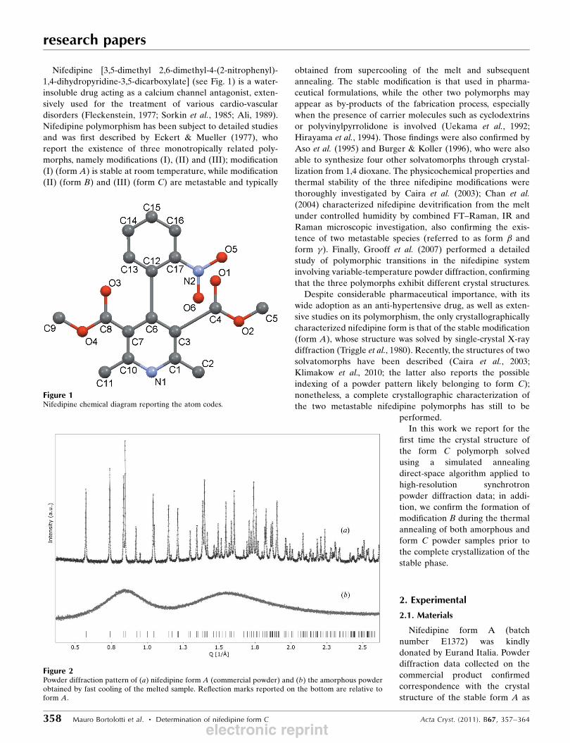
Nifedipine [3,5-dimethyl 2,6-dimethyl-4-(2-nitrophenyl)-
1,4-dihydropyridine-3,5-dicarboxylate] (see Fig. 1) is a water-
insoluble drug acting as a calcium channel antagonist, exten-
sively used for the treatment of various cardio-vascular
disorders (Fleckenstein, 1977; Sorkin et al., 1985; Ali, 1989).
Nifedipine polymorphism has been subject to detailed studies
and was first described by Eckert & Mueller (1977), who
report the existence of three monotropically related poly-
morphs, namely modifications (I), (II) and (III); modification
(I) (form A) is stable at room temperature, while modification
(II) (form B) and (III) (form C) are metastable and typically
obtained from supercooling of the melt and subsequent
annealing. The stable modification is that used in pharma-
ceutical formulations, while the other two polymorphs may
appear as by-products of the fabrication process, especially
when the presence of carrier molecules such as cyclodextrins
or polyvinylpyrrolidone is involved (Uekama et al., 1992;
Hirayama et al., 1994). Those findings were also confirmed by
Aso et al. (1995) and Burger & Koller (1996), who were also
able to synthesize four other solvatomorphs through crystal-
lization from 1,4 dioxane. The physicochemical properties and
thermal stability of the three nifedipine modifications were
thoroughly investigated by Caira et al. (2003); Chan et al.
(2004) characterized nifedipine devitrification from the melt
under controlled humidity by combined FT–Raman, IR and
Raman microscopic investigation, also confirming the exis-
tence of two metastable species (referred to as form � and
form �). Finally, Grooff et al. (2007) performed a detailed
study of polymorphic transitions in the nifedipine system
involving variable-temperature powder diffraction, confirming
that the three polymorphs exhibit different crystal structures.
Despite considerable pharmaceutical importance, with its
wide adoption as an anti-hypertensive drug, as well as exten-
sive studies on its polymorphism, the only crystallographically
characterized nifedipine form is that of the stable modification
(form A), whose structure was solved by single-crystal X-ray
diffraction (Triggle et al., 1980). Recently, the structures of two
solvatomorphs have been described (Caira et al., 2003;
Klimakow et al., 2010; the latter also reports the possible
indexing of a powder pattern likely belonging to form C);
nonetheless, a complete crystallographic characterization of
the two metastable nifedipine polymorphs has still to be
performed.
In this work we report for the
first time the crystal structure of
the form C polymorph solved
using a simulated annealing
direct-space algorithm applied to
high-resolution synchrotron
powder diffraction data; in addi-
tion, we confirm the formation of
modification B during the thermal
annealing of both amorphous and
form C powder samples prior to
the complete crystallization of the
stable phase.
2. Experimental
2.1. Materials
Nifedipine form A (batch
number E1372) was kindly
donated by Eurand Italia. Powder
diffraction data collected on the
commercial product confirmed
correspondence with the crystal
structure of the stable form A as
research papers
358 Mauro Bortolotti et al. � Determination of nifedipine form C Acta Cryst. (2011). B67, 357–364
Figure 1Nifedipine chemical diagram reporting the atom codes.
Figure 2Powder diffraction pattern of (a) nifedipine form A (commercial powder) and (b) the amorphous powderobtained by fast cooling of the melted sample. Reflection marks reported on the bottom are relative toform A.
electronic reprint

reported by Triggle et al. (1980); CSD entry BICCIZ, PDF
entry 02-060-4397 (Allen, 2002; Kabekkodu, 2011), space
group P21/c, a = 10.923 (5), b = 10.326 (6), c = 14.814 (7) A, �=
92.70 (6)�, V = 1669.03 (494) A3. No additional impurities
were detected in the diffraction pattern (see Fig. 2a).
2.2. Synchrotron high-resolution powder diffraction
Powder diffraction experiments were performed at the
Swiss–Norwegian beamline (BM01B) of the European
Synchrotron Radiation Facility in Grenoble, France. The
beamline is mainly designed for high-resolution powder
diffraction, with the additional capability to perform
combined diffraction/XAS experiments. The powder diffrac-
tion instrument consists of a two-circle goniometer equipped
with six counting chains, each consisting of an Na-I scintilla-
tion counter coupled with a Si-111 analyser crystal; detectors
are mounted with a 1.1� relative 2� offset (see Fig. 3a). This
instrumental setup allows fast data collection while main-
taining at the same time an excellent intrinsic resolution (0.01�
FWHM at � = 1 A).
Diffraction data were collected using 0.5 A radiation and a
0.002� angular step over a 0.5–25� 2� interval. Counting times
between 10 and 200 ms per step were used for the various
experiments, depending on the
kinetics of the phenomena
observed. Samples were loaded in a
0.7 mm capillary spinning in the
axial direction to improve particle
statistics. For in situ characteriza-
tion, a hot-air blower placed under
the capillary sample holder was
used, with a temperature range
from 298 to 1273 K (Fig. 3b). The
instrumental calibration was
performed using a NIST 640c
silicon standard.
Different sample preparation
methodologies and data collection
strategies were tested in an attempt
to isolate the pure polymorphs and
collect powder data of sufficient
quality to allow a reliable ab initio
structure solution. In a first experi-
ment the commercial nifedipine
powder (sample 1) was used
without further treatments, with the
goal of performing a complete in
situ characterization of the devi-
trification behaviour under
annealing. The sample was loaded
in the capillary and heated beyond
the melting point (� 463 K), then
kept at this temperature for 10 min;
the Bragg peaks disappearing in the
observed diffraction pattern
confirmed the complete melting of
the compound. The capillary was
then quickly removed from the
furnace and allowed to cool at room
temperature (� 298 K), inducing
the formation of an amorphous
glass. The sample was then reheated
to 323 K (heating rate 5 K min�1),
then subjected to variable-
temperature powder X-ray diffrac-
tion (PXRD) from 323 to 423 K in
2 K steps. A second sample (sample
2) consisting of the commercial
research papers
Acta Cryst. (2011). B67, 357–364 Mauro Bortolotti et al. � Determination of nifedipine form C 359
Figure 3Experimental station at BM01B (ESRF, Grenoble) with (a) the powder diffractometer and (b) a moredetailed view of the hot-air blower used during the annealing experiments.
Figure 4In situ thermal evolution of amorphous nifedipine (a) at 337 K showing the nucleation of the metastablepolymorph B after 10 min (b), and of the stable form A after 30 min (c). After 60 min the sample hascompletely transformed to form A (f). Reflection marks on the bottom show peak positions relative tothe stable modification.
electronic reprint
powder was heated ex situ in an electric oven to 463 K for
several minutes until completely melted, then allowed to cool
at room temperature. The solidified amorphous glass was then
pulverized with an agate mortar and pestle, and the obtained
powder subjected to variable-temperature PXRD with the
same annealing schedule described for sample 1. In a final
experiment (sample 3), the form C polymorph was obtained ex
situ following the procedure described in Burger & Koller
(1996); form A powder was placed on an aluminium foil and
heated on a hot-plate beyond the melting temperature, then
allowed to cool at room temperature; the amorphous glass
thus obtained was then reheated to � 333 K when the crys-
tallization of the first metastable polymorph (form B) was
observed. Once visual observation confirmed crystallization
was complete, the sample was allowed to cool at room
temperature to favour the form B ! form C transition,
visually confirmed by morphological and chromatic variations
of the sample (Grooff et al., 2007). The form C sample thus
obtained was then pulverized and loaded in the capillary; high-
quality powder data suitable for the ab initio structure solution
were collected at room temperature, then the thermal evolu-
tion of the sample was studied using the annealing schedule
previously described and applied to the other samples.
3. Results and discussion
3.1. In situ thermal behaviour of nifedipine
Nifedipine polymorphs are typically obtained starting from
the amorphous form by means of a non-equilibrium devi-
trification process. Nucleation of form B crystals is induced by
a thermal annealing of the amorphous phase; depending on
the heating rate and the physical characteristics of the sample
(solid glass or powder), a considerable variability in the
nucleation temperature is observed, varying from 333 to 363 K
(Aso et al., 1995; Zhou et al., 2003). Starting from the meta-
stable form B, polymorph C is then obtained by rapid cooling
to room temperature, while a further temperature increase
will invariably produce the nucleation and growth of the stable
form A (Grooff et al., 2007).
An in situ characterization of nifedipine polymorphism thus
presents the need to heat the stable modification above the
melting point and then rapidly cool it to room temperature to
obtain the amorphous state of the sample, before performing
the subsequent thermal annealing treatments that induce the
nucleation of the polymorphs. The relatively narrow nuclea-
tion and stability interval of the metastable forms (form B in
particular) presents a series of experimental challenges, firstly
accurate and continuous control of the sample temperature.
The thermal conditioning set-up adopted during this work,
based on the use of a hot-air blower heating a relatively large
sample region (Fig. 3b), did not allow a particularly sophisti-
cated temperature control; in fact, a considerable thermal
gradient was induced along the illuminated volume of the
sample, giving rise to a differential thermal history and,
consequently, inconsistent crystallization behaviour along the
capillary axis. On the other hand, the beam-sampling volume
had to be large enough to assure adequate counting statistics
during thermal evolution experiments, making it very difficult
to obtain high-quality diffraction patterns of the pure poly-
morphs.
In the case of nifedipine commercial powder (pure form A,
sample 1), solidification upon
rapid cooling of the melt produced
a discontinuous amorphous glass
inside the capillary, worsening the
thermal gradient issue and thus
making a complete in situ char-
acterization unfeasible. The amor-
phous powder was then obtained
ex situ, as described in x2 (sample
2), and subjected to thermal
annealing starting from room
temperature; diffraction patterns
were acquired every 0.5 K to
follow the devitrification process.
The first diffraction peaks, indi-
cating form B nucleation onset,
appeared at 337 K; at that point
the annealing was halted and the
temperature kept constant to
monitor the crystallization kinetics
evolution. Despite several
attempts it was not possible to
acquire high quality data of the
pure form B given the strong
competition with the stable form
A, which started to nucleate before
research papers
360 Mauro Bortolotti et al. � Determination of nifedipine form C Acta Cryst. (2011). B67, 357–364
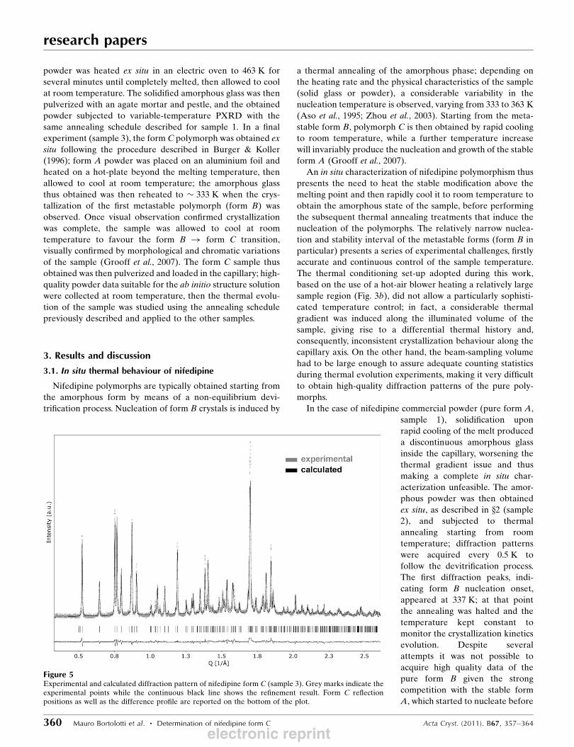
Figure 5Experimental and calculated diffraction pattern of nifedipine form C (sample 3). Grey marks indicate theexperimental points while the continuous black line shows the refinement result. Form C reflectionpositions as well as the difference profile are reported on the bottom of the plot.
electronic reprint

the complete transformation of the amorphous phase into
form B. Upon cooling, only the form B fraction transformed to
form C, while the stable modification fraction remained
unchanged, leaving again a contaminated sample. Fig. 4
reports the sample evolution at 337 K at 10 min intervals,
showing evidence of form B nucleation (peaks at Q = 0.52,
0.65, 0.76 A�1) gradually being substituted by the crystallizing
form A (peaks at Q = 0.58, 0.74, 0.84 and 0.85 A�1).
3.2. Ab initio structure solution of form C
Given the experimental difficulties in isolating the pure
metastable polymorphs during in situ characterization, the ex
situ preparation of form C following the procedure described
in the previous paragraph (sample 3) was chosen. Once crys-
tallized, form C is relatively stable at room temperature so
long acquisition times were possible, allowing high-quality
powder data suitable for an ab initio structure determination
to be collected.
Diffraction spectra were acquired at room temperature (T =
298 K) over a 0.5–25.5� 2� interval with a 0.002� step size; the
counting time for each data point was 50 ms. After a careful
examination to exclude formation of the stable modification,
the patterns were summed together to improve signal statis-
tics, for a total counting time of 1 s per data point (Fig. 5, grey
marks). Accurate peak positions in the diffraction pattern
were determined by fitting the individual reflections with the
ReX software (Bortolotti et al., 2009); the first 32 reflection
positions were determined and the first 20 were input into the
program DICVOL (Boultif & Louer, 2004). The indexing
algorithm produced one solution in the triclinic crystal system
with relatively high figures of merit M(20) = 33.3, F(20) = 280.1
(de Wolff, 1968; Smith & Snyder, 1979), and several additional
results in the monoclinic space group with very low figures of
merit [M(20) < 5]. None of the monoclinic solutions could
account for all the input line positions and were thus
discarded. The triclinic solution (a = 9.8698, b = 13.8935, c =
14.2862 A, � = 61.225, � = 79.824, � = 81.764�, V = 1686.25 A3)
indexed all the 20 input peaks as well as the other reflections
[F(32) = 147.9]; moreover, the cell volume, supposing an
occupancy of 4 molecules per cell, suggested an almost iden-
tical calculated density to that of the stable polymorph.
The ab initio structure solution was carried out with a
modified version of the ReX software, using a direct-space
approach implemented in the form of an extended search,
simulated annealing algorithm (Coelho, 2000). The structural
research papers
Acta Cryst. (2011). B67, 357–364 Mauro Bortolotti et al. � Determination of nifedipine form C 361
Table 1Crystallographic data of nifedipine form C after the final Rietveldrefinement.
Crystal dataChemical formula C17H18N2O6
Mr 346.34Crystal system, space group Triclinic, P�11Temperature (K) 298a, b, c (A) 9.864 (1), 13.893 (9), 14.287 (3)�, �, � (�) 61.227 (3), 79.827 (2), 81.782 (1)V (A3) 1685.5 (9)Z 4Radiation type Synchrotron, � = 0.50000 ASpecimen shape, size (mm) Cylinder, 14 � 0.7
Data collectionDiffractometer Two-theta/omega SnB diffractometer
– ESRFSpecimen mounting CapillaryData collection mode TransmissionScan method Step2� values (�) 2�min = 1.5, 2�max = 12.0, �step = 0.002
RefinementR factors and goodness of fit Rp = 0.151, Rwp = 0.172, Rexp = 0.017,
RBragg = 0.151, �2 = 19.536No. of data points 12 508No. of parameters 15No. of restraints 0H-atom treatment H-atom parameters not refined
Figure 6Comparison of the crystallographic structure of (a) nifedipine form A and(b) nifedipine form C (c axis orthogonal to view plane). While themolecular conformation remains substantially unchanged, the crystalpacking shows important differences, with form A (space group P21/c)exhibiting a parallel alignment of pyridine and nitrophenyl groups, andform C a substantially orthogonal configuration.
electronic reprint

optimization was attempted in the space group P�11, defining
two independent nifedipine molecules in the asymmetric unit.
For each molecule, nine geometrical parameters were defined,
namely the coordinates of the centre of mass and the molecule
orientation, as well as the relative orientation of the nitro-
phenyl group and the two carboxylate groups with respect to
the main pyridine fragment. The merit function used by the
solution algorithm was defined using the full powder profile;
although the procedure is considerably slower with respect to
working directly with integrated intensities, it avoids possible
errors introduced during the profile intensity extraction in
procedures like that of Pawley (Pawley, 1981) or Le Bail (Le
Bail et al., 1988). To maximize the convergence chance six trial
runs were performed, starting each time from a random
structural parameters set; each run evaluated a population of
106 individuals. Of the six optimization runs, four converged to
a final Rwp value < 0.25 before the 500 000 th step, a behaviour
which was considered a reliable indicator of an achieved
convergence; the two other runs reached a plateau at a Rwp
value of � 0.5 without further improvements. The solutions
obtained from the four successful runs were compared and
found to be equivalent from a crystallographic point of view,
displaying differences only in the absolute positioning of the
asymmetric unit inside the unit cell. The high Rwp solutions
were, on the other hand, clearly unsound from a chemical
point of view, showing overlapping
molecules inside the unit cell and
unreasonable bond angles.
Starting from the ab initio solu-
tion, the final Rietveld least-squares
optimization was performed with
the software Maud (Lutterotti et al.,
1999); refined parameters included
an intensity scale factor and a
fourth-order polynomial back-
ground, as well as unit-cell
constants and average crystallite
size; isotropic displacement para-
meters were globally refined. The
final value of Rwp obtained was
0.172; the calculated cell volume is
1685.54 A3, only 1% higher than
the stable modification. The
experimental pattern, as well as the
calculated profile and the error plot
are shown in Fig. 5; a crystal-
lographic data summary is reported
in Table 1.
H-atom positions, not taken into
account during the structure solu-
tion and the successive Rietveld
optimization steps, were deter-
mined and added to the refined
structure using the algorithm
implemented in the OpenBabel
software package (Guha et al.,
2006); the complete chemical
structure with atomic coordinates is reported in the supporting
material.1
The unit cell of nifedipine form C is shown in Fig. 6(b),
compared with the stable modification (Fig. 6a) as obtained
from the literature (Triggle et al., 1980). The asymmetric unit
contains two molecules in general positions; the most signifi-
cant difference between the two polymorphic structures lies in
the molecular packing. In modification A (space group P21/c)
both pyridine and nitrophenyl rings are parallel to each other,
showing a head-tail and head-head configuration; in form C,
on the contrary, molecules are aligned in an approximately
orthogonal configuration, with the pyridine groups of one
molecular fragment (C1) and the other (C2) forming an angle
of 89.13�, and the respective nitrophenyl groups slightly
misaligned (83.17�).
The molecular conformation, on the other hand, is
remarkably similar to that of form A, as well as the two
solvatomorphs reported in the literature (Caira et al., 2003;
Klimakow et al., 2010). A conformational comparison inves-
tigation was carried out through a search for similar
compounds inside the CSD (Allen, 2002). Using the Mogul
research papers
362 Mauro Bortolotti et al. � Determination of nifedipine form C Acta Cryst. (2011). B67, 357–364
Figure 7Comparison between torsion-angle distributions derived from the Cambridge Structural Database andobserved values in nifedipine form A and form C. Values are reported for the angles formed by thepyridine fragment with the two carboxylic groups (O2—C4—C3—C6 and O3—C8—C7—C6) and thenitrophenyl group (C7—C6—C12—C13), as well as the angle between the phenyl ring and the nitrogroup (C16—C17—N2—O6).
1 Supplementary data for this paper are available from the IUCr electronicarchives (Reference: KD5048). Services for accessing these data are describedat the back of the journal.
electronic reprint

software (Bruno et al., 2004) with an exact match search
criterion (R < 0.10), statistical distributions were retrieved for
the dihedral angles formed by the pyridine fragment with the
nitrophenyl and carboxylic groups, as well as the angle
between the phenyl ring and the nitro group; the distributions
were then compared with the actual values observed in form A
and form C (Fig. 7). The metastable polymorph exhibits values
fairly close to the distribution maxima; torsion angles relative
to the two carboxyl groups (O2—C4—C3—C6 and O3—C8—
C7—C6) in the molecular fragment C1 show a similar orien-
tation to form A, whereas in fragment C2 they are rotated by
� 180�. Additionally, the angle between the nitrophenyl and
the pyridine group (C7—C6—C12—C13) is slightly distorted
with respect to the stable modification, with a 14.15� average
difference. Table 2 summarizes the various dihedral angle
values for the two polymorphs.
A conformational energy comparison between the two
nifedipine forms was carried out using ab initio methods.
Single-point energy calculations were performed at the
B3LYP/6-31G* level of theory using the software GAMESS
(Schmidt et al., 1993); the obtained average energy for form C
shows a moderate 18.02 kJ mol�1 increase with respect to the
stable polymorph, suggesting a rather stable conformational
equilibrium.
The form C sample, structurally characterized as previously
described, was finally subjected to a thermal annealing process
starting from room temperature (298 K) up to the complete
crystallization of the stable modification (Fig. 8). Diffraction
spectra were collected at 1 K intervals to closely follow the
solid-state evolution of the material. Form C was stable up to
328 K, after which new diffraction peaks belonging to form B
(Q = 0.52, 0.65 and 0.76 A�1) started to appear in the
diffraction pattern. Even in this case, however, it was not
possible to avoid the contamination of the sample by the
stable form A, which started to nucleate immediately after
(peaks at Q = 0.58, 0.74, 0.84 and 0.85 A�1). Reflections
belonging to form B started to disappear at 358 K, when the
sample was almost completely composed of the stable modi-
fication.
4. Conclusions
We reported for the first time the structure of nifedipine form
C, solved by means of ab initio direct-space methods applied
to synchrotron powder diffraction data. The polymorph crys-
tallizes in the space group P�11, with a cell volume nearly equal
to that of the stable form; in addition, the internal confor-
mation of the nifedipine molecules resembles very closely that
observed in the stable form, with comparable bond lengths
and bond angles. On the other hand, the molecular packing
differs substantially, with molecules orthogonally aligned with
respect to each other.
The existence of another tran-
sient metastable polymorph (form
B) with a different crystal struc-
ture is confirmed; its occurrence
can be observed during thermal
annealing from room temperature
of the form C polymorph as well as
the amorphous form. Unfortu-
nately, it was not possible to
collect good quality diffraction
data of form B, which is stable
only in a very limited temperature
interval. The authors believe that
such fast transformation kinetics
could be successfully followed
using the new generation of real-
time multiple strip (RTMS)
detectors, which when coupled to
a high brilliance synchrotron
source can provide an almost real-
time sample monitoring while
maintaining the high instru-
mental resolution needed for
advanced structural investi-
gations.
research papers
Acta Cryst. (2011). B67, 357–364 Mauro Bortolotti et al. � Determination of nifedipine form C 363
Table 2Comparison between selected dihedral angles (�) in forms A and C.
Polymorph Form A Form C (1) Form C (2)
O2—C4—C3—C6 166.5 8.1 174.2O3—C8—C7—C6 164.7 5.1 150.0C7—C6—C12—C13 49.1 59.1 67.4C16—C17—N2—O6 38.0 41.5 13.1
Figure 8In situ thermal evolution of nifedipine form C (sample 3) from 298 to 363 K. Form C remains stable up to328 K, when peaks belonging to both form B and form A start to appear. Transformation to the stablemodification A is almost complete at 363 K.
electronic reprint
We would like to thank all the BM01B staff, in particular
Herman Emerich and Olga Safonova, for their helpful support
during the experiment preparation and the data acquisition.
Further thanks go to Lorenzo Magarotto (Eurand Italy) for
providing the nifedipine sample used during this work. The
research leading to these results has received funding from the
European Community’s Seventh Framework Program under
the I3 Project ELISA, grant agreement No. 226716.
References
Ali, S. L. (1989). Analytical Profile of Drug Substances, edited by K.Florey, Vol. 18, pp. 221–288. NewYork: Academic Press Inc.
Allen, F. H. (2002). Acta Cryst. B58, 380–388.Aso, Y., Yoshioka, S., Otsuka, T. & Kojima, S. (1995). Chem. Pharm.
Bull. 43, 300–303.Bernstein, J. (2002). Polymorphism in Molecular Crystals. New York:
Oxford University Press.Bortolotti, M., Lutterotti, L. & Lonardelli, I. (2009). J. Appl. Cryst.42, 538–539.
Boultif, A. & Louer, D. (2004). J. Appl. Cryst. 37, 724–731.Brittain, H. G. (2009). Polymorphism in Pharmaceutical Solids, 2nd
ed. New York: Informa Healthcare.Brittain, H. G. (2010). J. Pharm. Sci. 99, 3648–3664.Bruno, I. J., Cole, J. C., Kessler, M., Luo, J., Motherwell, W. D. S.,
Purkis, L. H., Smith, B. R., Taylor, R., Cooper, R. I., Harris, S. E. &Orpen, A. G. (2004). J. Chem. Inf. Comput. Sci. 44, 2133–2144.
Burger, A. & Koller, K. T. (1996). Sci. Pharm. 64, 293–301.Caira, M. R., Robbertse, Y., Bergh, J. J., Song, M. N. & De Villiers,
M. M. (2003). J. Pharm. Sci. 92, 2519–2533.Chan, K. L. A., Fleming, O. S., Kazarian, S. G., Vassou, D., Chryssikos,
G. D. & Gionis, V. (2004). J. Raman Spectrosc. 35, 353–359.Chemburkar, S. R. et al. (2000). Org. Proc. Res. Dev. 4, 413–417.Coelho, A. A. (2000). J. Appl. Cryst. 33, 899–908.David, W. I. F., Shankland, K., McCusker, L. B. & Baerlocher, Ch.
(2006). Editors. Structure Determination from Powder DiffractionData. Oxford University Press.
Eckert, T. & Mueller, J. (1977). Arch. Pharm. 310, 116–118.Fleckenstein, A. (1977). Annu. Rev. Pharmacol. Toxicol. 17, 149–
166.Grooff, D., De Villiers, M. M. & Liebenberg, W. (2007). Thermochim.
Acta, 454, 33–42.Guha, R., Howard, M. T., Hutchison, G. R., Murray-Rust, P., Rzepa,
H., Steinbeck, C., Wegner, J. K. & Willighagen, E. (2006). J. Chem.Inf. Model. 46, 991–998.
Hirayama, F., Wang, Z. & Uekama, K. (1994). Pharm. Res. 11, 1766–1770.
Kabekkodu, S (2011). Editor. PDF-4/Organics 2011 Database.International Centre for Diffraction Data, Newtown Square, PA,USA.
Klimakow, M., Leiterer, J., Kneipp, J., Rossler, E. A., Panne, U.,Rademann, K. & Emmerling, F. (2010). Langmuir, 26, 11233–11237.
Klimakow, M., Rademann, K. & Emmerling, F. (2010). Cryst. GrowthDes. 10, 2693–2698.
Le Bail, A., Duroy, H. & Fourquet, J. L. (1988). Mater. Res. Bull. 23,447–452.
Lutterotti, L., Matthies, S. & Wenk, H. R. (1999). Proc. of the 12th Int.Conf. on Textures of Materials (ICOTOM-12), Vol. 1, 1599, August1999. Montreal, Canada: NRC Research Press.
Pawley, G. S. (1981). J. Appl. Cryst. 14, 357–361.Schmidt, M. W., Baldridge, K. K., Boatz, J. A., Elbert, S. T., Gordon,
M. S., Jensen, J. H., Koseki, S., Matsunaga, N., Nguyen, K. A., Su,S. J., Windus, T. L., Dupuis, M. & Montgomery, J. A. (1993). J.Comput. Chem. 14, 1347–1363.
Smith, G. S. & Snyder, R. L. (1979). J. Appl. Cryst. 12, 60–65.Sorkin, E. M., Clissol, S. P. & Brogden, R. N. (1985). Drugs, 30, pp.
182–274.Triggle, A. M., Shefter, E. & Triggle, D. J. (1980). J. Med. Chem. 23,
1442–1445.Uekama, K., Ikegami, K., Wang, Z., Horiuchi, Y. & Hirayama, F.
(1992). J. Pharm. Pharmacol. 44, 73–78.Wolff, P. M. de (1968). J. Appl. Cryst. 1, 108–113.Zhou, D., Schmitt, E. A., Zhang, G. G., Law, D., Vyazovkin, S., Wight,
C. A. & Grant, D. J. W. (2003). J. Pharm. Sci. 92, 1779–1792.
research papers
364 Mauro Bortolotti et al. � Determination of nifedipine form C Acta Cryst. (2011). B67, 357–364
electronic reprint
Related Documents