Please cite this article in press as: S. Shafique, et al., Determination of Molecular and Biochemical Changes in Cotton Plants Mediated by Mealybug, NJAS - Wageningen J. Life Sci. (2014), http://dx.doi.org/10.1016/j.njas.2014.05.001 ARTICLE IN PRESS G Model NJAS-164; No. of Pages 7 NJAS - Wageningen Journal of Life Sciences xxx (2014) xxx–xxx Contents lists available at ScienceDirect NJAS - Wageningen Journal of Life Sciences jo ur nal homepage: www.elsevier.co m/locate/njas Determination of Molecular and Biochemical Changes in Cotton Plants Mediated by Mealybug Shazia Shafique a , Aqeel Ahmad a,∗ , Sobiya Shafique a , Tehmina Anjum a , Waheed Akram a , Zoobia Bashir b a Institute of Agricultural Sciences, University of the Punjab, Lahore, Pakistan b Department of Physics, Bahauddin Zakariya University, Multan, Pakistan a r t i c l e i n f o Article history: Received 27 June 2013 Received in revised form 6 April 2014 Accepted 2 May 2014 Available online xxx Keywords: Insect-plant system Induced defenses Biochemicals Histological studies Defensive enzymes a b s t r a c t Phenococcus solenopsis (cotton mealybug) is a devastating insect pest of many countries of the world including Pakistan. Due to its piercing sucking type feeding behavior, it injures cotton plants mechanically and induces several cytological and physiological changes in the host. These changes have been studied after subjecting healthy plants with mealybug under controlled conditions and it was recorded that mealybug attack enhanced lignin, cellulose and hemicellulose contents remarkably. It was also observed that defensive biochemicals of cotton i.e. phenolics and terpenoids were also significantly increased (up to 7 times) with progressing time (0-3 hr) in injured plants. Defensive enzymes i.e. phenyl ammonia lyase (PAL), polyphenol oxidase (PPO) and peroxidase (POD) also showed direct relationship with the passing time (0-3 hr) after mealybug feeding. Similarly, expression of thaumatin-like, metallothionein and profilin genes was enhanced with the elicitation of plant defenses due to insect herbivory. There was no connection found between pathogenesis related Pseml gene and plant defense against herbivory. Study concluded that mealybug did not modulate all plant defenses. There were specific biochemicals and defense related genes influenced by the attack of mealybug. © 2014 Royal Netherlands Society for Agricultural Sciences. Published by Elsevier B.V. All rights reserved. 1. Introduction Nature has armed plants with defense weapons to ensure their survival against harsh conditions; and which are generally cat- egorized as constitutive and induced responses of plants due to external factors. Those external factors which can modulate plant responses may be chemical, physical or biological in nature, among which the insect herbivory is also an external stimulus [1]. Her- bivory induces plant defense panel against biotic and abiotic factors [2]. Frequently reported changes in plant tissues due to insect feeding are modulation of defense chemicals [3,4]. Moreover, pro- duction of enhanced transcriptional rate of plant defense genes with concomitant increase in pathogenesis related proteins has also been reported in some insect plant interactions [5]. ∗ Corresponding author. E-mail addresses: [email protected] (S. Shafique), [email protected] (A. Ahmad), [email protected] (S. Shafique), [email protected] (T. Anjum), meher [email protected] (W. Akram), [email protected] (Z. Bashir). Cotton mealybug is an economically important pest of many ornamentals, medicinals, food crops and forest plants, with wide host spectrum [6]. Both the nymphs and adults of mealybug can harm crop plants by feeding upon phloem sap and egesting honey- dews [7]. Cotton is one of the preferred hosts of mealybug without any specification of crop variety [8]. Massive population, colos- sal host spectrum and huge economic losses give mealybug an identical position among agriculturally important insect pests. It is necessarily important to study its impact on physiology and defense systems of plants. Therefore, physiological effects of mealy- bug on innate defense system of cotton plants have been studied. It will develop better understandings among researchers about the plant behavior against this insect. 2. Methodology 2.1. Collection of samples Cotton plants were grown in green house of ‘Institute of Agri- cultural Sciences, University of the Punjab, Lahore, Pakistan’ and randomly divided into two equal sets (each set containing 10 http://dx.doi.org/10.1016/j.njas.2014.05.001 1573-5214/© 2014 Royal Netherlands Society for Agricultural Sciences. Published by Elsevier B.V. All rights reserved.

Welcome message from author
This document is posted to help you gain knowledge. Please leave a comment to let me know what you think about it! Share it to your friends and learn new things together.
Transcript
N
DM
SZa
b
a
ARRAA
KIIBHD
1
seerwb[fdwb
(((
h1
ARTICLE IN PRESSG ModelJAS-164; No. of Pages 7
NJAS - Wageningen Journal of Life Sciences xxx (2014) xxx–xxx
Contents lists available at ScienceDirect
NJAS - Wageningen Journal of Life Sciences
jo ur nal homepage: www.elsev ier .co m/locate /n jas
etermination of Molecular and Biochemical Changes in Cotton Plantsediated by Mealybug
hazia Shafiquea, Aqeel Ahmada,∗, Sobiya Shafiquea, Tehmina Anjuma, Waheed Akrama,oobia Bashirb
Institute of Agricultural Sciences, University of the Punjab, Lahore, PakistanDepartment of Physics, Bahauddin Zakariya University, Multan, Pakistan
r t i c l e i n f o
rticle history:eceived 27 June 2013eceived in revised form 6 April 2014ccepted 2 May 2014vailable online xxx
eywords:nsect-plant systemnduced defensesiochemicals
a b s t r a c t
Phenococcus solenopsis (cotton mealybug) is a devastating insect pest of many countries of the worldincluding Pakistan. Due to its piercing sucking type feeding behavior, it injures cotton plants mechanicallyand induces several cytological and physiological changes in the host. These changes have been studiedafter subjecting healthy plants with mealybug under controlled conditions and it was recorded thatmealybug attack enhanced lignin, cellulose and hemicellulose contents remarkably. It was also observedthat defensive biochemicals of cotton i.e. phenolics and terpenoids were also significantly increased (upto 7 times) with progressing time (0-3 hr) in injured plants. Defensive enzymes i.e. phenyl ammonialyase (PAL), polyphenol oxidase (PPO) and peroxidase (POD) also showed direct relationship with thepassing time (0-3 hr) after mealybug feeding. Similarly, expression of thaumatin-like, metallothionein
istological studiesefensive enzymes
and profilin genes was enhanced with the elicitation of plant defenses due to insect herbivory. Therewas no connection found between pathogenesis related Pseml gene and plant defense against herbivory.Study concluded that mealybug did not modulate all plant defenses. There were specific biochemicalsand defense related genes influenced by the attack of mealybug.
© 2014 Royal Netherlands Society for Agricultural Sciences. Published by Elsevier B.V. All rightsreserved.
. Introduction
Nature has armed plants with defense weapons to ensure theirurvival against harsh conditions; and which are generally cat-gorized as constitutive and induced responses of plants due toxternal factors. Those external factors which can modulate plantesponses may be chemical, physical or biological in nature, amonghich the insect herbivory is also an external stimulus [1]. Her-
ivory induces plant defense panel against biotic and abiotic factors2]. Frequently reported changes in plant tissues due to insecteeding are modulation of defense chemicals [3,4]. Moreover, pro-uction of enhanced transcriptional rate of plant defense genes
Please cite this article in press as: S. Shafique, et al., Determination of MMealybug, NJAS - Wageningen J. Life Sci. (2014), http://dx.doi.org/10.1
ith concomitant increase in pathogenesis related proteins has alsoeen reported in some insect plant interactions [5].
∗ Corresponding author.E-mail addresses: [email protected] (S. Shafique), [email protected]
A. Ahmad), [email protected] (S. Shafique), [email protected]. Anjum), meher [email protected] (W. Akram), [email protected]. Bashir).
ttp://dx.doi.org/10.1016/j.njas.2014.05.001573-5214/© 2014 Royal Netherlands Society for Agricultural Sciences. Published by Else
Cotton mealybug is an economically important pest of manyornamentals, medicinals, food crops and forest plants, with widehost spectrum [6]. Both the nymphs and adults of mealybug canharm crop plants by feeding upon phloem sap and egesting honey-dews [7]. Cotton is one of the preferred hosts of mealybug withoutany specification of crop variety [8]. Massive population, colos-sal host spectrum and huge economic losses give mealybug anidentical position among agriculturally important insect pests. Itis necessarily important to study its impact on physiology anddefense systems of plants. Therefore, physiological effects of mealy-bug on innate defense system of cotton plants have been studied.It will develop better understandings among researchers about theplant behavior against this insect.
2. Methodology
2.1. Collection of samples
olecular and Biochemical Changes in Cotton Plants Mediated by016/j.njas.2014.05.001
Cotton plants were grown in green house of ‘Institute of Agri-cultural Sciences, University of the Punjab, Lahore, Pakistan’ andrandomly divided into two equal sets (each set containing 10
vier B.V. All rights reserved.
IN PRESSG ModelN
2 Journal of Life Sciences xxx (2014) xxx–xxx
pawflhotaS
2
wmlvme
atceprt
tcctost
top(a
amtbwcAes
2
pn(wabaf
ab
Table 1Detailed primer sequences used to target pathogenesis related specific genes incotton plants. Primers were used to amplify specific genes from both treatments ofcotton plants through reverse transcriptase PCR.
Genes AccessionNumbers
Primer Sequences (5′----3′)
Metallothionein HM137170 F-GTACATGGGACACACCGACCAR-GTACAAAACTGTTTTCTGGACACGTAA
Pathogenesisrelated Pseml
AF211850 F-ATGGTGTCAGGGACTTCATCAAC
R-CTAGCAGTATAAGTTGGGATTGGAThaumatin-like HM202602 F-GCTTTGGGAGTTTCGTTGATATTCAT
R-ATGGTGGTGCCGCAGAAGAC
ARTICLEJAS-164; No. of Pages 7
S. Shafique et al. / NJAS - Wageningen
lants) at the age of 45 days. One set was subjected to mealybugttack at the rate of 10 mealybug adults per plant and second setas left as negative control. Both the plant sets were covered withoating net cover of propylene and placed side by side in the green-ouse under controlled conditions (26 ± 2 ◦C). Time course studiesf both plant sets were conducted regarding physiological and his-ological changes induced in cotton plants. All chemical reagentsnd lab equipment used in this study had been purchased fromigma Aldrich.
.2. Biochemical studies
Lignin was stained using phloroglucinol solution (0.1%) whichas prepared in aqueous HCl solution (20%). Immersing plantaterial for 1-2 min in this solution provided the cytostaining of
ignin in plant tissue. Then microtome was used to prepare trans-erse sections of the tissue and the sections were examined undericroscope [9]. The sections were cut at different time intervals for
valuating the rate of accumulation of lignin contents.Two percent of potassium iodide solution was prepared in water
nd 0.2 g of iodine was dissolved in it. Transverse sections of cot-on stem were placed in it for 15 min and then covered with glassoverslips on glass slides. Drops of H2SO4 were placed along thedges of glass coverslips (on the microscope slide) and allowed forassive penetration under them. After incubation of 5 minutes atoom temperature, stained cellulose contents of plant tissue sec-ions were observed under microscope [10].
Hemicellulose was quantified by Sulfuric acid method. Plantissue was immersed in 0.5% aqueous solution of H2SO4 and per-entage mass recovery provided the amount of hemicellulosicontents of cotton tissue which had been dissolved in acidic solu-ion [11]. Cytostaining of hemicellulose contents were also carriedut by adopting phenol sulphuric acid method [12], which alsotained phenolics accumulations as dark grey masses inside plantissue.
Time course studies for evaluating phenolic contents in plantissue were determined by adopting the recommended methodf Mujica et al. [13] with small modifications. Ten grams driedlant powder was extracted with aqueous methanol solution80%). Extracts were re-extracted with a solvent mixture of ethylecetate/diethyle ether (1:1).
Ten grams of cotton plant tissue was extracted with ethylecetate for 15 min at 40 ◦C for terpenoids extraction. Organicaterials were removed through filtration and acidic substances
hrough adding 5% KOH, 300 ml. Basic compounds were isolatedy adding 300 ml, 5% HCl solution. The remnants of extracts wereashed with water in a separating funnel. Washed extracts were
oncentrated up to 100 ml and centrifuged for 15 min at 5000 rpm.fter removing the sediments, remaining solution was rotaryvaporated at 40 ◦C and quantification was carried out throughpectrophotometer by dissolving it in CHCl3 [14].
.3. Histological studies
Defense related enzymes are the most effective weapons oflants to control invading pathogens. These are included polyphe-ol oxidase (PPO), phenyl ammonia lyase (PAL) and peroxidasePOD). For detection of the enzymes, protein extract of plant tissueas prepared by crushing unit mass of tissue in a pre-chilled pestle
nd mortar in the presence of ice cold 0.1 M sodium phosphateuffer of pH 6.8. After sufficient stirring, liquid was centrifugedt 10000 rpm for 20 min. The supernatant was taken and used in
Please cite this article in press as: S. Shafique, et al., Determination of MMealybug, NJAS - Wageningen J. Life Sci. (2014), http://dx.doi.org/10.1
urther experiments of individual enzyme detection.Activity of PAL was studied by using the method of Burrell
nd Rees [15]. The enzyme extract (0.2 ml) along with sodiumorate buffer (2.5 ml, 8.8 pH) was added to 0.03 ml phenylalanine
Profilin JQ697838 F-ATGAGTTGGCAGCAATTCGTGGAR-TCAGCCGTTACCTGCACCCTT
and let it undisturbed at 37 ◦C on water bath. Then 0.5 ml of 1 MTrichloroacetic acid was added to reaction mixture and lightabsorbance was recorded at 290 nm in spectrophotometer forformation of Trans-cinnamic acid.
PPO activity was determined by adopting the method of Mayeret al. [16]. The enzyme extract (0.2 ml) was accompanied with1.5 ml, 0.01 M Catechol and spectroscopy was carried out at 495 nm.
For determination of POD activity, method of Polle et al. [17]was adopted. Substrate solution was prepared by adding 5 mMPotassium Phosphate (pH 5.25), 10 mM H2O2 and 40 mM Guaiacoland 50 �l enzyme extract was added to it assuming the extinctioncoefficient 25.5 mM−1cm−1 [18]. Spectrophotometric results wererecorded at 436 nm.
2.4. Expression of defense genes
Expression of four different types of plant defense genes i.e.metallothionein, thaumatin-like, pathogenesis related Pseml andprofilin was studied in cotton plants by performing Multiplex RT-PCR. For the multiplication of cDNA templates of above mentionedgenes, primer sets were designed from NCBI database (Table 1)with the accession numbers of HM137170, HM202602, AF211850and JQ697838; respectively. RNA (1-2 �g) was subjected to reversetranscriptase at 25 ◦C for 6 minutes, 40 ◦C for 60 minutes and 70 ◦Cfor 9 minutes. Moreover, the reaction of 35 cycles was precededwith the 95 ◦C for 10 sec and annealing temperature for 15 sec (forMultiplex reaction annealing temperature was adjusted to 60 ◦C);and elongation at 72 ◦C for 15 sec. Reaction product was elec-trophorated on agarose gel and analyzed through GELANALYZER(Lazar, Hungary) to give a precise view of bands properties.
3. Results
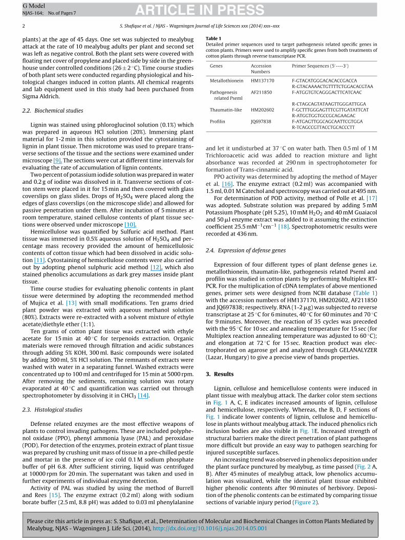
Lignin, cellulose and hemicellulose contents were induced inplant tissue with mealybug attack. The darker color stem sectionsin Fig. 1 A, C, E indicates increased amounts of lignin, celluloseand hemicellulose, respectively. Whereas, the B, D, F sections ofFig. 1 indicate lower contents of lignin, cellulose and hemicellu-lose in plants without mealybug attack. The induced phenolics richinclusion bodies are also visible in Fig. 1E. Increased strength ofstructural barriers make the direct penetration of plant pathogensmore difficult but provide an easy way to pathogen searching forinjured susceptible surfaces.
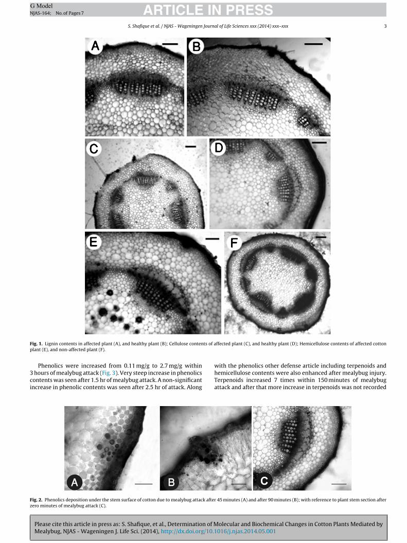
An increasing trend was observed in phenolics deposition underthe plant surface punctured by mealybug, as time passed (Fig. 2 A,B). After 45 minutes of mealybug attack, low phenolics accumu-
olecular and Biochemical Changes in Cotton Plants Mediated by016/j.njas.2014.05.001
lation was visualized, while the identical plant tissue exhibitedhigher phenolic contents after 90 minutes of herbivory. Deposi-tion of the phenolic contents can be estimated by comparing tissuesections of variable injury period (Figure 2).
ARTICLE IN PRESSG ModelNJAS-164; No. of Pages 7
S. Shafique et al. / NJAS - Wageningen Journal of Life Sciences xxx (2014) xxx–xxx 3
F ts of ap
3ci
Fz
ig. 1. Lignin contents in affected plant (A), and healthy plant (B); Cellulose contenlant (E), and non-affected plant (F).
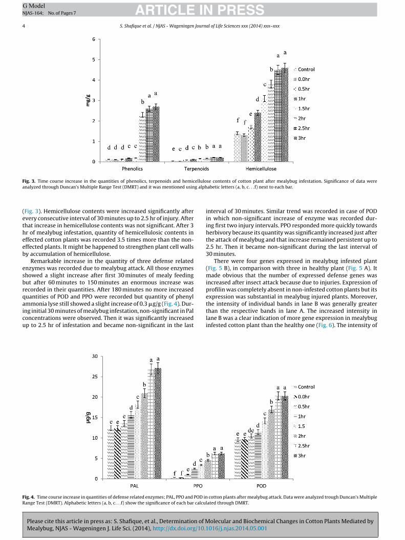
Phenolics were increased from 0.11 mg/g to 2.7 mg/g within
Please cite this article in press as: S. Shafique, et al., Determination of MMealybug, NJAS - Wageningen J. Life Sci. (2014), http://dx.doi.org/10.1
hours of mealybug attack (Fig. 3). Very steep increase in phenolicsontents was seen after 1.5 hr of mealybug attack. A non-significantncrease in phenolic contents was seen after 2.5 hr of attack. Along
ig. 2. Phenolics deposition under the stem surface of cotton due to mealybug attack afterero minutes of mealybug attack (C).
ffected plant (C), and healthy plant (D); Hemicellulose contents of affected cotton
with the phenolics other defense article including terpenoids and
olecular and Biochemical Changes in Cotton Plants Mediated by016/j.njas.2014.05.001
hemicellulose contents were also enhanced after mealybug injury.Terpenoids increased 7 times within 150 minutes of mealybugattack and after that more increase in terpenoids was not recorded
45 minutes (A) and after 90 minutes (B); with reference to plant stem section after
ARTICLE IN PRESSG ModelNJAS-164; No. of Pages 7
4 S. Shafique et al. / NJAS - Wageningen Journal of Life Sciences xxx (2014) xxx–xxx
Fig. 3. Time course increase in the quantities of phenolics, terpenoids and hemicellulose contents of cotton plant after mealybug infestation. Significance of data werea g alph
(etheeb
esbrqaicu
FR
nalyzed through Duncan’s Multiple Range Test (DMRT) and it was mentioned usin
Fig. 3). Hemicellulose contents were increased significantly aftervery consecutive interval of 30 minutes up to 2.5 hr of injury. Afterhat increase in hemicellulose contents was not significant. After 3r of mealybug infestation, quantity of hemicellulosic contents inffected cotton plants was recorded 3.5 times more than the non-ffected plants. It might be happened to strengthen plant cell wallsy accumulation of hemicellulose.
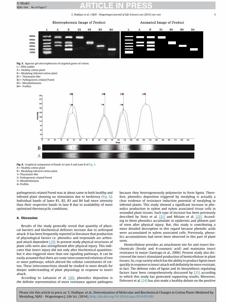
Remarkable increase in the quantity of three defense relatednzymes was recorded due to mealybug attack. All those enzymeshowed a slight increase after first 30 minutes of mealy feedingut after 60 minutes to 150 minutes an enormous increase wasecorded in their quantities. After 180 minutes no more increaseduantities of POD and PPO were recorded but quantity of phenylmmonia lyse still showed a slight increase of 0.3 �g/g (Fig. 4). Dur-
Please cite this article in press as: S. Shafique, et al., Determination of MMealybug, NJAS - Wageningen J. Life Sci. (2014), http://dx.doi.org/10.1
ng initial 30 minutes of mealybug infestation, non-significant in Paloncentrations were observed. Then it was significantly increasedp to 2.5 hr of infestation and became non-significant in the last
ig. 4. Time course increase in quantities of defense related enzymes; PAL, PPO and POD inange Test (DMRT). Alphabetic letters (a, b, c. . .f) show the significance of each bar calcu
abetic letters (a, b, c. . .f) next to each bar.
interval of 30 minutes. Similar trend was recorded in case of PODin which non-significant increase of enzyme was recorded dur-ing first two injury intervals. PPO responded more quickly towardsherbivory because its quantity was significantly increased just afterthe attack of mealybug and that increase remained persistent up to2.5 hr. Then it became non-significant during the last interval of30 minutes.
There were four genes expressed in mealybug infested plant(Fig. 5 B), in comparison with three in healthy plant (Fig. 5 A). Itmade obvious that the number of expressed defense genes wasincreased after insect attack because due to injuries. Expression ofprofilin was completely absent in non-infested cotton plants but itsexpression was substantial in mealybug injured plants. Moreover,the intensity of individual bands in lane B was generally greater
olecular and Biochemical Changes in Cotton Plants Mediated by016/j.njas.2014.05.001
than the respective bands in lane A. The increased intensity inlane B was a clear indication of more gene expression in mealybuginfested cotton plant than the healthy one (Fig. 6). The intensity of
cotton plants after mealybug attack. Data were analyzed trough Duncan’s Multiplelated through DMRT.
ARTICLE IN PRESSG ModelNJAS-164; No. of Pages 7
S. Shafique et al. / NJAS - Wageningen Journal of Life Sciences xxx (2014) xxx–xxx 5
Fig. 5. Agarose gel electrophoresis of targeted genes of cotton.L = DNA LadderA = Healthy cotton plantB = Mealybug infested cotton plantB1 = Thaumatin-likeB2 = Pathogenesis related PsemlB3 = MetallothioneinB4 = Profilin.
Fig. 6. Graphical comparison of bands in Lane A and Lane B of Fig. 5.A = Healthy cotton plantB = Mealybug infested cotton plant1= Thaumatin-like234
piIto
4
caoppcbeotda
t
= Pathogenesis related Pseml= Metallothionein= Profilin
athogenesis related Pseml was at about same in both healthy andnfested plant showing no stimulation due to herbivory (Fig. 6).ndividual bands of lanes B1, B2, B3 and B4 had more intensityhan their respective bands in lane B due to availability of moreptimized thermocyclic conditions.
. Discussion
Results of the study generally reveal that quantity of physi-al barriers and biochemical defenses increase due to arthropodttack. It has been frequently reported in literature that productionf physiological factors i.e. phenolics and terpenoids are arthro-od attack dependent [19]. In present study physical structures oflant cells were also strengthened after physical injury. This indi-ates that insect injury did not only alter biochemical quantities;ut it also triggered more than one signaling pathways. It can beasily assumed that there are some interconnected relations of twor more pathways, which altered the cellular constituents of cot-on. These interconnections should be studied in more details for
Please cite this article in press as: S. Shafique, et al., Determination of MMealybug, NJAS - Wageningen J. Life Sci. (2014), http://dx.doi.org/10.1
eeper understanding of plant physiology in response to insectttack.
According to Lattanzio et al. [20], phenolics deposition ishe definite representation of more resistance against pathogens
because they heterogeneously polymerize to form lignin. There-fore, phenolics deposition triggered by mealybug is actually aclear evidence of resistance induction potential of mealybug ininfested plants. This study showed a significant increase in phe-nolics production in xylem and xylem associated tissue cells inwounded plant tissues. Such type of increase has been previouslydescribed by Stotz et al. [21] and Miriam et al. [22]. Accord-ing to them phenolics accumulate in epidermis and phloem partof stem after physical injury. But, this study is contributing amore detailed description in this regard because phenolic acidswere accumulated in xylem associated cells. Previously, pheno-lics accumulations had never been observed in this part of plantstem.
Hemicellulose provides an attachment site for anti-insect bio-chemicals (ferulic and 4-coumaric acid) and maintains insectresistance in maize (Santiago et al., 2006). Present study also dis-covered the insect stimulated production of hemicellulose in planttissues. So, crop variety which has the ability to produce lignin morequickly in response to insect attack will definitely be more resistant,
olecular and Biochemical Changes in Cotton Plants Mediated by016/j.njas.2014.05.001
in fact. The defense roles of lignin and its biosynthesis regulatingfactors have been comprehensively discussed by [23] accordingto which this study has generated supporting results. Moreover,Delessert et al. [24] has also made a healthy debate on the positive
ING ModelN
6 Journ
ri
aiesstlstd
Ptrlqapbhddp
rtTiTod
awpipas
ituos[ofrt
iepgwf
5
c
[
[
[
[
[
[
[
[
[
[
[
[
[
ARTICLEJAS-164; No. of Pages 7
S. Shafique et al. / NJAS - Wageningen
ole of physical injuries in modulation of lignin biosynthesis whichs completely in the favor of this study.
Physical injury of host plant tissue induces a number of physicalnd biochemical changes in plants which are mostly defensiven nature. According to Korth [25], Arimura et al. [26] and Frostt al. [27], arthropods attack and mechanical wounds inducetructural defenses in plants. Present study also concluded theame phenomenon specifically with reference to piercing suckingype insect (mealybug). Increased quantities of cellulose hemicel-ulose and lignin contents of the plant tissue proved the inducedtrengthening of physical barriers of plant defenses. This boostedoughness of physical barriers may be to make plant tissuesifficult to digest by herbivores.
Significantly higher quantities of defense related proteins i.e.AL, PPO and POD in insect infested tissues confer stronger resis-ance. These enzymes are also involved in biosynthesis of defenseelated biochemicals e.g. PAL is involved in the formation of pheno-ics through phenylpropanoid pathway. Therefore, their increaseduantity can easily justify the enhanced plant resistance againstrthropods. All these increased defenses also deter the relish oflant tissue for their feeding insects [28]. These enzymes can alsoe used as resistance describing parameters of plants. This studyas a connection with Potter et al. [29]; who concluded that insectamage to plants may refer their metabolism towards higher pro-uction of proteins. Hence, elevated amount of defense relatedroteins is the reminiscent of his investigation.
Quicker response of PPO against herbivory has also beenecorded in this investigation. It generates two major possibili-ies. (i) PPO is an enzyme easy to be induced against herbivory.hus, plant tried to increase its quantity preferentially. (ii) PPO isnvolved in the essential most processes effective against herbivory.hus, it was imperative for plant to increase its quantity before anyther defensive enzyme. A lot of investigations are required in thisirection to figure out the reason behind this.
Plant insect interaction is a system of continuous evolution,rmed with biochemicals [30,31], proteins [32] and physicaleapons [33]. Significantly, increased amounts of biochemicals i.e.henolics, hemicellulose and terpenoids are also the evidences of
nduced defenses of cotton plant against herbivory. As lignin is aolymer of phenolics; so, it is very easy to correlate the increasedmounts of both of these biochemicals in same plant tissue by singletimulus.
Although all the defenses studied have a significant increase innjured plant on more or less same pattern but it is notable thathe expression of tested defense related genes had not always beenpregulated. This reveals that simultaneous enhanced expressionf these four genes is not compulsory to deter insect attack. Thistrictly follows the same pattern previously described by Bell et al.34] and Royo et al. [35] according to their studies upregulationf genes is not fully linked with the biosynthesis of plant defenseactors. Similarly, more or less equal expression of ‘pathogenesiselated pseml’ in two different treatments concludes that this par-icular gene has no role in plant defense against arthropod attack.
There is a need of a series of comprehensive studies to discovernterconnections of these genes with the biosynthesis of defenselements. The only connection developed between PR genes andlant defense is thaumatin-like locus expression. Expression of thisene is strictly in the favor of Wang et al. [36] and Safavi et al. [37]ho also observed the similar behavior in separate studies and
ound this locus as a defense defining of plants against herbivores.
Please cite this article in press as: S. Shafique, et al., Determination of MMealybug, NJAS - Wageningen J. Life Sci. (2014), http://dx.doi.org/10.1
. Conclusion
Quantities of plant defensive biochemicals are changed underotton mealybug attack; but not always increased. Mealybug
[
PRESSal of Life Sciences xxx (2014) xxx–xxx
herbivory also alters the rate of transcription of cotton plant cells,particularly of those genes transcribing thaumatin like proteinspecies. Thus, the plant pathways, especially related to thaumatinlike gene families are very important to understand physiolog-ical changes occurring in plants after mealybug herbivory. Theinterrelations of these pathways might disclose the facts behindaltered quantities of defense responses of plant induced by cottonmealybug.
References
[1] I. Kaplan, R. Halitschke, A. Kessler, S. Sardanelli, R.F. Denno, Constitutive andinduced defenses to herbivory in above- and belowground plant tissues, Ecol-ogy 89 (2) (2008) 392–406.
[2] S.D. Lawrence, N.G. Novak, Maize genes Induced by herbivory and volicitin,Journal of Chemical Ecology. 30 (2004) 2543–2557.
[3] P.M. Schenk, K. Kazan, I. Wilson, J.P. Anderson, T. Richmond, S.C.Somerville, J.M. Manners, Coordinated plant defence responses in Arabidop-sis revealed by microarray analysis, Proc. Nat. Acad. Sci. USA 97 (2000)11655–11660.
[4] M. De Vos, V.R. Van Oosten, R.M.P. Van Poecke, J.A. Van Pelt, M.J. Pozo, J.M.M.,J. Antony, A.J. Buchala, J.P. Métraux, L.C. Van Loon, M. Dicke, C.M.J. Pieterse,Signal signature and transcriptome changes of Arabidopsis during pathogenand insect attack, Mol. Plant Microbe Interact. 18 (2005) 923–937.
[5] R. Rakwal, G.K. Agrawal, N.S. Jwa, Characterization of rice (Oryza sativa L)Bowman-Birk protease inhibitor: tightly light regulated induction in responseto cut jasmonic acid, ethylene and protein phosphotase 2A inhibitors, Gene 263(2001) 189–198.
[6] Muhammad A (2007) Mealybug: cotton crop’s worst catastrophe’ published bythe Centre for Agro-Informatics Research (CAIR), Pakistan in October 2007.
[7] C. Hodgson, G. Abbas, M.J. Arif, S. Saeed, H. Karar, Phenacoccus solenopsis Tinsley(Sternorrhyncha: Coccoidea: Pseudococcidae), an invasive mealybug damag-ing cotton in Pakistan and India, with a discussion on seasonal morphologicalvariation, Zootaxa. 1913 (2008) 1–35.
[8] Dutt U (2007) Mealy bug infestation in Punjab: Bt cotton falls flat. EnvironmentNews Service, 21 August.
[9] A. Ahmad, S. Shafique, S. Shafique, Cytological and physiological basis fortomato varietal resistance against Alternaria alternata, J Sci Food Agric. 93(2013) 2315–2322.
10] Z. Bashir, S. Shafique, A. Ahmad, T. Anjum, S. Shafique, W. Akram, Quantificationof cellulose contents by transmission spectra of plant tissues, Cellulose ChemTechnol 47 (5–6) (2013) 509–513.
11] C. Schädel, A. Blöchl, A. Richter, G. Hoch, Quantification and monosaccha-ride composition of hemicelluloses from different plant functional types, PlantPhysiology and Biochemistry: (2009) 1–9.
12] M. Dubois, K.A. Gilles, J.K. Hamilton, P.A. Rebers, F. Smith, Colorimetric methodfor determination of sugars and related substances, Anal Chem. 28 (1956)350–435.
13] M. Mujica, M. Granito, N. Soto, Importance of the extraction method in thequantification of total phenolic compounds in Phaseolus vulgaris L, IntercienciaIndexada en el Science Citation Index. 34 (2009) 650–654.
14] P.H. Patil, J.Y. Patil, J.N. Mahale, J.B. Patel, S.J. Surana, Evaluation of antiulceractivity of the terpenoid fraction from the leaves of Thespesia populnea (L)(Malvaceae) in albino rats, Research Journal of Pharmaceutical, Biological andChemical Sciences. 1 (4) (2010) 495–513.
15] M.M. Burrell, T.A. Rees, Metabolism of phenylalanine and tyrosine in rice leavesinfected by Pyricularia oryzae, Physiol Plant Pathol. 4 (1974) 497–508.
16] A.M. Mayer, E. Harel, R.B. Shaul, Assay of catechol oxidase: a critical comparisonof methods, Phytochem. 5 (1965) 783–789.
17] A. Polle, K. Chakrabarti, W. Schumann, H. Rennenberg, Composition and prop-erties of hydrogen peroxidase decomposing systems In apoplastic and totalextracts of needles of Norway Spruce (Picea abies L., Karst.), Plant Physiology.94 (1990) 312–319.
18] Putter J (1970) Peroxidasen. In Methoden der enzymatischen Analysen. Editedby Bergmeyer, H. Vol. 1. Verlag Chemie, Weinheim.
19] C. Frankenstein, U. Schmitt, G. Koch, Topochemical studies on modified lignindistribution in the xylem of poplar (Populus spp.) after wounding, Annals ofBotany. 97 (2006) 195–204.
20] Lattanzio V, Lattanzio VMT, Cardinali A (2006) Role of phenolics in theresistance mechanisms of plants against fungal pathogens and insects. Phy-tochemistry: Advances in Research. 23-67. ISBN: 81-308-0034-9.
21] H.U. Stotz, B.R. Pittendrigh, J. Kroymann, K. Weniger, J. Fritsche, A. Bauke,T. Mitchell-Olds, induced plant defense responses against chewing insects.ethylene signaling reduces resistance of Arabidopsis against egyptian cot-ton worm but not diamondback moth, Plant Physiology. 124 (3) (2000)1007–1018.
22] M.I. Miriam, A.M. Carlos, S. AleS, T.B. Ian, L.B. Carlos, Solar Ultraviolet-B Radi-
olecular and Biochemical Changes in Cotton Plants Mediated by016/j.njas.2014.05.001
ation and Insect Herbivory Trigger Partially Overlapping Phenolic Responsesin Nicotiana attenuata and Nicotiana longiflora, Annals of Botany 99 (1) (2006)103–109.
23] H.V. Scheller, Ulvskov Peter, Hemicelluloses, Annual Review of Plant Biology61 (2010) 263–309.
ING ModelN
Journ
[
[
[
[
[
[
[
[
[
[
[
[
[
Plantarum. 139 (1) (2010) 27–38.[37] K. Safavi, R. Zareie, B.S. Ebrahim, Constitutive expression of thaumatin-like
ARTICLEJAS-164; No. of Pages 7
S. Shafique et al. / NJAS - Wageningen
24] C. Delessert, I. Wilson, D. Van Der Straeten, E. Dennis, R. Dolferus, Spatial andtemporal analysis of the local response to wounding, Plant Molecular Biology.55 (2004) 165–181.
25] K.L. Korth, Profiling the response of plants to herbivorous insects, GenomeBiology. 4 (2003) 221.
26] G. Arimura, C. Kost, W. Boland, Herbivore-induced, indirect plant defences,Biochim & Biophys Acta. 1734 (2005) 91–111.
27] C.J. Frost, M.C. Mescher, J.E. Carlson, C.M. De Moraes, Plant defense primingagainst herbivores: getting ready for a different battle, Plant Physiology. 146(2008) 818–824.
28] K. Hahlbrock, D. Scheel, Physiology andmolecular biology of phenylpropanoidmetabolism, Annual Review of Plant Physiology and Plant Molecular Biology40 (1989) 347–369.
29] S. Potter, S. Uknes, K. Lawton, A.M. Winter, D. Chandler, J. DiMaio, R. Novitzky,E. Ward, J. Ryals, Regulation of a hevein-like gene in Arabidopsis, Mol Plant-Microbe Interact 6 (1993) 680–685.
30] D.J. Kliebenstein, J. Kroymann, P. Brown, A. Figuth, D. Pedersen, J.Gershenzon, T. Mitchell-Olds, Genetic control of natural variation inArabidopsis glucosinolate accumulation, Plant Physiology. 126 (2001)811–825.
Please cite this article in press as: S. Shafique, et al., Determination of MMealybug, NJAS - Wageningen J. Life Sci. (2014), http://dx.doi.org/10.1
31] M.A. Birkett, C.A.M. Campbell, K. Chamberlain, E. Guerrieri, A.J. Hick, J.L. Martin,M. Matthes, J.A. Napier, J. Pettersson, J.A. Pickett, G.M. Poppy, E.M. Pow, B.J. Pye,L.E. Smart, G.H. Wadhams, L.J. Wadhams, C.M. Woodcock, New roles for cis-jasmone as an insect semiochemical and in plant defense, Proc Natl Acad SciUSA. 97 (2000) 9329–9334.
PRESSal of Life Sciences xxx (2014) xxx–xxx 7
32] M. Haruta, I.T. Major, M.E. Christopher, J.J. Patton, C.P. Constabel, A Kunitztrypsin inhibitor gene family from trembling aspen (Populus tremuloidesMichx.): cloning, functional expression, and induction by wounding and her-bivory, Plant Molecular Biology 46 (2001) 347–359.
33] J.A. Fordyce, A.A. Agrawal, The role of plant trichomes and caterpillar group sizeon growth and defense of the pipevine swallowtail Battus philenor, Journal ofAnimal Ecology. 70 (2001) 997–1005.
34] E. Bell, R.A. Creelman, J.E. Mullet, A chloroplast lipoxygenase is required forwound-induced jasmonic acid accumulation in Arabidopsis, Proc Natl Acad SciUSA. 92 (1995) 8675–8679.
35] J. Royo, J. Leon, G. Vancanneyt, J.P. Albar, S. Rosahl, F. Ortego, P. Cas-tanera, J.J. Sanchez-Serrano, Antisense-mediated depletion of a potatolipoxygenase reduces wound induction of proteinase inhibitors andincreases weight gain of insect pests, Proc Natl Acad Sci USA 96 (1999)1146–1151.
36] X. Wang, C. Tang, L. Deng, G. Cai, X. Liu, B. Liu, Q. Han, H. Buchenauer, G. Wei, D.Han, L. Huang, Z. Kang, Characterization of a pathogenesis-related thaumatin-like protein gene TaPR5 from wheat induced by stripe rust fungus, Physiologia
olecular and Biochemical Changes in Cotton Plants Mediated by016/j.njas.2014.05.001
protein (TLP-3) in transgenic tobacco plants leads to enhance resistance toAlternaria alternata, Archives of Phytopathology and Plant Protection. 45 (2)(2012) 161–169.
Related Documents