Pure Appl. Biol., 10(4):962-968, December, 2021 http://dx.doi.org/10.19045/bspab.2021.100100 Published by Bolan Society for Pure and Applied Biology 962 Research Article Determination of antibacterial activity of tea water concentrates against E. coli and S. aureus Rohan Ali 1 , Navid Iqbal 2 , Kiran Mukhtiar 3 , Saqlain Jehan 2 , Ali Abbas 1 , Muhmmad Lateef 4 , Abdur Raziq 5 , Muhammad Waseem Khan 6 and Hayat Ullah 5* 1. Department of Microbiology, Government College University Faisalabad, Punjab-Pakistan 2. Institute of Biotechnology and Genetic Engineering, The University of Agriculture Peshawar-Pakistan 3. Department of Environmental Sciences, Faculty of Biological Sciences, Quaid-i-Azam University, Islamabad- Pakistan 4. Biotechnology Department of Centre for Agricultural Biochemistry & Biotechnology Faculty of Agriculture, University of Agriculture-Faisalabad 5. Department of Bioinformatics & Biotechnology, Faculty of Life Science, Government College University Faisalabad-Pakistan 6. Department of Biotechnology, Faculty of Life Sciences and Informatics, Balochistan University of Information Technology Engineering and Management Sciences, Quetta-Pakistan *Corresponding author’s email: [email protected] Citation Rohan Ali, Navid Iqbal, Kiran Mukhtiar, Saqlain Jehan, Ali Abbas, Muhmmad Lateef, Abdur Raziq, Muhammad Waseem Khan and Hayat Ullah. Determination of antibacterial activity of tea water concentrates against E. coli and S. aureus. Pure and Applied Biology. Vol. 10, Issue 4, pp962-968. http://dx.doi.org/10.19045/bspab.2021.100100 Received: 17/10/2020 Revised: 18/12/2020 Accepted: 28/12/2020 Online First: 02/01/2021 Abstract Tea is one of the most common beverages consumed since centuries. It has antimicrobial activity against various pathogens. Current research study was designed to evaluate and compare the antibacterial activity of different commercially available tea (green and black) concentrates against pathogenic bacterial strains of gram-negative Escherichia coli (E. coli) and gram-positive Staphylococcus aureus (S. aureus). Modified broth micro dilution method was used for the evaluation and analysis of tea samples. For this purpose, stock solutions of green and black tea concentrates, mannitol broth and bacterial suspensions were prepared. E. coli and S. aureus 10 μl suspension was added according to labelled wells and incubated. Minimum inhibitory concentration was confirmed by well diffusion method. The results suggest that green tea water concentrate is effective against both strains of these pathogenic bacteria, inhibiting their growth at 6.2 mg/ml. The black tea water concentrate is found more efficient against E. coli than S. aureus, with the minimum inhibitory concentration at 6.2 mg/ml and 12.5 mg/ml respectively. The findings of the current study encourage and recommend the use of both green and black tea in normal routines as well as a traditional medicinal remedy for the treatment of various human ailments. Keywords: Antibacterial activity; Growth inhibition; Micro dilution; Modified broth; Tea ( Camellia sinensis) Introduction In beverages, tea (Camellia sinensis) is most commonly consumed in the world. Tea contains ingredients that refresh mind by stimulating and producing good feelings [1]. Tea has various types and flavours such as oolong tea, green tea and black tea. Green tea is most commonly used due to its antioxidant

Welcome message from author
This document is posted to help you gain knowledge. Please leave a comment to let me know what you think about it! Share it to your friends and learn new things together.
Transcript

Pure Appl. Biol., 10(4):962-968, December, 2021 http://dx.doi.org/10.19045/bspab.2021.100100
Published by Bolan Society for Pure and Applied Biology 962
Research Article
Determination of antibacterial activity of
tea water concentrates against E. coli and
S. aureus
Rohan Ali1, Navid Iqbal2, Kiran Mukhtiar3, Saqlain Jehan2, Ali Abbas1,
Muhmmad Lateef4, Abdur Raziq5, Muhammad Waseem Khan6 and Hayat
Ullah5*
1. Department of Microbiology, Government College University Faisalabad, Punjab-Pakistan
2. Institute of Biotechnology and Genetic Engineering, The University of Agriculture Peshawar-Pakistan
3. Department of Environmental Sciences, Faculty of Biological Sciences, Quaid-i-Azam University, Islamabad-
Pakistan
4. Biotechnology Department of Centre for Agricultural Biochemistry & Biotechnology Faculty of Agriculture,
University of Agriculture-Faisalabad
5. Department of Bioinformatics & Biotechnology, Faculty of Life Science, Government College University
Faisalabad-Pakistan
6. Department of Biotechnology, Faculty of Life Sciences and Informatics, Balochistan University of Information
Technology Engineering and Management Sciences, Quetta-Pakistan *Corresponding author’s email: [email protected]
Citation Rohan Ali, Navid Iqbal, Kiran Mukhtiar, Saqlain Jehan, Ali Abbas, Muhmmad Lateef, Abdur Raziq, Muhammad
Waseem Khan and Hayat Ullah. Determination of antibacterial activity of tea water concentrates against E. coli and
S. aureus. Pure and Applied Biology. Vol. 10, Issue 4, pp962-968. http://dx.doi.org/10.19045/bspab.2021.100100
Received: 17/10/2020 Revised: 18/12/2020 Accepted: 28/12/2020 Online First: 02/01/2021
Abstract Tea is one of the most common beverages consumed since centuries. It has antimicrobial activity against
various pathogens. Current research study was designed to evaluate and compare the antibacterial activity
of different commercially available tea (green and black) concentrates against pathogenic bacterial strains
of gram-negative Escherichia coli (E. coli) and gram-positive Staphylococcus aureus (S. aureus). Modified
broth micro dilution method was used for the evaluation and analysis of tea samples. For this purpose, stock
solutions of green and black tea concentrates, mannitol broth and bacterial suspensions were prepared. E.
coli and S. aureus 10 µl suspension was added according to labelled wells and incubated. Minimum
inhibitory concentration was confirmed by well diffusion method. The results suggest that green tea water
concentrate is effective against both strains of these pathogenic bacteria, inhibiting their growth at 6.2
mg/ml. The black tea water concentrate is found more efficient against E. coli than S. aureus, with the
minimum inhibitory concentration at 6.2 mg/ml and 12.5 mg/ml respectively. The findings of the current
study encourage and recommend the use of both green and black tea in normal routines as well as a
traditional medicinal remedy for the treatment of various human ailments.
Keywords: Antibacterial activity; Growth inhibition; Micro dilution; Modified broth; Tea (Camellia
sinensis)
Introduction
In beverages, tea (Camellia sinensis) is most
commonly consumed in the world. Tea
contains ingredients that refresh mind by
stimulating and producing good feelings [1].
Tea has various types and flavours such as
oolong tea, green tea and black tea. Green tea
is most commonly used due to its antioxidant
Ali et al.
963
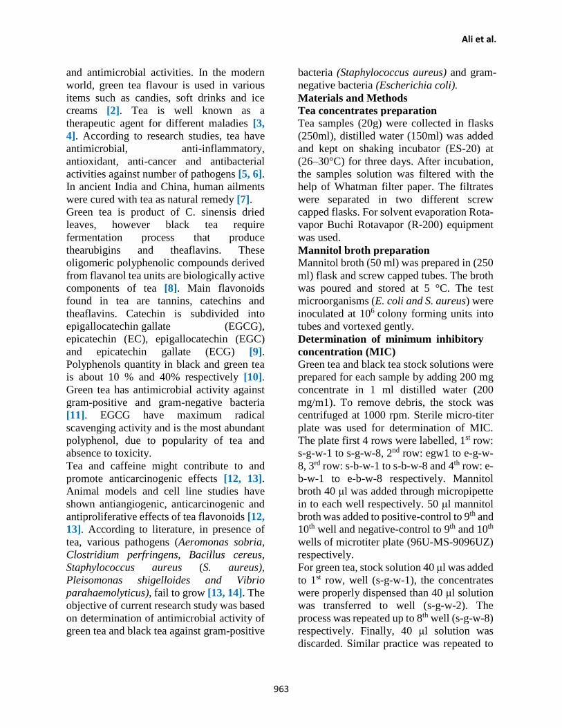
and antimicrobial activities. In the modern
world, green tea flavour is used in various
items such as candies, soft drinks and ice
creams [2]. Tea is well known as a
therapeutic agent for different maladies [3,
4]. According to research studies, tea have
antimicrobial, anti-inflammatory,
antioxidant, anti-cancer and antibacterial
activities against number of pathogens [5, 6].
In ancient India and China, human ailments
were cured with tea as natural remedy [7].
Green tea is product of C. sinensis dried
leaves, however black tea require
fermentation process that produce
thearubigins and theaflavins. These
oligomeric polyphenolic compounds derived
from flavanol tea units are biologically active
components of tea [8]. Main flavonoids
found in tea are tannins, catechins and
theaflavins. Catechin is subdivided into
epigallocatechin gallate (EGCG),
epicatechin (EC), epigallocatechin (EGC)
and epicatechin gallate (ECG) [9].
Polyphenols quantity in black and green tea
is about 10 % and 40% respectively [10].
Green tea has antimicrobial activity against
gram-positive and gram-negative bacteria
[11]. EGCG have maximum radical
scavenging activity and is the most abundant
polyphenol, due to popularity of tea and
absence to toxicity.
Tea and caffeine might contribute to and
promote anticarcinogenic effects [12, 13].
Animal models and cell line studies have
shown antiangiogenic, anticarcinogenic and
antiproliferative effects of tea flavonoids [12,
13]. According to literature, in presence of
tea, various pathogens (Aeromonas sobria,
Clostridium perfringens, Bacillus cereus,
Staphylococcus aureus (S. aureus),
Pleisomonas shigelloides and Vibrio
parahaemolyticus), fail to grow [13, 14]. The
objective of current research study was based
on determination of antimicrobial activity of
green tea and black tea against gram-positive
bacteria (Staphylococcus aureus) and gram-
negative bacteria (Escherichia coli).
Materials and Methods
Tea concentrates preparation
Tea samples (20g) were collected in flasks
(250ml), distilled water (150ml) was added
and kept on shaking incubator (ES-20) at
(26–30°C) for three days. After incubation,
the samples solution was filtered with the
help of Whatman filter paper. The filtrates
were separated in two different screw
capped flasks. For solvent evaporation Rota-
vapor Buchi Rotavapor (R-200) equipment
was used.
Mannitol broth preparation
Mannitol broth (50 ml) was prepared in (250
ml) flask and screw capped tubes. The broth
was poured and stored at 5 °C. The test
microorganisms (E. coli and S. aureus) were
inoculated at 106 colony forming units into
tubes and vortexed gently.
Determination of minimum inhibitory
concentration (MIC)
Green tea and black tea stock solutions were
prepared for each sample by adding 200 mg
concentrate in 1 ml distilled water (200
mg/m1). To remove debris, the stock was
centrifuged at 1000 rpm. Sterile micro-titer
plate was used for determination of MIC.
The plate first 4 rows were labelled, 1st row:
s-g-w-1 to s-g-w-8, 2nd row: egw1 to e-g-w-
8, 3rd row: s-b-w-1 to s-b-w-8 and 4th row: e-
b-w-1 to e-b-w-8 respectively. Mannitol
broth 40 μl was added through micropipette
in to each well respectively. 50 μl mannitol
broth was added to positive-control to 9th and
10th well and negative-control to 9th and 10th
wells of microtiter plate (96U-MS-9096UZ)
respectively.
For green tea, stock solution 40 μl was added
to 1st row, well (s-g-w-1), the concentrates
were properly dispensed than 40 μl solution
was transferred to well (s-g-w-2). The
process was repeated up to 8th well (s-g-w-8)
respectively. Finally, 40 μl solution was
discarded. Similar practice was repeated to
Pure Appl. Biol., 10(4):962-968, December, 2021 http://dx.doi.org/10.19045/bspab.2021.100100
964
8th (s-g-w-8) well respectively and 40 ml
solution was discarded from 8th (s-g-w-8)
well. Similar steps was done for 2nd row (e-
g-w-1 to e-g-w-8) wells. For dilutions of
black tea, similar practice was done to 3rd
row (s-b-w-1 to s-b-w-8) wells of plate and
4th row (e-b-w-1 to e-b-w-8). The
concentrates of tea (green and black) were
(0.78, 1.56, 3.12, 6.25, 12.5, 25, 50, and 100
mg / ml) respectively.
Suspension of Staphylococcus aureus (S.
aureus) 10 µl through micropipette was
added to 1st and 3rd row of plate. 10 µl
suspension of E.coli was added to 2nd and
4th row of microtiter plate and kept on
incubation for one day at temperature (35
°C). Methyl red dye 15 µl solution was
added to every well of microtiter plate
respectively. With the help of plate-reader
at (450 nm), optical density of wells was
noted.
MIC (minimum inhibitory
concentration)
Each concentrate stock solution (100
mg/ml) was prepared by adding 100 mg in
1 ml distilled water and centrifuged at
2000 rpm. Test microorganisms (S.
aureus and E. coli), suspensions (106
colony forming units) were inoculated on
warmed mueller-hinton agar plates.
Uniform size of wells was made on agar
plates and (50 µl) tea concentrates was
added to each well respectively and
incubated at 37 °C for 24 hours.
Results
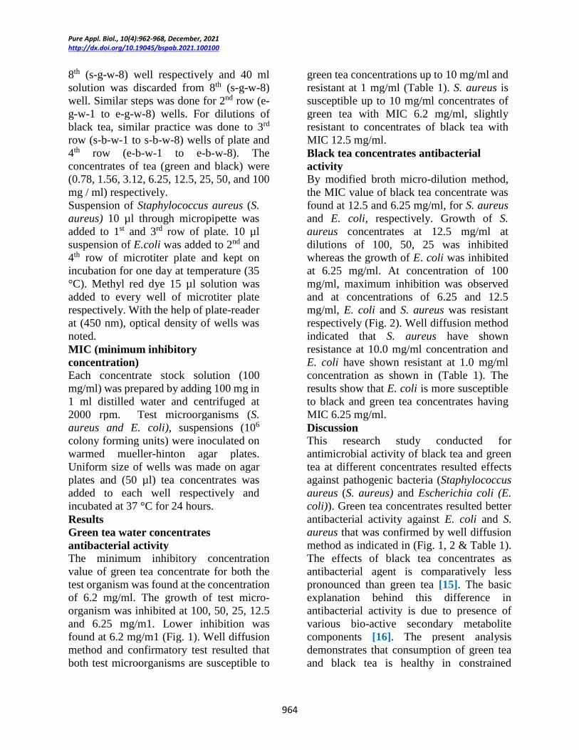
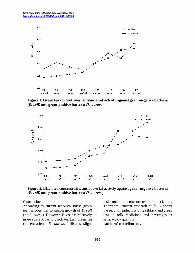
Green tea water concentrates
antibacterial activity
The minimum inhibitory concentration
value of green tea concentrate for both the
test organism was found at the concentration
of 6.2 mg/ml. The growth of test micro-
organism was inhibited at 100, 50, 25, 12.5
and 6.25 mg/m1. Lower inhibition was
found at 6.2 mg/m1 (Fig. 1). Well diffusion
method and confirmatory test resulted that
both test microorganisms are susceptible to
green tea concentrations up to 10 mg/ml and
resistant at 1 mg/ml (Table 1). S. aureus is
susceptible up to 10 mg/ml concentrates of
green tea with MIC 6.2 mg/ml, slightly
resistant to concentrates of black tea with
MIC 12.5 mg/ml.
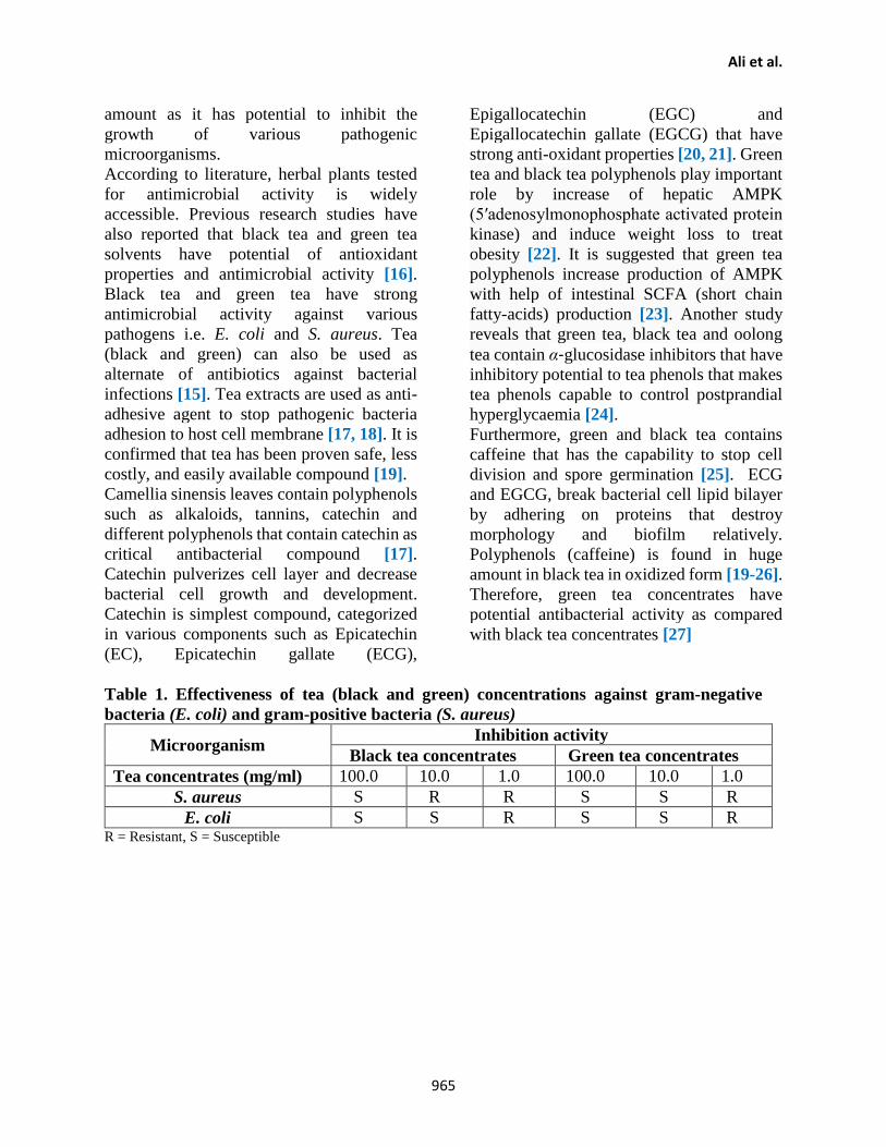
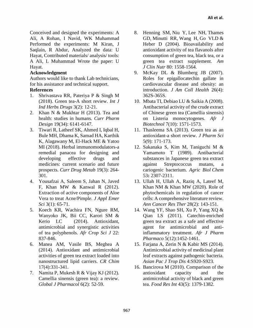
Black tea concentrates antibacterial
activity
By modified broth micro-dilution method,
the MIC value of black tea concentrate was
found at 12.5 and 6.25 mg/ml, for S. aureus
and E. coli, respectively. Growth of S.
aureus concentrates at 12.5 mg/ml at
dilutions of 100, 50, 25 was inhibited
whereas the growth of E. coli was inhibited
at 6.25 mg/ml. At concentration of 100
mg/ml, maximum inhibition was observed
and at concentrations of 6.25 and 12.5
mg/ml, E. coli and S. aureus was resistant
respectively (Fig. 2). Well diffusion method
indicated that S. aureus have shown
resistance at 10.0 mg/ml concentration and
E. coli have shown resistant at 1.0 mg/ml
concentration as shown in (Table 1). The
results show that E. coli is more susceptible
to black and green tea concentrates having
MIC 6.25 mg/ml.
Discussion
This research study conducted for
antimicrobial activity of black tea and green
tea at different concentrates resulted effects
against pathogenic bacteria (Staphylococcus
aureus (S. aureus) and Escherichia coli (E.
coli)). Green tea concentrates resulted better
antibacterial activity against E. coli and S.
aureus that was confirmed by well diffusion
method as indicated in (Fig. 1, 2 & Table 1).
The effects of black tea concentrates as
antibacterial agent is comparatively less
pronounced than green tea [15]. The basic
explanation behind this difference in
antibacterial activity is due to presence of
various bio-active secondary metabolite
components [16]. The present analysis
demonstrates that consumption of green tea
and black tea is healthy in constrained
Ali et al.
965
amount as it has potential to inhibit the
growth of various pathogenic
microorganisms.
According to literature, herbal plants tested
for antimicrobial activity is widely
accessible. Previous research studies have
also reported that black tea and green tea
solvents have potential of antioxidant
properties and antimicrobial activity [16].
Black tea and green tea have strong
antimicrobial activity against various
pathogens i.e. E. coli and S. aureus. Tea
(black and green) can also be used as
alternate of antibiotics against bacterial
infections [15]. Tea extracts are used as anti-
adhesive agent to stop pathogenic bacteria
adhesion to host cell membrane [17, 18]. It is
confirmed that tea has been proven safe, less
costly, and easily available compound [19].
Camellia sinensis leaves contain polyphenols
such as alkaloids, tannins, catechin and
different polyphenols that contain catechin as
critical antibacterial compound [17].
Catechin pulverizes cell layer and decrease
bacterial cell growth and development.
Catechin is simplest compound, categorized
in various components such as Epicatechin
(EC), Epicatechin gallate (ECG),
Epigallocatechin (EGC) and
Epigallocatechin gallate (EGCG) that have
strong anti-oxidant properties [20, 21]. Green
tea and black tea polyphenols play important
role by increase of hepatic AMPK
(5′adenosylmonophosphate activated protein
kinase) and induce weight loss to treat
obesity [22]. It is suggested that green tea
polyphenols increase production of AMPK
with help of intestinal SCFA (short chain
fatty-acids) production [23]. Another study
reveals that green tea, black tea and oolong
tea contain α‐glucosidase inhibitors that have
inhibitory potential to tea phenols that makes
tea phenols capable to control postprandial
hyperglycaemia [24].
Furthermore, green and black tea contains
caffeine that has the capability to stop cell
division and spore germination [25]. ECG
and EGCG, break bacterial cell lipid bilayer
by adhering on proteins that destroy
morphology and biofilm relatively.
Polyphenols (caffeine) is found in huge
amount in black tea in oxidized form [19-26].
Therefore, green tea concentrates have
potential antibacterial activity as compared
with black tea concentrates [27]
Table 1. Effectiveness of tea (black and green) concentrations against gram-negative
bacteria (E. coli) and gram-positive bacteria (S. aureus)
Microorganism Inhibition activity
Black tea concentrates Green tea concentrates
Tea concentrates (mg/ml) 100.0 10.0 1.0 100.0 10.0 1.0
S. aureus S R R S S R
E. coli S S R S S R R = Resistant, S = Susceptible
Pure Appl. Biol., 10(4):962-968, December, 2021 http://dx.doi.org/10.19045/bspab.2021.100100
966
Figure 1. Green tea concentrates, antibacterial activity against gram-negative bacteria
(E. coli) and gram-positive bacteria (S. aureus)
Figure 2. Black tea concentrates, antibacterial activity against gram-negative bacteria
(E. coli) and gram-positive bacteria (S. aureus)
Conclusion
According to current research study, green
tea has potential to inhibit growth of E. coli
and S. aureus. However, E. co1i is relatively
more susceptible to black tea than green tea
concentrations. S. aureus indicates slight
resistance to concentrates of black tea.
Therefore, current research study supports
the recommended use of tea (black and green
tea) in folk medicines and beverages in
satisfactory quantity.
Authors’ contributions
Ali et al.
967
Conceived and designed the experiments: A
Ali, A Rohan, I Navid, WK Muhammad
Performed the experiments: M Kiran, J
Saqlain, R Abdur, Analyzed the data: U
Hayat, Contributed materials/ analysis/ tools:
A Ali, L Muhammad Wrote the paper: U
Hayat.
Acknowledgment Authors would like to thank Lab technicians,
for his assistance and technical support.
References
1. Shrivastava RR, Pateriya P & Singh M
(2018). Green tea-A short review. Int J
Ind Herbs Drugs 3(2): 12-21.
2. Khan N & Mukhtar H (2013). Tea and
health: studies in humans. Curr Pharm
Design 19(34): 6141-6147.
3. Tiwari R, Latheef SK, Ahmed I, Iqbal H,
Bule MH, Dhama K, Samad HA, Karthik
K, Alagawany M, El-Hack ME & Yatoo
MI (2018). Herbal immunomodulators-a
remedial panacea for designing and
developing effective drugs and
medicines: current scenario and future
prospects. Curr Drug Metab 19(3): 264-
301.
4. Yousafzai A, Saleem S, Jahan N, Javed
F, Khan MW & Kanwal R (2012).
Extraction of active components of Aloe
Vera to treat Acne/Pimple. J Appl Emer
Sci 3(1): 65-71.
5. Koech KR, Wachira FN, Ngure RM,
Wanyoko JK, Bii CC, Karori SM &
Kerio LC (2014). Antioxidant,
antimicrobial and synergistic activities
of tea polyphenols. Afr Crop Sci J 22:
837-846.
6. Manea AM, Vasile BS, Meghea A
(2014). Antioxidant and antimicrobial
activities of green tea extract loaded into
nanostructured lipid carriers. CR Chim
17(4):331-341.
7. Namita P, Mukesh R & Vijay KJ (2012).
Camellia sinensis (green tea): a review.
Global J Pharmacol 6(2): 52-59.
8. Henning SM, Niu Y, Lee NH, Thames
GD, Minutti RR, Wang H, Go VLD &
Heber D (2004). Bioavailability and
antioxidant activity of tea flavanols after
consumption of green tea, black tea, or a
green tea extract supplement. Am
J Clin Nutr 80: 1558-1564.
9. McKay DL & Blumberg JB (2007).
Roles for epigallocatechin gallate in
cardiovascular disease and obesity: an
introduction. J Am Coll Health 26(4):
362S-365S.
10. Mbata TI, Debiao LU & Saikia A (2008).
Antibacterial activity of the crude extract
of Chinese green tea (Camellia sinensis)
on Listeria monocytogenes. Afr J
Biotechnol 7(10): 1571-1573.
11. Thasleema SA (2013). Green tea as an
antioxidant-a short review. J Pharm Sci
5(9): 171-173.
12. Sakanaka S, Kim M, Taniguchi M &
Yamamoto T (1989). Antibacterial
substances in Japanese green tea extract
against Streptococcus mutans, a
cariogenic bacterium. Agric Biol Chem
53: 2307-2311.
13. Ullah H, Ullah A, Raziq A, Lateef M,
Khan NM & Khan MW (2020). Role of
phytochemicals in regulation of cancer
cells: A comprehensive literature review.
Ann Cancer Res Ther 28(2): 143-151.
14. Wang YF, Shao SH, Xu P, Yang XQ &
Qian LS (2011). Catechin-enriched
green tea extract as a safe and effective
agent for antimicrobial and anti-
inflammatory treatment. Afr J Pharm
Pharmaco 5(12):1452-1461.
15. Farjana A, Zerin N & Kabir MS (2014).
Antimicrobial activity of medicinal plant
leaf extracts against pathogenic bacteria.
Asian Pac J Trop Dis 4:S920-S923.
16. Bancirova M (2010). Comparison of the
antioxidant capacity and the
antimicrobial activity of black and green
tea. Food Res Int 43(5): 1379-1382.

Pure Appl. Biol., 10(4):962-968, December, 2021 http://dx.doi.org/10.19045/bspab.2021.100100
968
17. Khan H, Khan MA & Abdullah (2015).
Antibacterial, antioxidant and cytotoxic
studies of total saponin, alkaloid and
sterols contents of decoction of
Joshanda: Identification of components
through thin layer chromatography.
Toxicol Ind Health 31(3): 202-208.
18. Khan H, Saeed M, Muhammad N &
Perviz S (2016). Phytochemical analysis,
antibacterial, and antifungal assessment
of aerial parts of Polygonatum
verticillatum. Toxicol Ind Health 32(5):
841-847.
19. Radji M, Agustama RA, Elya B &
Tjampakasari CR (2013). Antimicrobial
activity of green tea extract against
isolates of methicillin–resistant
Staphylococcus aureus and multi–drug
resistant Pseudomonas aeruginosa.
Asian Pac J Trop Biomed 3(8): 663-667.
20. Mbuthia SK, Wachira FN & Koech RK
(2014). In vitro antimicrobial and
synergistic properties of water soluble
green and black tea extracts. Afr J
Microbiol Res 8(14): 1527-1534.
21. Cho YS, Oh JJ & Oh KH (2010).
Antimicrobial activity and biofilm
formation inhibition of green tea
polyphenols on human teeth. Biotechnol
Bioproc E 15(2): 359-364.
22. Annunziata G, Maisto M, Schisano C,
Ciampaglia R, Daliu P, Narciso V,
Tenore GC, & Novellino E (2018).
Colon bioaccessibility and antioxidant
activity of white, green and black tea
polyphenols extract after in vitro
simulated gastrointestinal digestion.
Nutrients 10(11): 1711-1718.
23. Henning SM, Yang J, Hsu M, Lee R-P,
Grojean EM, Ly A, Tseng C-H, Heber D,
& Li Z (2018). Decaffeinated green and
black tea polyphenols decrease weight
gain and alter microbiome populations
and function in diet-induced obese mice.
Eur. J. Nutr 57(8): 2759-2769.
24. Yang X, Kong FJ (2016). Evaluation of
the in vitro α‐glucosidase inhibitory
activity of green tea polyphenols and
different tea types. J. Sci. Food Agric
96(3): 777-782.
25. Bagheri R, Rashidlamir A, Ashtary‐Larky D, Wong A, Alipour M &
Motevalli MS (2020). Does Green Tea
Extract Enhance the Anti‐inflammatory
Effects of Exercise on Fat Loss? Br J
Clin Pharmaco 86(4): 753-762.
26. Mohanpuria P, Kumar V & Yadav SK
(2010). Tea caffeine: metabolism,
functions, and reduction strategies. Food
Sci Biotechnol 19(2):275-287.
27. Archana S & Abraham J (2011).
Comparative analysis of antimicrobial
activity of leaf extracts from fresh green
tea, commercial green tea and black tea
on pathogens. JAPS 1(8): 149-154.
Related Documents