Biochem. J. (1995) 306, 63-69 (Printed in Great Britain) Determinants of the unusual cleavage specificity of lysyl-bradykinin- releasing kallikreins Jair R. CHAGAS, Fernanda C. V. PORTARO, Isaura Y. HIRATA, Paulo C. ALMEIDA, Maria A. JULIANO, Luiz JULIANO and Eline S. PRADO* Department of Biophysics, Escola Paulista de Medicina, Rua Tres de Maio, 100, Sao Paulo 04044-020, Brazil Kinetic data for the hydrolysis by human tissue kallikrein of fluorogenic peptides with o-aminobenzoyl-Phe-Arg (Abz-FR) as the acyl group and different leaving groups demonstrate that interactions with the S'l, S'2 and S'3 subsites are important for cleavage efficiency. In addition, studies on the hydrolysis of fluorogenic peptides with the human kininogen sequence span- ning the scissile Met-Lys bond [Abz-M-I-S-L-M-K-R-P-N-(2,4- dinitrophenyl)ethylenediamine] and analogues with different residues at positions P'1, P'2 and P'3 showed that (a) the presence of a proline residue at P'3 and the interactions with the tissue kallikrein-binding sites S2 to S'2 are determinants of Met-Lys INTRODUCTION Tissue (or glandular) kallikreins (EC 3.4.21.35) are serine proteinases involved in kininogen processing. They release lysyl- bradykinin (Pierce and Webster, 1961) by the highly specific cleavage of two dissimilar peptide bonds, Arg-Ser and Met-Lys, in bovine and human kininogens (Habermann, 1966; Han et al., 1976; Lottspeich et al., 1984) and Arg-Thr and Leu-Lys in horse kininogen (Sugo et al., 1981). The only exception known so far is the tissue kallikrein from rat which does not cleave the Met- Lys bond, but releases bradykinin by cleavage of the Lys-Arg and Arg-Arg bonds in bovine and rat kininogens respectively (Alhenc-Gelas et al., 1981; Kato et al., 1985). Tissue kallikreins have been suggested to have an important role in the control of blood pressure and in the local regulation of blood flow [see review by Bhoola et al. (1992)]. These enzymes have also been associated with tissue inflammatory reactions such as those seen in asthma, rhinitis and arthritis [see review by Polosa (1993)]. On the other hand, there is evidence that these enzymes may process precursor molecules other than kininogens, including prolactin in the pituitary gland (Powers, 1993) and prorenin (Sealey et al., 1978; Derkx et al., 1979; Yokosawa et al., 1979). Apart from their possible roles in relevant physiological processes, understanding the mechanism of limited kininogen proteolysis by tissue kallikreins is of interest, because these enzymes are usually considered to be a prototype for several other kallikrein gene family members involved in the processing of prohormones (Taylor et al., 1970) and growth factor precursors (Thomas et al, 1981). The development of specific inhibitors of tissue kallikreins and other kallikrein-like enzymes depends on detailed information about their substrate-subsite interactions. Some of these deter- minants were identified in previous studies on the hydrolysis by pig, horse and rat tissue kallikreins of the arginyl and lysyl bonds in synthetic substrates. The following observations were made: bond cleavage and (b) residues P3, P4 and/or P5 are important for cleavage efficiency. The substitution of phenylalanine for methionine or arginine in substrates with scissile Met-Lys or Arg-Xaa bonds demonstrated that lysyl-bradykinin-releasing tissue kallikreins also have a primary specificity for phenyl- alanine. The replacement of arginine by phenylalanine in (D)P-F- R-p-nitroanilide (pNA) produced an efficient and specific chromogenic substrate, (D)P-F-F-pNA, for the lysyl-bradykinin- releasing tissue kallikreins as it is resistant to plasma kallikrein and other arginine hydrolases. (a) arginine in position P1 [nomenclature of Schechter and Berger (1967)] is preferred over lysine (Fiedler, 1987); (b) the phenyl- alanine side chain binds best to the S2 subsite of the enzyme (Fiedler, 1987; Chen and Bode, 1983; Araujo-Viel et al., 1988); (c) D-amino acid residues at P3 interact favourably with human and horse kallikreins (Kettner and Shaw, 1981; Oliveira et al., 1987; Araujo-Viel et al., 1988); (d) proline in P'2 impairs the cleavage by horse and pig, but not by rat tissue kallikrein, and arginine in P'1 or P'2 increases the efficiency of cleavage by all three kallikreins (Prado et al., 1983, 1986; Chagas et al., 1991, 1992). Little is known about the determinants of the hydrolysis of the Met-Lys bond because of the resistance of the Met-Xaa linkage in short synthetic peptides to cleavage by tissue kallikreins (Araujo-Viel et al., 1981; Fiedler, 1983). The hydrolytic activity of tissue kallikreins at the Met-Lys bond is very unusual as these serine proteases have been well characterized as arginine hydrolases. In order to gain more information on the determinants of the cleavage specificity of lysyl-bradykinin-releasing kallikreins, we have studied the effects of S'l to S'3 interactions on the hydrolysis of Arg-Ser and Met- Lys bonds by human tissue kallikrein. For these studies, we have synthesized intramolecularly quenched fluorogenic peptides with the human kininogen sequences that flank the susceptible Met- Lys and Arg-Ser bonds. These model substrates, as well as peptides with analogous sequences, yielded important infor- mation on the determinants of this unusual cleavage specificity of tissue kallikreins, including data on the efficient hydrolysis of the Phe-Xaa bond in some of the synthetic peptide substrates. MATERIALS AND METHODS Enzymes Homogeneous human urinary kallikrein and rat urinary kalli- krein obtained as described by Shimamoto et al. (1980) and Chao Abbreviations used: Abz, o-aminobenzoic acid; EDDnp, N-(2,4-dinitrophenyl)ethylenediamine; pNA, p-nitroanilide; Tos, p-toluenesulphonyl. The peptides in this paper are identified by the letter of the P1 residue in the cleavage site (R, M, F, A) and a number. * To whom correspondence should be addressed. 63

Welcome message from author
This document is posted to help you gain knowledge. Please leave a comment to let me know what you think about it! Share it to your friends and learn new things together.
Transcript

Biochem. J. (1995) 306, 63-69 (Printed in Great Britain)
Determinants of the unusual cleavage specificity of lysyl-bradykinin-releasing kallikreinsJair R. CHAGAS, Fernanda C. V. PORTARO, Isaura Y. HIRATA, Paulo C. ALMEIDA, Maria A. JULIANO, Luiz JULIANOand Eline S. PRADO*Department of Biophysics, Escola Paulista de Medicina, Rua Tres de Maio, 100, Sao Paulo 04044-020, Brazil
Kinetic data for the hydrolysis by human tissue kallikrein offluorogenic peptides with o-aminobenzoyl-Phe-Arg (Abz-FR) asthe acyl group and different leaving groups demonstrate thatinteractions with the S'l, S'2 and S'3 subsites are important forcleavage efficiency. In addition, studies on the hydrolysis offluorogenic peptides with the human kininogen sequence span-ning the scissile Met-Lys bond [Abz-M-I-S-L-M-K-R-P-N-(2,4-dinitrophenyl)ethylenediamine] and analogues with differentresidues at positions P'1, P'2 and P'3 showed that (a) the presenceof a proline residue at P'3 and the interactions with the tissuekallikrein-binding sites S2 to S'2 are determinants of Met-Lys
INTRODUCTIONTissue (or glandular) kallikreins (EC 3.4.21.35) are serineproteinases involved in kininogen processing. They release lysyl-bradykinin (Pierce and Webster, 1961) by the highly specificcleavage of two dissimilar peptide bonds, Arg-Ser and Met-Lys,in bovine and human kininogens (Habermann, 1966; Han et al.,1976; Lottspeich et al., 1984) and Arg-Thr and Leu-Lys in horsekininogen (Sugo et al., 1981). The only exception known so faris the tissue kallikrein from rat which does not cleave the Met-Lys bond, but releases bradykinin by cleavage of the Lys-Argand Arg-Arg bonds in bovine and rat kininogens respectively(Alhenc-Gelas et al., 1981; Kato et al., 1985).
Tissue kallikreins have been suggested to have an importantrole in the control of blood pressure and in the local regulationof blood flow [see review by Bhoola et al. (1992)]. These enzymeshave also been associated with tissue inflammatory reactionssuch as those seen in asthma, rhinitis and arthritis [see review byPolosa (1993)]. On the other hand, there is evidence that theseenzymes may process precursor molecules other than kininogens,including prolactin in the pituitary gland (Powers, 1993) andprorenin (Sealey et al., 1978; Derkx et al., 1979; Yokosawa et al.,1979). Apart from their possible roles in relevant physiologicalprocesses, understanding the mechanism of limited kininogenproteolysis by tissue kallikreins is of interest, because theseenzymes are usually considered to be a prototype for severalother kallikrein gene family members involved in the processingofprohormones (Taylor et al., 1970) and growth factor precursors(Thomas et al, 1981).The development of specific inhibitors of tissue kallikreins and
other kallikrein-like enzymes depends on detailed informationabout their substrate-subsite interactions. Some of these deter-minants were identified in previous studies on the hydrolysis bypig, horse and rat tissue kallikreins of the arginyl and lysyl bondsin synthetic substrates. The following observations were made:
bond cleavage and (b) residues P3, P4 and/or P5 are importantfor cleavage efficiency. The substitution of phenylalanine formethionine or arginine in substrates with scissile Met-Lys orArg-Xaa bonds demonstrated that lysyl-bradykinin-releasingtissue kallikreins also have a primary specificity for phenyl-alanine. The replacement of arginine by phenylalanine in (D)P-F-R-p-nitroanilide (pNA) produced an efficient and specificchromogenic substrate, (D)P-F-F-pNA, for the lysyl-bradykinin-releasing tissue kallikreins as it is resistant to plasma kallikreinand other arginine hydrolases.
(a) arginine in position P1 [nomenclature of Schechter and Berger(1967)] is preferred over lysine (Fiedler, 1987); (b) the phenyl-alanine side chain binds best to the S2 subsite of the enzyme(Fiedler, 1987; Chen and Bode, 1983; Araujo-Viel et al., 1988);(c) D-amino acid residues at P3 interact favourably with humanand horse kallikreins (Kettner and Shaw, 1981; Oliveira et al.,1987; Araujo-Viel et al., 1988); (d) proline in P'2 impairs thecleavage by horse and pig, but not by rat tissue kallikrein, andarginine in P'1 or P'2 increases the efficiency of cleavage by allthree kallikreins (Prado et al., 1983, 1986; Chagas et al., 1991,1992). Little is known about the determinants of the hydrolysisof the Met-Lys bond because of the resistance of the Met-Xaalinkage in short synthetic peptides to cleavage by tissue kallikreins(Araujo-Viel et al., 1981; Fiedler, 1983).The hydrolytic activity of tissue kallikreins at the Met-Lys
bond is very unusual as these serine proteases have been wellcharacterized as arginine hydrolases. In order to gain moreinformation on the determinants of the cleavage specificity oflysyl-bradykinin-releasing kallikreins, we have studied the effectsof S'l to S'3 interactions on the hydrolysis of Arg-Ser and Met-Lys bonds by human tissue kallikrein. For these studies, we havesynthesized intramolecularly quenched fluorogenic peptides withthe human kininogen sequences that flank the susceptible Met-Lys and Arg-Ser bonds. These model substrates, as well aspeptides with analogous sequences, yielded important infor-mation on the determinants of this unusual cleavage specificity oftissue kallikreins, including data on the efficient hydrolysis of thePhe-Xaa bond in some of the synthetic peptide substrates.
MATERIALS AND METHODS
EnzymesHomogeneous human urinary kallikrein and rat urinary kalli-krein obtained as described by Shimamoto et al. (1980) and Chao
Abbreviations used: Abz, o-aminobenzoic acid; EDDnp, N-(2,4-dinitrophenyl)ethylenediamine; pNA, p-nitroanilide; Tos, p-toluenesulphonyl. Thepeptides in this paper are identified by the letter of the P1 residue in the cleavage site (R, M, F, A) and a number.
* To whom correspondence should be addressed.
63

64 J. R. Chagas and others
and Margolius (1979) respectively were a generous gift fromProfessor J. Chao, Medical University of South Carolina, SC,U.S.A. Porcine pancreatic kallikrein (1956 kallikrein units/mgof protein), prepared by Bayer A. C., Wuppertal, Germany, waskindly supplied by Dr. F. Fiedler, University of Munich, Munich,Germany. Dr. C. A. M. Sampaio, of the Department of Bio-chemistry, Escola Paulista de Medicina, Sao Paulo, Brazil, kindlysupplied the sample of human plasma kallikrein, which wasisolated as described previously (Oliva et al., 1982). Horse tissuekallikrein was purified to homogeneity as described by Giusti etal. (1988). Molar concentrations of the enzyme solutions weredetermined by active-site titration with 4-nitrophenyl-4-guanidinobenzoate by the methods of Fiedler et al. (1972) andSampaio et al. (1984).
Peptide substratesPreviously described procedures were used for the synthesis,purification and analysis of the intramolecularly quenchedfluorogenic peptide substrates (Chagas et al., 1991) and ofpeptidyl-p-nitroanilide (pNA) (Juliano and Juliano, 1985).The peptides containing glutamine as C-terminal residuewere synthesized by solid-phase peptide synthesis as follows:N2-Fmoc-Glu(aCO)-N-(2,4-dinitrophenyl)ethylenediamine(EDDnp) was obtained and attached to benzhydrylamino resinthrough p-{(R,S)- x-[1-(9H-fluoren-9-yl)methoxyformamido]-2,4-dimethoxybenzylphenoxyacetic acid as a linker and thepeptides were synthesized by Fmoc methodology using themultiple automated peptide synthesizer PSSM-8 (ShimadzuCo.). Methionine-containing peptides, before purification byh.p.l.c., were submitted to a 48 h incubation at 37 'C with 0.1 Mdithiothreitol to reduce the methionine sulphoxide introducedduring synthesis. Peptides and fragments for h.p.l.c. identificationof hydrolysis products were prepared with either methionine oroxidized methionine residues in order to allow the detection byh.p.l.c. of any oxidation product formed under the assayconditions. The stock solutions of the peptide substrates wereprepared in 1 mM HCI containing no more than 100%dimethylformamide.
H.p.l.c. analysis of the synthetic fluorogenic substrates and theirhydrolysis productsPeptide solutions (80-150 ,sM) in 20 mM Tris/HCl, pH 8.0 or9.0, were incubated with the different proteinases at 30 'C.Samples (20 ,tl) of the substrate and incubation mixtures wereperiodically removed for h.p.l.c. analysis until 100 0/ hydrolysiswas reached. The hydrolysis products were separated by h.p.l.c.and submitted to amino acid analysis. The scissile bonds werededuced from the sequences of the substrate fragments. Theh.p.l.c. conditions used for the analytical procedure were: (a)0.1 M NaH2PO4 (pH 4.2) as solvent A and acetonitrile/water(9: 1, v/v) as solvent B and (b) 0.1 0% trifluoroacetic acid in water(solvent A) and acetonitrile/water (9: 1, v/v) containing 0.1 %trifluoroacetic acid as solvent B. Separations were performed ata flow rate of I ml/min using a Novapack C18 column(3.9 mm x 150 mm). For peptide purification, an Ultraprep C18column (150 mm x 21.2 mm; 10 ,tm particles) was used andgradient elution was performed with 0.1 % trifluoroacetic acid(solvent A) and acetonitrile/water (9: 1 v/v) (solvent B) at a flowrate of 3 ml/min. In all cases, elution was followed by u.v.
absorption (214 nm) and by fluorescence (Aemissmon = 420 nm and
Enzyme assays
Hydrolysis of the peptidyl-pNA at 30 °C in 0.05 M Tris/HCl,pH 9.0, containing 1 mM EDTA was photometrically measuredas previously described (Oliveira et al., 1987) by determination ofthe 4-nitroaniline released after the addition of enzyme to 2 ml ofreaction buffer. Hydrolysis was carried out for 5-10 min and theinitial hydrolytic rates were calculated from the slope of the time-dependent absorbance curves extrapolated to zero time.
Hydrolysis of the fluorogenic peptide substrates at 30 °C in0.02 M Tris/HCl, pH 9.0, containing 1 mM EDTA was moni-tored by measuring the fluorescence at Aemission = 420 nm andAexcitation = 320 nm in an Hitachi F-2000 spectrofluorimeter, as
previously described (Chagas et al., 1991).The standard hydrolysis conditions were strictly maintained
for different substrates. Low buffer concentrations were usedbecause the activities of tissue kallikreins can be impaired by highconcentrations of cations (Oliveira et al., 1987; Araujo-Viel etal., 1988). EDTA was added to avoid proteinase inhibition byheavy metals. The enzyme concentration varied from 0.016 nM,for the best substrates, to 4 nM for the less susceptible ones. Inmost of the cases, the concentration of the substrates rangedfrom 10 times less than the Km to 10 times greater than the Km.The kinetic parameters were calculated as described by Wilkinson(1961). For substrates with Km lower than 0.2 ,uM, highersubstrate concentrations were used and the kcat./Km values were
confirmed from determinations under pseudo-first-order con-
ditions.
Inhibition studiesEnzyme assays were conducted at 30 °C in 20 mM Tris/HClcontaining 1 mM EDTA, at pH 9.0, using (D)P-F-R-pNAand aminobenzoyl (Abz)-F-R-S-S-R-EDDnp or Abz-F-R-S-R-EDDnp as substrates. Three inhibitor concentrations for eachone of four substrate concentrations were used. The substrateconcentration intervals used corresponded to 1-10 Km and thelimit for substrate hydrolysis was 10%. The Ki values were
calculated from Dixon plots (Dixon, 1972) and/or from theK, and the substrate Km values (Salvesen and Nagase, 1989).The type of inhibition was determined from Lineweaver-Burkplots as described by Segel (1974).
RESULTSHydrolysis by human tissue kalllkrein of Arg-Xaa bonds influorogenic peptides with Abz-F-R as the acyl group and differentleaving groups
Tabies 1-3 present the kinetic data for hydrolysis by humantissue kallikrein of internally quenched fluorogenic peptides,which varied in their content of hydrophilic (serine), hydrophobicaliphatic (leucine), aromatic (phenylalanine) and basic (arginine)residues at positions P'1, P'2 and P'3. H.p.l.c. analysis of thehydrolysates of all the substrates of the R series (Tables 1-3)demonstrated that Arg-Xaa was the only bond cleaved.The model peptide for these fluorogenic substrates, Abz-F-R-
S-S-R-EDDnp (peptide RI), has the sequence of humankininogen at the C-terminal side of bradykinin and was hydro-lysed by human tissue kallikrein (Km = 0.19,M and kcat =
1.35 s-1). The glutamine residue in the analogue R2 (Table 1)does not correspond to the P'4 residue in human kininogen butwas introduced as a requirement for the solid-phase synthesis ofthe substrates with systematic variations at the P'l, P'2 and P'3positions. The addition of glutamine to the C-terminus of peptideRI increased with Km and did not change the kcat values (peptide
Aexcitation = 320 nm). R2, Table 1).

Tissue kallikrein subsite interactions 65
Table 1 Kinetic parameters for the hydrolysis by human tissue kallikrein of fluorogenic peptides with different residues at P'1 (X)Hydrolysis conditions were 30 °C, 20 mM Tris/HCI/l mM EDTA, pH 9.0. Values are means+ S.D.
Kmk kI/K
Peptides Substrates (JIM) (s.1) (s-1 * mM-1)
Abz-F-R-X-S-R-Q-EDDnpR2 Abz-F-R-S-S-R-Q-EDDnpR3 Abz-F-R-L-S-R-Q-EDDnpR4 Abz-F-R-R-S-R-Q-EDDnpR5 Abz-F-R-F-S-R-Q-EDDnp
Abz-F-R-X-P-EDDnpR6 Abz-F-R-R-P-EDDnpR7 Abz-F-R-S-P-EDDnpR8 Abz-F-R-A-P-EDDnp
Abz-F-R-X-V-EDDnpR9 Abz-F-R-R-V-EDDnpR10 Abz-F-R-S-V-EDDnpRi 1 Abz-F-R-L-V-EDDnp
1.5 + 0.40.6+ 0.11.3 + 0.30.4+ 0.2
0.13 + 0.021.40 + 0.201.20 + 0.20
0.06 + 0.010.90 + 0.100.32 + 0.01
1.6 + 0.21.5 + 0.17.1 +0.20.8 + 0.1
0.06 + 0.010.22 + 0.01
0.041 + 0.004
0.45 + 0.020.88 + 0.01
0.006 + 0.001
1067250054622000
46215734
750098719
Table 2 Kinetic parameters for the hydrolysis by human tissue kallikrein of fluorogenic peptides with different residues at P'2 (X)
Hydrolysis conditions were 30 °C, 20 mM Tris/HCI/1 mM EDTA, pH 9.0. Values are means+S.D.
Km kut kat /KmPeptides Substrates (4uM) (s-1) (s- mM-1)
Abz-F-R-S-X-R-Q-EDDnpR2 Abz-F-R-S-S-R-Q-EDDnpR12 Abz-F-R-S-L-R-Q-EDDnpR13 Abz-F-R-S-R-R-Q-EDDnpR14 Abz-F-R-S-F-R-O-EDDnp
Abz-F-R-S-X-EDDnpRi 5 Abz-F-R-S-S-EDDnpR7 Abz-F-R-S-P-EDDnpR1 0 Abz-F-R-S-V-EDDnpR16 Abz-F-R-S-R-EDDnp
1.50 + 0.400.18 + 0.030.12 + 0.02
0.47 + 0.061.40 + 0.200.90 + 0.100.04 + 0.01
1.6 + 0.20.40 + 0.021.18+ 0.08
Not hydrolysed
0.53 + 0.020.22 + 0.010.88 + 0.011.01 + 0.07
1 0672 2229833
1128157978
25250
Table 3 Kinetic parameters for the hydrolysis by human tissue kallikrein of fluorogenic peptides with different residues at position P'3 (X
Hydrolysis conditions were 30 °C, 20 mM Tris/HCI/1 mM EDTA, pH 9.0. Values are means+ S.D.
Km k kat/KKPeptides Substrates (uM) (s1) (s- mM-1)
Abz-F-R-S-S-X-Q-EDDnpR2 Abz-F-R-S-S-R-Q-EDDnpR1 7 Abz-F-R-S-S-L-O-EDDnpR18 Abz-F-R-S-S-F-Q-EDDnpR1 9 Abz-F-R-S-S-S-Q-EDDnp
1.5 + 0.401.1 + 0.203.0 + 0.202.2 + 0.92
1.60 + 0.200.61 + 0.040.69 + 0.021.50 +0.03
1067554230681
Substitutions at P'1
(1) Abz-F-R-X-S-R-Q-EDDnp (X = S, L, R or F). The kineticparameters for the hydrolysis by human tissue kallikrein ofpeptides R2 and R5 (Table 1) demonstrate a preference of theenzyme S'l subsite for arginine over serine, leucine and phenyl-alanine and was characterized by a higher kcat/Km value for thepeptide with arginine in P'1 (peptide R4).
(2) Abz-F-R-X-P-EDDnp and Abz-F-R-X-V-EDDnp (X =R, S, A or L). The preference for arginine in P'1 was also
demonstrated by the lower Km values for the hydrolysis byhuman kallikrein 1 of the tetrapeptides R6 and R9 which haverespectively Arg-Pro and Arg-Val as the leaving group.
Substitutions at P'2(1) Abz-F-R-S-X-R-Q-EDDnp (X = S, L, R or F). In thehydrolysis by human tissue kallikrein of these peptides withsubstitutions at the P'2 position (R2, R12, R13 and R14 in Table

66 J. R. Chagas and others
Table 4 Hydrolysis by human tissue kallikrein of fluorogenic peptides with the human kininogen sequence flanking the scissile Met-Lys bond (peptides MIand M2) and analogues
Hydrolysis conditions were 30 °C, 20 mM Tris/HCI/1 mM EDTA, pH 9.0. Values are means + S.D. L.R.C., low rate of cleavage; see the text for hydrolysis conditions. N.H., no hydrolysis observedwith [E] = 7 nM and [S] = 4 1uM. Arrows (J ) indicate the cleavage sites.
SubstratesKm km1 kat/KK
Peptides P2 Pi P'1 P'2 P'3 P'4 (uM) (s-1) (s- mM-')
Ml Abz-M2 Abz-M-1-S-Fl Abz-F2 Abz-M-1-S-M3 Abz-M-1-S-Al Abz-M-1-S-M4 Abz-M-1-S-M5 Abz-M-1-S-M6 Abz-M-1-S-M7 Abz-M-1-S-M8 Abz-M-1-S-M9 Abz-M-1-S-M1o Abz-M-1-S-Ml1 Abz-M-1-S-M12 Abz-M-1-S-M13 Abz-M-1-S-
L - ML - ML - FL - FL 4 M(0)L - AL - ML - ML - ML - ML - ML - ML - ML - ML - ML - M
U K - R4 K - RJ K - RJ K - R1 K - R4 K 1 R4 R 1 RI A - RJ K - R(Tos)
J K - RJ K - K4 K - S4 K - L- K - F- K - P
J K 4 R
2), the highest kcat/Km value was shown by peptide RI 3 (X = R),which had a 12-fold lower Km value than peptide R2. Thesubstrate Rl 2 (X = L) was hydrolysed with a similarly low Kmvalue but its kcat value was 4-fold lower than that of R2. PeptideR14 (X = F) was resistant to hydrolysis by kallikrein 1.
(2) Abz-F-R-S-X-EDDnp (X = S, P, V or R). The data inTable 2 show that arginine at P'2 in peptide R16 led to a
significant decrease in Km and an increase in kcat values whichresulted in a 22-fold higher kcat./Km than for the peptide withserine at P'2 (R15). The tetrapeptide R7 (X = P) showed thelowest susceptibility to human tissue kallikrein in this peptideseries, and the kcat./Km value for substrate RIO (X = V) did notdiffer from that of peptide R15.
Substitutions at P'3
(1) Abz-F-R-S-S-X-Q-EDDnp (X = R, L, F or S). The kineticdata in Table 3 demonstrate a lower susceptibility to humankallikrein 1 of the substrates with leucine, serine (peptides R17and R19) and particularly phenylalanine (peptide R18) at pos-ition P'3 than peptide R2 (X = R).
Hydrolysis by human tissue kalflkrein of fluorogenic peptides withthe human kininogen sequence flanking the scissile Met-Lys bondand their analoguesH.p.l.c. analysis of the hydrolysates of peptides Abz-L-M-K-R-P-EDDnp (Ml) and of the longer homologue Abz-M-I-S-L-M-K-R-P-EDDnp demonstrated that the Met-Lys bond was theonly one cleaved by human, horse and pig tissue kallikrein. Asexpected the bradykinin-releasing enzymes, rat tissue and plasmakallikreins, trypsin and plasmin, cleaved only the Lys-Arg bond.The kinetic data in Table 4 demonstrate that the increase inpeptide size (Ml1 and M2) significantly increased the susceptibilityof the Met-Lys bond to human tissue kallikrein.The cleavage site(s) in this series of peptides with different P1
P'4 residues were determined by amino acid analysis of the
hydrolysis products separated by h.p.l.c. and are indicated inTable 4.
Substitutions at P1
(1) Abz-L-F-K-R-P-EDDnp (Fl) and Abz-M-I-S-L-F-K-R-P-EDDnp (F2). The Phe-Lys bond in these peptides was the onlybond susceptible to human, horse and pig tissue kallikreins. Thekcat values for the hydrolysis of these peptides by humankallikrein 1 were surprisingly higher than the values for thesubstrates with methionine at P1 (Table 4).
(2) Abz-M-I-S-L-M(O)-K-R-P-EDDnp (M3). Incubation of a60 1uM peptide solution with 2.5 x 10⁻⁴ uM human tissuekallikrein at pH 9.0 for approx. 15 h led to a 33 % hydrolysis ofthe peptide from which the hydrolysis products were identified as
being K-R-P-EDDnp (approx. 19 %) and M(O)-K-R-P-EDDnp(14 %). For the lower homologous peptide, Abz-L-M(O)-K-R-P-EDDnp, a 100-fold higher enzyme concentration was used andafter a 15 h incubation 34% of the substrate was hydrolysed viacleavage of the Met(O)-Lys and Leu-Met bonds. The higher K1values for both peptides with an oxidized methionine residue(Table 5) indicate less efficient binding of this residue to the Ssubsite of the tissue kallikrein.
(3) Abz-M-I-S-L-A-K-R-P-EDDnp (Al). This peptide was
also very resistant to tissue kallikrein. At [S] = 30,M and [E] =1 x 10⁻² #M, only 10% hydrolysis was detected after a 5 hincubation. Similar amounts of Lys-Arg-Pro-EDDnp and Arg-Pro-EDDnp were found, indicating cleavage at both the Ala-Lysand Lys-Arg bonds. The K, value for this peptide also indicatesa less efficient binding to the enzyme (Table 5).
Substitutions at P'1
(1) Abz-M-I-S-L-M-R-R-P-EDDnp (M4). Two bonds, Met-Argand Arg-Arg, were cleaved in this peptide by the tissue kallikrein,but the rates of hydrolysis were low. At a substrate concentrationof 60 1sM, 30% of the substrate was hydrolysed overnight, andthe hydrolysis products, identified by h.p.l.c., corresponded to
1.1 + 0.10.7+0.10.9+0.10.8 + 0.2
_ p_ p_ p_ p- p_ p_ p_ p_ p_ p- p_ p_ p
p- p- S
64471
22225250
1501578108
2
0.07 + 0.010.33 + 0.022.0+ 0.1
4.20 + 0.04L.R.C.L.R.C.L.R.C.L.R.C.L.R.C.0.33 + 0.011.42 + 0.080.04 + 0.010.02 + 0.01N.H.N.H.L.R.C.
2+10.9 + 0.1
0.37 + 0.049+2
-0-Q-Q-Q-Q-Q- S
Tissue kallikrein subsite interactions 67
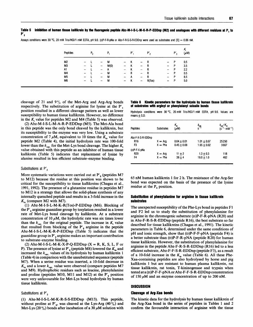
Table 5 Inhibition of human tissue kalllkrein by the fluorogenic peptfde Abz-M-I-S-L-M-K-R-P-EDDnp (M2) and analogues with dffferent residues at P1 toP'2Assays conditions were 30 °C, 20 mM Tris/HCI/1 mM EDTA, pH 9.0. (D)P-F-R-pNa or Abz-F-R-S-S-R-EDDnp were used as substrates and [E] = 0.06 nM.
KPPeptides P2 P1 P'1 P'2 P'3 (/,uM)
M2M3AlM4M5M6
- L - M- L - M(0)- L - A- L - M- L - M- L - M
cleavage of 21 and 9% of the Met-Arg and Arg-Arg bondsrespectively. The substitution of arginine for lysine at the P'1position resulted in a different cleavage pattern as well as lowersusceptibility to human tissue kallikrein. However, no differencein the K, value for peptides M2 and M4 (Table 5) was observed.
(2) Abz-M-I-S-L-M-A-R-P-EDDnp (M5). The Met-Ala bondin this peptide was the only bond cleaved by the kallikrein, butits susceptibility to the enzyme was very low. Using a substrateconcentration of 7 ,tM, equivalent to 10 times the Km value forpeptide M2 (Table 4), the initial hydrolysis rate was 100-foldlower than the kc.t for the Met-Lys bond cleavage. The higher Kivalue obtained with this peptide as an inhibitor of human tissuekallikrein (Table 5) indicates that replacement of lysine byalanine resulted in less efficient substrate-enzyme binding.
Substitutions at P'2More systematic variations were carried out at P'2 (peptides M7to M12) because the residue at this position was shown to becritical for the susceptibility to tissue kallikreins (Chagas et al.,1991, 1992). The presence of a glutamine residue in peptides M7to M12 is a strategy that allows the solid-phase synthesis of anyinternally quenched peptide and results in a 3-fold increase in theKm (compare M2 with M7).
(1) Abz-M-I-S-L-M-K-R(Tos)-P-EDDnp (M6). Blocking ofthe P'2 arginine guanidine group by tosylation resulted in a lowerrate of Met-Lys bond cleavage by kallikrein. At a substrateconcentration of 10,uM, the hydrolytic rate was six times lowerthan the kcat for the original peptide. The increase in K1 valuethat resulted from blocking of the P'2 arginine in the peptideAbz-M-I-S-L-M-K-R-P-EDDnp (Table 5) indicates that theguanidine group in P'2 arginine makes an important contributionto substrate-enzyme binding.
(2) Abz-M-I-S-L-M-K-X-P-Q-EDDnp (X = R, K, S, L, F orP). The presence of lysine at P'2 (peptide M8) lowered the Km andincreased the kcat values of its hydrolysis by human kallikrein 1(Table 4) in comparison with the unsubstituted sequence (peptideM7). When a serine residue was inserted, a 10-fold decrease inKm and a lower kcat value were observed (compare peptides M7and M9). Hydrophobic residues such as leucine, phenylalanineand proline (peptides M10, Ml 1 and M12) at the P'2 positionwere very unfavourable for Met-Lys bond hydrolysis by humantissue kallikrein.
Substitutions at P'3
(1) Abz-M-I-S-L-M-K-R-S-S-EDDnp (M13). This peptide,without proline at P'2, was cleaved at the Lys-Arg (40%) andMet-Lys (20 %) bonds after incubation of a 30 ,#M solution with
K - RK - RK - RR - RA - RK - R(Tos)
- P 0.5- P 3.5- P 2.2- P 0.5- P 8.5- P 5.0
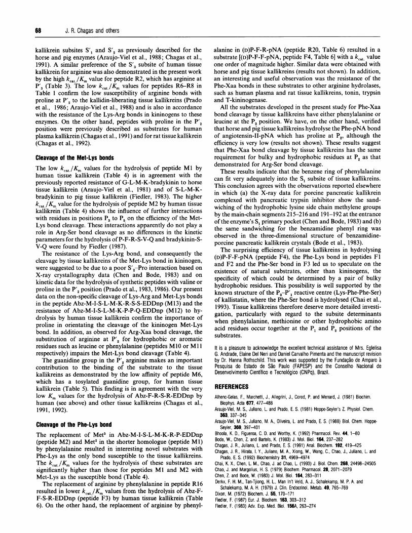
Table 6 Kinetic parameters for the hydrolysis by human tissue kallikreinof substrates with arginyl or phenylalanyl scissile bondsHydrolysis conditions were 30 °C, 20 mM Tris/HCI/1 mM EDTA, pH 9.0. Values aremeans+ S.D.
Km k kcat/KmPeptides Substrates (utM) s (s-1 . mM-1)
Abz-F-X-S-R-EDDnpR16 X = Arg 0.04 +0.01 1.01 + 0.07 25250F3 X = Phe 0.45+0.08 1.65+0.02 3667
(D)P-F-X-pNaR20 X = Arg 11+2 1.3+0.3 118F4 X = Phe 39+4 18.0+1.0 462
65 nM human kallikrein 1 for 2 h. The resistance of the Arg-Serbond was expected on the basis of the presence of the lysineresidue at the P2 position.
Substitution of phenylalanine for arginine In tissue kalllkreinsubstratesThe unexpected susceptibility of the Phe-Lys bond in peptides Fland F2 led us to study the substitution of phenylalanine forarginine in the chromogenic substrate (D)P-F-R-pNA (R20) andin Abz-F-R-S-R-EDDnp (peptide R16), the best substrate so fardescribed for tissue kallikreins (Chagas et al., 1991). The kineticparameters in Table 6, determined under the same conditions ofpH and ionic strength, show that (D)P-F-F-pNA (peptide F4) isa better substrate than (D)P-F-R-pNA (peptide R20) for humantissue kallikrein. However, the substitution of phenylalanine forarginine in the peptide Abz-F-R-S-R-EDDnp (R16) led to a lessefficient substrate, Abz-F-F-S-R-EDDnp (peptide F3), as a resultof a 10-fold increase in the Km value (Table 6). All these Phe-Xaa-containing peptides are also hydrolysed by horse and pigkallikrein 1 but are resistant to human plasma kallikrein, rattissue kallikrein, rat tonin, T-kininogenase and trypsin whentested at a (D)P-F-F-pNA or Abz-F-F-S-R-EDDnp concentrationof 150 ,uM and an enzyme concentration of up to 200 nM.
DISCUSSIONCleavage of Arg-Xaa bondsThe kinetic data for the hydrolysis by human tissue kallikrein ofthe Arg-Xaa bond in the series of peptides in Tables 1 and 2confirm the favourable interaction of arginine with the tissue
68 J. R. Chagas and others
kallikrein subsites S'1 and S'2 as previously described for thehorse and pig enzymes (Araujo-Viel et al., 1988; Chagas et al.,1991). A similar preference of the S'3 subsite of human tissuekallikrein for arginine was also demonstrated in the present workby the high kcat/Km value for peptide R2, which has arginine atP'3 (Table 3). The low kcat./Km values for peptides R6-R8 inTable 1 confirm the low susceptibility of arginine bonds withproline at P'2 to the kallidin-liberating tissue kallikreins (Pradoet al., 1986; Araujo-Viel et al., 1988) and is also in accordancewith the resistance of the Lys-Arg bonds in kininogens to theseenzymes. On the other hand, peptides with proline in the P'2position were previously described as substrates for humanplasma kallikrein (Chagas et al., 1991) and for rat tissue kallikrein(Chagas et al., 1992).
Cleavage of the Met-Lys bondsThe low kcat./Km values for the hydrolysis of peptide Ml byhuman tissue kallikrein (Table 4) is in agreement with thepreviously reported resistance of G-L-M-K-bradykinin to horsetissue kallikrein (Araujo-Viel et al., 1981) and of S-L-M-K-bradykinin to pig tissue kallikrein (Fiedler, 1983). The higherkcat/Km value for the hydrolysis of peptide M2 by human tissuekallikrein (Table 4) shows the influence of further interactionswith residues in positions P3 to P5 on the efficiency of the Met-Lys bond cleavage. These interactions apparently do not play arole in Arg-Ser bond cleavage as no differences in the kineticparameters for the hydrolysis of P-F-R-S-V-Q and bradykinin-S-V-Q were found by Fiedler (1987).The resistance of the Lys-Arg bond, and consequently the
cleavage by tissue kallikreins of the Met-Lys bond in kininogen,were suggested to be due to a poor S'2-Pro interaction based onX-ray crystallography data (Chen and Bode, 1983) and onkinetic data for the hydrolysis of synthetic peptides with valine orproline in the P'2 position (Prado et al., 1983, 1986). Our presentdata on the non-specific cleavage of Lys-Arg and Met-Lys bondsin the peptide Abz-M-I-S-L-M-K-R-S-S-EDDnp (Ml 3) and theresistance of Abz-M-I-S-L-M-K-P-P-Q-EDDnp (M 12) to hy-drolysis by human tissue kallikrein confirm the importance ofproline in orientating the cleavage of the kininogen Met-Lysbond. In addition, as observed for Arg-Xaa bond cleavage, thesubstitution of arginine at P'2 for hydrophobic or aromaticresidues such as leucine or phenylalanine (peptides M 10 or Ml 1respectively) impairs the Met-Lys bond cleavage (Table 4).The guanidine group in the P'2 arginine makes an important
contribution to the binding of the substrate to the tissuekallikreins as demonstrated by the low affinity of peptide M6,which has a tosylated guanidine group, for human tissuekallikrein (Table 5). This finding is in agreement with the verylow Km values for the hydrolysis of Abz-F-R-S-R-EDDnp byhuman (see above) and other tissue kallikreins (Chagas et al.,1991, 1992).
Cleavage of the Phe-Lys bondThe replacement of Met5 in Abz-M-I-S-L-M-K-R-P-EDDnp(peptide M2) and Met2 in the shorter homologue (peptide Ml)by phenylalanine resulted in interesting novel substrates withPhe-Lys as the only bond susceptible to the tissue kallikreins.The kcat./Km values for the hydrolysis of these substrates aresignificantly higher than those for peptides Ml and M2 withMet-Lys as the susceptible bond (Table 4).The replacement of arginine by phenylalanine in peptide R16
resulted in lower kcat /Km values from the hydrolysis of Abz-F-F-S-R-EDDnp (peptide F3) by human tissue kallikrein (Table6). On the other hand, the replacement of arginine by phenyl-
alanine in (D)P-F-R-pNA (peptide R20, Table 6) resulted in asubstrate [(D)P-F-F-pNA, peptide F4, Table 6] with a kcat valueone order of magnitude higher. Similar data were obtained withhorse and pig tissue kallikreins (results not shown). In addition,an interesting and useful observation was the resistance of thePhe-Xaa bonds in these substrates to other arginine hydrolases,such as human plasma and rat tissue kallikreins, tonin, trypsinand T-kininogenase.
All the substrates developed in the present study for Phe-Xaabond cleavage by tissue kallikreins have either phenylalanine orleucine at the P2 position. We have, on the other hand, verifiedthat horse and pig tissue kallikreins hydrolyse the Phe-pNA bondof angiotensin-II-pNA which has proline at P2, although theefficiency is very low (results not shown). These results suggestthat Phe-Xaa bond cleavage by tissue kallikreins has the samerequirement for bulky and hydrophobic residues at P2 as thatdemonstrated for Arg-Ser bond cleavage.
These results indicate that the benzene ring of phenylalaninecan fit very adequately into the Si subsite of tissue kallikreins.This conclusion agrees with the observations reported elsewherein which (a) the X-ray data for porcine pancreatic kallikreincomplexed with pancreatic trypsin inhibitor show the sand-wiching of the hydrophobic lysine side chain methylene groupsby the main-chain segments 215-216 and 191-192 at the entranceof the enzyme's S, primary pocket (Chen and Bode, 1983) and (b)the same sandwiching for the benzamidine phenyl ring wasobserved in the three-dimensional structure of benzamidine-porcine pancreatic kallikrein crystals (Bode et al., 1983).The surprising efficiency of tissue kallikreins in hydrolysing
(D)P-F-F-pNA (peptide F4), the Phe-Lys bond in peptides Fland F2 and the Phe-Ser bond in F3 led us to speculate on theexistence of natural substrates, other than kininogens, thespecificity of which could be determined by a pair of bulkyhydrophobic residues. This possibility is well supported by theknown structure of the P3-P'1 reactive centre (Lys-Phe-Phe-Ser)of kallistatin, where the Phe-Ser bond is hydrolysed (Chai et al.,1993). Tissue kallikreins therefore deserve more detailed investi-gation, particularly with regard to the subsite determinantswhen phenylalanine, methionine or other hydrophobic aminoacid residues occur together at the P1 and P2 positions of thesubstrates.
It is a pleasure to acknowledge the excellent technical assistance of Mrs. EglelisaG. Andrade, Elaine Del Neri and Daniel Carvalho Pimenta and the manuscript revisionby Dr. Hanna Rothschild. This work was supported by the Funda~ao de Amparo aPesquisa do Estado de Sao Paulo (FAPESP) and the Conselho Nacional deDesenvolvimento Cientifico e Tecnol6gico (CNPq), Brazil.
REFERENCESAlhenc-Gelas, F., Marchetti, J., Allegrini, J., Corod, P. and Menard, J. (1981) Biochim.
Biophys. Acta 677, 477-488Araujo-Viel, M. S., Juliano, L. and Prado, E. S. (1981) Hoppe-Seyler's Z. Physiol. Chem.
363, 337-345Araujo-Viel, M. S., Juliano, M. A., Oliveira, L. and Prado, E. S. (1988) Biol. Chem. Hoppe-
Seyler, 369, 397-401Bhoola, K. D., Figueroa, C. D. and Worthy, K. (1992) Pharmacol. Rev. 44,1-80Bode, W., Chen, Z. and Bartels, K. (1983) J. Mol. Biol. 164, 237-282Chagas, J. R., Juliano, L. and Prado, E. S. (1991) Anal. Biochem. 192, 419-425Chagas, J. R., Hirata, I. Y., Juliano, M. A., Xiong, W., Wang, C., Chao, J., Juliano, L. and
Prado, E. S. (1992) Biochemistry 31, 4969-4974Chai, K. X., Chen, L. M., Chao, J. ad Chao, L. (1993) J. Biol. Chem. 268, 24498-24505Chao, J. and Margolius, H. S. (1979) Biochem. Pharmacol. 28, 2071-2079Chen, Z. and Bode, W. (1983) J. Mol. Biol. 164, 283-311Derkx, F. H. M., Tan-Tjiong, H. L., Man In't Veld, A. J., Schalekamp, M. P. A. and
Schalekamp, M. A. H. (1979) J. Clin. Endocrinol. Metab. 49, 765-769Dixon, M. (1972) Biochem. J. 55, 170-171Fiedler, F. (1987) Eur. J. Biochem. 163, 303-312Fiedler, F. (1983) Adv. Exp. Med. Biol. 156A, 263-274

Tissue kallikrein subsite interactions
Fiedler, F., Muller, B. and Werle, E. (1972) FEBS Lett. 24, 41-44Giusti, E. P., Sampaio, C. A. M. Michelacci, Y. M., Stella, R. C. R., Oliveira, L. and Prado,
E. S. (1988) Biol. Chem. Hoppe-Seyler 369, 387-396Habermann, E. (1966) Arch. Exp. Pathol. Pharmakol. 253, 474-483Han, Y. N., Kato, H., Iwanaga, S. and Suzuki, T. (1976) FEBS Lett. 63, 197-200Juliano, M. A. and Juliano, L. (1985) Braz. J. Med. Biol. Res. 18, 435-445Kato, H., Enjyoji, K., Miyata, T., Hayashi, I., Oh-ishi, S. and Iwanaga, S. (1985) Biochem.
Biophys. Res. Commun. 127, 289-295Kettner, C. and Shaw, E. (1981) Methods Enzymol. 80, 826-842Lottspeich, F., Kellermann, J., Henschen, A., Rauth, G. and Muller-Esterl, W. (1984) Eur. J.
Biochem. 142, 227-232Oliva, L., Grisolia, D., Sampaio, M. U. and Sampaio, C. A. M. (1982) Agents Actions 9
(Suppl.), 52-57Oliveira, L., Araujo-Viel, M. S., Juliano, L. and Prado, E. S. (1987) Biochemistry 26,
5032-5035Pierce, J. W. and Webster, M. E. (1961) Biochem. Biophys. Res. Commun. 5, 353-357Polosa, R. (1993) Allergy 48, 217-225Powers, C. A. (1993) Mol. Cell. Endocrinol. 90, C15-C20
Prado, E. S., Prado de Carvalho, L., Araujo-Viel, M., Ling, N. and Rossier, J. (1983)Biochem. Biophys. Res. Commun. 122, 366-371
Prado, E. S., Araujo-Viel, M. S., Juliano, M. A., Juliano, L., Stella, R. C. R. and Sampaio,C. A. M. (1986) Biol. Chem. Hoppe-Seyler 367, 199-205
Salvesen, G. and Nagase, H. (1989) in Proteolytic Enzymes (Beynon, R. J. and Bond, J. S.,eds.), pp. 87-88, IRL Press, Oxford
Sampaio, C. A. M., Sampaio, M. U. and Prado, E. S. (1984) Hoppe-Seyler's Z. Physiol.Chem. 365, 297-302
Schechter, I. and Berger, A. (1967) Biochem. Biophys. Res. Commun. 127, 157-162Sealey, J. E., Atlas, S. A. and Laragh, J. H. (1978) Am. J. Med. 65, 994-1000Segel, I. H. (1974) in Enzyme Kinetics, pp. 18-111, J. Wiley and Sons, New YorkShimamoto, K., Chao, J. and Margolius, H. S. (1980) J. Endocrinol. Metab. 51, 840-859Sugo, T., Kato, H. and Iwanaga, S. (1981) Eur. J. Biochem. 115, 439-447Taylor, J. M., Cohen, S. and Mitchell, W. M. (1970) Proc. Natl. Acad. Sci. U.S.A. 67,
164-171Thomas, K. A., Baglas, N. C. and Bradshaw, R. A. (1981) J. Biol. Chem. 256, 9156-9166Wilkinson, G. N. (1961) Biochem. J. 80, 324-329Yokosawa, N., Takahashi, N., Inagami, T. and Page, D. L. (1979) Biochim. Biophys. Acta
569, 211-219
69
Received 20 December 1993/26 August 1994; accepted 13 September 1994
Related Documents