aromatic amines identification CLINICAL CHEMISTRY, Vol. 16, No. 11, 1970 931 Detectionof BasicOrganicDrugsand Their Metabolitesin Urine Milton L Bastos,1 Gerald E. Kananen, Randolph M. Young, Joseph R. Monforte, and Irving Sunshine Basic organic drugs were extracted into ethanol from urine saturated with potassium carbonate. Drugs in this concentrate were purified by extraction into ether at pH 8.5. The ether extract was evaporated and applied to several thin-layer chromatograms. Functional-group detection was used in qualita- tive and semiquantitative analysis of the developed chromatograms by spraying the plates sequentially with several group-specific reagents. This simple, sensitive, and inexpensive procedure permits simultaneous analy- sis of many samples. It is well suited for detection of drug abuse. Additional Keyphrases thin-layer chromatography drug misuse #{149}primary, secondary, and tertiary amines #{149} NH-heterocyclics hydraz ides #{149} primary phenothiazines #{149} indoles #{149} group-specific reagents for INCREASING USE of drugs has generated many demands for chemical analysis of biological specimens. These analyses help to ascertain if a poisoning has occurred, if a parolee has relapsed into abusing or misusing drugs, if an athlete or an animal has been “doped” before a sporting event, or if unfamiliar tablets contain proscribed drugs. Existing procedures (1-4) have various limitations that restrict their general use as a simple, rapid screening test for many drugs. Our first contribution is a novel procedure, developed to separate basic drugs and their metab- olites from urine. This separation is easily adapted to the simultaneous analysis of many samples. The urine sample, to which a small volume of ethanol is added, is saturated with potassium carbonate. The basic and neutral drugs (and their metabolites) concentrate in the ethanol, which, being less dense than the saturated potassium carbonate solution, is separated from it by cen- trifugation. Thus, drugs are simply, rapidly, and efficiently concentrated and separated from urine. Drugs isolated by this salting-out technique can then be identified by physical chemical methods. Currently, many such methods are used to identify drugs in extracts of biological samples. Gas chro- matography, spectrophotometry, and fluorometry are helpful if the analysis is restricted to identi- fying a small number of substances, if time is not an important consideration, and if the number of samples is small. When rapid, comprehensive testing of a large number of samples is desired, From the Institute of Pathology, Case-Western Reserve Uni- versity, and The Cuyahoga County Coroner’s Office, Cleveland, Ohio 44106. 1 Present address, Laboratory of Addictive Drugs, 520 First Ave., New York, N.Y. 10016. Address reprint requests to I.S., at. Coroner’s Office address. Received July 27, 1970; accepted Sept. 3, 1970. thin-layer chromatography is the procedure of choice. For this purpose, the ethanol concentrate is mixed with ether and the mixture is then washed with aqueous buffer. The supernatant washed ether, which contains the drugs in question, can be concentrated, thin-layer chromatographed, and identified by suitable detection reagents. Our second contribution is the application of functional-group analysis to the detection of basic drugs on thin-layer chromatograms. The replete thin-layer chromatographic literature (e.g., 5-7) describes many sprays used to make visible the colorless compounds involved, including multiple- sequential sprays (2, 3). The resulting colors help characterize many drugs. Reagents designed to detect functional groups may simplify the identi- fication process further. In our procedure, primary amines are first detected by use of a ninhydrin spray and exposure to ultraviolet light, and are confirmed by using the diazonium salt of p-nitroaniline. Secondary amines are detected by acid ninhydrin and heat and also by dithiocarbamate formation when the chro- matogram is exposed to CS2 and NH3. The dithio- carbamates are made visible by spraying the plate with cupric chloride. Tertiary amines are then detected by spraying with iodoplatinate. Over- spraying with p-dimethylaminobenzaldehyde then reveals primary aromatic amines and indoles. In addition, hydrazides or phenothiazines also appear. Thus both Rf data and characterizing func- tional group information are obtained, which may be sufficient for a tentative identification. Material and Methods Apparatus (a)Centrifuge. (b) Apparatus for thin-layer chromatography,

Welcome message from author
This document is posted to help you gain knowledge. Please leave a comment to let me know what you think about it! Share it to your friends and learn new things together.
Transcript
aromatic aminesidentification
CLINICAL CHEMISTRY, Vol. 16, No. 11, 1970 931
Detectionof BasicOrganicDrugsand Their Metabolitesin Urine
Milton L Bastos,1 Gerald E. Kananen, Randolph M. Young, Joseph R.Monforte, and Irving Sunshine
Basic organic drugs were extracted into ethanol from urine saturated withpotassium carbonate. Drugs in this concentrate were purified by extractioninto ether at pH 8.5. The ether extract was evaporated and applied to severalthin-layer chromatograms. Functional-group detection was used in qualita-tive and semiquantitative analysis of the developed chromatograms byspraying the plates sequentially with several group-specific reagents. Thissimple, sensitive, and inexpensive procedure permits simultaneous analy-sis of many samples. It is well suited for detection of drug abuse.
Additional Keyphrases thin-layer chromatography drug misuse #{149}primary,secondary, and tertiary amines #{149} NH-heterocyclics hydraz ides #{149} primary
phenothiazines #{149} indoles #{149} group-specific reagents for
INCREASING USE of drugs has generated manydemands for chemical analysis of biological
specimens. These analyses help to ascertain if apoisoning has occurred, if a parolee has relapsedinto abusing or misusing drugs, if an athlete oran animal has been “doped” before a sportingevent, or if unfamiliar tablets contain proscribeddrugs. Existing procedures (1-4) have variouslimitations that restrict their general use as asimple, rapid screening test for many drugs.
Our first contribution is a novel procedure,developed to separate basic drugs and their metab-olites from urine. This separation is easily adaptedto the simultaneous analysis of many samples.The urine sample, to which a small volume ofethanol is added, is saturated with potassiumcarbonate. The basic and neutral drugs (and theirmetabolites) concentrate in the ethanol, which,being less dense than the saturated potassiumcarbonate solution, is separated from it by cen-trifugation. Thus, drugs are simply, rapidly, andefficiently concentrated and separated from urine.
Drugs isolated by this salting-out technique canthen be identified by physical chemical methods.Currently, many such methods are used to identifydrugs in extracts of biological samples. Gas chro-matography, spectrophotometry, and fluorometryare helpful if the analysis is restricted to identi-fying a small number of substances, if time is notan important consideration, and if the number ofsamples is small. When rapid, comprehensivetesting of a large number of samples is desired,
From the Institute of Pathology, Case-Western Reserve Uni-versity, and The Cuyahoga County Coroner’s Office, Cleveland,Ohio 44106.
1 Present address, Laboratory of Addictive Drugs, 520 FirstAve., New York, N.Y. 10016. Address reprint requests to I.S.,at. Coroner’s Office address.
Received July 27, 1970; accepted Sept. 3, 1970.
thin-layer chromatography is the procedure ofchoice. For this purpose, the ethanol concentrate ismixed with ether and the mixture is then washedwith aqueous buffer. The supernatant washedether, which contains the drugs in question, can beconcentrated, thin-layer chromatographed, andidentified by suitable detection reagents.
Our second contribution is the application offunctional-group analysis to the detection of basicdrugs on thin-layer chromatograms. The repletethin-layer chromatographic literature (e.g., 5-7)describes many sprays used to make visible thecolorless compounds involved, including multiple-sequential sprays (2, 3). The resulting colors helpcharacterize many drugs. Reagents designed todetect functional groups may simplify the identi-fication process further.
In our procedure, primary amines are firstdetected by use of a ninhydrin spray and exposureto ultraviolet light, and are confirmed by using thediazonium salt of p-nitroaniline. Secondary aminesare detected by acid ninhydrin and heat and alsoby dithiocarbamate formation when the chro-matogram is exposed to CS2 and NH3. The dithio-carbamates are made visible by spraying the platewith cupric chloride. Tertiary amines are thendetected by spraying with iodoplatinate. Over-spraying with p-dimethylaminobenzaldehyde thenreveals primary aromatic amines and indoles. Inaddition, hydrazides or phenothiazines also appear.
Thus both Rf data and characterizing func-tional group information are obtained, whichmay be sufficient for a tentative identification.
Material and Methods
Apparatus(a)Centrifuge.(b) Apparatus for thin-layer chromatography,
932 CLINICAL CHEMISTRY, Vol. 16, No. 11, 1970
including plates coated with silica-gel G. A Desagaapparatus was used to apply “Silica-gel G nachStahl” on glass plates (Brinkmann Instr., Inc.,Westbury, N.Y. 11590)
(c) Screw-capped test tubes, 125 X 16 mm.(d) Scriber for chromatographic plates (no.
8705/975, Chemical Rubber Co., Cleveland, Ohio44114).
(e) Pasteur pipets.(f) diSPo Micro Pipets (Scientific Products Co.,
Evanston, Ill.)(g) Vortex R. mixer (Scientific Products).
Reagents
All chemicals are AR grade.
Potassium carbonate, anhydrous.Ethanolic KOH. Dissolve 2 g of potassium hy-
droxide in 250 ml of 95% ethanol.Buffer solution. Dissolve 53.5 g of ammonium
chloride in 950 ml of water. Add 18 ml of concen-trated ammonium hydroxide and adjust the pHto 8.5 with ammonium hydroxide or hydrochloricacid. This pH must be checked daily.
Acetic acid. Twenty milliliters of acetic acid plus80 ml of water.
Alkali mixture. Mix 30 g of anhydrous potassiumcarbonate with 20 g of anhydrous sodium bicar-bonate.
Developing solvents (proportions by volume). 81:chloroform-methanol (90: 10). S2: isopropyl ether-ethanol (80: 20). 83: methanol-ammonia (100:1.5)[Ammonia is “aqua ammonia” (28% NH3 inwater)].
Drugs. These were of pharmaceutical quality,10 mg/10 ml of ethanol. For those with limitedsolubility in ethanol, a few drops of dilute acid
were added to ensure solution.Spray reagents. NINHYDRIN IN ACETONE. Dis-
solve 0.4 g of ninhydrmn in acetone and dilute to100 ml. This reagent must be freshly prepared.
NINHYDRIN-ACID. Dissolve 0.3 g of ninhydrmn in100 ml of isopropyl alcohol and 1 ml of glacial
acetic acid.DITHIOCARBAMATE REAGENT. Pour 10 ml of
carbon disulfide into a 150-ml beaker containing10 ml of concentrated ammonium hydroxide.The size of the beaker is critical. The two chemicalsmust not stratify. Each must be exposed to air.The beaker is placed inside a glass developingchamber. In 10 to 15 mmthe chamber fills withdense white vapors and is ready to use. It can onlybe reused throughout one day, unless the contentsof the beaker become orange colored, in which casea fresh mixture must be prepared.
COPPER CHLORIDE. Dissolve 5 g in 100 ml ofwater.
POTASSIUM IODOPLATINATE. Platinum chloride,1 g, is dissolved in 100 ml of water containing 10 gof potassium iodide. This mixture is diluted to 500nil with water.
p-DIMETHYLAMINOBENZALDEHYDE (DMB) - Dis-solve (with gentle warming) 1 g of DMB in 100ml of iN hydrochloric acid.
p-NITH0ANILINE (PNA) REAGENT. Dissolve 2.5g of PNA in 250 ml of iN hydrochloric acid anddilute to 500 ml with ethanol. Just before use, add2 ml of a solution of sodium nitrite (5 g/100 ml) to10 ml of PNA. Cool this mixture to 5#{176}to 10#{176}C(re-frigerator) for 10 mm and use only when cold.
SODIUM HYDROXIDE REAGENT. Dissolve 50 gof sodium hydroxide in 100 ml of water. Tenmilliliters of this solution is diluted to 100 ml with95% ethanol just before use.
ProcedureSeparation of drugs from urine. To 1.0 ml of
ethanol in a screw-capped test tube add 10 ml ofurine and mix. Add solid potassium carbonateuntil the solution is saturated (about 12 g). Aftercapping the tube, mix its contents immediately byinversion. Agitate the tube with a Vortex R mixerfor 20 s. Since the process is exothermic, the tubemust be securely capped. Place the tube in an up-right position. After 5 mm, a creamy dark-brownsuspension should float to the surface. Centrifugethe tube for 3 mm. Usually 0.8 to 0.9 ml of alcoholseparates from the aqueous phase; if less separates,more potassium carbonate should be added andthe tube capped, mixed, and recentrifuged.
Some urmnes form a film of protein between thewater and alcohol phases. If this film is present,the tip of the Pasteur pipet should be used to pushthe film against the wall of the test tube. Thenaspirate the highly colored alcohol concentrateinto the pipet with a rubber bulb, rejecting anyaqueous phase that comes into the pipet by squeez-ing the bulb. Transfer the ethanol concentrateto a 10-mi test tube. If this volume is less than 0.8ml, add several drops of ethanol to the originaltube to wash the protein layer and add this washsolution to the ethanol concentrate in the testtube. Add 5 nil of ether and 1.0 ml of buffer to theethanol concentrate and agitate the mixture thor-oughly with a Vortex mixer. Two layers form onstanding. The (lower) ethanol-buffer mixturecontains the water-soluble fraction, which includesany morphine glucuronides that may be present.The (upper) ethanol-ether layer contains anybasic organic drugs, and is transferred to a secondtest tube. Save both for subsequent testing.
Chromatographic procedure. Add a drop of diluteacetic acid to the ether-ethanol. Place the testtube in a water bath at 50#{176}C,in a hood. To helpprevent bumping, add a sealed capillary tube to thetube containing the ether-ethanol mixture. Blow-ing air over the surface of the tubes facilitatesevaporation. Dissolve the residue in 50 il of ethan-olic potassium hydroxide. With a capillary pipet,transfer 10- to i5-M1 aliquots of this solution fromthe test tube to a chromatographic plate, previ-
CLINICAL CHEMISTRY, Vol. 16, No. 11, 1970 933
ously scribed so that 20 columns are formed. Mi-quots of the samples are applied to each of twoplates, to be developed simultaneously in two de-veloping systems. Save the test tubes and the cap-illaries, which usually contain sufficient materialfor another plate should this be necessary.
Aliquots of suitable reference drugs must beapplied to every plate. The number used dependson the circumstances, but at least one primaryamine (amphetamine), one secondary amine(methamphetamine), and one tertiary amine(morphine) should be included on each plate.
Develop one plate in developing solvent Siand the other in solvent 83. After the developingsolvent has ascended 10 to 12 cm above the applica-tion points, remove the plates from the developingtanks and air-dry them. Heat the plate that wasdeveloped in S3 at 90#{176}Cfor 5 mm. The other plateis not heated.
Identification procedures. Every colored spot thatforms should be recorded after each spray Opera-tion.
Expose the chromatogram developed in solventSi to 350-nm and 254-nm radiation and observeany absorption or fluorescence. Then spray thischromntogram with ninhydrmn-acetone and exposeit to 350-nm radiation for 10 mm, or until thebackground becomes slightly pink. Place the plateinside a chamber containing the dithiocarbamatereagent for 10 mm, withdraw it from the chamber.Then spray it lightly with the copper chloride solu-tion. Excessive spraying with copper chloride is un-desirable because this increases background colorand so makes spots harder to see when the iodo-platinate spray is applied. Heat the plate at 90#{176}Cfor 15 mm. Observe,2 then follow the copperchloride spray with iodoplatinate. Air-dry thechromatogram and again apply the iodoplatinate.Observation of the color intensity of the referencestandards serves as a guide to the amount of sprayrequired. Air-dry the chromatogram and overspraywith concentrated hydrochloric acid. Again air-dry, and spray the plate with DMB. The sprayedplate is observed immediately after this spray isapplied and 2 to 14 h later, when the backgroundhas faded.
Spray the chromatogram developed in 53 withacid ninhydrmn and again place it in an oven at90#{176}Cfor 5 mm. After the plate has cooled to roomtemperature use the iodoplatinate spray. Over-spray with concentrated hydrochloric acid, air-drythe plate, then spray with PNA reagent. The freshlyprepared PNA is sprayed lightly. A black back-ground should appear promptly. If it does not, thisindicates the PNA reagent was not diazotized.Allow the mixture of PNA and sodium nitrite to
‘Morphine forms a brown spot; this is an adaptation of Deniges
reaction for morphine. In the presence of copper ion, ninhydrinoxidizes the morphine to form a brown compound.
react for 5 to 10 mmmore, then respray the plate.The background should now become black. Avoidwetting the plate. Should this happen, the platemust be air-dried until the background turns grey.Then spray the chromatogram with the sodium hy-droxide reagent. This reagent should be sprayeduntil the black background disappears or until theamphetamine reference spot becomes red.
Those spots with Rf values of 0.80 or greaterwhen developed in the chloroform-methanol sol-vent can be better identified in a third systemwhich uses 82 as the developing solvent. Thus, thetubes containing aliquots of those samples withhigh Rf values should be separated from thosepreviously saved, and aliquots of each of theseshould be applied on another plate coated withsilica-gel G and developed in the isopropyl ether-ethanol system. When developed and dried, thisplate should be sprayed as in the 51 system.
Spraying of the chromatograms should be inter-digitated if one person is doing these operations;with two people, each could confine his activity toone spray sequence.
If morphine is suspected, the ethanol-buffersolution must be analyzed. This ethanol-buffersolution, which was separated from the ether-ethanol extract, contains ethanol-soluble glucuro-nides and those basic substances less soluble in theether-ethanol layer than in the ethanol-bufferlayer.
This solution should be hydrolyzed to separatemorphine from its glucuronide. To do so, add 0.5 mlof 6N hydrochloric acid and heat this solution in awater bath at 100#{176}Cfor 1 h. After cooling, neu-tralize the acid by slowly adding the alkali mixtureuntil there is no further effervescence. This shouldbring the pH of the solution to 8.8 ± 0.3. Extractthis solution with 10 ml of chloroform-methanol(9: 1 by volume). Separate the solvent from theaqueous phase and evaporate the solvent. Dissolvethe residue in 50 Miof ethanol.
Spot this ethanol solution on a chromatographyplate and develop it in SI, then spray successivelywith iodoplatinate and hydrochloric acid.
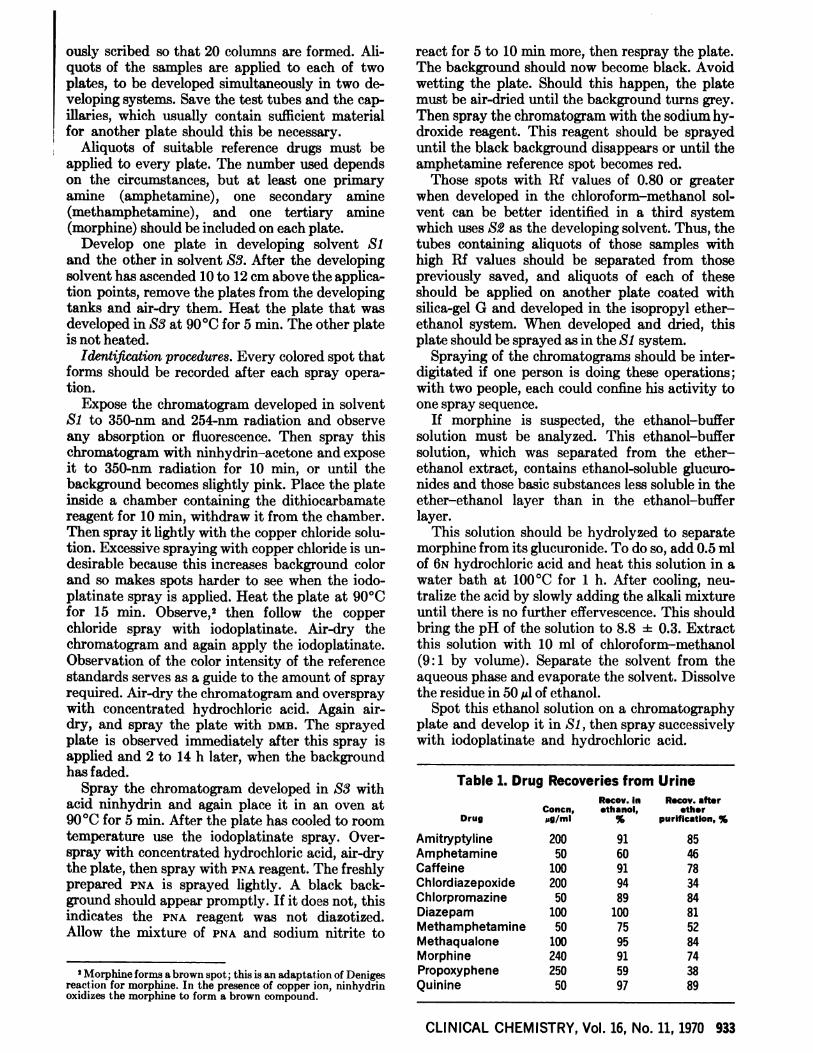
Table 1. Drug Recoveries from UrineRecov. in Rocoy. after
Concn, ethanol, etherDrug 5g/ml % purification, %
AmitryptylineAmphetamineCaffeineChiordiazepoxideChiorpromazineDiazepamMethamphetamineMethaqualoneMorphinePropoxypheneQuinine
50 60 46100 91 78200 94 3450 89 84
100 100 8150 75 52
100 95 84240 91 74250 59 3850 97 89
Nlnhydrin-acetone spray PNA sprayMn. amt.Mm. amt.
detected, pg Color detected, pg Color
35050202
10253
purple
pinkapurpletbluepink”purplebpurple
1
115121
Becomes pink after next spray.Not observed after ninhydrin and iodoplatinate spray.Fluorescence.
Dithiocarbamate reagent N inhyd nfl-addMm. amt. Mm. amt.
detected, pg Color detected, pg ColorSI S SS
Rf X 100
1 brown 10(uv)” purple 49 23 7010 brown 10(uv) purple 20 05 211 brown 3(uv) purple 28 06 331 brown 3 pink 10 07 301 brown 6(uv) pink 91 88 775 yellow 4(uv) purple 22 09 65
10 brown 15 purple 21 15 281 brown 5 purple 21 07 371 brown 3 pink 15 07 3310 brown 6(uv) purple 79 19 765 yellow 6(uv) purple 30 14 755 brown 2 purple 48 13 381 green 3(uv) purple 97 94 81
10 brown 10 purple 05 06 381 brown 1 purple 23 05 34
20 brown 5 purple 69 26 7510 brown 10(uv) purple 20 60 795 brown 6 purple 14 05 23
Also detected in the primary aromatic amine group.uv denotes uv absorption after application of ninhydrin-acid and heat.Fluorescence.
ResuIts
934 CLINICAL CHEMISTRY, Vol. 16, No. 11, 1970
To test the described procedure, known amountsof several representative drugs were added towater and analyzed according to this procedure.The amounts of each drug in the ethanol and in theresidue obtained after evaporation of the ether-ethanol mixture were determined by uv spectro-photometry or gas chromatography (Table 1).
Another measure of the efficiency of the pro-cedure is whether it will detect a given amount ofdrug added to a medication-free urine. The mini-mal concentration tested was 2 g of drug perml of urine. The ether-alcohol extracts of urinesamples containing the drugs tested were evapo-rated and the residues applied to thin-layer chroma-tograms and sprayed as described above.
The following substances were detected when10 ml of urine containing 2 g of drug per ml of
urine was analyzed: amitryptyline, amphetamine,butethamine, caffeine, chlordiazepoxide, chlor-phentermine, chlorpromazine, codeine, diazepam,hydromorphon, imipramine, lidocaine, meperidine,mephentermine, methadone, methamphetamine,methoxamine, morphine, phenylephrine, phenyl-propanolamine, procaine, propoxyphene, quinine,and strychnine. Other drugs were not included inthis evaluation.
Identification of the Drugs
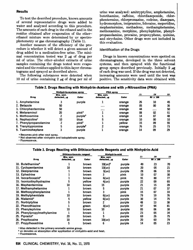
Drugs in known concentrations were spotted onchromatograms, developed in the three solventsystems, and then sprayed with the functionalgroup sprays described previously. Initially 2 gof each drug was used. If the drug was not detected,increasing amounts were used until the test waspositive. The sensitivity data were obtained with
Table 2. Drugs Reacting with Ninhydrin-Acetone and with p-Nitroaniline (PNA)
Drug
1. Amphetamine2. Betazole3. Chlorphentermine4. Metaraminol5. Methoxamine6. Naphazoline’7. Phenylpropanolamine8. Tranylcypromine9. Tuaminoheptane
orangeorangeorangeorangeorangeorangeyelloworangeorange
Si
Rf X 100
26 10 4505 00 3644 ... 5008 02 5114 07 4810 00 1015 09 5576 25 7314 09 29
Table 3. Drugs Reacting with Dithiocarbamate Reagents and with Ninhydrin-Acid
Drug
10. Butethamine”11. Cyclopentamine12. Desipramine13. Ephedrine14. lsocarboxazidc15. Isonicotinylhydrazine16. Mephentermine17. Methamphetamine18. Methoxyphenylamine19. Methylphenidate20. Nialamidc21. Nortriptyline22. Phenothiazine23. Phenylephrine24. Phenylpropylmethylamine25. Pipradolc26. Propitocaine27. Propylhexedrine
M In.amt.
detected,pg
Mmn.amt.
detected,Drug pg
All compounds yield a blue to purple product.“Fluorescence.
Not tertiary amines but can not be detected in amounts of 20 g with thepreviousreagents.
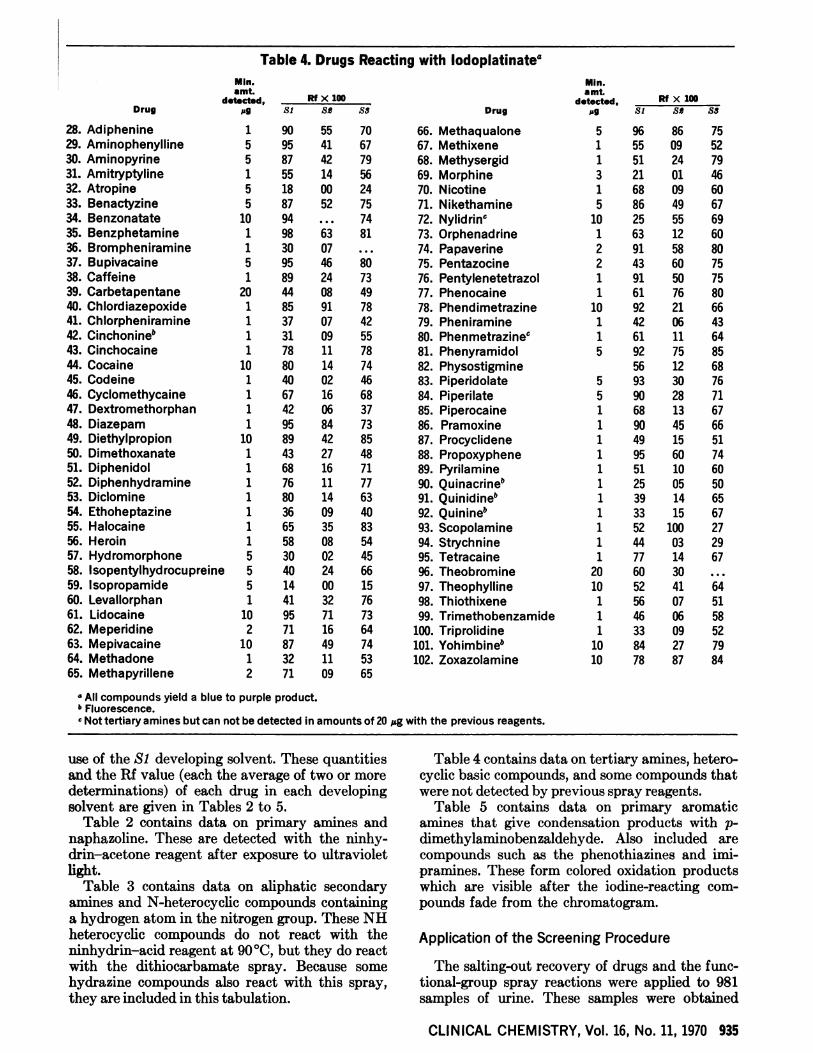
Table 4. Drugs Reacting with Iodoplatinate#{176}
CLINICAL CHEMISTRY, Vol. 16, No. 11, 1970 935
Drug
28. Adiphenine29. Aminophenylline30. Aminopyrine31. Amitryptyline32. Atropine33. Benactyzine34. Benzonatate35. Benzphetamine36. Brompheniramine37. Bupivacaine38. Caffeine39. Carbetapentane40. Chlordiazepoxide41. Chlorpheniramine42. Cinchonine”43. Cinchocaine44. Cocaine45. Codeine46. Cyclomethycaine47. Dextromethorphan48. Diazepam49. Diethylpropion50. Dimethoxanate51. Diphenidol52. Diphenhydramine53. Diclomine54. Ethoheptazine55. Halocaine56. Heroin57. Hydromorphone58. Isopentylhydrocupreine59. lsopropamide60. Levallorphan61. Lidocaine62. Meperidine63. Mepivacaine64. Methadone65. Methapyrillene
Rf>( 100
Si SS 85
1 90 55 705 95 41 675 87 42 791 55 14 565 18 00 245 87 52 75
10 94 ... 741 98 63 811 30 075 95 46 801 89 24 73
20 44 08 491 85 91 781 37 07 421 31 09 551 78 11 78
10 80 14 741 40 02 461 67 16 681 42 06 371 95 84 73
10 89 42 851 43 27 481 68 16 711 76 11 771 80 14 631 36 09 401 65 35 831 58 08 545 30 02 455 40 24 665 14 00 151 41 32 76
10 95 71 732 71 16 64
10 87 49 741 32 11 532 71 09 65
66. Methaqualone67. Methixene68. Methysergid69. Morphine70. Nicotine71. Nikethamine72. Nylidrinc73. Orphenadrine74. Papaverine75. Pentazocine76. Pentylenetetrazol77. Phenocaine78. Phendimetrazine79. Pheniramine80. Phenmetrazinec81. Phenyramidol82. Physostigmine83. Piperidolate84. Piperilate85. Piperocaine86. Pramoxine87. Procyclidene88. Propoxyphene89. Pyrilamine90. Quinacrine”91.Quinidine”92. Quinine”93. Scopolamine94.Strychnine95.Tetracaine96. Theobromine97. Theophylline98. Thiothixene99. Trimethobenzamide
100. Triprolidine101. Yohimbine”102. Zoxazolamine
RI X 100
Si S Ss
5 96 86 751 55 09 521 51 24 793 21 01 461 68 09 605 86 49 67
10 25 55 691 63 12 602 91 58 802 43 60 751 91 50 751 61 76 80
10 92 21 661 42 06 431 61 11 645 92 75 85
56 12 685 93 30 765 90 28 711 68 13 671 90 45 661 49 15 511 95 60 741 51 10 601 25 05 501 39 14 651 33 15 671 52 100 271 44 03 291 77 14 67
20 60 3010 52 41 641 56 07 511 46 06 581 33 09 52
10 84 27 7910 78 87 84
use of the Si developing solvent. These quantitiesand the Rf value (each the average of two or moredeterminations) of each drug in each developingsolvent are given in Tables 2 to 5.
Table 2 contains data on primary amines andnaphazoline. These are detected with the ninhy-din-acetone reagent after exposure to ultravioletlight.
Table 3 contains data on aliphatic secondaryamines and N-heterocycic compounds containinga hydrogen atom in the nitrogen group. These NHheterocyclic compounds do not react with theninhydrmn-acid reagent at 90#{176}C,but they do reactwith the dithiocarbamate spray. Because somehydrazine compounds also react with this spray,they are included in this tabulation.
Table 4 contains data on tertiary amines, hetero-cyclic basic compounds, and some compounds thatwere not detected by previous spray reagents.
Table 5 contains data on primary aromaticamines that give condensation products with p-dimethylaminobenzaldehyde. Also included arecompounds such as the phenothiazines and imi-pramines. These form colored oxidation productswhich are visible after the iodine-reacting com-pounds fade from the chromatogram.
Application of the Screening Procedure
The salting-out recovery of drugs and the func-tional-group spray reactions were applied to 981samples of urine. These samples were obtained
lodoplatInatespray, mm.
Drug amt. detected, pg
DMB spraySI 81Mm.
amt. detected, pg Color RI X 10088
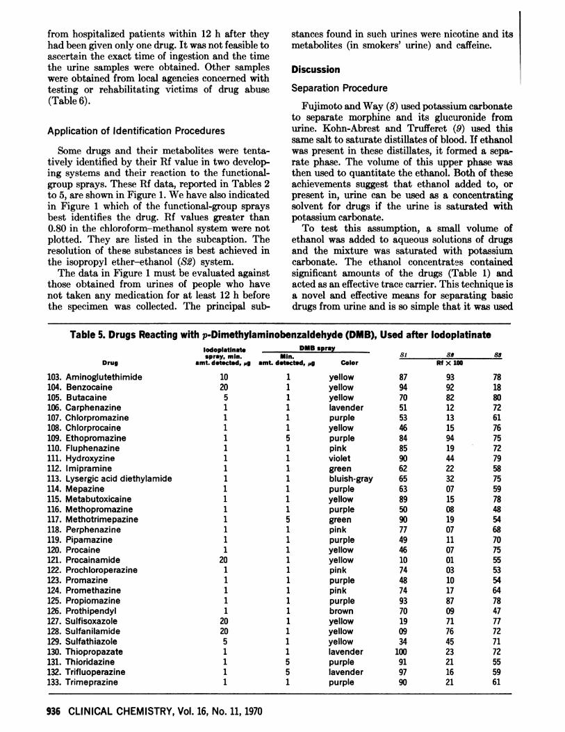
103.Aminoglutethimide 10 1 yellow 87 93 78104. Benzocaine 20 1 yellow 94 92 18105. Butacaine 5 1 yellow 70 82 80106. Carphenazine 1 1 lavender 51 12 72107. Chlorpromazine 1 1 purple 53 13 61108. Chlorprocaine 1 1 yellow 46 15 76109. Ethopromazine 1 5 purple 84 94 75110.Fluphenazine 1 1 pink 85 19 72111. Hydroxyzine 1 1 violet 90 44 79112. lmipramine 1 1 green 62 22 58113. Lysergic acid diethylamide 1 1 bluish-gray 65 32 75114. Mepazine 1 1 purple 63 07 59115. Metabutoxicaine 1 1 yellow 89 15 78116. Methopromazine 1 1 purple 50 08 48117.Methotrimepazine 1 5 green 90 19 54118. Perphenazine 1 1 pink 77 07 68119. Pipamazine 1 1 purple 49 11 70120. Procaine 1 1 yellow 46 07 75121. Procainamide 20 1 yellow 10 01 55122. Prochloroperazine 1 1 pink 74 03 53123. Promazine 1 1 purple 48 10 54124. Promethazine 1 1 pink 74 17 64125. Propiomazine 1 1 purple 93 87 78126. Prothipendyl 1 1 brown 70 09 47127. Sulfisoxazole 20 1 yellow 19 71 77128. Sulfanilamide 20 1 yellow 09 76 72129. Sulfathiazole 5 1 yellow 34 45 71130.Thiopropazate 1 1 lavender 100 23 72131. Thioridazine 1 5 purple 91 21 55132. Trifluoperazine 1 5 lavender 97 16 59133. Trimeprazine 1 1 purple 90 21 61
936 CLINICAL CHEMISTRY, Vol. 16, No. 11, 1970
from hospitalized patients within 12 h after theyhad been given only one drug. It was not feasible toascertain the exact time of ingestion and the timethe urine samples were obtained. Other sampleswere obtained from local agencies concerned withtesting or rehabilitating victims of drug abuse(Table 6).
Application of Identification Procedures
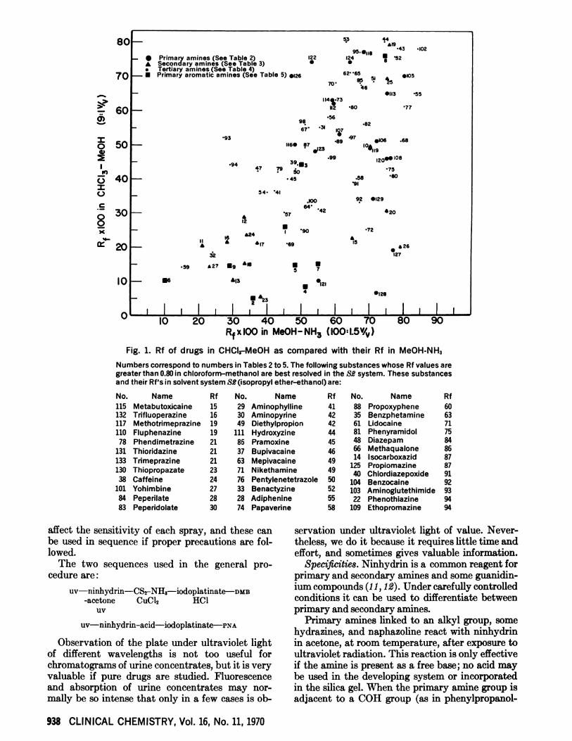
Some drugs and their metabolites were tenta-tively identified by their Rf value in two develop-ing systems and their reaction to the functional-group sprays. These Rf data, reported in Tables 2to 5, are shown in Figure 1. We have also indicatedin Figure 1 which of the functional-group spraysbest identifies the drug. Hf values greater than0.80 in the chloroform-methanol system were notplotted. They are listed in the subcaption. Theresolution of these substances is best achieved inthe isopropyl ether-ethanol (52) system.
The data in Figure 1 must be evaluated againstthose obtained from urines of people who havenot taken any medication for at least 12 h beforethe specimen was collected. The principal sub-
stances found in such urines were nicotine and itsmetabolites (in smokers’ urine) and caffeine.
Discussion
Separation Procedure
Fujimoto and Way (8) used potassium carbonateto separate morphine and its glucuronide fromurine. Kohn-Abrest and Trufferet (9) used thissame salt to saturate distillates of blood. If ethanolwas present in these distillates, it formed a sepa-rate phase. The volume of this upper phase wasthen used to quantitate the ethanol. Both of theseachievements suggest that ethanol added to, orpresent in, urine can be used as a concentratingsolvent for drugs if the urine is saturated withpotassium carbonate.
To test this assumption, a small volume ofethanol was added to aqueous solutions of drugsand the mixture was saturated with potassiumcarbonate. The ethanol concentrates containedsignificant amounts of the drugs (Table 1) andacted as an effective trace carrier. This technique isa novel and effective means for separating basicdrugs from urine and is so simple that it was used
Table 5. Drugs Reacting with p-Dimethylaminobenzaldehyde (DMB), Used after lodoplatinate
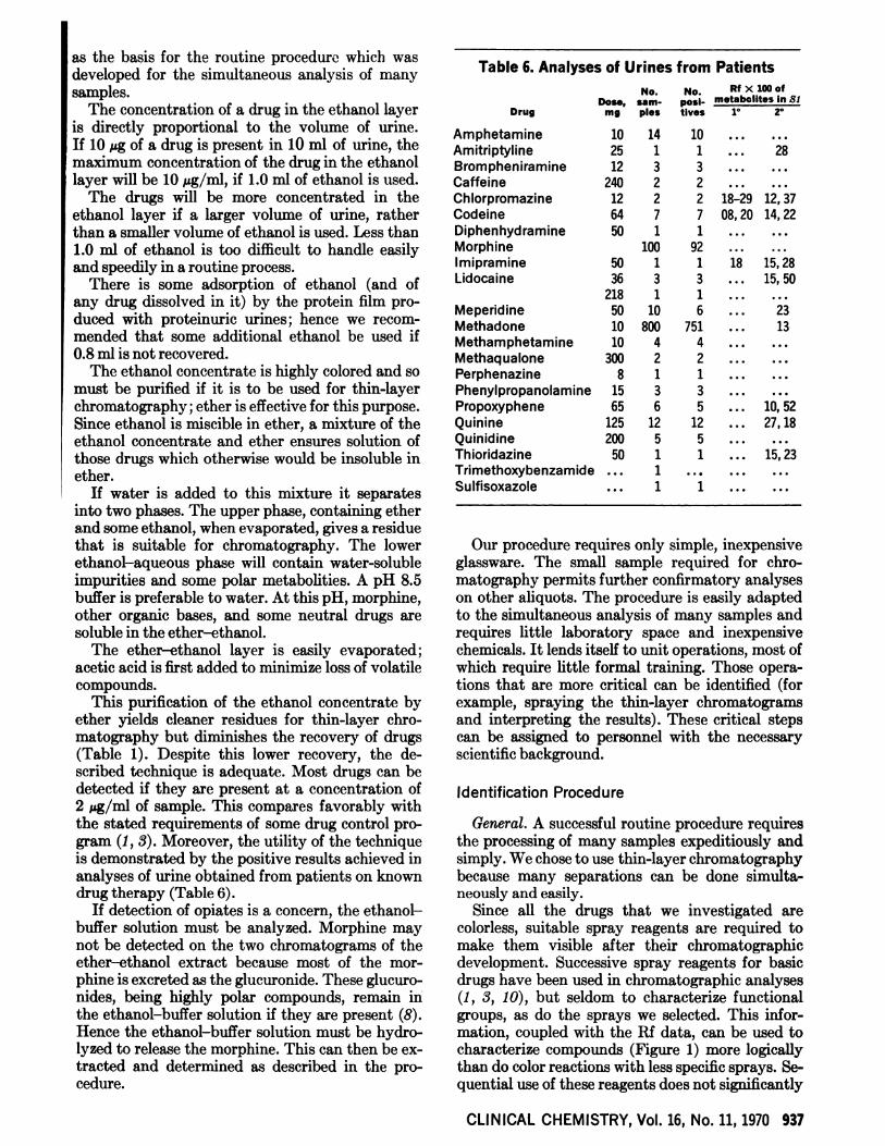
Table 6. Analyses of Urines from Patients
No. No. RI X 100 ofDose, sam- posi- metabclites in SI
Drug mg pies tives 1 Z
CLINICAL CHEMISTRY, Vol. 16, No. 11, 1970 937
as the basis for the routine procedure which wasdeveloped for the simultaneous analysis of manysamples.
The concentration of a drug in the ethanol layeris directly proportional to the volume of urine.If 10 g of a drug is present in 10 ml of urine, themaximum concentration of the dug in the ethanollayer will be 10 g/ml, if 1.0 ml of ethanol is used.
The drugs will be more concentrated in theethanol layer if a larger volume of urine, ratherthan a smaller volume of ethanol is used. Less than1.0 ml of ethanol is too difficult to handle easilyand speedily in a routine process.
There is some adsorption of ethanol (and ofany drug dissolved in it) by the protein film pro-duced with proteinuric urmnes; hence we recom-mended that some additional ethanol be used if0.8 ml is not recovered.
The ethanol concentrate is highly colored and somust be purified if it is to be used for thin-layerchromatography; ether is effective for this purpose.Since ethanol is miscible in ether, a mixture of theethanol concentrate and ether ensures solution ofthose drugs which otherwise would be insoluble inether.
If water is added to this mixture it separatesinto two phases. The upper phase, containing etherand some ethanol, when evaporated, gives a residuethat is suitable for chromatography. The lowerethanol-aqueous phase will contain water-solubleimpurities and some polar metabolities. A pH 8.5buffer is preferable to water. At this pH, morphine,other organic bases, and some neutral drugs aresoluble in the ether-ethanol.
The ether-ethanol layer is easily evaporated;acetic acid is first added to minimize loss of volatilecompounds.
This purification of the ethanol concentrate byether yields cleaner residues for thin-layer chro-matography but diminishes the recovery of drugs(Table 1). Despite this lower recovery, the de-scribed technique is adequate. Most drugs can bedetected if they are present at a concentration of2 Mg/mi of sample. This compares favorably withthe stated requirements of some drug control pro-gram (1, 3). Moreover, the utility of the tecimique
is demonstrated by the positive results achieved inanalyses of urine obtained from patients on knowndug therapy (Table 6).
If detection of opiates is a concern, the ethanol-buffer solution must be analyzed. Morphine maynot be detected on the two chromatograms of theether-ethanol extract because most of the mor-phine is excreted as the glucuronide. These glucuro-nides, being highly polar compounds, remain inthe ethanol-buffer solution if they are present (8).Hence the ethanol-buffer solution must be hydro-lyzed to release the morphine. This can then be ex-tracted and determined as described in the pro-cedure.
AmphetamineAmitri ptylineBrom phen iramineCaffeineChlorpromazineCodeineDiphen hyd ramineMorphineIm ipram in eLidocaine
MeperidineMethadoneMethamphetamineMethaqualonePerphenazinePhenyIpropa no Iam mePropoxypheneQuinineQuinidineThioridazineTrimethoxybenzamideSu Ifisoxazole
10 14 10 - -. . . -
25 1 1 - . - 28
12 3 3 ... . -.
240 2 2 - -. . . -
12 2 2 18-29 12, 3764 7 7 08,20 14,2250 1
100192
. - -
...
...
...
50 1 1 18 15,2836 3 3 ... 15,50
218 1 1 . - - ..
50 10 6 ... 23
10 800 751 ... 13
10 4 4 . . - ...
300 2 2 ... ..
8 1 1 .. ..
15 3 3 . . - ..
65 6 5 ... 10,52
125 12 12 ... 27,18
200 5 5 ... ...
50 1 1 ... 15,23
... 1 ... ... ...
... 1 1 ... ...
Our procedure requires only simple, inexpensiveglassware. The small sample required for chro-matography permits further confirmatory analyseson other aliquots. The procedure is easily adaptedto the simultaneous analysis of many samples andrequires little laboratory space and inexpensivechemicals. It lends itself to unit operations, most ofwhich require little formal training. Those opera-tions that are more critical can be identified (forexample, spraying the thin-layer chromatograms
and interpreting the results). These critical stepscan be assigned to personnel with the necessaryscientific background.
Identification Procedure
General. A successful routine procedure requiresthe processing of many samples expeditiously andsimply. We chose to use thin-layer chromatographybecause many separations can be done simulta-neously and easily.
Since all the drugs that we investigated arecolorless, suitable spray reagents are required tomake them visible after their chromatographicdevelopment. Successive spray reagents for basicdrugs have been used in chromatographic analyses(1, 3, 10), but seldom to characterize functionalgroups, as do the sprays we selected. This infor-mation, coupled with the Rf data, can be used tocharacterize compounds (Figure 1) more logicallythan do color reactions with less specific sprays. Se-quential use of these reagents does not significantly
80 53
- #{163}1943 .
- #{149}Primary amines (See Table 2) 22 24 52A Secondary amines (See Table 3) S. Tertiary amines (See Table 4)
70 - Primary aromatic amines (See Table 5) l26 62 65 #{149}I058S7O 46
- #{149}113 .55
> 60 114%.73
- 112 #{149}80
.82- 67 I7
50 - uue. 87 #{149}ioe
lO#{246}0. .23 119.99 20e 108
79 .3 40 - .se
.9’54. #{149}4
.100 92 #{149}I29
30 - 64 42 #{149}20Q 12
72*24
6*17 69 *26cr’20_ #{163}
32 127
.59 #{163}27#{149}9I$ #{149}#{149}5 7
10 - #{163}ms #{149}2I
#{149}128
0 I I I I I I i I I I10 20 30 40 50 60 70 80 90
Rfx 100 in MeOH-NH3 (l00:I.5V/)
Fig. 1. Rf of drugs in CHCI,-MeOH as compared with their Rf in MeOH-NH3
Numbers correspond to numbers in Tables 2 to 5. The following substances whose Rf values aregreater than 0.80 in chloroform-methanol are best resolved in the S system. These substancesand their Rf’s in solvent system S (isopropyl ether-ethanol) are:
938 CLINICAL CHEMISTRY, Vol. 16, No. 11, 1970
No. Name115 Metabutoxicaine132 Trifluoperazine117 Methotrimeprazine110 Fluphenazine78 Phendimetrazine
131 Thioridazine133 Trimeprazine130 Thiopropazate38 Caffeine
101 Yohimbine
84 Peperilate83 Peperidolate
Rf No. Name15 29 Aminophylline16 30 Aminopyrine19 49 Diethylpropion19 111 Hydroxyzine21 85 Pramoxine21 37 Bupivacaine21 63 Mepivacaine23 71 Nikethamine24 76 Pentylenetetrazole27 33 Benactyzine28 28 Adiphenine30 74 Papaverine
Rf No. Name41 88 Propoxyphene42 35 Benzphetamine42 61 Lidocaine44 81 Phenyramidol45 48 Diazepam
46 66 Methaqualone14 Isocarboxazid
125 Propiomazine40 Chlordiazepoxide
50 104 Benzocaine52 103 Aminoglutethimide55 22 Phenothiazine58 109 Ethopromazine
Rf60637175848687879192939494
affect the sensitivity of each spray, and these canbe used in sequence if proper precautions are fol-lowed.
The two sequences used in the general pro-cedure are:
uv-ninhydrin-CS2-NH,--iodoplatinate-DaIB-acetone CuCI2 HC1
uv
uv-ninhydrin-acid--iodoplatinate-PNA
Observation of the plate under ultraviolet lightof different wavelengths is not too useful forchromatograms of urine concentrates, but it is veryvaluable if pure drugs are studied. Fluorescenceand absorption of urine concentrates may nor-mally be so intense that only in a few cases is oh-
servation under ultraviolet light of value. Never-theless, we do it because it requires little time andeffort, and sometimes gives valuable information.
Specificities. Ninhydrmn is a common reagent forprimary and secondary amines and some guanidin-ium compounds (ii, 12). Under carefully controlledconditions it can be used to differentiate betweenprimary and secondary amines.
Primary amines linked to an alkyl group, somehydrazines, and naphazoline react with ninhydrmnin acetone, at room temperature, after exposure toultraviolet radiation. This reaction is only effectiveif the amine is present as a free base; no acid maybe used in the developing system or incorporatedin the silica gel. When the primary amine group isadjacent to a COil group (as in phenylpropanol-
CLINICAL CHEMISTRY, Vol. 16, No. 11, 1970 939
amine, metaraminol) the product of the reaction ispink instead of purple.
If the test for primary amines is positive insolvent system Si, the diazonium salt of p-nitro-aniline is used to confirm this on the chromato-gram developed in S3. When PNA is applied afterthe iodoplatinate spray, it is far more sensitivefor primary amines than when it is applied as asingle spray (1, 13). In such a sequence it can de-tect 1 g of amphetamine. It will also detect lowconcentrations of other primary amines such aschlorphentermine, betazole, and metaraminol,which the less sensitive ninhydrmn-acetone wouldnot detect in the 81 procedure.
WThen the ninhydrmn-acetone reaction is positiveand the PNA reaction is negative, the positive reac-tion observed in the plate developed in 81 is not
owing to primary amines but rather to naphazolineor a large amount (50 Mg) of some secondary amine.
The ninhydrmn-acid reagent is used as the firstspray on the 83-developed chromatogram becauseit is sensitive to secondary amines and not to mostprimary amines. As much as 20 g of primaryamines-e.g., amphetamine, metaraminol, tuamine,chlorphentermine-do not react with the acid-ninhydrmn even if the chromatogram is heated for5 to 10 mm at 90#{176}C.Under these conditionsnaphazoline and a few primary amines such asmethoxainine, tranylcypromine, and pheriylpro-panolamine may react. Despite this limitation,the acid-ninhydrmn will indicate whether a sec-ondary amine may be present, and will not interferewith the subsequent PNA test for primary amines.Presence of secondary amines is confirmed by apositive result with the dithiocarbamate reagent onthe chromatogram developed in Si: these twopositive results indicate that a secondary amineattached to an aliphatic group is probably present.If the ninhydrin-acid result is negative and thatfrom the dithiocarbamate reagent is positive, theneither a NH-heterocyclic compound that forms adithiocarbamate, or a hydrazide or a hydrazinecompound (both of which form a dithiocarbazide)may be present.
The dithiocarbamates formed from secondaryamines and NH-heterocyclic compounds can bedifferentiated from some of the dithiocarbazidesformed with hydazide derivatives. Dithiocar-bamates react with cupric ion to form brownproducts instead of the yellow dithiocarbazidederivatives formed by isonicotinyl hydrazine andnialamid. Other hydrazines-iproniazid, hydrala-zine, phenylethylhydrazine, phenylisopropylhydra-zine-which normally would be detected by theformation of a dithiocarbazide do not give theexpected reaction if the suggested spray sequence isused, because of the interference by ninhydrmn. Ifninhydmn is not used, these compounds do react.Some secondary amines and NH-heterocyclic com-pounds are not detected by the dithiocarbamate
reaction because of its low sensitivity for thesecompounds. These are detected by the subsequentiodoplatinate spray.
lodoplatinate is used to detect tertiary amines.Overspraying this reagent with concentratedhydrochloric acid increases its sensitivity and thusreveals compounds that otherwise would not beseen. A further increase in sensitivity is achievedon the Si-developed chromatogram because thecupric chloride spray precedes the iodoplatinate.Cupric ion is reduced to cuprous iodide by the iodo-platinate; and this compound and the iodineformed in the reaction, together with the othersprays, make many drugs and artifacts apparentthat would not be seen if only the iodoplatinatewere used.
Another advantage of coupling the cupric andiodoplatinate sprays is the fading of the colordeveloped on the chromatogram due to oxidationby the cupric ion if a light iodoplatinate spray isused. This clears the chromatogram so that anycolor the next spray generates can be seen. Thisoxidation also produces colored products if pheno-thiazines are present. These remain on the chro-matogram even after the next spray, DMB, is used.This characteristic behavior of phenothiazine com-pounds permits their inclusion in the classificationscheme as a group of drugs that gives coloredproducts after the DMB spray. Other substances re-acting with DMB are indoles, which form blue or violetcondensation products; sulfonamides and primaryaromatic amines, which form yellow Schiff bases.
Sensitivity of DMB is not impaired if the othersprays are used first, but small amounts of primaryaromatic amines and indolic compounds cannot bedetected until the background has faded com-pletely, which may take as long as 2 to 12 h.Storing these chromatograms overnight and ob-serving them in the morning may be rewarding.
There are some disadvantages with these spraysequences. In low concentration some organic basesare not detected. Some others, such as nylidrmn andphenmetrazine, had to be classified in groups otherthan those that predictably should have includedthem because the sensitivity of the characterizingfunctional group sprays was not high enough.
The ethanol-ether extract may also contain someneutral organic drugs. The proposed sequence ofsprays detects some neutral compounds such aschlordiazepoxide, diazepam, and sulfonamides, butmany other neutral compounds do not react withthe sprays used. The suggested sequences areprimarily designed to detect organic bases. Otherspray procedures should be used if one is concernedwith detecting neutral drugs.
Another disadvantage of the identification pro-cess is its sensitivity to caffeine and nicotine, aswell as to some normal components of urine. Thismakes interpretation of the chromatograms moredifficult for the inexperienced observer.
940 CLINICAL CHEMISTRY, Vol. 16, No. 11, 1970
To overcome these difficulties, one must make acareful choice of the chromatographic conditions.The Si system and the suggested sequence ofsprays provide maximum dispersion of the drugs onthe plate, good separation of morphine and codeine,minimum of interference in the ninhydrmn-acetonereaction, adequate reproducibility of Rf values,and, except for nicotine and caffeine, the inter-ference by normal components appears at eitherthe origin or the solvent front. Near the bottom ofof the plate the interferences are revealed by theninhydrin reagent, which reacts with those primaryamines normally present and those resulting fromputrefaction of the urine. The DMB spray revealsanother group of “normal” spots whose Rf’s are0.10 and 0.95 and one other, rather infrequently, atRf 0.40.
The 53 mixture is useful despite the concentra-tion of artifacts in the Rf region between 0.70 to0.85. Morphine has an Rf of 0.46 in this system andis more easily visualized, if present, because fewother common drugs have a similar Rf. Anotheradvantage of this system is its sensitivity for pri-mary amines, because PNA used in this spraysequence detects small amounts of amphetamine.
The 82 system was used to separate drugs whoseRf values were greater than 0.80 in the Si system,with excellent resolution and identification of manydrugs in this group. Most of the interfering sub-stances one can expect in the S2 system fromnormal urines remain at the origin.
Since primary amines are not found in thisgroup, the ninhydrmn-acetone may be omitted,thus removing some of the previously describedlimitations on the dithiocarbamate procedure;hydrazides or hydrazines present will now reactand be detected.
Applications. Smokers’ and coffee-drinkers’ urinesinclude several spotsobserved on plates developed inboth the Si and the S3 systems. These are nicotine,caffeine, and metabolites of nicotine. Two metabo-lites appear after the iodoplatinate spray. Thenicotine metabolites are only revealed by the acidoverspray. Identification of those durgs whose Rfvalues are in these regions is therefore more diffi-cult. Fortunately, drugs of primary interest inproblems of drug abuse and drug misuse do nothave Rf values in these regions. If a nonsmoker isinvolved, then interpretation is simpler because thenicotine spots are not found.
After becoming familiar with the pattern seenwith normal urine it is easier to distinguish smallamounts of drug among the normal artifacts, butspraying and interpreting require experience andskill.
Another advantage of the proposed identifica-tion procedure is that it can be used to detect manydrugs in lower concentrations than is possible withother procedures that use single-spray reagents. Ifa chromatogram reveals only “normal” spots, the
conclusion is warranted that no significant quan-tities of primary, secondary, or tertiary amines,NH-heterocyclic compounds, hydazide com-pounds, primary aromatic amines, phenothiazinecompounds, or indolic compounds are present inthe sample. This “negative” result is meaningfuland valuable.
If unusual spots are detected, drugs may bepresent. A tentative identification of the spots ob-tained from a given sample can be made by com-paring its Rf values in the two solvent systems andits reactions to the functional group sprays withthe data tabulated in Figure 1.
If two or more spots are found on one chrornato-gram, two drugs may be present. Alternatively, ifone of them is a tertiary amine with a high Rf andthe other is a secondary or primary amine, a dugand its metabolites may be present. The Rf valuesof metabolites of some common drugs are presentedin Table 6 and Figure 1. The relative intensity ofeach spot is significant. Sometimes higher concen-trations of the metabolites are found and onlysmall quantities of the unchanged drug. This isparticularly true for meperidine and propoxyphene.
Supported in part by grant no. 09863-07 from the National In-stitute of General Medical Science; grant FR-5410, General Re-search Support Branch, Division of Research Facilities and Re-sources, NIH; and The Research Corporation, New York, N.Y.
References1. Mule, S. J., Identification of narcotics, barbiturates, amphet-amines, tranquilizers and psychotomimetics in human urine. J.Chrornatogr. 39, 302 (1969).2. Dole, V. P., Kim, V. K., and Eglitis, I., Detection of narcoticdrugs, tranquilizers, amphetamines and barbiturates in urine.J. Amer. Med. Ass. 198, 349 (1966).3. Davidow, B., Petri, N. L., and Quame, B., A thin layerchromatographic procedure for detecting drug abuse. Amer. J.Clin. Pathol. 50, 714 (1968).4. Beckett, A. H., Tucker, G. T., and Moffat, A. C., Routine de-tection and identification in urine of stimulants and other drugs,some of which may be used to modify performance in sport. J.Pharm. Pharmacol. 19, 273 (1967).5. Sunshine, I., Handbook of Analytical Toxicology. The Chemi-cal Rubber Co., Cleveland, Ohio, 1969.
6. Clarke, E. G. C., Isolation and Identification of Drugs. ThePharmaceutical Press, London, 1969.7. Cochin, J., Analysis for narcotic analgesics and barbiturates inurine by mc techniques without previous extraction and concen-tration. Psychopharmacol. Bull. 3, 53 (1966).8. Fujimoto, J. M., and Way, E. L., Isolation and crystallizationof “bound” morphine from urine of human addicts. J. Pharmacol.Exp. Ther. 121, 340 (1957).
9. Kohn-Abrest, E., and Truffert, L., Methode pur la detectionet determination post-morten de l’alcohol dam le sang. Ann.Fraudes 30, 210 (1937).10. Smith, I., Chromatographic and Electrophoretic Techniques:Chromatography, 1, 2nd ed. William Heineman Medical BookLtd., London, 1968.
11. Conn, R. B., and Davis, R. B., Green fluorescence of guani-dinium compounds with ninhydrin. Nature 183, 1053 (1959).12. Yamagisha, M., and Yoshida, T., Ninhydrin reaction ofsecondary amines. Pharm. Soc. Japan 74, 1042 (1954).13. Wickstr#{246}m,A., and Salvesen, B., Separation and identifica-tion of sympathomemetic amines by paper chromatography. J.Pharm. Pharmacol. 4, 631 (1952).
Related Documents