Detection of Salmonella enterica serovar Typhi by nested peR using different procedures of nucleic acid extraction. A DISSERTATION SUBMITTED TO THE BRAC UNIVERSITY IN PARTIAL FULFILMENT OF THE REQUIREMENTS FOR THE DEGREE OF MASTER OF SCIENCE IN BIOTECHNOLOGY Submitted by Md Jahedul Islam ID No- 08376003 May 2010 MS Biotechnology Department of Mathematics and Natural Sciences BRAe University 66 MohakhaLi, Dhaka - 1212 Bangladesh

Welcome message from author
This document is posted to help you gain knowledge. Please leave a comment to let me know what you think about it! Share it to your friends and learn new things together.
Transcript

Detection of Salmonella enterica serovar Typhi by nested peR using different procedures of nucleic
acid extraction.
A DISSERTATION SUBMITTED TO THE BRAC UNIVE RSITY IN PARTIAL FULFILMENT OF THE REQUIREMENTS FOR THE DEGREE OF MASTER OF SCIENCE IN BIOTECHNOLOGY
Submitted by Md Jahedul Islam ID No- 08376003
May 2010
MS Biotechnology Department of Mathematics and Natural Sciences
BRAe University 66 MohakhaLi, Dhaka - 1212
Bangladesh

DEDICATED
TO
MY BELOVED PARENTS

ACKNOWLEDGEMENTS
At first I would like to pay gratitude to the most Gracious, most Merciful, Creator of this
universe the Almighty Allah who has kept me healthy, strong and energetic to complete
the thesis.
I wish to express my deepest sense of gratitude to my supervisor and honorable teacher
Professor Naiyyum Choudhury, Coordinator, Biotechnology Program, Department of
Mathematics and Natural Sciences (MNS), BRAC University, Dhaka for his able
guidance, suggestions, encouragement and his valuable advice during the course of study.
I would like to express my sincere gratitude to my supervisor Professor Firdausi Qadri,
Senior Scientist, Immunology Laboratory, Laboratory Sciences Division, ICDDR, B
Dhaka for her kind guidance and supervision. I am indebted for her sympathy and warm
encouragement throughout the time of study in ICDDR, B.
I am grateful to Professor A A Ziauddin Ahmad, Chairperson, MNS department, Dr
Aparna Islam, Assistant Professor, Biotechnology Program and teachers of Dept of
Mathematics and Natural Sciences, BRAC University, Dhaka, for their support, valuable
suggestions and inspiration in making such an effort to submit the thesi s.
I offer my heartiest honor to Alaullah Sheikh, Sarwar Bhuian, Md Arifuzzaman and Dr.
Farhana Khanom, Research officers, Immunology Laboratory, LSD, ICDDR, B, Dhaka
for their help and support. Without their help it would have been difficult for me to
achieve my goal.
And finally, earnest thanks from my heart to my family members especially to my
beloved parents for the encouragement to complete my thesis successfully.
I have my highest regards to all the subjects who consented without hesitation and gave
their valuable time to volunteer in this research work. Without their participation this
study would have not been possible at all.
CERTIFICATION OF ORIGINALITY OF THE WORK
This is to certify that, the Thesis entitled ' Detection of Salmonella enlerica serovar Typhi
by nested peR using different procedures of nucleic acid extraction., a requirement for
the degree of Master of Science (MS) in Biotechnology under the Dept of Mathematics
and Natural Sciences, BRAC University was carried out in the Immunology Laboratory
of the Laboratory Sciences Division of the International Centre for Diarrheal Disease
Research, Bangladesh (ICDDR, B), Dhaka during the period of October 2008 to January
20 I 0, under our joint supervision.
Supervisor
N~ro, 30,,'06;\ 0 (Professor Naiyyum Cboudbury) Coordinator MS Biotechnology MNS Department BRAC University Dhaka, Bangladesh
Supervisor
~~~Q~ (Dr. Firdausi QadrO. Senior Scientist Immunology Laboratory Laboratory Sciences Division ICDDR,B Dhaka, Bangladesh

Contents Pages
Chapter One: Introduction
1.1
1.2
1.3
1.4
1.5
1.6
1.7
1.7.1
1.8
1.9
1.10
Background
Structure & Comparisn of the Salmonella
Typhi Genome
Antigenic types of S. Typhi
Etiology
Pathophysiology
Diagnosis of typhoid fever
Serological diagnosis
Widal test
Diagnosis of typhoid by nested Polymerase
Chain Reaction (PCR)
Treatment of typhoid fever
Prevention of typhoid fever
Aim of the study
Chapter Two: Materials and methods
2.1
2.2
2.3
2.4
2.5.1
2.5.2
2.5.3
Study subjects
Sample Collection
DNA extraction from invitro grown Salmonella Typhi pure culture
Preparation of S. Typhi bacteria for spiking in blood DNA Extraction by a procedure described by Haque et al. DNA Extraction by direct Boiling
DNA Extraction by using Qiagen Kit
Page No. 1-15
1
2-3
4-5
5-8
8-9
9-10
10-11
11
11-12
\3
14
15
16-25
16
16
17
17
18
19
19

2.6.1
2.6.2
2.7.1
2.7.2
2.7.3
2.7.4
2.7.5
2.7.6
DNA extraction from patient's blood specimens by using Qiagen Kit
DNA extraction from blood of suspected typhoid fever patient by using a procedure previously described by R. Boom et al. Primer sets
Master mixture preparation for first round peR Thermal cycle for first round of peR
Master mixture for second round of peR
Thermal cycle for second round of peR
Agarose Gel (1.5%) electrophoresis
Chapter 3: Results
3.1
3.2
3.3
Detection of Salmonella enterica serovar Typhi by nested peR using different nucleic acid extraction methods
Optimization of DNA extraction procedure
Detection of typhi in blood in typhoid fever patients
Chapter 4: Discussion
21
22
23
23
24
24
25
25
26-30
26
26-28
29-30
31-32
---------------------------Chapter 5: References
Chapter 6: Appendix
33-41
42-46

List of Figures Pages
~ I
[-
Figure No.
I
2
3
Figure Name
I J Structur~of Salmonella Typhi
I -J-~ircular genome map of.!. Typhi
: Etiology of typhoid fever
+ 4 ! A Schematic diagram of nested peR I 5 I Patients blood in EDT A tube 1 6_ ; 8 Typhi ~'o";" gcOW" ;0 ~A pi,,, t 7
8
9
10
II
I Electrophoresis of the peR products performed I by usmg 1.5% agarose gel. Nested peR results of i DNA samples (only even number) extracted by a ! procedure described by Haque et a1 . I
-; Electrop-horesis of the peR ;oducts performed t I by using l.5% agarose gel. Nested peR results of I I DNA samples (only even number) extracted by ! I direct boiling procedlLre . _ __ I i Electrophoresis of the peR products performed I by using 1.5% agarose gel. Nested peR results of I DNA samples ext~acted by Qia~e~kjt.
i Electrophoresis of the peR products perfonned I by using 1.5% agarose gel. Nested peR was done i for typhi detection of four (4) patients of blood I culture positive samples.
I 1 Electrophoresis of the peR products- performed I
by using 1.5% agarose gel. Nested peR was done I
for typhi detection of all patients regardless of1 blood culture positive and negative.
~------~--------- ----
Page No.
4
3
7
J - J
12 J 16 I 17~ 25
25 l
26
27
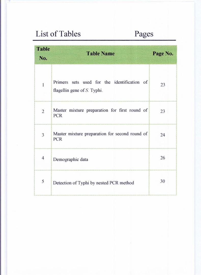
List of Tables Pages
Table Table Name Page No.
No.
I I I I I I
I Primers I I sets used for the identification of I
I 1 23 I I flagellin gene of S. Typhi. I I
f I
I 2J Mo"" mixture preparation for first round of 23 I peR
l- _._-- -I I 3 Master mixture preparation for second round of 24
I peR I
I I --
26 f~~ D,mog"phi, d,ta I I
I 5 I Detection ofTyphi by nested peR method 30 I I
l __ --'-_ J

List of Abbreviation
PBS - Phosphate buffered saline
LSD - Laboratory Sciences Division
S. Typhi - Salmonella Typhi
dH20 - Deionized water
~I - Micro liter
rpm - Rotation per minute
ICDDR,B - International Centre for Diarrheal Disease Research, Bangladesh
ORF - Open reading frame
Vi antigen - Virulence antigen
M cells - Microfold cells
TNF a - Tumor necrosis factor a
LPS - Lipopolysaccharide
PCR - Polymerase chain reaction
DNA - Deoxyribo nucleic acid
MDR - Multy drug resistant
LA - Luria agar
MgCh - Magnesium chloride
Taq - Therrnophillus aquaticus
dNTPs - Deoxyribo nucleotide tri phosphates
TBE - Tris borate EDTA

Abstract

Abstract
Typhoid fever is one of the lJll!ior health problems in Bangladesh. It is caused by the
bacteriwn SaJroone/la enlerica serover Typhi (S. Typhi). The infection rate of S. Typhi is
higher in children. The conventional diagnostic methods of typhoid have limitations.
Most commonly used Widal test gives a high rate of false positive results. As such
PCR method was tried to diagnose typhoid fever in some selected patients of
Bangladesh. Fifty three (53) patients were enroUed in this study from Dhaka hospital
of ICDDR, B and Kamalapur field site. Among them 27 (5 I %) persons are male.
Blood was coUected from all patients. Three different methods namely, a method by
Haque et ai , direct boiling method and commercial Qiagen kit method were tried to
extract Salmonella Typhi DNA from blood and invitro samples. To optimize the DNA
extraction from blood invitro grown S. Typhi bacteria was spiked into blood. From
the three different methods, commercial Qiagen kit method did well comparably to
extract DNA from spiked blood than the other methods. However, none of these
methods did appear promising to extract DNA from patient's blood. Because low
percentage of PCR positive were found from patient' s blood culture positive samples.
So another method was tried which was described earlier by Boom' s et aI. [1990] for
detection of S. Typhi from blood of suspected typhoid fever patient. This method
(Boom's et aI.[1990]) showed comparably satisfactory results than the other methods
to deal with patient' s blood. In this method the percentage of PCR positive was 35.7
% among the blood culture confinned patients. DNA extraction was also done in
blood culture negative samples by this method. Here 30.8% positive for PCR was
found among the culture negative samples. However there are some drawbacks in
DNA extraction procedures especially in low concentrated samples. In the method by
Haque et aI. [200 I] unexpected band was found and same phenomenon was observed
in direct boiling method. This might be due to contamination with S. Typhi. It seems
that, S. Typhi can be detected from patient' s blood specimen by PCR method for
diagnostic purpose. Howeve~ more study is needed to evaluate the efficacy of PCR
method with some modifications for detecting S. Typhi at low concentration in blood.

Chapter One
Introduction

Introduction
1.1 Background
Typhoid fever is caused by Salmonella enlerica serovar Typhi (S Typhi) which is an
important health problem in many developing countries [I]. S Typhi is a highly virulent
host restricted invasive pathogen that affects only humans [2]. Most of this burden occurs
among people of low-income regions, particularly in Asia, Africa, Lati~ America, the
Caribbean, and Oceania. About 80% of cases come from Bangladesh, China, India,
Indonesia, Laos, Nepal, Pakistan, and Vietnam [3]. It has been estimated that, 16-33 million
annual cases resulting in 216,000 deaths in endemic areas and its incidence is highest in
children and young adults between 5 and 19 years old [4]. Although young children are also
susceptible. In nonendemic areas, disease outbreak may occur from a contaminated food or
through carriers [9, 10].
The disease is characterized by the onset of prolonged high fever, severe headache, malaise
and abdominal pain [5]. The illness often causes diarrhea, especially in younger children,
whereas constipation is common in older children and adults. Serious complications occur in
up to 10% of typhoid fever patients, especially those who have been i II for more than two
weeks and have not received proper treatment [5]. Almost half of the treated patients
continue to excrete the pathogen for about one month after the symptoms have been
disappeared and 5% still continue upto five months [6, 7]. Approximately 3% become
chronic carriers and continue to excrete the or.ganism lifelong [8] . The encounters of S.
Typhi to humans are generally caused through fecal-oral route from infected individuals to
healthy ones [II].
Chapter 1: Introduction

1.2 Structure & Comparisn of the S. Typhi Genome
The genus Salmonella has 3 specIes, Salmonella enterica, Salmonella bongori and
Salmonella subterranean [15]. S. enlerica has seven subspecies consistently delineated by
sequence variation. The majority of diseases causing serovars are from subspecies,
Typhimurium, Typhi and Paratyphi [12].
Complete genome sequence data provides the genetic characterization of pathogens and
their hosts. The S. Typhi genome consists of 479 kb encoding around 4000 genes where over
200 genes are functionally inactive. Comparison of S. Typhi isolates from around the world
indicates that, they are highly related (clonal) and they emerged from a single point of
origin, around 30,000 - 50,000 years ago. Evidence suggests that S. Typhi undergoing gene
degradation and it has also recently acquired genes, such as those encoding the Vi antigen
by horizontal gene transfer [17].
Salmonella Typhi CTIS has a large circular chromosome consisting of 4.S Mb and two
plasm ids are pHCMI and pHCM2, which are 21S kb and 106 kb respectively. S. Typhi
CT IS has 4646 genes and 204 pseudogenes, nine of which resemble intact genes in strain
Ty2. Along with the 195 pseudogenes common with CTIS, strain Ty2 has also II unique
ones. S. Typhi Ty2 consists one large chromosome that is 4.7 Mb with an average G+C
content of 52.05% [16].
Chapter 1: Introduction 2

S el1leri('{/
~nnarT)pIH
~Im i ll l"y2
·":"'r'llf.l""
• •
Fig 2: Circular genome map of S. Typhi [IS]
The Ty2 genome has 4,545 ORFs and pseudogenes, where 4,516 are shared with CT IS
(outer circle, blue) and 29 of which are unique (pink). The second circle shows the locations
and orientations of rRNA operons (red) and tRNAs (turquoise). The third circle shows
insertion element distributions. The fourth circle shows the scale in base pairs. The fifth
circle shows the GIC skew, calculated for each sliding window of 10 kb along the genome.
The sixth and seventh (innermost) circles show the CTIS and Ty2 genome comparison
where, blue indicates collinear regions, red indicates inverted regions, green indicates a
region that is translocated and inverted again within the half-genome inversion region, and
yellow indicates unique regions [IS]. More discrepancies are found when looking at
prophages. Four prophages are located in identical parts of the genome relative to the
adjacent non phage genes, and both strains contain parts of prophages that are unique to one
another (16).
Chapter 1: Introduction 3

1.3 Antigenic types of Salmonella Typbi
Salmonella is a genus of rod-shaped, Gram-negative, non-spore forming, predominantly
motile bacteria belonging to the family of Enterobacteriaceae [12], with more than 2S0 I
serotypes [13]. Its diameter is around 0.7 to I.S 11m and lengths from 2 to Slim. The bacteria
contains nagella which projected along with all directions [12].
Figure I: Structure of Salmonella Typhi [67].
As with all Enterobacteriaceae, the genus Salmonella has three kinds of m:yor antigens with
identifying characteristics [14]-
a) Somatic or 0 antigens (bacterial endotoxins): These are heat stable and alcohol resistant.
Cross-absorption studies individualize a large number of antigenic factors, 67 of which are
used for serological identification.
Chapter 1: Introduction 4

b) H antigen: This is a protein structure associated with the flagella. Flagellar antigens are
heat-labile proteins. By mixing colonies of Salmonella with flagella-specific antisera gives
a characteristic pattern of agglutination.
c) Surface Vi (for virulence) antigen: This is a polysaccharide on the exterior of the cell
wall. The surface antigens (Vi) in Salmonella may mask 0 antigens, and the bacteria will
not be agglutinated with '0 ' antigen specific antisera [14].
1.4 Etiology
Most current understanding of S. Typhi is from observing the disease in humans, volunteer
research and animal models. S. Typhimurium in the murine model is the best characterization
of typhoid fever in humans [20]. In murine model during first exposure, the S. Typhi is
ingested and it enters into the small intestine through the microfold cells (M cells) of the
Peyer's patches. After being endocytosed by M cells, the bacterium is able to migrate to the
mesenteric lymph nodes and then multiply [20]. A fterwards the bacteria are released into
blood stream and circulate to cause a systemic infection [21]. S. Typhi is then taken up from
the blood by antigen presenting cells such as the macrophages that line over the liver,
spleen, and bone marrow sinusoids. The bacteria is able to replicate and stay in the
macrophages [22]. The macrophages can lose the ability to kill intracellular bacteria. The
clinical symptoms like fever, nausea, constipation and diarrhoea are observed when the
bacteria reenters into the circulation. S. Typhi is then removed from the blood via the gall
bladder to the small intestine.
Chapter 1: Introduction 5

are not sufficiently sensitive and specific [50]. Widal test has a presumptive diagnostic value
in non endemic areas but in endemic areas it ' s use is controversial [51].
1.7.1 Widal test
The Widal test is used to demonstrate rising titres of antibodies to flagellar (H) and somatic
(0) antigens in typhoid and paratyphoid fever [52, 53]. An increased 0 antibody level
signifies acute infection, whilst an increased H antibody level may indicate the serotype of
the infecting organism. Widal test has limited use, because H and 0 antibody levels may rise
non-speci fically due to cross reactions with other enterobacteriaceae.
1.8 Diagnosis of typhoid by nested Polymerase Chain Reaction (PCR)
The diagnosis of typhoid fever can be possible by Polymerase Chain Reaction (PCR) using
blood and stool as sole source of template DNA of Salmonella Typhi [54]. The flagellin
gene of S. Typhi can be detected by the Polymerase chain reaction (PCR). Nested
polymerase chain reaction is a modification of polymerase chain reaction intended to reduce
the contamination in products due to non specific primer binding. In conventional PCR a
commonly occurring problem is primers binding to incorrect regions of the DNA, giving
unexpected products (non specific bands). Nested polymerase chain reaction involves two
sets of primers, used in two successive runs of polymerase chain reaction, the second set
intended to amplify a secondary target within the first run product [55].
Chapter 1: Introduction II
U wanted primer annealing
No second run primer annealing
and
and
First PCR run. Cycle 1 DNA melting and primer binding.
l Fitst run majority PCR products
Seoond PCR run. Cycle 1 DNA melting and primer binding.
Unc::ontaJTlinalted final product
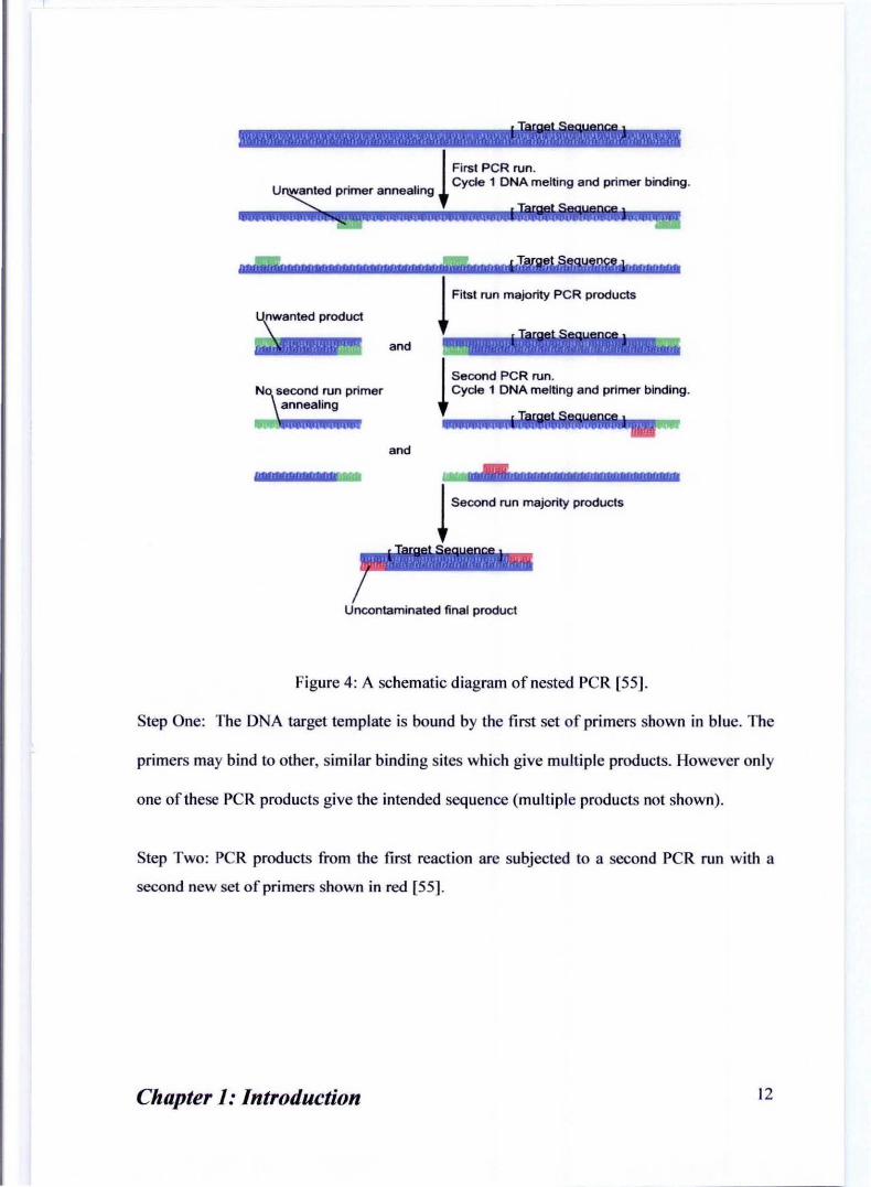
Figure 4: A schematic diagram of nested peR [55].
Step One: The DNA target template is bound by the first set of primers shown in blue. The
primers may bind to other, similar binding sites which give mUltiple products. However only
one of these peR products give the intended sequence (multiple products not shown).
Step Two: peR products from the first reaction are subjected to a second peR run with a
second new set of primers shown in red [55].
Chapter 1: Introduction 12

1.9 Treatment of typboid fever
1.9.1 General management
Supportive measures are important in the management of typhoid fever, such as oral or
intravenous hydration, the use of antipyretics, appropriate nutrition and blood transfusions if
indicated. More than 90"10 of patients can be managed at home with oral antibiotics, reliable
care and close medical follow-up [57]. However, patients with persistent vomiting, severe
diarrhea and abdominal distension may require hospitalization and parenteral antibiotic
therapy.
1.9.2 Antimicrobial therapy
In areas of endemic disease typhoid fever is managed at home with antibiotics and bed rest.
Effective antibiotic treatment for typhoid was developed in 1948, but even with modem
drugs typhoid still takes the life of about one in every hundred people [58]. Typhoid fever in
most cases is not fatal. Antibiotics, such as ampicillin, chloramphenicol, trimethoprim
sulfamethoxazole, amoxicillin and ciprofloxacin, have been commonly used to treat typhoid
fever in developed countries. Prompt treatment of the disease with antibiotics reduces the
case-fatality rate to approximately I %. When untreated, typhoid fever persists for three
weeks to a month. Death occurs in between 10% and 30% of untreated cases [59]. Since the
appearance of multidrug resistant strain, sensitivity test is very important for selecting drug.
Chapter 1: Introduction 13

1.9.3 Resistance
Resistance to ampicillin, chloramphenicol, trimethoprim-sulfamethoxazole and streptomycin
are now common, and these agents have not been used as first line treatment since almost 20
years. Typhoid that is resistant to these agents is known as multidrug-resistant typhoid
(MDR typhoid). Ciprofloxacin resistance is an increasing problem especially in the Indian
subcontinent and Southeast Asia. For these patients, the recommended fll"St line of treatment
is ceftriaxone. It has also been suggested that azithromycin is better at treating typhoid in
resistant populations than both fluoroquinolone and ceftriaxone drugs [60].
1.10 Prevention of typhoid fever
Sanitation and hygiene are the measures that can be taken to prevent typhoid. Careful food
preparation and washing of hands are therefore crucial for preventing typhoid. Health
education is of paramount importance to raise public awareness and induce behaviour
change [59).
There are two vaccines currently recommended by the World Health Organization for the
prevention of typhoid, these are the live, oral Ty21 a vaccine and the injectable Typhoid
polysaccharide vaccine. Both are between 50% to 80% protective and are recommended for
travelers to areas where typhoid is endemic [61].
1.1 0.1 Vaccination
Two types of typhoid vaccines are currently by WHO to preventing typhoid fever, (I) An
oral live-attenuated vaccine, (2) A Vi polysaccharide vaccine for parenteral use.
Chapter 1: Introduction 14
1.10.2 An oral live-attenuated vaccine
The oral vaccine containing live attenuated Salmonella Typhi Ty21 a strains in enteric coated
capsules, taken every two days, a total of 3 doses. The capsule must be refrigerated but not
frozen to achieve maximum efficacy. The vaccination should be completed within a week
and the booster dose should be given every 5 years. The vaccine recipient should not be
younger than 6 years [62]. The commercial vaccine is VivotifBema.
1.10.3 Vi polysaccharide vaccine
The vaccine is composed of purified Vi ("virulence") capsular polysaccharide antigen of
Salmonella Typhi isolated from blood cultures. This capsular polysaccharide of S. Typhi is
conjugated with nontoxic recombinant Pseudomonas aeruginosa exotoxin A and enhances
immunity. It is given by the parenteral route once a time. This vaccine must be kept at 4°C
temperature. Children below two years of age are not given this vaccine. A booster dose is
given every 2 years to maintain protection [63, 64]. The commercial vaccine is Typherix.
Aim ofthe study
To optimize the DNA isolation of Salmonella Typhi from blood of patients with enteric
fever which leads to rapid, sensitive and specific detection of Salmonella Typhi as well as
typhoid fever using nested PCR procedure.
Chapter 1: Introduction 15

Chapter Two
Materials and Methods
Materials and Metbods
The study was carried out at the Immunology Laboratory of the Laboratory Sciences Division
of the International Centre for Diarrheal Disease Research, Bangladesh (ICDDR, B).
2.1 Study Subjects
Patients with high fever (? 39°C) for 3-7 days with or without diarrhea who came to the
ICDDR, B hospital or from the Kamalapur field site in Dhaka were enrolled for this study.
Written consent was taken before taking blood from them.
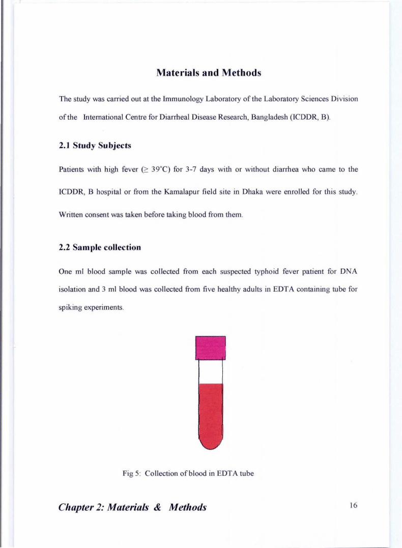
2.2 Sample coUection
One ml blood sample was collected from each suspected typhoid fever patient for DNA
isolation and 3 ml blood was collected from fi ve healthy adults in EDT A containing tube for
spiking experiments.
Fig 5: Collection of blood in EDTA tube
Chapter 2: Materials & MetllOds 16

2.3 DNA extraction from invitro grown Salmonella Typhi pure culture:
Salmonella el1lerica serovar Typhi, ST -004 (stored at -70"c) was streaked on MacConkey
agar plate and incubated over night at 37°C.
A loop of S. Typhi bacteria from the MacConkey agar plate was taken and suspended into an
eppendorf tube containing 100 fil of Phosphate Buffered Saline (PBS, 10 IllJ'v1. pH 7.4). The
suspension was boiled at 100°C in water bath for 10 minutes . The tube was then transferred
on ice and kept for I minute. After that the suspension was centrifuged at 16099.2 x g for 10
minutes. The supematant was collected and used as template and positive control for PCR
(positive control).
2.4 Prel>aration of S. Typhi bacteria for spiking in blood:
I . S. Typhi control strain (laboratory strain st-004) was streaked on Luria agar plate (LA)
and incubated ovemight :!t 37°C.
2. From the overnight grown plate 2 colonies were inoculated into eppendorf tube
containing I ml PBS. This was then vortexed for 15 seconds.
Fig 6: S. Typhi colonies cultu red on Luria Ag!!f med!C!!!!
Chapter 2: Materials & Methods 17

3. Bacterial suspension was then serially diluted down thirty times by 10 fold dilution
each time.
4. A IOOIlI of bacterial suspension from each dilution was spread on LA plate for
determining bacterial count.
5. Plates were incubated overnight at 3TC and colonjes were counted.
6. Rest of the bacterial suspensions from each dilution were centrifuged for 8 minutes at
16099.2 x g and the supernatent was discarded.
7. A IOOIlI of blood (from healthy subjects) was added on the bacterial pellet into each
eppendorftube and mixed by pulse vortexing for 5 seconds.
2.5.1 DNA Extraction procedure from spiked blood by Haque et al. [66]:-
I. S. Typhi spiked blood samples containing serially diluted bacteria were taken into
eppendorftubes serially.
2. The tubes were centrifuged for 5 minutes at 11180 x g.
3. The pellet was mixed with lysis buffer (I ml in each tube) and centrifuged for 6
minutes at 11180 x g . The supernatent was discarded.
4. Steps 2 & 3 were repeated.
5. The pellet was mixed with water (I ml in each tube), and centrifuged for I minute at
11180 x g. The supernatent was discarded.
6. Deionized water was added (30 III in each tube) to the pellet and boiled for 20
minutes.
7. The samples were kept at 4·C before use.
Chapter 2: Materials & Methods 18
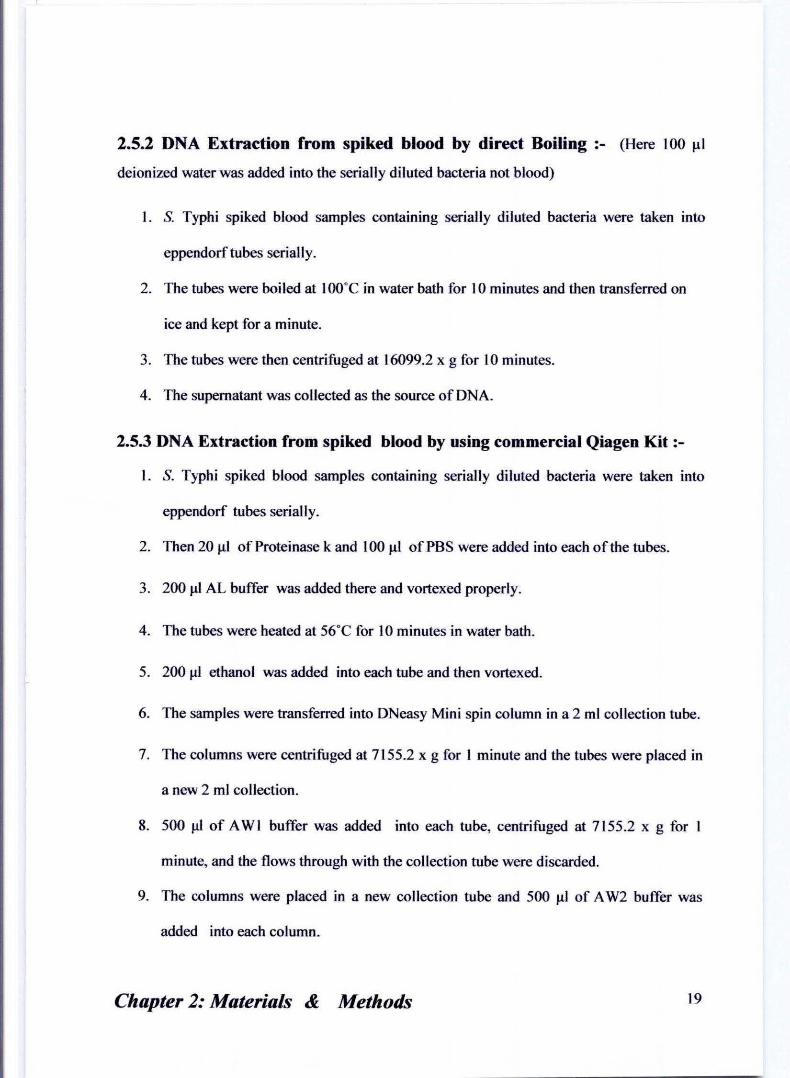
2.5.2 DNA Extraction from spiked blood by direct Boiling:- (Here 100 111
deionized water was added into the serially diluted bacteria not blood)
I. S. Typhi spiked blood samples containing serially diluted bacteria were taken into
eppendorftubes serially.
2. The tubes were boiled at 100' C in water bath for 10 minutes and then transferred on
ice and kept for a minute.
3. The tubes were then centrifuged at 16099.2 x g for 10 minutes.
4. The supernatant was collected as the source of DNA.
2.5.3 DNA Extraction from spiked blood by using commercial Qiagen Kit :-
I. S. Typhi spiked blood samples containing serially diluted bacteria were taken into
eppendorf tubes serially.
2. Then 20 111 of Proteinase k and 100111 of PBS were added into each of the tubes.
3. 200 j!I AL buffer was added there and vortexed properly.
4. The tubes were heated at 56' C for 10 minutes in water bath.
5. 200 111 ethanol was added into each tube and then vortexed.
6. The samples were transferred into DNeasy Mini spin column in a 2 ml collection tube.
7. The columns were centrifuged at 7155.2 x g for I minute and the tubes were placed in
a new 2 ml collection.
8. 500 111 of AWl buffer was added into each tube, centrifuged at 7155.2 x g for
minute, and the flows through with the collection tube were discarded.
9. The columns were placed in a new collection tube and 500 111 of A W2 buffer was
added into each column.
Chapter 2: Materials & Methods 19

10. The columns were centrifuged for 3 minutes at 21912.8 x g to dry the DNeasy
membrane and the flow through was discarded.
11. The columns were transferred to new eppendorf tubes and 50 /11 of AE buffer were
added into each column.
12. Then the tubes were incubated for 5 minutes at room temperature and centrifuged for
1 minute at I 1180 x g .
13. Finally the flow through fluids were collected as extracted DNA.
Chapter 2: Materials & Methods 20

2.6.1 DNA extraction from patient's blood specimens by using commercial
Qiagen Kit :-
I. Blood sample was collected (100 fll) from patients in each labeled eppendorf tube.
2. Proteinase k (20 fll) and PBS (100 fll) were added into each of the tubes.
3. AL buffer (200 fll) was added and vortexed properly.
4. The tubes were heated at 56°C for 10 minutes in water bath.
5. Ethanol was added (200 fIl) into each tube and then vortexed.
6. The samples were transferred into DNeasy Mini spin column in a 2 ml collection
tube (provided by the reagent company).
7. The columns were centrifuged at 7155.2 x g for I minute and the tubes were
placed in a new 2 ml collection tube (provided in the kit).
8. AWl buffer was added (500 fIl) into each tube, centrifuged at 7155.2 x g for I
minute, and the flows through with the collection tube were discarded.
9. The columns were placed in a new collection tube (provided by the reagent
company) and A W2 buffer was added (500 fIl) into each column.
10. The columns were centrifuged for 3 minutes at 21912.8 x g to dry the DNeasy
membrane and the flow through were discarded.
II. The columns were transferred to new eppendorf tubes and AE buffer were added
(50 fll) into each column.
12. Then the tubes were incubated for 5 minutes at room temperature and centrifuged
for I minute at 11180 x g .
13. Finally the flow through were collected as extracted DNA.
Chapter 2: Materials & Methods 21
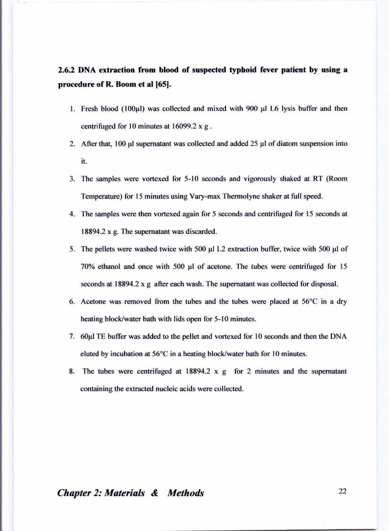
2.6.2 DNA extraction from blood of suspected typhoid fever patient by using a
procedure of R. Boom et aI165].
I. Fresh blood (l001i1) was collected and mixed with 900 iii L6 lysis buffer and then
centrifuged for 10 minutes at 16099.2 x g .
2. After that, 100 Jl.I supernatant was collected and added 25 iii of diatom suspension into
it.
3. The samples were vortexed for 5-10 seconds and vigorously shaked at RT (Room
Temperature) for 15 minutes using Vary-max Thermolyne shaker at full speed.
4. The samples were then vortexed again for 5 seconds and centrifuged for 15 seconds at
18894.2 x g. The supernatant was discarded.
5. The pellets were washed twice with 500 iii L2 extraction buffer, twice with 500 iii of
70% ethanol and once with 500 iii of acetone. The tubes were centrifuged for 15
seconds at 18894.2 x g after each wash. The supernatant was collected for disposal.
6. Acetone was removed from the tubes and the tubes were placed at 56°C in a dry
heating block/water bath with lids open for 5-1 0 minutes.
7. 60lil TE buffer was added to the pellet and vortexed for 10 seconds and then the DNA
eluted by incubation at 56°C in a heating block/water bath for 10 minutes.
8. The tubes were centrifuged at 18894.2 x g for 2 minutes and the supernatant
containing the extracted nucleic acids were collected.
Chapter 2: Materials & Methods 22
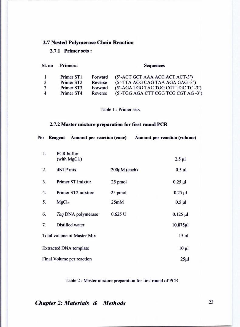
2.7 Nested Polymerase Chain Reaction
2.7.1 Primer sets :
SI. no Primers: Sequences
I Primer STI Forward (5 ' -ACT GCT AAA ACC ACT ACT-3') 2 Primer ST2 Reverse (5 ' -ITA ACG CAG TAA AGA GAG -3 ' ) 3 Primer sn Forward (5 ' -AGA TGG TAC TGG CGT TGC TC -3 ' ) 4 Primer ST4 Reverse (5 ' -TGG AGA CIT CGG TCG CGT AG -3')
Table I : Primer sets
2.7.2 Master mixture preparation for first round peR
No Reagent Amount per reaction (conc) Amount per reaction (volume)
I. PCR buffer (with MgCI2) 2.5 ~I
2. dNTPmix 200~M (each) 0.5 ~I
3. Primer STlmixtur 25 pmol 0.25 ~I
4. Primer ST2 mixture 25 pmol 0.25 ~I
5. MgCIz 25mM 0.5 ~I
6. Taq DNA polymerase 0.625 U 0.125 ~I
7. Distilled water 1O .875~1
Total volume of Master Mix 15 ~I
Extracted DNA template 10~1
Final Volume per reaction 25~1
Table 2 : Master mixture preparation for first round of PCR
Chapter 2: Materials & Methods 23

2.7.3 Thermal cycle for first round of peR:
First step 95°C for 5 minutes (initial denaturation)
Second step 94°C for denaturation-----I min
Third step 57°C for prime annealing-----I min
Fourth step n oc for elongation--I min
Then second step is repeated fourty times
Fifth step n oc for 7 minutes (final extension step)
2.7.4 Master mixture preparation for 2nd round PCR
No Reagent Amount per reaction (cone) Amount per reaction (volume)
I. peR buffer (with MgCh) 2.5 III
2. dNTPmix 2001lM (each) 0.5 III
3. Primer STI mixlur 25 pmol 0.25 III
4. Primer ST2 mixture 25 pmol 0.25 j!I
5. MgCh 25mM 0.5 III
6. Taq DNA polymerase 0.625 U 0. 125 111
7. Distilled water 15.875111
Total volume of Master Mix 20 III
Extracted DNA template 5 III
Final Volume per reaction 25fll
Table 3 : Master mixture preparation for second round of PCR
Chapter 2: Materials & Methods 24
2.7.5 Thermal cycle for 2nd round of peR:
First step
Second step
Third step
Fourth step
95°C for 5 minutes (initial denaturation)
94°C for denaturation-----I min
57°C for prime annealing----I min
72°C for e1ongation----1 min
Then second step is repeated fourty times
Fifth step 72°C for 7 minutes (final extension step)
2.7.6 Agarose Gel electropboresis :
Reagents: Agarose, Tris-Borate-EDT A (TBE) buffer, Ethidium bromide, 100bp ladder.
Preparation of 1.5% Agarose gel
I. TBE buffer was taken (100 ml) In a conical flask and added 1.5 gm
Agarose powder into it.
2. The flask was heated for 3 minutes in microwave oven.
3. Ethidium Bromide was added (4 Ill) and mixed properly.
4. The mixture was layed on gel tray for cooling.
5. PCR samples were stained with loading dye. (4 III dye with 10111 PCR
product).
6. Electrophoresis was performed for 90 minutes at 80V.
7. The gel was observed under UV light in gel-doc machine.
Chapter 2: Materials & Methods 25

Chapter Three
Results
Results
3.1 Detection of Salmonella enterica serovar Typbi by nested peR using
different nucleic acid extraction metbods.
Demographic Data :
Parameter Number
Patient enrolled 53
Blood collected From 53 patients
Gender Percentage
Male 27 (51%)
Female 26 (49%)
Fifty three (53) patients were enrolled in this study from Dhaka hospital of leDDR, Band
Kamalapur field site. Among them 27 (51 %) persons were male. Blood was collected from
all patients studied here.
3.2 Optimization of DNA extraction procedure:
For the evaluation of efficient DNA extraction procedure nested pe R was performed using
DNA samples extracted from spiked blood by using different extraction procedures.
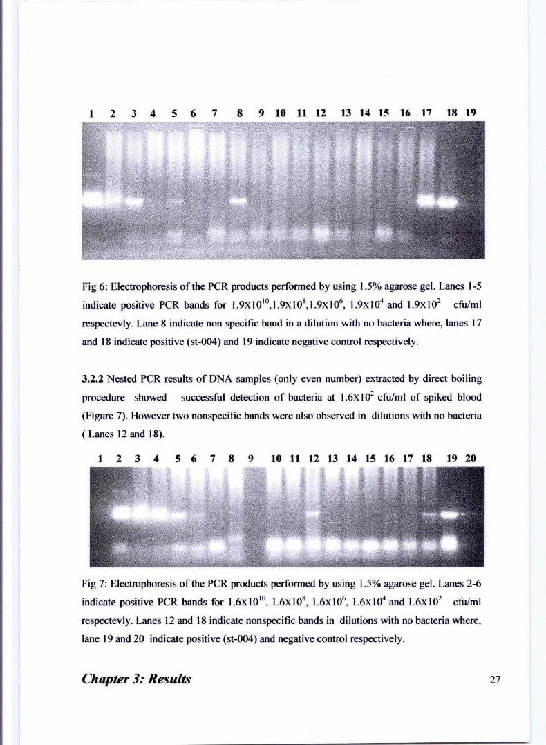
3.2.1 Nested peR results of DNA samples (on ly even number) extracted by a procedure
described by Haque et al. [66] showed successful detection of bacteria at 1.9 X 10² cfulml
of spiked blood (Figure 6). However a nonspecific band was also observed in a negative
control in similar dilutions with no bacteria ( Lane 8).
Chapter 3: Results 26
1234567 8 9 10 11 12 13 14 15 16 17 18 19
Fig 6: Electrophoresis of the peR products perfonned by using 1.5% agarose gel. Lanes 1-5
indicate positive peR bands for 1.9XI0' O, 1.9x 10⁸, 1.9x 10⁶, 1.9xI04 and 1.9xI02 cfu/ml
respectevly. Lane 8 indicate non specific band in a dilution with no bacteria where, lanes 17
and 18 indicate positive (st-004) and 19 indicate negative control respectively.
3.2.2 Nested peR results of DNA samples (only even number) extracted by direct boiling
procedure showed successful detection of bacteria at 1.6xl02 cfulml of spiked blood
(Figure 7). However two nonspecific bands were also observed in dilutions with no bacteria
( Lanes 12 and 18).
1 2 3 4 5 6 7 8 9 10 11 12 13 14 15 16 17 18 19 20
Fig 7: Electrophoresis of the peR products perfonned by using 1.5% agarose gel. Lanes 2-6
indicate positive peR bands for 1.6XIO IO, 1.6X10⁸
, 1.6X10⁶, 1.6xI04 and 1.6xI02 cfulml
respectevly. Lanes 12 and 18 indicate nonspecific bands in dilutions with no bacteria where,
lane 19 and 20 indicate positive (st-004) and negative control respectively.
Chapter 3: Results 27
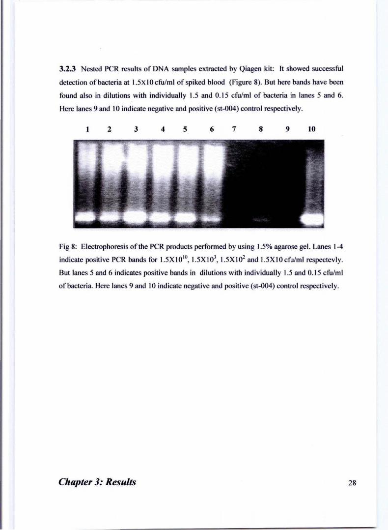
3.2.3 Nested peR results of DNA samples extracted by Qiagen kit: It showed successful
detection of bacteria at 1.5x I 0 cfulml of spiked blood (Figure 8). But here bands have been
found also in dilutions with individually 1.5 and 0. 15 cfulml of bacteria in lanes 5 and 6.
Here lanes 9 and 10 indicate negative and positive (st-004) control respectively.
1 2 3 4 5 6 7 8 9 10
Fig 8: Electrophoresis of the peR products performed by using 1.5% agarose gel. Lanes 1-4
indicate positive peR bands for 1.5XIO IO, 1.5X10³, 1.5XI02 and 1.5XIOcfulmi respectevly.
But lanes 5 and 6 indicates positive bands in dilutions with individually 1.5 and 0. 15 cfulml
of bacteria. Here lanes 9 and 10 indicate negative and positive (st-004) control respectively.
Chapter 3: Results 28

3.3 Detection of S. typhi in blood of patients with typhoid fever
Out of fifty three (53) patients 14 (26.4%) were positive by blood culture .
3.3.1 DNA extraction from patient's blood specimens by using commercial
Qiagen Kit.
Nested PCR was done for S. Typhi detection of four (4) patients of blood culture positive
samples. Only One (25%) patient was positive in PCR method among 4 blood culture
confirmed typhoid patients.
1 2 3 4 5 6 7
Fig II : Electrophoresis of the PCR products performed by using 1.5% agarose gel. Lanes 1-
4 indicate patient's blood sample where lane I shows only one band. Lane 6 is positive
control (st·OO4) and lane 7 indicates in negative control.
Chapter 3: Results 29

3.3.2 DNA extraction from patient's blood specimens by using a previously
described procedure:
Nested PCR was done for typhi detection of all patients regardless of blood culture positive
and negative using a procedure described earlier (R. Boom et ai, [65]). A total of5 (35.7%)
patients were positive in PCR method among 14 blood culture confirmed typhoid patients.
12 (30.8%) patients were also found positive by PCR among blood culture negative patients.
Parameters Number
Total Blood Samples 53
Culture (+) ve 14
Culture (-) ve 39
PCR positive of Culture (+) ve 5
PCR positive of Cu Iture (-) ve 12
Table 4 : Detection of Typhi by nested PCR method in blood culture (+)ve and (-)ve
patients.
1 2 3 4 5 6 7 8 9 10 11
Fig 10 : Electrophoresis of the PCR products performed by using 1.5% agarose gel. Lanes 1-
7 indicate blood specimen from patients. Lane 9 indicates a positive control (st-004) and,
lane II is negative control where no sample was inlcluded in lane 8 and 10.
Chapter 3: Results 30

Chapter Four
Discussion

Discussion
In this study peR method used by different scientists to diagnose typhoid fever patients
in Bangladesh was evaluated for devising a better procedure to diagnose the disease at
low bacterial titer.
Three different methods were tried for extracting Salmonella Typhi DNA from blood as
well as laboratory control specimens. To optimize the DNA extraction procedure from
blood, spiked invitro grown S. Typhi bacteria was added to blood. The blood was serially
diluted to detennine the minimum concentration of bacteria that could be extracted from
the blood specimens.
A method previously described by Haque et at. [66] was sensitive upto 1.9x I 02 cfu/ml in
blood. So this method is not sensitive enough to use for typhoid fever blood specimens,
because this is very high load of bacteria that might be necessary to be present in blood
for detection of S.Typhi. In natural infection, the concentration of S. Typhi is much
lower.
Another method that was tried, the direct boiling method which was sensitive up to
1.6x I 02 cfu/ml in blood. So this method was also not sensitive enough for diagnostic
purposes for typhoid fever from blood specimens. However, by this method the
separation on agarose gel gave distinct and sharp bands. The amount of bacteria was
however very high that might be necessarily being present in blood obtained from a
patient.
The third method tried was utilizing the commercial Qiagen kit method. Here sharp bands
obtained after electrophoresis and bacteria up to 1.5x I 0 cfulml could be detected. Thus
Chapter 4: Discussion 31

this was the best procedure and was comparatively more sensitive than the above
methods.
The commercial Qiagen kit was found to be the most sensitive method for DNA
extraction from blood. The other two methods were not able to extract DNA for detect of
S. Typhi as efficiently and was more sensitive. Based on this comparative study the
Qiagen kit method was used for DNA extraction from blood of patients suspected of
typhoid fever.
The DNA extraction from patient's blood using the Qiagen kit however did not look
promising because only one patient was positive by peR out of four blood culture
positive specimens tested. Thus, another method which was described earlier by Boom's
et al. [65] was used for detection of S. Typhi from blood of suspected typhoid fever
patient. In this method it was observed that 5 patients were positive for pe R out of 14
blood culture confirmed patients. DNA extraction was also done in blood culture
negative samples by this method (Boom's et al. [65]). Here, 12 (31%) positive for pe R
out of 39 culture negative samples were found.
There are some drawbacks in DNA extraction procedures especially when specimens
with low concentration of bacteria are used. In the method described by Haque et al [66]
one unexpected band was found and same was observed by the direct boiling method.
This could have been being due to contamination with S. Typhi.
In thi s exploratory study it was observed that, S. Typhi could be detected from the
patient's blood specimen by peR method for diagnostic purpose. More study is needed to
evaluate the efficacy of peR method for detecting S. Typhi at low concentration in blood.
Chapter 4: Discussion 32

Chapter Five
References

References
I. Velema JP, van Wijnen G, Bult P, Jota S. (1997). Typhoid fever in Ujung
Pandang, Indonesia-high risk groups and high risk behaviours. Trop Med Int
Health I I: 1088- 1094.
2. Background Paper on Vaccination against Typhoid Fever using New-Generation
Vaccines. WHO SAGE. (2007) . World Health Organization. Background
document: The diagnosis, treatment and prevention of typhoid fever. Geneva:
WHO, 2003 (WHOIV&B/03.07)
3. Birkhead GS, Morse Levine WC, Fudala JK, Kondracki SF, Chang HG,
Shayegani M, Novick L, Blake PA. (1993). Typhoid fever at a resort hotel in New
York: a large outbreak with an unusual vehicle. J Infect Dis 167: 1228- 1232.
4. Grunen E, Flepp M, Gabathuler U. (1997). Outbreak of typhoid fever in a non
endemic area: comparison of three molecular typing methods. J Microbiol
Methods 28: 179-185.
5. World Health Organization (WHO). (2003). Background document: Background
document: The diagnosis, treatment and prevention of typhoid fever.
Biologicals ... diagnosis, treatment and prevention of typhoid fever
whqlibdoc.who.intihq/2003/WHO _V &B _03.07.
6. Lai CW, Chan RC, Cheng AF, Sung JY, Leung JW. ( 1992). Common bile duct
stones: a cause of chronic salmonellosis. Am J Gastroenterol 87: I 198-1199.
Chapter 5: References 33

7. Dutta U, Garg PK, Kumar R, Tandon RK. (2000). Typhoid carriers among
patients with gallstones are at increased risk for carcinoma of the gallbladder. Am
J Gastroenterol95: 784-787.
8. Zavala Trujillo I, Quiroz C, Gutierrez MA, Arias J, Renteria M. (1991).
Fluoroquinolones in the treatment of typhoid fever and the carrier state. Eur J Clin
Microbiol Infect Dis 10: 334-341.
9. Chau IT, Campbell JI, Galindo eM, Van Minh Hoang N, Diep TS, Nga IT, et
al. (2007). Antimicrobial drug resistance of Salmonella enterica serovar typhi in
asia and molecular mechanism of reduced susceptibility to the
fluoroquinolones. An/imicrob Agents Chemo/her. 51 (12):4315-23.
10. World Health Organization. (2007). "Typhoid Fever". Retrieved 2007-08-28.
hnp:llwww.who.intlvaccine_research/diseasesldiarrhoeal/en/index7.html.
II. Salmonella typhi, Salmonella en/erica typhi . By: David V. Pollack. Copyright
Dennis Kunkel Microscopy, Inc. Introduction: Worldwide, typhoid fever affects
roughly 17 million. web.uconn.edu/ . ..ISalmonellatyphi/Salmonellatyphi.html.
12. Salmonella Information . Salmonella Research Today is a free monthly online
journal that collates and summarizes the latest research about Salmonella.
salmonella.researchtoday.netlabout-salmonella.htm.
13. Salmonella Typhi to primates. (1997). Prevalence of Salmonella on pig carcasses
at a Slaughter house In Salmonella Dublin to cattle, (Mousing).
www.vphcap.orgifileITHESIS/02nd-studentl . ..IChapter2.
14. Salmonella and Salmonellosis. Web Review of To dar's Online Textbook of
Bacteriology. In humans, Salmonella are the cause of two diseases
Chapter 5: References 34
called salmonellosis: enteric fever (typhoid),
www.textbookofbacteriology.neVsalmonella.html.
15. Salmonella Research Today. Salmonella Typhimurium, Food Poisoning,
Infection, Treatment. Salmonella Research Today is a free monthly online journal
that collates and summarizes the latest research about Salmonella, including
details on salmonella typhimurium, food poisoning, infection, treatment.
16. Salmonella - Mjcrobe. Genome Structure: Salmonella typhi CTI8 has a large
circular chromosome ... parts of the genome relative to the adjacent nonphage
genes, microbewiki .kenyon.edulindex.php/Salmonella
17. Stephen baker and Gordon Dougan. (2007). The Genome of Salmonella enlerica
Serovar Typhi. Stephen baker and Gordon Dougan. The Wellcome Trust Sanger
Institute, Hinxton, Cambridge, United Kingdom. The S. Typhi Genome . CID:45
(suppl I ).S29
18. Wen Deng, 1 Shian-Ren Liou, lt Guy Plunkett 1ll,1 George F. Mayhew, I Debra
J. Rose, I Valerie Burland,1 Voula Kodoyianni, I,2 David C. Schwartz,
Comparative Genomics of Salmonella enterica Serovar Typhi Strains Ty2 and
CT 18. www.pubmedcentral.nih.gov/articlerender.fcgi?blobtype=html.
19. Janeway, A.C, P. Travers, M. Walport., J.M. Shlomchik. (2001). 24 Apr
2003 ... Immunology: The Immune System in Health and Disease. New
York, NY: Elsevier Science Ltd. Garland Publishing, pp 387, 336, 586.
20. Marcela F. Pasetti, Myron M. Levine and Marcelo B. Sztein. (2003). Attenuated
Salmonella enlerica serovar Typhi (S. Typhi) strains can serve as safe and
effective oral vaccines to prevent typhoid fever Vaccine, 21 :5-6:401-418.
Chapter 5: References 35

21. Janeway, A.C, P. Travers, M. Walport., J.M. Shlomchik. (2001). Immunology:
The Immune System in Health and Disease. New York, NY: Elsevier Science
Ltd. Garland Publishing, pp 387, 336, 586.
22. Diagle, F., Graham, J.E., and C.R., Curtiss. (2001). Identification of Salmonella
typhi genes expressed within macrophages by selective capture of the transcribed
sequences (SCOTS). Molecular-Microbiology. 41(5):1211-1222.
23. Brennan, M.A., and Cookson BT (2000). Salmonella induces macrophage death
by caspase-I dependent necrosis. Molecular Microbiology. 38:31-40.
24. Salerone, G.R., Wyant, T.L, Passeti, M.F., Fernadez, V.M. , Tacket, C.O. , Levin,
M.M., and M.B, Sztein. (2003). Cocomitant induction of CD4+ and CD8+ T cell
responses in volunteers immunized with Salmonella enterica serovar Typhi CVD
908-htrA.170(5):2734-2741.
25. Shtrichman, R., Heithhoff, D.M., Mahan, MJ., and C.E., Samuel. (2002). Tissue
selectivty of interferon-stimulatied gene expression in mice infected with Dam +
versus Dam- Salmonella enterica serovar Typhimurium strains. Infection and
Immunity. 70(10):5579-5588.
26. Macdonald T.T., and L., Steidler. (2000). Recent developments in the
immunology of inflammatory bowel disease. Journal of Immunology. 51 :2-9.
27. Butler, T., [slam, A., Kabir, L., and P.K. , Jones. (1991). Patterns of morbidity and
mortality in typhoid fever dependent on age and gender.
28. Van Basten, J.P. and R. Stockenbrugger. (1994). Typhoid perforation. A review
of the literature since 1960.Trop. Geogr. Med. , 46:336-339.
Chapter 5: References 36
29. Chiu CH, Chuang CH, Chiu S, et al. (2009). Salmonella Infection:
eMedicine Pediatrics: General Medicine, Salmonella enterica serotype
Choleraesuis infections in pediatric patients. Pediatrics ; 117(6):e 1193-6.
30. Raffatellu M, Chessa D, Wilson RP, Tukel C, Ak~elik M, Baumler AJ. (2006).
Capsule-mediated immune evasion: a new hypothesis explaining aspects of
typhoid fever pathogenesis. Infect Immun. ; 74( I): 19-27.
31. Parry CM, Hien IT, Dougan G, et al. Typhoid fever. N Engl J Med. (2002).
347(22): 1770-82.
32. Ramsden AE, Mota LJ, Munter S, Shorte SL, Holden DW. (2007). The SPI-2
type III secretion system restricts motility of Salmonella-containing vacuoles. Cell
Microbiol. ;9(10):2517-29.
33. Typhoid Fever- The pathophysiology of typhoid fever is a complex process which
proceeds through several stages. (24;26;27) The disease begins with an
asymptomatic .. .Introduction - . uloronlo. ca/oisISW2006ItyphoidJever. him.
34. Profile of Typhoid Fever Caused by Salmonella Typhi. (2008). Carriers of
Typhoid Fever May Not be Sick. From Ingrid Koo, Ph.D., About.com Guide.
About.com, Health's Disease and content is reviewed by the Medical Review
Board.
35. Wain J, House D, Parkhill J, Parry C, Dougan G, Wain J et al. (2002). unlocking
the genome of the human typhoid bacillus. The Lancet Infectious Diseases,
2(3): 163-170.
Chapter 5: References 37
36. Andrade DR, Andrade Junior DR, de Andrade DR, de Andrade Junior DR.
(2001). Typhoid fever as cellular microbiological model. Revista do Instituto de
Medicina Tropical de Sao Paulo 2003; 45(4): 185-191 .
37. House D, Bishop A, Parry C, Dougan G, Wain J, House D et al. Typhoid fever:
pathogenesis and disease. Current Opinion in lnfectious Diseases; 14(5):573-578.
38. Parry CM, Hien TT, Dougan G, et al. Typhoid fever. N Engl J Med. (2002).
347(22): 1770-82.
39. Gotuzzo E, Frisancho 0 , Sanchez J, Liendo G, Carrillo C, Black RE, et
al. Typhoid Fever: Treatment & Medication eMedicine Infectious
DiseasesAssociation between the acquired immunodeficiency syndrome and
infection with Salmonella typhi . emedieine.medseape.eomlarlic/eI23 1135-
Irealmenl.
40. Ram PK, Naheed A, Brooks W A, Hossain MA, Mintz ED, Breiman RF. (2007).
Ri sk factors for typhoid fever in a slum in Dhaka, Bangladesh. Epidemiol Infeel
; 135(3):458-65.
41. Ali S, Vollaard AM, Widjaja S, Surjadi C, van de Vosse E, van Dissel JT.
(2006). PARK2IPACRG polymorph isms and susceptibility to typhoid and
paratyphoid fever. Clin Exp Immunol ; 144(3):425-31 .
42. Earampamoorthy S, Koff RS. (1975). Health hazards of bivalve-mollusk
ingestion. Ann Inlern Med. Jul 1975;83(1): I 07-10.
43. Levine MM, Tacket CO, Sztein MH. (200 1). Host-Salmonella interaction: human
trials. Microbes In/eel. Nov-Dec 200 I ;3( 14-15): 1271-9.
Chapter 5: References 38
•

44. Hornick, R. B. (1970). Pathogenesis of typhoid fever. J Egypt Public Health
Assoc 45:247-5.
45. World Health Organization. (2003). Background document: The diagnosis,
treatment and prevention of typhoid fever.
46. Wain, J., T. S. Diep, V. A. Ho, A. M. Walsh, T. T. Nguyen, C. M. Parry, and N. J.
White. (1998). Quantitation of bacteria in blood of typhoid fever patients and
relationship between counts and clinical features, transmissibility, and antibiotic
resistance. J Clin Microbiol 36: 1683-7.
47. Culture methods, http://www.geocities.com/avinash_abhyankar/culture.htm.
48. World Health Organization, The diagnosis, treatment and, prevention of typhoid
fever, Background document: Communicable Disease Surveillance and
Response,Vaccines and Biologicals.
49. Vallenas, C., H. Hernandez, B. Kay, R. Black, and E. Gotuzzo. (1985). Efficacy
of bone marrow, blood, stool and duodenal contents cultures for bacteriologic
confirmation of typhoid fever in children. Pediatr Infect Dis 4:496-8.
50. Abraham, G., B. Teklu, M. Gedebu, G. H. Selassie, and G. Azene. (1981).
Diagnostic value of the Widal test. Trop Geogr Med 33:329-33.
51 . Pang, T., and S. D. Puthucheary. (1983). Significance and value of the Widal test
in the diagnosis of typhoid fever in an endemic area. J Clin PathoI36:471-5.
52. Saha, S. K., M. Ruhulamin, M. Hanif, M. Islam, and W. A. Khan. ( 1996).
Interpretation of the Widal test in the diagnosis of typhoid fever in Bangladeshi
children. Ann Trop Paediatr 16:75-8.
Chapter 5: References 39

53. Schroeder, S. A., B. Aserkoff, and P. S. Brachman. (1%8). Epidemic
salmonellosis in hospitals and institutions. A five-year review. N Engl J Med
279:674-8.
54. Kumar, A., V. Arora, A. Bashamboo, and S. Ali. (2002). Detection of Salmonella
Typhi by polymerase chain reaction: implications in diagnosis of typhoid fever.
Infect Genet Evol 2: I 07-1 O.
55. http://en.wikipedia.orglwikifNested]CR.
56. www.pcrstation.comlimageslnested-pcr.gif. 450 x 505 - 14k - gif -Image may be
subject to copyright.Below is the image at: www.pcrstation.com/nVnested-pcr/
57. Punjabi NH. (2000). Typhoid fever. In: Rakel RE, editor. Conn 's Current therapy.
Fifty-second edition. Philadelphia: WB Saunders; 2000. P.161-5.
58. Chinh, N. T., C. M. Parry, N. T. Ly, H. D. Ha, M. X. Thong, T. S. Diep, J. Wain,
N. J. White, and J. J. Farrar. (2000). A randomized controlled comparison of
azithromycin and ofloxacin for treatment of multi drug-resistant or nalidixic acid
resistant enteric fever. Antimicrob Agents Chemother 44: 1855-9.
59. Typhoid fever - For a related disease which is caused by two different bacteria,
Paratyphoid fever. en.wikipedia.orglwiki/Typhoid _fever.
60. EfTa EE, Bukirwa H. (2008). "Azithromycin for treating uncomplicated typhoid
and paratyphoid fever (enteric fever)" . Cochrane Database of Systematic Reviews
(I). doi: 10. 100211465 I 858.CD006083.pub2.
61. 'Typhoid vaccines: WHO position paper". Wldy. 2008, Epidemiol. Rec. 83 (6):
49-59. PMID 18260212. February (2008).
www.who.intlwer/2008/wer8306/enlindex.html.
Chapter 5: References 40
62. Silva, B. A., C. Gonzalez, G. C. Mora, and F. Cabello. (1987). Genetic
characteristics of the Salmonella Typhi strain Ty2 1 a vaccine. J Infect Dis
155: 1077-8.
63. Levine, M. M. , and M. B. Sztein. (2004). Vaccine development strategies for
improving immunization: the role of modem immunology. Nat Immunol 5:460-4.
64. Levine, M. M., D. N. Taylor, and C. Ferreccio. (1989). Typhoid vaccines come of
age. Pediatr Lnfect Dis J 8:374-81.
65. R Boom, C J Sol, M M Salimans, C L Jansen, P M Wertheim-van Dillen and J
van der Noordaa.) \990). Rapid and simple method for purification of nucleic
acids. J Clin Microbiol. March; 28(3): 495-503.
66. Haque, A., N. Ahmed, A. Peerzada, A. Reza, S. Bashir, and G Abbas. (2001).
Utility of PCR in diagnosis of problematic cases of typhoid. Jpn. J. Infect. Dis.
54:237-239.
67. www.physorg.com/news83593133.html
Chapter 5: References 41

Chapter Six
Appendices
Appendices
Appendix 1
Preparation of Pbospbate buffered Saline (PBS) (PH-7.2) ( I Itr) :
NaCI 80.00 gm
Na2HP04 11.50 gm
KH2P04 2.00gm
KCI 2.00 gm
Deionized water 1000.00 ml
Appendix 2
Reagent Preparation for DNA extraction (DNA extraction by R. Boom et al. (651) :
Diatom Suspension
I. Deionized water (10 mL) was taken into a 50 mL Falcon tube.
2. Diatomaceous Earth (5 g) was added into it.
3. The tube was filled upto 50ml with deionized water (dH20).
4. The tube was shaken vigorously (to making MUD).
5. The diatoms allowed to settle down for 3 hours (30 minutes minimum).
6. The upper Liquid layer was removed.
7. The tube was refilled upto 50 mL with dH20 and mixed properly.
8. Then the tube was allowed to settle down for 2 hours (30 minutes minimum).
9. The upper liquid layer was removed.
10. A volume of dH20 equal to the semi-solid MUD was added.
Chapter 6: Appendices 42

II. The diatom suspension after vortexed aliquoted into small volumes (0.5-1.0
ml) and autoclaved (I SIbs for 15 minutes). Then stored at room temperature.
Buffers
Preparation ofSOmM lOOml Tris-HCI (PH 6.4):
I. Tris (0.606g) in a conical flask was taken and deionized water (90mJ) was
added into the flask.
2. The mixture was stired and the pH was adjusted to 6.4 by adding HCI.
3. The final volume was made 100mJ by adding dH20 .
Preparation of L2 extraction buffer (SOml):
I. GuSCN (32g) in a conical flask was taken and Tris-HCI was added uplo
50ml.
2. The flask was heated at 65°C for 10-20 minutes to dissolve GuSCN.
3. The solution was filtered through 1.2~m filter and transferred to a 50 ml
falcon tube. Then stored at RT.
4. The conical flask was covered by aluminium foil and falcon tube was
covered by brown paper.
Preparation of L6 extraction buffer (SOml):
5. GuSCN (32g) in a conical flask was taken and Tris-HCI was added uplo
50mJ.
6. The flask was heated at 65°C for 10-20 minutes to dissolve GuSCN.
7. EDTA (0.292g) and Triton X-loo (I00~1) were added. The mixture was
stired for 10 min for mixing.
8. The solution was filtered through 1 .2~m filter and transferred to a 50 ml
falcon tube. Then stored at RT.
Chapter 6: Appendices 43

9. The conical flask was covered by aluminium foil and falcon tube was
covered by brown paper.
Appendix 3
Reagents for DNA extraction using commercial Qiagen Kit :
The Reagents were provided by the Qiagen reagent company.
Appendix 4
Buffer preparation for DNA Extraction (DNA extraction by Haque et al. 166]) :
For SO mJ buffer
I. Tris Hel (10 ml of 50 mM) was taken in a bottle.
2. Deionized water (40 ml) was added into it.
3. The PH adjusted to 8.
4. EDTA (0.0146 g) was added into it and stired to dissolve.
5. Then triton X (200 111) was added and mixed properly.
Appendix 5
Preparation of Tris-EDT A (TE) buffer (l00ml)
I. Deionized water (105 ml) with 0.2 11m was filtered and 95 mI was taken in a
beaker.
2. Tris (0.1211 g) was added into it.
3. The solution was stired and the pH was adjusted at 8.
4. EDTA (0.0292g) was added there and stired to dissolve.
5. The filtered deionized water was added upto 100ml.
6. The solution was filtered with 0.2 11m filter and autoclaved.
7. Then stored at +4°C.
Chapter 6: Appendices 44

Appendix 6
Preparation ofTBE buffer (lOx):
Tris
Boric Acid
Na-EDTA
The deionized water was autoclaved upto 1 L
Appendix 7
Loading dye composition (6X)
0.25% BPB (Bromo Phenol Blue): 0.025 g
0.05% XC (Xylene Cyanol FF) : 0.5 ml
100 mM EDTA : 2 ml
: 5 mI
12.1g
6.0
0.74g
(Stock 1 % BPB
(Stock I%XC
(Stock 0.5 mM
2.5 mI)
0.5 ml)
2.0 rnl)
(Stock 100% Gly 5.0 m!) 50% Glycerol
Distilled water : Rest of water Nil
For 10 rnl IOmI
Chapter 6: Appendices 45
Related Documents


![SALMONELLA ENTERICA SUBSP. ENTERICA 1,4,[5],12:i:-](https://static.cupdf.com/doc/110x72/6297d8bb7423086b1b094e2e/salmonella-enterica-subsp-enterica-14512i.jpg)


