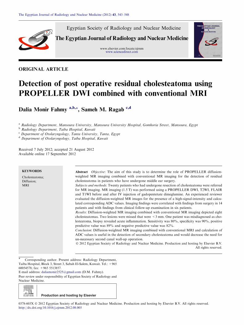
ORIGINAL ARTICLE Detection of post operative residual cholesteatoma using PROPELLER DWI combined with conventional MRI Dalia Monir Fahmy a,b, * , Sameh M. Ragab c,d a Radiology Department, Mansoura University, Mansoura University Hospital, Gomhoria Street, Mansoura, Egypt b Radiology Department, Taiba Hospital, Kuwait c Department of Otolaryngology, Tanta University, Tanta, Egypt d Department of Otolaryngology, Taiba Hospital, Kuwait Received 7 July 2012; accepted 21 August 2012 Available online 17 September 2012 KEYWORDS Cholesteatoma; Diffusion; MRI Abstract Objective: The aim of this study is to determine the role of PROPELLER diffusion- weighted MR imaging combined with conventional MR imaging for the detection of residual cholesteatoma in patients who have undergone middle ear surgery. Subjects and methods: Twenty patients who had undergone resection of cholesteatoma were referred for MR imaging. MR imaging (1.5 T) was performed using a PROPELLER DWI, T2WI, FLAIR and T1WI before and after IV injection of gadopentetate dimeglumine. An experienced reviewer evaluated the diffusion-weighted MR images for the presence of a high-signal-intensity and calcu- lated corresponding ADC values. Imaging findings were correlated with findings from surgery in 14 patients and with findings from clinical follow-up examination in six patients. Results: Diffusion-weighted MR imaging combined with conventional MR imaging depicted eight cholesteatomas. Two lesions were missed that were <3 mm. One patient was misdiagnosed as cho- lesteatoma, biopsy revealed acute inflammation. Sensitivity was 80%, specificity was 90%, positive predictive value was 89% and negative predictive value was 82%. Conclusion: Diffusion-weighted MR imaging combined with conventional MRI and calculation of ADC values is useful in the detection of secondary cholesteatoma and would decrease the need for un-necessary second canal wall-up operation. Ó 2012 Egyptian Society of Radiology and Nuclear Medicine. Production and hosting by Elsevier B.V. All rights reserved. * Corresponding author. Present address: Radiology Department, Taiba Hospital, Block 3, Street 3, Sabah El-Salem, Kuwait. Tel.: +965 60054578; fax: +965 5513857. E-mail address: [email protected] (D.M. Fahmy). Peer review under responsibility of Egyptian Society of Radiology and Nuclear Medicine. Production and hosting by Elsevier The Egyptian Journal of Radiology and Nuclear Medicine (2012) 43, 543–548 Egyptian Society of Radiology and Nuclear Medicine The Egyptian Journal of Radiology and Nuclear Medicine www.elsevier.com/locate/ejrnm www.sciencedirect.com 0378-603X Ó 2012 Egyptian Society of Radiology and Nuclear Medicine. Production and hosting by Elsevier B.V. All rights reserved. http://dx.doi.org/10.1016/j.ejrnm.2012.08.005

Welcome message from author
This document is posted to help you gain knowledge. Please leave a comment to let me know what you think about it! Share it to your friends and learn new things together.
Transcript
The Egyptian Journal of Radiology and Nuclear Medicine (2012) 43, 543–548
Egyptian Society of Radiology and Nuclear Medicine
The Egyptian Journal of Radiology andNuclearMedicine
www.elsevier.com/locate/ejrnmwww.sciencedirect.com
ORIGINAL ARTICLE
Detection of post operative residual cholesteatoma using
PROPELLER DWI combined with conventional MRI
Dalia Monir Fahmy a,b,*, Sameh M. Ragab c,d
a Radiology Department, Mansoura University, Mansoura University Hospital, Gomhoria Street, Mansoura, Egyptb Radiology Department, Taiba Hospital, Kuwaitc Department of Otolaryngology, Tanta University, Tanta, Egyptd Department of Otolaryngology, Taiba Hospital, Kuwait
Received 7 July 2012; accepted 21 August 2012Available online 17 September 2012
*
Ta
60E-
Pe
N
03
ht
KEYWORDS
Cholesteatoma;
Diffusion;
MRI
Corresponding author. Pr
iba Hospital, Block 3, Street
054578; fax: +965 5513857.mail address: daliamonir252
er review under responsibility
uclear Medicine.
Production an
78-603X � 2012 Egyptian So
tp://dx.doi.org/10.1016/j.ejrn
esent add
3, Sabah
5@gmail.
of Egyp
d hostin
ciety of
m.2012.0
Abstract Objective: The aim of this study is to determine the role of PROPELLER diffusion-
weighted MR imaging combined with conventional MR imaging for the detection of residual
cholesteatoma in patients who have undergone middle ear surgery.
Subjects and methods: Twenty patients who had undergone resection of cholesteatoma were referred
for MR imaging. MR imaging (1.5 T) was performed using a PROPELLER DWI, T2WI, FLAIR
and T1WI before and after IV injection of gadopentetate dimeglumine. An experienced reviewer
evaluated the diffusion-weighted MR images for the presence of a high-signal-intensity and calcu-
lated corresponding ADC values. Imaging findings were correlated with findings from surgery in 14
patients and with findings from clinical follow-up examination in six patients.
Results: Diffusion-weighted MR imaging combined with conventional MR imaging depicted eight
cholesteatomas. Two lesions were missed that were <3 mm. One patient was misdiagnosed as cho-
lesteatoma, biopsy revealed acute inflammation. Sensitivity was 80%, specificity was 90%, positive
predictive value was 89% and negative predictive value was 82%.
Conclusion: Diffusion-weighted MR imaging combined with conventional MRI and calculation of
ADC values is useful in the detection of secondary cholesteatoma and would decrease the need for
un-necessary second canal wall-up operation.� 2012 Egyptian Society of Radiology and Nuclear Medicine. Production and hosting by Elsevier B.V.
All rights reserved.
ress: Radiology Department,
El-Salem, Kuwait. Tel.: +965
com (D.M. Fahmy).
tian Society of Radiology and
g by Elsevier
Radiology and Nuclear Medicine. Production and hosting by Elsevier B.V. All rights reserved.
8.005
544 D.M. Fahmy, S.M. Ragab
1. Introduction
Unlike recurrent cholesteatoma, developing from recurring
retraction pockets or defects in the tympanic membrane recon-struction, residual cholesteatoma cannot be detected by a simpleclinical examination [1]. Several methods, such as eustachiantube endoscopy, have been proposed to detect residual choleste-
atomas [2]. However, these techniques are not routinely per-formed and canal wall-up (CWU) tympanoplasties for middleear cholesteatoma usually require second-look surgery to rule
out the presence of residual cholesteatoma [1]. Identificationof residual cholesteatoma and differentiation from postopera-tive granulation tissue by non invasive technique to avoid sec-
ond look surgery are of great value.Diffusion-weighted imaging is a technique that measures
the molecular diffusion of water (Brownian motion) within
the tissues [3]. Several reports have shown that diffusion-weighted single-shot spin-echo echo-planar sequences can beuseful in the diagnosis of cholesteatomas [4–6]. The main prob-lem facing DWI in this temporal bone region is susceptibility
artifacts. PROPELLER DWI is new technique developed toovercome these artifacts.
The aim of this study is to determine the role of PROPEL-
LER diffusion-weighted MR imaging combined with conven-tional MR imaging for the detection of residual cholesteatomain patients who have undergone middle ear surgery.
2. Patients and methods
This prospective study was performed after submission andapproval from the local ethics committee of our institution.It was conducted at Taiba hospital (Kuwait) with the co-oper-ation of radiology and ear, throat and larynx departments.
2.1. Patients
We evaluated 20 consecutive patients (7 female and 13 malepatients; mean age, 42 years; age range, 12–60 years) whohad undergone a canal wall-up mastoidectomy for a choleste-
atoma of the middle ear and underwent MR examination be-fore surgery from August 2004 to March 2012. Surgery wasperformed within 1–2 weeks after the MR examination and
18 months after the original operation.
2.2. Imaging technique
MR imaging was performed at 1.5 T (Optima 450w, GE,Healthcare, Milwaukee, Wis) using a standard head coil witheight elements (8 HR Brain; GE Healthcare).
The same protocol was used for all patients: axial fast SE T2-weighted imaging (TR, 3203 ms; TE, 102 ms; flip angle, 90�;bandwidth 41.7 kHz; matrix, 352 · 255, FOV 22 · 22, section
thickness 2 mm; intersection gap 0.2 mm, NEX 2, 20 sections,duration 2 min 6 s), coronal fast SE T2-weighted imaging (TR,3790 ms; TE, 102 ms; flip angle, 90�; bandwidth 35.7 kHz; ma-trix, 352 · 255, FOV 19 · 19, section thickness 2 mm; intersec-
tion gap 0.2 mm, NEX 8, 15 sections, duration 2 min 47 s),axial FLAIR imaging (TR 9000 ms; TE, 145 ms, TI 2200, band-width 31.2 kHz; matrix, 352 · 255, FOV 24 · 24, section thick-
ness 1.5 mm; intersection gap 0.8 mm, NEX 1, 20 sections;duration, 5 min 6 s), axial FSE T1-weighted imaging (TR
240 ms; TE, 10.3 ms; bandwidth 25 kHz; matrix 320 · 256,
FOV 18 · 18, section thickness 2.5 mm; intersection gap,0.5 mm,NEX3, 13 sections, duration 3 min 1 s), and axial PRO-PELLER (multishot fast spin-echo periodically rotated overlap-ping parallel lines with enhanced reconstruction) DWI (TR
5049 ms, TE 85.7 ms, EC 1/1, band width 83.3 kHz, FOV24 · 24, section thickness 3 mm; intersection gap 0.2 mm,NEX 1.5, 20 sections; duration: 4 min 23 s) were performed
using an echo train length of 24. Early axial FSE T1-weightedpostgadolinium (0.1 mmol/kg) and late axial FSE T1-weightedpostgadolinium (at least 45 min after contrast injection) with
the same parameters as axial FSE T1-weighted imaging.Apparent diffusion coefficient maps were used.
2.3. Imaging evaluation
The size of the lesion was measured in the greatest transversediameter on coronal T2-weighted SE images. High SI was con-
sidered in comparison to brain tissue. At least three ADC val-ues were calculated for each lesion and the average value wasconsidered.
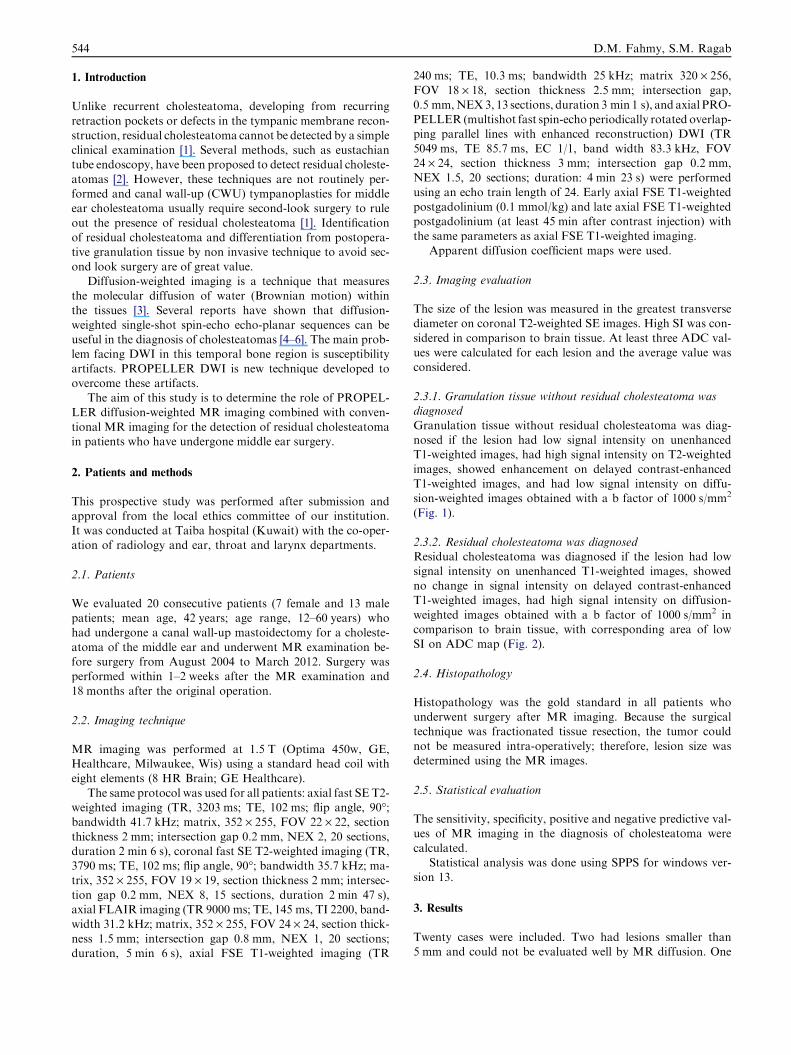
2.3.1. Granulation tissue without residual cholesteatoma wasdiagnosedGranulation tissue without residual cholesteatoma was diag-nosed if the lesion had low signal intensity on unenhancedT1-weighted images, had high signal intensity on T2-weighted
images, showed enhancement on delayed contrast-enhancedT1-weighted images, and had low signal intensity on diffu-sion-weighted images obtained with a b factor of 1000 s/mm2
(Fig. 1).
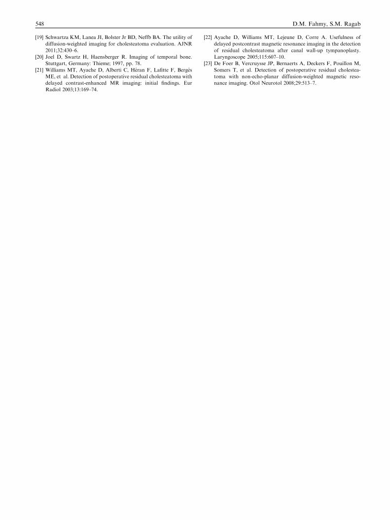
2.3.2. Residual cholesteatoma was diagnosedResidual cholesteatoma was diagnosed if the lesion had lowsignal intensity on unenhanced T1-weighted images, showedno change in signal intensity on delayed contrast-enhancedT1-weighted images, had high signal intensity on diffusion-
weighted images obtained with a b factor of 1000 s/mm2 incomparison to brain tissue, with corresponding area of lowSI on ADC map (Fig. 2).
2.4. Histopathology
Histopathology was the gold standard in all patients whounderwent surgery after MR imaging. Because the surgicaltechnique was fractionated tissue resection, the tumor could
not be measured intra-operatively; therefore, lesion size wasdetermined using the MR images.
2.5. Statistical evaluation
The sensitivity, specificity, positive and negative predictive val-ues of MR imaging in the diagnosis of cholesteatoma were
calculated.Statistical analysis was done using SPPS for windows ver-
sion 13.
3. Results
Twenty cases were included. Two had lesions smaller than5 mm and could not be evaluated well by MR diffusion. One

Fig. 1 (a) Axial T2 WI revealed high SI lesion within the meso-
tympanum. (b) Axial DWI; the lesion displays low SI in
comparison to brain tissue. Final diagnosis was granulation tissue
confirmed by surgery.
Fig. 2 (a) Axial T2 WI revealed: A well defined irregular lesion of ho
(b) Delayed post contrast T1 WI: the lesion shows thin marginal enh
comparison to the brain (white arrow). (d) ADC map: the lesion
granulation tissue.
Detection of post operative residual cholesteatoma using PROPELLER DWI combined with conventional MRI 545
was misdiagnosed as residual cholesteatoma as it showed high
SI on DWI and no significant enhancement on post contraststudy, but pathological analysis revealed acute inflammatoryprocess. The rest were nine granulation tissue and eight casesof residual cholesteatoma. All eight cases of granulation tissue
showed low to faint SI on DWI, ADC ranged from 0.541 to0.128 · 10�3 mm2/s (with a mean value of 0.33 ± 0.09),allshowed moderate enhancement on post contrast study. Apart
from the misdiagnosed case, all eight cases of residual choleste-atoma showed high SI on DWI, ADC value ranged from 0.984to 0. 563 · 10�3 mm2/s (with a mean value of 0.77 ± 0.13).
Three cases showed no significant enhancement, while remain-ing five showed marginal enhancement. No overlap was foundbetween ADC values of residual cholesteatoma and granula-
tion tissue with a cut off value of 0.55 · 10�3 mm2/s.Sensitivity was 80%, specificity was 90%, positive predic-
tive value was 89% and negative predictive value was 82%.(see Tables 1 and 2)
4. Discussion
Acquired cholesteatomas generally occur in the middle ear andmastoid, whereas congenital cholesteatomas or epidermoidscan occur in other locations, including the cerebellopontine an-
gle, suprasellar cistern, calvarium, and multiple sites in thetemporal bone. Congenital cholesteatomas compose only 2%of middle ear cholesteatomas [7].
Acquired cholesteatoma consists of epithelial debris thatresults from desquamation of the lining of the external audi-tory canal and outer lining of the tympanic membrane. The
mogenous high SI is seen in the right epitympanum. (white arrow).
ancement (white arrow). (c) DWI: the lesion displays high SI in
displays low SI (white arrow). . .final diagnosis is postoperative

Table 1 overall results of detection of cholesteatoma using
DWI and delayed post contrast MRI.
Percentage
Sensitivity 80%
Specificity 90%
Positive predictive value 89%
Negative predictive value 82%
Table 2 ADC value · 10� in cases of residual cholesteatoma
and granulation tissue.
Cholesteatoma Granulation tissue
1 0.56 0.23
2 0.83 0.34
3 0.98 0.29
4 0.80 0.54
5 0.68 0.30
6 0.73 0.26
7 0.67 0.33
8 0.88 0.42
9 0.28
Mean ± SD 0.77 ± 0.13 0.33 ± 0.09
NB: One case was misdiagnosed as cholesteatoma, yet biopsy
revealed acute inflammation it displayed low ADC
value = 0.241 · 10�3 mm2/s).
546 D.M. Fahmy, S.M. Ragab
treatment is surgical resection. However, complete surgicalextirpation may be difficult in advanced lesions. After surgery,
it is difficult to distinguish between recurrent cholesteatomaand granulation tissue from both clinical and radiologic stand-points. The middle ear cavity is difficult to visualize because ofpostoperative scarring and thickening of the tympanic mem-
brane. The imaging appearance on both MR images and CTscans is often nonspecific [8].
Patients with well-aerated postoperative mastoid bowls and
middle ear cavities can be easily evaluated with CT [6]. How-ever, if a soft-tissue mass in the cavity of the middle ear is seenon high-resolution CT, diagnosis of the mass is not possible
because cholesteatoma, mucoid secretion, granulation tissue,fibrous tissue, and cholesterol granuloma cannot be differenti-ated from one another on high-resolution CT [9]. As a result,
many surgeons have to perform a follow-up procedure todetermine the cause of the mucosal thickening.
Diffusion weighted MRI (DWI) is a newly developed differ-entiating tool between residual cholesteatoma and granulation
tissue. The differentiating point is that only cholesteatomashows high signal intensity on diffusion-weighted MR images.Other tissues that can be found in the middle ear cavity after
surgery such as granulation tissue, fibrous tissue, cholesterolgranuloma, or serous fluid show low signal intensity on diffu-sion-weighted MR images [10].
In this study we had correctly diagnosed 8 cases of residualcholesteatoma which displayed high SI on DWI, 2 cases weremissed owing to their small size (<3 mm) and one case was
misdiagnosed as residual cholesteatoma but revealed acuteinflammation on surgical resection. Fitzek et al. [4] describedsimilar false positive case in their study which turned out tobe acute otitis media. We believe that acute inflammation
displays high SI on DWI due to the presence of pus and high
protein content. On the other hand; that case showed very lowADC value (0.241 · 10�3 mm2/s) which is lower than the meanvalue calculated for other cholesteatoma lesions. We believethat relying on both DWI and ADC values would have in-
creased the specificity of MR in detecting residual cholestea-toma and decreased false positive cases.
Dremmen et al. [11] also described two false positive lesions
who showed high SI on DWI. Yet it was accompanied byhyperintense signal intensity on T1-weighted images, consis-tent with transplanted fat in the postoperative cavity. This
proves the value of combination of conventional MRI toDWI.
On calculating ADC values, cholesteatoma did not show
low values when compared to granulation tissue. ADC valuesranged from 0.984 to 0. 563 · 10�3 mm2/s (with a mean valueof 0.77 ± 0.13 · 10�3 mm2/s). Granulation tissue displayedlower values, as it ranged from 0.541 to 0.128 · 10�3 mm2/s
(with a mean value of 0.33 ± 0.09 · 10�3 mm2/s). This sup-ports the hypothesis that a T2 shine through effect is predom-inantly responsible for the hyperintensity on DWI not limited
diffusion. Similar results were reported by Vercruysse et al. [12]who found that mean ADC values for cholesteatomas were0.844 · 10�3 mm2/s).
We did not find overlap between residual cholesteatomaand granulation tissue on ADC values. A cut off value of0.55 · 10�3 mm2/s could be used to differentiate between resid-ual cholesteatoma and granulation tissue. Yet we still think
that small number of cases included in this study is not enoughto reach reliable statistical data regarding ADC values and lar-ger study group would be needed.
Both echo-planar and non-echo-planar DWI techniqueshave been utilized in detecting cholesteatoma. However, manyauthors have favoured non-echo-planar DWI as it is less sus-
ceptible to the skull base distortion that can occur becauseof the presence of an air–bone interface [12–16]. More recently,another solution for this problem has been described that com-
bines echo-planar imaging with an image motion suppressiontechnique known as PROPELLER DWI [16].
In this study we used multishot fast spin-echo DWI-PRO-PELLER technique. It showed sensitivity of 80%, specificity
of 90%, positive predictive value of 89% and negative predic-tive value of 82%. It is considered the most recent method forthe diagnosis of residual cholesteatoma [17]. As previously
mentioned, the ability of DWI to be used consistently to eval-uate the temporal bone is hindered by image distortion causedby susceptibility artifacts, chemical-shift artifacts, and ghosts
in the phase-encoding direction. This is due to the high boneattenuation of the inner ear and the numerous air-bone inter-faces present within the mastoid air cells and the middle ear
cavity. With PROPELLER MR imaging, the marked reduc-tion in off-resonance artifacts is primarily caused by the typeof sequence (fast SE): Fast SE imaging is less sensitive tochanges in the constant magnetic induction field, because of
multiple 180� refocusing pulses. Reduction of susceptibilityartifacts is particularly important for adequate visualizationof the middle ear [13].
The main idea of PROPELLER DWI is radial k-space fill-ing technique, so MR imaging datasets are acquired in multi-ple overlapping radial sections, each of which includes data
sampled from the center to the periphery of k-space [16].

Detection of post operative residual cholesteatoma using PROPELLER DWI combined with conventional MRI 547
Yet its main disadvantage is low spatial resolution, which
results from the fact that the periphery of k-space is more spar-sely filled than its central region. It is difficult with radial sam-pling techniques to achieve the high spatial resolutioncommonly expected in clinical practice because of the in-
creased acquisition time and decreased SNR [18]. Also it canbe performed only with axial sections, which does not optimizevisualization of the tegmen region [19].
Another drawback is increased scanning time when com-pared to DWI-EPI. This makes it more liable to the motionartifacts. But fortunately, because the evaluation of cholestea-
toma requires only limited coverage, the resulting 4 min 23 sscanning time was tolerated well by patients included in thisstudy.
As previously mentioned; cholesteatoma is composed of anenlarging collection of exfoliated keratin within a sac of strat-ified squamous epithelium that shows no change in signalintensity on contrast-enhanced MR images [20]. Conversely,
granulation tissue shows enhancement only on delayed con-trast-enhanced images owing to its fibrous nature and, possi-bly, to the microvascular thrombosis phenomenon [21]. It is
necessary to obtain delayed contrast-enhanced images with adelay of 30–45 min after contrast material administration.Ayache et al. [22] achieved high diagnostic results with this
technique as they have correctly detected 17 out of 19 residualcholesteatoma with overall sensitivity of 90%; specificity of100%; positive predictive value of 100%; negative predictivevalue of 92%. They missed two lesions which were <3 mm.
These results are slightly higher than ours.The main disadvantage to this technique is long duration.
In the usual practice patients would not accept to wait for
45 min.De Foer et al. [23] showed that the concurrent use of non-
echo planar HASTE DWI and delayed contrast-enhanced MR
yielded no significant increase in diagnostic performance overthe use of non-echo planar DWI alone. In the current studywe found similar findings as regard both the false positive
and false negative cases. Delayed post contrast MRI did notdiagnose lesions <3 mm. Also the case with acute inflamma-tion showed marginal enhancement which was misleading.We believe that the addition of ADC map to DWI will be more
helpful than delayed post contrast.Limitations to this included small number of cases. Suscep-
tibility artifacts made it difficult to have clear images. Also we
could not evaluate lesions less than 3 mm. Thus, additionalstudies including lesions smaller than 3 mm are needed to de-fine the role of the diffusion-weighted sequence in the detection
of small recurrent cholesteatoma.
5. Conclusion
DWI with ADC map is useful in the detection of secondarycholesteatoma and would decrease the need for un-necessary
second canal up operation.
References
[1] Venaila F, Bonafec A, Poirrierc V, Mondaina M, Uzie A.
Comparison of echo-planar diffusion-weighted imaging and
delayed postcontrast T1-weighted MR imaging for the detection
of residual cholesteatoma. AJNR 2008;29:1363–8.
[2] Tierney PA, Pracy P, Blaney SP, Bowdler DA. An assessment of
the value of the preoperative computed tomography scans prior to
otoendoscopic ‘second look’ in intact canal wall mastoid surgery.
Clin Otolaryngol Allied Sci 1999;24:274–6.
[3] Le Bihan D, Breton E, Lallemand D, Grenier P, Cabanis E,
Laval-Jeantet M. MR imaging of intravoxel incoherent motions:
application to diffusion and perfusion in neurologic disorders.
Radiology 1986;161:401–7.
[4] Fitzek C, Mewes T, Fitzek S, Mentzel H-J, Hunsche S, Stocler P.
Diffusion-weighted MRI of cholesteatomas of the petrous bone. J
Magn Reson Imag 2002;15:636–41.
[5] Maheshwari S, Mukherji SK. Diffusion-weighted imaging for
differentiating recurrent cholesteatoma from granulation tissue
after mastoidectomy: case report. AJNR Am J Neuroradiol
2002;23:847–9.
[6] Stasolla A, Magliulo G, Parrotto D, Luppi G, Marini M.
Detection of postoperative relapsing/residual with diffusion-
weighted echo-planar magnetic resonance imaging. Otol Neurotol
2004;25:879–84.
[7] Swartz JD. Cholesteatomas of the middle ear: diagnosis, etiology,
and complications. Radiol Clin North Am 1984;22:15–35.
[8] Maheshwari S, Mukherji SK. Diffusion-weighted imaging for
differentiating recurrent cholesteatoma from granulation tissue
after mastoidectomy: case report. AJNR Am J Neuroradiol
2002;23:847–9.
[9] Bowdler DA, Parcy P, Blany S. Otoscopic second look intact
canal wall mastoid surgery. In: Sanna M, editor. Proceedings of
the Vth international conference on cholesteatoma and mastoid
surgery. Rome: Edizioni Internationali; 1997. p. 795–8.
[10] Dubrulle F, Souillard R, Chechin D, Vaneecloo FM, Desaulty A,
Vincent C. Diffusion-weighted MR imaging sequence in the
detection of postoperative recurrent cholesteatoma. Radiology
2006;238:604–10.
[11] Dremmen MH, Hofman PA, Hof JR, Stokroos RJ, Postma AA.
The diagnostic accuracy of non-echo-planar diffusion-weighted
imaging in the detection of residual and/or recurrent cholestea-
toma of the temporal bone. AJNR Am J Neuroradiol
2012;33(3):439–44.
[12] Vercruysse J-P, De Foer B, Pouillon M, Somers T, Casselman J,
Offecierset E. The value of diffusion-weighted MR imaging in the
diagnosis of primary acquired and residual cholesteatoma: a
surgical verified study of 100 patients. Eur Radiol 2006;16:1461–7.
[13] Khemani S, Lingam RK, Kalan A, Singh A. The value of non-
echo planar HASTE diffusion-weighted MR imaging in the
detection, localisation and prediction of extent of postoperative
cholesteatoma. Clin Otolaryngol 2011;36(4):306–12.
[14] Yamashitaa K, Yoshiuraa T, Hiwatashia A, Kamanoa H,
Dashjamtsa T, Shibatab S, et al. Detection of middle ear
cholesteatoma by diffusion-weighted mr imaging: multishot
echo-planar imaging compared with single-shot echo-planar
imaging. AJNR 2011;32:1915–8.
[15] De Foer B, Vercruysse JP, Bernaerts A, Maes J, Deckers F,
Michiels J, et al. The value of single-shot turbo spin-echo
diffusion-weighted MR imaging in the detection of middle ear
cholesteatoma. Neuroradiology 2007;49:841–8.
[16] Lehmann P, Saliou G, Brochart C, Page C, Deschepper B, Vallee
JN, et al. 3T MR imaging of postoperative recurrent middle ear
cholesteatomas: value of periodically rotated overlapping parallel
lines with enhanced reconstruction diffusion-weighted MR imag-
ing. AJNR Am J Neuroradiol 2009;2:423–7.
[17] Barath K, Huber AM, Stampfli P, Varga Z, Kollias S Barath K,
Huber AM, et al. Neuroradiology of cholesteatomas. AJNR Am
J Neuroradiol 2011;32(2):221–9.
[18] Wintersperger BJ, Runge VM, Biswas J, Nelson CB, Stemmer A,
Simonetta AB, et al. Brain magnetic resonance imaging at 3 T
using BLADE compared with standard rectilinear data sampling.
Invest Radiol 2006;41(7):586–92.
548 D.M. Fahmy, S.M. Ragab
[19] Schwartza KM, Lanea JI, Bolster Jr BD, Neffb BA. The utility of
diffusion-weighted imaging for cholesteatoma evaluation. AJNR
2011;32:430–6.
[20] Joel D, Swartz H, Haensberger R. Imaging of temporal bone.
Stuttgart, Germany: Thieme; 1997, pp. 78.
[21] Williams MT, Ayache D, Alberti C, Heran F, Lafitte F, Berges
ME, et al. Detection of postoperative residual cholesteatoma with
delayed contrast-enhanced MR imaging: initial findings. Eur
Radiol 2003;13:169–74.
[22] Ayache D, Williams MT, Lejeune D, Corre A. Usefulness of
delayed postcontrast magnetic resonance imaging in the detection
of residual cholesteatoma after canal wall-up tympanoplasty.
Laryngoscope 2005;115:607–10.
[23] De Foer B, Vercruysse JP, Bernaerts A, Deckers F, Pouillon M,
Somers T, et al. Detection of postoperative residual cholestea-
toma with non-echo-planar diffusion-weighted magnetic reso-
nance imaging. Otol Neurotol 2008;29:513–7.
Related Documents