Detection of NO and S- nitrosocompounds using mid-IR CRDS Vitali Stsiapura 1 , Vincent K. Shuali 1 , Angela Ziegler 1 , Kevin K. Lehmann 1 , Benjamin M. Gaston 2 1 University of Virginia; 2 Case Western Reserve University

Detection of NO and S- nitrosocompounds using mid-IR CRDS Vitali Stsiapura 1, Vincent K. Shuali 1, Angela Ziegler 1, Kevin K. Lehmann 1, Benjamin M. Gaston.
Dec 16, 2015
Welcome message from author
This document is posted to help you gain knowledge. Please leave a comment to let me know what you think about it! Share it to your friends and learn new things together.
Transcript
Detection of NO and S-nitrosocompounds using mid-IR CRDS
Vitali Stsiapura1, Vincent K. Shuali1, Angela Ziegler1, Kevin K. Lehmann1,
Benjamin M. Gaston2
1University of Virginia; 2Case Western Reserve University
Biochemistry of NO-containing compounds
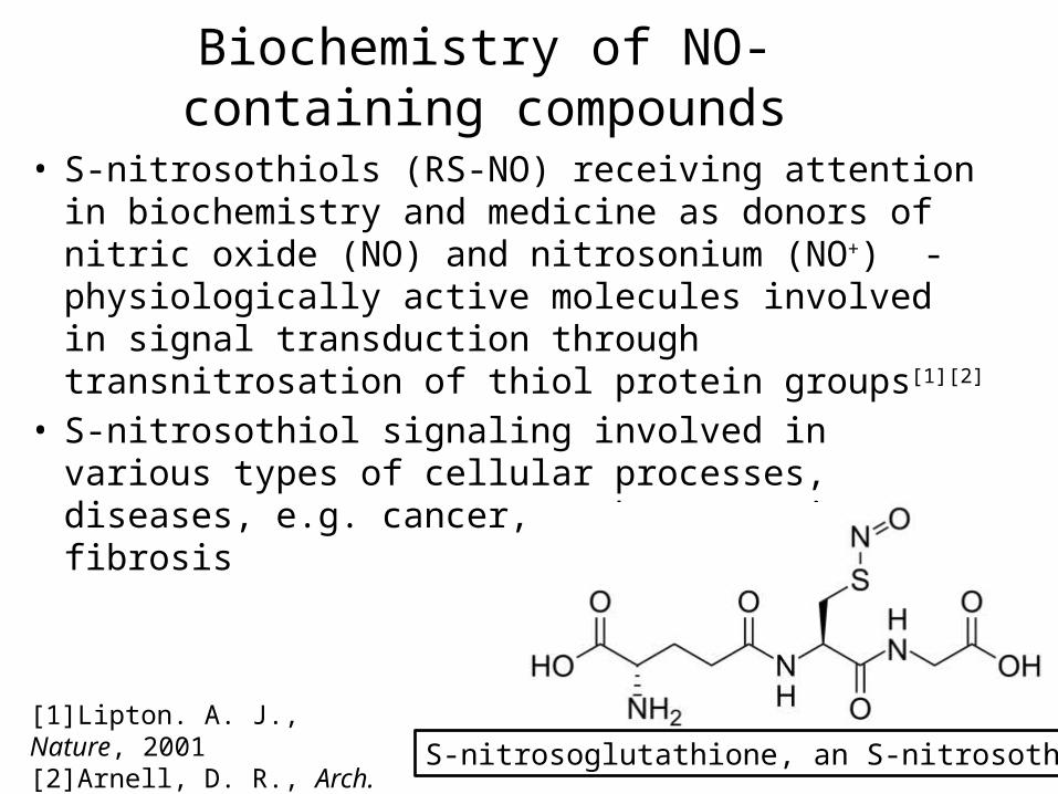
• S-nitrosothiols (RS-NO) receiving attention in biochemistry and medicine as donors of nitric oxide (NO) and nitrosonium (NO+) - physiologically active molecules involved in signal transduction through transnitrosation of thiol protein groups[1][2]
• S-nitrosothiol signaling involved in various types of cellular processes, diseases, e.g. cancer, asthma, cystic fibrosis
S-nitrosoglutathione, an S-nitrosothiol[1]Lipton. A. J., Nature, 2001[2]Arnell, D. R., Arch. Biochem. Biophys., 1995
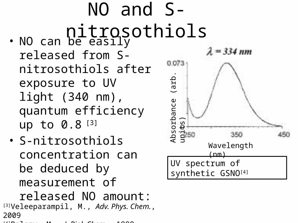
NO and S-nitrosothiols• NO can be easily released
from S-nitrosothiols after exposure to UV light (340 nm), quantum efficiency up to 0.8 [3]
• S-nitrosothiols concentration can be deduced by measurement of released NO amount:
UV spectrum of synthetic GSNO[4]
[3]Veleeparampil, M., Adv. Phys. Chem., 2009[4]Balazy, M., J. Biol. Chem., 1998
Wavelength (nm)
Abso
rban
ce (a
rb. u
nits
)
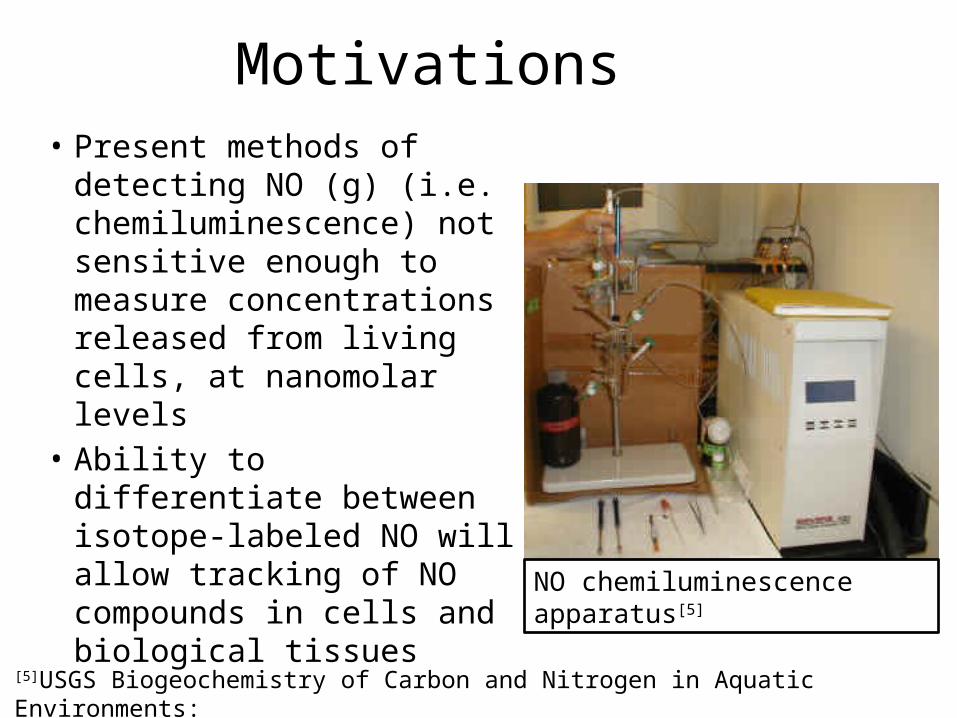
Motivations• Present methods of
detecting NO (g) (i.e. chemiluminescence) not sensitive enough to measure concentrations released from living cells, at nanomolar levels
• Ability to differentiate between isotope-labeled NO will allow tracking of NO compounds in cells and biological tissues
NO chemiluminescence apparatus[5]
[5]USGS Biogeochemistry of Carbon and Nitrogen in Aquatic Environments:
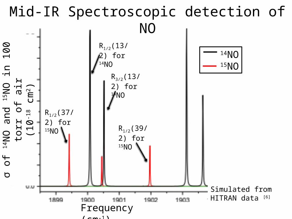
Mid-IR Spectroscopic detection of NO
14NO15NO
R1/2(13/2) for 14NO
R3/2(13/2) for 14NO
R1/2(37/2) for 15NO
R1/2(39/2) for 15NO
Simulated from HITRAN data [6]
Frequency (cm-1)
σ of
14N
O a
nd 15
NO
in 1
00 to
rr o
f air
(10-1
8 cm
2 )
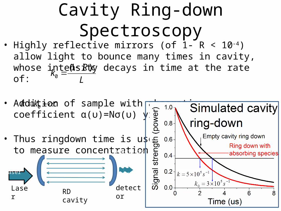
Cavity Ring-down Spectroscopy• Highly reflective mirrors (of 1- R < 10-4) allow light to bounce
many times in cavity, whose intensity decays in time at the rate of:
• Addition of sample with absorption coefficient α(υ)=Nσ(υ) yields:
• Thus ringdown time is usedto measure concentration N
L
cRk
)1(0
ckk 0
IR from laser
To detector
RD cavityLaser detector
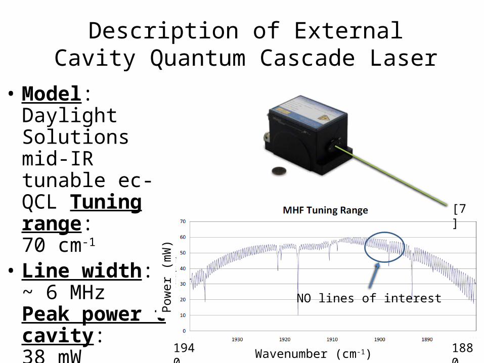
Description of External Cavity Quantum Cascade Laser
• Model: Daylight Solutions mid-IR tunable ec-QCL Tuning range:70 cm-1
• Line width:~ 6 MHzPeak power tocavity:38 mW
1940 1880
Pow
er (m
W)
Wavenumber (cm-1)
[7]
NO lines of interest
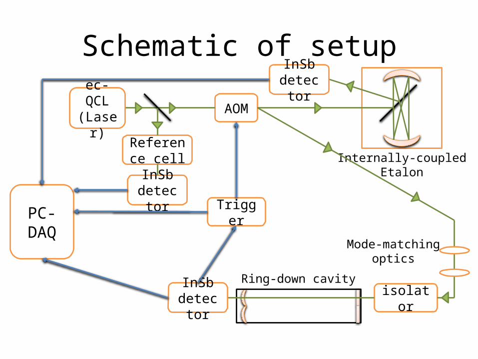
Schematic of setup
ec-QCL (Laser)
AOM
Reference cell
InSb detector
Internally-coupledEtalon
Ring-down cavity
isolatorInSb detector
Mode-matchingoptics
Trigger
InSb detector
PC-DAQ
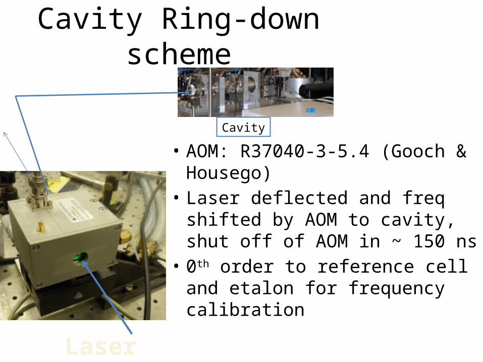
Cavity Ring-down scheme
• AOM: R37040-3-5.4 (Gooch & Housego)
• Laser deflected and freq shifted by AOM to cavity, shut off of AOM in ~ 150 ns
• 0th order to reference cell and etalon for frequency calibration
Laser
Cavity
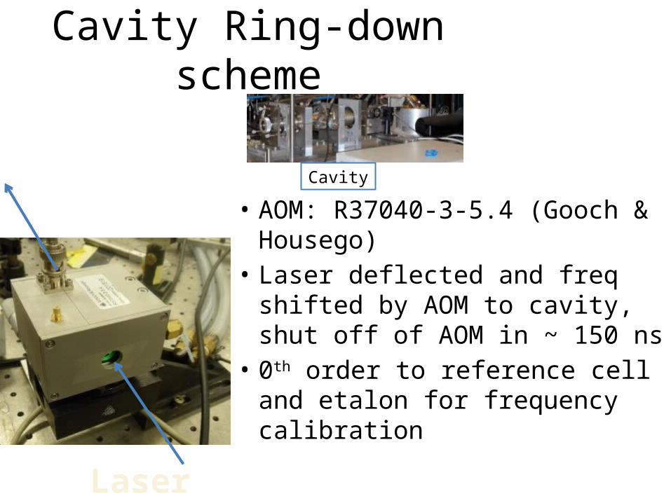
Cavity Ring-down scheme
• AOM: R37040-3-5.4 (Gooch & Housego)
• Laser deflected and freq shifted by AOM to cavity, shut off of AOM in ~ 150 ns
• 0th order to reference cell and etalon for frequency calibration
Laser
Cavity
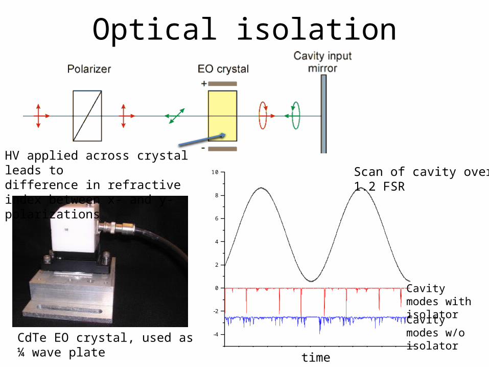
Optical isolation
CdTe EO crystal, used as ¼ wave plate
Returning beamblocked by polarizer
HV applied across crystal leads todifference in refractive index between x- and y- polarizations
0.00 0.02 0.04 0.06 0.08 0.10 0.12 0.14
-4
-2
0
2
4
6
8
10 Scan of cavity over1.2 FSR
Cavity modes with isolator
Cavity modes w/o isolator
time
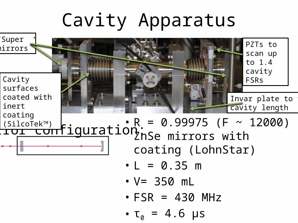
Cavity Apparatus
• R = 0.99975 (F ~ 12000)ZnSe mirrors with coating (LohnStar)
• L = 0.35 m• V= 350 mL• FSR = 430 MHz• τ0 = 4.6 μs
Mirror configuration:
Invar plate to fixcavity length
Cavity surfacescoated with inertcoating (SilcoTekTM)
“Supermirrors” PZTs to scan
up to 1.4 cavity FSRs
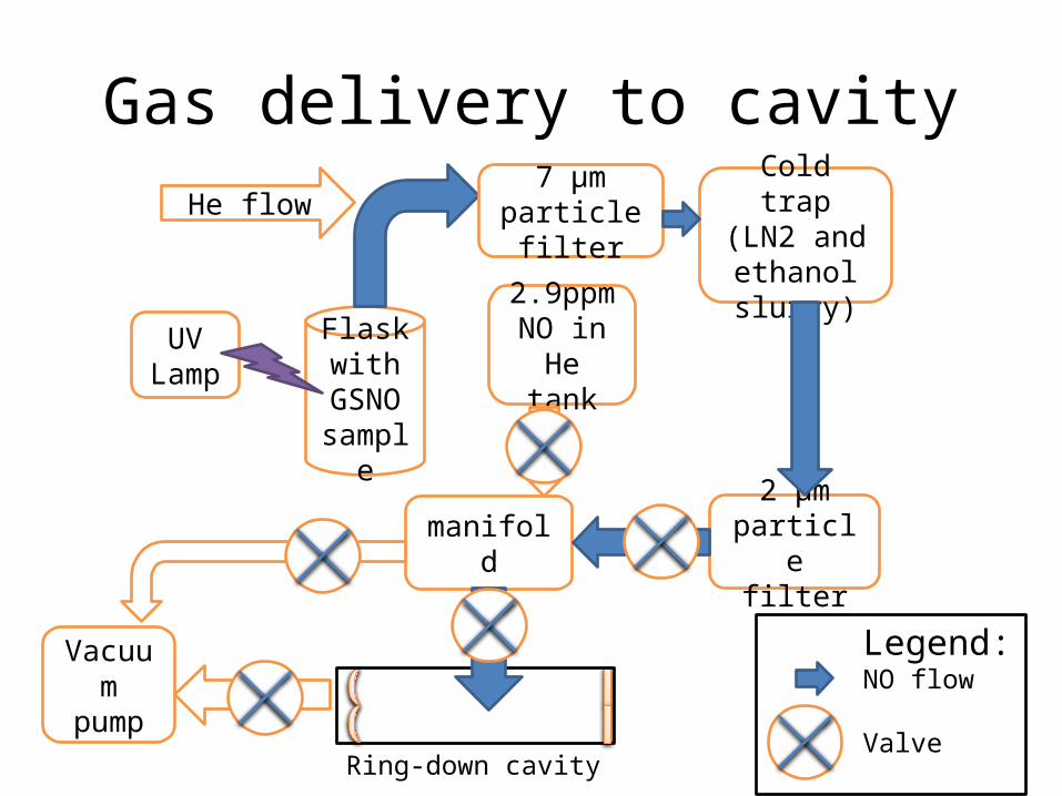
Gas delivery to cavity
UV Lamp
Flask with
GSNO sample
He flow7 μm
particle filter
Cold trap(LN2 and ethanol slurry)
2 μm particle
filtermanifold
Ring-down cavity
Vacuum pump
2.9ppm NO in He
tank
Legend:NO flow
Valve
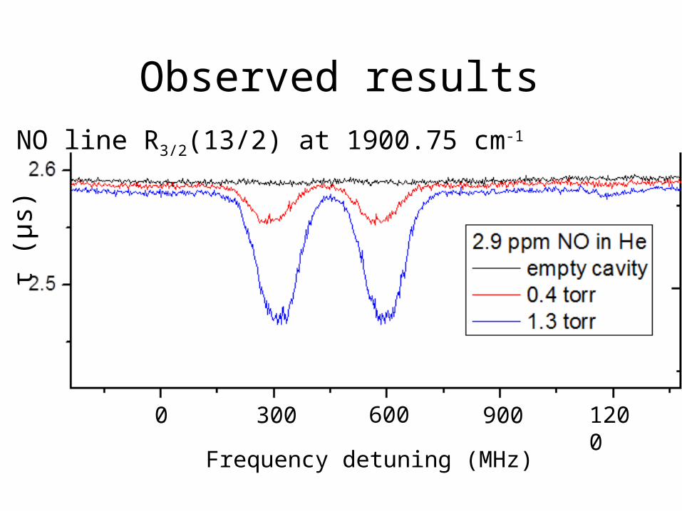
Observed resultsNO line R3/2(13/2) at 1900.75 cm-1
Frequency detuning (MHz)
τ (μ
s)
0 300 600 1200900
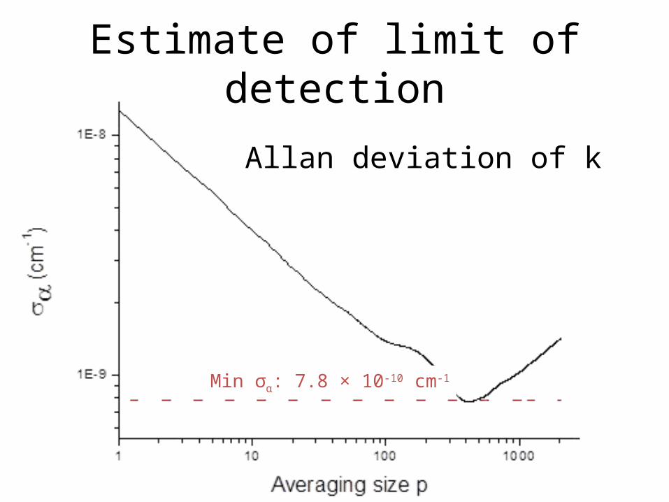
Estimate of limit of detection
Allan deviation of k
Min σα: 7.8 × 10⁻¹⁰ cm-1
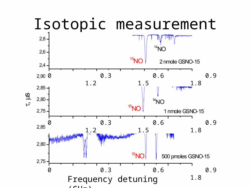
Isotopic measurement
0 0.3 0.6 0.9 1.2 1.5 1.8
0 0.3 0.6 0.9 1.2 1.5 1.8
0 0.3 0.6 0.9 1.2 1.5 1.8
Frequency detuning (GHz)
Conclusions
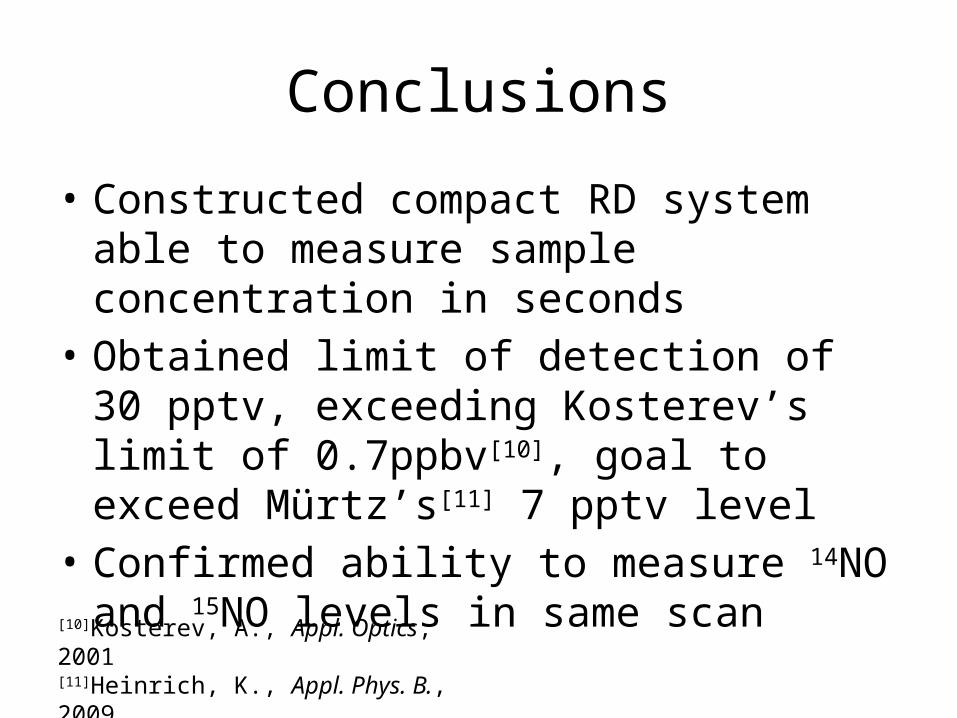
• Constructed compact RD system able to measure sample concentration in seconds
• Obtained limit of detection of 30 pptv, exceeding Kosterev’s limit of 0.7ppbv[10], goal to exceed Mürtz’s[11] 7 pptv level
• Confirmed ability to measure 14NO and 15NO levels in same scan
[10]Kosterev, A., Appl. Optics, 2001[11]Heinrich, K., Appl. Phys. B., 2009
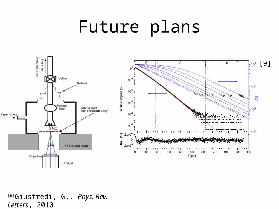
Future plans
[9]
[9]Giusfredi, G., Phys. Rev. Letters, 2010
Acknowledgments
• NIH and NSF: financial support• Dr. Joseph Hodges (NIST): advice and
assistance on cavity length and frequency stabilization
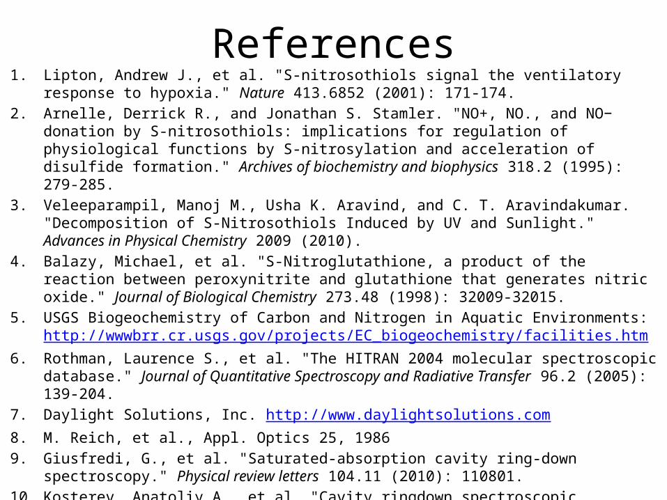
References1. Lipton, Andrew J., et al. "S-nitrosothiols signal the ventilatory response to hypoxia." Nature 413.6852
(2001): 171-174.2. Arnelle, Derrick R., and Jonathan S. Stamler. "NO+, NO., and NO− donation by S-nitrosothiols:
implications for regulation of physiological functions by S-nitrosylation and acceleration of disulfide formation." Archives of biochemistry and biophysics 318.2 (1995): 279-285.
3. Veleeparampil, Manoj M., Usha K. Aravind, and C. T. Aravindakumar. "Decomposition of S-Nitrosothiols Induced by UV and Sunlight." Advances in Physical Chemistry 2009 (2010).
4. Balazy, Michael, et al. "S-Nitroglutathione, a product of the reaction between peroxynitrite and glutathione that generates nitric oxide." Journal of Biological Chemistry 273.48 (1998): 32009-32015.
5. USGS Biogeochemistry of Carbon and Nitrogen in Aquatic Environments:http://wwwbrr.cr.usgs.gov/projects/EC_biogeochemistry/facilities.htm
6. Rothman, Laurence S., et al. "The HITRAN 2004 molecular spectroscopic database." Journal of Quantitative Spectroscopy and Radiative Transfer 96.2 (2005): 139-204.
7. Daylight Solutions, Inc. http://www.daylightsolutions.com8. M. Reich, et al., Appl. Optics 25, 19869. Giusfredi, G., et al. "Saturated-absorption cavity ring-down spectroscopy." Physical review letters
104.11 (2010): 110801.10. Kosterev, Anatoliy A., et al. "Cavity ringdown spectroscopic detection of nitric oxide with a continuous-
wave quantum-cascade laser." Applied optics 40.30 (2001): 5522-5529.11. Heinrich, K., et al. "Infrared laser-spectroscopic analysis of 14NO and 15NO in human breath." Applied
Physics B 95.2 (2009): 281-286.
Related Documents