BioMed Central Page 1 of 14 (page number not for citation purposes) Journal of Translational Medicine Open Access Research Detailed analysis of immunologic effects of the cytotoxic T lymphocyte-associated antigen 4-blocking monoclonal antibody tremelimumab in peripheral blood of patients with melanoma Begoña Comin-Anduix 1 , Yohan Lee 2 , Jason Jalil 1 , Alain Algazi 1 , Pilar de la Rocha 1 , Luis H Camacho 3 , Viviana A Bozon 4 , Cecile A Bulanhagui 4 , Elisabeth Seja 5 , Arturo Villanueva 5 , Bradley R Straatsma 6 , Antonio Gualberto 4 , James S Economou 1,7,8 , John A Glaspy 5,8 , Jesus Gomez- Navarro 4 and Antoni Ribas* 1,5,8 Address: 1 Department of Surgery, Division of Surgical Oncology, University of California Los Angeles (UCLA), Los Angeles, CA, USA, 2 Department of Human Genetics, UCLA, Los Angeles, CA, USA, 3 Oncology Consultants PA, Houston, TX, USA, 4 Pfizer Global Research and Development (PGRD), New London, CT, USA, 5 Department of Medicine, Division of Hematology/Oncology, UCLA; Los Angeles, CA, USA, 6 Department of Ophthalmology, Jules Stein Eye Institute, UCLA Los Angeles, CA, USA, 7 Department of Microbiology, Immunology and Molecular Genetics and 8 Jonsson Comprehensive Cancer Center, UCLA Los Angeles, CA, USA Email: Begoña Comin-Anduix - [email protected]; Yohan Lee - [email protected]; Jason Jalil - [email protected]; Alain Algazi - [email protected]; Pilar de la Rocha - [email protected]; Luis H Camacho - [email protected]; Viviana A Bozon - [email protected]; Cecile A Bulanhagui - [email protected]; Elisabeth Seja - [email protected]; Arturo Villanueva - [email protected]; Bradley R Straatsma - [email protected]; Antonio Gualberto - [email protected]; James S Economou - [email protected]; John A Glaspy - [email protected]; Jesus Gomez-Navarro - [email protected]; Antoni Ribas* - [email protected] * Corresponding author Abstract Background: CTLA4-blocking antibodies induce tumor regression in a subset of patients with melanoma. Analysis of immune parameters in peripheral blood may help define how responses are mediated. Methods: Peripheral blood from HLA-A*0201-positive patients with advanced melanoma receiving tremelimumab (formerly CP-675,206) at 10 mg/kg monthly was repeatedly sampled during the first 4 cycles. Samples were analyzed by 1) tetramer and ELISPOT assays for reactivity to CMV, EBV, MART1, gp100, and tyrosinase; 2) activation HLA-DR and memory CD45RO markers on CD4 + /CD8 + cells; and 3) real-time quantitative PCR of mRNA for FoxP3 transcription factor, preferentially expressed by T regulatory cells. The primary endpoint was difference in MART1-specific T cells by tetramer assay. Immunological data were explored for significant trends using clustering analysis. Results: Three of 12 patients eligible for immune monitoring had tumor regression lasting > 2 years without relapse. There was no significant change in percent of MART1-specific T cells by tetramer assay. Additionally, there was no generalized trend toward postdosing changes in other antigen-specific CD8 + cell populations, FoxP3 transcripts, or overall changes in surface expression Published: 1 May 2008 Journal of Translational Medicine 2008, 6:22 doi:10.1186/1479-5876-6-22 Received: 31 January 2008 Accepted: 1 May 2008 This article is available from: http://www.translational-medicine.com/content/6/1/22 © 2008 Comin-Anduix et al; licensee BioMed Central Ltd. This is an Open Access article distributed under the terms of the Creative Commons Attribution License (http://creativecommons.org/licenses/by/2.0 ), which permits unrestricted use, distribution, and reproduction in any medium, provided the original work is properly cited.

Welcome message from author
This document is posted to help you gain knowledge. Please leave a comment to let me know what you think about it! Share it to your friends and learn new things together.
Transcript
BioMed CentralJournal of Translational Medicine
ss
Open AcceResearchDetailed analysis of immunologic effects of the cytotoxic T lymphocyte-associated antigen 4-blocking monoclonal antibody tremelimumab in peripheral blood of patients with melanomaBegoña Comin-Anduix1, Yohan Lee2, Jason Jalil1, Alain Algazi1, Pilar de la Rocha1, Luis H Camacho3, Viviana A Bozon4, Cecile A Bulanhagui4, Elisabeth Seja5, Arturo Villanueva5, Bradley R Straatsma6, Antonio Gualberto4, James S Economou1,7,8, John A Glaspy5,8, Jesus Gomez-Navarro4 and Antoni Ribas*1,5,8Address: 1Department of Surgery, Division of Surgical Oncology, University of California Los Angeles (UCLA), Los Angeles, CA, USA, 2Department of Human Genetics, UCLA, Los Angeles, CA, USA, 3Oncology Consultants PA, Houston, TX, USA, 4Pfizer Global Research and Development (PGRD), New London, CT, USA, 5Department of Medicine, Division of Hematology/Oncology, UCLA; Los Angeles, CA, USA, 6Department of Ophthalmology, Jules Stein Eye Institute, UCLA Los Angeles, CA, USA, 7Department of Microbiology, Immunology and Molecular Genetics and 8Jonsson Comprehensive Cancer Center, UCLA Los Angeles, CA, USA
Email: Begoña Comin-Anduix - [email protected]; Yohan Lee - [email protected]; Jason Jalil - [email protected]; Alain Algazi - [email protected]; Pilar de la Rocha - [email protected]; Luis H Camacho - [email protected]; Viviana A Bozon - [email protected]; Cecile A Bulanhagui - [email protected]; Elisabeth Seja - [email protected]; Arturo Villanueva - [email protected]; Bradley R Straatsma - [email protected]; Antonio Gualberto - [email protected]; James S Economou - [email protected]; John A Glaspy - [email protected]; Jesus Gomez-Navarro - [email protected]; Antoni Ribas* - [email protected]
* Corresponding author
AbstractBackground: CTLA4-blocking antibodies induce tumor regression in a subset of patients withmelanoma. Analysis of immune parameters in peripheral blood may help define how responses aremediated.
Methods: Peripheral blood from HLA-A*0201-positive patients with advanced melanomareceiving tremelimumab (formerly CP-675,206) at 10 mg/kg monthly was repeatedly sampledduring the first 4 cycles. Samples were analyzed by 1) tetramer and ELISPOT assays for reactivityto CMV, EBV, MART1, gp100, and tyrosinase; 2) activation HLA-DR and memory CD45ROmarkers on CD4+/CD8+ cells; and 3) real-time quantitative PCR of mRNA for FoxP3 transcriptionfactor, preferentially expressed by T regulatory cells. The primary endpoint was difference inMART1-specific T cells by tetramer assay. Immunological data were explored for significant trendsusing clustering analysis.
Results: Three of 12 patients eligible for immune monitoring had tumor regression lasting > 2years without relapse. There was no significant change in percent of MART1-specific T cells bytetramer assay. Additionally, there was no generalized trend toward postdosing changes in otherantigen-specific CD8+ cell populations, FoxP3 transcripts, or overall changes in surface expression
Published: 1 May 2008
Journal of Translational Medicine 2008, 6:22 doi:10.1186/1479-5876-6-22
Received: 31 January 2008Accepted: 1 May 2008
This article is available from: http://www.translational-medicine.com/content/6/1/22
© 2008 Comin-Anduix et al; licensee BioMed Central Ltd. This is an Open Access article distributed under the terms of the Creative Commons Attribution License (http://creativecommons.org/licenses/by/2.0), which permits unrestricted use, distribution, and reproduction in any medium, provided the original work is properly cited.
Page 1 of 14(page number not for citation purposes)
Journal of Translational Medicine 2008, 6:22 http://www.translational-medicine.com/content/6/1/22
of T-cell activation or memory markers. Unsupervised hierarchical clustering based on immunemonitoring data segregated patients randomly. However, clustering according to T-cell activationor memory markers separated patients with clinical response and most patients with inflammatorytoxicity into a common subgroup.
Conclusion: Administration of CTLA4-blocking antibody tremelimumab to patients withadvanced melanoma results in a subset of patients with long-lived tumor responses. T-cellactivation and memory markers served as the only readout of the pharmacodynamic effects of thisantibody in peripheral blood.
Clinical trial registration number: NCT00086489
BackgroundCytotoxic T lymphocyte-associated antigen 4 (CTLA4) isan activation-induced, type I transmembrane protein ofthe immunoglobulin superfamily, expressed by recentlyactivated T lymphocytes as a covalent homodimer. It func-tions as an inhibitory receptor for the costimulatory mol-ecules B7.1 (CD80) and B7.2 (CD86), efficientlycompeting with the positive costimulatory receptor CD28[1-5]. Crosslinking of CTLA4 by B7 in the context of T-cellantigen receptor (TCR) engagement inhibits T-cell activa-tion, interleukin (IL)-2 gene transcription, and T-cell pro-liferation by directly inhibiting TCR signal transduction[3,6].
CTLA4 blockade using the specific antagonistic mono-clonal antibodies ipilimumab (formerly known asMDX010 and BMS734016) and tremelimumab (formerlyknown as CP-675,206 and ticilimumab) reproduciblyinduce objective tumor responses in a subset of patientswith melanoma [7-15]. Despite a wealth of knowledgeabout the antitumor activity induced by CTLA4 blockadein animal models, the mechanisms that mediate tumorregression in human patients are currently not fullyunderstood [16,17]. Several mechanisms have been pos-tulated: 1) Blocking the negative signaling from CTLA4expressed on recently activated tumor antigen-specific Tcells may boost natural or induced immune responses tocancer cells [3,18]; 2) Anti-CTLA4 antibodies may depleteCD4+CD25+ T regulatory cells (Treg) [19], which constitu-tively express CTLA4 [20], or inhibit reverse signaling toB7 costimulatory molecules expressed by immune sup-pressive plasmacytoid dendritic cells (pDC) [21-23] oractivated T cells [24]; 3) Anti-CTLA4 antibodies may resultin the presence of high titers of antibodies against solublemajor histocompatibility complex (MHC) class I chain-related protein A (MICA), an immune suppressive MHCclass I-like molecule shed by tumor cells [25]; 4) Expres-sion of CTLA4 on T cells increases their motility and inter-feres with establishment of durable interactions with cellsexpressing their cognate antigen [26], which may bereverted with monoclonal antibodies; or 5) Anti-CTLA4
antibodies may have direct cytotoxic effects on tumor cellsthat express CTLA4 [27].
Some of these hypotheses can be studied using modernimmune monitoring assays in peripheral blood. Quantifi-cation of antigen-specific T-cell responses by MHCtetramer and enzyme-linked immunospot (ELISPOT)assays is often used to assess immune activation in exper-imental cancer immunotherapy trials [28]. Definition ofkey methodological parameters (ie, accuracy, precision,and reproducibility) is critical to determine the extent ofT-cell expansion that represents a positive immuneresponse. The magnitude of minimum statistically signif-icant changes in the number of circulating antigen-specificT cells compared with baseline levels (defined as the refer-ence change value [RCV]) was recently reported for thetetramer and ELISPOT assays [29]. This calculation pro-vides a robust definition of immune response (either pos-itive or negative) that can be reliably applied to themonitoring of immunomodulatory effects of CTLA4-blocking monoclonal antibodies.
Tremelimumab is a fully human immunoglobulin (Ig)G2monoclonal antibody with high CTLA4 specificity thatantagonizes binding of CTLA4 to B7 costimulatory mole-cules, resulting in enhanced T-cell activation as demon-strated by increased cytokine production in vitro.Tremelimumab has demonstrated antitumor activity inpatients with metastatic melanoma [12]. As with anystudy using patient-derived samples, the ability torobustly test or rule out a hypothesis is limited by practicalconstraints of human experimentation [30]. Within theselimitations, we set up to test the hypothesis that tremeli-mumab may alter the number, functional activation orphenotype of immune cells in peripheral blood that mayprovide information on the mechanism of action of thisCTLA4-blocking monoclonal antibody. Therefore, in thisreport we analyzed immune parameters in the peripheralblood of patients receiving tremelimumab for the treat-ment of locally advanced or metastatic melanoma withthe goal of studying the mechanism of immune activationleading to objective tumor responses.
Page 2 of 14(page number not for citation purposes)
Journal of Translational Medicine 2008, 6:22 http://www.translational-medicine.com/content/6/1/22
Materials and methodsStudy Design and AssessmentsHuman leukocyte antigen (HLA)-A*0201-positivepatients with metastatic melanoma who received tremeli-mumab monthly at the maximum tolerated dose in aphase I trial [31] were enrolled in an open-label expan-sion cohort between June 2004 and April 2005, and con-sented to donate repeated peripheral blood samples whilereceiving tremelimumab (10 mg/kg monthly) intrave-nously for up to 24 cycles. This study was conductedaccording to the Declaration of Helsinki and its amend-ments and relevant International Conference on Harmo-nization Good Clinical Practice guidelines. The protocoland consent forms, and all modifications, were approvedby the University of California Los Angeles and Universityof Texas MD Anderson institutional review boards(approval numbers 03-01-059 and IDO3-0090, respec-tively). All patients provided written informed consentprior to any study procedures. Blood samples (40 mL)were collected from a peripheral vein during the screeningperiod, on day 1 before the first dose of antibody (bothbaseline samples), on the day of each new dose in subse-quent cycles, and 1 and 2 weeks after each monthly doseduring the first 4 monthly cycles of therapy. When possi-ble, a final blood sample was collected at the time thepatient went off study for whatever reason. All patientsunderwent baseline and follow-up eye exams every 2 to 4months throughout study participation following our pre-viously-published eye surveillance protocol [32,33]. Theprimary endpoint was the determination of immuneresponse to the melanoma-associated antigen recognizedby T cells (MART1)-derived epitope MART126–36 by MHCtetramer assay across multiple time points. This wasdefined as detecting a 99% RCV for this assay and antigen,which corresponds to a minimum 80% change from base-line in the percentage of circulating antigen-specific T cells[29]. Clinical responses were assessed by Response Evalu-ation Criteria in Solid Tumors (RECIST) [34]. In addition,a clinical benefit response was defined for patients whowere felt by the study investigators to have derived une-quivocal clinical benefit after tremelimumab despite notmeeting the standard criteria for response followingRECIST.
Patient EligibilityPatients with surgically incurable stage IIIc or IVmelanoma were eligible if they met the following majoreligibility criteria: HLA-A*0201 positive, baseline level ofcirculating MART126–36-specific T cells above the lowerlimit of detection (LLD) by tetramer assay, previouslydefined as 0.03% of CD8+ T cells [29], disease measurableby RECIST, Eastern Cooperative Oncology Group per-formance status ≤ 1, and having received at least 2 dosesof tremelimumab with samples for immune monitoringcollected weekly between both doses. Major exclusion cri-
teria were history of chronic inflammatory or autoim-mune disease and presence of active brain metastases.
Sample Processing, Cryopreservation, and ThawingPeripheral blood mononuclear cells (PBMC) were iso-lated by Ficoll-Hypaque (Amersham Pharmacia, Piscata-way, NJ) and cryopreserved in liquid nitrogen in RoswellPark Memorial Institute medium (RPMI) (Gibco-BRL,Gaithersburg, MD) supplemented with 20% (all percent-ages represent v/v) heat-inactivated human AB serum(Omega Scientific, Tarzana, CA) and 10% dimethylsulfox-ide (Sigma, St. Louis, MO). Cryopreserved PBMC aliquotswere thawed and immediately diluted with RPMI com-plete media consisting of 10% human AB serum and 1%penicillin, streptomycin, and amphotericin (Omega Sci-entific). Cells were washed and subjected to enzymatictreatment with DNAse (0.002%, Sigma) for 1 hour at37°C. Cells were washed again and used immediately orwere rested overnight in RPMI complete media in a 6%CO2 incubator.
MHC Tetramer AssayThe following peptide epitopes were used: 1) 2 negativecontrol epitopes: an HLA-A*0201-binding nonrelevantpeptide (referred to as Negative peptide from here on)[35], and the HLA-A*0201 immunodominant peptidealpha fetoprotein (AFP)325–332 (GLSPNLNRFL) derivedfrom the oncofetal antigen AFP [36]; 2) 2 infectious dis-ease epitopes as positive controls, cytomegalovirus(CMV)pp65495–503 (NLVPMVATV) and Epstein-Barr virus(EBV) BMLF1259–267 (GLCTLVAML); and 3) 3 HLA-A*0201 immunodominant peptides derived from tumorrejection antigens: MART126–35 (ELAGIGILTV),tyrosinase368–376 (YMDGTMSQV) and gp100209–217(ITDQVPFSV). All HLA-A*0201 tetramers were purchasedfrom Beckman Coulter Inc., San Diego, CA, as peptidepreloaded reagents, and the assay was performed follow-ing the manufacturer's instructions with minor modifica-tions as previously described [29].
ELISPOT AssaysInterferon gamma (IFN-γ) ELISPOT assays were also per-formed as previously described [29]. Briefly, PBMC werethawed from different time points and treated withDNAse. HLA-A*0201-transfected K562 (K562/A*0201),provided by Drs. Wolfgang Herr and Cedrik M. Britten(Johannes Gutenberg University, Mainz, Germany), werepulsed with the same peptide epitopes described for thetetramer assay and used as antigen-presenting cells. Then1 × 105 PBMC were mixed with 1 × 10⁴ peptide-pulsedK562/A*0201 in X-Vivo 10 media (BioWhittaker, Walk-ersville, MD) supplemented with 10% heat-inactivatedhuman AB serum and seeded directly into anti-IFN-γ anti-body coated ELISPOT plates for 20 hours.
Page 3 of 14(page number not for citation purposes)
Journal of Translational Medicine 2008, 6:22 http://www.translational-medicine.com/content/6/1/22
Multiplex Cytokine AssayCocultures of thawed PBMC and peptide pulsed K562/A*0201 cells were plated as described for ELISPOT assaysbut were placed in triplicate in flat-bottom 96-well plates.Twenty hours later, supernatants were collected and fro-zen after centrifugation at 468 g for 10 minutes. Thawedsupernatants from different time points were analyzed fol-lowing the manufacturer's instructions using a 17-plexassay (Bio-Plex Human Cytokine 17-Plex Panel, Bio-RadLaboratories, Hercules, CA). The cytokines quantifiedwere IL-2, IL-4, IL-6, IL-8, IL-10, granulocyte-macrophagecolony-stimulating factor (GM-CSF), IFN-γ, tumor necro-sis factor alpha (TNF-α), IL-1β, IL-5, IL-7, IL-12 (p70), IL-13, IL-17, granulocyte colony-stimulating factor (G-CSF),monocyte chemoattractant protein 1 (MCP-1/MCAF),and macrophage inflammatory protein 1 α (MIP-1α).Data were analyzed using Bio-Plex manager software with5PL curve fitting.
Multiparameter Surface Flow Cytometry AnalysisUnspecific antibody binding to Fc receptors from thawedPBMC was blocked with 100% adult bovine serum(Omega Scientific). PMBC were then stained using a panelof fluorescein-labeled antibodies against the following T-cell surface antigens: FITC-conjugated-UCHL1 (anti-CD45RO), APC-CY7-conjugated-SK3 (anti-CD4), APC-conjugated-FN50 (anti-CD69) (all from BD Biosciences,San Jose, CA), Pacific Blue-conjugated-S4.1 (anti-CD3)(Invitrogen, Carlsbad, CA), and ECD-conjugated-Immu-357 (anti-HLA-DR) (Beckman Coulter). Cells were fixedwith 0.5% paraformaldehyde. Immediately before flowcytometric analysis, 5 μL of 7-amino-actinomycin D (7-AAD) was added to gate out dead cells. The 5 color flowcytometry staining was acquired by a FACSAria using Flu-orescence Minus One approach [37]. Analysis was per-formed with FCS Express (DeNovo Software, Thornhill,Ontario, Canada) software.
FoxP3 Intracellular StainingPBMC were first labeled with the following surface anti-bodies: Pacific Blue-conjugated-S4.1 (anti-CD3),AlexaFluor467-conjugated-RPA-T4 (anti-CD4), APC-CY7-conjugated-M-A251 (anti-CD25). Intracellular stainingfor FoxP3 protein was performed following the manufac-turer's instructions using the PE-conjugated-PCH101 anti-FoxP3 antibody (eBioscience, San Diego, CA). Flow cyto-metric analysis was performed as described above.
Real-Time Quantitative Polymerase Chain Reaction (QPCR) for FoxP3Total RNA was extracted from thawed PBMC using RNe-asy mini kit (Qiagen, Valencia, CA). Human FoxP3 mRNAexpression was quantified using the iScript One-StepQuantitative reverse-transcriptase PCR kit (Bio-Rad) in anOpticon 2 (MJ Research, Ramsey, MN), and sample con-
centrations were corrected with human 18S rRNA [38].Amplification was conducted in a total volume of 25 μLfor 40 cycles of 15 seconds at 95°C, 30 seconds at 60°Cand 30 seconds at 72°C, with 2 beginning steps: 10 min-utes at 50°C (to convert RNA to cDNA) and 15 seconds at95°C (to inactivate reverse transcriptase). Samples wererun in triplicate. FoxP3 primers were forward 5'-CAA GTTCCA CAA CAT GCG AC-3'; and reverse, 5'-ATT GAC TGTCCG CTG CTT CT-3' [39]. 18S rRNA, primers were for-ward 5'-GC-CGA-AGC-GTT-TAC-TTT-GA-3' and reverse5'-TCC-ATT-ATT-CCT-AGC-TGC-GGT-ATC-3' [38].
Tumor Processing for Tetramer AssayTo generate a single-cell suspension and analysis of tumorinfiltrating lymphocytes (TIL), tumors obtained from anoutpatient excisional biopsy were decapsulated, mincedwith sterile surgical blades and enzymatically digested for1 to 2 hours with DNAse I (0.1 mg/mL, Sigma) and colla-genase D (1 mg/mL, Boehringer Mannheim, Indianapolis,IN) in 100 mL of AIM-V® media (Gibco-BRL). Cells wereplated in flasks in RPMI culture media and allowed toadhere for 2 hours. At that time, nonadherent cells,enriched for TIL, were collected and cryopreserved fortetramer analysis.
Statistical AnalysisThe previously defined 99% RCV [29] was applied todetect statistically significant changes in values above thelower limit of detection for the tetramer and ELISPOTassays. Data from all assays were normalized to an arbi-trary scale from 0 to -4 or +4 change from baseline for thegeneration of heat maps for unsupervised and supervisedhierarchical clustering of results. The mean of the resultsof 2 predosing samples was given a value of 0, and post-dosing changes could be either positive or negative. Fortetramer and ELISPOT assay results, positive changes <99% RCV were scored as 1, twice the RCV was scored as 2,3 times the RCV as 3, and 4 was scored for any resultbeyond this point. Negative changes compared with base-line were scored with the corresponding negative valuesfrom -1 to -4. The percentage change from baseline forsurface flow cytometry results and QPCR was calculatedfor FoxP3 mRNA as described by Maker et al [40]. A valueof 1 was assigned for a positive or negative percentagechange between 0 and 50% from baseline, 2 for percent-age changes between 51% and 100%, 3 for percentagechanges between 101% and 150%, and 4 for changesbeyond 151%. Samples were analyzed by pairwise aver-age-linkage cluster analysis [41] using dChip software ini-tially developed for genome-wide microarray expressiondata analysis. In this analysis, data vectors typicallyassigned as gene probe sets were substituted with theaforementioned transformed tetramer, ELISPOT, flowcytometry, and QPCR serial data points. Samples wereassigned vector columns, and assay points were assigned
Page 4 of 14(page number not for citation purposes)
Journal of Translational Medicine 2008, 6:22 http://www.translational-medicine.com/content/6/1/22
vector rows. In addition, a paired t test was used to deter-mine significant changes in pre- and postdosing levels ofthe surface markers HLA-DR and CD45RO.
Results and DiscussionPatient CharacteristicsTwenty-three HLA-A*0201-positive patients with surgi-cally incurable stage IIIc or IV MART1-positive melanomaprovided a baseline blood sample for screening analysisof circulating CD8+ T cells specific for MART126–35epitope. Patients with a baseline value within the measur-able range for the tetramer analysis (≥ 0.03%) wereselected. The inclusion of this criterion was based on thegoal of quantification of both positive and negativechanges in the number of circulating MART126–35-specificT cells after treatment with tremelimumab. Patients withbaseline levels of MART126–35-specific T cells below theassay LLD would not contribute to assessing a potentialdecrease in circulating T cells specific for this antigen upontreatment with CTLA4 blocking antibodies [29], a possi-bility that has not been excluded in prior studies[8,9,14,19,40,42]. Fifteen patients were enrolled and 12received 2 or more monthly doses of tremelimumab; 2patients experienced rapid tumor progression before thesecond scheduled dose, and 1 patient never received thefirst dose due to rapid deterioration of performance status.The 12 patients who received at least 2 doses of tremeli-mumab were considered evaluable for immune-monitor-ing assays, and their characteristics are summarized inTable 1.
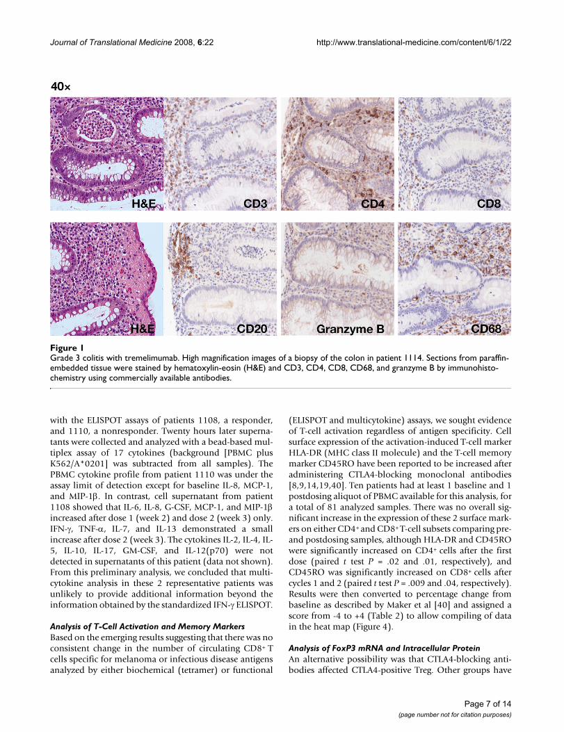
ToxicitiesNone of these patients developed a grade 4 treatment-related toxicity; however, 5 patients experienced treat-ment-related grade 3 toxicities. Two (1105A and 1108)developed marked increase in transaminases after 11 and5 doses, consistent with hepatitis (Table 1). Acute viralhepatitis was ruled out in both cases. In both cases thiswas a reason for study discontinuation. Although neitherpatient received corticosteroids, both fully recoveredwithin 2 months. One of these patients underwent 5 dailytherapeutic plasma exchange procedures with efficientclearance of detectable levels of circulating tremelimu-mab. Two patients (1106 and 1114) developed grade 3diarrhea after 4 and 2 doses. One underwent colonoscopyand biopsy, which demonstrated histologically provencolitis with lymphomonocytic infiltrates predominantlyby CD4+ T helper cells and CD68+ macrophages (Figure1). A final patient (1109) developed a generalized rash,which was described as leukocytoclastic vasculitis atbiopsy. Clinically significant grade 2 toxicities included 2cases of panhypopituitarism suggestive of hypophysitis,patient 1108 after 5 doses and patient 1113 after 22 dosesof tremelimumab. This later patient also developed ante-
rior uveitis after 21 doses that rapidly improved followingtreatment with corticosteroid-containing eye drops.
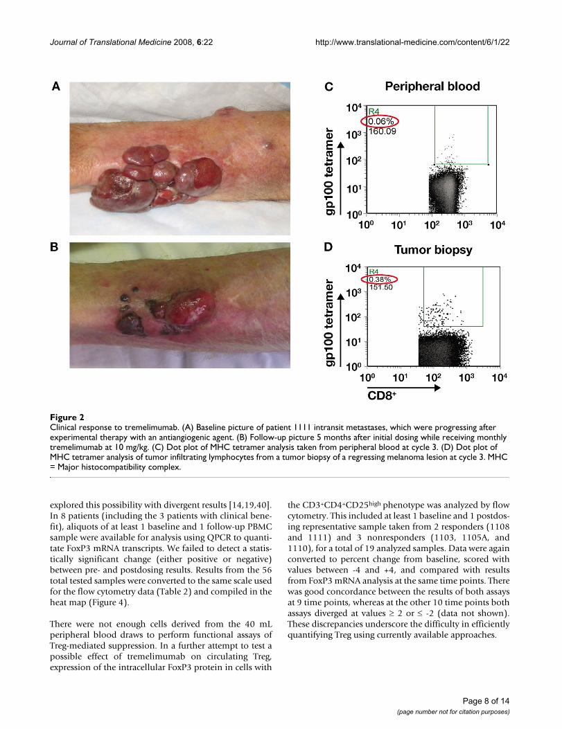
Clinical ResponsesOne patient (1108) achieved a complete response of 2nodal metastases that were proven to be melanoma atbaseline by fine needle aspiration. This response is ongo-ing 39+ months later. One patient (1111) had a majorpartial response of bulky in-transit metastasis, andremains relapse-free at 34+ months (Figure 2A and 2B). Athird patient (1113) did not qualify as a responder byRECIST criteria but was determined to have unequivocalevidence of clinical benefit. A [18F]fluorodeoxy-glucose(FDG) positron-emission tomography (PET)-positive 4-cm psoas muscle mass became PET negative after 9 doses.Upon surgical resection, pathological analysis showed a90% regression of melanoma with no disease progressionat 33+ months. This patient is listed as having a patholog-ical partial response. Only 1 of 5 patients with grade 3 tox-icity had an objective clinical response.
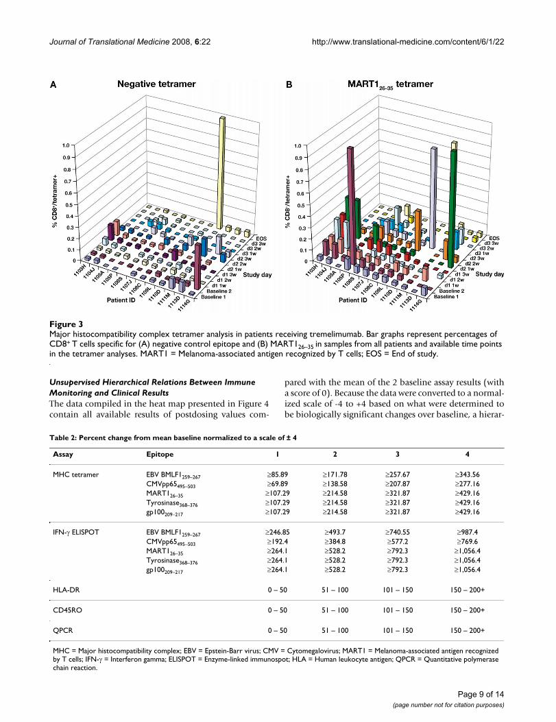
MHC Tetramer AssayT cells from treated patients were screened to determinewhether responses to tumor-specific antigens developedas a result of treatment with tremelimumab. A mean of 2baseline and 7 follow-up (range, 5 to 9) blood draws perpatient were tested. Samples were analyzed by flowcytometry for negative tetramer-specific MART126–35-spe-cific CD8+ T cells. Percentages of negative tetramer-specificand MART126–35-specific CD8+ T cells at each time pointindicated increases beyond the 99% RCV in 4 patients onmore than 1 occasion (Figure 3). However, these werespikes of peripheral appearance of melanoma antigen-specific T cells at isolated time points, as opposed to per-sistent elevations in several blood draws taken after dos-ing. Patient 1111, who had an objective clinical responseto therapy, underwent biopsy of a responding lesion.MHC tetramer analysis of nonadherent cells from thislesion demonstrated a 6-fold enrichment of gp100209–217-specific CD8+ T cells among total CD8+ T cells comparedwith peripheral blood (Figure 2C and 2D).
To efficiently present all of the data describing positive ornegative changes across 12 patients, multiple time points,and the different test epitopes (a total of 719 assayresults), the data were converted to a -4 to +4 scale basedon the 99% RCV (as described in the Materials and Meth-ods and Table 2) and color coded in shades of blue fornegative changes and red for positive changes. These datawere compiled in a heat map similar to the ones used toanalyze data from gene expression profiling (Figure 4). Asdescribed for MART126–35-specific T cells, no definitetrend was observed for changes over time in the propor-tion of CD8+ T cells reacting to any melanoma-(gp100209–
217, tyrosinase368–376) or infectious disease-(EBV
Page 5 of 14(page number not for citation purposes)
Journal of Translational Medicine 2008, 6:22 http://www.translational-medicine.com/content/6/1/22
BMLF1259–267, CMVpp65495–503) specific antigen in anypatient.
IFN-γ ELISPOT AssayThe heat map in Figure 4 also presents all the data gener-ated by ELISPOT assay enumerating cells producing IFN-γafter short ex vivo peptide stimulation. Results are colorcoded following the -4 to +4 scale range with negative andpositive changes defined by the 99% RCV compared withthe results from the 2 baseline samples (Table 2). The datasummarize replicate results from a mean of 9 (range, 6 to11) time points analyzed from the 12 study patients test-
ing reactivity to the negative control and test antigens fora total of 1,694 sample results. As with the tetramer assay,there was no evidence of a consistent change over time inthe assay results for any of the tested epitopes.
Multiplex Cytokine Production Upon Antigen ExposureThe possibility that IFN-γ may not be the most significantcytokine to define the functional status of antigen-specificT cells after CTLA4 blockade was explored. Therefore, weset up cocultures of 2 × 10⁵ PBMC and 2 × 10⁴ K562/A*0201 cells pulsed with either negative, MART126–35,tyrosinase368–376, and gp100209–217 peptides in parallel
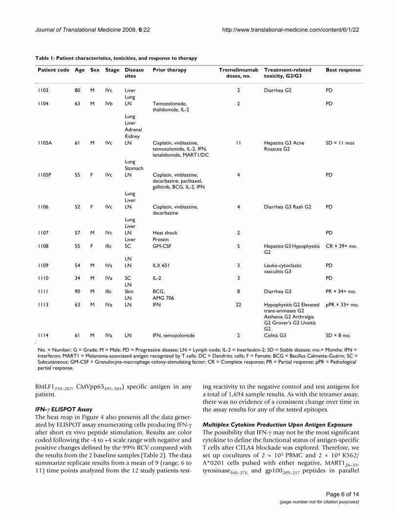
Table 1: Patient characteristics, toxicities, and response to therapy
Patient code Age Sex Stage Disease sites
Prior therapy Tremelimumab doses, no.
Treatment-related toxicity, G2/G3
Best response
1103 80 M IVc Liver 2 Diarrhea G2 PDLung
1104 63 M IVb LN Temozolomide, thalidomide, IL-2
2 PD
LungLiverAdrenalKidney
1105A 61 M IVc LN Cisplatin, vinblastine, temozolomide, IL-2, IFN, lenalidomide, MART1/DC
11 Hepatitis G3 Acne Rosacea G2
SD × 11 mos
LungStomach
1105P 55 F IVc LN Cisplatin, vinblastine, dacarbazine, paclitaxel, gefitinib, BCG, IL-2, IFN
4 PD
LungLiver
1106 52 F IVc LN Cisplatin, vinblastine, dacarbazine
4 Diarrhea G3 Rash G2 PD
LungLiver
1107 57 M IVc LN Heat shock 2 PDLiver Protein
1108 55 F IIIc SC GM-CSF 5 Hepatitis G3 Hypophysitis G2
CR × 39+ mo.
LN1109 54 M IVa LN ILX 651 3 Leuko-cytoclastic
vasculitis G3PD
1110 34 M IVa SC IL-2 3 PDLN
1111 90 M IIIc Skin BCG, 8 Diarrhea G2 PR × 34+ mo.LN AMG 706
1113 63 M IVa LN IFN 22 Hypophysitis G2 Elevated trans-aminases G2 Asthenia G2 Arthralgia G2 Grover's G2 Uveitis G2
pPR × 33+ mo.
1114 61 M IVa LN IFN, temozolomide 2 Colitis G3 SD × 8 mo.
No. = Number; G = Grade; M = Male; PD = Progressive disease; LN = Lymph node; IL-2 = Interleukin-2; SD = Stable disease; mo.= Months; IFN = Interferon; MART1 = Melanoma-associated antigen recognized by T cells; DC = Dendritic cells; F = Female; BCG = Bacillus Calmette-Guérin; SC = Subcutaneous; GM-CSF = Granulocyte-macrophage colony-stimulating factor; CR = Complete response; PR = Partial response; pPR = Pathological partial response.
Page 6 of 14(page number not for citation purposes)
Journal of Translational Medicine 2008, 6:22 http://www.translational-medicine.com/content/6/1/22
with the ELISPOT assays of patients 1108, a responder,and 1110, a nonresponder. Twenty hours later superna-tants were collected and analyzed with a bead-based mul-tiplex assay of 17 cytokines (background [PBMC plusK562/A*0201] was subtracted from all samples). ThePBMC cytokine profile from patient 1110 was under theassay limit of detection except for baseline IL-8, MCP-1,and MIP-1β. In contrast, cell supernatant from patient1108 showed that IL-6, IL-8, G-CSF, MCP-1, and MIP-1βincreased after dose 1 (week 2) and dose 2 (week 3) only.IFN-γ, TNF-α, IL-7, and IL-13 demonstrated a smallincrease after dose 2 (week 3). The cytokines IL-2, IL-4, IL-5, IL-10, IL-17, GM-CSF, and IL-12(p70) were notdetected in supernatants of this patient (data not shown).From this preliminary analysis, we concluded that multi-cytokine analysis in these 2 representative patients wasunlikely to provide additional information beyond theinformation obtained by the standardized IFN-γ ELISPOT.
Analysis of T-Cell Activation and Memory MarkersBased on the emerging results suggesting that there was noconsistent change in the number of circulating CD8+ Tcells specific for melanoma or infectious disease antigensanalyzed by either biochemical (tetramer) or functional
(ELISPOT and multicytokine) assays, we sought evidenceof T-cell activation regardless of antigen specificity. Cellsurface expression of the activation-induced T-cell markerHLA-DR (MHC class II molecule) and the T-cell memorymarker CD45RO have been reported to be increased afteradministering CTLA4-blocking monoclonal antibodies[8,9,14,19,40]. Ten patients had at least 1 baseline and 1postdosing aliquot of PBMC available for this analysis, fora total of 81 analyzed samples. There was no overall sig-nificant increase in the expression of these 2 surface mark-ers on either CD4+ and CD8+ T-cell subsets comparing pre-and postdosing samples, although HLA-DR and CD45ROwere significantly increased on CD4+ cells after the firstdose (paired t test P = .02 and .01, respectively), andCD45RO was significantly increased on CD8+ cells aftercycles 1 and 2 (paired t test P = .009 and .04, respectively).Results were then converted to percentage change frombaseline as described by Maker et al [40] and assigned ascore from -4 to +4 (Table 2) to allow compiling of datain the heat map (Figure 4).
Analysis of FoxP3 mRNA and Intracellular ProteinAn alternative possibility was that CTLA4-blocking anti-bodies affected CTLA4-positive Treg. Other groups have
Grade 3 colitis with tremelimumabFigure 1Grade 3 colitis with tremelimumab. High magnification images of a biopsy of the colon in patient 1114. Sections from paraffin-embedded tissue were stained by hematoxylin-eosin (H&E) and CD3, CD4, CD8, CD68, and granzyme B by immunohisto-chemistry using commercially available antibodies.
Page 7 of 14(page number not for citation purposes)
Journal of Translational Medicine 2008, 6:22 http://www.translational-medicine.com/content/6/1/22
explored this possibility with divergent results [14,19,40].In 8 patients (including the 3 patients with clinical bene-fit), aliquots of at least 1 baseline and 1 follow-up PBMCsample were available for analysis using QPCR to quanti-tate FoxP3 mRNA transcripts. We failed to detect a statis-tically significant change (either positive or negative)between pre- and postdosing results. Results from the 56total tested samples were converted to the same scale usedfor the flow cytometry data (Table 2) and compiled in theheat map (Figure 4).
There were not enough cells derived from the 40 mLperipheral blood draws to perform functional assays ofTreg-mediated suppression. In a further attempt to test apossible effect of tremelimumab on circulating Treg,expression of the intracellular FoxP3 protein in cells with
the CD3+CD4+CD25high phenotype was analyzed by flowcytometry. This included at least 1 baseline and 1 postdos-ing representative sample taken from 2 responders (1108and 1111) and 3 nonresponders (1103, 1105A, and1110), for a total of 19 analyzed samples. Data were againconverted to percent change from baseline, scored withvalues between -4 and +4, and compared with resultsfrom FoxP3 mRNA analysis at the same time points. Therewas good concordance between the results of both assaysat 9 time points, whereas at the other 10 time points bothassays diverged at values ≥ 2 or ≤ -2 (data not shown).These discrepancies underscore the difficulty in efficientlyquantifying Treg using currently available approaches.
Clinical response to tremelimumabFigure 2Clinical response to tremelimumab. (A) Baseline picture of patient 1111 intransit metastases, which were progressing after experimental therapy with an antiangiogenic agent. (B) Follow-up picture 5 months after initial dosing while receiving monthly tremelimumab at 10 mg/kg. (C) Dot plot of MHC tetramer analysis taken from peripheral blood at cycle 3. (D) Dot plot of MHC tetramer analysis of tumor infiltrating lymphocytes from a tumor biopsy of a regressing melanoma lesion at cycle 3. MHC = Major histocompatibility complex.
Page 8 of 14(page number not for citation purposes)
Journal of Translational Medicine 2008, 6:22 http://www.translational-medicine.com/content/6/1/22
Unsupervised Hierarchical Relations Between Immune Monitoring and Clinical ResultsThe data compiled in the heat map presented in Figure 4contain all available results of postdosing values com-
pared with the mean of the 2 baseline assay results (witha score of 0). Because the data were converted to a normal-ized scale of -4 to +4 based on what were determined tobe biologically significant changes over baseline, a hierar-
Major histocompatibility complex tetramer analysis in patients receiving tremelimumabFigure 3Major histocompatibility complex tetramer analysis in patients receiving tremelimumab. Bar graphs represent percentages of CD8+ T cells specific for (A) negative control epitope and (B) MART126–35 in samples from all patients and available time points in the tetramer analyses. MART1 = Melanoma-associated antigen recognized by T cells; EOS = End of study.
Table 2: Percent change from mean baseline normalized to a scale of ± 4
Assay Epitope 1 2 3 4
MHC tetramer EBV BMLF1259–267 ≥85.89 ≥171.78 ≥257.67 ≥343.56CMVpp65495–503 ≥69.89 ≥138.58 ≥207.87 ≥277.16MART126–35 ≥107.29 ≥214.58 ≥321.87 ≥429.16Tyrosinase368–376 ≥107.29 ≥214.58 ≥321.87 ≥429.16gp100209–217 ≥107.29 ≥214.58 ≥321.87 ≥429.16
IFN-γ ELISPOT EBV BMLF1259–267 ≥246.85 ≥493.7 ≥740.55 ≥987.4CMVpp65495–503 ≥192.4 ≥384.8 ≥577.2 ≥769.6MART126–35 ≥264.1 ≥528.2 ≥792.3 ≥1,056.4Tyrosinase368–376 ≥264.1 ≥528.2 ≥792.3 ≥1,056.4gp100209–217 ≥264.1 ≥528.2 ≥792.3 ≥1,056.4
HLA-DR 0 – 50 51 – 100 101 – 150 150 – 200+
CD45RO 0 – 50 51 – 100 101 – 150 150 – 200+
QPCR 0 – 50 51 – 100 101 – 150 150 – 200+
MHC = Major histocompatibility complex; EBV = Epstein-Barr virus; CMV = Cytomegalovirus; MART1 = Melanoma-associated antigen recognized by T cells; IFN-γ = Interferon gamma; ELISPOT = Enzyme-linked immunospot; HLA = Human leukocyte antigen; QPCR = Quantitative polymerase chain reaction.
Page 9 of 14(page number not for citation purposes)
Journal of Translational Medicine 2008, 6:22 http://www.translational-medicine.com/content/6/1/22
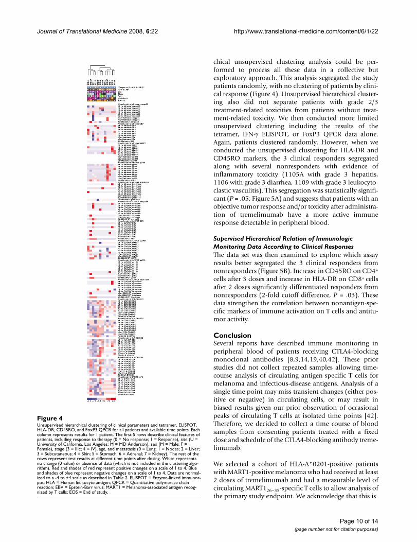
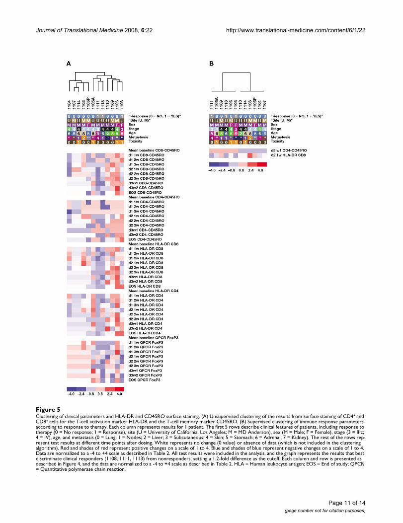
chical unsupervised clustering analysis could be per-formed to process all these data in a collective butexploratory approach. This analysis segregated the studypatients randomly, with no clustering of patients by clini-cal response (Figure 4). Unsupervised hierarchical cluster-ing also did not separate patients with grade 2/3treatment-related toxicities from patients without treat-ment-related toxicity. We then conducted more limitedunsupervised clustering including the results of thetetramer, IFN-γ ELISPOT, or FoxP3 QPCR data alone.Again, patients clustered randomly. However, when weconducted the unsupervised clustering for HLA-DR andCD45RO markers, the 3 clinical responders segregatedalong with several nonresponders with evidence ofinflammatory toxicity (1105A with grade 3 hepatitis,1106 with grade 3 diarrhea, 1109 with grade 3 leukocyto-clastic vasculitis). This segregation was statistically signifi-cant (P = .05; Figure 5A) and suggests that patients with anobjective tumor response and/or toxicity after administra-tion of tremelimumab have a more active immuneresponse detectable in peripheral blood.
Supervised Hierarchical Relation of Immunologic Monitoring Data According to Clinical ResponsesThe data set was then examined to explore which assayresults better segregated the 3 clinical responders fromnonresponders (Figure 5B). Increase in CD45RO on CD4+
cells after 3 doses and increase in HLA-DR on CD8+ cellsafter 2 doses significantly differentiated responders fromnonresponders (2-fold cutoff difference, P = .03). Thesedata strengthen the correlation between nonantigen-spe-cific markers of immune activation on T cells and antitu-mor activity.
ConclusionSeveral reports have described immune monitoring inperipheral blood of patients receiving CTLA4-blockingmonoclonal antibodies [8,9,14,19,40,42]. These priorstudies did not collect repeated samples allowing time-course analysis of circulating antigen-specific T cells formelanoma and infectious-disease antigens. Analysis of asingle time point may miss transient changes (either pos-itive or negative) in circulating cells, or may result inbiased results given our prior observation of occasionalpeaks of circulating T cells at isolated time points [42].Therefore, we decided to collect a time course of bloodsamples from consenting patients treated with a fixeddose and schedule of the CTLA4-blocking antibody treme-limumab.
We selected a cohort of HLA-A*0201-positive patientswith MART1-positive melanoma who had received at least2 doses of tremelimumab and had a measurable level ofcirculating MART126–35-specific T cells to allow analysis ofthe primary study endpoint. We acknowledge that this is
Unsupervised hierarchical clustering of clinical parameters and tetramer, ELISPOT, HLA-DR, CD45RO, and FoxP3 QPCR for all patients and available time pointsFigure 4Unsupervised hierarchical clustering of clinical parameters and tetramer, ELISPOT, HLA-DR, CD45RO, and FoxP3 QPCR for all patients and available time points. Each column represents results for 1 patient. The first 5 rows describe clinical features of patients, including response to therapy (0 = No response; 1 = Response), site (U = University of California, Los Angeles; M = MD Anderson), sex (M = Male; F = Female), stage (3 = IIIc; 4 = IV), age, and metastasis (0 = Lung; 1 = Nodes; 2 = Liver; 3 = Subcutaneous; 4 = Skin; 5 = Stomach; 6 = Adrenal; 7 = Kidney). The rest of the rows represent test results at different time points after dosing. White represents no change (0 value) or absence of data (which is not included in the clustering algo-rithm). Red and shades of red represent positive changes on a scale of 1 to 4. Blue and shades of blue represent negative changes on a scale of 1 to 4. Data are normal-ized to a -4 to +4 scale as described in Table 2. ELISPOT = Enzyme-linked immunos-pot; HLA = Human leukocyte antigen; QPCR = Quantitative polymerase chain reaction; EBV = Epstein-Barr virus; MART1 = Melanoma-associated antigen recog-nized by T cells; EOS = End of study.
Page 10 of 14(page number not for citation purposes)
Journal of Translational Medicine 2008, 6:22 http://www.translational-medicine.com/content/6/1/22
Clustering of clinical parameters and HLA-DR and CD45RO surface stainingFigure 5Clustering of clinical parameters and HLA-DR and CD45RO surface staining. (A) Unsupervised clustering of the results from surface staining of CD4+ and CD8+ cells for the T-cell activation marker HLA-DR and the T-cell memory marker CD45RO. (B) Supervised clustering of immune response parameters according to response to therapy. Each column represents results for 1 patient. The first 5 rows describe clinical features of patients, including response to therapy (0 = No response; 1 = Response), site (U = University of California, Los Angeles; M = MD Anderson), sex (M = Male; F = Female), stage (3 = IIIc; 4 = IV), age, and metastasis (0 = Lung; 1 = Nodes; 2 = Liver; 3 = Subcutaneous; 4 = Skin; 5 = Stomach; 6 = Adrenal; 7 = Kidney). The rest of the rows rep-resent test results at different time points after dosing. White represents no change (0 value) or absence of data (which is not included in the clustering algorithm). Red and shades of red represent positive changes on a scale of 1 to 4. Blue and shades of blue represent negative changes on a scale of 1 to 4. Data are normalized to a -4 to +4 scale as described in Table 2. All test results were included in the analysis, and the graph represents the results that best discriminate clinical responders (1108, 1111, 1113) from nonresponders, setting a 1.2-fold difference as the cutoff. Each column and row is presented as described in Figure 4, and the data are normalized to a -4 to +4 scale as described in Table 2. HLA = Human leukocyte antigen; EOS = End of study; QPCR = Quantitative polymerase chain reaction.
Page 11 of 14(page number not for citation purposes)
Journal of Translational Medicine 2008, 6:22 http://www.translational-medicine.com/content/6/1/22
a selected population of patients, and this analysis wasaimed at testing both positive and negative changes inantigen-specific T cells in peripheral blood. Contrary towhat has been reported by others [9,14], we did notobserve a significant correlation between the develop-ment of toxicities that may have an autoimmune mecha-nism of action with clinical responses. A limitation of ouranalysis is the small number of patients, resulting in lowerpower to detect statistically significant correlations. How-ever, we did note that certain toxicities, like hepatitis andanterior uveitis, may be more common with repeated dos-ing, something that has been previously described forhypophysitis [43]. Two unique skin toxicities with treme-limumab were noted in our patients, leukocytoclastic vas-culitis and Grover's disease of the skin.
Our data confirm observations by others [8,9,19,40] thatadministration of CTLA4-blocking antibodies to patientswith melanoma does not result in the sustained expan-sion of circulating melanoma antigen-specific CD8+ Tcells. It further rules out that this intervention decreasesthe number of circulating melanoma antigen-specific Tcells. However, the observed marked enrichment ofgp100209–217-specific CD8+ T cells in regressing tumor tis-sue compared with peripheral blood in 1 patient suggeststhat sampling of circulating antigen-specific T cells mightmiss overall melanoma antigen-specific T-cell expansionin the tumor. This may be particularly relevant for studieswithout time-course sampling. The occasional peaks ofincreased numbers of melanoma-circulating antigen-spe-cific T cells detected in several patients may reflect peaksof T-cell expansion at different time points that may notpersist because the cells accumulate in tumors. We rea-soned that this possibility could be better analyzed whenthe data were studied in an unbiased manner using unsu-pervised clustering analysis of data presented in heatmaps. However, even with this analysis, we were unableto correlate the activation or expansion of circulatingmelanoma antigen-specific CD8+ T cells with clinical ben-efit to CTLA4 blockade. It is certainly possible that the 3melanoma antigens studied may not be relevant forCTLA4 blockade-induced melanoma regressions. How-ever, these 3 antigen epitopes have been repeatedly recog-nized as HLA-A*0201 immunodominant peptides inpatients with immunotherapy-induced tumor regressions[44].
The constitutive expression of CTLA4 on Treg has raisedthe possibility that administration of anti-CTLA4 antibod-ies may deplete or modulate the function of these profes-sional immune suppressive cells [45,46]. However,neither ipilimumab nor tremelimumab were selected asdepleting antibodies for CTLA4-positive cells. In fact, bothantibodies were selected as blocking antibodies to CTLA4intended to activate cells expressing their target but not
kill them. Furthermore, tremelimumab is an IgG2 anti-body, an immunoglobulin subtype unlikely to fix com-plement or induce antibody-mediated cellularcytotoxicity [47]. Treg are difficult to study at the cellularlevel because they have a surface phenotype that is indis-tinguishable from chronically activated T helper cells.They can be detected by the intracellular expression of theTreg-specific transcription factor FoxP3 [48,49]. FoxP3mRNA transcripts in a population of PBMC can be quan-titated by QPCR; however, this technique does not allowefficient enumeration of Treg at the cellular level. Intracel-lular FoxP3 protein staining can be achieved by multi-color flow cytometry after cell permeabilization, but it iscurrently unclear how these 2 techniques compare and thebias of each analytical methodology. The gold standardassay for detecting Treg is the determination of their func-tional ability to inhibit the proliferation of clonallyexpanded T cells [20]. However, insufficient quantities ofblood were collected to perform this type of analysis.Therefore, our findings of no evidence of Treg depletion inperipheral blood samples by FoxP3 QPCR and intracellu-lar staining should be interpreted with caution. PairedFoxP3 mRNA quantitation and Treg functional analysishave been reported by Maker et al [40]. These investiga-tors concluded that administration of the CTLA4-blockingantibody ipilimumab to patients with melanoma neitherdepleted Treg nor downregulated their functionalimmune suppressive activity. Our data and the datareported by Maker et al are in contrast to data presentedby Reuben et al [14] who suggested that Treg defined byflow cytometry may be preferentially depleted in patientswith an objective response to the CTLA4 blocking anti-body tremelimumab, and O'Mahony et al [19] who sug-gested that Treg defined by FoxP3 QPCR may transientlydecrease in peripheral circulation 3 days after dosing andrecover 4 weeks later.
We analyzed tumor and infectious disease antigen-spe-cific CD8+ T-cell immune monitoring, FoxP3 mRNAquantitation, and T-cell activation/memory marker sur-face staining concurrently to detect trends relating to clin-ical parameters. We considered applying a stepwiseregression analysis to search for variables that are signifi-cantly associated with clinical response. However, oursample size was too small for such an analysis, especiallyconsidering that this cohort included only 3 clinicalresponders. Therefore, to accomplish the goal of generat-ing an overall evaluation of immune monitoring data, weturned to cluster analysis as a means to look at the data inan exploratory fashion. Markers of T-cell activation (HLA-DR) and memory phenotype (CD45RO) after dosing withtremelimumab segregated the 3 patients with clinical ben-efit together with 4 patients with inflammatory toxicity.Therefore, we conclude that tremelimumab does notincrease the number or function of antigen-specific CD8+
Page 12 of 14(page number not for citation purposes)
Journal of Translational Medicine 2008, 6:22 http://www.translational-medicine.com/content/6/1/22
T cells in peripheral blood nor decrease FoxP3 transcriptsbut appears to generally enhance T-cell activation and dif-ferentiation. Detection of T-cell activation and memorymarkers may be useful as readout of this antibody's abilityto activate the immune system, which may lead to phar-macodynamic effects consistent with its observed antitu-mor activity. Sampling of T cells in tumors may provideadditional information, and future studies should includeimmune monitoring in tumor tissues.
Competing interestsThe following authors have no competing interests toreport: Begoña Comin-Anduix, Yohan Lee, Jason Jalil,Alain Algazi, Pilar de la Rocha, Elisabeth Seja, Arturo Vil-lanueva, Bradley R. Straatsma, James S. Economou, andJohn A. Glaspy. Viviana A. Bozon, Cecile A. Bulanhagui,Antonio Gualberto, and Jesus Gomez-Navarro areemployed by and (with the exception of V. Bozon) ownstock in Pfizer, Inc. Luis Camacho and Antoni Ribasreceive honoraria and funding for research from Pfizer,Inc., and Antoni Ribas is also compensated for being anexpert on the advisory board for Pfizer, Inc.
Authors' contributionsJG-N, AG, and AR designed the study. LHC, B RS, JSE, JAG,and AR were responsible for patient care. BCA, YL, JJ, AA,and PdlR generated the immunologic data. BCA, VAB,CAB, ES, and AV collected these data. Data were analyzedby BCA, YL, VAB, CAB, JGN, and AR. The manuscript waswritten by BCA, JGN, and AR; and all co-authors reviewedthe final manuscript.
ConsentWritten informed consent approved by the University ofCalifornia Los Angeles and University of Texas MD Ander-son institutional review boards was obtained from thepatients for publication of this case report and any accom-panying images. A copy of the written consent is availablefor review by the Editor-in-Chief of this journal.
AcknowledgementsWe would like to acknowledge Dennis Noe, MD; Dmitri Pavlov, PhD; and Kelly Page, MBA; Pfizer Global Research and Development, New London, CT; and Clara Armengol and Imran Ahmad from University of California Los Angeles (UCLA) for their support in the conduct of this study. The UCLA Flow Cytometry Core Facility is supported by the National Insti-tutes of Health awards CA-16042 and AI-28697, and by the Jonsson Com-prehensive Cancer Center, the Center for AIDS Research from the UCLA AIDS Institute, and the David Geffen School of Medicine at UCLA. The authors would also like to thank Todd Parker, PhD, at ProEd Communica-tions, Inc.® for his medical editorial assistance. Financial support for medical editorial assistance was provided by Pfizer, Inc. Antoni Ribas is supported in part by the Jonsson Comprehensive Cancer Center and the Melanoma Research Foundation.
References1. Brunet JF, Denizot F, Luciani MF, Roux-Dosseto M, Suzan M, Mattei
MG, Golstein P: A new member of the immunoglobulin super-family – CTLA-4. Nature 1987, 328:267-270.
2. Walunas TL, Lenschow DJ, Bakker CY, Linsley PS, Freeman GJ, GreenJM, Thompson CB, Bluestone JA: CTLA-4 can function as a neg-ative regulator of T cell activation. Immunity 1994, 1:405-413.
3. Chambers CA, Kuhns MS, Egen JG, Allison JP: CTLA-4-mediatedinhibition in regulation of T cell responses: mechanisms andmanipulation in tumor immunotherapy. Annu Rev Immunol2001, 19:565-594.
4. Wang XB, Giscombe R, Yan Z, Heiden T, Xu D, Lefvert AK: Expres-sion of CTLA-4 by human monocytes. Scand J Immunol 2002,55:53-60.
5. Teft WA, Kirchhof MG, Madrenas J: A molecular perspective ofCTLA-4 function. Annu Rev Immunol 2006, 24:65-97.
6. Lee KM, Chuang E, Griffin M, Khattri R, Hong DK, Zhang W, StrausD, Samelson LE, Thompson CB, Bluestone JA: Molecular basis of Tcell inactivation by CTLA-4. Science 1998, 282:2263-2266.
7. Hodi FS, Mihm MC, Soiffer RJ, Haluska FG, Butler M, Seiden MV, DavisT, Henry-Spires R, MacRae S, Willman A, et al.: Biologic activity ofcytotoxic T lymphocyte-associated antigen 4 antibody block-ade in previously vaccinated metastatic melanoma and ovar-ian carcinoma patients. Proc Natl Acad Sci USA 2003,100:4712-4717.
8. Phan GQ, Yang JC, Sherry RM, Hwu P, Topalian SL, SchwartzentruberDJ, Restifo NP, Haworth LR, Seipp CA, Freezer LJ, et al.: Cancerregression and autoimmunity induced by cytotoxic T lym-phocyte-associated antigen 4 blockade in patients with met-astatic melanoma. Proc Natl Acad Sci USA 2003, 100:8372-8377.
9. Attia P, Phan GQ, Maker AV, Robinson MR, Quezado MM, Yang JC,Sherry RM, Topalian SL, Kammula US, Royal RE, et al.: Autoimmu-nity correlates with tumor regression in patients with meta-static melanoma treated with anti-cytotoxic T-lymphocyteantigen-4. J Clin Oncol 2005, 23:6043-6053.
10. Maker AV, Phan GQ, Attia P, Yang JC, Sherry RM, Topalian SL, Kam-mula US, Royal RE, Haworth LR, Levy C, et al.: Tumor regressionand autoimmunity in patients treated with cytotoxic T lym-phocyte-associated antigen 4 blockade and interleukin 2: aphase I/II study. Ann Surg Oncol 2005, 12:1005-1016.
11. Sanderson K, Scotland R, Lee P, Liu D, Groshen S, Snively J, Sian S,Nichol G, Davis T, Keler T, et al.: Autoimmunity in a phase I trialof a fully human anti-cytotoxic T-lymphocyte antigen-4 mon-oclonal antibody with multiple melanoma peptides and Mon-tanide ISA 51 for patients with resected stages III and IVmelanoma. J Clin Oncol 2005, 23:741-750.
12. Ribas A, Camacho LH, Lopez-Berestein G, Pavlov D, Bulanhagui CA,Millham R, Comin-Anduix B, Reuben JM, Seja E, Parker CA, et al.:Antitumor activity in melanoma and anti-self responses in aphase I trial with the anti-cytotoxic T lymphocyte-associatedantigen 4 monoclonal antibody CP-675,206. J Clin Oncol 2005,23:8968-8977.
13. Maker AV, Yang JC, Sherry RM, Topalian SL, Kammula US, Royal RE,Hughes M, Yellin MJ, Haworth LR, Levy C, et al.: Intrapatient doseescalation of anti-CTLA-4 antibody in patients with meta-static melanoma. J Immunother 2006, 29:455-463.
14. Reuben JM, Lee BN, Li C, Gomez-Navarro J, Bozon VA, Parker CA,Hernandez IM, Gutierrez C, Lopez-Berestein G, Camacho LH: Bio-logic and immunomodulatory events after CTLA-4 blockadewith ticilimumab in patients with advanced malignantmelanoma. Cancer 2006, 106:2437-2444.
15. Downey SG, Klapper JA, Smith FO, Yang JC, Sherry RM, Royal RE,Kammula US, Hughes MS, Allen TE, Levy CL, et al.: Prognostic fac-tors related to clinical response in patients with metastaticmelanoma treated by CTL-associated antigen-4 blockade.Clin Cancer Res 2007, 13:6681-6688.
16. Korman AJ, Peggs KS, Allison JP: Checkpoint blockade in cancerimmunotherapy. Adv Immunol 2006, 90:297-339.
17. Peggs KS, Quezada SA, Korman AJ, Allison JP: Principles and use ofanti-CTLA4 antibody in human cancer immunotherapy. CurrOpin Immunol 2006, 18:206-213.
18. Leach DR, Krummel MF, Allison JP: Enhancement of antitumorimmunity by CTLA-4 blockade. Science 1996, 271:1734-1736.
19. O'Mahony D, Morris JC, Quinn C, Gao W, Wilson WH, Gause B, Pit-taluga S, Neelapu S, Brown M, Fleisher TA, et al.: A pilot study of
Page 13 of 14(page number not for citation purposes)
http://www.ncbi.nlm.nih.gov/entrez/query.fcgi?cmd=Retrieve&db=PubMed&dopt=Abstract&list_uids=3496540
http://www.ncbi.nlm.nih.gov/entrez/query.fcgi?cmd=Retrieve&db=PubMed&dopt=Abstract&list_uids=3496540
http://www.ncbi.nlm.nih.gov/entrez/query.fcgi?cmd=Retrieve&db=PubMed&dopt=Abstract&list_uids=7882171
http://www.ncbi.nlm.nih.gov/entrez/query.fcgi?cmd=Retrieve&db=PubMed&dopt=Abstract&list_uids=7882171
http://www.ncbi.nlm.nih.gov/entrez/query.fcgi?cmd=Retrieve&db=PubMed&dopt=Abstract&list_uids=9856951
http://www.ncbi.nlm.nih.gov/entrez/query.fcgi?cmd=Retrieve&db=PubMed&dopt=Abstract&list_uids=9856951
http://www.ncbi.nlm.nih.gov/entrez/query.fcgi?cmd=Retrieve&db=PubMed&dopt=Abstract&list_uids=8596936
Journal of Translational Medicine 2008, 6:22 http://www.translational-medicine.com/content/6/1/22
Publish with BioMed Central and every scientist can read your work free of charge
"BioMed Central will be the most significant development for disseminating the results of biomedical research in our lifetime."
Sir Paul Nurse, Cancer Research UK
Your research papers will be:
available free of charge to the entire biomedical community
peer reviewed and published immediately upon acceptance
cited in PubMed and archived on PubMed Central
yours — you keep the copyright
Submit your manuscript here:http://www.biomedcentral.com/info/publishing_adv.asp
BioMedcentral
CTLA-4 blockade after cancer vaccine failure in patientswith advanced malignancy. Clin Cancer Res 2007, 13:958-964.
20. Shevach EM: CD4+ CD25+ suppressor T cells: more questionsthan answers. Nat Rev Immunol 2002, 2:389-400.
21. Grohmann U, Orabona C, Fallarino F, Vacca C, Calcinaro F, FalorniA, Candeloro P, Belladonna ML, Bianchi R, Fioretti MC, Puccetti P:CTLA-4-Ig regulates tryptophan catabolism in vivo. NatImmunol 2002, 3:1097-1101.
22. Fallarino F, Grohmann U, Hwang KW, Orabona C, Vacca C, BianchiR, Belladonna ML, Fioretti MC, Alegre ML, Puccetti P: Modulationof tryptophan catabolism by regulatory T cells. Nat Immunol2003, 4:1206-1212.
23. Munn DH, Sharma MD, Hou D, Baban B, Lee JR, Antonia SJ, MessinaJL, Chandler P, Koni PA, Mellor AL: Expression of indoleamine2,3-dioxygenase by plasmacytoid dendritic cells in tumor-draining lymph nodes. J Clin Invest 2004, 114:280-290.
24. Paust S, Lu L, McCarty N, Cantor H: Engagement of B7 on effec-tor T cells by regulatory T cells prevents autoimmune dis-ease. Proc Natl Acad Sci USA 2004, 101:10398-10403.
25. Jinushi M, Hodi FS, Dranoff G: Therapy-induced antibodies toMHC class I chain-related protein A antagonize immunesuppression and stimulate antitumor cytotoxicity. Proc NatlAcad Sci USA 2006, 103:9190-9195.
26. Schneider H, Downey J, Smith A, Zinselmeyer BH, Rush C, BrewerJM, Wei B, Hogg N, Garside P, Rudd CE: Reversal of the TCR stopsignal by CTLA-4. Science 2006, 313:1972-1975.
27. Contardi E, Palmisano GL, Tazzari PL, Martelli AM, Fala F, Fabbi M,Kato T, Lucarelli E, Donati D, Polito L, et al.: CTLA-4 is constitu-tively expressed on tumor cells and can trigger apoptosisupon ligand interaction. Int J Cancer 2005, 117:538-550.
28. Keilholz U, Weber J, Finke JH, Gabrilovich DI, Kast WM, Disis ML,Kirkwood JM, Scheibenbogen C, Schlom J, Maino VC, et al.: Immu-nologic monitoring of cancer vaccine therapy: results of aworkshop sponsored by the Society for Biological Therapy. JImmunother 2002, 25:97-138.
29. Comin-Anduix B, Gualberto A, Glaspy JA, Seja E, Ontiveros M, Rear-don DL, Renteria R, Englahner B, Economou JS, Gomez-Navarro J,Ribas A: Definition of an immunologic response using themajor histocompatibility complex tetramer and enzyme-linked immunospot assays. Clin Cancer Res 2006, 12:107-116.
30. Marincola FM: In support of descriptive studies; relevance totranslational research. J Transl Med 2007, 5:21.
31. Ribas A, Bozon VA, Lopez-Berestein G, Pavlov D, Reuben JM, ParkerCA, Seja E, Glaspy JA, Gomez-Navarro J, Camacho LH: Phase 1 trialof monthly doses of the human anti-CTLA4 monoclonalantibody CP-675,206 in patients with advanced melanoma. JClin Oncol 2005, 23(suppl):716s. Abstract 7524
32. Gordon LK, Ribas A, Nusinowitz S, Butterfield LH, Glaspy JA, Econo-mou JS, Straatsma BR: Surveillance of the eye and vision in aclinical trial of MART1-transformed dendritic cells for meta-static melanoma. Control Clin Trials 2004, 25:400-407.
33. Straatsma BR, Nusinowitz S, Young TA, Gordon LK, Chun MW,Rosen C, Seja E, Economou JS, Glaspy JA, Bozon V, et al.: Surveil-lance of the eye and vision in clinical trials of CP-675,206 formetastatic melanoma. Am J Ophthalmol 2007 in press.
34. Therasse P, Arbuck SG, Eisenhauer EA, Wanders J, Kaplan RS, Rubin-stein L, Verweij J, Van Glabbeke M, van Oosterom AT, Christian MC,Gwyther SG: New guidelines to evaluate the response totreatment in solid tumors. European Organization forResearch and Treatment of Cancer, National Cancer Insti-tute of the United States, National Cancer Institute of Can-ada. J Natl Cancer Inst 2000, 92:205-216.
35. Maecker HT, Moon J, Bhatia S, Ghanekar SA, Maino VC, Payne JK,Kuus-Reichel K, Chang JC, Summers A, Clay TM, et al.: Impact ofcryopreservation on tetramer, cytokine flow cytometry, andELISPOT. BMC Immunol 2005, 6:17.
36. Butterfield LH, Meng WS, Koh A, Vollmer CM, Ribas A, Dissette VB,Faull K, Glaspy JA, McBride WH, Economou JS: T cell responses toHLA-A*0201-restricted peptides derived from human alphafetoprotein. J Immunol 2001, 166:5300-5308.
37. Perfetto SP, Chattopadhyay PK, Roederer M: Seventeen-colourflow cytometry: unravelling the immune system. Nat RevImmunol 2004, 4:648-655.
38. Muthukumar T, Dadhania D, Ding R, Snopkowski C, Naqvi R, Lee JB,Hartono C, Li B, Sharma VK, Seshan SV, et al: Messenger RNA for
FOXP3 in the urine of renal-allograft recipients. N Engl J Med2005, 353:2342-2351.
39. Baratelli F, Lin Y, Zhu L, Yang SC, Heuze-Vourc'h N, Zeng G, Reck-amp K, Dohadwala M, Sharma S, Dubinett SM: Prostaglandin E2induces FOXP3 gene expression and T regulatory cell func-tion in human CD4+ T cells. J Immunol 2005, 175:1483-1490.
40. Maker AV, Attia P, Rosenberg SA: Analysis of the cellular mech-anism of antitumor responses and autoimmunity in patientstreated with CTLA-4 blockade. J Immunol 2005, 175:7746-7754.
41. Eisen MB, Spellman PT, Brown PO, Botstein D: Cluster analysisand display of genome-wide expression patterns. Proc NatlAcad Sci USA 1998, 95:14863-14868.
42. Comin-Anduix B, Lee Y, Jalil J, de la Rocha P, Seja E, Bozon VA, Cama-cho LH, Economou JS, Glaspy JA, Gomez-Navarro J, Ribas A: Immu-nological assays to differentiate responders fromnonresponders after CTLA blockade with CP-675,206[abstract]. Proc Int Soc Biol Ther Cancer 2006.
43. Blansfield JA, Beck KE, Tran K, Yang JC, Hughes MS, Kammula US,Royal RE, Topalian SL, Haworth LR, Levy C, et al.: Cytotoxic T-lym-phocyte-associated antigen-4 blockage can induce autoim-mune hypophysitis in patients with metastatic melanomaand renal cancer. J Immunother 2005, 28:593-598.
44. Rosenberg SA: Progress in human tumour immunology andimmunotherapy. Nature 2001, 411:380-384.
45. Liu H, Hu B, Xu D, Liew FY: CD4+CD25+ regulatory T cells curemurine colitis: the role of IL-10, TGF-beta, and CTLA4. JImmunol 2003, 171:5012-5017.
46. Read S, Greenwald R, Izcue A, Robinson N, Mandelbrot D, FranciscoL, Sharpe AH, Powrie F: Blockade of CTLA-4 on CD4+CD25+regulatory T cells abrogates their function in vivo. J Immunol2006, 177:4376-4383.
47. Bruggemann M, Williams GT, Bindon CI, Clark MR, Walker MR, Jef-feris R, Waldmann H, Neuberger MS: Comparison of the effectorfunctions of human immunoglobulins using a matched set ofchimeric antibodies. J Exp Med 1987, 166:1351-1361.
48. Hori S, Nomura T, Sakaguchi S: Control of regulatory T celldevelopment by the transcription factor Foxp3. Science 2003,299:1057-1061.
49. Fontenot JD, Rasmussen JP, Williams LM, Dooley JL, Farr AG, Ruden-sky AY: Regulatory T cell lineage specification by the fork-head transcription factor foxp3. Immunity 2005, 22:329-341.
Page 14 of 14(page number not for citation purposes)
http://www.ncbi.nlm.nih.gov/entrez/query.fcgi?cmd=Retrieve&db=PubMed&dopt=Abstract&list_uids=9843981
http://www.ncbi.nlm.nih.gov/entrez/query.fcgi?cmd=Retrieve&db=PubMed&dopt=Abstract&list_uids=9843981
http://www.ncbi.nlm.nih.gov/entrez/query.fcgi?cmd=Retrieve&db=PubMed&dopt=Abstract&list_uids=3500259
http://www.ncbi.nlm.nih.gov/entrez/query.fcgi?cmd=Retrieve&db=PubMed&dopt=Abstract&list_uids=3500259
Related Documents