Organic & Biomolecular Chemistry PAPER Cite this: Org. Biomol. Chem., 2014, 12, 73 Received 15th August 2013, Accepted 19th September 2013 DOI: 10.1039/c3ob41676c www.rsc.org/obc Design, synthesis and characterization of novel inhibitors against mycobacterial β-ketoacyl CoA reductase FabG4† Deb Ranjan Banerjee, a Debajyoti Dutta, b Baisakhee Saha, b Sudipta Bhattacharyya, b Kalyan Senapati, a Amit K. Das* b and Amit Basak* a We report the design and synthesis of triazole-polyphenol hybrid compounds 1 and 2 as inhibitors of the FabG4 (Rv0242c) enzyme of Mycobacterium tuberculosis for the first time. A major advance in this field occurred only a couple of years ago with the X-ray crystal structure of FabG4, which has helped us to design these inhibitors by the computational fragment-based drug design (FBDD) approach. Compound 1 has shown competitive inhibition with an inhibition constant (K i ) value of 3.97 ± 0.02 μM. On the other hand, compound 2 has been found to be a mixed type inhibitor with a K i value of 0.88 ± 0.01 μM. Ther- modynamic analysis using isothermal titration calorimetry (ITC) reveals that both inhibitors bind at the NADH co-factor binding domain. Their MIC values, as determined by resazurin assay against M. smegmatis, indicated their good anti-mycobacterial properties. A preliminary structure–activity relationship (SAR) study supports the design of these inhibitors. These compounds may be possible candidates as lead com- pounds for alternate anti-tubercular drugs. All of the reductase enzymes of the Mycobacterium family have a similar ketoacyl reductase (KAR) domain. Hence, this work may be extrapolated to find structure- based inhibitors of other reductase enzymes. Introduction Finding a new target to fight an existing disease has always been at the center of research in medicinal chemistry. Tuber- culosis (TB) is one such existing disease which is still causing problems for mankind. 1 The World Health Organization (WHO) has estimated that about one-third of the world popu- lation is infected by its causative agent Mycobacterium tuber- culosis (Mtb). 2 The emergence of multi drug resistant (MDR) and extremely drug resistant (XDR) Mtb strains has complicated the scenario. 3 First line TB drugs such as isoniazid, rifampicin, pyraniazid, ethambutol and streptomycin have failed in recent TB cases, so it is important to find alternative drugs to fight against tuberculosis. M. tuberculosis possesses a lipid-rich cell envelope which is required for its survival within the host cell. The main com- ponent of this virtually impenetrable cell wall, mycolic acids, is produced through fatty acid synthesis (FAS). Two fatty acid synthesis pathways, FAS-I and FAS-II, have been reported in Mycobacterium. 4 FAS-I, which is involved in the de novo syn- thesis of fatty acids, is a multi-domain enzyme. The FAS-II pathway is necessary for the synthesis of long chain fatty acids. These long-chain fatty acids are finally used in mycolic acid synthesis. Hence, the enzymes involved in FAS are attractive targets for drug designing. FabG, a ketoacyl reductase, is one of the major enzymes involved in FAS. The Mtb genome con- tains five FabG genes, 5 but only two, FabG1 (Rv1483) and FabG4 (Rv0242c), are conserved among the mycobacterial species. FabG1 is a well-known β-ketoacyl CoA reductase 6 and has been a good candidate for alternate drug discovery in the last decade. 7 FabG4 is the less explored gene, which has recently been reported to be an essential and functional gene for bacterial growth, survival 8 and fatty acid synthesis. 9 FabG4 may also have a role in the drug resistance of mycobac- terial species as it is over-expressed in sub-inhibitory concen- trations of Streptomycin. 10 These reports in the last three years have made FabG4 a new attractive target to fight tuberculosis. FabG4 is a high molecular weight ketoacyl reductase (HMwFabG). The crystal structure of FabG4 shows the presence of two distinct domains, domain I and II. 11 Domain I is an extra N-terminal domain, and domain II is a typical ‘ketoacyl CoA reductase (KAR) domain’. FabG4 is a NADH-dependent ketoacyl reductase, whereas FabG1 is a NADPH-dependent ketoacyl reductase enzyme. The conserved catalytic tetrad † Electronic supplementary information (ESI) available. See DOI: 10.1039/c3ob41676c a IIT Kharagpur, Chemistry, Kharagpur, West Bengal, India. E-mail: [email protected] b IIT Kharagpur, Biotechnology, Kharagpur, West Bengal, India This journal is © The Royal Society of Chemistry 2014 Org. Biomol. Chem. , 2014, 12, 73–85 | 73 Published on 19 September 2013. Downloaded on 25/02/2015 17:28:39. View Article Online View Journal | View Issue

Welcome message from author
This document is posted to help you gain knowledge. Please leave a comment to let me know what you think about it! Share it to your friends and learn new things together.
Transcript
Organic &Biomolecular Chemistry
PAPER
Cite this: Org. Biomol. Chem., 2014,12, 73
Received 15th August 2013,Accepted 19th September 2013
DOI: 10.1039/c3ob41676c
www.rsc.org/obc
Design, synthesis and characterization of novelinhibitors against mycobacterial β-ketoacyl CoAreductase FabG4†
Deb Ranjan Banerjee,a Debajyoti Dutta,b Baisakhee Saha,b Sudipta Bhattacharyya,b
Kalyan Senapati,a Amit K. Das*b and Amit Basak*a
We report the design and synthesis of triazole-polyphenol hybrid compounds 1 and 2 as inhibitors of the
FabG4 (Rv0242c) enzyme of Mycobacterium tuberculosis for the first time. A major advance in this field
occurred only a couple of years ago with the X-ray crystal structure of FabG4, which has helped us to
design these inhibitors by the computational fragment-based drug design (FBDD) approach. Compound 1
has shown competitive inhibition with an inhibition constant (Ki) value of 3.97 ± 0.02 μM. On the other
hand, compound 2 has been found to be a mixed type inhibitor with a Ki value of 0.88 ± 0.01 μM. Ther-
modynamic analysis using isothermal titration calorimetry (ITC) reveals that both inhibitors bind at the
NADH co-factor binding domain. Their MIC values, as determined by resazurin assay against M. smegmatis,
indicated their good anti-mycobacterial properties. A preliminary structure–activity relationship (SAR)
study supports the design of these inhibitors. These compounds may be possible candidates as lead com-
pounds for alternate anti-tubercular drugs. All of the reductase enzymes of the Mycobacterium family
have a similar ketoacyl reductase (KAR) domain. Hence, this work may be extrapolated to find structure-
based inhibitors of other reductase enzymes.
Introduction
Finding a new target to fight an existing disease has alwaysbeen at the center of research in medicinal chemistry. Tuber-culosis (TB) is one such existing disease which is still causingproblems for mankind.1 The World Health Organization(WHO) has estimated that about one-third of the world popu-lation is infected by its causative agent Mycobacterium tuber-culosis (Mtb).2 The emergence of multi drug resistant (MDR) andextremely drug resistant (XDR) Mtb strains has complicatedthe scenario.3 First line TB drugs such as isoniazid, rifampicin,pyraniazid, ethambutol and streptomycin have failed in recentTB cases, so it is important to find alternative drugs to fightagainst tuberculosis.
M. tuberculosis possesses a lipid-rich cell envelope which isrequired for its survival within the host cell. The main com-ponent of this virtually impenetrable cell wall, mycolic acids,is produced through fatty acid synthesis (FAS). Two fatty acidsynthesis pathways, FAS-I and FAS-II, have been reported in
Mycobacterium.4 FAS-I, which is involved in the de novo syn-thesis of fatty acids, is a multi-domain enzyme. The FAS-IIpathway is necessary for the synthesis of long chain fatty acids.These long-chain fatty acids are finally used in mycolic acidsynthesis. Hence, the enzymes involved in FAS are attractivetargets for drug designing. FabG, a ketoacyl reductase, is oneof the major enzymes involved in FAS. The Mtb genome con-tains five FabG genes,5 but only two, FabG1 (Rv1483) andFabG4 (Rv0242c), are conserved among the mycobacterialspecies. FabG1 is a well-known β-ketoacyl CoA reductase6 andhas been a good candidate for alternate drug discovery in thelast decade.7 FabG4 is the less explored gene, which hasrecently been reported to be an essential and functional genefor bacterial growth, survival8 and fatty acid synthesis.9
FabG4 may also have a role in the drug resistance of mycobac-terial species as it is over-expressed in sub-inhibitory concen-trations of Streptomycin.10 These reports in the last three yearshave made FabG4 a new attractive target to fight tuberculosis.
FabG4 is a high molecular weight ketoacyl reductase(HMwFabG). The crystal structure of FabG4 shows the presenceof two distinct domains, domain I and II.11 Domain I is anextra N-terminal domain, and domain II is a typical ‘ketoacylCoA reductase (KAR) domain’. FabG4 is a NADH-dependentketoacyl reductase, whereas FabG1 is a NADPH-dependentketoacyl reductase enzyme. The conserved catalytic tetrad
†Electronic supplementary information (ESI) available. See DOI:10.1039/c3ob41676c
aIIT Kharagpur, Chemistry, Kharagpur, West Bengal, India.
E-mail: [email protected] Kharagpur, Biotechnology, Kharagpur, West Bengal, India
This journal is © The Royal Society of Chemistry 2014 Org. Biomol. Chem., 2014, 12, 73–85 | 73
Publ
ishe
d on
19
Sept
embe
r 20
13. D
ownl
oade
d on
25/
02/2
015
17:2
8:39
.
View Article OnlineView Journal | View Issue
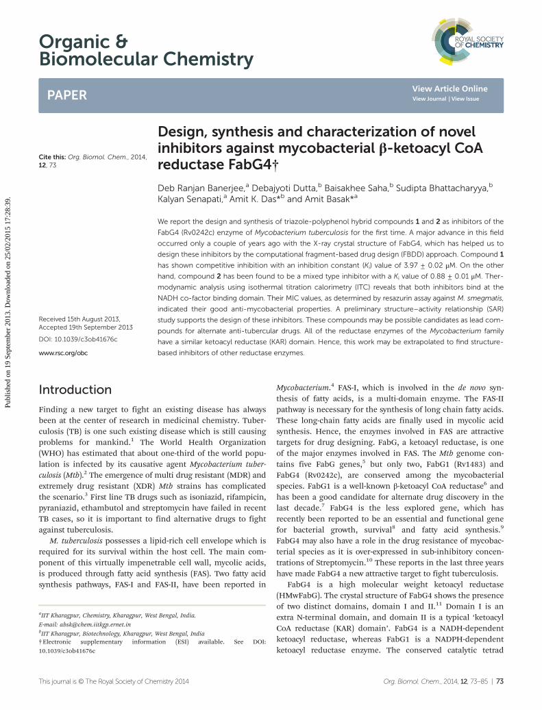
Ser347, Tyr360, Lys364 and Asn319 constitute the active siteresidues of the KAR domain. The active site is covered by loopI and loop II. The structure of FabG4 co-crystallized with hexa-noyl-CoA (HXC) and NAD+ (PDB ID: 3V1U) suggests that theactive site of FabG4 could be accessed from two differentsides: the major portal and minor portal.12 The major portal iswide open and is accessed by the cofactor to bind with theenzyme. The minor portal is narrower due to the helices of thedimeric interface and is accessed by the 4 phosphopan-tetheine-bound fatty acyl substrates. The active site is coveredfrom one side by two conserved loops, loop I and loop II. Theconserved NAG triad (Asn295, Ala296, Gly297) of loop I inter-acts with the pyrophosphate section of the substrate and guidesthe cofactor towards the active site. Asp244 and Val268 have acrucial role to hold NADH at the major portal by interactingwith the adenine section of the coenzyme. Asp244 and Arg223are the major factors behind NADH selectivity (Fig. 1).
Based on the information about the overall structure andspecificity of the FabG4 enzyme, we have attempted to designnovel structure-based inhibitors of FabG4. The synthesis of thedesigned compounds were carried out and their bio-activitieswere measured though biochemical assays. The drug suscepti-bilities of these inhibitors were tested against Mycobacterium
smegmatis. A preliminary structure activity relationship (SAR)has been carried out to justify the design of the inhibitors.
Results and discussionDesign strategy
The recently-solved X-ray crystal structure of FabG4 with co-factor NADH provides the way to design the first inhibitorsagainst this enzyme. We targeted the co-factor (NADH)-binding domain for design purposes. Inhibitors which targetthe NADH binding site can interact with the three subsites ofthe coenzyme binding domain: the nicotinamide-bindingsubsite (N-subsite), the adenosine-binding subsite (A-subsite)and the pyrophosphate-binding subsite (P-subsite). Forstructure-based design purposes, we followed the compu-tational fragment-based drug design (FBDD) strategy.13 In thisapproach, fragments and linkers were selected from a litera-ture survey and merged to form a library. The selection of thelead compounds was performed through computationaldocking studies. The advanced molecular grid-based dockingprogram Autodock4.214 was used for the docking of a flexibleligand within a flexible protein.
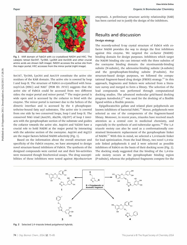
Epigallocatechin gallate and related plant polyphenols areknown inhibitors of bacterial FabG.15 Hence, polyphenols wereselected as one of the components of the fragment-basedlibrary. Moreover, in recent years, triazoles have received muchattention as a central core in medicinal chemistry, andespecially in the synthesis of anti-tubercular agents.16 The 1,4-triazole moiety can also be used as a conformationally con-strained bioisosteric replacement of the pyrophosphate linkerof NADH.17 With this in mind, we selected a 1,4-triazole linkerfor lead optimization. From the lead library, two novel 1,4-tria-zole linked polyphenols 1 and 2 were selected as possibleinhibitors of FabG4 on the basis of their docking score (Fig. 2).The docking study suggested that the binding of the 1,4-tria-zole moiety occurs at the pyrophosphate binding region(P-subsite), whereas the polyphenol fragments compete for the
Fig. 1 KAR domain of FabG4 with co-crystallized NADH and HXC. Thecatalytic tetrad (Ser347, Tyr360, Lys364 and Asn319) and other crucialamino acids are shown (left image). NADH accesses the active site fromthe major portal; HXC accesses from the minor portal (right image).
Fig. 2 Selected 1,4-triazole linked polyphenol hybrids.
Paper Organic & Biomolecular Chemistry
74 | Org. Biomol. Chem., 2014, 12, 73–85 This journal is © The Royal Society of Chemistry 2014
Publ
ishe
d on
19
Sept
embe
r 20
13. D
ownl
oade
d on
25/
02/2
015
17:2
8:39
. View Article Online
N- and A-subsites of the NADH binding region. The detailedinteractions from the docking studies are discussed later inthis report. To show the importance of fragments linked by the1,4-triazole moiety, two more hybrids, 3 and 4, were designedto draw preliminary structure–activity relationships.
Synthesis
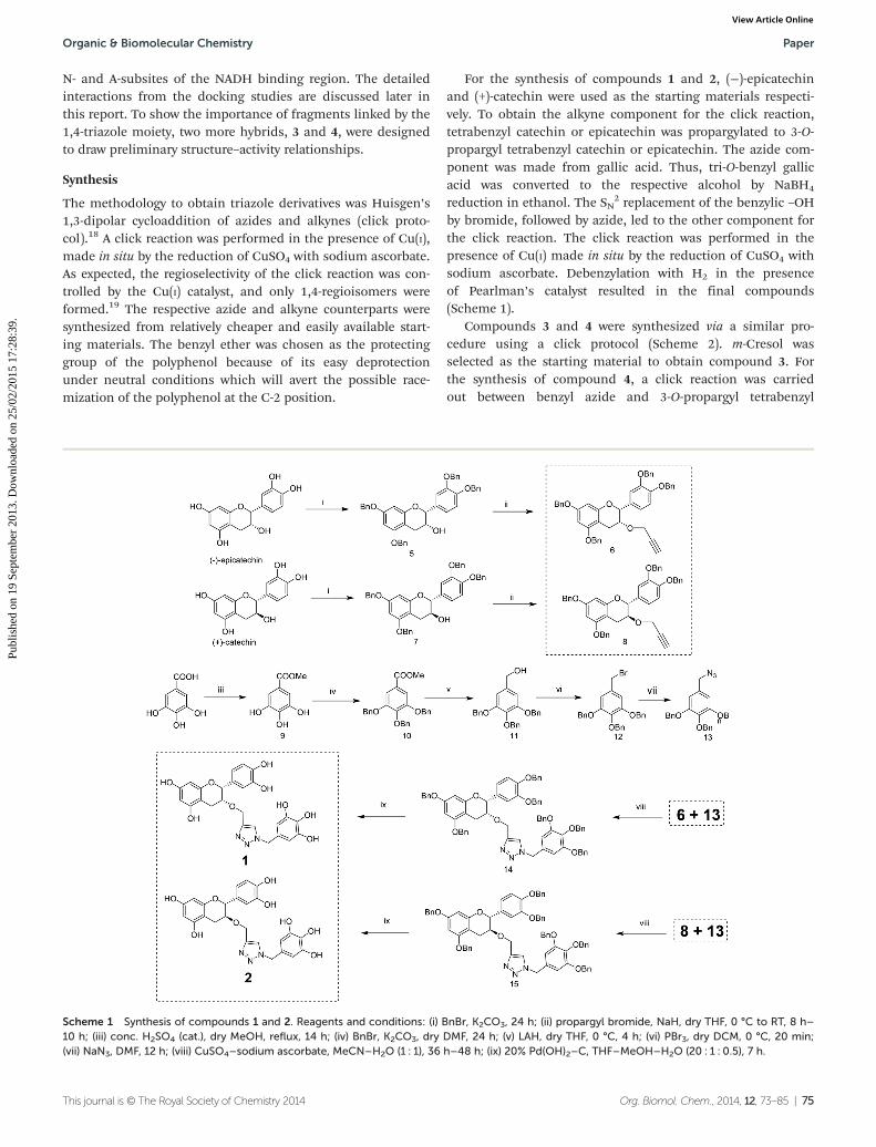
The methodology to obtain triazole derivatives was Huisgen’s1,3-dipolar cycloaddition of azides and alkynes (click proto-col).18 A click reaction was performed in the presence of Cu(I),made in situ by the reduction of CuSO4 with sodium ascorbate.As expected, the regioselectivity of the click reaction was con-trolled by the Cu(I) catalyst, and only 1,4-regioisomers wereformed.19 The respective azide and alkyne counterparts weresynthesized from relatively cheaper and easily available start-ing materials. The benzyl ether was chosen as the protectinggroup of the polyphenol because of its easy deprotectionunder neutral conditions which will avert the possible race-mization of the polyphenol at the C-2 position.
For the synthesis of compounds 1 and 2, (−)-epicatechinand (+)-catechin were used as the starting materials respecti-vely. To obtain the alkyne component for the click reaction,tetrabenzyl catechin or epicatechin was propargylated to 3-O-propargyl tetrabenzyl catechin or epicatechin. The azide com-ponent was made from gallic acid. Thus, tri-O-benzyl gallicacid was converted to the respective alcohol by NaBH4
reduction in ethanol. The SN2 replacement of the benzylic –OH
by bromide, followed by azide, led to the other component forthe click reaction. The click reaction was performed in thepresence of Cu(I) made in situ by the reduction of CuSO4 withsodium ascorbate. Debenzylation with H2 in the presenceof Pearlman’s catalyst resulted in the final compounds(Scheme 1).
Compounds 3 and 4 were synthesized via a similar pro-cedure using a click protocol (Scheme 2). m-Cresol wasselected as the starting material to obtain compound 3. Forthe synthesis of compound 4, a click reaction was carriedout between benzyl azide and 3-O-propargyl tetrabenzyl
Scheme 1 Synthesis of compounds 1 and 2. Reagents and conditions: (i) BnBr, K2CO3, 24 h; (ii) propargyl bromide, NaH, dry THF, 0 °C to RT, 8 h–10 h; (iii) conc. H2SO4 (cat.), dry MeOH, reflux, 14 h; (iv) BnBr, K2CO3, dry DMF, 24 h; (v) LAH, dry THF, 0 °C, 4 h; (vi) PBr3, dry DCM, 0 °C, 20 min;(vii) NaN3, DMF, 12 h; (viii) CuSO4–sodium ascorbate, MeCN–H2O (1 : 1), 36 h–48 h; (ix) 20% Pd(OH)2–C, THF–MeOH–H2O (20 : 1 : 0.5), 7 h.
Organic & Biomolecular Chemistry Paper
This journal is © The Royal Society of Chemistry 2014 Org. Biomol. Chem., 2014, 12, 73–85 | 75
Publ
ishe
d on
19
Sept
embe
r 20
13. D
ownl
oade
d on
25/
02/2
015
17:2
8:39
. View Article Online
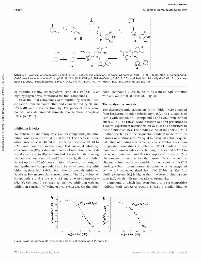
epicatechin. Finally, debenzylation using 20% Pd(OH)2–C athigh hydrogen pressure afforded the final compounds.
All of the final compounds were purified by repeated pre-cipitation from methanol–ether and characterized by 1H and13C NMR, and mass spectrometry. The purity of these com-pounds was determined through reverse-phase analyticalHPLC (see ESI†).
Inhibition kinetics
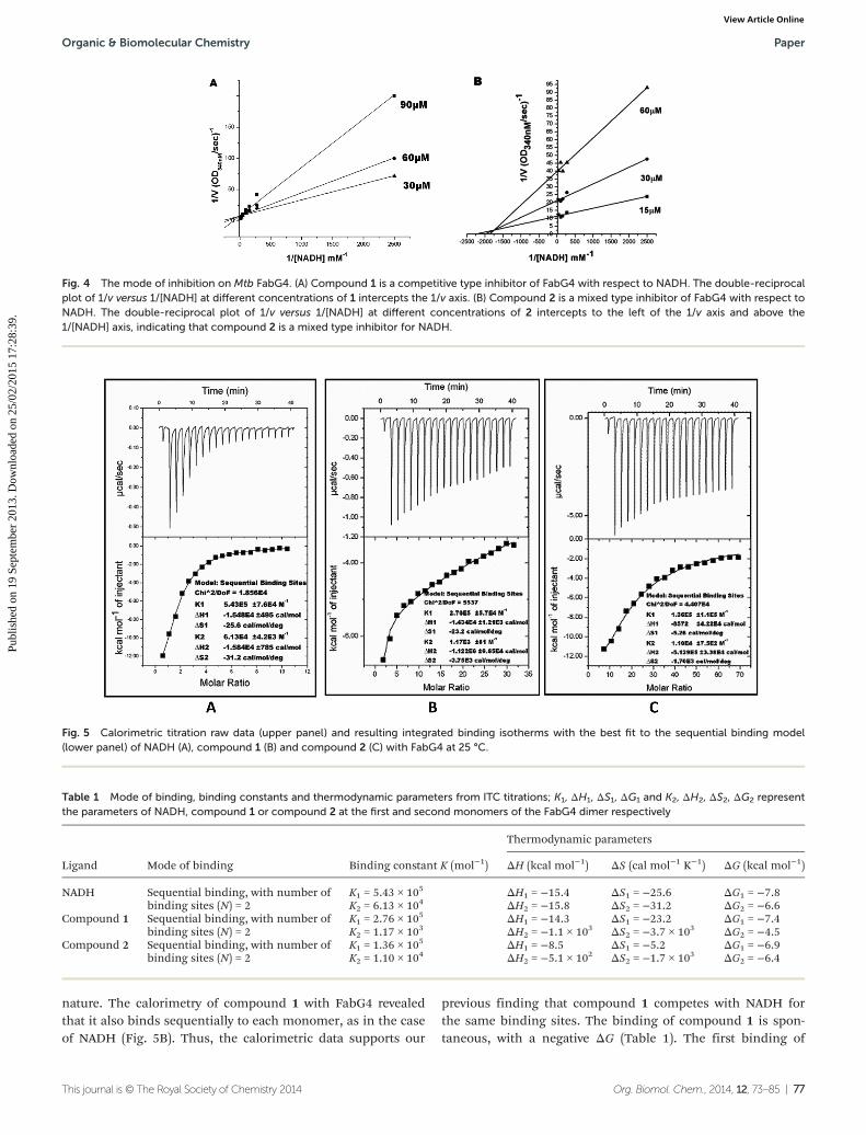
To evaluate the inhibitory effects of our compounds, the inhi-bition kinetics were carried out at 25 °C. The decrease in theabsorbance value at 340 nM due to the conversion of NADH toNAD+ was monitored in this assay. Half maximal inhibitoryconcentration (IC50) values and modes of inhibition were eval-uated kinetically. (−)-Epicatechin and (+)-catechin, the startingmaterials of compounds 1 and 2 respectively, did not inhibitFabG4 up to a 100 μM concentration. However, our designedand synthesized compounds 1 and 2 showed promising inhi-bition against Mtb FabG4. Both the compounds inhibitedFabG4 at low micromolar concentrations. The IC50 values ofcompounds 1 and 2 are 44.7 μM and 34.9 μM respectively(Fig. 3). Compound 1 showed competitive inhibition with aninhibition constant (Ki) value of 3.97 ± 0.02 μM. On the other
hand, compound 2 was found to be a mixed type inhibitorwith a Ki value of 0.88 ± 0.01 μM (Fig. 4).
Thermodynamic analysis
The thermodynamic parameters for inhibition were obtainedfrom isothermal titration calorimetry (ITC). The ITC studies ofFabG4 with compound 1, compound 2 and NADH were carriedout at 25 °C. The FabG4–NADH titration was first performed asa control experiment because NADH was used as a substrate inthe inhibition studies. The binding curve of the FabG4–NADHtitration nicely fits to the ‘sequential binding’ mode, with thenumber of binding sites (N) equal to 2 (Fig. 5A). This sequen-tial nature of binding is reasonable because FabG4 exists as aninseparable homo-dimer in solution. NADH binding to onemonomeric unit regulates the binding of a second NADH tothe second monomer, and this is co-operative in nature. Thisphenomenon is similar to other known FabGs where theoligomeric interface is responsible for cooperativity.20 NADHbinding to both the monomers is spontaneous, as suggestedby the ΔG values obtained from ITC (Table 1). The firstbinding constant (K1) is higher than the second binding con-stant (K2), which indicates negative co-operativity.
Compound 1, which has been found to be a competitiveinhibitor with respect to NADH, showed a similar binding
Scheme 2 Synthesis of compounds 3 and 4 for SAR. Reagents and conditions: (i) propargyl bromide, NaH, THF, 0 °C to RT, 48 h; (ii) compound 13,CuSO4–sodium ascorbate, MeCN–H2O (1 : 1), 24 h; (iii) Pd(OH)2–C, THF–MeOH–H2O (20 : 1 : 0.5), H2 (2 bar), 5 h; (iv) NaN3, dry DMF, 12 h; (v) com-pound 6, CuSO4–sodium ascorbate, MeCN–H2O, 4 h; (vi) Pd(OH)2–C, THF–MeOH–H2O (20 : 1 : 0.5), H2 (2.5 bar), 7 h.
Fig. 3 Dose–response plots to determine the IC50 of compounds 1 (A) and 2 (B).
Paper Organic & Biomolecular Chemistry
76 | Org. Biomol. Chem., 2014, 12, 73–85 This journal is © The Royal Society of Chemistry 2014
Publ
ishe
d on
19
Sept
embe
r 20
13. D
ownl
oade
d on
25/
02/2
015
17:2
8:39
. View Article Online
nature. The calorimetry of compound 1 with FabG4 revealedthat it also binds sequentially to each monomer, as in the caseof NADH (Fig. 5B). Thus, the calorimetric data supports our
previous finding that compound 1 competes with NADH forthe same binding sites. The binding of compound 1 is spon-taneous, with a negative ΔG (Table 1). The first binding of
Fig. 4 The mode of inhibition on Mtb FabG4. (A) Compound 1 is a competitive type inhibitor of FabG4 with respect to NADH. The double-reciprocalplot of 1/v versus 1/[NADH] at different concentrations of 1 intercepts the 1/v axis. (B) Compound 2 is a mixed type inhibitor of FabG4 with respect toNADH. The double-reciprocal plot of 1/v versus 1/[NADH] at different concentrations of 2 intercepts to the left of the 1/v axis and above the1/[NADH] axis, indicating that compound 2 is a mixed type inhibitor for NADH.
Fig. 5 Calorimetric titration raw data (upper panel) and resulting integrated binding isotherms with the best fit to the sequential binding model(lower panel) of NADH (A), compound 1 (B) and compound 2 (C) with FabG4 at 25 °C.
Table 1 Mode of binding, binding constants and thermodynamic parameters from ITC titrations; K1, ΔH1, ΔS1, ΔG1 and K2, ΔH2, ΔS2, ΔG2 representthe parameters of NADH, compound 1 or compound 2 at the first and second monomers of the FabG4 dimer respectively
Ligand Mode of binding Binding constant K (mol−1)
Thermodynamic parameters
ΔH (kcal mol−1) ΔS (cal mol−1 K−1) ΔG (kcal mol−1)
NADH Sequential binding, with number ofbinding sites (N) = 2
K1 = 5.43 × 10⁵ ΔH1 = −15.4 ΔS1 = −25.6 ΔG1 = −7.8K2 = 6.13 × 10⁴ ΔH2 = −15.8 ΔS2 = −31.2 ΔG2 = −6.6
Compound 1 Sequential binding, with number ofbinding sites (N) = 2
K1 = 2.76 × 10⁵ ΔH1 = −14.3 ΔS1 = −23.2 ΔG1 = −7.4K2 = 1.17 × 10³ ΔH2 = −1.1 × 10³ ΔS2 = −3.7 × 10³ ΔG2 = −4.5
Compound 2 Sequential binding, with number ofbinding sites (N) = 2
K1 = 1.36 × 10⁵ ΔH1 = −8.5 ΔS1 = −5.2 ΔG1 = −6.9K2 = 1.10 × 10⁴ ΔH2 = −5.1 × 10² ΔS2 = −1.7 × 10³ ΔG2 = −6.4
Organic & Biomolecular Chemistry Paper
This journal is © The Royal Society of Chemistry 2014 Org. Biomol. Chem., 2014, 12, 73–85 | 77
Publ
ishe
d on
19
Sept
embe
r 20
13. D
ownl
oade
d on
25/
02/2
015
17:2
8:39
. View Article Online
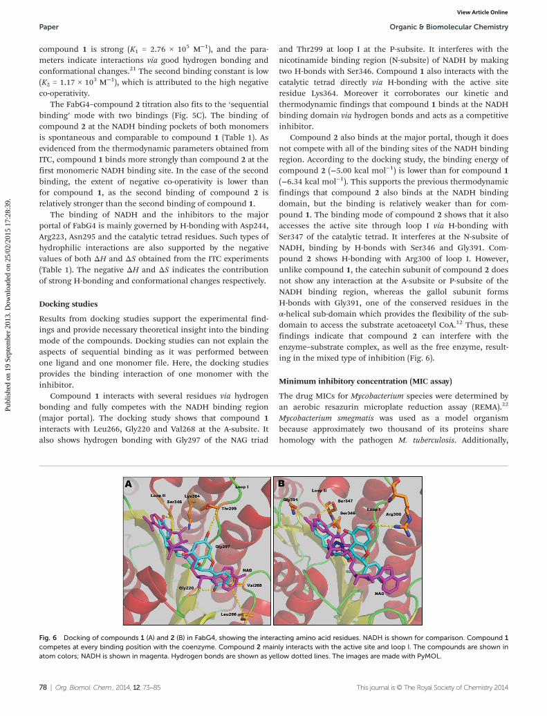
compound 1 is strong (K1 = 2.76 × 10⁵ M−1), and the para-meters indicate interactions via good hydrogen bonding andconformational changes.21 The second binding constant is low(K2 = 1.17 × 10³ M−1), which is attributed to the high negativeco-operativity.
The FabG4–compound 2 titration also fits to the ‘sequentialbinding’ mode with two bindings (Fig. 5C). The binding ofcompound 2 at the NADH binding pockets of both monomersis spontaneous and comparable to compound 1 (Table 1). Asevidenced from the thermodynamic parameters obtained fromITC, compound 1 binds more strongly than compound 2 at thefirst monomeric NADH binding site. In the case of the secondbinding, the extent of negative co-operativity is lower thanfor compound 1, as the second binding of compound 2 isrelatively stronger than the second binding of compound 1.
The binding of NADH and the inhibitors to the majorportal of FabG4 is mainly governed by H-bonding with Asp244,Arg223, Asn295 and the catalytic tetrad residues. Such types ofhydrophilic interactions are also supported by the negativevalues of both ΔH and ΔS obtained from the ITC experiments(Table 1). The negative ΔH and ΔS indicates the contributionof strong H-bonding and conformational changes respectively.
Docking studies
Results from docking studies support the experimental find-ings and provide necessary theoretical insight into the bindingmode of the compounds. Docking studies can not explain theaspects of sequential binding as it was performed betweenone ligand and one monomer file. Here, the docking studiesprovides the binding interaction of one monomer with theinhibitor.
Compound 1 interacts with several residues via hydrogenbonding and fully competes with the NADH binding region(major portal). The docking study shows that compound 1interacts with Leu266, Gly220 and Val268 at the A-subsite. Italso shows hydrogen bonding with Gly297 of the NAG triad
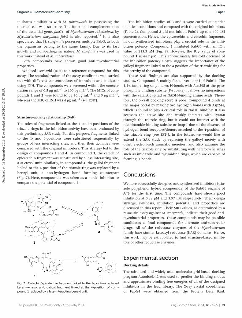
and Thr299 at loop I at the P-subsite. It interferes with thenicotinamide binding region (N-subsite) of NADH by makingtwo H-bonds with Ser346. Compound 1 also interacts with thecatalytic tetrad directly via H-bonding with the active siteresidue Lys364. Moreover it corroborates our kinetic andthermodynamic findings that compound 1 binds at the NADHbinding domain via hydrogen bonds and acts as a competitiveinhibitor.
Compound 2 also binds at the major portal, though it doesnot compete with all of the binding sites of the NADH bindingregion. According to the docking study, the binding energy ofcompound 2 (−5.00 kcal mol−1) is lower than for compound 1(−6.34 kcal mol−1). This supports the previous thermodynamicfindings that compound 2 also binds at the NADH bindingdomain, but the binding is relatively weaker than for com-pound 1. The binding mode of compound 2 shows that it alsoaccesses the active site through loop I via H-bonding withSer347 of the catalytic tetrad. It interferes at the N-subsite ofNADH, binding by H-bonds with Ser346 and Gly391. Com-pound 2 shows H-bonding with Arg300 of loop I. However,unlike compound 1, the catechin subunit of compound 2 doesnot show any interaction at the A-subsite or P-subsite of theNADH binding region, whereas the gallol subunit formsH-bonds with Gly391, one of the conserved residues in theα-helical sub-domain which provides the flexibility of the sub-domain to access the substrate acetoacetyl CoA.12 Thus, thesefindings indicate that compound 2 can interfere with theenzyme–substrate complex, as well as the free enzyme, result-ing in the mixed type of inhibition (Fig. 6).
Minimum inhibitory concentration (MIC assay)
The drug MICs for Mycobacterium species were determined byan aerobic resazurin microplate reduction assay (REMA).22
Mycobacterium smegmatis was used as a model organismbecause approximately two thousand of its proteins sharehomology with the pathogen M. tuberculosis. Additionally,
Fig. 6 Docking of compounds 1 (A) and 2 (B) in FabG4, showing the interacting amino acid residues. NADH is shown for comparison. Compound 1competes at every binding position with the coenzyme. Compound 2 mainly interacts with the active site and loop I. The compounds are shown inatom colors; NADH is shown in magenta. Hydrogen bonds are shown as yellow dotted lines. The images are made with PyMOL.
Paper Organic & Biomolecular Chemistry
78 | Org. Biomol. Chem., 2014, 12, 73–85 This journal is © The Royal Society of Chemistry 2014
Publ
ishe
d on
19
Sept
embe
r 20
13. D
ownl
oade
d on
25/
02/2
015
17:2
8:39
. View Article Online
it shares similarities with M. tuberculosis in possessing theunusual cell wall structure. The functional complementationof the essential gene, fabG1, of Mycobacterium tuberculosis byMycobacterium smegmatis fabG is also reported.23 It is alsospeculated that M. smegmatis possesses multiple FabG, as boththe organisms belong to the same family. Due to its fastgrowth and non-pathogenic nature, M. smegmatis was used inthis work instead of M. tuberculosis.
Both compounds have shown good anti-mycobacterialproperties.
We used isoniazid (INH) as a reference compound for thisassay. The standardization of the assay conditions was carriedout with different concentrations of inoculum and indicatorusing INH. The compounds were screened within the concen-tration range of 0.5 μg mL−1 to 100 μg mL−1. The MICs of com-pounds 1 and 2 were found to be 20 μg mL−1 and 5 μg mL−1,whereas the MIC of INH was 4 μg mL−1 (see ESI†).
Structure–activity relationship (SAR)
The roles of fragments linked at the 1- and 4-positions of thetriazole rings in the inhibition activity have been evaluated bythis preliminary SAR study. For this purpose, fragments linkedto the 1- and 4-positions were substituted sequentially bygroups of less interacting sites, and then their activities werecompared with the original inhibitors. This strategy led to thedesign of compounds 3 and 4. In compound 3, the catechin/epicatechin fragment was substituted by a less interacting site,a m-cresol unit. Similarly, in compound 4, the gallol fragmentlinked to the 4-position of the triazole ring was replaced by abenzyl unit, a non-hydrogen bond forming counterpart(Fig. 7). Here, compound 1 was taken as a model inhibitor tocompare the potential of compound 4.
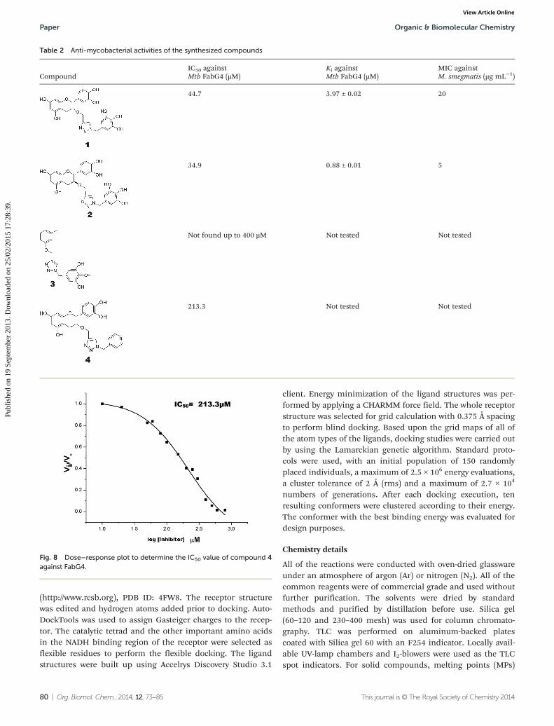
The inhibition studies of 3 and 4 were carried out underidentical conditions and compared with the original inhibitors(Table 2). Compound 3 did not inhibit FabG4 up to a 400 µMconcentration. Hence, the epicatechin and catechin fragmentsin our synthesized inhibitors play a crucial role in the inhi-bition potency. Compound 4 inhibited FabG4 with an IC50
value of 213.3 µM (Fig. 8). However, the IC50 value of com-pound 1 is 44.7 µM. This approximately five-fold decrease ofthe inhibition potency clearly suggests the importance of thegalloyl fragment linked to the 4-position of the triazole ring forthe activity of the compound.
These SAR findings are also supported by the dockingstudies. Compound 3 mainly floats over loop I of FabG4. The1,4-triazole ring only makes H-bonds with Asn295 at the pyro-phosphate binding subsite (P-subsite); it shows no interactionswith the catalytic tetrad or NADH-binding amino acids. There-fore, the overall docking score is poor. Compound 4 binds atthe major portal by making two hydrogen bonds with Asp244,which is found to play a crucial role in NADH binding. It alsoaccesses the active site and weakly interacts with Tyr360through the triazole ring, but it could not interact with thenicotinamide-binding subsite or loop I due to the absence ofhydrogen bond acceptors/donors attached to the 4-position ofthe triazole ring (see ESI†). In the future, we would like toextend the SAR study by replacing the galloyl moiety withother electron-rich aromatic moieties, and also examine therole of the triazole ring by substituting with heterocyclic ringssuch as imidazole and pyrimidine rings, which are capable offorming H-bonds.
Conclusions
We have successfully designed and synthesized inhibitors (tria-zole polyphenol hybrid compounds) of the FabG4 enzyme ofMtb for the first time. The compounds have shown goodinhibition at 0.88 μM and 3.97 μM respectively. Their designstrategy, synthesis, inhibition potential and properties arediscussed in this report. Their MIC values, as determined by aresazurin assay against M. smegmatis, indicate their good anti-mycobacterial properties. These compounds may be possiblecandidates as lead compounds for alternate anti-tuberculardrugs. All of the reductase enzymes of the Mycobacteriumfamily have similar ketoacyl reductase (KAR) domains. Hence,this work may be extrapolated to find structure-based inhibi-tors of other reductase enzymes.
Experimental sectionDocking details
The advanced and widely used molecular grid-based dockingprogram Autodock4.2 was used to predict the binding modesand approximate binding free energies of all of the designedinhibitors in the lead library. The X-ray crystal coordinatesof FabG4 were obtained from the Protein Data Bank
Fig. 7 Catechin/epicatechin fragment linked to the 1-position replacedby a m-cresol unit, galloyl fragment linked at the 4-position of com-pound 1 replaced by a less-interacting benzyl unit.
Organic & Biomolecular Chemistry Paper
This journal is © The Royal Society of Chemistry 2014 Org. Biomol. Chem., 2014, 12, 73–85 | 79
Publ
ishe
d on
19
Sept
embe
r 20
13. D
ownl
oade
d on
25/
02/2
015
17:2
8:39
. View Article Online
(http://www.rcsb.org), PDB ID: 4FW8. The receptor structurewas edited and hydrogen atoms added prior to docking. Auto-DockTools was used to assign Gasteiger charges to the recep-tor. The catalytic tetrad and the other important amino acidsin the NADH binding region of the receptor were selected asflexible residues to perform the flexible docking. The ligandstructures were built up using Accelrys Discovery Studio 3.1
client. Energy minimization of the ligand structures was per-formed by applying a CHARMM force field. The whole receptorstructure was selected for grid calculation with 0.375 Å spacingto perform blind docking. Based upon the grid maps of all ofthe atom types of the ligands, docking studies were carried outby using the Lamarckian genetic algorithm. Standard proto-cols were used, with an initial population of 150 randomlyplaced individuals, a maximum of 2.5 × 10⁶ energy evaluations,a cluster tolerance of 2 Å (rms) and a maximum of 2.7 × 10⁴
numbers of generations. After each docking execution, tenresulting conformers were clustered according to their energy.The conformer with the best binding energy was evaluated fordesign purposes.
Chemistry details
All of the reactions were conducted with oven-dried glasswareunder an atmosphere of argon (Ar) or nitrogen (N2). All of thecommon reagents were of commercial grade and used withoutfurther purification. The solvents were dried by standardmethods and purified by distillation before use. Silica gel(60–120 and 230–400 mesh) was used for column chromato-graphy. TLC was performed on aluminum-backed platescoated with Silica gel 60 with an F254 indicator. Locally avail-able UV-lamp chambers and I2-blowers were used as the TLCspot indicators. For solid compounds, melting points (MPs)
Table 2 Anti-mycobacterial activities of the synthesized compounds
CompoundIC50 againstMtb FabG4 (μM)
Ki againstMtb FabG4 (μM)
MIC againstM. smegmatis (μg mL−1)
44.7 3.97 ± 0.02 20
34.9 0.88 ± 0.01 5
Not found up to 400 μM Not tested Not tested
213.3 Not tested Not tested
Fig. 8 Dose–response plot to determine the IC50 value of compound 4against FabG4.
Paper Organic & Biomolecular Chemistry
80 | Org. Biomol. Chem., 2014, 12, 73–85 This journal is © The Royal Society of Chemistry 2014
Publ
ishe
d on
19
Sept
embe
r 20
13. D
ownl
oade
d on
25/
02/2
015
17:2
8:39
. View Article Online
were measured using melting point apparatus twice andreported without further calibration. The NMR spectra wererecorded on Bruker 200 MHz and 400 MHz machines. The fol-lowing abbreviations are used to describe the peak patternswhere appropriate: s = singlet, d = doublet, t = triplet, q =quartet, m = multiplet, dd = double doublet, ABq = AB quartet.
General procedure for the synthesis of compounds 6 and 8
In solutions of compounds 5 and 7 (1 eq.) in dry THF, sodiumhydride (4 eq.) was added at 0 °C and the mixture was stirredfor 30 minutes at 0 °C. Then propargyl bromide (2 eq.), dis-solved in dry THF, was added dropwise. The mixture wasstirred for 12 hours at room temperature, then quenched withNH4Cl solution, and extracted with EtOAc. The organic layerwas washed with brine and dried over anhydrous sodium sul-phate. Evaporation under vacuum gave an oil, from which thedesired compounds were isolated by column chromatography(Si-gel 60–120 mesh, PE–EA = 7 : 1 as the eluent).
(2R,3R)-5,7-Bis(benzyloxy)-2-(3,4-bis(benzyloxy)phenyl)-3-(prop-2-ynyloxy)chroman (6). Yield: 63%; state: white solid; mp:94 °C; δH (CDCl3, 400 MHz): 7.49–7.19 (21H, m, Ar–H),7.01–6.91 (2H, m, Ar–H), 6.29 (2H, s, H-6 & H-8), 5.19 (4 h, m,2 × CH2Ph), 5.04–4.97 (5H, m, H-2 & 2 × CH2Ph), 4.17 (1H, s,H-3), 3.99 (2H, m, OCH2), 3.04 (1H, m, H-4), 2.86 (1H, dd, J =17.2 Hz & 4 Hz), 2.24 (1H, broad s, –CCH).
(2R,3S)-5,7-Bis(benzyloxy)-2-(3,4-bis(benzyloxy)phenyl)-3-(prop-2-ynyloxy)chroman (8). Yield: 60%; state: off-white solid; mp:101 °C; δH (CDCl3, 400 MHz): 7.42–7.13 (20H, m, Ar–H), 7.00(1H, s, Ar–H), 6.87 (2H, broad s, Ar–H), 6.25 (1H, s, H-6/H-8),6.21 (1H, s, H-6/H-8), 5.17 (4H, m, 2 × CH2Ph), 4.99 (4H, m, 2 ×CH2Ph), 4.80 (1H, d, J = 7.2 Hz, H-2), 3.93 (1H, m, H-3), 3.74(2H, m, OCH2), 2.97 (1H, dd, J = 16.4 Hz & 5.2 Hz, H-4), 2.68(1H, dd, J = 16.4 Hz & 8 Hz, H-4), 2.32 (1H, broad s, CCH).
Methyl 3,4,5-tris(benzyloxy)benzoate (10). 1.6 g of com-pound 9 (8.7 mmol) was dissolved in dry DMF (30 mL). 9.6 ganhydrous K2CO3 (8 eq.) was added. 4.13 mL benzyl bromide(4 eq.) was added portion-wise under stirring. After 24 h, thereaction mixture was extracted with EtOAc (3 × 30 mL) andwashed with water. It was dried by sodium sulphate, concen-trated and subjected to column chromatography (Si-gel60–120 mesh, PE–EA = 10 : 1 as the eluent).
Yield: 2.9 g (75%); state: white solid; mp: 81 °C; δH (CDCl3,200 MHz): 7.45–7.26 (17H, m, Ar–H), 5.17 (6H, s, 3 × OCH2Ph),3.92 (3H, s, CH3); δC (CDCl3, 50 MHz): 166.5, 152.5, 137.4,136.6, 128.4, 128.1, 127.9, 127.9, 127.4, 125.1, 108.9, 75.0, 71.1,52.1.
(3,4,5-Tris(benzyloxy)phenyl)methanol (11). Compound 10(800 mg, 1.76 mmol) was dissolved in dry THF (25 mL) andcooled to 0 °C. Lithium aluminium hydride (80 mg, 1.2 eq.)was added very slowly under inert conditions. After 4 h, thereaction mixture was diluted with EtOAc and an acid workupwas carried out under cold conditions. The compound wasextracted with EtOAc (3 × 25 mL), washed with water and puri-fied by column chromatography (Si-gel 60–120 mesh, PE–EA =7 : 1 as the eluent).
Yield: 600.5 mg (80%); state: white solid; mp: 90 °C; δH(CDCl3, 400 MHz): 7.44–7.27 (15H, m, Ar–H), 6.67 (2H, s,Ar–H), 5.10 (4H, s, 2 × –CH2Ph), 5.06 (2H, s, –CH2Ph), 4.56(2H, s, –CH2OH); δC (CDCl3, 50 MHz): 152.8, 137.7, 137.5,137.0, 136.6, 128.5, 128.4, 128.2, 128.1, 127.8, 127.7, 127.3,106.2, 75.1, 65.2.
1,2,3-Tris(benzyloxy)-5-(bromomethyl)benzene (12). Com-pound 11 (511 mg, 1.2 mmol) was dissolved in dry CH2Cl2(20 mL) and stirred in ice. Then PBr3 (1.5 eq.) solution in dryDCM was added. The reaction mixture was stirred for 20 min,and was then carefully quenched with NaHCO3 solution andextracted with CH2Cl2 (3 × 30 mL). The CH2Cl2 layer waswashed with water 3–4 times, dried over sodium sulphate, fil-tered and concentrated under vacuum. The pure compoundwas isolated as a gummy liquid by flash column chromato-graphy (Si-gel 60–120 mesh, PE–EA = 20 : 1 as the eluent).
Yield: 499 mg (85%); state: yellow liquid; δH (CDCl3,200 MHz): 7.40–7.25 (15H, m, Ar–H), 6.69 (2H, s, Ar–H), 5.09(4H, s, 2 × CH2Ph), 5.03 (2H, s, CH2Ph), 4.39 (2H, s, CH2Br); δC(CDCl3, 50 MHz): 152.8, 137.7, 136.8, 133.1, 128.5, 128.1,127.9, 127.8, 127.4, 108.7, 75.2, 71.2, 34.2.
5-(Azidomethyl)-1,2,3-tris(benzyloxy)benzene (13). 300 mgof sodium azide (5 eq.) was added to 450 mg of compound 12(0.92 mmol) in 30 mL dry DMF, and stirred at room tempera-ture for 12 h. Extraction was carried out with EtOAc (2 ×25 mL), and the resultant solvent was washed by water, driedby sodium sulphate and subjected to column chromatography(Si-gel 230–400 mesh, PE–EA = 15 : 1 as the eluent).
Yield: 373.8 mg (90%); state: light yellow liquid; δH (CDCl3,200 MHz): 7.46–7.30 (15H, m, Ar–H), 6.66 (2H, s, Ar–H), 5.17(4H, s, 2 × CH2Ph), 5.11 (2H, s, CH2Ph), 4.28 (2H, s, –CH2N3);δC: 153.2, 137.9, 137.0, 131.1, 128.7, 128.3, 128.1, 128.0, 127.6,108.0, 75.3, 71.4, 55.1.
1-Methyl-3-(prop-2-ynyloxy)benzene (16). 500 mg m-cresol(9.3 mmol) was dissolved in 25 mL dry THF and cooled to0 °C. 500 mg sodium hydride (4 eq.) was added under stirringand left for 20 min for anion generation. The reaction wasagain cooled to 0 °C, and 1.1 mL of propargyl bromide (2 eq.)was added. Then the reaction was stirred at room temperatureand monitored by TLC. After 48 h, the reaction was almostcompleted. Then the excess sodium hydride was quenched byadding saturated NH4Cl solution, extracted with EtOAc (3 ×40 mL) and purified through column chromatography (Si-gel60–120 mesh, PE–EA = 10 : 1 as the eluent).
Yield: 1.1 g (78%); state: reddish liquid; δH (CDCl3,200 MHz): 7.19 (1H, m, Ar–H), 6.81 (3H, m, Ar–H), 4.64 (2H, d,J = 2.4 Hz, OCH2CCH), 2.49 (1H, t, J = 2.4 Hz, CCH), 2.35 (3H,s, CH3); δC (CDCl3, 50 MHz): 157.7, 139.6, 129.3, 122.5, 115.9,111.8, 78.9, 75.5, 55.7, 21.6.
General procedure for Huisgen’s 1,3-dipolar cycloaddition
Compounds 14, 15, 17 and 19 were synthesized using this pro-cedure. The respective alkyne and azide counterparts were dis-solved in acetonitrile and degasified for 15 min. Coppersulphate (1 eq.) was dissolved in an equal volume of water anddegassed for 30 min. Then sodium ascorbate (2 eq.) was
Organic & Biomolecular Chemistry Paper
This journal is © The Royal Society of Chemistry 2014 Org. Biomol. Chem., 2014, 12, 73–85 | 81
Publ
ishe
d on
19
Sept
embe
r 20
13. D
ownl
oade
d on
25/
02/2
015
17:2
8:39
. View Article Online
added, and the solution was stirred for 15 min. Then thedegassed acetonitrile solution was slowly added. The mixturewas stirred at room temperature until TLC indicated the dis-appearance of the starting materials. After that, the mixturewas poured into saturated aqueous NH4Cl solution and theproduct was extracted four times with EtOAc. The organic layerwas dried with sodium sulphate and filtered, and the solventwas removed under reduced pressure. The residue was purifiedby flash chromatography.
4-(((2R,3R)-5,7-Bis(benzyloxy)-2-(3,4-bis(benzyloxy)phenyl)-chroman-3-yloxy)methyl)-1-(3,4,5-tris(benzyloxy)benzyl)-1H-1,2,3-triazole (14). Yield: 70%, state: off-white solid; mp:103 °C; δH (CDCl3, 400 MHz): 7.46–7.24 (35H, m, Ar–H), 7.06(1H, s, Ar–H/olefinic H), 6.87 (2H, s, Ar–H), 6.48 (1H, s, Ar–H/olefinic H), 6.41 (2H, s, Ar–H), 6.28 (2H, s, H-6 & H-8),5.29–4.89 (17H, m, 7 × CH2Ph & NCH2 & H-2), 4.52 (2H, m,–OCH2), 3.99 (1H, broad s, H-3), 3.08 (1H, m, H-4), 2.83 (1H,dd, J = 17.2 Hz & 4 Hz, H-4); δC (CDCl3, 100 MHz): 158.7, 158.1,155.4, 153.0, 148.7, 148.1, 145.5, 137.6, 137.1, 137.1, 137.0,136.9, 136.7, 132.7, 130.2, 128.6, 128.6, 128.5, 128.4, 128.1,127.9, 127.9, 127.8, 127.5, 127.5, 127.4, 127.1, 122.5, 119.6,115.1, 113.4, 107.4, 101.4, 94.8, 93.5, 77.9, 75.1, 72.0, 71.7,71.0, 71.0, 70.1, 69.9, 62.6, 53.8, 24.7; HRMS (ES+): calcd forC74H65N3O9 + H+ 1140.4794, found 1140.4791.
4-(((2R,3S)-5,7-Bis(benzyloxy)-2-(3,4-bis(benzyloxy)phenyl)-chroman-3-yloxy)methyl)-1-(3,4,5-tris(benzyloxy)benzyl)-1H-1,2,3-triazole (15). Yield: 65%, state: off-white solid; mp:107 °C; δH (CDCl3, 400 MHz): 7.41–7.23 (35H, m, Ar–H), 6.96(1H, s, Ar–H/olefinic H), 6.88 (2H, s, Ar–H), 6.80 (1H, s, Ar–H/olefinic H), 6.46 (2H, s, Ar–H), 6.26 (2H, d, J = 2 Hz, H-6/H-8),6.21 (1H, d, J = 2 Hz, H-6/H-8), 5.32–4.98 (16H, m, 7 × CH2Ph &NCH2), 4.78 (1H, d, J = 7.6 Hz, H-2), 4.43 (2H, m, OCH2), 3.86(1H, m, H-3), 3.07 (1H, dd, J = 16.4 Hz & 5.2 Hz, H-4), 2.70 (1H,dd, J = 16.4 Hz & 8.4 Hz, H-4); δC (CDCl3, 100 MHz): 158.7,157.6, 155.0, 153.0, 148.7, 148.5, 145.4, 138.4, 137.5, 137.0,136.9, 136.8, 136.8, 136.6, 132.4, 130.0, 128.5, 128.5, 128.5,128.4, 128.1, 127.9, 127.9, 127.8, 127.5, 127.4, 127.4, 127.3,127.2, 122.4, 120.3, 114.9, 113.3, 107.5, 101.9, 94.1, 93.6, 79.5,75.1, 74.5, 71.4, 71.0, 70.9, 70.0, 69.8, 62.9, 53.9, 25.3. HRMS(ES+): calcd for C74H65N3O9 + H+ 1140.4794, found 1140.4861.
4-(m-Tolyloxymethyl)-1-(3,4,5-tris(benzyloxy)benzyl)-1H-1,2,3-triazole (17). Yield: 75%; state: yellow floppy solid; mp:132 °C; δH (CDCl3, 400 MHz): 7.34–7.16 (16H, m, Ar–H), 6.70(3H, m, Ar–H), 6.44 (2H, s, Ar–H), 5.27 (2H, s, NCH2), 5.07 (2H,s, OCH2), 4.96 (6H, s, 3 × CH2Ph), 2.23 (3H, s, CH3); δC (CDCl3,100 MHz): 158.1, 153.1, 144.6, 139.6, 138.5, 137.5, 136.6, 129.7,129.2, 128.5, 128.5, 128.1, 127.9, 127.9, 127.4, 122.4, 122.0,115.5, 111.4, 107.7, 75.1, 71.1, 61.8, 54.2, 21.5. HRMS (ES+):calcd for C38H35N3O4 + H+ 598.2700, found 598.2696.
1-Benzyl-4-(((2R,3R)-5,7-bis(benzyloxy)-2-(3,4-bis(benzyloxy)-phenyl)chroman-3-yloxy)methyl)-1H-1,2,3-triazole (19). Yield:70%, state: white solid; mp: 97 °C; δH (CDCl3, 400 MHz):7.48–7.26 (23H, m, Ar–H), 7.17–7.11 (3H, m, Ar–H), 6.89 (2H, s,Ar–H), 6.59 (1H, s, olefinic H), 6.30 (2H, s, H-6 & H-8),5.37–5.02 (10H, m, 4 × CH2Ph & NCH2), 4.90 (1H, s, H-2), 4.53(2H, m, OCH2), 3.99 (1H, s, H-3), 3.08 (1H, m, H-4), 2.81 (1H,
dd, J = 17.2 Hz & 3.6 Hz, H-4); δC (CDCl3, 100 MHz): 158.6,158.0, 155.4, 148.5, 148.1, 145.6, 137.1, 137.1, 137.0, 136.9,134.6, 132.4, 128.9, 128.6, 128.5, 128.5, 128.5, 128.4, 128.4,127.9, 127.8, 127.8, 127.8, 127.5, 127.4, 127.3, 127.2, 127.1,122.5, 119.6, 114.5, 113.5, 101.3, 94.7, 93.8, 77.9, 72.1, 71.3,70.9, 70.0, 69.8, 62.6, 53.7, 24.6. HRMS (ES+): calcd forC53H47N3O6 + H+ 822.3538, found 822.3494.
General procedure of debenzylation
Debenzylation was carried out in a parr apparatus. Solutionsof compounds 14, 15, 17 and 19 in a mixture of THF–MeOH–
H2O (20 : 1 : 0.5) were hydrogenated at 2–3 bar H2 pressure over20% Pd (OH)2–C for 5–7 h at room temperature. The reactionmixtures were filtered through celite using 1 : 1 ethyl acetate–methanol and concentrated in vacuo. The gummy liquid wasdissolved in a few drops of methanol and precipitation wascarried out by adding diethyl ether. The compounds were puri-fied by repeated precipitation. The pure compounds appearedas yellow gummy liquids in the absence of ether. All of the sol-vents were distilled prior to use and inert conditions weremaintained for most of the time to avoid oxidation.
5-((4-(((2R,3R)-2-(3,4-Dihydroxyphenyl)-5,7-dihydroxychroman-3-yloxy)methyl)-1H-1,2,3-triazol-1-yl)methyl)benzene-1,2,3-triol(1). Yield: 43%; state: yellow gummy liquid; δH (acetone-d6,400 MHz): 7.34 (1H, s, olefinic H, Ar–H), 7.11 (1H, s, olefinicH, Ar–H), 6.88 (2H, d, J = 1.6 Hz, Ar–H), 6.46 (2H, d, J = 4.4 Hz,Ar–H), 6.11 (1H, d, J = 2 Hz, H-6/H-8), 6.00 (1H, d, J = 2 Hz,H-6/H-8), 5.33 (2H, ABq, J = 14.6 Hz, –NCH2), 5.02 (1H, s, H-2),4.57 (2H, ABq, J = 12.8 Hz, –OCH2), 4.10 (1H, merged withethyl acetate peak, H-3), 2.95 (1H, dd, J = 16.8 Hz, 3.2 Hz, H-4),2.85 (1H, dd, J = 16.8 Hz, 4.2 Hz, H-4); δC (acetone-d6,100 MHz): 156.7, 156.6, 156.3, 145.9, 145.2, 144.5, 144.4, 133.0,131.3, 126.7, 122.7, 118.6, 114.6, 114.5, 107.3, 98.8, 95.4, 94.8,77.5, 73.4, 62.8, 53.3, 24.4; DEPT-135 (acetone-d6, 100 MHz):122.7, 118.5, 114.6, 114.4, 107.2, 95.3, 94.8, 77.4, 73.3, 62.7,53.3, 24.3. LCMS (ES−): calcd for C25H23N3O9 509.1434, found508.0723.
5-((4-(((2R,3S)-2-(3,4-Dihydroxyphenyl)-5,7-dihydroxychroman-3-yloxy)methyl)-1H-1,2,3-triazol-1-yl)methyl)benzene-1,2,3-triol(2). Yield: 40%; state: yellow gummy liquid; δH (acetone-d6,400 MHz): 7.47 (1H, s, olefinic H), 6.86–6.80 (2H, m, Ar–H),6.69 (1H, m, Ar–H), 6.38 (2H, s, Ar–H), 6.01 (1H, d, J = 2 Hz,H-6/H-8), 5.88 (1H, d, J = 2.4 Hz, H-6/H-8), 5.29 (2H, ABq, J =14.4 Hz, –NCH2), 4.76 (1H, d, J = 7.2 Hz, H-2), 4.47 (2H, ABq,J = 12.4 Hz, –OCH2), 3.87 (1H, q, J = 7.2 Hz), 2.82 (1H, dd, J =16 Hz & 5.2 Hz, H-4), 2.55 (1H, dd, J = 16 Hz & 7.2 Hz, H-4); δC(acetone-d6, 100 MHz): 157.8, 157.2, 156.7, 146.8, 145.7, 133.8,132.4, 127.8, 123.6, 119.6, 115.9, 115.0, 108.2, 108.1, 100.1,96.2, 95.4, 80.1, 75.5, 63.5, 54.2, 25.5; DEPT-135 (acetone-d6,100 MHz): 122.6, 118.6, 114.9, 114.0, 107.2, 95.2, 94.5, 79.1,74.5, 62.5, 53.2, 24.5; LCMS (ES−): calcd for C25H23N3O9
509.1434, found 508.0962.5-((4-(m-Tolyloxymethyl)-1H-1,2,3-triazol-1-yl)methyl)benzene-
1,2,3-triol (3). Yield: 55%; state: deep yellow gummy liquid; δH(acetone-d6, 400 MHz): 7.95 (1H, s, olefinic H), 7.13 (1H, t, J =7.8 Hz, Ar–H), 6.83–6.73 (3H, m, Ar–H), 6.41 (2H, s, Ar–H), 5.38
Paper Organic & Biomolecular Chemistry
82 | Org. Biomol. Chem., 2014, 12, 73–85 This journal is © The Royal Society of Chemistry 2014
Publ
ishe
d on
19
Sept
embe
r 20
13. D
ownl
oade
d on
25/
02/2
015
17:2
8:39
. View Article Online
(2H, s, –NCH2), 5.10 (2H, s, –OCH2), 2.27 (3H, s, –CH3); δC(acetone-d6, 100 MHz): 158.8, 146.0, 143.9, 139.3, 133.0, 129.2,126.9, 123.5, 121.7, 115.5, 111.7, 107.4, 61.5, 53.4, 20.7. HRMS(ES+): calcd for C17H17N3O4 + H+ 328.1292, found 328.1256.
(2R,3R)-3-((1-Benzyl-1H-1,2,3-triazol-4-yl)methoxy)-2-(3,4-dihydroxyphenyl)chroman-5,7-diol (4). Yield: 53%; state:yellow gummy liquid; δH (acetone-d6, 400 MHz): 7.35–7.29 (6H,m, Ar–H), 7.04 (1H, s, olefinic H), 6.78 (2H, s, Ar–H), 6.01 (1H,s, Ar–H), 5.89 (1H, s, Ar–H), 5.49 (2H, ABq, J = 14.8 Hz,–NCH2), 4.94 (1H, s, H-2), 4.49 (2H, ABq, J = 12.8 Hz, –OCH2),4.01 (1H, s, H-3), 2.86–2.72 (2H, merged with water peak, 2 ×H-4); δC (acetone-d6, 100 MHz): 156.8, 156.7, 156.4, 145.6,144.5, 136.2, 131.4, 128.9, 128.2, 128.1, 122.9, 118.7, 114.6,114.6, 98.9, 95.3, 94.8, 77.6, 73.3, 62.9, 53.3, 24.4. HRMS (ES+):calcd for C25H23N3O6 + H+ 462.1660, found 462.1671.
FabG4 inhibition assay
The inhibition studies were carried out in vitro at 25 °C bymonitoring the decrease of OD340 in the kinetic mode as afunction of conversion of NADH to NAD+ using an Evolution™300 UV-visible spectrophotometer (Thermo Fisher scientific).Protein purification was carried out as described elsewhere.24
Briefly, the recombinant protein was over-expressed in E. coliM15 cells using IPTG induction. Cells were harvested andlysed in a re-suspension buffer using ultra-sonication. Thelysate was centrifuged at 14 000 rpm for 40 min to discard celldebris. The supernatant was loaded onto a Ni-NTA columnand eluted with 300 mM imidazole solution. The elutedprotein was further purified by gel filtration chromatographyusing a Superdex200 column. The elutant was pooled and con-centrated, and extensively dialyzed against HEPES buffer(50 mM, pH 7.4) containing 50 mM NaCl prior to the experi-ments. The inhibitor compounds were dissolved in HEPESbuffer (50 mM, pH 7.4) with 3% methanol (HPLC grade) toprepare a stock solution of 1 mM, which was further dilutedrepeatedly by HEPES buffer to achieve the desired concen-trations. The reaction mixture contained the inhibitor solutionin HEPES buffer, 0.5 mM acetoacetyl-CoA, 0.2 mM β-NADHand 1 μM of FabG4 protein in the final volume of 500 μL. Amixture of FabG4, NADH and acetoacetyl CoA was used as apositive control when no inhibitor was added. The reactionwas initiated by the addition of acetoacetyl-CoA. The decreasein absorbance was recorded in 5 min intervals. The IC50 valueswere determined by varying the inhibitor concentrations untilfull inhibition occurred. To determine the mode of inhibition,we screened the enzyme activity with varying NADH concen-tration at three different concentrations of inhibitor. All of theexperiments were repeated thrice. The IC50 values were calcu-lated graphically from the dose–response plots. The modes ofinhibition were determined from Lineweaver–Burk plots. Theinhibition constants were determined from secondary plots.
ITC experiments
Isothermal titration calorimetry (ITC) was carried out using aMicrocal ITC200 instrument. Titrations of FabG4 enzyme withcompound 1, compound 2 and NADH were performed at
25 °C. A reference power of 5 µcal s−1, stirring speed of800 rpm and 120 s spacing were selected. The protein solutionwas prepared in 50 mM HEPES buffer at pH 7.4. Protein solu-tions of 10 μM, 4.3 μM and 3 μM concentrations were recordedin the sample cell for the NADH (500 μM), compound 1(1 mM) and compound 2 (1 mM) titrations respectively. Theligand solutions were prepared in the same buffer with 3%HPLC grade methanol. This extra methanol was also added tothe sample cell to avoid large heat changes due to solvent mis-match (Getting Started, MicroCal ITC200). Separate buffer–ligand titrations were carried out as reference runs for eachcompound by recording the buffer in the sample cell. Thisreference value was subtracted from the protein–ligand titra-tion to nullify the heat of dissolution. One injection of 0.4 μL,followed by nineteen injections of 2 μL of the ligand solutions,were titrated into the FabG4 solution. All of the experimentswere repeated twice and the data were solved using MicroCal,LLC ITC 200 software. All of the parameters, such as tolerancelimit (zero), derivative delta (0.08) and weighting method (noweighting), were default values. The data points of the firstinjections (0.4 μL) were neglected. The heat changes of the 19injections (for the NADH and compound 1 titrations) and18 injections (for the compound 2 titration) were plottedagainst the molar ratio of ligand.
MIC study
The Mycobacterium smegmatis mc2155 strain was grown inMiddlebrook 7H9 broth with 0.2% glycerol and 0.05% Tween80 for 20 hours at 37 °C with shaking at 120 rpm, until thecells reached the mid-logarithmic phase (OD595 ∼ 0.5). Theculture was diluted to OD595 = 0.0005 prior to addition into asterile 96-well microtitre plate. Isoniazid (INH) was purchasedfrom Aldrich and used as a reference compound for this assay.An INH stock solution of 1 mg mL−1 was prepared in distilledwater and serially diluted in MB 7H9 media. 0.5 mg mL−1
stock solutions of compound 1 and 2 (in 50 mM HEPESbuffer, pH 7.4, 3% HPLC grade methanol) were subjected toserial two fold dilutions in MB 7H9 media to make the desiredconcentrations. Resazurin sodium salt was purchased fromSisco Research Laboratories, India. 0.5% (w/v) stock solutionof resazurin was prepared in distilled water, filter-sterilizedand diluted to 0.02% in distilled water.
One hundred microlitres of diluted culture (OD595 = 0.0005)was added to each well of the microtitre plate. The totalvolume was two hundred microlitres. The inoculum wasomitted from the negative control (Row A), and the inhibitorswere omitted from the growth control (Row B). INH, the refer-ence compound, was used as a positive control. After 24 hoursof post-drug incubation at 37 °C, 60 μL of 0.02% resazurinsolution was added to each well and incubated again for40 min at 37 °C. A color change from blue (resazurin) to pink(resorufin) indicated the growth of bacteria. The lowest con-centrations of drugs which prevented such a color change wererecorded as the MIC values. The assay was repeated twice andan average of both the experiments was calculated to find theMIC values.
Organic & Biomolecular Chemistry Paper
This journal is © The Royal Society of Chemistry 2014 Org. Biomol. Chem., 2014, 12, 73–85 | 83
Publ
ishe
d on
19
Sept
embe
r 20
13. D
ownl
oade
d on
25/
02/2
015
17:2
8:39
. View Article Online
Acknowledgements
DRB thanks the Council of Scientific and Industrial Research(CSIR) for the fellowship. Central Research Facility and Depart-ment of Chemistry, IIT Kharagpur are thanked for providingall of the instrumental facilities. AB and AKD gratefullyacknowledge the support of DBT for providing the necessaryresearch grant. AB is also grateful to DST for the JC BoseNational Fellowship. BS and KS are thankful to DST for fast-track young scientist award.
References
1 (a) B. H. Herzog, Respiration, 1998, 65, 5; (b) R. P. Tripathi,N. Tewari, N. Dwivedi and V. K. Tiwari, Med. Res. Rev., 2005,25, 93; (c) R. Vohra, M. Gupta, R. Chaturvedi and Y. Singh,Recent Pat. Anti-Infect. Drug Discovery, 2006, 1, 95;(d) Y. L. Janin, Bioorg. Med. Chem., 2007, 15, 2479.
2 (a) World Health Organization, World Health Forum, 1993,14, 1; (b) World Health Organization, Global tuberculosiscontrol, 2012, WHO/HTM/TB/2012.6.
3 (a) S. H. E. Kaufmann and E. Rubin, Handbook of Tuber-culosis: Clinics, Diagnostics, Therapy and Epidemiology,Wiley-VCH, 2008, pp. XXIII–XXVII; (b) M. A. Espinal, Tuber-culosis, 2003, 83, 44; (c) C. Lienhardt, M. Raviglione,M. Spigelman, R. Hafner, E. Jaramillo, M. Hoelscher,A. Zumla and J. Gheuens, J. Infect. Dis., 2012, 205, S241;(d) P. Bemer-Melchior, A. Bryskier and H. B. Drugeon,J. Antimicrob. Chemother., 2000, 46, 571; (e) A. Jain andR. Mondal, FEMS Immunol. Med. Microbiol., 2008, 53, 145;(f ) A. S. Fauci, J. Infect. Dis., 2008, 197, 1493–1498;(g) G. Maartens and R. J. Wilkinson, Lancet, 2007, 370,2030; (h) C. E. Barry 3rd and J. S. Blanchard, Curr. Opin.Chem. Biol., 2010, 14, 456.
4 K. Takayama, C. Wang and G. S. Besra, Clin. Microbiol. Rev.,2005, 18, 81.
5 S. T. Cole, R. Brosch, J. Parkhill, T. Garnier, C. Churcher,D. Harris, S. V. Gordon, K. Eiglmeier, S. Gas, C. E. Barry3rd, F. Tekaia, K. Badcock, D. Basham, D. Brown,T. Chillingworth, R. Connor, R. Davies, K. Devlin,T. Feltwell, S. Gentles, N. Hamlin, S. Holroyd, T. Hornsby,K. Jagels, A. Krogh, J. McLean, S. Moule, L. Murphy,K. Oliver, J. Osborne, M. A. Quail, M. A. Rajandream,J. Rogers, S. Rutter, K. Seeger, J. Skelton, R. Squares,S. Squares, J. E. Sulston, K. Taylor, S. Whitehead andB. G. Barrell, Nature, 1998, 393, 537.
6 (a) H. Marrakchi, S. Ducasse, G. Labesse, E. Margeat,H. Montrozier, L. Emorine, X. Charpentier, G. Laneelle andA. Quemard, Microbiology, 2002, 148, 951; (b) M. Cohen-Gonsaud, S. Ducasse, F. Hoh, D. Zerbib, G. Labesse andA. Quemard, J. Mol. Biol., 2002, 320, 249.
7 (a) S. Ducasse-Cabanot, M. Cohen-Gonsaud, H. Marrakchi,M. Nguyen, D. Zerbib, J. Bernadou, M. Daffe, G. Labesseand A. Quemard, Antimicrob. Agents Chemother., 2004, 48,242; (b) A. R. Rendina and D. Cheng, Biochem. J., 2005, 388,
895; (c) M. J. Vazquez, W. Leavens, R. Liu, B. Rodriguez,M. Read, S. Richards, D. Winegar and J. M. Dominguez,FEBS J., 2008, 275, 1556; (d) K. Kristan, T. Bratkovic,M. Sova, S. Gobec, A. Prezelj and U. Urleb, Chem.-Biol. Inter-act., 2009, 178, 310.
8 D. J. Beste, M. Espasa, B. Bonde, A. M. Kierzek,G. R. Stewart and J. McFadden, PLoS One, 2009, 4,e5349.
9 (a) A. Gurvitz, Mol. Genet. Genomics, 2009, 282, 407;(b) H. I. Boshoff, T. G. Myers, B. R. Copp, M. R. McNeil,M. A. Wilson and C. E. Barry 3rd, J. Biol. Chem., 2004, 279,40174.
10 P. Sharma, B. Kumar, N. Singhal, V. M. Katoch,K. Venkatesan, D. S. Chauhan and D. Bisht, Indian J. Med.Res., 2010, 132, 400.
11 D. Dutta, S. Bhattacharyya, S. Mukherjee, B. Saha andA. K. Das, J. Struct. Biol., 2011, 174, 147.
12 D. Dutta, S. Bhattacharyya, A. Roychowdhury, R. Biswasand A. K. Das, Biochem. J., 2013, 450, 127.
13 W. Yu, H. Xiao, J. Lin and C. Li, J. Med. Chem., 2013, 56,4402–4412.
14 G. M. Morris, R. Huey, W. Lindstrom, M. F. Sanner,R. K. Belew, D. S. Goodsell and A. J. Olson, J. Comput.Chem., 2009, 30, 2785.
15 (a) Y. M. Zhang and C. O. Rock, J. Biol. Chem., 2004, 279,30994; (b) D. Tasdemir, G. Lack, R. Brun, P. Ruedi,L. Scapozza and R. Perozzo, J. Med. Chem., 2006, 49, 3345;(c) S. K. Sharma, P. Parasuraman, G. Kumar, N. Surolia andA. Surolia, J. Med. Chem., 2007, 50, 765.
16 (a) M. S. Costa, N. Boechat, E. A. Rangel, F. C. Da Silva,M. T. De Souza, C. R. Rodrigues, H. C. Castro, I. N. Junior,M. C. S. Lourenco, S. M. S. V. Wardell and V. F. Ferreira,Bioorg. Med. Chem., 2006, 14, 8644; (b) N. Boechat,V. F. Ferreira, S. B. Ferreira, M. L. G. Ferreira, F. C. Da Silva,M. M. Bastos, M. S. Costa, M. C. S. Lourenco, A. C. Pinto,A. U. Krettli, A. C. Aguiar, B. M. Teixeira, N. V. Da Silva,P. R. C. Martins, F. A. F. M. Bezerra, A. L. S. Camilo,G. P. Da Silva and C. C. P. Costa, J. Med. Chem., 2011, 54,5988; (c) G. R. Labadie, A. Iglesia and H. R. Morbidoni,Mol. Diversity, 2011, 15, 1017; (d) P. Shanmugavelan,S. Nagarajan, M. Sathishkumar, A. Ponnuswamy,P. Yogeeswari and D. Sriram, Bioorg. Med. Chem. Lett.,2011, 21, 7273; (e) C. Menendez, A. Chollet, F. Rodriguez,C. Inard, M. R. Pasca, C. Lherbet and M. Baltas,Eur. J. Med. Chem., 2012, 52, 275.
17 L. Chen, D. J. Wilson, Y. Xu, C. C. Aldrich, K. Felczak,Y. Y. Sham and K. W. Pankiewicz, J. Med. Chem., 2010, 53,4768.
18 V. V. Rostovtsev, L. G. Green, V. V. Fokin andK. B. Sharpless, Angew. Chem., Int. Ed., 2002, 41, 2596.
19 Y. Lu and J. Gervay-Hague, Carbohydr. Res., 2007, 342, 1636.20 D. Dutta, S. Bhattacharyya and A. K. Das, Proteins: Struct.,
Funct., Bioinf., 2012, 80, 1250.21 Isothermal titration calorimetry and drug design by
Microcal. http://www.philadelphia.edu.jo/academics/mba-dawneh/uploadsITC-and-Drug-Design.pdf
Paper Organic & Biomolecular Chemistry
84 | Org. Biomol. Chem., 2014, 12, 73–85 This journal is © The Royal Society of Chemistry 2014
Publ
ishe
d on
19
Sept
embe
r 20
13. D
ownl
oade
d on
25/
02/2
015
17:2
8:39
. View Article Online
22 (a) J. C. Palomino, A. Martin, M. Camacho, H. Guerra,J. Swings and F. Portaels, Antimicrob. Agents Chemother.,2002, 46, 2720; (b) N. K. Taneja and J. S. Tyagi, J. Anti-microb. Chemother., 2007, 60, 288.
23 T. Parish, G. Roberts, F. Laval, M. Schaeffer, M. Daffe andK. Duncan, J. Bacteriol., 2007, 189, 3721.
24 D. Dutta, S. Bhattacharyya and A. K. Das, Acta Crystallogr.,Sect. F: Struct. Biol. Cryst. Commun., 2012, 68, 786.
Organic & Biomolecular Chemistry Paper
This journal is © The Royal Society of Chemistry 2014 Org. Biomol. Chem., 2014, 12, 73–85 | 85
Publ
ishe
d on
19
Sept
embe
r 20
13. D
ownl
oade
d on
25/
02/2
015
17:2
8:39
. View Article Online
Related Documents