Design, Synthesis, and Biological Evaluation of O‑2-Modified Indenoisoquinolines as Dual Topoisomerase I−Tyrosyl-DNA Phosphodiesterase I Inhibitors Peng-Cheng Lv, † Keli Agama, ‡ Christophe Marchand, ‡ Yves Pommier, ‡ and Mark Cushman* ,† † Department of Medicinal Chemistry and Molecular Pharmacology, College of Pharmacy, and the Purdue Center for Cancer Research, Purdue University, West Lafayette, Indiana 47907, United States ‡ Developmental Therapeutics Branch and Laboratory of Molecular Pharmacology, Center for Cancer Research, National Cancer Institute, Bethesda, Maryland 20892-4255, United States * S Supporting Information ABSTRACT: Tyrosyl-DNA phosphodiesterase I (TDP1) repairs stalled topoisomerase I (Top1)−DNA covalent complexes and has been proposed to be a promising and attractive target for cancer treatment. Inhibitors of TDP1 could conceivably act synergistically with Top1 inhibitors and thereby potentiate the effects of Top1 poisons. This study describes the successful design and synthesis of 2-position-modified indenoisoquinolines as dual Top1−TDP1 inhibitors using a structure-based drug design approach. Enzyme inhibition studies indicate that indenoisoquinolines modified at the 2-position with three-carbon side chains ending with amino substituents show both promising Top1 and TDP1 inhibitory activity. Molecular modeling of selected target compounds bound to Top1 and TDP1 was used to rationalize the enzyme inhibition results and structure−activity relationship analysis. ■ INTRODUCTION The topoisomerase I (Top1) family of eukaryotic enzymes is required to relax DNA supercoiling generated by replication, transcription, and chromatin remodeling. 1−4 Human Top1 acts through a nucleophilic tyrosine residue (Tyr723), which nicks the phosphodiester backbone of double-stranded, supercoiled DNA and forms a transient cleavage complex in which the 3′ end of the broken DNA strand is covalently linked to the enzyme (Scheme 1). 5−8 Camptothecin (1, Figure 1) is a natural product for which Top1 is its only cellular target. 9 Two camptothecin derivatives, irinotecan (2) and topotecan (3), are the only current Top1 inhibitors approved by the U.S. Food and Drug Administration for the treatment of cancer. 10,11 However, these camptothecin derivatives have several major drawbacks. First, camptothecins are compromised by the reversibility of the Top1−DNA cleavage complex, which necessitates long infusion times for maximum activity. 12−14 Second, the lactone ring is inherently unstable and hydrolyzes to form an inactive hydroxy acid. 10,15,16 In addition, the anti- cancer activities of the camptothecins are compromised by R364H 17 and N722S 18 mutations as well as by induction of the ABCG2 19−21 and MXR 21 ATP-binding cassette drug efflux transporters. Myelosuppression is dose-limiting with topotecan (2), 22 whereas the major dose-limiting toxicities of irinotecan (3) are neutropenia and diarrhea. 23 These limitations have stimulated the search for non-camptothecin Top1 inhibitors as anti-cancer agents. Indenoisoquinoline NSC314622 (4) was found to be a Top1 inhibitor with anti-cancer activity after a COMPARE analysis of its cytotoxicity profile in the National Cancer Institute’s 60 (NCI60)-cell screen indicated a high degree of correlation with camptothecin. Subsequent studies confirmed that the mecha- nism of action of the indenoisoquinolines is identical to that of the camptothecins. 24,25 Specifically, they stabilize the ternary cleavage complex by intercalation between the DNA base pairs at the cleavage site after single-strand cleavage by Top1, thus preventing religation of the broken phosphodiester backbone. These inhibitors are therefore classified as Top1 poisons as opposed to Top1 suppressors, which inhibit the DNA cleavage reaction. 6,7,26−28 Received: February 24, 2014 Published: May 6, 2014 Article pubs.acs.org/jmc © 2014 American Chemical Society 4324 dx.doi.org/10.1021/jm500294a | J. Med. Chem. 2014, 57, 4324−4336 Open Access on 05/06/2015

Welcome message from author
This document is posted to help you gain knowledge. Please leave a comment to let me know what you think about it! Share it to your friends and learn new things together.
Transcript
Design, Synthesis, and Biological Evaluation of O‑2-ModifiedIndenoisoquinolines as Dual Topoisomerase I−Tyrosyl-DNAPhosphodiesterase I InhibitorsPeng-Cheng Lv,† Keli Agama,‡ Christophe Marchand,‡ Yves Pommier,‡ and Mark Cushman*,†
†Department of Medicinal Chemistry and Molecular Pharmacology, College of Pharmacy, and the Purdue Center for CancerResearch, Purdue University, West Lafayette, Indiana 47907, United States‡Developmental Therapeutics Branch and Laboratory of Molecular Pharmacology, Center for Cancer Research, National CancerInstitute, Bethesda, Maryland 20892-4255, United States
*S Supporting Information
ABSTRACT: Tyrosyl-DNA phosphodiesterase I (TDP1) repairs stalled topoisomerase I (Top1)−DNA covalent complexes andhas been proposed to be a promising and attractive target for cancer treatment. Inhibitors of TDP1 could conceivably actsynergistically with Top1 inhibitors and thereby potentiate the effects of Top1 poisons. This study describes the successful designand synthesis of 2-position-modified indenoisoquinolines as dual Top1−TDP1 inhibitors using a structure-based drug designapproach. Enzyme inhibition studies indicate that indenoisoquinolines modified at the 2-position with three-carbon side chainsending with amino substituents show both promising Top1 and TDP1 inhibitory activity. Molecular modeling of selected targetcompounds bound to Top1 and TDP1 was used to rationalize the enzyme inhibition results and structure−activity relationshipanalysis.
■ INTRODUCTION
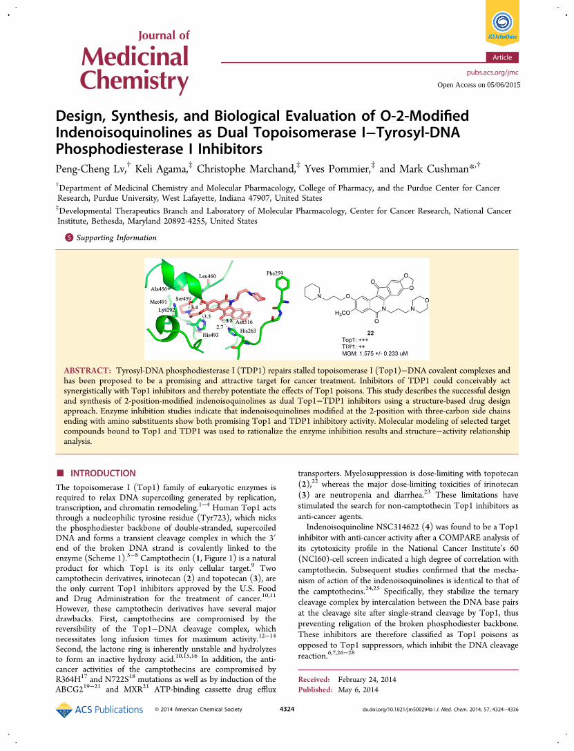
The topoisomerase I (Top1) family of eukaryotic enzymes isrequired to relax DNA supercoiling generated by replication,transcription, and chromatin remodeling.1−4 Human Top1 actsthrough a nucleophilic tyrosine residue (Tyr723), which nicksthe phosphodiester backbone of double-stranded, supercoiledDNA and forms a transient cleavage complex in which the 3′end of the broken DNA strand is covalently linked to theenzyme (Scheme 1).5−8 Camptothecin (1, Figure 1) is a naturalproduct for which Top1 is its only cellular target.9 Twocamptothecin derivatives, irinotecan (2) and topotecan (3), arethe only current Top1 inhibitors approved by the U.S. Foodand Drug Administration for the treatment of cancer.10,11
However, these camptothecin derivatives have several majordrawbacks. First, camptothecins are compromised by thereversibility of the Top1−DNA cleavage complex, whichnecessitates long infusion times for maximum activity.12−14
Second, the lactone ring is inherently unstable and hydrolyzesto form an inactive hydroxy acid.10,15,16 In addition, the anti-cancer activities of the camptothecins are compromised byR364H17 and N722S18 mutations as well as by induction of theABCG219−21 and MXR21 ATP-binding cassette drug efflux
transporters. Myelosuppression is dose-limiting with topotecan(2),22 whereas the major dose-limiting toxicities of irinotecan(3) are neutropenia and diarrhea.23 These limitations havestimulated the search for non-camptothecin Top1 inhibitors asanti-cancer agents.Indenoisoquinoline NSC314622 (4) was found to be a Top1
inhibitor with anti-cancer activity after a COMPARE analysis ofits cytotoxicity profile in the National Cancer Institute’s 60(NCI60)-cell screen indicated a high degree of correlation withcamptothecin. Subsequent studies confirmed that the mecha-nism of action of the indenoisoquinolines is identical to that ofthe camptothecins.24,25 Specifically, they stabilize the ternarycleavage complex by intercalation between the DNA base pairsat the cleavage site after single-strand cleavage by Top1, thuspreventing religation of the broken phosphodiester backbone.These inhibitors are therefore classified as Top1 poisons asopposed to Top1 suppressors, which inhibit the DNA cleavagereaction.6,7,26−28
Received: February 24, 2014Published: May 6, 2014
Article
pubs.acs.org/jmc
© 2014 American Chemical Society 4324 dx.doi.org/10.1021/jm500294a | J. Med. Chem. 2014, 57, 4324−4336
Open Access on 05/06/2015
The indenoisoquinolines have several advantages over thecamptothecins. First, the cleavage complexes induced by theindenoisoquinolines are more stable than those formed in thepresence of camptothecin derivatives.25 Second, in contrast tothe camptothecin derivatives, which have an unstable lactonering, the indenoisoquinolines are chemically stable. Moreover,the DNA cleavage site specificity of indenoisoquinolines isdifferent from that of camptothecin, so they target the genomedifferently, and the indenoisoquinolines are less affected by theR364H and N722N Top1-resistance mutations than iscamptothecin.25,29,30 After lead optimization and SAR studies,two indenoisoquinoline topoisomerase I inhibitors [indotecan(6, also known as LMP400 or NSC 724998) and indimitecan(7, also known as LMP776 or NSC 725776)] have enteredphase I clinical trials for the treatment of cancer patients at theNational Cancer Institute, and definite plans are beingformulated to commence phase II clinical trials.31−33 Indotecan(6) is not a substrate for the ABCG2 and MDR-1 efflux pumps,whereas indimitecan (7) is less affected than the camptothe-cins.29
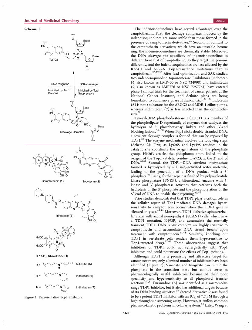
Tyrosyl-DNA phosphodiesterase I (TDP1) is a member ofthe phospholipase D superfamily of enzymes that catalyzes thehydrolysis of 3′ phosphotyrosyl linkers and other 3′-endblocking lesions.34−38 When Top1 nicks double-stranded DNA,a covalent cleavage complex is formed that can be repaired byTDP1.39 The enzyme mechanism involves the following steps(Scheme 2): First, as Lys265 and Lys495 residues in thecatalytic site coordinate the oxygen atoms of the phosphategroup, His263 attacks the phosphorus atom linked to theoxygen of the Top1 catalytic residue, Tyr723, at the 3′ end ofDNA.40,41 Second, the TDP1−DNA covalent intermediateformed is hydrolyzed by a His493-activated water molecule,leading to the generation of a DNA product with a 3′phosphate.42 Lastly, further repair is finished by polynucleotidekinase phosphatase (PNKP), a bifunctional enzyme with 5′kinase and 3′ phosphatase activities that catalyzes both thehydrolysis of the 3′ phosphate and the phosphorylation of the5′ end of DNA to enable their rejoining.43,44
Prior studies demonstrated that TDP1 plays a critical role inthe cellular repair of Top1-mediated DNA damage: hyper-sensitivity to camptothecin occurs when the TDP1 gene issilenced in yeast.45,46 Moreover, TDP1-defective spinocerebel-lar ataxia with axonal neuropathy-1 (SCAN1) cells, which havea TDP1 mutation, N493R, and accumulate the normallytransient TDP1−DNA repair complex, are highly sensitive tocamptothecin and accumulate DNA strand breaks upontreatment with camptothecin.47,48 Similarly, knocking outTDP1 in vertebrate cells renders them hypersensitive toTop1-targeted drugs.37,49 These observations suggest thatinhibitors of TDP1 could act synergistically with Top1inhibitors and could potentiate the effects of Top1 poisons.Although TDP1 is a promising and attractive target for
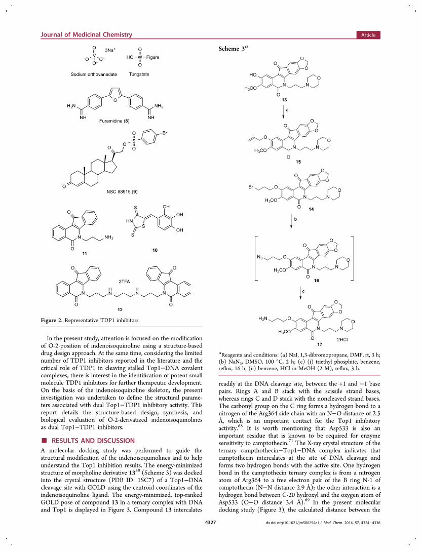
cancer treatment, only a limited number of inhibitors have beenidentified (Figure 2). Vanadate and tungstate can mimic thephosphate in the transition state but cannot serve aspharmacologically useful inhibitors because of their poorspecificity and hypersensitivity to all phosphoryl transferreactions.50,51 Furamidine (8) was identified as a micromolar-range TDP1 inhibitor, but it also has additional targets becauseof its DNA-binding activities.52 Steroid derivative 9 was foundto be a potent TDP1 inhibitor with an IC50 of 7.7 μM through ahigh-throughput screening assay. However, it suffers commonpharmacokinetic problems in cellular systems.53 Later, Wang et
Scheme 1
Figure 1. Representative Top1 inhibitors.
Journal of Medicinal Chemistry Article
dx.doi.org/10.1021/jm500294a | J. Med. Chem. 2014, 57, 4324−43364325
al. reported that arylidene thioxothiazolidinones can inhibitTDP1 and identified compound 10 as a submicromolarinhibitor of TDP1.54 Recently, indenoisoquinoline 11 andbis(indenoisoquinoline) 12 were found to be dual Top1−TDP1 inhibitors.55,56
After indenoisoquinoline 4 was found to be a Top1 inhibitorwith anti-cancer activity, a number of structure−activityrelationship studies were performed on the indenoisoquino-lines, including modifications on the A ring,57 B ring (side chainon the lactam nitrogen),58−62 C ring,63,64 and Dring.57,61−63,65−68 However, there is very limited study on themodification of the A ring of indenoisoquinoline, especially onthe 2-position. To date, the methoxy group is the only
substituent that has been placed at the 2-position. The crystalstructure of an indenoisoquinoline in a ternary complex withDNA and Top1 (PDB ID: 1SC7) indicates that the A ring isnext to the cleaved DNA strand, where there is more room toaccommodate substituents on the drug.69 Recent studies of themetabolism of indotecan (LMP400, 6) and indimitecan(LMP776, 7) involved the synthesis of 2-hydroxylatedindenoisoquinolines, thus providing a strategically placedhandle for the attachment of a variety of side chains.68 At thesame time, prior molecular modeling studies based on thecrystal structure of TDP1 (PDB ID: 1RFF)70 provide afoundation for the structure-based design of indenoisoquinolineTop1 inhibitors that also inhibit TDP1.55,56
Scheme 2
Journal of Medicinal Chemistry Article
dx.doi.org/10.1021/jm500294a | J. Med. Chem. 2014, 57, 4324−43364326
In the present study, attention is focused on the modificationof O-2-position of indenoisoquinoline using a structure-baseddrug design approach. At the same time, considering the limitednumber of TDP1 inhibitors reported in the literature and thecritical role of TDP1 in cleaving stalled Top1−DNA covalentcomplexes, there is interest in the identification of potent smallmolecule TDP1 inhibitors for further therapeutic development.On the basis of the indenoisoquinoline skeleton, the presentinvestigation was undertaken to define the structural parame-ters associated with dual Top1−TDP1 inhibitory activity. Thisreport details the structure-based design, synthesis, andbiological evaluation of O-2-derivatized indenoisoquinolinesas dual Top1−TDP1 inhibitors.
■ RESULTS AND DISCUSSIONA molecular docking study was performed to guide thestructural modification of the indenoisoquinolines and to helpunderstand the Top1 inhibition results. The energy-minimizedstructure of morpholine derivative 1358 (Scheme 3) was dockedinto the crystal structure (PDB ID: 1SC7) of a Top1−DNAcleavage site with GOLD using the centroid coordinates of theindenoisoquinoline ligand. The energy-minimized, top-rankedGOLD pose of compound 13 in a ternary complex with DNAand Top1 is displayed in Figure 3. Compound 13 intercalates
readily at the DNA cleavage site, between the +1 and −1 basepairs. Rings A and B stack with the scissile strand bases,whereas rings C and D stack with the noncleaved strand bases.The carbonyl group on the C ring forms a hydrogen bond to anitrogen of the Arg364 side chain with an N−O distance of 2.5Å, which is an important contact for the Top1 inhibitoryactivity.68 It is worth mentioning that Asp533 is also animportant residue that is known to be required for enzymesensitivity to camptothecin.71 The X-ray crystal structure of theternary campthothecin−Top1−DNA complex indicates thatcamptothecin intercalates at the site of DNA cleavage andforms two hydrogen bonds with the active site. One hydrogenbond in the camptothecin ternary complex is from a nitrogenatom of Arg364 to a free electron pair of the B ring N-1 ofcamptothecin (N−N distance 2.9 Å); the other interaction is ahydrogen bond between C-20 hydroxyl and the oxygen atom ofAsp533 (O−O distance 3.4 Å).69 In the present moleculardocking study (Figure 3), the calculated distance between the
Figure 2. Representative TDP1 inhibitors.
Scheme 3a
aReagents and conditions: (a) NaI, 1,3-dibromopropane, DMF, rt, 3 h;(b) NaN3, DMSO, 100 °C, 2 h; (c) (i) triethyl phosphite, benzene,reflux, 16 h, (ii) benzene, HCl in MeOH (2 M), reflux, 3 h.
Journal of Medicinal Chemistry Article
dx.doi.org/10.1021/jm500294a | J. Med. Chem. 2014, 57, 4324−43364327
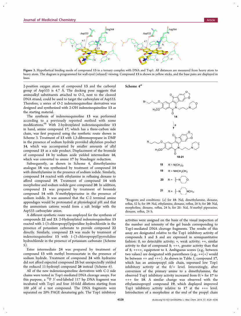
2-position oxygen atom of compound 13 and the carbonylgroup of Asp533 is 4.7 Å. The docking pose suggests thataminoalkyl substituents attached to O-2, next to the cleavedDNA strand, could be used to target the carboxylate of Asp533.Therefore, a series of O-2 indenoisoquinoline derivatives wasdesigned and synthesized with 2-OH indenoisoquinoline 13 asthe starting material.The synthesis of indenoisoquinoline 13 was performed
according to a previously reported method with somemodifications.68 With 2-hydroxylated indenoisoquinoline 13in hand, amine compound 17, which has a three-carbon sidechain, was first prepared using the synthetic route shown inScheme 3. Treatment of 13 with 1,3-dibromopropane in DMFin the presence of sodium hydride provided alkylation product14, which was accompanied by smaller amounts of allylcompound 15 as a side product. Displacement of the bromideof compound 14 by sodium azide yielded intermediate 16,which was converted to amine 17 by Staudinger reduction.Subsequently, as shown in Scheme 4, dimethylamino
analogue 18 was synthesized by treatment of compound 14with dimethylamine in the presence of sodium iodide. Similarly,compound 14 reacted with ethylamine in refluxing dioxane toafford compound 19. Treatment of compound 14 withmorpholine and sodium iodide gave compound 20. In addition,compound 21 was prepared by treatment of bromidecompound 14 with N-methylpiperazine in the presence ofsodium iodide. It was assumed that the C-2 terminal amineappendages would be protonated at physiological pH and thatthe ammonium cations would form a salt bridge with theAsp533 carboxylate anion.A different synthetic route was employed for the synthesis of
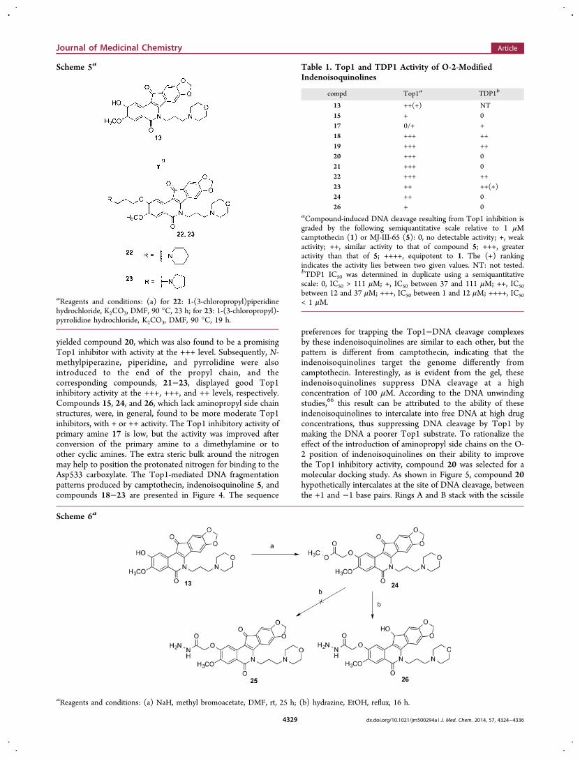
compounds 22 and 23. 2-Hydroxylated indenoisoquinoline 13reacted with 1-(3-chloropropyl)piperidine hydrochloride in thepresence of potassium carbonate to provide compound 22directly. Similarly, compound 23 was made by treatment ofindenoisoquinoline 13 with 1-(3-chloropropyl)pyrrolidinehydrochloride in the presence of potassium carbonate (Scheme5).Ester intermediate 24 was prepared by treatment of
compound 13 with methyl bromoacetate in the presence ofsodium hydride. Treatment of compound 24 with hydrazinedid not afford expected compound 25 but unexpectedly yieldedthe reduced 11-hydroxyl compound 26 instead (Scheme 6).All of the new indenoisoquinoline derivatives with C-2 side
chains were tested in Top1-mediated DNA cleavage assays. Forthis purpose, a 32P 3′-end-labeled 117 bp DNA fragment wasincubated with Top1 and four 10-fold dilutions starting from100 μM of a test compound. The DNA fragments wereseparated on 20% PAGE denaturing gels. The Top1 inhibitory
activities were assigned on the basis of the visual inspection ofthe number and intensity of the gel bands corresponding toTop1-mediated DNA cleavage fragments. The results of thisassay are designated relative to the Top1 inhibitory activity ofcompounds 1 and 5 and are expressed in semiquantitativefashion: 0, no detectable activity; +, weak activity; ++, similaractivity to that of compound 5; +++, greater activity than thatof 5; ++++, equipotent to 1. Ambiguous scores (e.g., betweentwo values) are designated with parentheses (e.g., ++(+) wouldbe between ++ and +++). As shown in Table 1, compound 17,which has an aminopropyl side chain, expressed low Top1inhibitory activity at the 0/+ level. Interestingly, afterconversion of the primary amine to a dimethylamine, theobserved Top1 inhibitory activity increased from 0/+ for 17 to+++ for 18. A similar change was observed with theethylaminopropyl compound 19, which displayed improvedTop1 inhibitory activity relative to 17 at the +++ level.Introduction of a morpholine at the end of the propyl chain
Figure 3. Hypothetical binding mode of compound 13 in a ternary complex with DNA and Top1. All distances are measured from heavy atom toheavy atom. The diagram is programmed for wall-eyed (relaxed) viewing. Compound 13 is shown in yellow sticks, and the base pairs are displayed inlines.
Scheme 4a
aReagents and conditions: (a) for 18: NaI, dimethylamine, dioxane,reflux, 52 h; for 19: NaI, ethylamine, dioxane, reflux, 26 h; for 20: NaI,morpholine, dioxane, reflux, 24 h; for 21: NaI, N-methyl piperazine,dioxane, reflux, 24 h.
Journal of Medicinal Chemistry Article
dx.doi.org/10.1021/jm500294a | J. Med. Chem. 2014, 57, 4324−43364328
yielded compound 20, which was also found to be a promisingTop1 inhibitor with activity at the +++ level. Subsequently, N-methylpiperazine, piperidine, and pyrrolidine were alsointroduced to the end of the propyl chain, and thecorresponding compounds, 21−23, displayed good Top1inhibitory activity at the +++, +++, and ++ levels, respectively.Compounds 15, 24, and 26, which lack aminopropyl side chainstructures, were, in general, found to be more moderate Top1inhibitors, with + or ++ activity. The Top1 inhibitory activity ofprimary amine 17 is low, but the activity was improved afterconversion of the primary amine to a dimethylamine or toother cyclic amines. The extra steric bulk around the nitrogenmay help to position the protonated nitrogen for binding to theAsp533 carboxylate. The Top1-mediated DNA fragmentationpatterns produced by camptothecin, indenoisoquinoline 5, andcompounds 18−23 are presented in Figure 4. The sequence
preferences for trapping the Top1−DNA cleavage complexesby these indenoisoquinolines are similar to each other, but thepattern is different from camptothecin, indicating that theindenoisoquinolines target the genome differently fromcamptothecin. Interestingly, as is evident from the gel, theseindenoisoquinolines suppress DNA cleavage at a highconcentration of 100 μM. According to the DNA unwindingstudies,66 this result can be attributed to the ability of theseindenoisoquinolines to intercalate into free DNA at high drugconcentrations, thus suppressing DNA cleavage by Top1 bymaking the DNA a poorer Top1 substrate. To rationalize theeffect of the introduction of aminopropyl side chains on the O-2 position of indenoisoquinolines on their ability to improvethe Top1 inhibitory activity, compound 20 was selected for amolecular docking study. As shown in Figure 5, compound 20hypothetically intercalates at the site of DNA cleavage, betweenthe +1 and −1 base pairs. Rings A and B stack with the scissile
Scheme 5a
aReagents and conditions: (a) for 22: 1-(3-chloropropyl)piperidinehydrochloride, K2CO3, DMF, 90 °C, 23 h; for 23: 1-(3-chloropropyl)-pyrrolidine hydrochloride, K2CO3, DMF, 90 °C, 19 h.
Scheme 6a
aReagents and conditions: (a) NaH, methyl bromoacetate, DMF, rt, 25 h; (b) hydrazine, EtOH, reflux, 16 h.
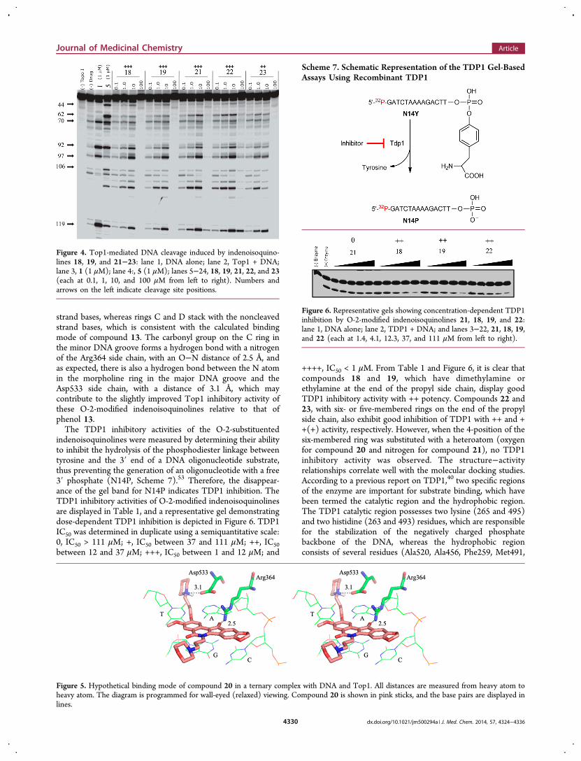
Table 1. Top1 and TDP1 Activity of O-2-ModifiedIndenoisoquinolines
compd Top1a TDP1b
13 ++(+) NT15 + 017 0/+ +18 +++ ++19 +++ ++20 +++ 021 +++ 022 +++ ++23 ++ ++(+)24 ++ 026 + 0
aCompound-induced DNA cleavage resulting from Top1 inhibition isgraded by the following semiquantitative scale relative to 1 μMcamptothecin (1) or MJ-III-65 (5): 0, no detectable activity; +, weakactivity; ++, similar activity to that of compound 5; +++, greateractivity than that of 5; ++++, equipotent to 1. The (+) rankingindicates the activity lies between two given values. NT: not tested.bTDP1 IC50 was determined in duplicate using a semiquantitativescale: 0, IC50 > 111 μM; +, IC50 between 37 and 111 μM; ++, IC50between 12 and 37 μM; +++, IC50 between 1 and 12 μM; ++++, IC50< 1 μM.
Journal of Medicinal Chemistry Article
dx.doi.org/10.1021/jm500294a | J. Med. Chem. 2014, 57, 4324−43364329
strand bases, whereas rings C and D stack with the noncleavedstrand bases, which is consistent with the calculated bindingmode of compound 13. The carbonyl group on the C ring inthe minor DNA groove forms a hydrogen bond with a nitrogenof the Arg364 side chain, with an O−N distance of 2.5 Å, andas expected, there is also a hydrogen bond between the N atomin the morpholine ring in the major DNA groove and theAsp533 side chain, with a distance of 3.1 Å, which maycontribute to the slightly improved Top1 inhibitory activity ofthese O-2-modified indenoisoquinolines relative to that ofphenol 13.The TDP1 inhibitory activities of the O-2-substituented
indenoisoquinolines were measured by determining their abilityto inhibit the hydrolysis of the phosphodiester linkage betweentyrosine and the 3′ end of a DNA oligonucleotide substrate,thus preventing the generation of an oligonucleotide with a free3′ phosphate (N14P, Scheme 7).53 Therefore, the disappear-ance of the gel band for N14P indicates TDP1 inhibition. TheTDP1 inhibitory activities of O-2-modified indenoisoquinolinesare displayed in Table 1, and a representative gel demonstratingdose-dependent TDP1 inhibition is depicted in Figure 6. TDP1IC50 was determined in duplicate using a semiquantitative scale:0, IC50 > 111 μM; +, IC50 between 37 and 111 μM; ++, IC50between 12 and 37 μM; +++, IC50 between 1 and 12 μM; and
++++, IC50 < 1 μM. From Table 1 and Figure 6, it is clear thatcompounds 18 and 19, which have dimethylamine orethylamine at the end of the propyl side chain, display goodTDP1 inhibitory activity with ++ potency. Compounds 22 and23, with six- or five-membered rings on the end of the propylside chain, also exhibit good inhibition of TDP1 with ++ and ++(+) activity, respectively. However, when the 4-position of thesix-membered ring was substituted with a heteroatom (oxygenfor compound 20 and nitrogen for compound 21), no TDP1inhibitory activity was observed. The structure−activityrelationships correlate well with the molecular docking studies.According to a previous report on TDP1,40 two specific regionsof the enzyme are important for substrate binding, which havebeen termed the catalytic region and the hydrophobic region.The TDP1 catalytic region possesses two lysine (265 and 495)and two histidine (263 and 493) residues, which are responsiblefor the stabilization of the negatively charged phosphatebackbone of the DNA, whereas the hydrophobic regionconsists of several residues (Ala520, Ala456, Phe259, Met491,
Figure 4. Top1-mediated DNA cleavage induced by indenoisoquino-lines 18, 19, and 21−23: lane 1, DNA alone; lane 2, Top1 + DNA;lane 3, 1 (1 μM); lane 4:, 5 (1 μM); lanes 5−24, 18, 19, 21, 22, and 23(each at 0.1, 1, 10, and 100 μM from left to right). Numbers andarrows on the left indicate cleavage site positions.
Figure 5. Hypothetical binding mode of compound 20 in a ternary complex with DNA and Top1. All distances are measured from heavy atom toheavy atom. The diagram is programmed for wall-eyed (relaxed) viewing. Compound 20 is shown in pink sticks, and the base pairs are displayed inlines.
Scheme 7. Schematic Representation of the TDP1 Gel-BasedAssays Using Recombinant TDP1
Figure 6. Representative gels showing concentration-dependent TDP1inhibition by O-2-modified indenoisoquinolines 21, 18, 19, and 22:lane 1, DNA alone; lane 2, TDP1 + DNA; and lanes 3−22, 21, 18, 19,and 22 (each at 1.4, 4.1, 12.3, 37, and 111 μM from left to right).
Journal of Medicinal Chemistry Article
dx.doi.org/10.1021/jm500294a | J. Med. Chem. 2014, 57, 4324−43364330
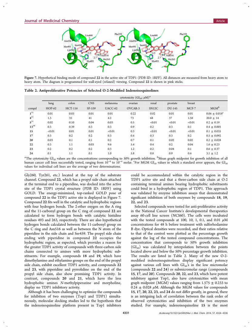
Gly260, Tyr261, etc.) located at the top of the substratechannel. Compound 22, which has a propyl side chain attachedat the terminal end to a piperidine, was docked into the activesite of the TDP1 crystal structure (PDB ID: 1RFF) usingGOLD. The energy-minimized, top-ranked GOLD pose ofcompound 22 in the TDP1 active site is displayed in Figure 7.Compound 22 fits well in the catalytic and hydrophobic regionswith four hydrogen bonds. The 2-ether oxygen on the A ringand the 11-carbonyl group on the C ring of compound 22 arecalculated to form hydrogen bonds with catalytic histidineresidues 493 and 263, respectively. There are also hypotheticalhydrogen bonds calculated between the 11-carbonyl group onthe C ring and Asn516 as well as between the N atom of thepiperidine in the side chain and Ser459. The propyl side chainending with piperidine in compound 22 occupies thehydrophobic region, as expected, which provides a reason forthe greater TDP1 activity of compounds with three-carbon sidechains connected to amines with more hydrophobic sub-stituents. For example, compounds 18 and 19, which havedimethylamino and ethylamino groups on the end of the propylside chain, exhibit excellent TDP1 activity, and compounds 22and 23, with piperidine and pyrrolidine on the end of thepropyl side chain, also show promising TDP1 activity. Incontrast, compounds 20 and 21, which have the lesshydrophobic amines N-methylpiperazine and morpholine,display no TDP1 inhibitory activity.Although it has been challenging to optimize the compounds
for inhibition of two enzymes (Top1 and TDP1) simulta-neously, molecular docking studies led to the hypothesis thatthe indenoisoquinoline platform present in Top1 inhibitors
could be accommodated within the catalytic region in theTDP1 active site and that a three-carbon side chain at O-2containing terminal amines bearing hydrophobic substituentscould bind in a hydrophobic region of TDP1. This approachwas validated by enzyme inhibition assays that demonstratedsignificant inhibition of both enzymes by compounds 18, 19,22, and 23.Selected compounds were tested for anti-proliferative activity
in the National Cancer Institute’s developmental therapeuticsassay 60-cell line screen (NCI60). The cells were incubatedwith the tested compounds at 100, 10, 1, 0.1, and 0.01 μMconcentrations for 48 h before treatment with sulforhodamineB dye. Optical densities were recorded, and their ratios relativeto that of the control were plotted as the percentage growthagainst the log of the tested compound concentrations. Theconcentration that corresponds to 50% growth inhibition(GI50) was calculated by interpolation between the pointslocated above and below the 50% percentage growth inhibition.The results are listed in Table 2. Many of the new O-2-modified indenoisoquinolines display significant potencyagainst various cell lines with GI50’s in the low micromolar(compounds 22 and 24) or submicromolar range (compounds15, 17, and 20). Compounds 20, 22, and 23, which have potentinhibitory against Top1, also have cytotoxicities with meangraph midpoint (MGM) values ranging from 1.575 ± 0.233 to0.24 ± 0.028 μM. Although the MGM values for compounds15, 17, 20, 22, 23, and 24 do not differ greatly, in general, thereis an intriguing lack of correlation between the rank order ofobserved cytotoxicities and inhibition of the two enzymesstudied. For example, indenoisoquinoline 15 is the most
Figure 7. Hypothetical binding mode of compound 22 in the active site of TDP1 (PDB ID: 1RFF). All distances are measured from heavy atom toheavy atom. The diagram is programmed for wall-eyed (relaxed) viewing. Compound 22 is shown in pink sticks.
Table 2. Antiproliferative Potencies of Selected O-2-Modified Indenoisoquinolines
cytotoxicity (GI50; μM)a
lung colon CNS melanoma ovarian renal prostate breast
compd HOP-62 HCT-116 SF-539 UACC-62 OVCAR-3 SN12C DU-145 MCF-7 MGMb
151 0.01 0.03 0.01 0.01 0.22 0.02 0.01 0.01 0.04 ± 0.018c
451 1.3 35 41 4.2 73 68 37 1.58 20.0 ± 14551 0.02 0.10 0.04 0.03 0.5 <0.01 <0.01 <0.01 0.2 ± 0.191358 0.3 0.39 0.3 0.3 0.9 0.2 0.3 0.1 0.4 ± 0.00515 <0.01 0.01 0.01 <0.01 0.3 <0.01 <0.01 <0.01 0.1 ± 0.03517 0.3 0.2 0.2 0.3 0.4 0.3 0.3 0.2 0.5 ± 0.09220 0.03 0.1 0.1 0.2 0.7 0.1 0.02 0.02 0.2 ± 0.02822 0.3 1.1 0.03 9.6 5.4 0.4 0.2 0.04 1.6 ± 0.2323 0.2 0.2 0.2 0.3 1.2 0.2 0.04 0.1 0.6 ± 0.3724 0.1 1.1 0.1 1.0 5.8 0.8 0.9 0.4 3.1 ± 1.2
aThe cytotoxicity GI50 values are the concentrations corresponding to 50% growth inhibition. bMean graph midpoint for growth inhibition of allhuman cancer cell lines successfully tested, ranging from 10−8 to 10−4 molar. cFor MGM GI50 values in which a standard error appears, the GI50values for individual cell lines are the average of two determinations.
Journal of Medicinal Chemistry Article
dx.doi.org/10.1021/jm500294a | J. Med. Chem. 2014, 57, 4324−43364331
cytotoxic compound, but it has low Top1 inhibitory activity andno detectable TDP1 inhibitory activity. In contrast, thecytotoxicity of 22 is comparatively low, but it has relativelyhigh activity versus both enzymes. The GI50 values in individualcell lines vary more widely than the MGM values, and moresignificant differences are observed. For example, compound 24is the most cytotoxic of the indenoisoquinolines versus the lungHOP-62 cell line, but it has the lowest overall cytotoxicity asindicated by the MGM value. Therefore, the lack of a strongcorrelation between enzyme inhibition and cytotoxicity is acomplicated matter that may be influenced by the particular cellline under investigation as well as by differences in cellularpenetration, distribution within the cell, metabolism, ejectionfrom the cell, and possible off-target effects.
■ CONCLUSIONS
A series of 2-position-substituted indenoisoquinolines with athree-carbon side chain linked at the end to amines wasdesigned and synthesized for the development of dual Top1and TDP1 inhibitors based on the hypotheses that (a) 2-OHindenoisoquinolines substituted with a three-carbon side chainending with amino substitutions could bind to Asp533 in theTop1 active site, thus improving the Top1 inhibitory activity,and (b) the indenoisoquinoline core could be accommodatedwithin the catalytic region in the TDP1 active site while a three-carbon side chain attached to amines with hydrophobicsubstitutions could bind to the hydrophobic region of TDP1.Top1 inhibition results reveal that the attachment of amino-propyl side chains targeting Asp533 results in a slight butconsistent improvement in activity in the cases of 18, 19, 20,21, and 22, but in the cases of primary amine 17 andpyrrolidine derivative 23, there was an unexpected drop inTop1 inhibitory activity. Enzyme inhibition results with bothTop1 and TDP1 indicate that compounds 18, 19, 22, and 23have good inhibitory activity against Top1 and also showpromising TDP1 inhibitory activity.
■ EXPERIMENTAL SECTIONGeneral. Solvents and reagents were purchased from commercial
vendors and were used without any further purification. Melting pointswere determined using capillary tubes with a Mel-Temp apparatus andare uncorrected. Infrared spectra were obtained using KBr pellets. IRspectra were recorded using a PerkinElmer 1600 series or SpectrumOne FTIR spectrometer. 1H NMR spectra were recorded at 300 MHzusing a Bruker ARX300 spectrometer with a QNP probe. Massspectral analyses were performed at the Purdue University Campus-Wide Mass Spectrometry Center. ESI−MS studies were performedusing a FinniganMAT LCQ Classic mass spectrometer. EI/CI−MSstudies were performed using a Hewlett-Packard Engine or GCQFinniganMAT mass spectrometer. APCI−MS studies were carried outusing an Agilent 6320 ion trap mass spectrometer. Analytical thin-layerchromatography was carried out on Baker-flex silica gel IB2-F plates,and compounds were visualized with short-wavelength UV light andninhydrin staining. Silica gel flash chromatography was performedusing 230−400 mesh silica gel. HPLC analyses were performed on aWaters 1525 binary HPLC pump/Waters 2487 dual λ absorbancedetector system using a 5 μM C18 reverse-phase column. Compoundpurities were estimated by reversed-phase C18 HPLC with a UVdetector at 254 nm, and the major peak area of each tested compoundwas ≥95% of the combined total peak area. All yields refer to isolatedcompounds.2-(3-Bromopropoxy)-3-methoxy-6-(3-morpholinopropyl)-
5H-[1,3]dioxolo[4′,5′:5,6]indeno[1,2-c]isoquinoline-5,12(6H)-dione (14). A solution of compound 1358 (0.100 g, 0.216 mmol) inDMF (6 mL) was treated with sodium hydride (0.011 g, 2.16 mmol).
After 10 min, 1,3-dibromopropane was added. The mixture was stirredat room temperature for 3 h. The mixture was diluted to a volume of200 mL with CHCl3, washed with H2O (2 × 50 mL) and saturatedaqueous NaCl (50 mL), dried over anhydrous sodium sulfate, andconcentrated. The residue was purified by flash column chromatog-raphy (SiO2, ∼40 g), eluting with 1% MeOH in CHCl3 to yieldcompound 14 as a solid (0.071 g, 56%). mp 197−199 °C. IR (film)3434, 2102, 1638, 1498, 1304, 1115, 1032 cm−1. 1H NMR (CDCl3) δ8.02 (s, 1H), 7.62 (s, 1H), 7.38 (s, 1H), 7.04 (s, 1H), 6.15 (s, 2H),5.58−5.37 (m, 2H), 4.79−4.77 (d, J = 5.1 Hz, 2H), 4.69−4.52 (t, J =8.4 Hz, 2H), 3.97 (s, 3H), 3.77 (s, 4H), 2.55 (s, 6H), 2.02 (s, 2H),1.33 (s, 2H). ESI−MS m/z 585/587 (MH+), 505 (MH+ − HBr).HRESI−MS m/z 585.1236 (MH+); calcd for C28H30BrN2O7,585.1243.
2-(Allyloxy)-3-methoxy-6-(3-morpholinopropyl)-5H-[1,3]-dioxolo[4′,5′:5,6]indeno [1,2-c]isoquinoline-5,12(6H)-dione(15). Column chromatography of the mixture described above alsoyielded side-product 15 as a solid (0.013 g, 10%). mp 199−200 °C(dec). IR (film) 2936, 1749, 1698, 1651, 1304, 1034, 786 cm−1. 1HNMR (CDCl3) δ 8.01 (s, 1H), 7.61 (s, 1H), 7.37 (s, 1H), 7.03 (s, 1H),6.08 (s, 3H), 5.53−5.40 (m, 2H), 4.79−4.77 (d, J = 5.7 Hz, 2H),4.51−4.45 (t, J = 7.5 Hz, 2H), 3.97 (s, 3H), 3.77 (s, 4H), 2.54 (s, 6H),2.01 (s, 2H). ESI−MS m/z 505 (MH+). HRESI−MS m/z 505.1967(MH+); calcd for C28H29N2O7, 505.1975. HPLC purity: 97.57% (C18reverse phase, MeOH/H2O, 90:10).
2-(3-Azidopropoxy)-3-methoxy-6-(3-morpholinopropyl)-5H-[1,3]dioxolo[4′,5′:5,6]indeno[1,2-c]isoquinoline-5,12(6H)-dione (16). Sodium azide (0.021 g, 0.22 mmol) and compound 14(0.128 g, 0.22 mmol) were diluted with DMSO (4 mL), and themixture was heated at 100 °C for 2 h. The mixture was diluted to avolume of 200 mL with CHCl3, washed with H2O (2 × 60 mL) andsaturated aqueous NaCl (50 mL), dried over anhydrous sodiumsulfate, and concentrated. The resulting residue was purified by flashcolumn chromatography (SiO2, ∼40 g), eluting with 0.5% MeOH inCHCl3 to yield product 16 as a solid (0.036 g, 60%). The solid wasused for the next step without further purification. ESI−MS m/z 548(MH+). HRESI−MS m/z 548.2141 (MH+); calcd for C28H30N5O7,548.2145.
2-(3-Aminopropoxy)-3-methoxy-6-(3-morpholinopropyl)-5H-[1,3]dioxolo[4′,5′:5,6]indeno[1,2-c]isoquinoline-5,12(6H)-dione (17). Triethyl phosphite (0.022 mL, 0.183 mmol) was added toa solution of compound 16 (0.040 g, 0.073 mmol) in benzene (4 mL),and the mixture was heated at reflux for 24 h. The mixture was dilutedto a volume of 200 mL with CHCl3, washed with H2O (2 × 50 mL)and saturated aqueous NaCl (50 mL), dried over anhydrous sodiumsulfate, and concentrated. The resulting residue was purified by flashcolumn chromatography (SiO2, ∼40 g), eluting with 0.5% MeOH inCHCl3 to yield the title compound as a solid. The solid (0.015 g, 0.023mmol) was diluted with benzene (4 mL), and 2 M HCl in methanol (6mL) was added to the solution. The mixture was heated at reflux for 3h. The reaction mixture was allowed to cool to room temperature, andthe precipitate was filtered to provide desired compound 17 as a solid(0.007 g, 58%). mp > 350 °C. IR (film) 3413, 2346, 1751, 1651, 1559,1437, 1309, 737 cm−1. 1H NMR (D2O) δ 6.77 (s, 1H), 6.62 (s, 1H),6.56 (s, 1H), 6.16 (s, 1H), 5.96 (s, 2H), 4.86−4.82 (m, 2H), 4.01 (s,6H), 3.78 (s, 2H), 3.67 (s, 3H), 3.32 (s, 4H), 3.24−3.19 (t, J = 7.5 Hz,2H), 2.19−2.17 (m, 4H). ESI−MS m/z 522 (MH+). HRESI−MS m/z522.2249 (MH+); calcd for C28H32N3O7, 522.2246. HPLC purity:95.23% (C18 reverse phase, MeOH/H2O, 85:15).
2-(3-(Dimethylamino)propoxy)-3-methoxy-6-(3-morpholi-nopropyl)-5H-[1,3]dioxolo[4′,5′:5,6]indeno[1,2-c]isoquinoline-5,12(6H)-dione (18). Sodium iodide (0.061 g, 0.408 mmol) andcompound 14 (0.020 g, 0.034 mmol) were diluted with dioxane (10mL), and dimethylamine (0.023 mL, 0.408 mmol) was addeddropwise. The mixture was stirred at reflux for 52 h. The mixturewas diluted to a volume of 250 mL with CHCl3, washed with H2O (2× 50 mL) and saturated aqueous NaCl (50 mL), dried over anhydroussodium sulfate, and concentrated. The residue was purified by flashcolumn chromatography (SiO2, ∼40 g), eluting first with 0.5% MeOHin CHCl3 and then with 1% MeOH in CHCl3 to yield product 18 as asolid (0.009 g, 47%). mp 176−178 °C (dec). IR (film) 2922, 1698,
Journal of Medicinal Chemistry Article
dx.doi.org/10.1021/jm500294a | J. Med. Chem. 2014, 57, 4324−43364332
1650, 1483, 1306, 1032, 786 cm−1. 1H NMR (CDCl3) δ 8.02 (s, 1H),7.63 (s, 1H), 7.43 (s, 1H), 7.08 (s, 1H), 6.09 (s, 2H), 4.54−4.48 (t, J =8.4 Hz, 2H), 4.29−4.25 (t, J = 6.3 Hz, 2H), 3.95 (s, 3H), 3.78−3.74 (t,J = 4.8 Hz, 2H), 2.57−2.53 (m, 14H), 2.27 (s, 2H), 2.03−2.02 (m,2H). ESI−MS m/z 550 (MH+). HRESI−MS m/z 550.2558 (MH+);calcd for C30H36N3O7, 550.2553. HPLC purity: 97.57% (C18 reversephase, MeOH/H2O, 85:15).2-(3-(Ethylamino)propoxy)-3-methoxy-6-(3-morpholino-
propyl)-5H-[1,3]dioxolo[4′,5′:5,6]indeno[1,2-c]isoquinoline-5,12(6H)-dione (19). Sodium iodide (0.060 g, 0.408 mmol) andcompound 14 (0.020 g, 0.034 mmol) were diluted with dioxane (8mL), and ethylamine (0.020 mL, 0.408 mmol, 70 wt % solution inwater) was added dropwise. The mixture was stirred at reflux for 26 h.The mixture was diluted to a volume of 200 mL with CHCl3, washedwith H2O (2 × 50 mL) and saturated aqueous NaCl (50 mL), driedover anhydrous sodium sulfate, and concentrated. The residue waspurified by flash column chromatography (SiO2, ∼40 g), eluting with0.5% MeOH in CHCl3 to yield product 19 as a solid (0.009 g, 46%).mp 187−188 °C (dec). IR (film) 1648, 1553, 1392, 1254, 1116, 1033,785 cm−1. 1H NMR (CDCl3) δ 7.53 (s, 1H), 7.24 (s, 1H), 6.90 (s,1H), 6.60 (s, 1H), 5.89 (s, 2H), 4.06 (s, 2H), 3.85 (s, 6H), 3.72 (s,3H), 3.49−3.38 (m, 4H), 3.19−3.12 (m, 9H), 2.25−2.19 (m, 4H).ESI−MS m/z 550 (MH+). HRESI−MS m/z 550.2553 (MH+); calcdfor C30H36N3O7, 550.2553. HPLC purity: 98.36% (C18 reverse phase,MeOH/H2O, 80:20).3-Methoxy-2-(3-morpholinopropoxy)-6-(3-morpholino-
propyl)-5H-[1,3]dioxolo[4′,5′:5,6]indeno[1,2-c]isoquinoline-5,12(6H)-dione (20). Sodium iodide (0.139 g, 0.924 mmol) andcompound 14 (0.045 g, 0.077 mmol) were diluted with dioxane (10mL), and morpholine (0.08 mL, 0.924 mmol) was added dropwise.The mixture was stirred at reflux for 24 h. The mixture was diluted to avolume of 200 mL with CHCl3, washed with H2O (2 × 60 mL) andsaturated aqueous NaCl (50 mL), dried over anhydrous sodiumsulfate, and concentrated. The residue was purified by flash columnchromatography (SiO2, ∼40 g), eluting first with 0.5% MeOH inCHCl3 and then with 1% MeOH in CHCl3 to yield product 20 as asolid (0.018 g, 41%). mp 188−189 °C (dec). IR (film) 2956, 1869,1749, 1650, 1508, 1307, 1032, 865 cm−1. 1H NMR (CDCl3) δ 7.99 (s,1H), 7.61 (s, 1H), 7.39 (s, 1H), 7.04 (s, 1H), 6.08 (s, 2H), 4.64−4.51(t, J = 7.5 Hz, 2H), 4.28−4.24 (t, J = 6.6 Hz, 2H), 3.95 (s, 3H), 3.76(s, 8H), 2.53 (s, 12H), 2.15−2.11 (t, J = 6.3 Hz, 2H), 2.01 (s, 2H).ESI−MS m/z 592 (MH+). HRESI−MS m/z 592.2664 (MH+); calcdfor C32H38N3O8, 592.2659. HPLC purity: 95.38% (C18 reverse phase,MeOH/H2O, 85:15).3-Methoxy-2-(3-(4-methylpiperazin-1-yl)propoxy)-6-(3-mor-
pholinopropyl)-5H-[1,3]dioxolo[4′,5′:5,6]indeno[1,2-c]-isoquinoline-5,12(6H)-dione (21). Sodium iodide (0.061 g, 0.408mmol) and compound 14 (0.020 g, 0.034 mmol) were diluted withdioxane (5 mL), and N-methyl piperazine (0.041 mL, 0.408 mmol)was added dropwise. The mixture was stirred at reflux for 24 h. Themixture was diluted to a volume of 200 mL with CHCl3, washed withH2O (2 × 50 mL) and saturated aqueous NaCl (50 mL), dried overanhydrous sodium sulfate, and concentrated. The residue was purifiedby flash column chromatography (SiO2, ∼40 g), eluting first with 0.5%MeOH in CHCl3 and then with 3% MeOH in CHCl3 to yield product21 as a solid (0.010 g, 49%). mp 181−183 °C (dec). IR (film) 2924,1870, 1650, 1508, 1307, 1032, 868 cm−1. 1H NMR (CDCl3) δ 8.01 (s,1H), 7.62 (s, 1H), 7.42 (s, 1H), 7.06 (s, 1H), 6.09 (s, 2H), 4.53−4.47(t, J = 8.4 Hz, 2H), 4.28−4.23 (t, J = 6.6 Hz, 2H), 3.95 (s, 3H), 3.77−3.74 (t, J = 4.5 Hz, 2H), 2.62−2.53 (m, 16H), 2.35 (s, 3H), 2.13−2.06(m, 2H), 2.05−2.00 (m, 2H). ESI−MS m/z 605 (MH+). HRESI−MSm/z 605.2986 (MH+); calcd for C33H41N4O7, 605.2975. HPLC purity:100% (C18 reverse phase, MeOH/H2O, 90:10).3-Methoxy-6-(3-morpholinopropyl)-2-(3-(piperidin-1-yl)-
propoxy)-5H-[1,3]dioxolo[4′,5′:5,6]indeno[1,2-c]isoquinoline-5,12(6H)-dione (22). 1-(3-Chloropropyl)piperidine hydrochloride(214 mg, 1.08 mmol) and K2CO3 (298 mg, 2.16 mmol) were added toa DMF (5 mL) solution of compound 13 (0.100 g, 0.216 mmol). Themixture was heated at 90 °C for 23 h. The mixture was diluted to avolume of 300 mL with CHCl3, washed with H2O (2 × 80 mL) andsaturated aqueous NaCl (80 mL), dried over anhydrous sodium
sulfate, and concentrated. The residue was purified by flash columnchromatography (SiO2, ∼40 g), eluting with 0.25% MeOH in CHCl3to yield product 22 as a solid (0.072 g, 58%). mp 156−157 °C (dec).IR (film) 3399, 2091, 1645, 1392, 1305 cm−1. 1H NMR (CDCl3) δ7.99 (s, 1H), 7.60 (s, 1H), 7.39 (s, 1H), 7.04 (s, 1H), 6.08 (s, 2H),4.51−4.46 (t, J = 7.8 Hz, 2H), 4.25−4.21 (t, J = 6.6 Hz, 2H), 3.95 (s,3H), 3.77−3.74 (m, 4H), 2.58−2.46 (m, 12H), 2.18−2.09 (m, 2H),2.04−1.95 (m, 2H), 1.61−1.58 (m, 4H), 1.46−1.44 (m, 2H). ESI−MSm/z 590 (MH+). HRESI−MS m/z 590.2859 (MH+); calcd forC33H40N3O7, 590.2866. HPLC purity: 95.88% (C18 reverse phase,MeOH/H2O, 95:05).
3-Methoxy-6-(3-morpholinopropyl)-2-(3-(pyrrolidin-1-yl)-propoxy)-5H-[1,3]dioxolo[4′,5′:5,6]indeno[1,2-c]isoquinoline-5,12(6H)-dione (23). 1-(3-Chloropropyl)piperidine hydrochloride(158 mg, 0.86 mmol) and K2CO3 (237 mg, 1.72 mmol) were added toa DMF (5 mL) solution of compound 13 (0.80 g, 0.172 mmol). Themixture was heated at 90 °C for 19 h. The mixture was diluted to avolume of 300 mL with CHCl3, washed with H2O (2 × 80 mL) andsaturated aqueous NaCl (80 mL), dried over anhydrous sodiumsulfate, and concentrated. The residue was purified by flash columnchromatography (SiO2, ∼40 g), eluting with 0.25% MeOH in CHCl3to yield product 23 as a solid (0.051 g, 55%). mp 151−152 °C (dec).IR (film) 3418, 2936, 2119, 1660, 1392, 1225, 1105, 1063 cm−1. 1HNMR (CDCl3) δ 8.02 (s, 1H), 7.63 (s, 1H), 7.42 (s, 1H), 7.07 (s, 1H),6.09 (s, 2H), 4.52−4.48 (t, J = 8.1 Hz, 2H), 4.29−4.24 (t, J = 6.6 Hz,2H), 3.95 (s, 3H), 3.77−3.74 (t, J = 4.5 Hz, 2H), 2.80−2.77 (m, 2H),2.70 (s, 2H), 2.57−2.53 (m, 6H), 2.25−2.20 (m, 2H), 2.03−1.98 (m,2H), 1.87 (s, 6H). ESI−MS m/z 576 (MH+). HRESI−MS m/z576.2705 (MH+); calcd for C32H38N3O7, 576.2710; HPLC purity:96.29% (C18 reverse phase, MeOH/H2O, 80:20).
Methyl 2-((3-Methoxy-6-(3-morpholinopropyl)-5,12-dioxo-6,12-dihydro-5H-[1,3]dioxolo[4′ ,5′ :5,6]indeno[1,2-c]-isoquinolin-2-yl)oxy)acetate (24). Sodium hydride (0.067 g, 2.8mmol) and compound 13 (0.130 g, 0.28 mmol) were diluted withDMF (8 mL), and methyl bromoacetate (0.106 mL, 1.12 mmol) wasadded dropwise. The mixture was stirred at room temperature for 7 h.The mixture was diluted to a volume of 250 mL with CHCl3, washedwith H2O (2 × 60 mL) and saturated aqueous NaCl (50 mL), driedover anhydrous sodium sulfate, and concentrated. The residue waspurified by flash column chromatography (SiO2, ∼40 g), eluting with0.5% MeOH in CHCl3 to yield product 24 as a solid (0.077 g, 51%).mp 226−227 °C. IR (film) 2345, 1869, 1749, 1650, 1508, 1031, 737cm−1. 1H NMR (CDCl3) δ 7.91 (s, 1H), 7.65 (s, 1H), 7.40 (s, 1H),7.04 (s, 1H), 6.09 (s, 2H), 4.87 (s, 2H), 4.52−4.47 (t, J = 7.2 Hz, 2H),3.98 (s, 3H), 3.86 (s, 3H), 3.78 (s, 4H), 2.54 (s, 6H), 2.01 (s, 2H).ESI−MS m/z 537 (MH+). HRESI−MS m/z 537.1875 (MH+); calcdfor C28H29N2O9, 537.1873. HPLC purity: 96.60% (C18 reverse phase,MeOH/H2O, 90:10).
2-((12-Hydroxy-3-methoxy-6-(3-morpholinopropyl)-5-oxo-6,12-dihydro-5H-[1,3]dioxolo[4′ ,5′ :5,6]indeno[1,2-c]-isoquinolin-2-yl)oxy)acetohydrazide (26). Hydrazine (0.028 mL,0.056 mmol) and compound 13 (0.015 g, 0.028 mmol) were dilutedwith EtOH (10 mL), and the mixture was heated at reflux for 16 h.The precipitate obtained was washed with hexane (10 mL) and ether(10 mL) to yield product 26 as a light yellow solid (0.006 g, 40%). mp266−268 °C. IR (film) 2365, 1869, 1773, 1648, 1508, 1032, 738 cm−1.1H NMR (CDCl3) δ 9.30 (s, 1H), 7.62 (s, 1H), 7.45 (s, 1H), 7.36 (s,1H), 7.21 (s, 1H), 6.10 (s, 2H), 5.34 (s, 2H), 4.61 (s, 2H), 4.48 (s,2H), 3.88 (s, 3H), 3.61 (s, 4H), 2.49 (m, 6H), 1.96 (m, 2H). ESI−MSm/z 539 (MH+). HRESI−MS m/z 539.2146 (MH+); calcd forC27H30N4O8, 539.2142. HPLC purity: 95.19% (C18 reverse phase,MeOH/H2O, 85:15).
Topoisomerase I-Mediated DNA Cleavage Reactions. Humanrecombinant Top1 was purified from baculovirus as previouslydescribed.26 DNA cleavage reactions were prepared as previouslyreported with the exception of the DNA substrate.23 Briefly, a 117 bpDNA oligonucleotide (Integrated DNA Technologies) encompassingthe previously identified Top1 cleavage sites in the 161 bp fragmentfrom pBluescript SK(−) phagemid DNA was employed. This 117 bpoligonucleotide contains a single 5′ cytosine overhang, which was 3′-
Journal of Medicinal Chemistry Article
dx.doi.org/10.1021/jm500294a | J. Med. Chem. 2014, 57, 4324−43364333
end-labeled by a fill-in reaction with [α-32P]dGTP in React 2 buffer(50 mM Tris-HCl, pH 8.0, 100 mM MgCl2, and 50 mM NaCl) and0.5 units of DNA polymerase I (Klenow fragment, New EnglandBiolabs). Unincorporated [32P]dGTP was removed using mini QuickSpin DNA columns (Roche, Indianapolis, IN), and the eluatecontaining the 3′-end-labeled DNA substrate was collected. Approx-imately 2 nM radiolabeled DNA substrate was incubated withrecombinant Top1 in 20 μL of reaction buffer [10 mM Tris-HCl,pH 7.5, 50 mM KCl, 5 mM MgCl2, 0.1 mM EDTA, and 15 μg/mLBSA] at 25 °C for 20 min in the presence of various concentrations ofcompounds. The reactions were terminated by adding SDS (0.5% finalconcentration) followed by the addition of two volumes of loading dye(80% formamide, 10 mM sodium hydroxide, 1 mM sodium EDTA,0.1% xylene cyanol, and 0.1% bromphenol blue). Aliquots of eachreaction mixture were subjected to 20% denaturing PAGE. Gels weredried and visualized using a phosphoimager and ImageQuant software(Molecular Dynamics). For simplicity, cleavage sites were numberedas previously described in the 161 bp fragment.Gel-Based Assay Measuring the Inhibition of Recombinant
TDP1. A 5′-[32P]-labeled single-stranded DNA oligonucleotidecontaining a 3′ phosphotyrosine (N14Y) was generated as describedby Dexheimer et al.53 The DNA substrate was then incubated with 5pM recombinant TDP1 in the absence or presence of inhibitor for 15min at room temperature in a buffer containing 50 mM Tris HCl, pH7.5, 80 mM KCl, 2 mM EDTA, 1 mM DTT, 40 μg/mL BSA, and0.01% Tween-20. Reactions were terminated by the addition of onevolume of gel loading buffer [99.5% (v/v) formamide, 5 mM EDTA,0.01% (w/v) xylene cyanol, and 0.01% (w/v) bromophenol blue].Samples were subjected to 16% denaturing PAGE, and gels wereexposed after drying to a PhosphorImager screen (GE Healthcare).Gel images were scanned using a Typhoon 8600 (GE Healthcare), anddensitometric analyses were performed using ImageQuant software(GE Healthcare).Molecular Modeling. The Top1 crystal structure for docking was
prepared, and the docking protocol was validated as previouslydescribed.58 The ternary complex ligand centroid coordinates fordocking were defined using the ligand in the Top1−DNA−MJ238crystal structure (PDB ID: 1SC7) as the center of the binding pocket(x = 21.3419, y = −3.9888, and z = 28.2163). The ligand was thendeleted. Indenoisoquinolines to be modeled were constructed inSYBYL. Atom types were assigned using SYBYL atom typing.Hydrogens were added, and the ligands were minimized by theconjugate gradient method using the MMFF94s force field withMMFF94 charges, a distance-dependent dielectric function, and a 0.01kcal mol−1 Å−1 energy gradient convergence criterion. Each ligand wasdocked into the mutant crystal structure using GOLD 3.2 with defaultparameters, and the coordinates were defined by the crystal structureas described above. The top four poses for each ligand were examined.The highest-ranked poses for these ligands were merged into thecrystal structure, and the entire complex was subsequently subjected tominimization using a standard Powell method, the MMFF94s forcefield and MMFF94 charges, a distance-dependent dielectric function,and a 0.05 kcal mol−1 Å−1 energy gradient convergence criterion.During the energy minimization, the ligand and a 7 Å spheresurrounding the ligand were allowed to move while the structuresoutside this sphere were frozen in an aggregate.The TDP1 crystal structure (PDB ID: 1RFF) was prepared by
removing one of the monomers along with all crystallized waters, thepolydeoxyribonucleotide 5′-D-(*AP*GP*TP*T)-3′, the Top1-derivedpeptide residues 720−727 (mutation L724Y), and all metal ions. TheLys265, Lys495, and His493 residues were protonated. Missinghydrogens were added as needed. GOLD docking was performedusing centroid coordinates x = 7.194, y = 52.407, and z = 0.704. Thehydrogen-bond length was set to 4 Å, and the van der Waals parameterwas set to 10 Å. The top ligand-binding pose (highest GOLD score)was selected and merged with the prepared protein. The ligand wassurrounded by a sphere with a 12 Å radius and energy-minimized bythe conjugate gradient method using the MMFF94s force field andMMFF94 charges with SYBYL software. The calculation wasterminated when the gradient reached a value of 0.05 kcal mol−1 Å−1.
■ ASSOCIATED CONTENT*S Supporting Information1H NMR spectra, HPLC traces, and SMILES molecularformula strings. This material is available free of charge viathe Internet at http://pubs.acs.org.
■ AUTHOR INFORMATIONCorresponding Author*Phone: 765-494-1465. Fax: 765-494-6970. E-mail: [email protected] authors declare no competing financial interest.
■ ACKNOWLEDGMENTSThis work was made possible by the National Institutes ofHealth (NIH) through support from research grant UO1CA89566 and was supported by the Intramural ResearchProgram, Center for Cancer Research, National CancerInstitute, NIH (Z01 BC 006161 and Z01 BC 006150). Invitro cytotoxicity testing was performed by the DevelopmentalTherapeutics Program at the National Cancer Institute undercontract NO1-CO-56000.
■ ABBREVIATIONS USEDAPCI−MS, atmospheric-pressure chemical ionization massspectrometry; CI/EI−MS, chemical ionization/electron impactmass spectrometry; CPT, camptothecin; DMAP, 4-dimethyla-minopyridine; DMSO-d6, dimethyl sulfoxide-d6; ESI−MS,electrospray ionization mass spectrometry; HRMS, high-resolution mass spectrometry; SCAN1, spinocerebellar ataxiawith axonal neuropathy; TDP1, tyrosyl-DNA phosphodiester-ase I; TFA, trifluoroacetic acid; Top1, topoisomerase type I
■ REFERENCES(1) Pommier, Y. DNA Topoisomerase I Inhibitors: Chemistry,Biology, and Interfacial Inhibition. Chem. Rev. 2009, 109, 2894−2902.(2) Pommier, Y. Topoisomerase I Inhibitors: Camptothecins andBeyond. Nat. Rev. Cancer 2006, 6, 789−802.(3) Champoux, J. J. DNA Topoisomerases: Structure, Function, andMechanism. Annu. Rev. Biochem. 2001, 70, 369−413.(4) Stewart, L.; Redinbo, M. R.; Qiu, X.; Hol, W. G. J.; Champoux, J.J. A Model for the Mechanism of Human Topoisomerase I. Science1998, 279, 1534−1541.(5) Pommier, Y.; Barcelo, J. A.; Rao, V. A.; Sordet, O.; Jobson, A. G.;Thibaut, L.; Miao, Z.-H.; Seiler, J. A.; Zhang, H.; Marchand, C.;Agama, K.; Redon, C. Repair of Topoisomerase I-Mediated DNADamage. Prog. Nucleic Acid Res. Mol. Biol. 2006, 81, 179−229.(6) Sordet, O.; Khan, Q. A.; Kohn, K. W.; Pommier, Y. ApoptosisInduced by Topoisomerase Inhibitors. Curr. Med. Chem.: Anti-CancerAgents 2003, 3, 271−290.(7) Staker, B. L.; Hjerrild, K.; Feese, M. D.; Behnke, C. A.; Burgin, A.B.; Stewart, L. The Mechanism of Topoisomerase I Poisoning by aCamptothecin Analogue. Proc. Natl. Acad. Sci. U.S.A. 2002, 99, 15387−15392.(8) Wang, J. C. Cellular Roles of DNA Topoisomerases: A MolecularPerspective. Nat. Rev. Mol. Cell Biol. 2002, 3, 430−440.(9) Wall, M. E.; Wani, M. C.; Cook, C. E.; Palmer, K. H.; McPhail, A.T.; Sim, G. A. Plant Antitumor Agents. I. The Isolation and Structureof Camptothecin, a Novel Alkaloidal Leukemia and Tumor Inhibitorfrom Camptotheca acuminata. J. Am. Chem. Soc. 1966, 88, 3888−3890.(10) Teicher, B. Next Generation of Topoisomerase I Inhibitors:Rationale and Biomarker Strategies. Biochem. Pharmacol. 2008, 75,1262−1271.(11) Thomas, C. J.; Rahier, N. J.; Hecht, S. M. Camptothecin:Current Perspectives. Bioorg. Med. Chem. 2004, 12, 1585−1604.
Journal of Medicinal Chemistry Article
dx.doi.org/10.1021/jm500294a | J. Med. Chem. 2014, 57, 4324−43364334
(12) Li, C. J.; Averboukh, L.; Pardee, A. B. Beta-Lapachone, a NovelDNA Topoisomerase I Inhibitor with a Mode of Action Different fromCamptothecin. J. Biol. Chem. 1993, 268, 22463−22468.(13) Jaxel, C.; Kohn, K. W.; Wani, M. C.; Pommier, Y. Structure−Activity Study of the Actions of Camptothecin Derivatives onMammalian Topoisomerase I: Evidence for a Specific Receptor Siteand a Relation to Antitumor Activity. Cancer Res. 1989, 49, 1465−1469.(14) Minami, H.; Beijen, J. H.; Verwij, J.; Ratain, M. J. LimitedSampling Model for Area under the Concentration Time Curve ofTotal Topotecan. Clin. Cancer Res. 1996, 2, 43−46.(15) Luzzio, M. J.; Besterman, J. M.; Emerson, D. L.; Evans, M. G.;Lackey, K.; Leitner, P. L.; McIntyre, G.; Morton, B.; Myers, P. L.; Peel,M.; Sisco, J. M.; Sternbach, D. D.; Tong, W. Q.; Truesdale, A.;Uehling, D. E.; Vuong, A.; Yates, J. Synthesis and Antitumor-Activityof Novel Water-Soluble Derivatives of Camptothecin as SpecificInhibitors of Topoisomerase-I. J. Med. Chem. 1995, 38, 395−401.(16) Mi, Z.; Burke, T. G. Differential Interactions of CamptothecinLactone and Carboxylate Forms with Human Blood Components.Biochemistry 1994, 33, 10325−10336.(17) Urasaki, Y.; Laco, G. S.; Pourquier, P.; Takebayashi, Y.;Kohlhagen, G.; Gioffre, C.; Zhang, H. L.; Chatterjee, D.; Pantazis, P.;Pommier, Y. Characterization of a Novel Topoisomerase I Mutationfrom a Camptothecin-Resistant Human Prostate Cancer Cell Line.Cancer Res. 2001, 61, 1964−1969.(18) Fujimori, A.; Harker, W. G.; Kohlhagen, G.; Hoki, Y.; Pommier,Y. Mutation at the Catalytic Site of Topoisomerase I in CEM/C2, aHuman Leukemia Cell Resistant to Camptothecin. Cancer Res. 1995,55, 1339−1346.(19) Bates, S. E.; Medina-Perez, W. Y.; Kohlhagen, G.; Antony, S.;Nadjem, T.; Robey, R. W.; Pommier, Y. ABCG2 Mediates DifferentialResistance to SN-38 (7-Ethyl-10-hydroxycamptothecin) and Homo-camptothecins. J. Pharmacol. Exp. Ther. 2004, 310, 836−842.(20) Hoki, Y.; Fujimori, A.; Pommier, Y. Differential Cytotoxicity ofClinically Important Camptothecin Derivatives in P-GlycoproteinOverexpressing Cell Lines. Cancer Chemother. Pharmacol. 1997, 40,433−438.(21) Brangi, M.; Litman, T.; Ciotti, M.; Nishiyama, K.; Kohlhagen,G.; Takimoto, C.; Robey, R.; Pommier, Y.; Fojo, T.; Bates, S. E.Camptothecin Resistance: Role of the ATP-Binding Cassette (ABC),Mitoxantrone-Resistance Half-Transporter (MXR), and Potential forGlucuronidation in MXR-Expressing Cells. Cancer Res. 1999, 59,5938−5946.(22) Armstrong, D. K. Topotecan Dosing Guidelines in OvarianCancer: Reduction and Management of Hematologic Toxicity.Oncologist 2004, 9, 33−42.(23) Saltz, L. B. Clinical Use of Irinotecan: Current Status and FutureConsiderations. Oncologist 1997, 2, 402−409.(24) Paull, K. D.; Shoemaker, R. H.; Hodes, L.; Monks, A.; Scudiero,D. A.; Rubinstein, L.; Plowman, J.; Boyd, M. R. Display and Analysis ofPatterns of Differential Activity of Drugs Against Human Tumor CellLines: Development of Mean Graph and COMPARE Algorithm. J.Natl. Cancer Inst. 1989, 81, 1088−1092.(25) Kohlhagen, G.; Paull, K.; Cushman, M.; Nagafuji, P.; Pommier,Y. Protein-Linked DNA Strand Breaks Induced by NSC 314622, aNovel Noncamptothecin Topoisomerase I Poison. Mol. Pharmacol.1998, 54, 50−58.(26) Bailly, C. Topoisomerase I Poisons and Suppressors asAnticancer Drugs. Curr. Med. Chem. 2000, 7, 39−58.(27) Hsiang, Y. H.; Hertzberg, R.; Hecht, S.; Liu, L. F. CamptothecinInduces Protein-linked DNA Breaks via Mammalian DNA Topo-isomerase I. J. Biol. Chem. 1985, 260, 14873−14878.(28) Pommier, Y.; Marchand, C. Interfacial Inhibitors: TargetingMacromolecular Complexes. Nat. Rev. Drug Discovery 2012, 11, 25−36.(29) Antony, S.; Agama, K. K.; Miao, Z. H.; Takagi, K.; Wright, M.H.; Robles, A. I.; Varticovski, L.; Nagarajan, M.; Morrell, A.; Cushman,M.; Pommier, Y. Novel Indenoisoquinolines NSC 725776 and NSC
724998 Produce Persistent Topolsomerase I Cleavage Complexes andOvercome Multidrug Resistance. Cancer Res. 2007, 67, 10397−10405.(30) Chrencik, J. E.; Staker, B. L.; Burgin, A. B.; Pourquier, P.;Pommier, Y.; Stewart, L.; Redinbo, M. R. Mechanisms ofCamptothecin Resistance by Human Topoisomerase I Mutations. J.Mol. Biol. 2004, 339, 773−784.(31) Nagarajan, M.; Morrell, A.; Ioanoviciu, A.; Antony, S.;Kohlhagen, G.; Agama, K.; Hollingshead, M.; Pommier, Y.;Cushman, M. Synthesis and Evaluation of IndenoisoquinolineTopoisomerase I Inhibitors Substituted with Nitrogen Heterocycles.J. Med. Chem. 2006, 49, 6283−6289.(32) Pourquier, P.; Ueng, L.-M.; Fertala, J.; Wang, D.; Park, H.-J.;Essigmann, J. M.; Bjornsti, M.-A.; Pommier, Y. Induction of ReversibleComplexes between Eukaryotic DNA Topoisomerase I and DNA-Containing Oxidative Base Damages. 7,8-Dihydro-8-oxoguanine and5-Hydroxycytosine. J. Biol. Chem. 1999, 274, 8516−8523.(33) ClinicalTrials.gov. A Phase I Study of IndenoisoquinolinesLMP400 and LMP776 in Adults With Relapsed Solid Tumors andLymphomas. http://clinicaltrials.gov/ct2/show/study/NCT01051635(accessed Feb 22, 2014).(34) Interthal, H.; Pouliott, J. J.; Champoux, J. J. The Tyrosyl-DNAPhosphodiesterase Tdp1 Is a Member of the Phospholipase DSuperfamily. Proc. Natl. Acad. Sci. U.S.A. 2001, 98, 12009−12014.(35) Waite, M. The PLD Superfamily: Insights into Catalysis.Biochim. Biophys. Acta, Mol. Cell Biol. Lipids 1999, 1439, 187−197.(36) Raymond, A. C.; Rideout, M. C.; Staker, B.; Hjerrild, K.; Burgin,A. B. Analysis of Human Tyrosyl-DNA Phosphodiesterase I CatalyticResidues. J. Mol. Biol. 2004, 338, 895−906.(37) Murai, J.; Huang, S. Y. N.; Das, B. B.; Dexheimer, T. S.; Takeda,S.; Pommier, Y. Tyrosyl-DNA Phosphodiesterase 1 (TDP1) RepairsDNA Damage Induced by Topoisomerases I and II and BaseAlkylation in Vertebrate Cells. J. Biol. Chem. 2012, 287, 12848−12857.(38) Huang, S. Y. N.; Murai, J.; Dalla Rosa, I.; Dexheimer, T. S.;Naumova, A.; Gmeiner, W. H.; Pommier, Y. TDP1 Repairs Nuclearand Mitochondrial DNA Damage Induced by Chain-TerminatingAnticancer and Antiviral Nucleoside Analogs. Nucleic Acids Res. 2013,41, 7793−7803.(39) Pommier, Y.; Leo, E.; Zhang, H. L.; Marchand, C. DNATopoisomerases and Their Poisoning by Anticancer and AntibacterialDrugs. Chem. Biol. 2010, 17, 421−433.(40) Dexheimer, T. S.; Antony, S.; Marchand, C.; Pommier, Y.Tyrosyl-DNA Phosphodiesterase as a Target for Anticancer Therapy.Anti-Cancer Agents Med. Chem. 2008, 8, 381−389.(41) Davies, D. R.; Interthal, H.; Champoux, J. J.; Hol, W. G. J.Insights into Substrate Binding and Catalytic Mechanism of HumanTyrosyl-DNA Phosphodiesterase (Tdp1) from Vanadate and Tung-state-Inhibited Structures. J. Mol. Biol. 2002, 324, 917−932.(42) Strumberg, D.; Pilon, A. A.; Smith, M.; Hickey, R.; Malkas, L.;Pommier, Y. Conversion of Topoisomerase 1 Cleavage Complexes onthe Leading Strand of Ribosomal DNA into 5′-Phosphorylated DNADouble-Strand Breaks by Replication Runoff. Mol. Cell. Biol. 2000, 20,3977−3987.(43) Gottlin, E. B.; Rudolph, A. E.; Zhao, Y.; Matthews, H. R.; Dixon,J. E. Catalytic Mechanism of the Phospholipase D SuperfamilyProceeds via a Covalent Phosphohistidine Intermediate. Proc. Natl.Acad. Sci. U.S.A. 1998, 95, 9202−9207.(44) Yang, S. W.; Burgin, A. B.; Huizenga, B. N.; Robertson, C. A.;Yao, K. C.; Nash, H. A. A Eukaryotic Enzyme that Can Disjoin Dead-End Covalent Complexes between DNA and Type I Topoisomerases.Proc. Natl. Acad. Sci. U.S.A. 1996, 93, 11534−11539.(45) Pouliot, J. J.; Robertson, C. A.; Nash, H. A. Pathways for Repairof Topoisomerase I Covalent Complexes in Saccharomyces cerevisiae.Genes Cells 2001, 6, 677−687.(46) Liu, C. Y.; Pouliot, J. J.; Nash, H. A. The Role of TDP1 fromBudding Yeast in the Repair of DNA Damage. DNA Repair 2004, 3,593−601.(47) Miao, Z. H.; Agama, K.; Sordet, O.; Povirk, L.; Kohn, K. W.;Pommier, Y. Hereditary Ataxia SCAN1 Cells Are Defective for the
Journal of Medicinal Chemistry Article
dx.doi.org/10.1021/jm500294a | J. Med. Chem. 2014, 57, 4324−43364335
Repair of Transcription-Dependent Topoisomerase I CleavageComplexes. DNA Repair 2006, 5, 1489−1494.(48) Interthal, H.; Chen, H. J.; Kehl-Fie, T. E.; Zotzmann, J.;Leppard, J. B.; Champoux, J. J. SCAN1 Mutant Tdp1 Accumulates theEnzyme-DNA Intermediate and Causes Camptothecin Hypersensi-tivity. EMBO J. 2005, 24, 2224−2233.(49) Maede, Y.; Shimizu, H.; Fukushima, T.; Kogame, T.; Nakamura,T.; Miki, T.; Takeda, S.; Pommier, Y.; Murai, J. Differential andCommon DNA Repair Pathways for Topoisomerase I- and II-Targeted Drugs in a Genetic DT40 Repair Cell Screen Panel. Mol.Cancer Ther. 2014, 13, 214−220.(50) Davies, D. R.; Interthal, H.; Champoux, J. J.; Hol, W. G. J. TheCrystal Structure of Human Tyrosyl-DNA Phosphodiesterase, Tdp1.Structure 2002, 10, 237−248.(51) Davies, D. R.; Interthal, H.; Champoux, J. J.; Hol, W. G. J.Crystal Structure of a Transition State Mimic for Tdp1 Assembledfrom Vanadate, DNA, and a Topoisomerase I-Derived Peptide. Chem.Biol. 2003, 10, 139−147.(52) Antony, S.; Marchand, C.; Stephen, A. G.; Thibaut, L.; Agama,K. K.; Fisher, R. J.; Pommier, Y. Novel High-Throughput Electro-chemiluminescent Assay for Identification of Human Tyrosyl-DNAPhosphodiesterase (Tdp1) Inhibitors and Characterization ofFuramidine (NSC 305831) as an Inhibitor of Tdp1. Nucleic AcidsRes. 2007, 35, 4474−4484.(53) Dexheimer, T. S.; Gediya, L. K.; Stephen, A. G.; Weidlich, I.;Antony, S.; Marchand, C.; Interthal, H.; Nicklaus, M.; Fisher, R. J.;Njar, V. C.; Pommier, Y. 4-Pregnen-21-ol-3,20-dione-21-(4-bromo-benzenesulfonate) (NSC 88915) and Related Novel SteroidDerivatives as Tyrosyl-DNA Phosphodiesterase (Tdp1) Inhibitors. J.Med. Chem. 2009, 52, 7122−7131.(54) Sirivolu, V. R.; Vernekar, S. K. V.; Marchand, C.; Naumova, A.;Chergui, A.; Renaud, A.; Stephen, A. G.; Chen, F.; Sham, Y. Y.;Pommier, Y.; Wang, Z. 5-Arylidenethioxothiazolidinones as Inhibitorsof Tyrosyl-DNA Phosphodiesterase I. J. Med. Chem. 2012, 55, 8671−8684.(55) Nguyen, T. X.; Morrell, A.; Conda-Sheridan, M.; Marchand, C.;Agama, K.; Bermingam, A.; Stephen, A. G.; Chergui, A.; Naumova, A.;Fisher, R.; O’Keefe, B. R.; Pommier, Y.; Cushman, M. Synthesis andBiological Evaluation of the First Dual Tyrosyl-DNA Phosphodiester-ase I (Tdp1)−Topoisomerase I (Top1) Inhibitors. J. Med. Chem.2012, 55, 4457−4478.(56) Conda-Sheridan, M.; Reddy, P. V. N.; Morrell, A.; Cobb, B. T.;Marchand, C.; Agama, K.; Chergui, A.; Renaud, A.; Stephen, A. G.;Bindu, L. K.; Pommier, Y.; Cushman, M. Synthesis and BiologicalEvaluation of Indenoisoquinolines That Inhibit Both Tyrosyl-DNAPhosphodiesterase I (Tdp1) and Topoisomerase I (Top1). J. Med.Chem. 2013, 56, 182−200.(57) Cushman, M.; Jayaraman, M.; Vroman, J. A.; Fukunaga, A. K.;Fox, B. M.; Kohlhagen, G.; Strumberg, D.; Pommier, Y. Synthesis ofNew Indeno[1,2-c]isoquinolines: Cytotoxic Non-Camptothecin Topo-isomerase I Inhibitors. J. Med. Chem. 2000, 43, 3688−3698.(58) Nagarajan, M.; Xiao, X.; Antony, S.; Kohlhagen, G.; Pommier,Y.; Cushman, M. Design, Synthesis, and Biological Evaluation ofIndenoisoquinoline Topoisomerase I Inhibitors Featuring PolyamineSide Chains on the Lactam Nitrogen. J. Med. Chem. 2003, 46, 5712−5724.(59) Nagarajan, M.; Morrell, A.; Fort, B. C.; Meckley, M. R.; Antony,S.; Kohlhagen, G.; Pommier, Y.; Cushman, M. Synthesis andAnticancer Activity of Simplified Indenoisoquinoline TopoisomeraseI Inhibitors Lacking Substituents on the Aromatic Rings. J. Med. Chem.2004, 47, 5651−5661.(60) Morrell, A.; Placzek, M. S.; Steffen, J. D.; Antony, S.; Agama, K.;Pommier, Y.; Cushman, M. Investigation of the Lactam Side ChainLength Necessary for Optimal Indenoisoquinoline Topoisomerase IInhibition and Cytotoxicity in Human Cancer Cell Cultures. J. Med.Chem. 2007, 50, 2040−2048.(61) Morrell, A.; Placzek, M.; Parmley, S.; Antony, S.; Dexheimer, T.S.; Pommier, Y.; Cushman, M. Nitrated Indenoisoquinolines as
Topoisomerase I Inhibitors: A Systematic Study and Optimization. J.Med. Chem. 2007, 50, 4419−4430.(62) Strumberg, D.; Pommier, Y.; Paull, K.; Jayaraman, M.; Nagafuji,P.; Cushman, M. Synthesis of Cytotoxic IndenoisoquinolineTopoisomerase I Poisons. J. Med. Chem. 1999, 42, 446−457.(63) Fox, B. M.; Xiao, X.; Antony, S.; Kohlhagen, G.; Pommier, Y.;Staker, B. L.; Stewart, L.; Cushman, M. Design, Synthesis, andBiological Evaluation of Cytotoxic 11-AlkenylindenoisoquinolineTopoisomerase I Inhibitors and Indenoisoquinoline−CamptothecinHybrids. J. Med. Chem. 2003, 46, 3275−3282.(64) Kiselev, E.; Dexheimer, T. S.; Pommier, Y.; Cushman, M.Design, Synthesis, and Evaluation of Dibenzo[c,h][1,6]naphthyridinesas Topoisomerase I Inhibitors and Potential Anticancer Agents. J. Med.Chem. 2010, 53, 8716−8726.(65) Morrell, A.; Placzek, M.; Parmley, S.; Grella, B.; Antony, S.;Pommier, Y.; Cushman, M. Optimization of the Indenone Ring ofIndenoisoquinoline Topoisomerase I Inhibitors. J. Med. Chem. 2007,50, 4388−4404.(66) Kiselev, E.; DeGuire, S.; Morrell, A.; Agama, K.; Dexheimer, T.S.; Pommier, Y.; Cushman, M. 7-Azaindenoisoquinolines asTopoisomerase I Inhibitors and Potential Anticancer Agents. J. Med.Chem. 2011, 54, 6106−6116.(67) Kiselev, E.; Agama, K.; Pommier, Y.; Cushman, M.Azaindenoisoquinolines as Topoisomerase I Inhibitors and PotentialAnticancer Agents: A Systematic Study of Structure−ActivityRelationships. J. Med. Chem. 2012, 55, 1682−1697.(68) Cinelli, M. A.; Reddy, P. V. N.; Lv, P. C.; Liang, J. H.; Chen, L.;Agama, K.; Pommier, Y.; van Breemen, R. B.; Cushman, M.Identification, Synthesis, and Biological Evaluation of Metabolites ofthe Experimental Cancer Treatment Drugs Indotecan (LMP400) andIndimitecan (LMP776) and Investigation of Isomerically Hydroxy-lated Indenoisoquinoline Analogues as Topoisomerase I Poisons. J.Med. Chem. 2012, 55, 10844−10862.(69) Staker, B. L.; Feese, M. D.; Cushman, M.; Pommier, Y.;Zembower, D.; Stewart, L.; Burgin, A. B. Structures of Three Classesof Anticancer Agents Bound to the Human Topoisomerase I−DNACovalent Complex. J. Med. Chem. 2005, 48, 2336−2345.(70) Davies, D. R.; Interthal, H.; Champoux, J. J.; Hol, W. G. J.Explorations of Peptide and Oligonucleotide Binding Sites of Tyrosyl-DNA Phosphodiesterase Using Vanadate Complexes. J. Med. Chem.2004, 47, 829−837.(71) Andoh, T.; Ishii, K.; Suzuki, Y.; Ikegami, Y.; Kusunoki, Y.;Takemoto, Y.; Okada, K. Characterization of a Mammalian Mutantwith a Camptothecin Resistant DNA Topoisomerase I. Proc. Natl.Acad. Sci. U.S.A. 1987, 84, 5565−5569.
Journal of Medicinal Chemistry Article
dx.doi.org/10.1021/jm500294a | J. Med. Chem. 2014, 57, 4324−43364336
Related Documents