DOI: 10.1002/cmdc.201402015 Design, Synthesis, and Biological Evaluation of 1,3- Diarylpropenones as Dual Inhibitors of HIV-1 Reverse Transcriptase Rita Meleddu, [a] Valeria Cannas, [b] Simona Distinto,* [a] Giorgia Sarais, [a] Claudia Del Vecchio, [c] Francesca Esposito, [b] Giulia Bianco, [a] Angela Corona, [b] Filippo Cottiglia, [a] Stefano Alcaro, [d] Cristina Parolin, [e] Anna Artese, [d] Daniela Scalise, [d] Massimo Fresta, [d] Antonella Arridu, [a] Francesco Ortuso, [d] Elias Maccioni,* [a] and Enzo Tramontano [b] Introduction The design of multiple-acting ligands has become a fascinating challenge for the therapy of diseases with multifarious patho- logical mechanisms such as human immunodeficiency virus (HIV) and acquired immunodeficiency syndrome (AIDS). [1] The inhibition of multiple targets with a single molecule could im- prove patient compliance and decrease the occurrence of drug resistance. [2] Since the identification of HIV-1 as the causative agent of AIDS, more than 20 antiretroviral drugs targeting different steps of the HIV replication cycle have been approved for the clinical treatment of HIV-infected patients. [3] Among these, one of the most attractive and explored targets is HIV-1 reverse transcriptase (RT), which is responsible for retrotranscription. This process converts the viral single-stranded RNA genome into integration-competent double-stranded DNA through the formation of an RNA/DNA hybrid intermediate. RT consists of two subunits of different length, p66 and p51, which are com- bined in a stable asymmetric heterodimer. [4] Currently, two classes of RT inhibitors (RTIs) are included in approved combination treatments used for HIV-1 handling, namely, nucleoside/nucleotide RT inhibitors (NRTIs/NtRTIs) and non-nucleoside RT inhibitors (NNRTIs). [3, 5] Notably, despite its critical relevance for the HIV life cycle, [6] no drugs are clinically available for the inhibition of the RT-associated RNase H func- tion, even though some RNase H inhibitors have recently been designed and studied. [7] Most of the RNase H inhibitors identi- fied so far chelate divalent metal ions [magnesium(II), Mg II ] that are coordinated in the active site by the catalytic residues D443, E478, D498, and D549. These compounds, however, show toxicity as a result of the lack of specific binding. [8] Inter- estingly, it was recently reported that some hydrazones, [9] naphthyridinone, [8c] and anthraquinone derivatives [10] inhibit the HIV-1 RNase H function by recognizing an allosteric pocket located between the polymerase catalytic region and the NNRTI binding pocket (NNRTIBP), which is 50 ĸ away from the RNase H catalytic site, and by directly communicating with the NNRTIBP. [9] Clearly, the development of compounds that inhibit both RT-associated RNA-dependent DNA polymerase (RDDP) and RNase H activities would have several advantages that would lead to a complete block of RT functions, new favorable drug- resistance profiles, a decrease in the use of drug combinations, and a reduction in toxic side effects. However, almost all classes of RTIs are selective toward one of the two main RT-as- A small library of 1,3-diarylpropenones was designed and syn- thesized as dual inhibitors of both HIV-1 reverse transcriptase (RT) DNA polymerase (DP) and ribonuclease H (RNase H) associ- ated functions. Compounds were assayed on these enzyme ac- tivities, which highlighted dual inhibition properties in the low-micromolar range. Interestingly, mutations in the non-nu- cleoside RT inhibitor binding pocket strongly affected RNase H inhibition by the propenone derivatives without decreasing their capacity to inhibit DP activity, which suggests long-range RT structural effects. Biochemical and computational studies in- dicated that the propenone derivatives bind two different in- terdependent allosteric pockets. [a] Dr. R. Meleddu, Dr. S. Distinto, Dr. G. Sarais, Dr. G. Bianco, Dr. F. Cottiglia, Dr. A. Arridu, Prof. E. Maccioni Department of Life and Environmental Sciences University of Cagliari, Via Ospedale 72, 09124 Cagliari (Italy) E-mail : [email protected] [email protected] [b] Dr. V. Cannas, Dr. F. Esposito, Dr. A. Corona, Prof. E. Tramontano Department of Life and Environmental Sciences University of Cagliari, Cittadella Universitaria di Monserrato, SS554 09042 Monserrato, Cagliari (Italy) [c] Dr. C. Del Vecchio Department of Molecular Medicine University of Padua, Via Gabelli, 63, 35121 Padua (Italy) [d] Prof. S. Alcaro, Dr. A. Artese, Dr. D. Scalise, Prof. M. Fresta, Dr. F. Ortuso Dipartimento di Scienze della Salute UniversitȤ Magna Graecia di Catanzaro, Campus “S. Venuta” Viale Europa, 88100 Catanzaro (Italy) [e] Prof. C. Parolin Department of Biology University of Padua, via U. Bassi 58/b, 35121 Padua (Italy) Supporting information for this article is available on the WWW under http://dx.doi.org/10.1002/cmdc.201402015. # 2014 Wiley-VCH Verlag GmbH & Co. KGaA, Weinheim ChemMedChem 2014, 9, 1869 – 1879 1869 CHEMMEDCHEM FULL PAPERS

Welcome message from author
This document is posted to help you gain knowledge. Please leave a comment to let me know what you think about it! Share it to your friends and learn new things together.
Transcript
DOI: 10.1002/cmdc.201402015
Design, Synthesis, and Biological Evaluation of 1,3-Diarylpropenones as Dual Inhibitors of HIV-1 ReverseTranscriptaseRita Meleddu,[a] Valeria Cannas,[b] Simona Distinto,*[a] Giorgia Sarais,[a] Claudia Del Vecchio,[c]
Francesca Esposito,[b] Giulia Bianco,[a] Angela Corona,[b] Filippo Cottiglia,[a] Stefano Alcaro,[d]
Cristina Parolin,[e] Anna Artese,[d] Daniela Scalise,[d] Massimo Fresta,[d] Antonella Arridu,[a]
Francesco Ortuso,[d] Elias Maccioni,*[a] and Enzo Tramontano[b]
Introduction
The design of multiple-acting ligands has become a fascinatingchallenge for the therapy of diseases with multifarious patho-logical mechanisms such as human immunodeficiency virus(HIV) and acquired immunodeficiency syndrome (AIDS).[1] Theinhibition of multiple targets with a single molecule could im-prove patient compliance and decrease the occurrence of drugresistance.[2]
Since the identification of HIV-1 as the causative agent ofAIDS, more than 20 antiretroviral drugs targeting differentsteps of the HIV replication cycle have been approved for theclinical treatment of HIV-infected patients.[3] Among these, oneof the most attractive and explored targets is HIV-1 reverse
transcriptase (RT), which is responsible for retrotranscription.This process converts the viral single-stranded RNA genomeinto integration-competent double-stranded DNA through theformation of an RNA/DNA hybrid intermediate. RT consists oftwo subunits of different length, p66 and p51, which are com-bined in a stable asymmetric heterodimer.[4]
Currently, two classes of RT inhibitors (RTIs) are included inapproved combination treatments used for HIV-1 handling,namely, nucleoside/nucleotide RT inhibitors (NRTIs/NtRTIs) andnon-nucleoside RT inhibitors (NNRTIs).[3, 5] Notably, despite itscritical relevance for the HIV life cycle,[6] no drugs are clinicallyavailable for the inhibition of the RT-associated RNase H func-tion, even though some RNase H inhibitors have recently beendesigned and studied.[7] Most of the RNase H inhibitors identi-fied so far chelate divalent metal ions [magnesium(II), MgII]that are coordinated in the active site by the catalytic residuesD443, E478, D498, and D549. These compounds, however,show toxicity as a result of the lack of specific binding.[8] Inter-estingly, it was recently reported that some hydrazones,[9]
naphthyridinone,[8c] and anthraquinone derivatives[10] inhibitthe HIV-1 RNase H function by recognizing an allosteric pocketlocated between the polymerase catalytic region and theNNRTI binding pocket (NNRTIBP), which is 50 � away from theRNase H catalytic site, and by directly communicating with theNNRTIBP.[9]
Clearly, the development of compounds that inhibit bothRT-associated RNA-dependent DNA polymerase (RDDP) andRNase H activities would have several advantages that wouldlead to a complete block of RT functions, new favorable drug-resistance profiles, a decrease in the use of drug combinations,and a reduction in toxic side effects. However, almost allclasses of RTIs are selective toward one of the two main RT-as-
A small library of 1,3-diarylpropenones was designed and syn-thesized as dual inhibitors of both HIV-1 reverse transcriptase(RT) DNA polymerase (DP) and ribonuclease H (RNase H) associ-ated functions. Compounds were assayed on these enzyme ac-tivities, which highlighted dual inhibition properties in thelow-micromolar range. Interestingly, mutations in the non-nu-
cleoside RT inhibitor binding pocket strongly affected RNase Hinhibition by the propenone derivatives without decreasingtheir capacity to inhibit DP activity, which suggests long-rangeRT structural effects. Biochemical and computational studies in-dicated that the propenone derivatives bind two different in-terdependent allosteric pockets.
[a] Dr. R. Meleddu, Dr. S. Distinto, Dr. G. Sarais, Dr. G. Bianco, Dr. F. Cottiglia,Dr. A. Arridu, Prof. E. MaccioniDepartment of Life and Environmental SciencesUniversity of Cagliari, Via Ospedale 72, 09124 Cagliari (Italy)E-mail : [email protected]
[b] Dr. V. Cannas, Dr. F. Esposito, Dr. A. Corona, Prof. E. TramontanoDepartment of Life and Environmental SciencesUniversity of Cagliari, Cittadella Universitaria di Monserrato, SS55409042 Monserrato, Cagliari (Italy)
[c] Dr. C. Del VecchioDepartment of Molecular MedicineUniversity of Padua, Via Gabelli, 63, 35121 Padua (Italy)
[d] Prof. S. Alcaro, Dr. A. Artese, Dr. D. Scalise, Prof. M. Fresta, Dr. F. OrtusoDipartimento di Scienze della SaluteUniversit� Magna Graecia di Catanzaro, Campus “S. Venuta”Viale Europa, 88100 Catanzaro (Italy)
[e] Prof. C. ParolinDepartment of BiologyUniversity of Padua, via U. Bassi 58/b, 35121 Padua (Italy)
Supporting information for this article is available on the WWW underhttp://dx.doi.org/10.1002/cmdc.201402015.
� 2014 Wiley-VCH Verlag GmbH & Co. KGaA, Weinheim ChemMedChem 2014, 9, 1869 – 1879 1869
CHEMMEDCHEMFULL PAPERS
sociated activities[3, 5, 7d] and onlya few are active on both ofthem.[9–11]
Recently, we reported theidentification of HIV-1 RT single-site dual inhibitors (SSDIs) bya combined shape-, 2D-finger-print-, and pharmacophore-based virtual screening ap-proach.[12] Pursuing the strategy of developing new anti-HIV in-hibitors, we designed and synthesized a series of 3-(1-methox-ynaphthalen-2-yl)-1-arylprop-2-en-1-one derivatives, evaluatedtheir activity against both HIV-1 RT-associated functions, andcharacterized their mechanism of action.
Results and Discussion
Design and synthesis of new compounds
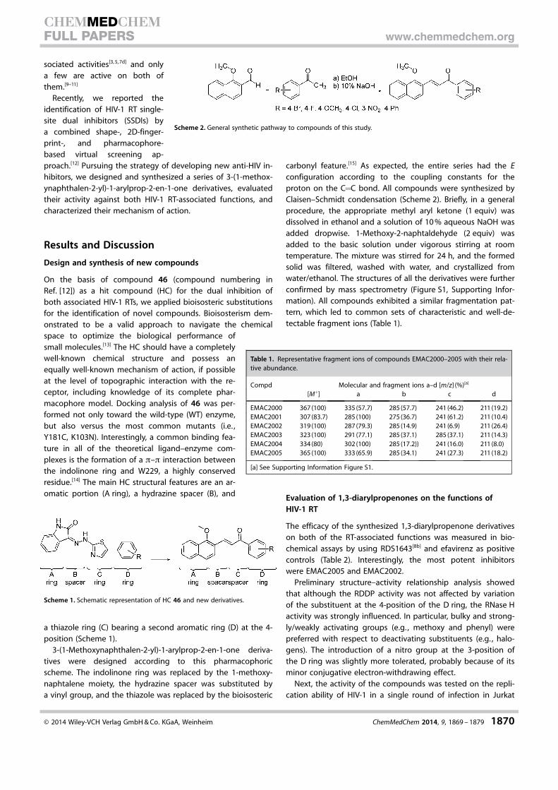
On the basis of compound 46 (compound numbering inRef. [12]) as a hit compound (HC) for the dual inhibition ofboth associated HIV-1 RTs, we applied bioisosteric substitutionsfor the identification of novel compounds. Bioisosterism dem-onstrated to be a valid approach to navigate the chemicalspace to optimize the biological performance ofsmall molecules.[13] The HC should have a completelywell-known chemical structure and possess anequally well-known mechanism of action, if possibleat the level of topographic interaction with the re-ceptor, including knowledge of its complete phar-macophore model. Docking analysis of 46 was per-formed not only toward the wild-type (WT) enzyme,but also versus the most common mutants (i.e. ,Y181C, K103N). Interestingly, a common binding fea-ture in all of the theoretical ligand–enzyme com-plexes is the formation of a p–p interaction betweenthe indolinone ring and W229, a highly conservedresidue.[14] The main HC structural features are an ar-omatic portion (A ring), a hydrazine spacer (B), and
a thiazole ring (C) bearing a second aromatic ring (D) at the 4-position (Scheme 1).
3-(1-Methoxynaphthalen-2-yl)-1-arylprop-2-en-1-one deriva-tives were designed according to this pharmacophoricscheme. The indolinone ring was replaced by the 1-methoxy-naphtalene moiety, the hydrazine spacer was substituted bya vinyl group, and the thiazole was replaced by the bioisosteric
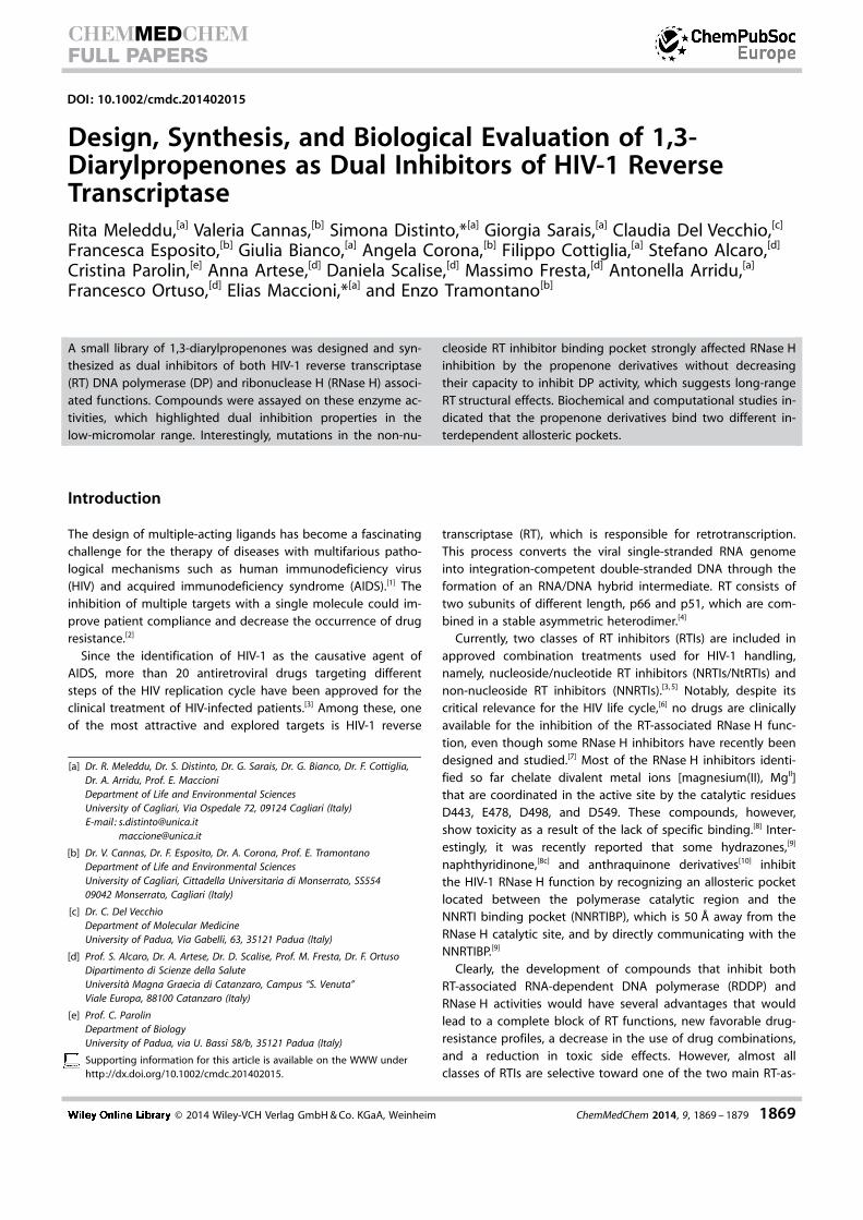
carbonyl feature.[15] As expected, the entire series had the Econfiguration according to the coupling constants for theproton on the C=C bond. All compounds were synthesized byClaisen–Schmidt condensation (Scheme 2). Briefly, in a generalprocedure, the appropriate methyl aryl ketone (1 equiv) wasdissolved in ethanol and a solution of 10 % aqueous NaOH wasadded dropwise. 1-Methoxy-2-naphtaldehyde (2 equiv) wasadded to the basic solution under vigorous stirring at roomtemperature. The mixture was stirred for 24 h, and the formedsolid was filtered, washed with water, and crystallized fromwater/ethanol. The structures of all the derivatives were furtherconfirmed by mass spectrometry (Figure S1, Supporting Infor-mation). All compounds exhibited a similar fragmentation pat-tern, which led to common sets of characteristic and well-de-tectable fragment ions (Table 1).
Evaluation of 1,3-diarylpropenones on the functions ofHIV-1 RT
The efficacy of the synthesized 1,3-diarylpropenone derivativeson both of the RT-associated functions was measured in bio-chemical assays by using RDS1643[8b] and efavirenz as positivecontrols (Table 2). Interestingly, the most potent inhibitorswere EMAC2005 and EMAC2002.
Preliminary structure–activity relationship analysis showedthat although the RDDP activity was not affected by variationof the substituent at the 4-position of the D ring, the RNase Hactivity was strongly influenced. In particular, bulky and strong-ly/weakly activating groups (e.g. , methoxy and phenyl) werepreferred with respect to deactivating substituents (e.g. , halo-gens). The introduction of a nitro group at the 3-position ofthe D ring was slightly more tolerated, probably because of itsminor conjugative electron-withdrawing effect.
Next, the activity of the compounds was tested on the repli-cation ability of HIV-1 in a single round of infection in Jurkat
Scheme 1. Schematic representation of HC 46 and new derivatives.
Scheme 2. General synthetic pathway to compounds of this study.
Table 1. Representative fragment ions of compounds EMAC2000–2005 with their rela-tive abundance.
Compd Molecular and fragment ions a–d [m/z] (%)[a]
[M+] a b c d
EMAC2000 367 (100) 335 (57.7) 285 (57.7) 241 (46.2) 211 (19.2)EMAC2001 307 (83.7) 285 (100) 275 (36.7) 241 (61.2) 211 (10.4)EMAC2002 319 (100) 287 (79.3) 285 (14.9) 241 (6.9) 211 (26.4)EMAC2003 323 (100) 291 (77.1) 285 (37.1) 285 (37.1) 211 (14.3)EMAC2004 334 (80) 302 (100) 285 (17.2)) 241 (16.0) 211 (8.0)EMAC2005 365 (100) 333 (65.9) 285 (34.1) 241 (27.3) 211 (18.2)
[a] See Supporting Information Figure S1.
� 2014 Wiley-VCH Verlag GmbH & Co. KGaA, Weinheim ChemMedChem 2014, 9, 1869 – 1879 1870
CHEMMEDCHEMFULL PAPERS www.chemmedchem.org
cells. Given that the CC50 values (cytotoxic concentration for50 % of cells) ranged between 3 and 15 mm, compound con-centrations were set at 10 mm for EMAC2000; 5, 0.5, and0.05 mm for EMAC2000 and EMAC2003; and 0.5 and 0.05 mm
for EMAC2004 and EMAC2005. All compounds were not ableto inhibit HIV-1 replication within these experimental condi-tions (Figure 1). Thus, we performed in vitro permeabilityassays to asses if a reduced or absent transmembrane permea-tion could explain these results.
Biochemical characterization of the mechanism of HIV-1 RTinhibition by EMAC2005
Several classes of HIV-1 RNase H have been reported, and ingeneral, they act by chelating the MgII ions within the active
site, which is essential for enzyme function. To verify the possi-bility that 1-(4-biphenyl)-3-(1-methoxynaphthalen-2-yl)prop-2-en-1-one (EMAC2005) could chelate the divalent ions in theRNase H catalytic site, we determined its absorbance spectra inthe absence and presence of MgCl2, but we did not observeany differences (data not shown).
Subsequently, we further investigated if EMAC2005 and thediketo acid derivative RDS1643, an RNase H catalytic site inhibi-tor,[8b] were able to simultaneously bind to the RT. Such anevaluation was performed by means of the Yonetani-revisedYonetani–Theorell model, which allows the competition be-tween two inhibitors of a certain enzyme for the same bindingsite or two non-overlapping binding sites to be determined. Inthis revised model, the plot of the inverse of the reaction ve-locity (1/V) observed in the presence of various concentrationsof the first inhibitor, in the absence or in the contemporaneouspresence of the second inhibitor, leads to a series of lines thatare parallel if the two inhibitors compete for the same bindingsite or a series of lines that intersect if the inhibitors bind todifferent enzyme sites.[16]
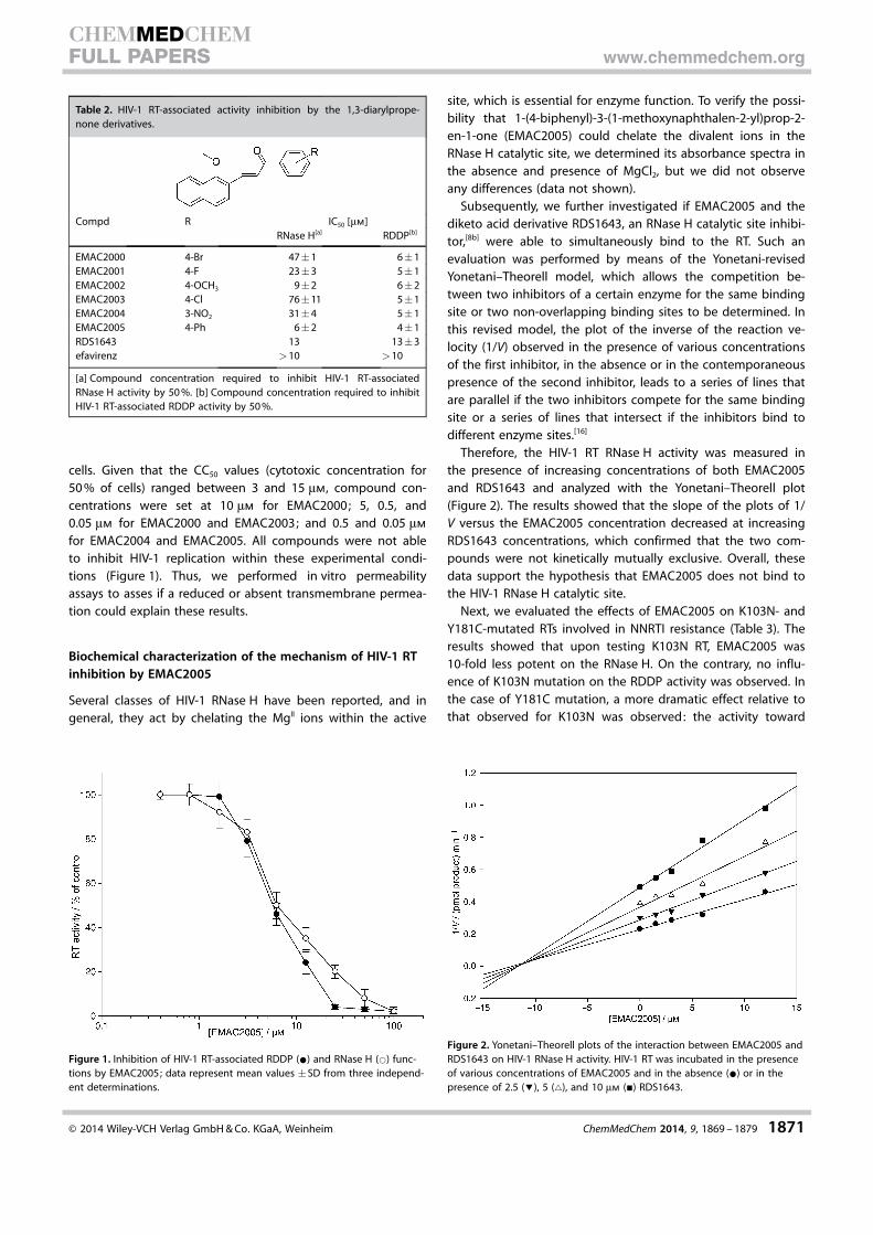
Therefore, the HIV-1 RT RNase H activity was measured inthe presence of increasing concentrations of both EMAC2005and RDS1643 and analyzed with the Yonetani–Theorell plot(Figure 2). The results showed that the slope of the plots of 1/V versus the EMAC2005 concentration decreased at increasingRDS1643 concentrations, which confirmed that the two com-pounds were not kinetically mutually exclusive. Overall, thesedata support the hypothesis that EMAC2005 does not bind tothe HIV-1 RNase H catalytic site.
Next, we evaluated the effects of EMAC2005 on K103N- andY181C-mutated RTs involved in NNRTI resistance (Table 3). Theresults showed that upon testing K103N RT, EMAC2005 was10-fold less potent on the RNase H. On the contrary, no influ-ence of K103N mutation on the RDDP activity was observed. Inthe case of Y181C mutation, a more dramatic effect relative tothat observed for K103N was observed: the activity toward
Table 2. HIV-1 RT-associated activity inhibition by the 1,3-diarylprope-none derivatives.
Compd R IC50 [mm]RNase H[a] RDDP[b]
EMAC2000 4-Br 47�1 6�1EMAC2001 4-F 23�3 5�1EMAC2002 4-OCH3 9�2 6�2EMAC2003 4-Cl 76�11 5�1EMAC2004 3-NO2 31�4 5�1EMAC2005 4-Ph 6�2 4�1RDS1643 13 13�3efavirenz >10 >10
[a] Compound concentration required to inhibit HIV-1 RT-associatedRNase H activity by 50 %. [b] Compound concentration required to inhibitHIV-1 RT-associated RDDP activity by 50 %.
Figure 1. Inhibition of HIV-1 RT-associated RDDP (*) and RNase H (*) func-tions by EMAC2005; data represent mean values �SD from three independ-ent determinations.
Figure 2. Yonetani–Theorell plots of the interaction between EMAC2005 andRDS1643 on HIV-1 RNase H activity. HIV-1 RT was incubated in the presenceof various concentrations of EMAC2005 and in the absence (*) or in thepresence of 2.5 (!), 5 (~), and 10 mm (&) RDS1643.
� 2014 Wiley-VCH Verlag GmbH & Co. KGaA, Weinheim ChemMedChem 2014, 9, 1869 – 1879 1871
CHEMMEDCHEMFULL PAPERS www.chemmedchem.org

RNase H activity was almost suppressed, whereas the activitytoward the RT-associated RDDP was only slightly affected.This behavior might be explained either by the binding ofEMAC2005 to a single site close to Y181, the hydrazonespocket,[9] or the NNRTIBP, or by the interaction of the com-pound with two interdependent pockets, the conformations ofwhich are affected by the Y181C mutation.
In silico modeling of the interaction of EMAC2005 withHIV-1 RT
A computational study based on molecular docking experi-ments in tandem with molecular dynamics simulations wasperformed to understand the possible mechanism of inhibi-tion of this series of diarylpropenone derivatives. The studieswere focused on the most active compound only, that is,EMAC2005.
Molecular docking approaches have become very useful andlargely widespread for the prediction of feasible bindingmodes of a ligand, the target site of which is either known orunknown (blind docking). According to the available literature,dual inhibitory activity could be achieved either by inhibitor
binding into two different sites[17] or by its binding intoa single site.[7d, 8c, 9, 10c, 12] Therefore, we investigated both possi-bilities.
The very diverse group of NNRTIs bind allosterically in thehydrophobic NNRTIBP and lock the enzyme into an inactiveform. Owing to the flexibility of the target and to the differentshapes of the known inhibitors (Figure 3 a), we decided to per-form ensemble docking experiments.[18] The major conforma-tional changes in the NNRTIBP were taken into account to per-form a clustering of the available RT-NNRTI complexes. In par-ticular, the orientation of Y181, Y188, Y183, and primer gripb12–b13 hairpin were considered (Figure 3 b).[19] A representa-tive of each different cluster was picked, and the 3D structureof HIV RT was retrieved from the RCSB Protein Data Bank(PDB).[20]
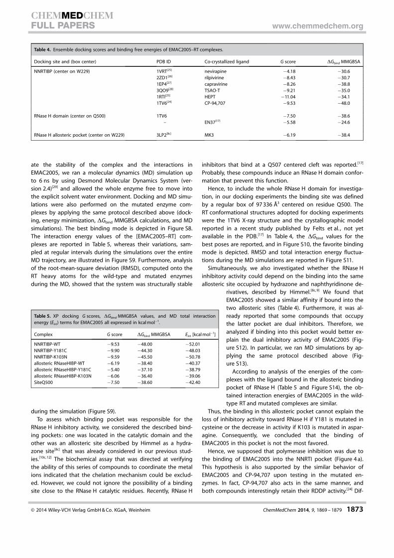
QM (quantum mechanical) polarized docking was per-formed.[21] This recognition workflow combines docking withsemi-empirical methods to calculate charges within the proteinenvironment. This methodology performs significantly betterthan classical molecular mechanics approaches.[22] We validatedthe protocol for this target by performing re-docking experi-ments (data not shown). The obtained [EMAC2005–RT] com-plexes were subjected to a post-docking procedure based onenergy minimization and successive binding free energy calcu-lations. The binding free energies (DGbind) were obtained byapplying molecular mechanics and continuum solvationmodels by using the molecular mechanics generalized Born/surface area (MMGBSA) method.[23] As reported in Table 4, bycomparing the DGbind MMGBSA values, we could assert thatthe most probable binding mode in the NNRTIBP was obtainedby docking the compound into the RT conformation model re-ported in PDB ID 1TV6.[24]
The best docking pose of EMAC2005 and its comparisonwith respect to the relative co-crystallized compounds are re-ported in the Supporting Information (Figures S2–S7). To evalu-
Table 3. Inhibition of drug-resistant HIV-1 mutated RT-associated func-tions by EMAC2005.
Compd IC50 [mm]Y181C RT K103N RT
RNase H[a] RDDP[b] RNase H[a] RDDP[b]
EMAC2005 >100 8�3 59�8 3�1efavirenz – 0.40�0.03 – 0.68�0.05
[a] Compound concentration required to inhibit HIV-1 RT-associatedRNase H activity by 50 %. [b] Compound concentration required to inhibitHIV-1 RT-associated RDDP activity by 50 %.
Figure 3. a) Structures and PDB IDs of co-crystallized NNRTIs selected for the ensemble docking procedure. b) Superimposition of the primer grip region andof residues 181, 183, and 188 of employed RT PDB structures for ensemble docking experiments.
� 2014 Wiley-VCH Verlag GmbH & Co. KGaA, Weinheim ChemMedChem 2014, 9, 1869 – 1879 1872
CHEMMEDCHEMFULL PAPERS www.chemmedchem.org
ate the stability of the complex and the interactions inEMAC2005, we ran a molecular dynamics (MD) simulation upto 6 ns by using Desmond Molecular Dynamics System (ver-sion 2.4)[29] and allowed the whole enzyme free to move intothe explicit solvent water environment. Docking and MD simu-lations were also performed on the mutated enzyme com-plexes by applying the same protocol described above (dock-ing, energy minimization, DGbind MMGBSA calculations, and MDsimulations). The best binding mode is depicted in Figure S8.The interaction energy values of the [EMAC2005–RT] com-plexes are reported in Table 5, whereas their variations, sam-pled at regular intervals during the simulations over the entireMD trajectory, are illustrated in Figure S9. Furthermore, analysisof the root-mean-square deviation (RMSD), computed onto theRT heavy atoms for the wild-type and mutated enzymesduring the MD, showed that the system was structurally stable
during the simulation (Figure S9).To assess which binding pocket was responsible for the
RNase H inhibitory activity, we considered the described bind-ing pockets: one was located in the catalytic domain and theother was an allosteric site described by Himmel as a hydra-zone site[8c] that was already considered in our previous stud-ies.[10c, 12] The biochemical assay that was directed at verifyingthe ability of this series of compounds to coordinate the metalions indicated that the chelation mechanism could be exclud-ed. However, we could not ignore the possibility of a bindingsite close to the RNase H catalytic residues. Recently, RNase H
inhibitors that bind at a Q507 centered cleft was reported.[17]
Probably, these compounds induce an RNase H domain confor-mation that prevent this function.
Hence, to include the whole RNase H domain for investiga-tion, in our docking experiments the binding site was definedby a regular box of 97 336 �3 centered on residue Q500. TheRT conformational structures adopted for docking experimentswere the 1TV6 X-ray structure and the crystallographic modelreported in a recent study published by Felts et al. , not yetavailable in the PDB.[17] In Table 4, the DGbind values for thebest poses are reported, and in Figure S10, the favorite bindingmode is depicted. RMSD and total interaction energy fluctua-tions during the MD simulations are reported in Figure S11.
Simultaneously, we also investigated whether the RNase Hinhibitory activity could depend on the binding into the sameallosteric site occupied by hydrazone and naphthyridinone de-
rivatives, described by Himmel.[8c, 9] We found thatEMAC2005 showed a similar affinity if bound into thetwo allosteric sites (Table 4). Furthermore, it was al-ready reported that some compounds that occupythe latter pocket are dual inhibitors. Therefore, weanalyzed if binding into this pocket would better ex-plain the dual inhibitory activity of EMAC2005 (Fig-ure S12). In particular, we ran MD simulations by ap-plying the same protocol described above (Fig-ure S13).
According to analysis of the energies of the com-plexes with the ligand bound in the allosteric bindingpocket of RNase H (Table 5 and Figure S14), the ob-tained interaction energies of EMAC2005 in the wild-type RT and mutated complexes are similar.
Thus, the binding in this allosteric pocket cannot explain theloss of inhibitory activity toward RNase H if Y181 is mutated incysteine or the decrease in activity if K103 is mutated in aspar-agine. Consequently, we concluded that the binding ofEMAC2005 in this pocket is not the most favored.
Hence, we supposed that polymerase inhibition was due tothe binding of EMAC2005 into the NNRTI pocket (Figure 4 a).This hypothesis is also supported by the similar behavior ofEMAC2005 and CP-94,707 upon testing in the mutated en-zymes. In fact, CP-94,707 also acts in the same manner, andboth compounds interestingly retain their RDDP activity.[24] Dif-
Table 4. Ensemble docking scores and binding free energies of EMAC2005–RT complexes.
Docking site and (box center) PDB ID Co-crystallized ligand G score DGbind MMGBSA
NNRTIBP (center on W229) 1VRT[25] nevirapine �4.18 �30.62ZD1[26] rilpivirine �8.43 �30.71EP4[27] capravirine �8.26 �38.83QO9[28] TSAO-T �9.21 �35.01RTI[25] HEPT �11.04 �34.11TV6[24] CP-94,707 �9.53 �48.0
RNase H domain (center on Q500) 1TV6 �7.50 �38.6– EN37[17] �5.58 �24.6
RNase H allosteric pocket (center on W229) 3LP2[8c] MK3 �6.19 �38.4
Table 5. XP docking G scores, DGbind MMGBSA values, and MD total interactionenergy (Eint) terms for EMAC2005 all expressed in kcal mol�1.
Complex G score DGbind MMGBSA Eint [kcal mol�1]
NNRTIBP-WT �9.53 �48.00 �52.01NNRTIBP-Y181C �9.90 �44.30 �48.03NNRTIBP-K103N �9.59 �45.50 �50.78allosteric RNaseHIBP-WT �6.19 �38.40 �40.37allosteric RNaseHIBP-Y181C �5.40 �37.10 �38.79allosteric RNaseHIBP-K103N �6.06 �36.40 �39.06SiteQ500 �7.50 �38.60 �42.40
� 2014 Wiley-VCH Verlag GmbH & Co. KGaA, Weinheim ChemMedChem 2014, 9, 1869 – 1879 1873
CHEMMEDCHEMFULL PAPERS www.chemmedchem.org
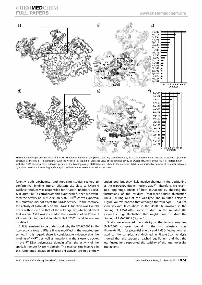
ferently, both biochemical and modeling studies seemed toconfirm that binding into an allosteric site close to RNase Hcatalytic residues was responsible for RNase H inhibitory activi-ty (Figure 4 b). To corroborate this hypothesis further, we evalu-ated the activity of EMAC2005 on A502F RT.[30] As we expected,this mutation did not affect the RDDP activity. On the contrary,the activity of EMAC2005 on the RNase H function was fivefoldlower with respect to that of the wild-type RT, which indicatedthat residue A502 was involved in the formation of an RNase Hallosteric binding pocket in which EMAC2005 could be accom-modated.
Still, it remained to be understood why the EMAC2005 inhib-itory activity toward RNase H was modified in the mutated en-zymes. In this regard, there is considerable evidence that thebinding of NNRTIs as well as mutations in the allosteric pocketin the RT DNA polymerase domain affect the activity of thespatially remote RNase H domain. The mechanisms involved inthis long-range alteration of RNase H activity are not entirely
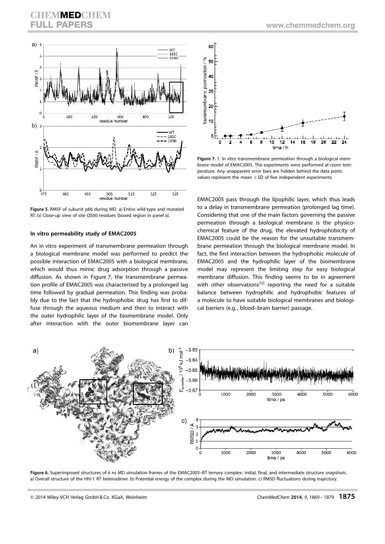
understood, but they likely involve changes in the positioningof the RNA/DNA duplex nucleic acid.[31] Therefore, we exam-ined long-range effects of both mutations by checking thefluctuations of the residues [root-mean-square fluctuation(RMSF)] during MD of the wild-type and mutated enzymes(Figure 5 a). We noticed that although the wild-type RT did notshow relevant fluctuations in the Q500 site involved in thebinding of EMAC2005, some residues in the mutated RTsshowed a huge fluctuation that might have disturbed thebinding of EMAC2005 (Figure 5 b).
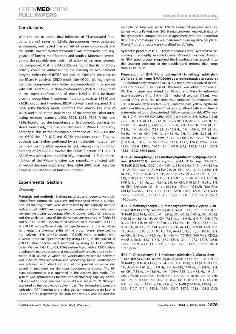
Finally, we evaluated the stability of the ternary enzyme–EMAC2005 complex bound in the two allosteric sites(Figure 6). Plots for potential energy and RMSD fluctuations re-lated to the complex are depicted in Figure 6 b,c. Analysisshowed that the structure reached equilibrium and that thelow fluctuations supported the stability of the intermolecularinteractions.
Figure 4. Superimposed structures of 6 ns MD simulation frames of the [EMAC2005–RT] complex: initial, final, and intermediate structure snapshots. a) Overallstructure of the HIV-1 RT heterodimer with the NNRTIBP occupied. b) Close-up view of the binding cavity. d) Overall structure of the HIV-1 RT heterodimerwith the Q500 site occupied. e) Close-up view of the binding cavity. c,f) Residues involved in the complex stabilization sorted by number of contacts betweenligand and receptor. Interacting and catalytic residues are represented as stick structures.
� 2014 Wiley-VCH Verlag GmbH & Co. KGaA, Weinheim ChemMedChem 2014, 9, 1869 – 1879 1874
CHEMMEDCHEMFULL PAPERS www.chemmedchem.org
In vitro permeability study of EMAC2005
An in vitro experiment of transmembrane permeation througha biological membrane model was performed to predict thepossible interaction of EMAC2005 with a biological membrane,which would thus mimic drug adsorption through a passivediffusion. As shown in Figure 7, the transmembrane permea-tion profile of EMAC2005 was characterized by a prolonged lagtime followed by gradual permeation. This finding was proba-bly due to the fact that the hydrophobic drug has first to dif-fuse through the aqueous medium and then to interact withthe outer hydrophilic layer of the biomembrane model. Onlyafter interaction with the outer biomembrane layer can
EMAC2005 pass through the lipophilic layer, which thus leadsto a delay in transmembrane permeation (prolonged lag time).Considering that one of the main factors governing the passivepermeation through a biological membrane is the physico-chemical feature of the drug, the elevated hydrophobicity ofEMAC2005 could be the reason for the unsuitable transmem-brane permeation through the biological membrane model. Infact, the first interaction between the hydrophobic molecule ofEMAC2005 and the hydrophilic layer of the biomembranemodel may represent the limiting step for easy biologicalmembrane diffusion. This finding seems to be in agreementwith other observations[32] reporting the need for a suitablebalance between hydrophilic and hydrophobic features ofa molecule to have suitable biological membranes and biologi-cal barriers (e.g. , blood–brain barrier) passage.
Figure 5. RMSF of subunit p66 during MD. a) Entire wild-type and mutatedRT. b) Close-up view of site Q500 residues (boxed region in panel a).
Figure 6. Superimposed structures of 6 ns MD simulation frames of the EMAC2005–RT ternary complex: initial, final, and intermediate structure snapshots.a) Overall structure of the HIV-1 RT heterodimer. b) Potential energy of the complex during the MD simulation. c) RMSD fluctuations during trajectory.
Figure 7. 1. In vitro transmembrane permeation through a biological mem-brane model of EMAC2005. The experiments were performed at room tem-perature. Any unapparent error bars are hidden behind the data point;values represent the mean �SD of five independent experiments.
� 2014 Wiley-VCH Verlag GmbH & Co. KGaA, Weinheim ChemMedChem 2014, 9, 1869 – 1879 1875
CHEMMEDCHEMFULL PAPERS www.chemmedchem.org
Conclusions
With the aim to obtain dual inhibitors of RT-associated func-tions, a small series of 1,3-diarylpropenones were designed,synthesized, and tested. The activity of some compounds andthe profile toward mutated enzymes was remarkable and sug-gestive of further modifications and studies. Moreover, investi-gating the possible mechanism of action of the most-promis-ing compound, that is, EMAC2005, we found that its inhibitoryactivity could be addressed to the binding at two differentenzyme clefts : the NNRTIBP site and an allosteric site close tothe RNase H catalytic DEDD motif (site Q500). We highlightedthat the compound was better accommodated in a pocketwith Y181 and Y188 in close conformation (PDB ID: 1TV6) thanin the open conformation of most NNRTIs. This facilitatesenzyme recognition if common mutations, such as Y181C andK103N, occur, and therefore, RDDP activity is not impaired. TheEMAC2005 binding mode confirms the known key role ofW229 and Y188 in the stabilization of the complex. Other inter-acting residues, namely, L100, P225, L234, Y318, V106, andP236, highlighted the importance of hydrophobic contacts. In-stead, most likely, the loss and decrease in RNase H inhibitorypotency is due to the improbable entrance of EMAC2005 intothe Q500 site if Y181C and K103N mutations occur. This hy-pothesis was further confirmed by a single-point mutation ex-periment on the A502 residue. In fact, whereas the inhibitionpotency of EMAC2005 toward the RDDP function of HIV-1 RTA502F was almost not modified (IC50 increased 1.5-fold), the in-hibition of the RNase function was remarkably affected witha fivefold decrease in potency. Thus, EMAC2005 most likely be-haves as a dual-site dual-function inhibitor.
Experimental Section
Chemistry
Materials and methods: Starting materials and reagents were ob-tained from commercial suppliers and were used without purifica-tion. All melting points were determined by the capillary methodwith a Stuart SMP11 melting point apparatus or a B�chi-540 capil-lary melting points apparatus. Melting points, yields of reactions,and the analytical data of the derivatives are reported in Tables S1and S2. The 1H NMR spectra of all samples were measured in CDCl3
at 278.1 K with a Varian Unity 300 spectrometer. In the signal as-signments, the chemical shifts of the proton were referenced tothe solvent (1 H: d= 7.24 ppm). 13C NMR were recorded witha Varian Unity 500 spectrometer by using CDCl3 as the solvent at278.1 K. Mass spectra were recorded by using an HPLC–MS/MSVarian (Varian, Palo Alto, CA, USA) system fitted with a 1200 L triplequadrupole mass spectrometer equipped with an electrospray ioni-zation (ESI) source. A Varian MS workstation version 6.8 softwarewas used for data acquisition and processing. Rapid identificationwas achieved with direct infusion of the purified molecule, dis-solved in methanol, on the mass spectrometer source. The ESImass spectrometer was operated in the positive ion mode. Thesystem was optimized as follows: the electrospray capillary poten-tial was set to 65 V, whereas the shield was set at 725 V. Nitrogenwas used as the desolvation solvent gas. The atmospheric pressureionization (API) housing and drying gas temperatures were kept at54 and 375 8C, respectively. The scan time was 1 s, and the detector
multiplier voltage was set to 1700 V. Elemental analyses were ob-tained with a PerkinElmer 240 B microanalyzer. Analytical data ofthe synthesized compounds are in agreement with the theoreticaldata. TLC chromatography was performed by using silica gel plates(Merck F254), and spots were visualized by UV light.
Synthetic procedures: 1,3-Diarylpropenones were synthesized ac-cording to a slightly modified Claisen–Schmidt reaction. Analysisby NMR spectroscopy supported the E configuration according tothe coupling constants of the double-bond protons that rangefrom 15 to 16 Hz.
Preparation of (E)-1-(4-bromophenyl)-3-(1-methoxynaphthalen-2-yl)prop-2-en-1-one (EMAC2000) as a representative procedure:1-(4-Bromophenyl)ethanone (0.9 g, 4.5 mmol) was dissolved in eth-anol (15 mL) and a solution of 10 % NaOH was added dropwise atRT. The mixture was stirred for 10 min, and then 1-methoxy-2-naphthaldehyde (1 g, 5.4 mmol) in ethanol solution (15 mL) wasadded. After 24 h, the reaction was complete (as monitored byTLC, n-hexane/ethyl acetate = 2:1), and the pale yellow crystallinesolid was filtered, washed with water, crystallized with a mixture ofwater/ethanol, and characterized. Yellow crystals ; yield: 67 %; mp:110–112 8C; 1H NMR (300 MHz, CDCl3): d= 4.06 (s, 3 H, OCH3), 7.3 (d,J = 9.1 Hz, 1 H, Ar�CH), 7.41 (t, J = 7.5 Hz, 1 H, Ar�CH), 7.55 (t, J =7.5 Hz, 1 H, Ar�CH), 7.65 (d, J = 8.3 Hz, 2 H, Ar�CH), 7.82 (d, J =7.5 Hz, 1 H, Ar�CH), 7.85 (d, J = 15.6 Hz, 1 H, �CH=), 7.9 (d, J =9,0 Hz, 1 H, Ar�CH), 7.93 (d, J = 8.3 Hz, 2 H, Ar�CH), 8.25 (d, J =8.6 Hz, 1 H, Ar�CH), 8.51 ppm (d, J = 15.6 Hz, 1 H, �CH=) ; 13C NMR(100 MHz, CDCl3): d= 56.1 112.7, 117.1, 123.3, 124.1, 126.6, 127.6,128.7, 129.0, 130.0, 130.1 (2 C), 131.8 (2 C), 132.1, 133.1, 137.3,138.3, 157.2, 190.2 ppm.
(E)-1-(4-Fluorophenyl)-3-(1-methoxynaphthalen-2-yl)prop-2-en-1-one (EMAC2001): Yellow crystals; yield: 81 %; mp: 93–95 8C;1H NMR (300 MHz, CDCl3): d= 4.07 (s, 3 H, OCH3), 7.11 (t, J = 8.5 Hz,1 H, Ar�CH), 7.26 (d, J = 9.1 Hz, 2 H, Ar�CH), 7.42 (d, J = 7.1 Hz, 1 H,Ar�CH), 7.50 (t, J = 8.5 Hz, 1 H, Ar�CH), 7.57 (d, J = 7.1 Hz, 1 H, Ar�CH), 7.78 (d, J = 15.9 Hz, 1 H, �CH=), 7.93 (d, J = 8.6 Hz, 1 H, Ar�CH),8.09 (t, JH,H = 9.1 Hz, JH,F = 9.3 Hz, 2 H, Ar�CH), 8.18 (d, J = 8.5 Hz, 1 H,Ar�CH), 8.43 ppm (d, 1 H, J = 15.9 Hz, �CH=) ; 13C NMR (100 MHz,CDCl3): d= 56.5, 112.7, 115.7, 123.3, 124.0, 126.8, 127.6, 128.6 (2 C),129.0, 130.0 (2 C), 131.1, 131.3, 132.0, 133.0, 136.2, 138.3, 157.2,190.0 ppm.
(E)-1-(4-Methoxyphenyl)-3-(1-methoxynaphthalen-2-ylprop-2-en-1-one (EMAC2002): Yellow crystals ; yield: 83 %; mp: 137–139 8C;1H NMR (300 MHz, CDCl3): d= 3.9 (s, 3 H, OCH3), 4.05 (s, 3 H, OCH3),7.00 (d, J = 8.9 Hz, 1 H, Ar�CH), 7.33 (d, J = 9.0 Hz, 2 H, Ar�CH), 7.70(t, J = 8 Hz, 1 H, Ar�CH), 7.47 (d, J = 16.0 Hz, 1 H, �CH=), 7.55 (t, J =8 Hz, 1 H, Ar�CH), 7.82 (d, J = 8.0 Hz, 1 H, Ar�CH), 7.89 (d, J = 9.0 Hz,1 H, Ar�CH), 8.08 (d, J = 9.0 Hz, 1 H, Ar�CH), 8.28 (d, J = 9.0 Hz, 2 H,Ar�CH), 8.45 (d, J = 16.0 Hz, 1 H, �CH=) ; 13C NMR (100 MHz, CDCl3):d= 55.4, 56.5, 112.7, 113.5, 117.7, 123.5, 124.1, 127.2, 127.6, 128.6,129.1, 130.9 (2 C), 131.5 (2 C), 131.7, 133.1, 137.0, 156.9, 163.3,189.5 ppm.
(E)-1-(4-Chlorophenyl)-3-(1-methoxynaphthalen-2-yl)prop-2-en-1-one (EMAC2003): Yellow crystals ; yield: 53 %; mp: 108–109 8C;1H NMR (300 MHz, CDCl3): d= 4.06 (s, 3 H, OCH3), 7.33 (d, J = 9.1 Hz,1 H, Ar�CH), 7.41 (t, J = 7.8 Hz, 1 H, Ar�CH), 7.49 (d, J = 8.4 Hz, 2 H,Ar�CH), 7.53 (d, J = 15.6 Hz, 1 H, �CH=), 7.55 (t, J = 7.8 Hz, 1 H, Ar�CH), 7.74 (d, J = 8.1 Hz, 1 H, Ar�CH), 7.90 (d, J = 9.6 Hz, 1 H, Ar�CH),8.01 (d, J = 8.3 Hz, 2 H, Ar�CH), 8.25 (d, J = 8.6 Hz, 1 H, Ar�CH),8.51 ppm (d, J = 15.6 Hz, 1 H,�CH=) ; 13C NMR (500 MHz, CDCl3): d=56.3, 112.7, 117,1, 123.3, 124.0, 126.7, 127.6, 128.6, 128.8 (2 C),
� 2014 Wiley-VCH Verlag GmbH & Co. KGaA, Weinheim ChemMedChem 2014, 9, 1869 – 1879 1876
CHEMMEDCHEMFULL PAPERS www.chemmedchem.org
129.0, 130.0 (2 C), 132.1, 133.1, 136.9, 138.3, 139.0, 157.2,190.0 ppm.
(E)-1-(3-Nitrophenyl)-3-(1-methoxynaphthalen-2-yl)prop-2-en-1-one (EMAC2004): Pale orange crystals; yield: 64 %; mp: 143–145 8C; 1H NMR (300 MHz, CDCl3): d= 4.10 (s, 3 H, OCH3), 7.35 (d,J = 8.9 Hz, 1 H, Ar�CH), 7.42 (t, J = 7.8 Hz, 1 H, Ar�CH), 7.58 (t, J =8.5 Hz, 1 H, Ar�CH), 7.72 (t, J = 7.8 Hz, 1 H, Ar�CH), 7.83 (d, J =8.0 Hz, 1 H, Ar�CH), 7.93 (d, J = 8.5 Hz, 1 H, Ar�CH), 7.94 (d, J =15.6 Hz, 1 H, �CH=), 8.25 (d, J = 8.5 Hz, 1 H, Ar�CH), 8.39 (d, J =8.5 Hz, 1 H, Ar�CH), 8.44 (d, J = 8.0 Hz, 1 H, Ar�CH), 8.60 (d, J =15.6 Hz, <1 H, C->CH=), 8.89 ppm (s, 1 H, Ar�CH); 13C NMR(500 MHz, CDCl3): d= 56.3, 112.6, 116.6, 123.1, 123.4, 124.1, 125.7,126.8, 127.8, 128.8, 129.0, 129.8, 132.7, 133.1, 134.1, 139.5, 140.0,148.3, 157.6, 189.0 ppm.
(E)-1-(4-Biphenyl)-3-(1-methoxynaphthalen-2-yl)prop-2-en-1-one(EMAC2005): Yellow crystals; yield: 87 %; mp: 104–105 8C; 1H NMR(300 MHz, CDCl3): d= 4.07 (s, 3 H, OCH3), 7.34–7.50 (m, 2 H, Ar�CH),7.56 (t, J = 8.1 Hz, 1 H, Ar�CH), 7.69 (d, J = 7.8 Hz, 2 H, Ar�CH), 7.75(d, J = 7.8 Hz, 2 H, Ar�CH), 7.83(d, J=8.8 Hz, 1 H, Ar�CH), 7.88 (d, J =8.0 Hz, 1 H, Ar�CH), 7.91 (d, J = 8.2 Hz, 2 H, Ar�CH), 7.95 (d, J =15.9 Hz, 1 H, �CH=), 8.04 (d, J = 8.2 Hz, 2 H, Ar�CH), 8.19 (d, J =
8.8 Hz, 1 H, Ar�CH), 8.29 (d, J = 8.1 Hz, 1 H, Ar�CH), 8.54 ppm (d, J =15.9 Hz, 1 H, �CH=) ; 13C NMR (100 MHz, CDCl3): d= 56.4, 112.8,117.4, 123.5, 124.0, 127.2 (3 C), 127.3 (3 C), 127.5, 128.1, 128.6, 128.9(2 C), 129.1, 129.2 (2 C), 131.8, 137.3, 137.7, 140.1, 145.3, 157.1,190.7 ppm.
Biology
Protein expression and purification: The recombinant HIV-1 RT pro-tein, the coding gene of which was subcloned in the p6HRT_protplasmid, was expressed in E. coli strain M15.[33] The bacteria cellswere grown up to an optical density (at 600 nm) of 0.8 and in-duced with 1.7 mm isopropyl b-d-1-thiogalactopyranoside (IPTG)for 5 h. HIV-1 RT purification was performed as described.[8h] Briefly,cell pellets were re-suspended in lysis buffer (20 mm HEPES,pH 7.5; 0.5 m NaCl; 5 mm b-mercaptoethanol; 5 mm imidazole;0.4 mg mL�1 lysozyme), incubated on ice for 20 min, sonicated, andcentrifuged at 30 000 g for 1 h. The supernatant was applied toa His-binding resin column and washed thoroughly with washbuffer (20 mm HEPES, pH 7.5; 0.3 m NaCl; 5 mm b-mercaptoetha-nol; 60 mm imidazole; 10 % glycerol). RT was eluted by imidazolegradient, and the enzyme-containing fractions were pooled and di-alyzed and aliquots were stored at �80 8C.
RNase H polymerase-independent cleavage assay: The HIV-1 RT-asso-ciated RNase H activity was measured as described[12] in 100 mL re-action volume containing 50 mm Tris HCl, pH 7.8; 6 mm MgCl2 ;1 mm dithiothreitol (DTT); 80 mm KCl; hybrid RNA/DNA (5’-GTTTTC TTT TCC CCC CTG AC-3’-fluorescein; 5’-CAA AAG AAA AGGGGG GAC UG-3’-dabcyl) and 3.8 nm RT. The reaction mixture wasincubated for 1 h at 37 8C. The enzymatic reaction was stoppedwith the addition of ethylenediaminetetraacetic acid (EDTA) andmeasured with a Victor3 instrument (Perkin) at 490/528 nm.
DNA polymerase assay: The HIV-1 RT-associated (RDDP) activity wasmeasured by using an Invitrogen EnzCheck Reverse TranscriptaseAssay Kit, in 50 mL volume containing 60 mm Tris HCl, pH 8.1;8 mm MgCl2 ; 60 mm KCl; 13 mm DTT; 100 mm dTTP; 2 nm HIV-1 RT;poly(A)-oligo(dT). The reaction mixture was incubated for 30 min at37 8C. The enzymatic reaction was stopped with the addition ofEDTA and measured with Victor3 (Perkin) at 502/523 nm after theaddition of picogreen.
Cell lines and virus: The human embryonic c kidney cells 293T andthe human T-lymphoid Jurkat cell line (clone E6-1) were from theAmerican Type Culture Collection and maintained in DMEM orRPMI medium (Invitrogen), respectively, containing 10 % fetalbovine serum (FBS, Invitrogen), at 37 8C under a humidified 5 %CO2 atmosphere. Recombinant viral stock was produced by transi-ent transfection of 293T cells as previously described[34] and usedto transduce Jurkat cells. In this context, an env-defective provirusencoding the bacterial chloramphenicol acetyltransferase (CAT)gene was complemented in trans by the envelope glycoproteinderived from the laboratory-adapted T-cell-tropic strain HXBc2. Thelevel of CAT expression in the infected cells reflects the efficiencyof a single round of the retroviral infection cycle.
Cytotoxicity assay: For cytotoxicity assays, cell lines were seeded in96-well plates (Falcon) at an initial density of 105 cells per 100 mL inmedium containing 10 % FBS, in the absence or presence of serialdilutions of test compounds. Plates were incubated for 72 h at37 8C in under a humidified 5 % CO2 atmosphere. Cell viability wasdetermined by using Cell Proliferation Kit I (MTT) (Roche).
Molecular modeling
Ligand preparation: Theoretical 3D models of the most active de-rivative EMAC2005 was built by means of Maestro.[35] The inhibitorstructure was optimized by means of an energy minimization per-formed by using the MMFFs force field,[36] the GB/SA[37] water im-plicit solvation model, and the Polak–Ribier Conjugate Gradient(PRCG) method by converging on gradient with a threshold of0.05 kJ (mol �)�1
.
Protein preparation: The coordinates for reverse transcriptase en-zymes were taken from the RCSB Protein Data Bank[20] (PDB IDs:1VRT,[25] 2ZD1,[26] 1EP4,[27] 3QO9,[28] 1RTI,[25] 1TV6,[24] and 3LP2).[8c]
The proteins were prepared by using the Maestro Protein Prepara-tion Wizard. Original water molecules were removed and terminiwere capped. The bond orders and formal charges were added forhetero groups, and all the hydrogen atoms were added in thestructure. Missing atoms and residues were included. After prepa-ration, the structures were refined to optimize the hydrogen-bondnetwork by using OPLS_2005[38] force field. The minimization wasterminated once the energy converged or the RMSD reacheda maximum cutoff of 0.30 �.
Docking protocol: Molecular docking studies were performed byusing the QMPL workflow protocol.[21] Grids were defined aroundthe refined structure by centering on the residue indicated inTable 3 and fixing the box volume at 97 336 �3. The extra precision(XP) docking algorithm was applied for scoring theoretical poses.The other settings were left as default. The same protocol was ap-plied for all the simulations indicated in Tables 3 and 4.
Post-docking protocol: A total of 10 000 steps of the Polak–Ribierconjugate gradient (PRCG) minimization method were conductedon the top-ranked theoretical complexes by using the OPLS_2005force field. The optimization process was performed up to the de-rivative convergence criterion equal to 0.01 kJ (mol �)�1. The bind-ing free energies (DGbind) were computed by applying molecularmechanics and continuum solvation models with the molecularmechanics generalized Born/surface area (MMGBSA) method.[23]
Best docking complexes were submitted to 6 ns of MD by usingDesmond (version 2.4).[39] The complexes were solvated witha TIP3P (transferable intermolecular potential 3-Point)[40] box ofwater and counter ions were added to neutralize the system netcharge. The solvated models were optimized, and subsequently
� 2014 Wiley-VCH Verlag GmbH & Co. KGaA, Weinheim ChemMedChem 2014, 9, 1869 – 1879 1877
CHEMMEDCHEMFULL PAPERS www.chemmedchem.org
the MTK_NPT (Martyna–Tobias–Klein with constant number of par-ticles, pressure and temperature) ensemble was employed.[41] Thedefault stages in the relaxation process for the NPT ensemble in-cluded two energy minimizations and four simulation steps.During the energy minimizations, two runs of 2000 iteration wereprocessed by using the steepest descent method: during the firstrun, the protein structure was fixed by a force restraint constant of50 kcal (mol �)�1, and in the second run all restraints were removed.With the first simulation, at NVT (constant number of particles,volume, and temperature) ensemble, the system reached a temper-ature of 10 K. In the following three simulations in the NPT ensem-ble, the system was heated up to 300 K and the pressure was keptconstant at 100 kPa by using the Berendsen thermostat–barostat.During the production phase, temperature and pressure were keptconstant by using the Nos�–Hoover thermostat–barostat. Theenergy and trajectory were recorded every 1.2 and 4.8 ps, respec-tively. For multiple time step integration, a RESPA (reversible refer-ence system propagator algorithm)[42] was applied to integrate theequation of motion with Fourier-space electrostatics computedevery 6 fs, and all remaining interactions computed every 2 fs. Allchemical bond lengths involving hydrogen atoms were fixed withSHAKE.[43] The short-range cutoff was set to 9 �, and the smoothparticle mesh Ewald method (PME)[44] was used for long-range elec-trostatic interactions. The resulting seven trajectories were ana-lyzed in terms of interaction energies and geometries. The sameprotocol was applied for the EMAC2005–RT ternary complex. Mo-lecular modeling figures were depicted by LigandScout[45] andVMD (version 1.8.7).[46]
In vitro membrane permeation studies
HPLC determination of EMAC2005: The sensitive HPLC methodwith UV detection was developed for the quantitative determina-tion of EMAC2005. The chromatographic system was a HPLC Jascomodel PU-1580 (Tokyo, Japan) with a 20 mL loop injection valve.The chromatographic system was equipped with a Jasco MD 1510diode array detector, which was set at lmax = 296 for EMAC2005.The separation was performed by using a C18 reverse-phase Phe-nomenex column (Jupiter 250 � 4.60 mm, 5 mm particle size), whichwas maintained at room temperature. The mobile phase was pH 9water (eluent A) and acetonitrile (eluent B) and it was delivered ata flow rate of 1 mL min�1. Solvents were degased by sonication for15 min. A gradient elution method was applied for the determina-tion of EMAC2005. The gradient was set as follows: eluent A/eluent B = 70:30, 0–7 min; linear increase of eluent B to 60 %, 7–9 min; linear increase of eluent B to 70 %, 9–12 min; eluent A/eluent B = 30:70, 12–19 min; linear decrease of eluent B to 50 %,19–21 min; and then the system was linearly returned to the origi-nal conditions, 21–25 min (see the Supporting Information for fur-ther details). HPLC data were processed by using the Borwin chro-matography software (version 1.5) from Jasco. A pure ethanol solu-tion of EMAC2005 was prepared (1 mg mL�1) and used as a stocksolution for the calibration curve. EMAC2005 quantification wasperformed by using a calibration curve in a linear concentration in-terval ranging from 0.1 to100 mg mL�1, according to Equation (1):
AUC ¼ 0:5598x�1:94010 ð1Þ
in which AUC is the area under the curve (mAu � min) and x is thedrug concentration (mg mL�1).
In vitro transmembrane permeation: A model of biological mem-brane was prepared as elsewhere reported.[47] Briefly, a poly-
carbonate membrane (50 nm pore size) was presoaked in pH 7.4isotonic phosphate buffer for 3 h and layered on a synthetic cellu-lose nitrate membrane (molecular weight cutoff = 10 000 Da),which was previously impregnated with a liquid paraffin/lauryl al-cohol (2.1:10 w/w) mixture up to the doubling of the weight. FlowFranz diffusion cells were used for the transmembrane permeationof EMAC2005, and they were characterized by a surface area of0.75 cm2 and a nominal receiving volume of 4.75 mL. The model ofbiological membrane was placed horizontally between the donorand receptor compartments. An ethanol/water mixture (50:50 v/v)was used to fill the receptor compartment. The same mixture(200 mL) was used to suspend the drug. This suspension wasplaced into the donor compartments. The receptor fluid was con-stantly stirred at 600 rpm during the experiments by means ofa magnetic anchor and warmed (GR 150 thermostat, Grant Instru-ments Ltd, Cambridge, UK) to 37 8C. These conditions were main-tained throughout the experiments. At predetermined times,400 mL of the receptor compartment was withdrawn by usinga Minipuls 3 peristaltic pump [Gilson Italia S.r.l. , Cinisello Balsamo(MI), Italy] connected to a FC 204 fraction collector [Gilson ItaliaS.r.l. , Cinisello Balsamo (MI), Italy] and immediately replaced withthe same volume of fresh solution. The amount of EMAC2005 thatpermeated through the membranes was immediately analyzed byHPLC. Experiments were performed in triplicate, and results are themean �SD of five different experiments.
Acknowledgements
This work was supported by RAS (Regione Autonoma della Sarde-gna) grant LR 7/2007 CRP2 450, CRP-24915 and Istituto Superi-ore di Sanit� grant RF-PAD-2009-1304305 n.40H98. The PhDgrant of R.M. was funded by Fondazione Banco di Sardegna. F.E.was supported by RAS fellowships, co-financed with funds fromPO Sardinia FSE 2007–2013 and of LR 7/2007, project CRP2 683.
Keywords: antiviral agents · dual inhibitors · enzymes · HIV-1 ·molecular modeling · RNase H
[1] a) P. Zhan, X. Liu, Z. Li, C. Pannecouque, E. De Clercq, Curr. Med. Chem.2009, 16, 3903 – 3917; b) R. Morphy, Z. Rankovic, J. Med. Chem. 2005, 48,6523 – 6543.
[2] B. L. Roth, D. J. Sheffler, W. K. Kroeze, Nat. Rev. Drug Discovery 2004, 3,353 – 359.
[3] Y. Mehellou, E. De Clercq, J. Med. Chem. 2010, 53, 521 – 538.[4] G. N. Nikolenko, S. Palmer, F. Maldarelli, J. W. Mellors, J. M. Coffin, V. K.
Pathak, Proc. Natl. Acad. Sci. USA 2005, 102, 2093 – 2098.[5] F. Esposito, A. Corona, E. Tramontano, Mol. Biol. Int. 2012, 586401.[6] O. Schatz, F. V. Cromme, F. Gr�ninger-Leitch, S. F. J. Le Grice, FEBS Lett.
1989, 257, 311 – 314.[7] a) K. Klumpp, T. Mirzadegan, Curr. Pharm. Des. 2006, 12, 1909 – 1922;
b) E. Tramontano, Mini-Rev. Med. Chem. 2006, 6, 727 – 737; c) E. Tramon-tano, R. Di Santo, Curr. Med. Chem. 2010, 17, 2837 – 2853; d) S. Distinto,E. Maccioni, R. Meleddu, A. Corona, S. Alcaro, E. Tramontano, Curr.Pharm. Des. 2013, 19, 1850 – 1859.
[8] a) J. Q. Hang, S. Rajendran, Y. Yang, Y. Li, P. W. K. In, H. Overton, K. E. B.Parkes, N. Cammack, J. A. Martin, K. Klumpp, Biochem. Biophys. Res.Commun. 2004, 317, 321 – 329; b) E. Tramontano, F. Esposito, R. Badas,R. Di Santo, R. Costi, P. La Colla, Antiviral Res. 2005, 65, 117 – 124;c) D. M. Himmel, K. A. Maegley, T. A. Pauly, J. D. Bauman, K. Das, C.Dharia, A. D. Clark, Jr. , K. Ryan, M. J. Hickey, R. A. Love, S. H. Hughes, S.Bergqvist, E. Arnold, Structure 2009, 17, 1625 – 1635; d) T. A. Kirschberg,M. Balakrishnan, N. H. Squires, T. Barnes, K. M. Brendza, X. Chen, E. J. Ei-senberg, W. Jin, N. Kutty, S. Leavitt, A. Liclican, Q. Liu, X. Liu, J. Mak, J. K.Perry, M. Wang, W. J. Watkins, E. B. Lansdon, J. Med. Chem. 2009, 52,
� 2014 Wiley-VCH Verlag GmbH & Co. KGaA, Weinheim ChemMedChem 2014, 9, 1869 – 1879 1878
CHEMMEDCHEMFULL PAPERS www.chemmedchem.org
5781 – 5784; e) H.-P. Su, Y. Yan, G. S. Prasad, R. F. Smith, C. L. Daniels, P. D.Abeywickrema, J. C. Reid, H. M. Loughran, M. Kornienko, S. Sharma, J. A.Grobler, B. Xu, V. Sardana, T. J. Allison, P. D. Williams, P. L. Darke, D. J.Hazuda, S. Munshi, J. Virol. 2010, 84, 7625 – 7633; f) S. Chung, D. M.Himmel, J.-K. Jiang, K. Wojtak, J. D. Bauman, J. W. Rausch, J. A. Wilson,J. A. Beutler, C. J. Thomas, E. Arnold, S. F. J. Le Grice, J. Med. Chem. 2011,54, 4462 – 4473; g) E. B. Lansdon, Q. Liu, S. A. Leavitt, M. Balakrishnan,J. K. Perry, C. Lancaster-Moyer, N. Kutty, X. Liu, N. H. Squires, W. J. Wat-kins, T. A. Kirschberg, Antimicrob. Agents Chemother. 2011, 55, 2905 –2915; h) V. Suchaud, F. Bailly, C. Lion, E. Tramontano, F. Esposito, A.Corona, F. Christ, Z. Debyser, P. Cotelle, Bioorg. Med. Chem. Lett. 2012,22, 3988 – 3992.
[9] D. M. Himmel, S. G. Sarafianos, S. Dharmasena, M. M. Hossain, K. McCoy-Simandle, T. Ilina, A. D. Clark, J. L. Knight, J. G. Julias, P. K. Clark, K.Krogh-Jespersen, R. M. Levy, S. H. Hughes, M. A. Parniak, E. Arnold, ACSChem. Biol. 2006, 1, 702 – 712.
[10] a) T. Kharlamova, F. Esposito, L. Zinzula, G. Floris, Y.-C. Cheng, G. Dutsch-man, E. Tramontano, Med. Chem. 2009, 5, 398 – 410; b) E. Tramontano, T.Kharlamova, L. Zinzula, F. Esposito, J. Chemother. 2011, 23, 273 – 276;c) F. Esposito, T. Kharlamova, S. Distinto, L. Zinzula, Y.-C. Cheng, G.Dutschman, G. Floris, P. Markt, A. Corona, E. Tramontano, FEBS J. 2011,278, 1444 – 1457.
[11] F. Esposito, A. Corona, L. Zinzula, T. Kharlamova, E. Tramontano, Chemo-therapy 2012, 58, 299 – 307.
[12] S. Distinto, F. Esposito, J. Kirchmair, M. C. Cardia, M. Gaspari, E. Maccioni,S. Alcaro, P. Markt, G. Wolber, L. Zinzula, E. Tramontano, Eur. J. Med.Chem. 2012, 50, 216 – 229.
[13] L. M. A. Lima, E. J. Barreiro, Curr. Med. Chem. 2005, 12, 23 – 49.[14] H. Pelemans, R. Esnouf, E. De Clercq, J. Balzarini, Mol. Pharmacol. 2000,
57, 954 – 960.[15] T. Rosen, A. A. Nagel, J. P. Rizzi, J. L. Ives, J. B. Daffeh, A. H. Ganong, K.
Guarino, J. Heym, S. McLean, J. Med. Chem. 1990, 33, 2715 – 2720.[16] T. Yonetani, Methods Enzymol. 1982, 87, 500 – 509.[17] A. K. Felts, K. LaBarge, J. D. Bauman, D. V. Patel, D. M. Himmel, E. Arnold,
M. A. Parniak, R. M. Levy, J. Chem. Inf. Model. 2011, 51, 1986 – 1998.[18] S.-Y. Huang, X. Zou, Proteins: Struct. Funct. Bioinf. 2007, 66, 399 – 421.[19] K. A. Paris, O. Haq, A. K. Felts, K. Das, E. Arnold, R. M. Levy, J. Med. Chem.
2009, 52, 6413 – 6420.[20] H. M. Berman, J. Westbrook, Z. Feng, G. Gilliland, T. N. Bhat, H. Weissig,
I. N. Shindyalov, P. E. Bourne, Nucleic Acids Res. 2000, 28, 235 – 242.[21] Schrçdinger LLC, “QM Polarized Protocol” in Schrçdinger Suite, New
York, NY, USA.[22] J. Y. Chung, J.-M. Hah, A. E. Cho, J. Chem. Inf. Model. 2009, 49, 2382 –
2387.[23] P. A. Kollman, I. Massova, C. Reyes, B. Kuhn, S. Huo, L. Chong, M. Lee, T.
Lee, Y. Duan, W. Wang, O. Donini, P. Cieplak, J. Srinivasan, D. A. Case,T. E. Cheatham, Acc. Chem. Res. 2000, 33, 889 – 897.
[24] J. D. Pata, W. G. Stirtan, S. W. Goldstein, T. A. Steitz, Proc. Natl. Acad. Sci.USA 2004, 101, 10548 – 10553.
[25] J. S. Ren, R. Esnouf, E. Garman, D. Somers, C. Ross, I. Kirby, J. Keeling, G.Darby, Y. Jones, D. Stuart, D. Stammers, Nat. Struct. Biol. 1995, 2, 293 –302.
[26] K. Das, J. D. Bauman, A. D. Clark, Y. V. Frenkel, P. J. Lewi, A. J. Shatkin,S. H. Hughes, E. Arnold, Proc. Natl. Acad. Sci. USA 2008, 105, 1466 –1471.
[27] J. S. Ren, C. Nichols, L. E. Bird, T. Fujiwara, H. Sugimoto, D. I. Stuart, D. K.Stammers, J. Biol. Chem. 2000, 275, 14316 – 14320.
[28] K. Das, J. D. Bauman, A. S. Rim, C. Dharia, A. D. Clark, M.-J. Camarasa, J.Balzarini, E. Arnold, J. Med. Chem. 2011, 54, 2727 – 2737.
[29] K. J. Bowers, E. Chow, H. Xu, R. O. Dror, M. P. Eastwood, B. A. Gregersen,J. L. Klepeis, I. Kolossvary, M. A. Moraes, F. D. Sacerdoti, J. K. Salmon, Y.Shan, D. E. Shaw in Proceedings SC06 ACM/IEEE Conference, Tampa, FL(USA), 2006, p. 84.
[30] Q. Gong, L. Menon, T. Ilina, L. G. Miller, J. Ahn, M. A. Parniak, R. Ishima,Chem. Biol. Drug Des. 2011, 77, 39 – 47.
[31] a) J. M. Seckler, M. D. Barkley, P. L. Wintrode, Biophys. J. 2011, 100, 144 –153; b) T. Ilina, K. LaBarge, S. G. Sarafianos, R. Ishima, M. A. Parniak, Biol-ogy 2012, 1, 521 – 541.
[32] C. Celia, D. Cosco, D. Paolino, M. Fresta, Med. Res. Rev. 2011, 31, 716 –756.
[33] a) E. Tramontano, Y. c. Cheng, Biochem. Pharmacol. 1992, 43, 1371 –1376; b) J. W. Mellors, G. J. Im, E. Tramontano, S. R. Winkler, D. J. Medina,G. E. Dutschman, H. Z. Bazmi, G. Piras, C. J. Gonzalez, Y. C. Cheng, Mol.Pharmacol. 1993, 43, 11 – 16.
[34] C. Parolin, B. Gatto, C. Del Vecchio, T. Pecere, E. Tramontano, V. Cecchet-ti, A. Fravolini, S. Masiero, M. Palumbo, G. Pal�, Antimicrob. Agents Che-mother. 2003, 47, 889 – 896.
[35] Schrçdinger LLC, Maestro GUI, New York, NY, USA, 2012.[36] T. Halgren, J. Comput. Chem. 1996, 17, 520 – 552.[37] W. Hasel, T. F. Hendrickson, W. C. Still, Tetrahedron Comput. Methodol.
1988, 1, 103 – 116.[38] G. A. Kaminski, R. A. Friesner, J. Tirado-Rives, W. L. Jorgensen, J. Phys.
Chem. B 2001, 105, 6474 – 6487.[39] K. J. Bowers, R. O. Dror, D. E. Shaw, J. Chem. Phys. 2006, 124, 184109 –
184111.[40] W. L. Jorgensen, J. Chandrasekhar, J. D. Madura, R. W. Impey, M. L. Klein,
J. Chem. Phys. 1983, 79, 926 – 935.[41] G. J. Martyna, D. J. Tobias, M. L. Klein, J. Chem. Phys. 1994, 101, 4177 –
4189.[42] D. A. Gibson, E. A. Carter, J. Phys. Chem. 1993, 97, 13429 – 13434.[43] J.-P. Ryckaert, G. Ciccotti, H. Berendsen, J. Comp. Physiol. 1977, 23, 327 –
341.[44] T. Darden, D. York, L. Pedersen, J. Chem. Phys. 1993, 98, 10089 – 10092.[45] a) InteLigand Software GmbH, LigandScout 3.0, Maria Enzersdorf, Aus-
tria, 2009 ; b) G. Wolber, T. Langer, J. Chem. Inf. Model. 2005, 45, 160 –169.
[46] W. Humphrey, A. Dalke, K. Schulten, J. Mol. Graphics 1996, 14, 33 – 38.[47] G. Cavallaro, M. Fresta, G. Giammona, G. Puglisi, A. Villari, Int. J. Pharm.
1994, 111, 31 – 41.
Received: February 6, 2014
Published online on May 21, 2014
� 2014 Wiley-VCH Verlag GmbH & Co. KGaA, Weinheim ChemMedChem 2014, 9, 1869 – 1879 1879
CHEMMEDCHEMFULL PAPERS www.chemmedchem.org
Related Documents