Design, Prototyping, Validation, and Testing of a Wearable Surface Electromyography Acquisition System by Adam Freed A thesis submitted to the Faculty of Graduate and Postdoctoral Affairs in partial fulfillment of the requirements for the degree of Masters of Applied Science in Biomedical Engineering Carleton University Ottawa, Ontario ©2012, Adam Freed

Welcome message from author
This document is posted to help you gain knowledge. Please leave a comment to let me know what you think about it! Share it to your friends and learn new things together.
Transcript

Design, Prototyping, Validation, and Testing of a Wearable Surface Electromyography Acquisition System
by
Adam Freed
A thesis submitted to the Faculty of Graduate and Postdoctoral Affairs in partial fulfillment of the requirements for the degree of
Masters of Applied Science
in
Biomedical Engineering
Carleton University Ottawa, Ontario
©2012, Adam Freed

1+1Library and Archives Canada
Published Heritage Branch
Bibliotheque et Archives Canada
Direction du Patrimoine de I'edition
395 Wellington Street Ottawa ON K1A0N4 Canada
395, rue Wellington Ottawa ON K1A 0N4 Canada
Your file Votre reference
ISBN: 978-0-494-93475-3
Our file Notre reference ISBN: 978-0-494-93475-3
NOTICE:
The author has granted a nonexclusive license allowing Library and Archives Canada to reproduce, publish, archive, preserve, conserve, communicate to the public by telecommunication or on the Internet, loan, distrbute and sell theses worldwide, for commercial or noncommercial purposes, in microform, paper, electronic and/or any other formats.
AVIS:
L'auteur a accorde une licence non exclusive permettant a la Bibliotheque et Archives Canada de reproduire, publier, archiver, sauvegarder, conserver, transmettre au public par telecommunication ou par I'lnternet, preter, distribuer et vendre des theses partout dans le monde, a des fins commerciales ou autres, sur support microforme, papier, electronique et/ou autres formats.
The author retains copyright ownership and moral rights in this thesis. Neither the thesis nor substantial extracts from it may be printed or otherwise reproduced without the author's permission.
L'auteur conserve la propriete du droit d'auteur et des droits moraux qui protege cette these. Ni la these ni des extraits substantiels de celle-ci ne doivent etre imprimes ou autrement reproduits sans son autorisation.
In compliance with the Canadian Privacy Act some supporting forms may have been removed from this thesis.
While these forms may be included in the document page count, their removal does not represent any loss of content from the thesis.
Conformement a la loi canadienne sur la protection de la vie privee, quelques formulaires secondaires ont ete enleves de cette these.
Bien que ces formulaires aient inclus dans la pagination, il n'y aura aucun contenu manquant.
Canada

Abstract
Surface electromyography (sEMG) can provide clinicians an objective measure of
muscle function. Despite strong evidence of its utility, clinical use of sEMG is limited
because of the time, costs, and complexity associated with conventional acquisition
systems. To address the shortcomings of conventional sEMG systems, we introduce the
WEAR (Wearable EMG Analysis for Rehabilitation), a compact, wearable sEMG
acquisition system with wireless capabilities.
In this thesis, a user-centred design (UCD) process is introduced as a means to
research end-user (physiotherapist) needs and limitations, validate initial design concepts,
and capture design requirements. A functional prototype WEAR system was
implemented based on two novel concepts: 1) a wearable electrode mount housing a
reusable polarizable electrode array, and 2) a multi-channel integrated analog front end
solution, originally intended for use in electrocardiography and electroencephalography
applications.
Functional performance of the WEAR prototype was compared against a
conventional sEMG acquisition system, which employed a single pair of disposable pre
gelled Ag/AgCl electrodes and discreet component design. Data from isotonic, isometric
contractions and walking trials from 10 participants were used to evaluate sEMG signal
quality. Results suggest that sEMG output from the WEAR prototype was comparable to
the conventional sEMG acquisition system output, even with the use of an array of
reusable polarizable electrodes and the integrated analog front end.

Statement of Originality
This thesis describes the results of the author’s research conducted at Carleton University
and TOHRC during the course of the M.ASc. program. Results presented herein have
been published in the conference proceedings - 34 th Conference o f the Canadian
Medical & Biological Engineering Society and Festival o f International Conferences on
Caregiving, Disability, Aging and Technology and IEEE International Workshop on
thMedical Measurements and Applications and have also been accepted to the 35
Conference o f the Canadian Medical & Biological Engineering Society. The details of
the location in the thesis of the results of these publications are summarized below (along
with a detailed description of the author’s contributions to these publications).
A. Freed, A. Parush, A. D. C. Chan, and E. D. Lemaire, “A user-centered design
case study: Design of a wearable sEMG system”, 34th Conference of the Canadian
Medical & Biological Engineering Society and Festival of International Conferences
on Caregiving, Disability, Aging and Technology, Toronto, Canada, 69374, pp. 1-4,
2011.
The results of this conference paper constitute the first portion of Chapter 3. The author
prepared all interview questions and focus group discussion topics, performed all user
research, carried out the preliminary data analysis, prepared the manuscript for
publication, and made all necessary revisions based on feedback from the co-authors.
A. Freed, A. D. C. Chan, E. D. Lemaire, A. Parush, "Wearable EMG analysis for
rehabilitation (WEAR)", IEEE International Workshop on Medical Measurements
and Applications, Bari, Italy, pp. 601-604,2011.

The results of this conference paper constitute the first portion of Chapter 4. The author
implemented the prototype WEAR system based on system concept by Dr. Chan and Dr.
Lemaire, prepared the manuscript for publication, and made all necessary revisions based
on feedback from the co-authors.
A. Freed, A. D. C. Chan, E .D. Lemaire, A. Parush, and E. Richard, “Pilot test of the
prototype wearable EMG analysis for rehabilitation (WEAR) system”, accepted to
35th Conference of the Canadian Medical & Biological Engineering Society, Halifax,
Canada, 2012.
The results of this conference paper constitute the latter portion of Chapter 4. The author
implemented the prototype WEAR system based on system concept by Dr. Chan and Dr.
Lemaire, conducted system validation, carried out data analysis, prepared the manuscript
for publication, and made all necessary revisions based on feedback from the co-authors.
v

To my wife
vi

Acknowledgments
I would like to thank my supervisors, Adrian Chan, Ed Lemaire, and Avi Parush for
their time, patience, and guidance throughout the course of my research. In particular, I
would like to acknowledge their open mindedness in allowing me to take a multi
disciplinary approach with this thesis. In addition, I would like to thank Joao Thomas and
Emile Richard for their technical assistance and advice.
Finally, I dedicate this thesis to Michelle, without whom this would never have
become a reality. Her constant support and love inspired me to push through the hard
times and propelled me to the completion of this journey.

Table of Contents
Abstract..............................................................................................................................iii
Statement of Originality.................................................................................................... iv
Acknowledgments............................................................................................................ vii
Table of Contents............................................................................................................viii
List of Tables....................................................................................................................xv
List of Figures................................................................................................................ xvii
List of Abbreviations....................................................................................................... xx
1. Introduction................................................................................................................ 1
1.1. Motivation............................................................................................................ 1
1.2. Thesis Objectives................................................................................................ 2
1.3. Summary of Contributions.................................................................................. 2
1.4. Thesis Outline..................................................................................................... 5
2. Literature Review...................................................................................................... 6
2.1. Clinical Gait Analysis......................................................................................... 6
2.1.1. Gait Analysis Techniques...........................................................................7
2.2. Surface Electromyography..................................................................................8
2.3. sEMG Acquisition Systems................................................................................9
2.3.1. Electronic Hardware................................................................................... 10
2.3.2. Surface Electrodes.....................................................................................11
2.3.3. Electrode Array............................................................................................12
2.4. Wearable Systems.............................................................................................. 13viii

2.4.1. Wearability................................................................................................. 13
2.5. User-Centred Design......................................................................................... 14
2.5.1. UCD Methods............................................................................................. 15
2.5.1.1. Analyze.................................................................................................. 16
2.5.1.2. Design/Re-design................................................................................. 17
2.5.1.3. Test/Re-test........................................................................................... 18
2.5.2. UCD in Biomedical Applications.............................................................. 19
2.5.2.1. UCD Case Study #1: Patient Safety in Hospitals............................... 19
2.5.2.2. UCD Case Study #2: Rehabilitation Technology............................. 20
2.6. Wearable Biosignal Monitoring Systems....................................................... 21
3. User Research............................................................................................................24
3.1. Planning Stage...................................................................................................25
3.2. One-on-One Interviews..................................................................................... 27
3.2.1. Materials and Methods............................................................................... 27
3.2.1.1. Demographics....................................................................................... 29
3.2.2. Results..........................................................................................................31
3.2.2.1. Needs and Limitations..........................................................................31
3.2.3. Discussion....................................................................................................35
3.2.3.1. Verification of Initial Requirements...................................................35
3.2.3.2. Emerging Requirements.......................................................................37
3.3. Focus Groups.....................................................................................................40
3.3.1. Materials and Methods............................................................................... 41
ix

3.3.1.1. Demographics.......................................................................................41
3.3.1.2. Data Collection.................................................................................... 42
3.3.2. Results......................................................................................................... 46
3.3.2.1. Key Focus Group Findings (End-User Needs and Limitations).......47
3.3.2.2. Topic #1 - Observational and Technology Aided Gait Analysis.....47
3.3.2.3. Topic #2 - sEMG and Assessment Processes................................... 48
3.3.2.4. Topic #3 - Biofeedback......................................................................49
3.3.2.5. Extras.....................................................................................................52
3.3.3. Discussion................................................................................................... 52
3.3.3.1. Detailed Requirements for a Biofeedback Feature............................ 54
3.3.3.2. Requirements for System Output Handling.......................................54
3.3.3.3. User Interface Requirements............................................................... 55
3.3.3.4. Hygienic Requirements....................................................................... 56
3.4. User Research Outcomes...................................................................................56
3.4.1. End-User Needs and Limitations...............................................................60
3.4.2. Functional Requirements........................................................................... 61
3.4.3. Usability Requirements...............................................................................62
3.5. Conclusions.........................................................................................................63
4. Design and Implementation................................................................................... 65
4.1. Introduction.........................................................................................................65
4.2. System Design....................................................................................................65
4.2.1. Physical Interface........................................................................................ 66
x

4.2.1.1. Electrode Array and Mount.................................................................66
4.2.1.2. Foot Switches/Accelerometers........................................................... 66
4.2.2. Electronics................................................................................................... 67
4.2.3. Signal Conditioning................................................................................... 68
4.3. Prototype Implementation................................................................................. 68
4.3.1. Physical Interface........................................................................................68
4.3.1.1. Electrode Array and Mount.................................................................68
4.3.1.2. Foot Switches.......................................................................................70
4.3.2. Electronics................................................................................................... 71
4.3.2.1. ADS 1298.............................................................................................. 72
4.3.2.2. PIC24.....................................................................................................73
4.3.2.3. Embedded Software.............................................................................73
4.3.3. Signal Conditioning....................................................................................74
4.4. Methodology for Pilot Validation.................................................................... 75
4.4.1. sEMG Acquisition Systems....................................................................... 75
4.4.1.1. WEAR Prototype..................................................................................75
4.4.1.2. The Ottawa Hospital Rehabilitation Centre System......................... 75
4.4.1.3. Modified TRC System.........................................................................77
4.4.2. Electrodes.................................................................................................... 78
4.4.3. Pilot Data Collection...................................................................................78
4.4.3.1. Task#l: Resting................................................................................... 79
4.4.3.2. Task #2: Isometric and Isotonic Contractions....................................79
xi

4.4.3.3. Task #3: Walking................................................................................80
4.4.4. Data Processing.......................................................................................... 80
4.5. Results.................................................................................................................80
4.5.1. Task #1: Resting....................................................................................... 80
4.5.1.1. Noise Root Mean Square Comparison...............................................81
4.5.2. Task #2: Isometric and Isotonic Contractions.........................................82
4.5.2.1. Signal RMS Comparison.....................................................................83
4.5.2.2. Signal-to-Noise Ratio Comparison.................................................... 84
4.5.2.3. Power Spectral Density Comparison..................................................86
4.5.3. Task #3: Walking....................................................................................... 87
4.5.3.1. Data Segmentation.............................................................................. 87
4.5.3.2. Signal Strength Comparison................................................................88
4.6. Discussion.......................................................................................................... 93
4.6.1. Task #1: Resting....................................................................................... 93
4.6.1.1. Noise RMS Comparison......................................................................93
4.6.2. Task #2: Isometric and Isotonic Contractions......................................... 94
4.6.2.1. Signal RMS Comparison..................................................................... 94
4.6.2.2. SNR Comparison..................................................................................96
4.6.2.3. PSD Comparison..................................................................................96
4.6.3. Task #3: Walking........................................................................................96
4.6.3.1. Data Segmentation...............................................................................96
4.6.3.2. SS Comparison.....................................................................................97
xii

4.7. Conclusion.........................................................................................................97
5. Participant Testing...................................................................................................99
5.1. Introduction........................................................................................................99
5.2. Methodology......................................................................................................99
5.2.1. Participant Demographics.......................................................................... 99
5.2.2. sEMG Acquisition Systems.......................................................................99
5.2.3. Electrodes...................................................................................................100
5.2.4. Data Collection......................................................................................... 101
5.2.5. Statistical Analysis.................................................................................... 102
5.3. Results...............................................................................................................103
5.3.1. Task#l: Resting....................................................................................... 103
5.3.2. Task #2: Isometric and Isotonic Contractions.......................................103
5.3.2.1. RMS Comparison................................................................................ 104
5.3.2.2. SNR Comparison................................................................................. 105
5.3.2.3. PSD Comparison................................................................................. 107
5.3.3. Task #3: Walking...................................................................................... 108
5.3.3.1. SS Comparison....................................................................................108
5.4. Discussion.........................................................................................................I l l
5.4.1. Task#1: Resting........................................................................................I l l
5.4.2. Task #2: Isometric and Isotonic Contractions.......................................114
5.4.2.1. RMS Comparison................................................................................ 114
5.4.2.2. SNR Comparison..................................................................................115
xiii

5.4.2.3. PSD Comparison............................................................................... 115
5.4.3. Task #3: Walking..................................................................................... 115
5.4.3.1. SS Comparison.................................................................................... 115
5.4.4. Secondary Outcomes............................................................................... 116
5.5. Conclusion.......................................................................................................117
6. Conclusions and Future Work................................................................................ 118
6.1. Conclusions......................................................................................................118
6.2. Recommendations for Future Work............................................................. 119
6.2.1. Design Process..........................................................................................119
6.2.2. Physical System Design........................................................................... 120
6.2.3. Automation of Signal Analysis/Interpretation........................................120
6.2.4. User Experience/Interface Design..........................................................121
List of References...........................................................................................................123
Appendix A User Research Background Questionnaire..........................................131
Appendix B User Research Interview Questions..................................................... 132
Appendix C Responses to Interview Questions....................................................... 134
Appendix D Focus Group Spreadsheets................................................................... 138
Appendix E Focus Group Raw Data.........................................................................140
Appendix F User Research Information and Consent Form...................................148
Appendix G Participant Testing Information and Consent Form........................... 151
xiv

List of Tables
Table 3.1: High level system specifications................................................................... 26
Table 3.2: Demographic comparison of PT groups (mean and standard deviation).. 30
Table 3.3: High level participant needs and limitations based on interview outcomes.
......................................................................................................................... 32
Table 3.4: Percentage of total staff and patients per clinic who would benefit from
muscle function analysis (mean and standard deviation)............................35
Table 3.5: Verification of initial requirements through interview outcomes............... 36
Table 3.6: Emerging requirements based on interview outcomes.................................38
Table 3.7: Focus group discussion topics....................................................................... 41
Table 3.8: Summary of pros and cons of technology-aided gait analysis.................... 48
Table 3.9: Summary of pros and cons of purely observational gait analysis...............49
Table 3.10: Summary of topic #2 calibration discussion............................................. 50
Table 3.11: Summary of topic #2 assessment discussion.............................................50
Table 3.12: Summary of topic #2 results analysis discussion......................................51
Table 3.13: Summary of topic #3 patient use of biofeedback discussion................... 51
Table 3.14: Summary of topic #3 PT use of biofeedback discussion..........................52
Table 3.15: Summary of focus group extras discussion............................................... 52
Table 3.16: New system requirements based focus group outcomes.......................... 53
Table 3.17: Functional requirements listed in order of priority................................... 57
Table 3.18: Functional requirements listed in order of priority (cont’d).....................58
Table 3.19: Usability requirements listed in order of priority......................................58
xv

Table 3.20: Usability requirements listed in order of priority (cont’d)...................... 59
Table 4.1: Foot switch voltage states.............................................................................. 71
Table 4.2: Validation test order...................................................................................... 79
Table 4.3: SS data for all actions and all trials................................................................92
Table 5.1: Participant demographics.............................................................................100
Table 5.2: Validation test order.....................................................................................102
Table 5.3: Electrode channel with highest mean RMS per trial for each participant.
106
Table 5.4: Walking SS for all four actions and both systems across all participants.
....................................................................................................................... I l l
Table 5.5: Walking SS statistics for all four actions and both systems across all
participants....................................................................................................113

List of Figures
Figure 2.1: The gait cycle [10]...........................................................................................6
Figure 2.2: sEMG activity (blue line) captured through bipolar surface electrodes
(white and black; green is ground) and dynamometer response (red line)
during grip test [13]........................................................................................8
Figure 2.3: (a) Flexible material dry electrode; (b) steel dry electrode; (c) Ag/AgCl
wet electrode................................................................................................11
Figure 2.4: Iterative UCD lifecycle [33]..........................................................................15
Figure 4.1: WEAR system architecture...........................................................................66
Figure 4.2: (a) Electrode channel configuration and (b) electrode mount....................69
Figure 4.3: (a) WEAR prototype hardware and (b) WEAR prototype as worn by
participant..................................................................................................... 72
Figure 4.4: (a) TRC system block diagram, (b) custom EMG unit, and (c) Vicon A/D.
.......................................................................................................................76
Figure 4.5: (a) MTRC system block diagram and (b) M15LT system with one
M15A54 quad amplifier and a F-15EB/B1 electrode board..................... 77
Figure 4.6: Experimental setup for TA muscle activation............................................. 80
Figure 4.7: Resting noise from channel 3 for all trials, (a) WEAR system, (b) TRC
system, and c) MTRC system......................................................................81
Figure 4.8: Noise RMS amplitude, for all 6 channels of sEMG................................... 82
Figure 4.9: Isometric and isotonic contractions from channel 3 of WEAR with dry
electrodes.......................................................................................................83
xvii

Figure 4.10: Signal mean RMS amplitude, averaged across 10 contractions (1
standard deviation error bars), for all 6 channels of sEMG................... 84
Figure 4.11: Isometric and isotonic contractions from all channels of TRC with dry
electrodes.....................................................................................................85
Figure 4.12: Mean SNR, averaged across 10 contractions for channel 3 for all trials.
86
Figure 4.13: PSD averaged across 10 contractions for channel 3 for all trials 87
Figure 4.14: Data segmentation, WEAR channel 3 with dry electrodes: (a) stand-to-
sit, (b) sit-to-stand, (c) loading response and swing phase...................... 89
Figure 4.15: Mean SS boxplots across all five repetitions of each trial for (a) sit-to-
stand, (b) stand-to-sit, (c) loading response, and (d) swing phase 90
Figure 5.1: SENIAM electrode placement for TA [60]............................................... 101
Figure 5.2: Mean noise RMS, averaged across 3 trials (1 standard deviation error
bars), from selected channel for all 10 subjects......................................104
Figure 5.3: Abnormal noise signals from (a) participant 1, (b) participant 6, (c)
participant 4, and (d) participant 5...........................................................105
Figure 5.4: Mean RMS, averaged across 10 contractions and 3 trials (1 standard
deviation error bars) per system, for all 10 participants.........................106
Figure 5.5: Mean SNR, averaged across 10 contractions and 3 trials (1 standard
deviation error bars) per system, for all 10 participants.........................107
Figure 5.6: (a) WEAR and (b) TRC system PSD averaged across 10 contractions and
3 trials per system for all 10 participants.................................................108
xviii

Figure 5.7: (a) Sit-to-stand and (b) stand-to-sit signal strength comparisons of both
systems for all 10 subjects..........................................................................110
Figure 5.8: (a) Loading response and (b) swing phase signal strength comparisons of
both systems for all 10 subjects................................................................. 112
xix

List of Abbreviations
A/D Analog-to-Digital
ADC Analog-to-Digital Converter
AFE Analog Front End
COTS Commercial Off-The-Shelf
EMG Electromyography
FR Functional Requirements
GUI Graphics User Interface
IQR Interquartile Range
MPU Microprocessor Unit
MTRC Modified TRC System
NG NeuroGym Rehabilitation
OASIS Open architecture for Accessible Services Integration and Standardization
PGA Programmable Gain Amplifier
PSD Power Spectral Density
PT Physiotherapist
PVT Private
RMS Root Mean Square
sEMG Surface Electromyography
SENIAM Surface Electromyography for the Non-Invasive Assessment of Muscles
SNR Signal-to-Noise Ratio

SPI Serial Port Interface
SS Signal Strength
TA Tibialis Anterior
TOHRC The Ottawa Hospital Rehabilitation Centre
TRC Conventional sEMG Acquisition System Located in The Ottawa Hospital
Rehabilitation Centre’s Rehabilitation Technology Laboratory
UCD User-Centred Design
UR Usability Requirement
WEAR Wearable EMG Analysis for Rehabilitation
xxi
1. Introduction
1.1. Motivation
For over 70 years, surface electromyography (sEMG) has been used in research and
clinical rehabilitation to gain more insight into a participant or patient’s muscle function
patterns during motion [1]. Walking is commonly analyzed in a systematic process,
called gait analysis, to assess the effects of injuries or neuromuscular diseases (i.e.,
cerebral palsy, muscular dystrophy, Parkinson’s) [2]. Although observational gait
analysis methods, without the aid of technology, can be highly efficient and cost-
effective, they can also be subjective. Implementing technology into gait analysis can
provide objective measures. By acquiring sEMG data from muscle groups activated
throughout walking, relative strength and timing of the muscle contractions can be
quantified [3].
sEMG acquisition is not without its drawbacks. sEMG acquisition systems can be
complicated to operate and signals can be difficult to interpret, often requiring a
specialized technician or engineer to assist in a gait analysis session. A whole session,
including setup, system calibration and patient assessment can take from two to four
hours [4]. In addition, the high equipment purchasing and operating costs tend to be
prohibitive, especially for small, private rehabilitation clinics. Given the potential
benefits of sEMG in rehabilitation, a need exists for a sEMG acquisition system that can
address the drawbacks associated with conventional sEMG acquisition [5].
This thesis presents the Wearable EMG Analysis for Rehabilitation (WEAR) system,
a wearable sEMG acquisition system that aims to improve upon cost and time factors,1

enabling widespread availability of sEMG analysis at the point of patient care. The
proposed system will be intuitive to learn and use, thus mitigating the need for support
personnel and their associated fees. The WEAR system will also take advantage of
leading-edge technology to reduce equipment costs.
1.2. Thesis Objectives
The overall objective for this ongoing research program is the developments of
portable, wearable, and easy to use sEMG acquisition system that will support more
widespread clinical use of sEMG. The objectives for this thesis are related to the
development of the WEAR system, a prototype portable, wearable sEMG acquisition
system that employs a dry electrode array. Specific objectives are:
1) Capture a set of functional and usability requirements for WEAR system
development via a user-centred design (UCD) process [6] [7] [8].
2) Implement, validate and test a functional proof-of-concept WTAR prototype
system.
1.3. Summary of Contributions
The following is a list of major contributions presented in this thesis:
1. Overall WEAR system design, prototype implementation, validation, and
testing
WEAR employs an array of dry surface electrodes in a reusable electrode
mount, or sleeve, instead of conventional, self-adhesive wet electrodes. The
reusable electrode mount hastens system set-up and reduces electrode
placement complexity, since electrode placement based on measured distances

between anatomical markers would not be required. This is the first electrode
array for clinical applications in biomechanical movement analysis.
Additionally, by employing an integrated analog front end solution rather than
using discrete components (i.e. bioamplifiers, analog-to-digital converters),
the system will be compact in size, thus wearable.
Implementation of the WEAR prototype demonstrated both system feasibility
and physical interface effectiveness. Prototype validation was carried out on
one participant to compare the WEAR signal quality with two conventional
sEMG acquisition systems. Further pilot testing was carried out on 10
participants. Results of validation and participant testing showed comparable
performance between the WEAR prototype and conventional systems.
2. Identification of a list of functional and usability requirements for a
clinically feasible sEMG acquisition system
The thesis describes the UCD process undertaken to perform user research
with a group of physiotherapists, who were identified as potential end-users of
a sEMG acquisition system. Physiotherapist feedback in a series of one-on-
one interviews and focus groups was analyzed and translated into a list of
functional and usability design requirements. System design following the
captured requirements should result in a system that addresses end-user needs
and limitations in terms of muscle function analysis. Addressing end-user
needs and limitations should aid in the acceptance of a new system by
healthcare professionals.
3

3. Demonstrated ADS1298 viability as an integrated analog front end in a
wearable sEMG application
The ADS 1298 is a compact, low-power, integrated analog front end solution
intended for use in biosignal acquisition. Originally designed for
electrocardiography and electroencephalography applications, the eight
channels, programmable gain amplifiers, and high-resolution, 24-bit analog-
to-digital converters proved to be highly effective for sEMG acquisition. The
eight channels were particularly suited for the WEAR prototype to
accommodate the electrode array.
Portions of the research have been disseminated as conference papers:
• A. Freed, A. Parush, A. D. C. Chan, and E. D. Lemaire, “A user-centered design
case study: Design of a wearable sEMG system”, 34th Conference o f the
Canadian Medical & Biological Engineering Society and Festival o f
International Conferences on Caregiving, Disability, Aging and Technology,
Toronto, Canada, 69374, pp. 1-4,2011.
• A. Freed, A. D. C. Chan, E. D. Lemaire, A. Parush, "Wearable EMG analysis
for rehabilitation (WEAR)", IEEE International Workshop on Medical
Measurements and Applications, Bari, Italy, pp. 601-604,2011.
• A. Freed, A. D. C. Chan, E .D. Lemaire, A. Parush, and E. Richard, “Pilot test
of the prototype wearable EMG analysis for rehabilitation (WEAR) system”,
accepted to 35th Conference o f the Canadian Medical & Biological Engineering
Society, Halifax, Canada, 2012.
4

1.4. Thesis Outline
The remaining chapters in this thesis are organized as follows. Chapter 2 presents a
high-level review of the literature pertaining to gait analysis, sEMG, wearable systems,
UCD, and practical applications of wearable systems for biosignal analysis. Chapter 3
discusses the user research performed to capture the list of functional and usability
requirements. Chapter 4 discusses the overall WEAR system design, prototype
implementation, and validation. Chapter 5 presents prototype testing with a group of 10
participants. Conclusions and recommendations for future work are presented in Chapter
6 .
5

2. Literature Review
To understand the technical and usability issues with conventional sEMG systems for
gait analysis, the following sections review and examine the relevant literature on clinical
gait analysis, sEMG systems, wearable technology, and user-centred design (UCD). This
literature review describes key issues for developing a clinically viable sEMG acquisition
system.
2.1. Clinical Gait Analysis
Clinical gait analysis is the systematic study of human walking, using observational
and measurable information to understand and implement treatment plans for gait
abnormalities [2]. Clinicians strive to detect inconsistencies in a person’s gait cycles,
which can be divided into stance and swing phases. Stance phase begins with an initial
heel strike and ends as the toe lifts off the ground (toe off), after the opposite foot has
planted. The swing phase begins at toe off and ends at heel strike (Figure 2.1). The
normal gait cycle can be disrupted by a number of factors; including, aging, stroke,
neurological damage, joint injury, or muscle fatigue [9].
Stance Swing0 60 100
Figure 2.1: The gait cycle [10].
6

2.1.1. Gait Analysis Techniques
Observational gait analysis involves visual assessment by a clinician, coupled with
patient oral feedback. While observational gait analysis is a relatively fast process and
costs no more than a clinician’s time, it is highly subjective, possibly resulting in biased
results and missed information. In addition, a clinician lacking in experience or
possessing certain biases due to recent training could misdiagnose gait deficiencies while
performing an observational gait analysis [5]. To bypass some of this subjectivity,
clinicians can perform technology aided gait analysis. Technology aided gait analysis can
provide clinicians with objective information about the patient’s gait and provide a means
of storing data for a detailed analysis without the patient’s presence [5].
Video recording can be combined with visual assessment to review gait cycles
repeatedly and in slow motion. Quantifiable information can be obtained from video to
improve assessment quality; such as stride length, stride event timing, velocity, cadence,
and stance/swing proportion [9]. Advanced laboratories can use motion capture
technology (e.g., passive, reflective marker systems) for body orientation analysis or
compression foot switches to align motion information to the gait cycle [11]. Motion
capture software, such as the Vicon 3D motion analysis system, generates 3D body
orientation data that can be used to compare movements with previously published
normal motion. 3D motion analysis can capture gait abnormalities that clinicians miss
with observational gait analysis [12]. Force plates installed in a walkway are often used
in conjunction with motion capture to measure reactionary torques and forces between
the foot and the ground, providing an extra level of information [4]. Despite their
7

advantages, advanced technological modalities are quite expensive in terms of equipment
purchases, the engineer or technologist operating the system, and the time for set up, data
capture, and analysis.
2.2. Surface Electromyography
EMG signals are bioelectric signals associated with muscle contractions. Amplitudes
of EMG signals vary in sympathy with the strength of muscle contractions. sEMG is a
technique used to non-invasively acquire EMG signals, using electrodes placed on the
skin, as opposed to needle or wire EMG electrodes that are inserted into the muscle of
interest. Figure 2.2 shows the sEMG signal recorded from surface electrodes on the
forearm during grip testing.
sEMG can be used to determine the relationships between muscle activation signals
and biomechanical variables [14]. These relationships are upheld throughout dynamic
voluntary contractions and isometric contractions [14]. By acquiring sEMG data from
Figure 2.2: sEMG activity (blue line) captured through bipolar surface electrodes (white and black; green is ground) and dynamometer response (red line) during grip test [13].
muscle groups activated during the gait cycle, relative strength and timing of the muscle
8

contractions can be quantified, rather than simply estimated [3], sEMG is useful for
people rehabilitating from injury, adjusting to new prostheses, or suffering from
neuromuscular diseases such as cerebral palsy, muscular dystrophy, and Parkinson’s [2].
2.3. sEMG Acquisition Systems
sEMG signal amplitudes are small (in the order of a few mV) and can be affected by
a variety of contaminants (e.g., power line interference, motion artifact) [14][15].
Therefore, sEMG acquisition requires specialized bioinstrumentation amplifiers with
high input impedance, high common mode rejection ratio, and low noise to ensure a high
signal-to-noise ratio [14]. sEMG signals often resemble filtered white Gaussian noise,
making it difficult to verify signal quality [15]. As a result, sEMG is often limited to
laboratory settings with specially trained personnel operating the acquisition system,
verifying the signal quality, and interpreting the results.
sEMG acquisition also has a long set-up time that includes skin preparation (e.g.,
cleaning with alcohol and often shaving or abrading the area to reduce the electrode-skin
impedance [16]) and electrode placement based on anatomical landmarks [17]. A whole
session, including setup, system calibration, and patient assessment can take two to four
hours [4], Conventional sEMG systems also tend to be wired and bulky units, limiting the
context of use to a particular area of a lab or clinic and to movements that do not interfere
with the wires. Equipment and associated personnel costs can also be an issue. sEMG is
not widely used in clinical gait analysis despite its proven ability to provide more detailed
quantifiable information on muscle activation [5].
9

A need exists for an innovative sEMG acquisition system for gait analysis that is
clinically feasible [5]. Such a system would employ portable, wearable, and wireless
technology incorporating reusable dry electrodes as opposed to the standard disposable
gelled electrodes [18]. A more flexible sEMG system could also be used in applications
such as long term muscle fatigue detection [19] or home based movement analysis after a
brain injury [20].
2.3.1. Electronic Hardware
Many conventional sEMG acquisition system setups follow the same basic
configuration. A pre-amplifier located very close to the electrodes can help mitigate the
effects of noise and motion artifact [14]. After pre-amplification, signals are sent through
shielded wires to a central hub for further amplification (potentially adjustable) and signal
filtering (e.g., high-pass filters can be used to eliminate low frequency noise, anti-aliasing
filtering). Data are then sent through an analog-to-digital converter (ADC) and then to a
computer for processing. With the advent of new technology, a similar approach can be
taken to produce a compact, lightweight, and power-efficient sEMG system.
Manufacturers such as Texas Instruments (Dallas, TX, USA) and Analog Devices
(Norwood, MA, USA) have recently developed integrated analog front end (AFE)
solutions. For example, the ADS 1298 (Texas Instruments) is a low-power, 8-channel
biopotential amplifier with 24-bit analog-to-digital converters and a built in multiplexer
to simplify data transfer [21]. Employing an AFE in the design of a novel, compact
sEMG system would eliminate the need for a series of discrete components. In addition
to the AFE, such a novel system would require a microprocessor for system configuration
10

and control, a data storage module (i.e. SD memory card reader/writer), a wireless data
transmission module (i.e. Wi-Fi, Bluetooth), and a power source. These electronics could
be housed in a compact package and placed close to the electrodes due to the small size
and low power requirements. The proximity between the electronics and electrodes
would reduce the noise associated with long wires between electrodes and
amplification/analog-to-digital conversion.
2.3.2. Surface Electrodes
Surface electrodes can be grouped broadly into wet electrodes (e.g., Ag/AgCl) and
dry electrodes (e.g., stainless steel). The advantages of wet electrodes are reduced motion
artifact, reduced contact impedance, and typically low cost [22]. The main disadvantages
of wet electrodes are that their performance can degrade over time, notably in the
commonly used disposable, pre-gelled Ag/AgCl types. Dry electrodes, which are often
reusable, have shown comparable performance to wet electrodes, including those made of
steel [22] and more advanced flexible materials [23]. While dry electrodes tend to be
expensive, cost differences could be realized over time since dry electrodes, such as those
made of steel, could be cleaned and reused rather than disposed after each use. Figure 2.3
depicts a few of the electrodes that have been used for sEMG acquisition.
Figure 2.3: (a) Flexible material dry electrode; (b) steel dry electrode; (c) Ag/AgCl wet electrode.
11

Conventional sEMG analysis systems typically utilize disposable electrodes, affixed
to areas related to the muscle being analyzed. Electrode placement schemes, such as the
guidelines developed in the Surface Electromyography for the Non-Invasive Assessment
of Muscles (SENIAM) project, use anatomical landmarks that must be precisely
measured for each individual [16]. Although effective, electrode placement based on
anatomical measurement can be time consuming and inconsistent between clinician and
even between patients, since anatomical markers can be more difficult to discern on some
patients [24]. To reduce the time for inconsistent placement of electrodes, an electrode
array can be employed.
2.3.3. Electrode Array
Electrode arrays reduce the setup time and complexity of electrode placement
because they use a series of evenly spaced electrodes. A two-dimensional electrode array,
consisting of m rows and n columns, can be quickly placed over the muscle area to
collect data from pairs across the whole array. Automated software can be used to
analyze each pair based on time and frequency domain characteristics to determine the
“optimal” electrode pair to be used in the motion analysis [24]. This array approach is in
contrast with the time consuming conventional approach of a carefully placing a single
electrode pair.
Different schemes can be employed to designate electrode pairs within an array. An
easily implemented method uses dedicated pairs. Electrode pairs can also be designated
in a more complex setup whereby electrodes are analyzed as bipolar pairs with any other
electrode in the array. The advantage of the more complex method is that there are more
12

pair options within the array. Regardless of the method of pair designation, SENIAM
guidelines specify an inter-electrode distance of 20mm (centre-to-centre) along the length
of the muscle fibre [16].
2.4. Wearable Systems
Wearable systems in healthcare originated through physicians’ need to gather long
term information about their patients. By incorporating miniature sensors into non-
obtrusive accessories attached to the body (i.e., clothing, rings, or straps) and integrating
small handheld units to store data, wearable systems can always be ready to record
adverse events. Data from handheld units or event loggers can be transmitted wirelessly,
or via a docking station, to a computer for transfer to a central server where clinicians can
access the information for analysis [25].
Health monitoring systems have advanced past the confines of point-of-care and
home-based environments due in part to advances in materials and sensor technology. In
addition to biosignals (i.e. heart rate, skin temperature, respiration rate, etc), information
can be gathered and monitored in real-time on movement, posture, location, pressure, and
fabric damage/ballistic penetration [26]. Monitoring technologies employing wearable
sensors are being used in such fields as rehabilitation, emergency response, and athletics.
Regardless of the domain, wearability is an essential factor to consider while designing
wearable systems.
2.4.1. Wearability
Gemperle, et al. [27] defined wearability as the “interaction between the human body
and the wearable object”. Low weight, flexibility, and unobtrusiveness can have a
13

positive effective on wearability. Wearability can be rated on a number of levels. Knight
and Baber [28] proposed six comfort rating scales to assess wearability:
1. Emotion: Does the wearer worry about how they look while wearing the
device? If they feel tense or nervous, biometrics and movement patterns can
be altered.
2. Attachment: Does the wearer feel the device moving around on their body (i.e.
swinging, pulling, etc.)? This is important in terms of sensor placement.
3. Harm: Is the device painful to wear? Is it causing the wearer any type of
physical damage?
4. Perceived change: Does wearing the device make the wearer feel physically
different or weird in some way?
5. Movement: Does the device impede or restrict the movement of the wearer?
6. Anxiety: Does the wearer feel secure wearing the device?
Wearability considerations stem from the field of human factors, which concerns
itself with the interactions between humans and technology. By following a process that
incorporates end-users into design flow, usability goals, such as wearability, can be
factored into the design requirements.
2.5. User-Centred Design
User-Centered Design (UCD), a process developed by researchers in the field of
Human Factors, can improve productivity, reduce operator errors, reduce the amount of
training and support required, and improve acceptance of a product or system by the
users [7] [8]. UCD is an iterative design process incorporating end-user feedback and
14

validation at each stage. UCD methodology has made deep inroads in sectors such as
defense and aviation, where human factors or usability engineers are an integral part of
design teams [29]. Multidisciplinary fields, such as biomedical engineering, lend
themselves particularly well to UCD since the systems being designed must be used by
people with varying levels of technology expertise.
2.5.1. UCD Methods
Figure 2.4 shows the five phases involved in the UCD process: 1) analyze, 2) design,
3) test, 4) re-design, and 5) re-test. During the analyze phase, end-users participate in
field observations, questionnaires, interviews, and/or focus groups. Analyze phase
information is used to generate a list of usability goals and design requirements to meet
the end-users’ needs and limitations. The system design phase can result in conceptual or
physical prototypes based on a sub-set of the design requirements generated in the
analyze phase. The test phase then involves end-users in usability testing to acquire direct
feedback on the prototype that can be incorporated into the re-design and re-test phases
[8]. UCD is an iterative process, where iterations continue until all, or the highest
priority, requirements are met.
Figure 2.4: Iterative UCD lifecycle [33]

The following sub-sections provide an overview of each phase, but are not intended
to be a comprehensive review of UCD. For a more in-depth knowledge, works such as
those by Nielsen [31] and Norman [32] should be consulted.
2.5.1.1. Analyze
Effective use of UCD requires early user involvement since identification of context
of use, specific end-user groups, and an initial set of usability goals can help steer
development [30]. Context of use defines where the system will be used and under what
conditions, while the end-users are those who will be most frequently employing the
system within the usage context (e.g., farmers are the primary end-users of a plow). A
system may also have a set of secondary end- users, a group that is affected by the system
but not necessarily driving the system through its main workflow (e.g., passengers in an
airplane are secondary end-users, but the pilot is the primary end-user) [34]. Usability
goals are a set of measurable parameters such as effectiveness (i.e., can users successfully
achieve their goals) [35], efficiency (i.e., time and effort it takes to successfully complete
certain tasks) [35], and learnability (i.e., time required to learn to confidently use a
system) [36]. Some UCD information can be derived during an initial planning stage
involving members of the design team (i.e. technology experts) [8], however, subject
matter experts [37] (end-users), can provide much greater depth of information.
Various methods can be employed to obtain detailed information on end-users and
their needs and limitations. A field observation is a study during which the researcher
silently observes work flow to gather contextual information for the system being
designed [8]. Questionnaires can be developed from field observation information to get
16

direct feedback on researcher-perceived issues or difficulties [8]. While questionnaires
may allow more users to be involved, one-on-one interviews in the user’s place of work
tend to be more informative. A semi-structured interview consisting of open-ended
questions allows the interviewer to expand and to tease out more detailed information on
current limitations and shortcomings [8]. Another way to gather information on needs,
limitations, and requirements is through focus groups that allow a group of end-users to
interact in a researcher-moderated forum. Focus groups encourage end-users to
brainstorm on topics presented by the researcher who can then determine priority
requirements based on converging or consensus opinions [8].
Information gathered during the analyze phase enables researchers to define an
encompassing set of usability and technical requirements based on user needs. This
information can also be used to develop task based scenarios (e.g. crane operator must
move a shipping container from one place to another). Requirements generated from
scenarios remain stable throughout the system design lifecycle as opposed to those
generated by functions (e.g. crane arm must have variable speed) and are more
universally understandable [37]. With a comprehensive set of requirements outlining how
the system can satisfy usability goals, system design can begin.
2.5.I.2. Design/Re-design
The UCD design phase combines technical requirements with usability requirements.
The outcome of an early design phase can take many forms, since prototypes are not
necessarily physical or functional. Paper prototypes or mock-ups based on the scenarios
generated from the gathered requirements can be implemented for early usability testing
17

[8]. Paper prototypes are inexpensive, quick to create, and help ensure that analysis of
user needs and limitations was done effectively prior to practical development [30].
After early usability testing, a practical and functional proof-of-concept prototype can
be developed during the re-design phase. A proof-of-concept prototype does not
necessarily incorporate all usability requirements gathered during the analyze phase.
Whether mock-ups or practical prototypes are developed, usability tests conducted at
each development stage can help to confirm the usability goals and define further
technical or usability requirements.
2.5.1.3. Test/Re-test
During usability tests, end-users interact with the system to accomplish prescribed
tasks [8]. Usability tests can be conducted in a silent or think-aloud manner, with the
subject expressing his/her thoughts on what they are seeing, doing, and feeling [8]. The
researcher should not guide the user, but encourage them to express their thoughts during
a think-aloud test or to continue trying different options in the event of difficulties [8].
Information gathered during usability testing can include the time required to complete
the tasks, number of times the subject expresses frustration, and different paths to success
[8].
Usability tests results can be used to further refine the design specifications for the
next design iteration. Usability testing performed in early UCD iterations is referred to as
formative testing and improves the system during development, while testing in later
iterations is referred to as summative testing to determine if the system can be used
successfully [38]. Frequent early testing can be relatively inexpensive to implement,
18

since system development is more flexible early on. Focusing end-user testing on one
large, summative test just before, or just after the completion of system development
tends to be more expensive and takes longer to implement, thus delaying time to market
[31].
2.5.2. UCD in Biomedical Applications
UCD has been proven to provide benefits in a variety of fields and biomedical
technology is among them. Healthcare is a multi-disciplinary field where clinicians are
not only expected to maintain an up-to-date knowledge base of medical conditions related
to their specializations, but also expected to be current with treatment methods and
medical technology. Given the time pressures placed on clinicians, it is important to
design technology that can address clinicians’ needs in a way that is intuitive to them.
The following case studies demonstrate where UCD could have made a difference or has
been used to implement a new technology.
2.5.2.I. VCD Case Study #1: Patient Safety in Hospitals
Ensuring patient safety is a requirement in hospitals. Gosbee [39] described a
particular case in which a patient with an abnormal ECG was connected to an ambulatory
monitor on the way to the ICU. With a reported blood pressure of 120/80 mm Hg and a
heart rate of 72 bpm, clinicians did not assume any immediate risk to the patient. Upon
arrival at the ICU, a 4th year medical student remarked on the lack of correlation between
the readings on the monitor and the fact that the patient had a respiratory rate of 24 bpm.
Inspection of the monitor revealed that it was in demonstration mode, indicated by a
small ‘D’ on the screen. It turned out that the patient had a “real” blood pressure of 80/60
19

and heart rate of 140 bpm. After implementing emergency procedures, the patient was
stabilized after a few hours of treatment.
Blame could have been shared by a number of parties in Gosbee’s case study and
more detailed training and procedures could have been implemented to avoid similar
situations. Analysis of the situation revealed that certain equipment employed in that
hospital was “tricky” to use and specialty teams were informally created to operate
specific devices. On the day of the incident involving the monitor in demo mode, the
specialty team was not available and the healthcare team was using the equipment for the
first time. Also, the equipment could be set in demo mode indefinitely and several
unclear steps were necessary to switch it into or out of demo mode.
Early implementation of UCD could have helped avoid patient risk. By understanding
the end-user’s technological knowledge and the distractions and pressures placed on
clinicians using the monitors, system designers may have taken another approach. For
example, demo mode could have been exited automatically after 15 minutes, or similar
tactics from aviation could have been implemented, with displays in simulation mode
showing a large ‘X’ in the background [39].
2.S.2.2. UCD Case Study #2: Rehabilitation Technology
Cerebral palsy is a condition that occurs in children due to brain injuries in the womb
and is the most common cause of physical disability in children [40]. Children with CP
show muscle weakness, a lack of dexterity, and coordination difficulties, among other
symptoms. Many CP sufferers have problems reaching for and grasping objects, in
addition to being unable to use their arms to properly brace for a fall. Therapeutic
20

exercises have been developed to increase arm strength and dexterity in CP patients.
While the treatments have proven effective, children do not perform the repetitive,
difficult routines often enough for full benefit [40].
Weightman et al. [40] employed a UCD methodology to develop a new home-based
rehabilitation system for children with CP. The system incorporated a computer game
with a force feedback interface aimed at 5-12 year olds to help them complete therapeutic
routines to improve arm strength and dexterity. They employed a three stage approach: a
requirements capture stage, an iterative design/test stage including multiple formative
usability testing sessions with pre- and post- test user interviews, and a summative test
involving long term home use by subjects followed by interviews and questionnaires.
After the third development stage, Weightman et al. [40] concluded that they had
achieved a much higher level of user acceptance by employing the UCD process during
rehabilitation system development. In addition to the positive usage statistics, feedback
from parents and children involved in the study showed that the technology was
perceived to be beneficial and that the process was enjoyable.
2.6. Wearable Biosignal Monitoring Systems
Bringing the concept of wearability to biosignal (i.e. EMG and ECG) monitoring has
generated a number of innovative systems. Sensors have been integrated into thin
materials, gigabytes of information can be stored in postage-stamp sized cards, and
energy storage has decreased in size while increasing in efficiency. Designers have
combined different design techniques, including UCD, to develop a number of wearable
systems for biosignal monitoring.
21
In 2004, Pozzo et al. [41] developed a 64-channel wearable sEMG acquisition system
for a European project called ‘Neuromuscular Assessment in the Elderly Worker’
(NEW). The NEW searched for a user-friendly, small-sized EMG acquisition system for
field use, but no commercial system met all the requirements. The system developed by
Pozzo et al. [41] was successfully implemented in field studies, but given the reduced
size of electronics, memory storage, and batteries, the NEW system would seem bulky by
today’s standards.
Another European project called the Open architecture for Accessible Services
Integration and Standardization (OASIS) began in 2008 and was scheduled to run for
four years [42]. OASIS was a large scale project using UCD to develop a common open
architecture platform to integrate information from a variety of plug-and-play systems to
enable independent and autonomous living for the elderly. As one of the plug-and-play
technologies, Piogga et al. [42] developed a wearable system to monitor posture and
kinematic information along with acquiring sEMG data. Using sensors integrated into a
lycra garment, the collected data was merged into a dynamic measure. By implementing
a Predictive Dynamic Model, the single dynamic measure was used to determine
muscular fatigue and physiological conditions.
Innovative approaches to the design of wearable biosignal monitors have been
employed in a variety of domains. A wearable computing platform called wearIT@work
was developed for use in multiple fields, including emergency response [43]. By
monitoring information such as EMG, heart rate, and location, firemen can be kept safer
during emergency situations. The wearIT@work system for emergency response used a
22

UCD process to develop simulation environments to properly test the equipment.
Through simple board games, the researchers obtained detailed information on scenarios
commonly faced by firemen, which were then used to create virtual environments. The
virtual environments were effective enough that the efficacy of the wearIT@worlc system
could be thoroughly tested in a controlled environment [43].
23

3. User Research
Previous research on electrode arrays [24] and reusable dry electrodes [44]
demonstrated the technical potential for sEMG muscle analysis in a clinical setting.
However, despite the technical feasibility, a sEMG system developed without direct input
from target end-users could fail to gain acceptance. A UCD methodology was
implemented to engage end-users in the design of a wearable sEMG system.
Employing a UCD process in this thesis entailed additional planning, participant
recruitment, and data gathering and analysis. Since the project timeline was limited, UCD
activities were restricted to performing user-research to obtain a comprehensive list of
end-user needs and associated design requirements. Further steps of a UCD approach
would have included an iterative design stage to incorporate the list of design
requirements, followed by end-user testing. The end-user testing would then provide
another list of requirements to be analyzed and incorporated into subsequent design
iterations. The user research conducted for this thesis focused on planning sessions, semi
structured one-on-one interviews, and focus groups. The goals of each stage directly
affected the strategy applied to the subsequent stage, ultimately leading to a list of
usability and functional design requirements.
The user-research planning stage had three main goals: 1) identification of end-user
groups (i.e., who will use the system), 2) defining the contextual variables (i.e., where,
why, and how the system will be used), and 3) defining the WEAR system usability
goals. The appropriate end-user group was identified during the planning stage and was
recruited for the interviews and focus groups. The semi-structured interviews were
24

conducted in the end-user’s work environment to allow the researchers to gain more
insight into the space and tools currently available to the end-users (i.e. to verify some
assumptions made about the contextual variables). The interviews provided a preliminary
list of user needs based on the perceived limitations and frustrations of the end-users. The
list of needs generated with the interview results was then used to create three discussion
topics about implementing the WEAR system in a clinical setting. The three topics were
discussed openly between participants in the subsequent focus groups. Participant
feedback was analyzed using non-parametric frequency oriented statistics by associating
the discovered needs with potential system features and generating a set of end-user
requirements, with a focus on usability design goals. The final list of end-user
requirements was also used to validate the original usability goals derived in the planning
meeting.
3.1. Planning Stage
User research planning was conducted by the research team (Adam Freed, Dr. Adrian
D.C. Chan, Dr. Avi Parush, and Dr. Edward D. Lemaire) prior to participant recruitment
to specify target end-users, preliminary contextual variables, and to determine the main
usability goals for the system. Additionally, the end-user requirements gathering strategy
was developed. Decisions made regarding the high level system specifications can be
seen in Table 3.1.
25

Table 3.1: High level system specifications.
Primary end- user
Secondaryend-user Contextual variables Usability goals
Physiotherapist Rehabilitationpatient
•Where: Physiotherapy Clinic •Why: sEMG muscle analysis •How: Gait analysis in
rehabilitation
•Effectiveness•Efficiency•Leamability•Wearability
Various clinical groups could have been identified as target end-users for the WEAR
system (i.e., physicians, occupational therapists, etc.). By leading and participating in a
number of other research projects with physiotherapists (PT) at TOHRC, Dr. Lemaire
learned of the importance of motion analysis in PTs’ patient assessment and treatment
plans. Given that PTs currently employ motion analysis and the availability of a
participant pool at the TOHRC physiotherapy service clinic, they were designated as the
primary end-user of the nascent WEAR system for the purpose of this thesis. PT patients,
being the ones who would be wearing the system were then identified as secondary end-
users. Since statistical significance was not required in this type of qualitative research, a
decision was made to follow sample size criteria often used in qualitative methods
[45][46].
Since the intent of the WEAR system was to increase the accessibility of sEMG
analysis, the expected context of use was a physiotherapy clinic. Based on the knowledge
of TOHRC PT processes, we expected that the WEAR would be used to obtain
quantitative measures of muscle function during motion analysis for clinical
rehabilitation. Gait analysis is a common task performed by PTs and the tibialis anterior
26

(TA) is active throughout much of the gait cycle, thus the TA was selected as the target
test site.
Analysis of conventional sEMG acquisition systems showed that, while they provide
important information, set-up is long, they are complicated to use and learn, and free
movement can be affected by cables and wires. To transition sEMG analysis from a lab to
a clinical setting, the usability goals were set to achieve effectiveness, efficiency,
leamability, and wearability. The effectiveness goal can be achieved through error free
system operation and proven quality results [35]. Efficiency is defined in terms of speed
of use when compared with the average time required to setup and operate a conventional
sEMG system [35]. In terms of leamability, WEAR should require little time (-20-30
minutes) for new users to confidently use the system, including error avoidance/recovery
[36]. Finally, secondary end-users must be able to maintain natural movement patterns
throughout data acquisition [27]. Aside from leamability, measurable goals of usability
were not defined during the planning meetings.
A two step process of requirements gathering was planned: 1) one-on-one interviews
with a volunteer participant group of PTs, and 2) focus group discussions with the same
group of participants. Since system development in this thesis was to the functional
prototype stage, the requirements derived from the interviews and focus groups were
documented for implementation in subsequent design iterations of the WEAR system.
3.2. One-on-One Interviews
3.2.1. Materials and Methods
27

As the planning stage neared an end, a group of PTs was recruited for requirements
gathering purposes and to validate the decisions made in the planning meetings (Table
3.1). Eight PTs were recruited from various clinics throughout Ottawa, Canada (5 from
private clinics and 3 from the publicly funded TOHRC). The selection criteria included
PTs who routinely worked with clients exhibiting neuromuscular abnormalities in the
lower leg and were familiar with industry accepted assessment and rehabilitation
techniques for such deficiencies. Since the goal for this qualitative and exploratory
approach was to reach a point of data saturation in the generation of ideas to support the
design process, more subjects could have been recruited if data saturation had not been
reached.
The exploratory, one-on-one interviews were conducted between March 16th and
April 26th, 2011 at the PTs’ clinics. The interview goals were to gain familiarity with the
PTs’ work environments, standard workflows, commonly used tools and methods, roots
of their frustration, and to gauge the level of their understanding of EMG. The interviews
were also used to gather information on the PTs’ high level needs and their limitations in
muscle function analysis. Audio recordings of the interviews were taken and detailed
notes were inscribed. At the conclusion of each interview, the responses were reviewed
with the participant to ensure that they were understood and to provide an opportunity for
them to expand on or alter their responses.
After reading and signing the information and consent forms, the participants were
asked to fill out a short questionnaire (Appendix A). The questionnaire was designed to
get a brief overview of the participants’ level of experience in the healthcare industry and
28
in their current role, as well as a snapshot of their patient populations and experience with
EMG. Once the questionnaire was completed, the 14 question semi-structured interview
was conducted through a series of open ended questions (Appendix B). Interview
questions were designed to encourage the participants to describe their roles and provide
scenarios that detailed their muscle function assessment procedures.
The open-endedness of the questions and semi-structured nature of the interviews
allowed for follow-on questioning to further expand on new points of interest, which
could help in the discovery of different facets of the participants’ role. The overall goal of
the interviews was to identify the needs of the PTs with regards to functional muscle
assessment. The interview was designed to allow the research team to obtain greater
insight into the work environment, workflow, restrictions/limitations and technical
knowledge level of the end users. The line of questioning started with general inquiries
about how they assess patients and then lead towards EMG and EMG technology.
Aspects of their daily tasks that cause frustration were explored; such as, time delays,
level of complexity, and functional limitations.
By grouping common elements from the questionnaires and answers from interview
questions 1-4, general descriptions of the physiotherapists and their respective workdays
were constructed. Information gathered from questions 5-14 was compiled and a list of
high priority needs was produced to develop discussion topics for the focus groups.
3.2.1.1. Demographics
The participants consisted of two groups: 1) four PTs working in long-term
rehabilitation of more complex/chronic issues either at the TOHRC, or at NeuroGym
29

(NG) Rehabilitation in Ottawa ON (TOHRC/NG), and 2) four PTs from private out
patient clinics whose patient populations presented a more typical level of deficiency
(PVT). Table 3.2 shows comparisons of the two groups in terms of their experience levels
and how they use their time throughout the day. The TOHRC/NG PTs spent more years
in the healthcare industry, specifically in their current roles than the PVT PTs and
reported that their level of experience gave them a high degree of confidence in their
skills and observational abilities. The PT from NG fit with the group of PVT PTs in terms
of the clinic’s business model and his years of experience but, due to the patient
populations associated with NG, the modalities and strategies used were closer to those
used at the TOHRC.
Table 3.2: Demographic comparison of PT groups (mean and standarddeviation).
TOHRC/NG PVTYears in Healthcare 13.5 +/- 10.8 6.2 +/- 3.8Years in Current Role 10.0 +/- 9.4 3.5 +/- 3.8Average Level of Comfort with Technology (1 = Uncomfortable, 5 = Very comfortable) 3.0 +/- 0.0 4.5+/- 1.0
Time with clients (min) 53.1 +/- 9.4 24.4 +/- 8.3Time spent on muscle function testing (min) 21.7+/-8.8 5.1 +/- 1.8Time outside patient contact (min/day) 51.8+/- 18.2 59.6 +/- 19.0Clients/day 6.1 +/-1.3 13.5 +/- 4.7
TOHRC/NG therapists spent approximately twice as long with their clients as the
PVT PTs. Within a patient visit, TOHRC/NG PTs spent approximately four times the
number of minutes assessing muscle function (Table 3.2). The extra time per patient was
required due to the complexity for typical TOHRC/NG patients (i.e. complex orthopedic,
neurologic, long-term pain). The TOHRC/NG group had access to a large array of tools
30

and facilities (i.e. rehab pool, gym, sit-to-stand machines, sEMG/biofeedback equipment,
etc.). However, TOHRC/NG PTs indicated that they only had a moderate level of
comfort with new technology. Finally, also due to the nature of their patients’ conditions,
TOHRC/NG PTs worked in a more interdisciplinary/team environment, often dealing
with other healthcare providers (i.e. occupational therapists, physicians, etc.), as well as
their fellow PTs to contribute to an overall rehabilitation plan.
Although the four PVT PTs were less experienced than the TOHRC/NG group, they
indicated a greater inclination towards obtaining further education and training. Given the
simpler nature of their patients’ conditions (i.e. simple orthopedic, musculoskeletal,
amateur athletes) PVT PTs were generally dealing with a greater number of patients in a
given day (Table 3.2). PVT PTs rarely had the advantage of large facilities to store extra
equipment. New technology acquisitions at PVT clinics, as with TOHRC/NG facilities,
had to show proven benefits over traditional methods to justify the added cost, but the
limited budgets and low overhead of the PVT clinics further restricted such purchases. In
contrast to the TOHRC/NG PTs, the PVT PTs indicated a high level of comfort with new
technology. Along with the smaller facilities, the private clinics tended to have smaller
staffs who were often the only clinician working with a particular patient, many of whom
did not come to the facility through a doctor’s prescription.
3.2.2. Results
3.2.2.I. Needs and Limitations
A number of common needs and limitations between the two groups became evident
through analysis of the one-on-one interviews. By the sixth interview, information from
31

the interviews grew more repetitive and data saturation had been reached by the eighth
interview (e.g., all participants discussed how objective measures of muscle function
could satisfy one or more of their need/limitations). See Table 3.3 for a list of key
findings from the interviews, listed in order of the total number of PTs that touched on
the particular topic. An overview of individual interview question results is reported in
Appendix C. Due to the open-endedness of the interviews, certain information was
brought out as conversations branched from the initial interview questions
Table 3.3: High level participant needs and limitations based on interview outcomes.
Key interview findings (End-user needs/limitations) TOHRC/NG PVT TotalLack of modalities that provide objective measures 4 4 8PTs possess little to no EMG knowledge 3 4 7Time restrictions 4 3 7Small operating budgets 1 3 4Difficulty communicating with other healthcare providers, patients and insurance companies 2 1 3
A common theme throughout all eight interviews was the lack of objective measures
in the PTs’ testing methodology. The following quotes show that physiotherapy is a
subjective field and that while experience can lead to dependable opinions, hard numbers
are desired by therapists, patients, and private and public insurance companies:
“How do we really know? There’s nothing to prove our assumptions. We need
tools to give us more precision. ”
“With our current objective muscle measures, we can see range o f motion and
symmetry, but muscle testing is not precise. ”
32

“I don 7 want to administer a treatment for no reason. I need an indicator to
justify my choices. ”
Except for the one participant from NG, the lack of sEMG knowledge was evident.
Some of the participants recalled getting a very basic overview of EMG during their
education, had seen it briefly in their professional career, or had read articles on the
subject. The following quotes show the general lack of PT EMG knowledge:
“I had one class on what it is, but not about its use. "
“I don 7 know anything [about EMG]. ”
“I know that i t ’s a way to get objective information on muscle activation used
in research, but I never thought o f it as a clinical tool. ”
Another heavily emphasized factor across both groups of PTs was the lack of time.
Despite having just under an hour with each patient, the complex nature of the
TOHRC/NG PTs’ patients’ conditions meant that they had to rehabilitate multiple
muscles/joints in an effort to help them regain a functional quality of life. The PVT PTs
were under pressure to see as many patients as possible while still providing a high level
of care, so despite the shorter time per patient, they were seeing more than twice as many
patients as the TOHRC/NG PTs. As can be seen in Table 3.2, neither group of PTs was
left with very much time in their day for any paperwork (i.e. charting, reporting, research,
etc.). The following illustrate the PTs’ lack of time, specifically in the use of new tools
and equipment:
“Time is money in private clinics ”
“We don 7 use a lot o f tools because it takes too long to use them. ”
33

"EMG takes too long to set up. ”
The low overhead and small operating budgets, specifically in private clinics were
also common elements from the interviews. Even though there were no questions
specifically addressing clinic budgets in the interviews, the following quotes are samples
of budget issues mentioned throughout the interviews:.
“New technologies cost too much fo r our clinic. ”
"We would need evidence o f the value fo r the money. How useful is it to our
patient population? ”
"It’s all about getting the best bang for your buck. A high cost has to be
justified. "
The last common element on Table 3.3 is the inability to communicate with other
healthcare providers, the patients, and insurance providers. PTs expressed difficulties
with conveying their message to the different parties involved with their patients’ rehab
programs and thought that having objective measures could enable smoother
communication. The following quotes show the PTs’ frustration in communicating with
different parties:
"Manual muscle testing is subjective and can lead to differences o f opinion
between therapists. ”
"Subjective feedback is very important, but some patients are less aware o f
patternings and how to describe their issues. ”
“WSIB (Workplace Safety and Insurance Board) needs good measures. A tool
that could show baselining and improvement would be o f major value. ”
34

When asked about the potential beneficiaries of muscle function analysis technology
within their clinics, both groups indicated that a majority of their staffs would be able to
make use of such technology with almost all their patient populations. PVT PTs indicated
a higher rate of potential use than the TOHRC/NG PTs, but as can be seen in Table 3.4,
muscle function analysis technology could be widely used by both groups. The following
quotes illustrate how many staff and patients the participants believed would benefit from
objective measures of muscle function:
‘‘Most o f the staff would use it and at least 50-60% o f lower extremity
patients, but all groups could benefit since rehab is better surgery most o f the
time. ”
“Most o f the PTs, any clients with motor control issues, the elderly since it
would be more interesting than simply resistive training. For neuro patients, it
would be gold! ’’
"Half o f the staff and half o f the patients. ”
Table 3.4: Percentage of total staff and patients per clinic who would benefit from muscle function analysis (mean and standard deviation).
Participant Group Staff Usage Patient UsageTOHRC/NG 59.1% +/- 27.5 56.3% +/- 7.5PVT 100.0% +/- 0.0 87.5% +/- 25.0
3.2.3. Discussion
3.2.3.1. Verification o f Initial Requirements
Based on the key interview findings (Table 3.3) an efficient, cost-effective sEMG
acquisition system could be implemented in a clinical physiotherapy environment. Of the
35

four initial usability requirements, efficiency, effectiveness, and learnability were verified
in the interviews and matched to identified needs and limitations (Table 3.5). Wearability
was not discussed during the interviews since the focus was placed on gaining an
increased understanding of the PTs and how they use current modalities and tools as
primary end-users.
Table 3.5: Verification of initial requirements through interview outcomes.
Requirement Verified Need/LimitationEffective • Lack of modalities that provide objective measuresEfficient • Time restrictionsLeamable • PTs possess little to no EMG knowledge
• Time restrictionsWearable • Not yet verified
As previously discussed, Bevan defined effectiveness as error free system operation
and proven quality results [35]. Although the nature of the information provided by
sEMG would implicitly satisfy the need for objective muscle activation information, an
ineffective system would not be adopted by PTs for clinical use. Throughout the
interviews the need for reliability, accuracy, and/or precision in muscle function analysis
was mentioned seven times. A system that operates error free and produces proven
quality results would provide the effectiveness required by PTs to satisfy their need for
modalities that provide objective measures
Efficiency was also verified as a usability requirement by the emphasized lack of time
in a PT’s work day. Any new modality could not take more time than a modality it would
be replacing. A sEMG system that could provide a greater amount of quality information
36

in a shorter time span than current methods of muscle function analysis could be highly
adoptable.
Learnability was previously stated to be the measure of time for new users to
confidently use a system, including error avoidance/recovery [36]. The PTs indicated
their level of comfort with technology to be average to high (Table 3.2), therefore, they
should not intimidated by the thought of learning to use a new system in a short time.
However, since PTs possess little to no understanding of EMG, new sEMG system
should require little to no previous subject knowledge to make learning effective and
efficient. As such, it may prove important to limit the usage of technical terminology
pertaining to sEMG (i.e., gain, filtering, and quantization levels) in any system
documentation to avoid confusion or discouragement.
3.2.3.2. Emerging Requirements
By associating certain common themes from the interviews with various identified
needs and limitations, a list of new requirements was compiled (Table 3.6). Simple and
easy to read output reports are essential to the success of a new system and that has been
identified as one of the emerging requirements. An application frequently mentioned in
the interviews was biofeedback, defined by Gartha in [47] as “any techniques using
instrumentation to give a person moment to moment information about a specific
physiologic process which is under the control of the autonomic nervous system, but not
clearly or accurately perceived,” which has also been identified as an emerging
requirement to provide added value to the WEAR system. Finally, cost-effectiveness was
identified as a requirement, since budgetary constraints were expressed in the interviews.
37

Table 3.6: Emerging requirements based on interview outcomes.
Emerging requirement Need/LimitationSimple and easy to read output reports
• PTs possess little to no EMG knowledge• Difficulty communicating with other healthcare
providers, patients and insurance companies• Time restrictions
Biofeedback feature which requires little to no PT supervision
• Lack of modalities that provide objective measures• Difficulty communicating with other healthcare
providers, patients and insurance companies• Time restrictions
Cost effective system • Small operating budgets
With regards to the lack of EMG knowledge and lack of time, output screens and
reports from the WEAR system must provide information that is meaningful to the end-
users. Raw EMG data would have little meaning to PTs and it would take too long for
them to attempt to analyze the data. A data set that shows relative change in muscle
activation (i.e., compared to the patient’s baseline/previous results, statistical norms,
bilaterally, etc.) could be understood at a glance.
Simplified data representation could also satisfy the need to improve communication
between healthcare providers who could use the quantitative information to discuss their
patients’ improvements in more absolute terms rather than attempting to understand each
other’s interpretation of subjective test results (i.e. manual muscle tests). The hard
numbers could also help PTs provide information to their patients and their patients’
insurance companies. Patients could get a better understanding of how they are
improving and the insurance companies could get specific measures of improvement to
help them decide whether or not to continue providing reimbursements in the case of an
accident claim, for example.
38

In an effort to satisfy the need to quantify functional improvements, a biofeedback
feature could be included. Biofeedback could provide muscle activation information in
real-time for activities and motions, rather than simply specific isometric muscle
contractions. Use of real-time biofeedback was a feature deemed a necessity by the
therapists in order for the system to be attractive to them and their practices. Six out of
eight participants specifically mentioned biofeedback at some point in their interviews.
The PTs discussed how biofeedback could allow them to spend more time making use of
the system, how they would definitely employ a system that their patients could take
home and use every day, and how it could help to educate their patients in locating
particular muscles and ensuring that they are exercising properly. Of the two participants
who did not specifically mention biofeedback, one spoke of needing objective measures
for functional activities, a requirement addressable with biofeedback.
Walking is a basic functional activity for which biofeedback could be used; however,
other functional motions that could be quantified include climbing and descending stairs,
carrying grocery bags, swinging a golf club, or riding a bicycle. Biofeedback could also
be used to educate patients how to properly perform certain motions by enabling them to
see the co-contractions occurring throughout the action. With this type of visual
feedback, patients could be taught to perform a particular exercise correctly, thus
improving the PT’s ability to communicate with the patient. Finally, with a biofeedback
system that enables the patient to perform exercises and self-monitor their performance,
the PTs would not have to supervise for the duration of the exercises and would be free to
see other patients, perform other duties, or take a break.
39

The last requirement derived from the interview feedback is a system that is cost
effective. PTs, especially those from private clinics, deal with low overheads and limited
budgets. Design complexity and component selection must allow for a system that would
prove economically feasible and provide enough “bang for their buck”, which ties back in
with the efficiency and effectiveness requirements.
3.3. Focus Groups
Using the key findings and associated requirements derived from the one-on-one
interviews, three main topics for discussion in the focus groups were developed: 1) a
discussion on the pros and cons of purely observational gait analysis and those of
technology aided gait analysis, 2) how the PTs would incorporate a new sEMG analysis
system into their assessment process, and 3) a discussion on the potential role of
biofeedback the PTs’ rehab programs (Table 3.7). Most of the focus group’s time was
used for the three main discussion topics. Due to time constraints, three other requirement
topics (referred to as “extras”) were only briefly discussed: system appearance, hygienic
implications, and cost (Table 3.7).
To ease focus group facilitation, two sessions were conducted at TOHRC. Each
session was on a separate day, was scheduled to take approximately 1.5 hours, and was
recorded for quality purposes. By involving multiple participants in the discussions,
rather than one-on-one sessions, the hope was to have them expand on ideas as the
conversations progressed. The goals of the focus groups were to expand on the needs and
limitations discovered in the interviews as well as to obtain a more comprehensive list of
design requirements for the WEAR system.
40

Table 3.7: Focus group discussion topics.
Topic DescriptionObservational and technology aided gait analysis
You are going to conduct a gait analysis on a patient. You have the option of performing it observationally (purely subjective), or by using a combination of observation and technology (EMG, Video/Motion capture, force plates, etc.). Explain your reasoning behind using one or the other.
sEMG and assessment processes
A new patient comes into your clinic complaining of problems in her left leg. She doesn’t remember how they started, but feels weaker in the lower part of her leg when she jogs. It’s time to assess your new patient and in addition to your conventional tools and methods, you have a system to quickly and easily get EMG based information. Let’s discuss how you would use such a system in your assessment process.
Biofeedback You are working with a patient on a long-term rehabilitation program. He has had a stroke and is very slowly regaining mobility in his right forearm. You have a biofeedback system in your clinic which you would like to use as a modality in this patient’s rehab. Discuss how you would see your patient and yourself interacting with such a system.
Extras System appearance, hygienic implications, cost
3.3.1. Materials and Methods
3.3.1.1. Demographics
. The first focus group session consisted of three participants (2 TOHRC, 1 NG). A
fourth participant from the PVT group could not attend due to a scheduling conflict. One
participant arrived 15 minutes late and another advised the facilitator that she had to leave
approximately 30 minutes early, but ended up staying until the end of the session. While
it took extra prompting to initiate interaction, once the conversation began to flow, the
PTs became quite talkative. The first focus group was dominated by the most experienced
participant, but all participants were active. The climate and culture of the second session,
which consisted of five participants (4 PVT, 1 TOHRC) was more enthusiastic. There
41

was a high level of participation from all participants and even though the demographics
skewed towards PVT PTs, the single TOHRC participant was a consistent contributor.
3.3.1.2. Data Collection
The sessions began with a brief overview of sEMG, followed by a short presentation
of the main discoveries from the one-on-one interviews. The bulk of the time during each
focus group session was spent on interactive conversations based around the three main
discussion topics (Table 3.7). The “extras” were only briefly covered after the main topic
discussions had been completed since the scheduled time had been elapsed.
After presenting a discussion topic, the facilitator displayed an associated spreadsheet
on the screen (Appendix D). The spreadsheets contained headings used to guide the
discussions towards particular end-user needs or system requirements (Table 3.7). As
consensus on particular points of interest was reached, the facilitator would record the
point on the spreadsheet. This enabled the participants to see what had already been
discussed, kept the discussions on topic, allowed dispute or re-examination of particular
points, and helped when building on particular topics.
The interviews revealed that current technologies were considered to be too time
consuming for the amount of extra information they provided. In addition, experienced
PTs reported that they had developed strong observational skills, which allowed them to
accurately analyze their patients’ motions in a short time. Therefore, discussion topic #1
was created to gain greater insight into the PTs’ impressions of purely observational
versus technology aided gait analysis in order to discover more of their needs in terms of
making technology-based gait/motion analysis usable for PTs.
42

Topic #2 was intended to allow a discussion of how a wearable sEMG acquisition
system would be used by the PTs in a functional assessment. Since the main intent was to
develop a set of requirements for a highly usable sEMG system, topic #2 was created to
focus a discussion on three types of interactions with the system: 1) calibration (i.e. the
process of setting up the system for use on a patient and then verifying that it was applied
correctly and is ready to capture EMG data), 2) using the system during a patient
assessment, 3) and results reporting.
Topic #2 initially focused on the calibration procedure for a wearable sEMG
acquisition system. Details of calibration discussed were:
• System application: how to affix the system on the patient’s leg.
• Interaction: how the PT could physically control the system during
calibration.
• Feedback: how the system could communicate with the PT.
• Time: how long it could take to perform a calibration.
After calibration, topic #2 covered performing an initial patient assessment with the
WEAR system. Topics discussed were:
• Interaction: how the PT could interact with the system during an assessment.
• Feedback: how the system could communicate with the PT during an
assessment.
• Type of tests: could the PT use the system for the same tests he/she would
otherwise use in an assessment, or would he/she use it for additional/different
tests.
43

• Frequency of reassessment: how often the PT could use the system to reassess
their patients.
• Time: how long it could take to use the system.
Finally, topic #2 covered the results analysis phase of the initial assessment. Details
of the discussion addressed:
• Wireless vs. physical data transfer: would the PTs need the data to be
transferred wirelessly to a PC or laptop, or could they be amenable to
physically transferring a memory card.
• Real-time vs. offline data visualization: would the PTs need to see assessment
data in real-time, or could it be sufficient to simply analyze offline at a later
time.
• Images/charts/numbers: how the PTs would need to see the data.
• Decision support system: would they want a system to suggest treatment
based on assessment results.
• Reports: what information would have to be on the output reports.
• Time: how long it could take for output reports to be generated and analyzed.
Six of the eight PTs specifically mentioned biofeedback as an important feature for a
sEMG system to be useful, thus biofeedback was deemed an important feature to
investigate. As one of the emerging features from the interviews, biofeedback was the
focus of discussion topic #3. Topic #3 covered the aspects patient use and of PT use of a
biofeedback feature as part of the WEAR system.
In terms of patient use of biofeedback, details discussed in topic #3 were:
44

• When: the time frame in the rehabilitation process that biofeedback could be
used.
• Patient population: which types of patients could make use of such a feature.
• Game based vs. functional: the preference of a game based system (i.e. patient
performs a particular movement in response to a situation occurring on a
screen and accumulates a score for success/failure to properly perform the
movement) or a “functional” system (i.e. patient performs a particular
movement a certain number of times at their own pace and observes an
outcome measure).
• Standalone vs. GUI: Could the system be self contained for purposes of
biofeedback, or would it require a graphics user interface (GUI) on a
monitor/screen.
• Take home vs. clinic: Would the biofeedback system be best employed in the
clinic or at home.
• Time: Amount of time the PTs would want their patients using a biofeedback
system.
For PT use of biofeedback, topic #3 focused on the setup of a potential biofeedback
feature:
• Custom vs. Generic programs: Would PTs create their own biofeedback
programs, or would they stick to a set of generic pre-programmed ones.
45

• Custom vs. generic thresholds: For generic biofeedback programs/games,
would thresholds/parameters for success or failure be standard or
customizable based on the patients’ ability levels.
• Decision support system: Would the PTs make use of a pre-programmed
decision support system to guide them through a biofeedback system setup.
• Time: Amount of time that would be acceptable for biofeedback system setup.
Due to time restrictions, only a brief portion of each session was dedicated to cover
the “extras”. This portion of the discussion delved into the user needs and system
requirements associated with the appearance, cleaning and cost of the system. Although
system appearance was not discussed in the interviews, it was an aspect of wearability
that merited investigation. Cleaning of the WEAR system was included since the system
could be used by multiple patients, therefore it was important to discover the concerns the
PTs had in terms of transfer of germs and bacteria in their clinics. Finally, cost
effectiveness was an emerging system requirement from the interviews. Since some of
the participants were also involved in ownership or management of their clinics, they had
a good view of the clinics’ budgets and were able to help provide direction for a potential
system cost.
3.3.2. Results
The outcomes from each focus group topic are summarized in this section. The
summary tables from each topic show the details that received the most attention during
the respective discussions, listed in the order of emphasis given during the focus groups.
46

Complete spreadsheets of raw results compiled from both focus group sessions can be
found in Appendix E.
3.3.2.1. Key Focus Group Findings (End-User Needs and Limitations)
Outcomes from the focus groups were analyzed and amalgamated into a list of
additional needs and limitations in order of participant emphasis:
1. Lack of trust in new modalities/technologies.
2. Less experienced PTs have trouble recognizing visual cues.
3. Clinics have limited space.
4. Personal biases can affect PTs’ judgment.
5. Functional testing unrestricted by environment or activity level.
6. Circumstantial/External factors affect performance of patients.
7. Greatest gains achieved through frequency and intensity of exercise.
8. Patients not always motivated.
9. Inability of patients to frequently access clinic.
10. People come in all shapes and sizes.
11. Hospital privacy regulations.
12. Limited washing facilities.
13. Certain populations afraid of unfamiliar technology.
14. Need means to generate extra income.
3.3.2.2. Topic #1 - Observational and Technology Aided Gait Analysis
Discussion topic #1 covered the pros and cons of technology aided gait analysis
(Table 3.8) and the pros and cons of purely observational gait analysis (Table 3.9). In
47

both tables, the pros were placed opposite corresponding cons where possible. Where no
direct contrast for a pro or con was discussed, the opposite box in the table has been
greyed out. Among the new needs and limitations discovered were trust issues with
technology performing as advertised and the effect of external or circumstantial factors
on patients’ movement patterns, such as white coat syndrome (a psychological factor that
causes increased tension in people being observed in a clinical setting [48]).
Table 3.8: Summary of pros and cons of technology-aided gait analysis.
Pros ConsProvides objective measures to guide the rehab process
Added financial and time costs are not worth the amount of information provided
Can provide a measure of even small improvements
Very difficult to get normalization of results (mechanics vary from person to person)
Motivation - Concrete way to show improvement
With no improvement, objective measures can be de-motivating
Improved feedback between therapist and patient (and insurers)
Incorrectly applied or faulty technology could result in misdiagnoses
Biofeedback as teaching tool White coat syndromeCan generate extra income from patients and private insurers
Can't charge more to WSIB for extra information
3.3.2.3. Topic #2 - sEMG and Assessment Processes
The discussion of topic #2 brought out more specific design requirements, rather than
new needs or limitations. Outcomes from the calibration discussion included sizing,
hygienic requirements, and effective sEMG recording when sweating due to rigorous
activity. Requirements included a simple, basic display with obvious calibration
success/failure indicators. The time issues facing PTs were reinforced and guidelines for
how long system calibration could take were requested (Table 3.10). Discussion of the
assessment process revealed that the PTs would need to gain trust of the system through
48

evidence of effective operation and that the frequency and duration of its use would be
highly dependent on system efficiency (Table 3.11). Finally, reports would have to be
customizable, produced quickly, easily interpreted, and show comparative representations
of rehab progress rather than raw EMG data. Of note, hospital regulations on data
security could restrict wireless data transmission (Table 3.13).
Table 3.9: Summary of pros and cons of purely observational gait analysis.
Pros
An experienced PT can quickly assess gait with an acceptable degree of accuracy
No added cost or space required forequipment_____________________A well regarded clinician can be more motivating without technology
3.3.2.4. Topic #3 - Biofeedback
The discussion of patient use of biofeedback indicated that a well-designed system
could be used with most patients and that a true game based system could be important to
the success of WEAR as a product. Of note, issues patient compliance, inaccessibility of
clinics/unrestricted environment, and unsupervised biofeedback games providing more
time for the PTs were introduced (Table 3.12). PTs expressed a need for simple and fast
customization of biofeedback programs (Table 3.14).
__________________ Cons_______________Personal biases can affect PT opinions_____Circumstantial factors can affect patientperformance___________________________Less objective without technology, even ifaccurate_______________________________Can't necessarily correlate to other therapistsopinions______________________________Takes a lot of experience to see many of the jhysical cues
49

Table 3.10: Summary of topic #2 calibration discussion.
Application One size fits all
Three size options
Must stay on during motion/ sweating
Must be hygienic
Onesleeveperpatient
Interaction
Feedback
Time
Simple,basicdisplay
If too much work, won’t be used
Different preset options
No guessingIndication of success/ failure
View of input (raw orrepresentative)
2-5 minutes
If too long, would quickly stop using______
Same ascurrentmodalities
Few buttons or touch screen
Must be unobtrusive in practice
Scrollmenu
Table 3.11: Summary of topic #2 assessment discussion.
InteractionRemote (i.e. with tablet PC/smart phone)
Single start/stop buttonCould start and walk away, or have patient start
Feedback Indicator of system activity
Patient should not see for assessment
Screen with muscle activity to ensure correct operation
Type of tests Would originally be additional tests
Need evidence of system performance to replace old processes
Could add different tests, time permitting
Frequency of reassessment
Depends how long it takes to calibrate
Depends how long to get results
Frequency would depend on patient
Time Depends on value of results (up to 15min) 5 minutes
50

Table 3.13: Summary of topic #2 results analysis discussion.
Wireless vs. Physical Memory card Both
Data security 1 issues with wirelesstransmission |
Real-time vs. Offline Both good forOffline marketability
Images/Charts/Numbers Comparative data Scalable levels of
detail
Representation 1 s of raw data (not raw data)
DecisionSupportSystem
Would have to confirm until trust established
Good for teaching facility
Updated based on current research
ReportsViews of progress, % change in a table or graph
Visuals to show where problems lie in gait
Customizable
TimeLess than 5 minutes (1 minute ideal)
If similar to current gait assessment charts, would be faster to read
Based onpatientcomplexity
Table 3.12: Summary of topic #3 patient use of biofeedback discussion.
When Right away Within 1st year Until no more progress
Patientpopulation
Any with attention span
Difficult for cognitive issues or brain injury
Languagebarriers
Game based vs. functional
Game basedScores are a good measure of improvement
Both important
Standalone vs. GUI
Standalone could become boring
GUI based for use in clinic
Difficult toprogramstandalone
Take home vs. clinic
Take home good for those with mobility issues
Improved recovery with intensity and frequency
Can track compliance with take home
Time
If supervision not required, time not an issue
If effective, could replace current modalities
PTs can play against competitive patients
Could have “app” to program and play
Cost could dictate
51

Table 3.14: Summary of topic #3 PT use of biofeedback discussion.
Custom vs. Generic programsCustom vs.Generic thresholdsDecision Support System_________
Setup time
Reusable and simple to program custom
Some generic Menu basedprograms programming
Custom - varies between patientsGood for new users/students
Under 1 minute
Can be disabled
No time to pre program
As fast as any current modality
3.3.2.5. Extras
In terms of appearance, the key point raised by the participants was that the system
could not look “scary”. Limitations of PVT cleaning facilities and some cost guidelines
were also discussed (Table 3.15).
Table 3.15: Summary of focus group extras discussion.
Appearance Cannot look “scary” Compact with few exposed wires
Like EMS-TENS device
CleaningStrict requirements in hospital, lesser in PVT
PVT only have access to soap and water, alcohol, and towels
Cost Must be versatile to justify the cost
Starting at $200-$500, but ideal cost would be $99
Realistic pricing would be -$1000, high for PVT
3.3.3. Discussion
Both focus group sessions uncovered new needs and limitations (section 3.3.2.1), as
well as an expanded set of system requirements (Table 3.16). The new set of
requirements in Table 3.16 is listed opposite the new needs and limitation that they
address. Since there were common elements tying together certain requirements, these
elements have been grouped together in the following subsections.
52

Table 3.16: New system requirements based focus group outcomes.
New requirement Need/limitationGame based option for biofeedback • Patients not always motivated
• Greatest gains achieved through frequency and intensity of exercise
Standalone indicator of achieved goals (i.e. sound/LED)
• Functional testing unrestricted by environment or activity level
• Inability of patients to frequently access clinic
• Greatest gains achieved through frequency and intensity of exercise
Setup “wizard”, with generic use options and customizable goals/thresholds
• Lack of trust in new modalities/technologies
DSS to help interpret results/suggest appropriate treatments
• Lack of trust in new modalities/technologies
Real-time wireless data transmission and memory storage
• Lack of trust in new modalities/technologies• Functional testing unrestricted by
environment or activity level• Inability of patients to frequently access
clinicData security/information privacy • Hospital privacy regulationsRemote start/stop recording • Functional testing unrestricted by
environment or activity level• Circumstantial/External factors affect
performance of patientsSimple interface • Certain populations afraid of unfamiliar
technologySetup can be performed on the system, or through wireless “app”
• Clinics have limited space• Functional testing unrestricted by
environment or activity levelSystem activity indicator • Lack of trust in new modalities/technologiesObvious calibration results • Lack of trust in new modalitiesUnique mount per patient (cost dependent)
• People come in all shapes and sizes• Limited washing facilities
Non porous and easy to clean mount • Limited washing facilities
53

3.3.3.1. Detailed Requirements for a Biofeedback Feature
A game-based option could increase the level of fun involved with rehabilitation and
since game play releases endorphins in the brain, patients could benefit from pain and
stress reduction, among other benefits. A game-based option could also provide a
competitive aspect for goal oriented patients (i.e. passing levels, accomplishing more
difficult tasks, or playing head-to-head against their therapist or other patients), which
could help to ensure compliance with treatment programs. With one or more patients
performing exercises self-monitored through biofeedback, the clinics could generate
more income per PT.
Another requirement along the lines of biofeedback is a standalone indicator of
achieved goals Sound or light feedback could allow patients to work away from the
game-based biofeedback screen (i.e. flex a particular muscle until the indicator is
activated). Standalone biofeedback could also help those who cannot attend a clinic
regularly to proceed with their rehabilitation from their homes. For system use in
different situations, a “wizard” could guide the PT through system options and configure
customizable goals/thresholds for biofeedback. The setup wizard could increase clinician
confidence that the system is correctly configured to accomplish the rehabilitation goals.
3.3.3.2. Requirements for System Output Handling
A flexible decision support system based on current research (updatable to reflect
new findings) could aid in interpreting results and suggesting treatment options based on
assessment data. Like the setup wizard, the decision support system, could alleviate trust
issues, especially in experienced PTs by confirming some of their observational
54

suspicions in terms of patients’ physical problems. Additionally, a decision support
system would be quite beneficial in a teaching facility for trainees and new graduates.
Having sEMG data transmitted wirelessly in real-time and stored locally on a
memory card enables the system to be used in a variety of environments. Within a clinic
or location equipped with a computer (i.e. PC, laptop, Smartphone, etc.), real-time
wireless transmission would facilitate biofeedback applications and give the PT a live
visual representation of the output, thereby helping to eliminate trust issues by showing
that the system is functioning properly. Since data would also be stored locally, on a
memory card, remote testing could be performed in any environment with data analysis
performed at a later date, which would also benefit those unable to frequently access a
clinic. All data recording and transmission should be secure to ensure information
privacy, especially in healthcare settings where privacy regulations are strict. Data
security would also help to alleviate trust issues for PTs and patients worried about their
information being stolen.
3.3.3.3. User Interface Requirements
An important aspect of any successfully implemented system is a well designed user
interface. Remote operation was identified as a requirement for interfacing with WEAR.
The PTs discussed how circumstantial or external factors can affect patient performance.
With the ability to remotely control data capture, the patients would not know at which
point the system is active, hopefully removing some performance anxiety. Additionally,
this could provide opportunities for PTs to have their patients perform a number of
55

uninterrupted and varied activities in succession with only the important portions being
recorded.
Implementing a simple control interface could reduce perceived complexity and help
eliminate some of the fears certain populations face when dealing with new technologies
(e.g., minimal buttons and an easily navigable menu rather than a keypad). An alternative
method for set up, calibration and feedback while in use could be through a wireless
application. Since clinics tend to have space limitations, clinic areas may not have
computer workstations available. Wirelessly interfacing to a laptop computer,
Smartphone, or tablet pc could also provide the PTs the opportunity to obtain real-time
feedback in any location. While a wireless application could also provide an indication
that the system has been properly calibrated, a visual or audio indicator on the system
could increase user trust that the system is ready to collect data or in the process of data
collection.
3.3.3.4. Hygienic Requirements
Two different ideas were suggested to aid in the cleanliness aspect of the mounting
sleeve. The first is a unique sleeve for each patient, which would manage the PTs lack of
washing facilities. If one sleeve per patient is not practical, the second idea was for a non-
porous and easily cleanable material could be implemented to fight the spread of bacteria
between patients. Hospital regulations for cleanliness could make the reusable sleeve
approach difficult. Often, the only washing options are soap and water and alcohol swabs.
A proper material must be chosen to satisfy the needs for effective cleaning.
3.4. User Research Outcomes
56

Results from the interviews (section 3.2) and focus groups (section 3.3) were
analyzed to generate a final set of end-user needs and limitations. To address the needs
and limitations a list of functional requirements (Table 3.17, Table 3.18) and a list of
usability requirements (Table 3.19, Table 3.20) were generated. Functional requirements
are those that focus on how the system operates to address needs and limitations [6].
Non-functional (usability) requirements focus on how the user interacts with the system
and its ease of use [6].
Table 3.17: Functional requirements listed in order of priority.
Rank FunctionalRequirement Need/Limitation
1 Real-time wireless data transmission and memory storage
• Lack of trust in new modalities/technologies• Functional testing unrestricted by environment
or activity level• Inability to frequently access clinic
2 Biofeedback feature which requires little to no PT supervision
• Lack of modalities that provide objective measures
• Difficulty communicating with other healthcare providers, patients and insurance companies
• Time restrictions• Functional testing unrestricted by environment
or activity level• Patients not always motivated• Need means to generate extra income• Greatest gains achieved through frequency and
intensity of exercise3 Game based option for
biofeedback• Difficulty communicating with other healthcare
providers, patients and insurance companies• Time restrictions• Patients not always motivated• Greatest gains achieved through frequency and
intensity of exercise
57

Table 3.18: Functional requirements listed in order of priority (cont’d).
Rank FunctionalRequirement Need/Limitation
4 Standalone indicator of achieved goals (i.e. sound/LED)
• Functional testing unrestricted by environment or activity level
• Inability of patients to frequently access clinic• Greatest gains achieved through frequency and
intensity of exercise5 Setup “wizard” with
generic use options and customizable goals/thresholds
• Time restrictions• Lack of trust in new modalities/technologies• PTs possess little to no EMG knowledge
6 Remote start/stop recording
• Functional testing unrestricted by environment or activity level
• Circumstantial/External factors affect performance of patients
7 Decision support system to help interpret results/suggest appropriate treatments
• Time restrictions• Lack of trust in new modalities/technologies• PTs possess little to no EMG knowledge
8 Datasecurity/informationprivacy
• Hospital privacy regulations
Table 3.19: Usability requirements listed in order of priority.
Rank Usability Requirement Need/Limitation1 Effective sEMG
analysis system• Lack of modalities that provide objective
measures• Lack of trust in new modalities/technologies• Less experienced PTs have trouble recognizing
visual cues• Personal biases can affect PTs’ judgment• Circumstantial/External factors affect
performance of patients• Need means to generate extra income
2 Efficient sEMG analysis system (5 minute guideline including setup/calibration and result reporting)
• Time restrictions
58

Table 3.20: Usability requirements listed in order of priority (cont’d).
Rank Usability Requirement Need/Limitation3 Cost effective system • Small operating budgets4 Simple interface • Time restrictions
• Certain populations afraid of unfamiliar technology
5 Setup can be performed on the system, or through wireless “app”
• Time restrictions• Clinics have limited space• Functional testing unrestricted by environment
or activity level6 System activity
indicator• Lack of trust in new modalities/technologies
7 Obvious calibration results
• Time restrictions• Lack of trust in new modalities
8 Simple and easy to read output reports
• PTs possess little to no EMG knowledge• Difficulty communicating with other
healthcare providers, patients and insurance companies
• Time restrictions9 System must be
portable and wearable, with an innocuous appearance
• Clinics have limited space• Functional testing unrestricted by environment
or activity level• Certain populations afraid of unfamiliar
technology• Inability of patients to frequently access clinic• Greatest gains achieved through
frequency/intensity of exercise10 Mount must remain
stationary during vigorous activity/sweating
• Functional testing unrestricted by environment or activity level
• Lack of trust in new modalities/technologies
11 One size fits all, or three size options
• People come in all shapes and sizes
12 Unique mount per patient (cost dependent)
• People come in all shapes and sizes• Limited washing facilities• Time restrictions
13 Non porous and easy to clean mount
• Limited washing facilities• Time restrictions
14 Leamable • PTs possess little to no EMG knowledge• Time restrictions
59

3.4.1. End-User Needs and Limitations
The amalgamated list of needs and limitations is ordered based on how much each
was emphasized over the course of the user research (eight one-on-one interviews and
two focus groups):
1. Lack of modalities that provide objective measures
2. PTs possess little to no EMG knowledge
3. Time restrictions
4. Difficulty communicating with other healthcare providers, patients and
insurance companies
5. Lack of trust in new modalities/technologies
6. Less experienced PTs have trouble recognizing visual cues
7. Personal biases can affect PTs’ judgment
8. Functional testing unrestricted by environment or activity level
9. Small operating budgets
10. Circumstantial/External factors affect performance of patients
11. Greatest gains achieved through frequency and intensity of exercise
12. Patients not always motivated
13. Inability of patients to frequently access clinic
14. People come in all shapes and sizes
15. Hospital privacy regulations
16. Limited washing facilities
17. Clinics have limited space
60

18. Certain populations afraid of unfamiliar technology
19. Need means to generate extra income
3.4.2. Functional Requirements
The eight functional requirements (FR) derived through information collected during
the user research sessions satisfy 13 unique needs or limitations (Table 3.18, Table 3.18).
Table 3.18 and Table 3.18 are in order of FR importance based on participant emphasis
throughout the user research sessions.
The first four FRs deal with aspects of biofeedback, since it was heavily discussed
throughout the user research. A biofeedback feature could address the need for objective
measures of improvement for a variety of functional motions and a variety of patient
ability levels. The PTs also stated that the extra motivation provided by an interactive
biofeedback system and the ability for patients to take a system home with them to
increase the frequency of rehab exercises would greatly decrease the overall rehab
timeline. Time restrictions could also be addressed by aspects of a biofeedback feature,
particularly if game based. A biofeedback feature could allow PTs to leave their patients
unsupervised, providing time to treat multiple patients at once, or take care of other tasks,
such as charting, or taking a break. Providing a setup wizard for biofeedback
configuration could alleviate time issues since the PT would not have to spend a lot of
time learning the system, or constantly searching menus for different options.
A decision support system could also help alleviate some time restrictions, since the
PTs would not have to interpret all the results from a sEMG assessment themselves. PTs
would also not have to figure out how to incorporate the information into a new rehab
61

programs or exercises, since that could be suggested by the decision support system. The
decision support system would also be a benefit because PTs possess little EMG
knowledge; they would not have to learn about EMG signals.
3.4.3. Usability Requirements
The 13 usability requirements (URs) listed in order of priority in Table 3.19 and
Table 3.20 satisfy 16 unique needs or limitations. The URs are ordered based on
participant emphasis throughout the user research sessions.
As previously discussed, effectiveness is an essential usability requirement. In
addition to satisfying the need for modalities that provide objective measures, as
discovered in the interviews (Table 3.6), effectiveness also satisfies a number of needs
and limitations discovered in the focus groups. Trust of new technology was often
emphasized as a limitation and an effective system could help to alleviate the lack of
trust. Also, by providing reliable and timely objective results, human factors, such as PT
bias or inexperience and patients’ mental state could be removed from the assessment
equation. Finally, a system that could reliably provide objective measures of
rehabilitation progress could be offered as a service to patients and insurance companies
at an additional cost, as mentioned in 3.4.2.
In addition to effectiveness, efficiency was also a theme that carried weight
throughout the span of the user research. The results from the focus groups provided a
more precise requirement measure for efficiency, with a maximum 5 minute guideline
comprising system setup, calibration, and result reporting.
62

One of the initial requirements was that the system be wearable (Table 3.5). Needs
and limitations and more detailed requirements for wearability were defined in the focus
group sessions. Having a portable and wearable system with an innocuous appearance
would help to address a number of needs and limitations. Since clinics tend to have
limited space, a system with a small form factor would not add clutter. A wearable
system could be used outside of the clinical setting, brought home for patients who
cannot regularly access the clinic, and the innocuous appearance could help with patient
populations who are fearful or nervous around unfamiliar or “scary” looking technology.
Patients come in multiple sizes, so the system must accommodate anyone. Having a
one-size fits all, or three sizes to cover a range of people is an important UR. Regardless
of the sizing design option, the mounting sleeve must remain stationary during vigorous
activity and sweating. If the UR for a non-slipping sleeve is satisfied, PTs could have
their patients perform a variety of activities in different locations and trust that the results
will be consistent.
3.5. Conclusions
Employing a UCD process to capture design requirements for the WEAR system
facilitated the definition of functional and non-functional (usability) requirements. While
many of our initial assumptions were validated during the interview process, the in depth
knowledge gained from the interviews allowed the discussion topics for the focus groups
to be created. Coupling the outcomes of the interviews with those from the focus groups,
enabled the development of comprehensive lists of functional and usability requirements
along with the associated needs and limitations that they addressed.
63

The goal of the WEAR prototype (Chapter 4) was to produce results comparable to
conventional sEMG systems in functional testing (Chapter 5), thus the prototype did not
incorporate the requirements developed in this chapter. However, since UCD is an
iterative process, a subsequent design session should implement many of the FRs and
URs specified herein. It is expected that a more fully developed WEAR system
incorporating these design requirements could result in a product with an advantage over
conventionally designed sEMG systems in terms of ease of use, cost, efficiency,
learnability, and wearability.
64

4. Design and Implementation
4.1. Introduction
A need for objective muscle function measures in a clinical setting has been identified
through end-user research (Chapter 3). sEMG can non-invasively provide information on
the timing and force of muscle contractions, co-contractions, and indicators of spasticity
and muscle fatigue [14]. The time, equipment, and expertise requirements, traditionally
associated with sEMG data acquisition, are the main barriers to wide-spread clinical use
[5] [49] [50]. The WEAR system, introduced in this chapter, is a novel, wearable sEMG
acquisition system that aims to overcome these barriers [50].
A functional WEAR prototype was implemented as a proof-of-concept using
modified commercial off-the-shelf (COTS) components. To provide initial validation of
the WEAR system, pilot tests were performed on one participant. Pilot validation
consisted of comparisons between the WEAR and two conventional sEMG systems. In
addition to comparing the performances of the three different systems, two types of
electrodes were used to distinguish differences electronics (e.g., biopotential amplifiers)
and electrodes.
4.2. System Design
Figure 4.1 shows a conceptual view of the WEAR system. The WEAR system is
composed of three main sub-systems: 1) physical interface used to collect sEMG
information through electrodes; 2) electronics for bioamplification, analog-to-digital
65

conversion, and data storage and transmission; and 3) signal processing to filter unwanted
noise. Each of these sub-systems are discussed in the following sub-sections.
memory
Foot Switch/ ^ Accelerometer '
1) Physical Interface 2) Electronics 3) Signal processing
Figure 4.1: WEAR system architecture.
4.2.1. Physical Interface
4.2.1.1. Electrode Array and Mount
Conventional sEMG acquisition is performed by placing a single electrode pair above
the muscle of interest, based on anatomical landmarks and physical measurements (e.g.,
SENIAM project guidelines [16]). Using anatomical landmarks for electrode placement
is time consuming and can lead to inter- and intra-patient variability since the landmarks
are not always obvious and different clinicians may place electrodes in slightly different
locations [24] [17]. Instead of a single electrode pair, the WEAR system would employ an
electrode array that can be quickly positioned above the muscle of interest; a software
algorithm can later select an appropriate electrode pair from the array. Electrodes could
be integrated into a wearable sleeve to hasten the process of affixing the electrode array
to the patient.
4.2.1.2. Foot Switches/Accelerometers
66

Discerning the various gait cycle phases from sEMG can be difficult since the signal
lacks a specific pattern, such as the PQRST wave associated with the ECG signal. WEAR
would employ either foot switches or accelerometers which have both been shown to
provide adequate information to identify events in the gait cycle (e.g., foot strike, toe off,
and swing) [51].
4.2.2. Electronics
Rather than using discrete components to perform bioamplification and analog-to-
digital conversion, the WEAR system would employ one of the new integrated analog
front end (AFE) solutions, such as the ADS 1298 AFE (Texas Instruments, Dallas, TX,
USA), or the ADAS 1000 AFE (Analog Devices, Norwood, MA, USA). Advantages of an
integrated front end solution include small package size (i.e. under 10 mm X 10 mm),
light weight, and low power (i.e. under 22 mW). A general purpose microprocessor unit
(MPU) would be used to initialize the system and control data flow. Data would pass
through the MPU for storage and/or forwarding to a PC or central hub for processing.
Storage requirements for an eight channel AFE, with 24-bit resolution and 1000 Hz
sampling recording sEMG data for an hour would require 86.4 MB. To handle memory
requirements, small flash memory cards (i.e. microSD) are readily available with storage
capacities up to 32 GB. As reported in chapter 3, visibility of real-time sEMG data is a
usability requirement for the WEAR system. To provide capability for immediate data
forwarding without the measurement interference inherent with being tethered to a
stationary system, a wireless module (i.e. WiFi, Bluetooth, ZigBee) would be selected
based on size, power requirements, and range.
67

4.2.3. Signal Conditioning
A common type of noise associated with sEMG is power line interference from
nearby electrical sources [52]. This would appear as 60 Hz noise and potentially its
harmonics (60 Hz is the power line frequency in North America; in some other parts of
the world it is 50 Hz). Another type of noise is motion artifact, originating from
impedance changes in electrode-skin interface and/or movement of electrode leads during
locomotion [14]. An effect of motion artifact is caused by changes in the electrode-skin
interface (e.g., half cell potential values) due to motion [14]. Noise can also come in the
form of baseline drifts caused by slow shifting in the electrode-skin interface (e.g.,
channel gel concentrations) and electronics drift. Motion artifact and baseline drift are
low frequency (< 20 Hz) components of the sEMG signal and can be suppressed by the
application of a of a 15-20 Hz high-pass filter (HPF) [14]. The spectral content in such
low frequency ranges includes information related to the firing rate of active motor units.
While firing rate information is important to some applications, in the case of movement
analysis, it is inconsequential [23].
4.3. Prototype Implementation
A WEAR prototype was designed to validate the physical interface and bioamplifiers;
as such, wireless data transmission was not necessary for this stage of implementation.
4.3.1. Physical Interface
4.3.1.1. Electrode Array and Mount
An array of 8 electrodes was used for the WEAR prototype, comprised of 2 columns
of 4 electrodes aligned in parallel with the muscle fibers. Electrodes were paired to form
68
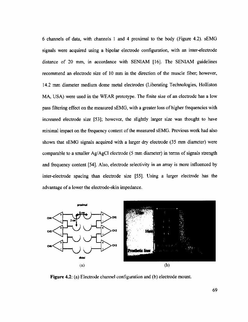
6 channels of data, with channels 1 and 4 proximal to the body (Figure 4.2). sEMG
signals were acquired using a bipolar electrode configuration, with an inter-electrode
distance of 20 mm, in accordance with SENIAM [16]. The SEN1AM guidelines
recommend an electrode size of 10 mm in the direction of the muscle fiber; however,
14.2 mm diameter medium dome metal electrodes (Liberating Technologies, Holliston
MA, USA) were used in the WEAR prototype. The finite size of an electrode has a low
pass filtering effect on the measured sEMG, with a greater loss of higher frequencies with
increased electrode size [53]; however, the slightly larger size was thought to have
minimal impact on the frequency content of the measured sEMG. Previous work had also
shown that sEMG signals acquired with a larger dry electrode (35 mm diameter) were
comparable to a smaller Ag/AgCl electrode (5 mm diameter) in terms of signals strength
and frequency content [54], Also, electrode selectivity in an array is more influenced by
inter-electrode spacing than electrode size [55]. Using a larger electrode has the
advantage of a lower the electrode-skin impedance.
proximal
(a) (t>)
Figure 4.2: (a) Electrode channel configuration and (b) electrode mount.
69

The WEAR prototype utilized a modified silicone prosthetic liner to help reduce
electrode shifting and mitigate the amount of motion artifact. An 8cm x 16cm piece
of Cordura 1000 fabric (J. Ennis Fabrics, Edmonton, AB, Canada) was selected for its
non-stretching properties to ensure consistent electrode spacing within the array and for
its 100% polyurethane backing which has been shown to be non-irritating to the skin. A
13cm x 16cm piece of 3mm thick Ossur Iceross prosthetic liner (Ossur Americas,
Foothill Ranch, CA, USA) was sewn onto the left and right hand sides of the Cordura.
The liner was chosen for its elastic properties to ensure a custom and secure fit. The liner
was secured at the back by two strips of hook-and-loop fabric. A view of the inside of the
electrode mount can be seen in Figure 4.2.
4.3.1.2. Foot Switches
Foot switches were commonly employed in the TOHRC's Rehabilitation Technology
Laboratory. This motivated the use of foot switches in the WEAR prototype, rather than
the accelerometers; identical switches were used. Four switch pads were taped to the
bottom of the shoe, three pads for the forefoot and one pad for the heel. Different voltage
states were produced based on which pads were pressed (Table 4.1). The foot switches
interfaced to the electronics via a 3-pin (i.e. signal, power, ground) male LEMO
connector (LEMO Connectors, Morges, Switzerland).
70

Table 4.1: Foot switch voltage states.
Pad pressed Voltage stateNone 01 or more forefoot 1Heel 21 or more forefoot & heel 3
4.3.2. Electronics
The prototype WEAR electronics included the ADS 1298 (Texas Instruments, Dallas
TX, USA), an 8-channel, 24-bit analog front end designed specifically for biopotential
measurements; the PIC24FJ128GA010 (PIC24; Microchip, Chandler AZ, USA), a
general-purpose 16-bit flash microcontroller, and a SD flash memory card. To simplify
prototyping, the ADS1298ECG-FE demonstration board (Texas Instruments, Dallas TX,
USA), Explorer 16 development board, and Microchip PICtail daughter board for SD and
MMC (Microchip, Chandler AZ, USA) were used. These boards integrated the
ADS 1298, PIC24, and SD card interface module, respectively. The system was powered
by six AA batteries. Figure 4.3 shows the WEAR prototype, highlighting the main
hardware components.
To provide a wearable system, the ADS1298FE was mounted to a molded plastic
bracket that could be strapped to the lower leg, just above the electrode mount. The
proximity of the ADS 1298 to the electrodes allowed for minimal lead lengths between
the sensors, bioamplification, and analog-to-digital (A/D) conversion, thereby helping to
reduce noise. The Explorer 16 and power supply were worn in a pouch strapped around
the waist (Figure 4.3). Since all communications between the Explorer 16 and
ADS1298FE were digital, distance was not a concern.
71
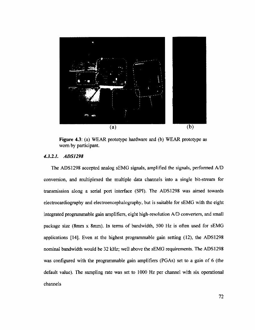
Figure 4.3: (a) WEAR prototype hardware and (b) WEAR prototype as worn by participant.
4.3.2.1. ADS1298
The ADS 1298 accepted analog sEMG signals, amplified the signals, performed A/D
conversion, and multiplexed the multiple data channels into a single bit-stream for
transmission along a serial port interface (SPI). The ADS 1298 was aimed towards
electrocardiography and electroencephalography, but is suitable for sEMG with the eight
integrated programmable gain amplifiers, eight high-resolution A/D converters, and small
package size (8mm x 8mm). In terms of bandwidth, 500 Hz is often used for sEMG
applications [14]. Even at the highest programmable gain setting (12), the ADS 1298
nominal bandwidth would be 32 kHz; well above the sEMG requirements. The ADS 1298
was configured with the programmable gain amplifiers (PGAs) set to a gain of 6 (the
default value). The sampling rate was set to 1000 Hz per channel with six operational
channels

The ADS 1298 used for the prototype was part of the ADS1298ECG-FE
demonstration board, designed to interface with the Texas Instruments MMBO mother
board. Since the ADS1298ECG-FE was expecting three different power supplies (1.8V,
3.3V, and 5V) from the MMBO, the Explorer 16 development board was modified to
supply these voltages. Additionally, the differential channel inputs to the ADS 1298 were
not directly accessible since the ADS1298ECG-FE was originally designed to
demonstrate ECG operation in a limited configuration. To bypass the ADS1298ECG-FE
on-board circuitry prior to the ADS 1298 differential inputs, surge protecting circuitry was
added.
4.32.2. P1C24
The PIC24 is a 16-bit RISC microcontroller with an on-chip oscillator and low
operational power requirements that makes it suitable for portable and weight restricted
applications. The PIC24 interfaced with the ADS 1298 through an SPI interface. A
separate SPI was used to interface the PIC24 to a SD flash memory card on the PICtail
board, where digitized sEMG signals were stored. Foot switch information was acquired
by the PIC24 through one of its analog-to-digital converters via an added 3-pin female
LEMO connector. System operation was controlled by a series of pushbuttons on the
Explorer 16 with the operation mode indicated on a LCD screen also located on the
Explorer 16.
4.3.2.3. Embedded Software
Embedded software was implemented on the PIC24 to program and command the
ADS 1298, and manage data flow throughout the WEAR prototype. On power-up, the
73

PIC24 sent commands to the AD 1298 to initialize its programmable registers so that it
could operate with the required settings. Once the ADS 1298 was initialized, it
automatically began amplifying analog sEMG data and converting them to digital format.
Since the specific PIC24 used lacked direct memory access (DMA), custom flow control
had to be implemented.
The user initialized data recording by pressing one of the buttons on the Explorer 16.
Once the PIC24 had been commanded to begin recording data, the PIC24 waited for the
interrupt signal from the ADS 1298 prior enabling data transfer. The interrupt signal
generated by the ADS 1298 was used to indicate when it had amplified and A/D
converted a single 216 bit sEMG data packet (24 status bits, followed by 24-bits per
channel: six active channels and two inactive channels for the WEAR prototype). sEMG
data packets transmitted from the ADS 1298 via the SPI interface, were temporarily
stored in a circular, or ring, buffer in the PIC24. Once a complete data packet was
received, the PIC24 wrote the sEMG data from the circular buffer to the SD flash
memory card. Concurrently, analog data from the footswitches were converted to digital
format by a PIC24 internal A/D converter, temporarily stored in the ring buffer, and then
written to the SD card with the sEMG data. Lack of DMA combined with slow write
speeds of the SD card resulted in the ADS 1298 being configured for operation on six
channels, with 1000 Hz sampling rate. Attempts to use all eight channels and/or a higher
sampling frequency resulted in lost data since the PIC24 could not write packets to the
SD card fast enough to keep up with the input rate.
4.3.3. Signal Conditioning
74
WEAR data files on the SD memory card were manually transferred to a computer
for offline signal processing performed in MatLab (The Mathworks, Natick, MA, USA).
Data were digitally high-pass filtered using a 3rd order Butterworth filter, with a
frequency cutoff of 30 Hz to eliminate motion artifact and any baseline drift. Next, notch
filtering was performed on the data using a second-order digital filter, with a center
frequency of 60 Hz and a Q-factor of 35. No harmonics were observed and thus only 60
Hz filtering was used. Comb filtering could have easily been implemented if necessary.
Filtering was performed in the forward and reverse direction for zero-phase filtering.
4.4. Methodology for Pilot Validation
4.4.1. sEMG Acquisition Systems
To validate the WEAR prototype we compared it to two conventional sEMG systems.
Data was collected for all three systems with a dry electrode array, a wet electrode array
to separate out the effects of the acquisition system and the electrodes. The same
reference electrode placement was used for each set-up, a single wet electrode on left
fibular head.
4.4.1.1. WEAR Prototype
The WEAR prototype used in pilot validation was as described in section 4.3.
4.4.1.2. The Ottawa Hospital Rehabilitation Centre System
The first conventional sEMG acquisition system, was located in the TOHRC’s
Rehabilitation Technology Laboratory (TRC system). The TRC acquisition system was a
custom built amplifier system comprised of four AD524 instrumentation amplifiers
(Analog Devices, Norwood MA, USA) each connected to a pair of electrodes, each with
75

a gain of 100, connected to a junction box. The junction box was then connected to a
OP27 precision operational amplifier (Analog Devices, Norwood, MA, USA) with an
adjustable gain set to 20 (total gain of 2000), followed by an electrical isolation circuit,
and then a high-pass, 3rd order Butterworth filter, with a frequency cutoff of 30 Hz. Data
were sampled at 2000 Hz using a Vicon MX Ultranet HD console with a 16-bit A/D
converter (Figure 4.4). Data were resampled in Matlab to 1000 Hz to be consistent with
the WEAR prototype’s sampling rate.
Electrodes were located 10 cm from the AD524 amplifiers to help minimize the
noise. 130 cm leads connected the AD524 to the junction box, which was connected to
the OP27 through a heavily shielded ~57 m long cable. The OP27, isolation circuit and
3rd order Butterworth filter were housed in a custom made unit in a shelving rack. The
Vicon MX Ultranet HD and A/D were also located in the shelving rack (Figure 4.4).
Junction Box
» OP27AD524
Electrode
FootSwitches
Iso 3ld Order ViconCircuit BWFilt. A/D
(a)
Figure 4.4: (a) TRC system block diagram, (b) custom EMG unit, and (c) Vicon A/D.
76

4.4.1.3. Modified TRC System
The second conventional sEMG acquisition system, the Modified TRC (MTRC)
system (Figure 4.5), had four electrode pairs connected via 150 cm leads to a junction
box (F-15EB/B1 16-channel electrode board, Grass Technologies, West Warwick, RI,
USA), which was connected by a 4.2m cable to the biopotential amplifiers (M15LT
system using a M15A54 quad amplifier, Grass Technologies). The M15LT system was
connected via a 4.5m cable to the same junction box used by the TRC system to align the
sEMG signals with the foot switches. The remaining parts of the system were the same as
the TRC system. The variable gain on the M l5 amplifier was set to 100 and the OP27
adjustable gain was set to 10 (total gain of 1000). As with the TRC system, MTRC
system data were sampled at 2000 Hz using a Vicon MX Ultranet HD console with a 16-
bit A/D converter. Data were resampled in Matlab to 1000 Hz for consistency with the
WEAR prototype.
^ = 1Electrodes
JunctionBox
M15A54
Foot Switches
JunctionBox * OP27
Iso 3rd Order ViconCircuit BW Filt. A/D
(a) (b)
Figure 4.5: (a) MTRC system block diagram and (b) M15LT system with one M15A54 quad amplifier and a F-15EB/B1 electrode board.
77

4.4.2. Electrodes
Dry and wet electrodes were used in the pilot validation to observe the effects of
electrode type on system performance. The dry electrodes, described in section 4.2.1.1,
were the LTI medium dome electrodes, housed in the electrode mount. The wet
electrodes were self adhesive 5 mm diameter pre-gelled Ag/AgCl electrodes (Meditrace
130, Kendall, Mansfield MA, USA). Since the wet electrodes were self adhesive, the wet
electrode array did not require the electrode mount. Both arrays used eight electrodes
aligned as seen in Figure 4.2.
4.4.3. Pilot Data Collection
Data were collected from the TA muscle of the left leg of one male participant (age
35, weight 74.8kg, calf diameter 39 cm), with no known neuromuscular disorders. Six
trials were performed in succession: the first three with the dry electrode array and the
last three using the wet electrode array (Table 4.2). Three tasks were performed in each
trial: 1) resting, 2) isometric and isotonic contractions, and 3) walking. The resting task
was performed to collect noise data. The isometric and isotonic contractions were
performed to compare inter- and intra-system signal quality. The walking task was
conducted to compare how the systems performed on a moving participant, since the
intent of the WEAR was for gait/motion analysis.
Prior to each trial, the skin’s surface above the TA was cleaned with an alcohol wipe.
The appropriate electrode array was placed on the participant, positioning the center of
the array at the position recommended by SENIAM. Positioning for trial #1 was done
visually; that is, no measurements were made to guide the placement, enabling rapid
78

Table 4.2: Validation test order.
Trial # System Electrodes1 WEAR Dry2 TRC Dry3 MTRC Dry4 WEAR Wet5 TRC Wet6 MTRC Wet
positioning of the electrode mount (< 20 s). Since the dry electrodes left a temporary
physical depression on the skin surface, the appropriate electrode array could be
positioned in the same location for each trial.
4.4.3.1. Task #1: Resting
The participant began seated on a bench with their legs extended and 10 s of data
were acquired while the participant’s leg was relaxed. These data should be
representative of the noise associated with the acquisition system (i.e. no sEMG present).
4.4.3.2. Task #2: Isometric and Isotonic Contractions
While seated on a bench, a 5 lb weight was tethered via a pulley to the top of the left
foot (Figure 4.6). The weight rested on a support block while the participant’s muscle
was relaxed. Ankle dorsiflexion raised the weight, which provided a counter force of
approximately 22 N. The participant performed 10 repetitions of dorsiflexions, holding
an isometric contraction for 3 seconds, plantarflexion to the starting position, and 3
seconds with the muscle relaxed before the next repetition.
79

Figure 4.6: Experimental setup for TA muscle activation.
4.4.3.3. Task #3: Walking
The participant began seated on a stool, stood up, walked straight for 5m at a self
selected pace, turned around, walked 5m back to the starting point, turned and sat back
down. The participant performed five repetitions of the walking task.
4.4.4. Data Processing
The mean of each data recording was subtracted to remove any offsets. To enable
direct comparisons of the amplitudes between the WEAR and conventional systems,
recorded amplitudes were converted to voltages and were divided by the total gain of
their respective acquisition systems. Since significant power line interference noise at 60
Hz was observed in both the WEAR and MTRC, the 60 Hz notch filter (as described in
section 4.2.3) was applied to all data. Significant noise was also observed at 300 Hz in
the MTRC trials, thus a second-order digital notch filter, with a center frequency of 300
Hz and a Q-factor of 35 was applied only to MTRC trial data. All data processing was
performed in Matlab.
4.5. Results
4.5.1. Task #1: Resting

Ten seconds of data were captured for each system, with eight seconds extracted, one
second after the start to avoid any effects from the digital filtering. There were no notable
differences between electrode types within each system, however, the noise amplitudes
varied between systems (Figure 4.7).
TRC Dry TRC Wet
WEAR Dry WEAR Wet
> 0.04
a 0.02
5! -0.04
4 5 6 7Time (sec)
3 4 5 6Time (sec)
MTRC Dry MTRC Wet
> 0.04ea 0.02
_ - 0.02
4 5 6Time (sec)
(C )
Figure 4.7: Resting noise from channel 3 for all trials, (a) WEAR system, (b) TRC system, and c) MTRC system.
4.5.1.1. Noise Root Mean Square Comparison
. There was very little difference in root mean square (RMS) voltages between
electrode types for any of the systems, with mean differences of 8.42% +/- 8.60%,
12.06% +/- 10.98%, and 4.75% +/- 3.45% across the six channels for the WEAR
81

prototype, TRC, and MTRC systems, respectively (Figure 4.8). The low variability
between electrode types showed that the larger electrode sizes of the dry electrodes did
not seem to have an impact on system performance. WEAR had the lowest noise RMS
values for every channel. The TRC system had the second highest RMS noise. The
MTRC system’s RMS noise was much higher than either the TRC system or WEAR
noise RMS levels (103.13% and 166.01% difference, respectively, for the means across
all channels and trials).
0.020
0.018
0.016o■g 0.014
J 0.012
| 0.010 g*% 0.008
| 0.006
0.004
0.002
0.0001 2 3 4 5 6
Channel Number
Figure 4.8: Noise RMS amplitude, for all 6 channels of sEMG.
4.5.2. Task #2: Isometric and Isotonic Contractions
For each isometric, isotonic contraction, 2048 ms segments of data were extracted,
512 ms after the start of the contraction; the starting time of each contraction was
identified manually through visual inspection of the recorded sEMG signals. This
segmentation avoided the transient portions of each contraction (i.e., segments
82
r ir ir ir ir ir i
■WEAR Dry ■WEAR Wet ■TRC Dry ■TRC Wet ■MTRC Dry ■MTRC Wet

correspond to isometric, isotonic contractions). An example of the WEAR with dry
electrodes output from the channel 3 isometric and isotonic contractions post-processing
can be seen in Figure 4.9.
^ -0.5
Time (sec)
Figure 4.9: Isometric and isotonic contractions from channel 3 of WEAR with dry electrodes.
4.5.2.1. Signal RMS Comparison
The RMS value of each sEMG segment was computed. Figure 4.10 is a bar chart of
the RMS values, averaged over the 10 contractions, for each channel and trial. The
WEAR prototype using the wet electrodes had the highest overall RMS values (0.25mV
+/- 0.04), while, except for the TRC system channel 6, the MTRC system using the wet
electrodes had the overall lowest RMS values (0.14mV +/- 0.003). Channel 3 had the
highest RMS values for each trial (0.24 mV +/- 0.05) and channel 5 had the lowest (0.15
mV +/-). As channel 3 had the highest mean RMS value, it was deemed the most
reasonable electrode pair for further analysis.
83

Although relative RMS values were consistent across the different systems and
electrodes, the TRC system did not always perform well with the dry electrodes. An
outlier can be seen in Figure 4.10, where channel 6 on the TRC dry trial had a RMS value
close to zero for all contractions. As can be seen in Figure 4.11, it appeared as though
channel 6 was not functioning (i.e. no contractions visible). The non-functioning channel
behavior of the TRC with dry electrodes was visible in previous tests (on different and/or
multiple channels) not reported here.
0.4
0.35
0.3
1 0.25<u■o
f 0.2
m 0.15
0.1
0.05
0
■ WEAR Dry■ WEAR Wet
- ■ TRC Dry■ TRC Wet■ MTRC Dry
‘ ■ MTRC Weti i T
JI 1 i |i « ntL t T 11 ti i
■ I
2 3 4Channel Number
Figure 4.10: Signal mean RMS amplitude, averaged across 10 contractions (1 standard deviation error bars), for all 6 channels of sEMG.
4.5.2.2. Signal-to-Noise Ratio Comparison
An estimate of the noise was established in task #1 (section 4.5.1). A signal-to-noise
ratio (SNR) can be derived by taking the ratio of the mean RMS value, averaged across
84

the 10 contractions, and the RMS value of the noise. Figure 4.12 is a bar chart of the
mean SNR over the 10 contractions for channel 3 of each trial. Based on the high RMS
and low noise associated with the WEAR prototype with wet electrodes, it was expected
that WEAR with wet electrodes would have the highest SNR, which proved to be the
case (46.7 dB +/- 1.4). The SNR of the WEAR with dry electrodes (43.9 dB +/- 0.9)
surpassed the SNR of the TRC system with either electrode type (22.77% difference and
34.45% difference for dry and wet electrodes, respectively). Based on showing the lowest
RMS and highest noise values, the MTRC system trials had the lowest SNR values. The
mean SNR across both MTRC system trials was 78.02% different than WEAR SNR
(mean across both electrode types) and 49.54% different than the TRC system’s SNR
(mean across both electrode types).
Channel I Channel 2 Channel 3
IIc/i -2
20 40 60Tim e (sec) Channel 4
20 40 60Tim e (sec) Channel 5
20 40 60Tim e (sec) Channel 6
20 40 60Tim e (sec)
20 40 60Tim e (sec)
20 40 60Tim e (sec)
Figure 4.11: Isometric and isotonic contractions from all channels of TRC with dry electrodes.
85

■WEAR Dry ■WEAR Wet ■TRC Dry ■TRC Wet ■MTRC Dry ■MTRC Wet
Channel 3
Figure 4.12: Mean SNR, averaged across 10 contractions for channel 3 for all trials.
4. S. 2.3. Power Spectral Density Comparison
Power spectral densities (PSD) for each 2048ms data segment of the isometric and
isotonic contractions were calculated using Welch’s method [56], using Hamming
windows of 512 samples, with 50% window overlap. A PSD plot (power vs. frequency)
for each trial type with the PSD values averaged over all 10 isometric and isotonic
contractions from channel 3 can be seen in Figure 4.13.
Based on correlation coefficients calculated for all six PSD curves, which ranged
from 0.95 to 0.99 (where 1.00 is an exact match), the spectral content was consistent. The
maximum power of the curves followed the same patterns seen in the RMS results, with
WEAR with wet electrodes showing the highest maximum power (-87.18 dB), followed
by the two TRC system maximum powers (-88.92 dB and -89.79 dB for dry and wet
electrodes, respectively), then the WEAR with dry electrodes (-90.00 dB), and finally the
two MTRC system maximum powers (-91.26 dB and -93.67 dB for dry and wet
electrodes, respectively).

-80 WEAR Dry WEAR Wet TRC Dry TRC Wet MTRC Dry MTRC Wet
-90
-100
00■o- 1 1 0
-120
-130
-140200 300Frequency (Hz)
400 500100
Figure 4.13: PSD averaged across 10 contractions for channel 3 for all trials.
The effects of the 60 Hz notch filters can be seen in each curve and the effects of the
300 Hz notch filter can be seen in the MTRC system curves (Figure 4.13).
4.5.3. Task #3: Walking
4.5.3.1. Data Segmentation
To analyze the walking trials, a linear envelope was computed for the sEMG data.
Based on the method developed by Barrett, Donnelly, and Olaighin in [57], raw sEMG
data were rectified and digitally low-pass filtered using a fifth-order Butterworth filter,
with a cutoff frequency of 5 Hz. By aligning the filtered and rectified sEMG data with the
foot switch data, different actions from the walking trials were segmented. Four actions
from each trial were used for analysis: sit-to-stand, stand-to-sit, loading response of every
step (i.e. the loading response of the TA from the beginning to the end of its contraction),
87

and swing phase of every step (i.e. TA activity beginning from push off and ending
during terminal swing) (Figure 4.14). The loading response and swing phase were
selected for analysis since they represent the periods with the most TA activity in the gait
cycle [58]. Sit-to-stand and stand-to-sit were selected since they are distinct actions in the
sequence; however, since the actions themselves varied between trials, they were
analyzed as a whole rather than segmented.
4.5.3.2. Signal Strength Comparison
To calculate the signal strength (SS) of the portion being analyzed, the normalized
integral of the sEMG signal was calculated. The area under the linear envelope curve was
estimated using trapezoidal numerical integration and then normalized by dividing the
relevant area under a curve by the number of samples in the area [44]. To compare intra-
and inter-system performance, boxplots for each action were created in Matlab (Figure
4.15).
Boxplots show variability and median SS relative to each movement action within
each trial. The top of box represented the upper 75th percentile (<y?) of the values and the
bottom of the box represented 25th percentile (qi). Interquartile ranges (qrqu IQR) are a
measure of variability within a set of data. The median value is indicated by the red line
inside the box. The “whiskers” show minimum and maximum values (i.e. the vertical
lines coming off the top and bottom of the boxes ending in horizontal lines) [59].
88

S tan d -to -S il repe tition 2, raw da ta
% ° 06
1 0 0 4
■I" 0.02 1
-4* -*4~
-C h 3 sB M G - F oo t S w itch
37 .2 37 .4 3 7 .6 37 .8 38 38 .2 38 .4 38 .6 38 .8T im e (sec)
S lan d -lo -S il rep e titio n 2, recti tied and Filtered d a ta
------- C h3 sE M G1 Foot Sw itch
.
37.2 37 .4 37 .6 37.8 38 38.2 38.4l im e (sec)
(a)
3 8 .6 38 .8
S *t-to-S tand rep e titio n 3, raw da ta
>* 0.2
0- 0 .2
-C h 3 sE M G - F o o t S w itch
44.1 44 .2 44 .3 4 4 .4 4 4 .5 4 4 .6 4I'im c (sec)
S it-to -S tan d rep itio n 3 , re c tified and filte red d a ta
C h3 sliM Ci F o o t S w itch
e 0 .06
0 .04“S.
0.02
4 4 .6 44 .744 44.1 44 .2 44 .3 4 4 .4 44 .5 44 .8
I Loh
I'im c (sec )<b)
3rd s tep , repe tition 3 , raw d a ta T
3.4
1's ftonse I
3.8
C h3 sE M G Foot S w itch
I . 1 1 |I Swing Phase
4.44 4.2T im e (sec)
3rd step , repe tition 3 , rec tified and filtered data
>5 C h3 sE M G
F oot Sw itch
0.05
ILoadlng Renponi
3.4 4 4.2 4.4 4.6I’im e (sec)
( C )
Figure 4.14: Data segmentation, WEAR channel 3 with dry electrodes: (a) stand-to-sit, (b) sit-to-stand, (c) loading response and swing phase.
89

To determine the limit of the upper whisker, Matlab found the nearest data point below
qi + w(IQR). To determine the limit of the lower whisker, Matlab found the nearest point
above qi - w(IQR). In both cases, w was set to the default value of 1.5 which
corresponded to an approximate standard deviation of +1-2.1 and coverage of 99.3% for
normally distributed data. Outliers (data points beyond the whiskers) are represented by
red plus symbols. [59].
x io4 x io-4
Sbl.4
5,0.8 't/i 0 .6
0.4 0.2
1 0.8 c/3 0.6
0.4 0.2 ©
,-4 x 10x 10
H> 1.4
2 i1 0.8 53 0.6
0.4 0.2
Figure 4.15: Mean SS boxplots across all five repetitions of each trial for (a) sit-to- stand, (b) stand-to-sit, (c) loading response, and (d) swing phase.
90

. Intra-system variability for the WEAR prototype was comparable to those seen in
both the TRC system and MTRC system (Figure 4.15). Values from the boxplots shown
in Table 4.3 reveal that except for the WEAR with wet electrodes IQR for the stand-to-sit
action (0.70 x 10'4), WEAR IQRs ranged from 0.10 x 10'4 to 0.33 x 10'4. TRC system
IQRs had a slightly lower range than WEAR IQRs, going from 0.09 x 10'4 to 0.29 x 10'4.
The MTRC system showed the lowest variability of the three systems, with IQRs ranging
from 0.06 x 10⁻⁴ to 0.21 x 10'4. No outliers were present in the sit-to-stand or stand-to-sit
actions. Except for WEAR with wet electrodes during the loading response action and the
TRC system with dry electrodes during swing phase, all systems had from one to five
outliers in the loading response and swing phase actions.
Comparing inter-system results for the sit-to-stand action revealed inconsistency in
both IQR and median value comparisons. For the sit-to-stand action, WEAR with wet
electrodes and the TRC system with dry electrodes showed the highest percent IQR
differences with the other trials (ranging from 24.42% to 121.56% differences). Between
the other four trials, IQRs for the sit-to-stand action only showed a range of 8.92% to
45.43% difference. Sit-to-stand median SS values for the MTRC system with wet
electrodes and MTRC system with dry electrodes were very close (4.33% difference), but
both MTRC system trial medians differed more from all other trials (from 20.73% to
50.70% difference). Outside of the MTRC system trials, sit-to-stand median SS
differences ranged from 5.22% to 30.78%.
91

Table 4.3: SS data for all actions and all trials.
MTRC MTRC Wet
WEAR Dr
WEAR Wet
Sit-to-standIQR (x 1(T)Median (x 10 J# of outliersStand-to-sitIQR (x 101Median (x 10 )# of outliersLoading responseIQR (x IQ-4)Median (x 10~4)# of outliersSwing phaseIQR (x IQ'4)Median (x 10')# of outliers
i ~n0.17 0.200.57 0.85
0
0.13 0.130.51 0.54
1 1
0.290.78
II0.150.48
0
0.180.64
0.100.48
0.68
0.160.49
1
0.190.47
0.090.32
1
Comparing inter-system results for the stand-to-sit action also revealed inconsistency
in both IQR and median value comparisons. For the stand-to-sit action, WEAR with wet
electrodes and the MTRC system with dry electrodes showed the highest percent IQR
differences with the other trials (ranging from 49.69% to 141.19% differences). Between
the other four trials, IQRs for the stand-to-sit action showed a range of 3.90% to 48.59%
difference. Stand-to-sit median SS values for WEAR with dry electrodes and the MTRC
system with wet electrodes were very close (5.25% difference), but medians compared to
all other trials differed to a higher degree (from 67.25% to 112.30% difference). Outside
92

of the WEAR with dry electrodes and MTRC system with wet electrodes trials, stand-to-
sit median SS differences ranged from 0.06% to 50.65%.
For the loading response action inter-system comparisons, only the TRC system with
dry electrodes showed IQR differences higher than other trials. IQR differences for the
TRC system with dry electrodes compared to all other trials ranged from 37.12% to
60.95%. Between the five other trials, IQR differences ranged from 2.05% to 25.62%.
Median SS differences for the loading response actions in the MTRC system with wet
electrodes trial showed the highest differences versus other trials (ranging from 19.36%
to 57.65%). Between all other trials, median SS differences ranged from 5.58% to
39.38%.
In terms of the swing phase action inter-system comparisons, the TRC system with
wet electrodes and MTRC system with wet electrodes showed IQR differences higher
than other trials. Although the IQR of the TRC system with wet electrodes and MTRC
system with wet electrodes only differed by 7.78% from each other, compared with all
other trials, differences ranged from 23.67% to 53.13%. Between the other four trials,
IQR differences ranged from 2.56% to 22.80%. Median SS differences for the swing
phase actions in the MTRC system with wet electrodes trial showed the highest
differences with other trials (ranging from 38.43% to 50.48%). Between all other trials,
median SS differences ranged from 1.44% to 12.66%.
4.6. Discussion
4.6.1. Task#l: Resting
4.6.1.1. Noise RMS Comparison
93

The noise RMS voltage levels while the participant was at rest showed differences
between trials (Figure 4.8). Inaccurate gain settings in one or all three systems was a
likely explanation for some of the differences (i.e., the amplifier gain was not exactly
what it was programmed or designed to be). Elevated noise in the TRC system could also
be explained in part by the fact that the TRC system had the highest input impedance of
all three systems (TRC input impedance was 1GGI, WEAR was 500MQ, and MTRC was
20MI2). High amplifier input impedance has been associated with increased motion
artifact due to movement of electrode leads and power line interference [52]. The MTRC
system showed the highest noise RMS of all three systems. MTRC system noise may
have been due to the 5.5 m between electrodes and the M15 amplifier. Pre-amplification
is generally placed close to the electrode to reduce noise [52].
4.6.2. Task #2: Isometric and Isotonic Contractions
4.6.2.1. Signal RMS Comparison
Since electrode positioning was maintained for both dry and wet electrode arrays for
all three systems, it was expected that the channel with the highest RMS amplitudes
would be the same. This is precisely what was observed (Figure 4.10). This result showed
good intra-system repeatability for the WEAR prototype. With the same channel resulting
in the highest mean RMS value for each trial, WEAR showed similar sEMG acquisition
capabilities to the two conventional systems. Since the same electrode array was used,
differences in RMS between systems were attributed to inaccurate gain settings in one or
all three systems as described in section 4.6.1.1.
94

The RMS amplitude for the WEAR prototype with the dry electrode array was
notably lower than the RMS amplitude of the WEAR prototype using wet electrodes
(37.60% difference).Dry electrodes are polarizable and tend to have much larger
electrode-skin impedances than wet electrodes, which are non-polarizable [22]. The
higher electrode-skin impedance could account for the decrease in RMS amplitude, since
signal strength loss is associated with higher impedance. However, the same behavior
was not seen in the TRC or MTRC systems, where the RMS values varied by channel,
with certain channels showing higher RMS amplitude for the dry electrodes. This may
have been attributed to individual anomalies from a single-case study.
In the TRC trial with dry electrodes, no signal was recorded on channel 6 (Figure
4.11). The non-functioning channel occurred over 75% of the time in preliminary
prototype testing. The non-functioning channel varied position in the array and
sometimes occurred on multiple channels. Dry electrodes are known for having higher
electrode-skin impedance than wet electrodes, which could result in a large impedance
mismatch between the two electrodes; this in turn could result in a large differential
voltage at the AD524 amplifier causing it to saturate. A non-functioning channel was
never on TRC seen with wet electrodes, or on either the WEAR or MTRC systems. Since
the non-functioning electrode pair behavior was never seen in MTRC results, the AD524,
which is the difference between the TRC and MTRC systems, in combination with the
dry electrodes was a likely enabler the problem. An investigation into the root cause of
the intermittent, nonfunctioning channels, when using the TRC system with dry
electrodes, is outside the scope of this thesis.
95

4.6.2.2. SNR Comparison
The SNR bar chart (Figure 4.12) showed similar patterns to channel 3 in the signal
RMS bar chart (Figure 4.10) except for the WEAR with wet electrodes trial. The WEAR
prototype exhibited less noise, resulting in the highest SNR. Similar to the noise and
signal RMS values, the differences in SNR may be explained by the differences in gain
settings, amplifier input impedances, and distance between electrodes and pre-
amplification.
Despite not suffering from the TRC system amplifier saturation issues with dry
electrodes, the MTRC system’s results were not as high quality as expected. The SNR
values for the MTRC system trials were lower than the other two systems. Since the main
difference between the TRC and MTRC systems was the proven, COTS bioamplifier, one
would have expected performance to be similar. The lower MTRC system SNR may be
explained by the added distance between the electrodes and the amplifier allowing more
noise into the signal.
4.6.2.3. PSD Comparison
Spectral content was similar for all trials with minor differences attributable to
differences in configured versus actual amplifications.
4.6.3. Task #3: Walking
4.6.3.I. Data Segmentation
Gait sEMG output was rectified and filtered, producing linear envelope curves, before
segmentation. Since sit-to-stand and stand-to-sit linear envelopes did not show consistent
patterns between repetitions and trials, it was difficult to segment them into specific
96

portions, thus they were analyzed as a whole. The other two actions analyzed, the
loading response and the swing phase had more distinct patters in the linear envelope
curves and were thus simpler to segment from each step. The foot switch data enabled
segmentation to be more obvious, although the linear envelopes showed some variation
from step to step and system to system.
4.6.3.2. SS Comparison
Comparing the WEAR prototype’s performance to the TRC and MTRC systems for
IQR across the four actions did not reveal any glaring differences. For each action, there
were one to two trials that showed much higher or much lower IQR values than the other
trials. WEAR with wet electrodes had IQR values that were substantially different in two
actions (sit-to-stand and stand-to-sit), while the TRC system had three such actions (two
with dry electrodes, one with wet) and the MTRC system had two such actions (one with
dry electrodes, one with wet). Median values across all trials and actions had lower
percent differences than with the IQR values. WEAR with dry electrodes stood out only
in the stand-to-sit action, while the MTRC system had substantially different medians
five times (twice with dry electrodes, three times with wet). WEAR also had slight less
outliers combined across all trials and actions (five for WEAR and eight each for the
TRC and MTRC systems).
4.7. Conclusion
In response to a proven need for objective measures of muscle function in clinical
rehabilitation, a functional prototype of the WEAR sEMG acquisition system was
designed and implemented. Pilot tests validated the functionality of the WEAR prototype
97

and suggest that its performance is comparable to two conventional sEMG acquisition
systems. Additional testing was conducted to provide a more thorough comparison
(Chapter 5).
98

5. Participant Testing
5.1. Introduction
Following successful validation of the WEAR prototype (section 4.4), further testing
with was needed to gather comparative information across multiple participants. This
chapter describes the methodology and reports results from participant testing carried out
at the TOHRC Rehabilitation Technology Laboratory.
5.2. Methodology
5.2.1. Participant Demographics
Ten able-bodied individuals were recruited from TOHRC and Carleton University.
Exclusion criteria included presence of neurological, orthopaedic, or cardio respiratory
issues that affected gait. After reading and signing information and consent forms, the
testing procedure was explained and participants were given the opportunity to ask
questions. Prior to testing, participant height, weight, and calf circumference were
recorded (Table 5.1).
5.2.2. sEMG Acquisition Systems
For participant testing, two sEMG acquisition systems were compared: 1) the WEAR
prototype system (section 4.3), which uses a dry LTI electrode array (section 4.3.1.1) and
2) the conventional TRC system (section 4.4.1.2) using one wet Ag/AgCl electrode pair
(section 4.4.2). A MTRC system (section 4.4.1.3) evaluation was not required for this
testing phase since only wet electrodes outcomes were used as a comparator (i.e., MTRC
testing was required in due to issues with dry electrode use with the TRC system
99

Table 5.1: Participant demographics.
Participant # Gender Ht (cm) Wt(kg) Calf diameter (cm)1 M 180.0 65.0 35.02 M 167.6 64.4 35.03 F 162.5 58.9 33.04 F 160.0 49.9 34.55 F 175.2 58.9 35.56 F 167.6 74.8 43.07 M 185.4 74.8 38.58 M 175.2 90.7 40.09 F 167.6 63.5 36.510 M 185.4 99.8 42.0
described in section 4.5.2.1). The WEAR prototype was described in section 4.3 and the
TRC system was described in section 4.4.1.2.
5.2.3. Electrodes
Wet electrode placement with the TRC system followed the process outlined in the
SENIAM project for TA: the first electrode was placed “at 1/3 on the line between the tip
of the fibula and the tip of the medial malleolus” [60]. The second electrode was placed
two centimeters down the same line from the first electrode. Figure 5.1 shows the
electrode placement method with the blue dot representing the tip of the fibula and the
orange ‘x’ representing the first electrode location.
100

m
Figure 5.1: SENIAM electrode placement for TA [60].
5.2.4. Data Collection
Data collection was as described in section 4.4.3, except that the TRC trials were
performed using a single, SENIAM placed, wet electrode pair and WEAR trials were
performed with the dry electrode array. Array positioning for each WEAR trial was done
visually (i.e., no measurements were made to guide array placement). Unlike the dry
electrode placement method described in section 4.4.3, there was no effort made to place
the electrode mount in the same position for each WEAR trial.
Each participant underwent a total of six trials, three for each sEMG acquisition
system. Participant 1 was tested with the TRC system for the first three trials and the
WEAR system for the last three trials. The starting system was alternated for subsequent
participants (Table 5.2).
101

Table 5.2: Validation test order.
Participants 1 ,3 ,5 ,7 ,9 Participants 2 ,4 ,6 ,8 ,1 0Trial# System System
1 TRC WEAR2 TRC WEAR3 TRC WEAR4 WEAR TRC5 WEAR TRC6 WEAR TRC
As with the experimental methods described in section 4.4.3, three tasks were
performed in each trial: 1) resting, 2) isometric and isotonic contractions, and 3) walking.
The resting task was performed as described in section 4.4.3.1 and isometric and isotonic
contractions were performed as section in chapter 4.4.3.2. The walking task was
performed as described in section 4.4.3.3. Data processing was performed as described in
section 4.4.4.
5.2.5. Statistical Analysis
The null hypothesis was that signal RMS and SNR means from task #2, and mean SS
IQR, median value, and number of outliers from task #3 for both the WEAR and TRC
systems were the same. Two tailed t-tests were used to disprove the null hypothesis and
calculate the statistical significance of the difference between the two systems (a = 0.05),
since it was unknown which system would have the higher mean values prior to data
collection. Paired t-tests were used since the same participants were tested with each
system [59]. Statistical power, which provides a measure of the likelihood that the null
hypothesis would be rejected given that another outcome is true, was also measured for
102

all five data sets [59]. Finally, ideal sample sizes for achieving good statistical power in
the testing of the five data sets were calculated [59].
5.3. Results
5.3.1. Task #1: Resting
The mean noise RMS across the three trials per system was lower with the WEAR
than the TRC system for each participant (Figure 5.2). Mean noise RMS across all 10
participants was 88.80% different between WEAR trials (2.16 x 10'3 mV +/- 6.57 x 10‘4)
and TRC system trials (5.16 x 10'3 mV +/- 1.83 x 10‘3). Figure 5.2 displays noise data for
the channels as selected in section 5.3.2.1. Output from five WEAR resting trials
(participant 1, trial 1; participant 6, trial 1; participant 4, trials 2 and 3; and participant 5,
trial 1) showed anomalies and were thus discounted from further analysis (Figure 5.3).
Since only one WEAR noise trial was used for participant 4, there was no standard
deviation, thus no error bars on the corresponding bar in Figure 5.2. The standard
deviation for the two WEAR noise trials used for participant 5 (+/- 1.27 x 10'6) was too
small to be viewable on the corresponding bar in Figure 5.2.
5.3.2. Task #2: Isometric and Isotonic Contractions
2048 ms data segments were extracted per contraction, as described in section 4.5.2.
103

1.20E-02
1.00E-02
g 8.00E-03C/D| 6.00E-03d>
• 5 4.00E-03 Z
2.00E-03
O.OOE+OO HI
WEAR
ru4 5 6 7Participant number
Figure 5.2: Mean noise RMS, averaged across 3 trials (1 standard deviation error bars), from selected channel for all 10 subjects.
5.3.2.1. RMS Comparison
For each trial, the RMS value of each sEMG segment was computed and averaged
across all 10 contractions per channel (Figure 5.4). The channel with the highest RMS
value (selected channel) for each WEAR trial was used in analysis for comparison to the
single TRC system electrode pair (Table 5.3).
The WEAR system had a higher mean RMS voltage for every participant except
participant 10. WEAR mean signal RMS across all participants (0.274 mV +/- 0.126) and
TRC system mean signal RMS (0.159 mV +/- 0.077) differed by 52.95%. T-test results
revealed a significant difference between mean RMS values for the WEAR and TRC
systems (p = 0.0114).
104

Trial I Trial 1 Trial 2 Trial 3
Trial 2Trial 3
- -0.05
4 5 6 7Time (sec)
(a)4 5 6
Tim e (sec)
(b)Trial Trial 2 Trial 3
Trial 1 Trial 2 Trial 3
Tim e (sec)2 3 4 5 6 7
Tim e (sec)
(d)
Figure 5.3: Abnormal noise signals from (a) participant 1, (b) participant 6, (c) participant 4, and (d) participant 5.
5.3.2.2. SNR Comparison
SNR was calculated as in section 4.5.2.2. Based on the generally higher RMS (nine
out of ten participants) and lower noise associated with the WEAR system, the higher
WEAR SNR was as expected for all participants (Figure 5.5). WEAR mean SNR across
all participants (41.697 dB +/- 3.073) and TRC system mean SNR (28.299 mV +/- 4.224)
differed by 38.28%. T-test results revealed a significant difference between mean SNR
values for the WEAR and TRC systems (p < 0.0001).
105

WEAR
<si 0.4
1 3
4 5 6 7Participant number
10
Figure 5.4: Mean RMS, averaged across 10 contractions and 3 trials (1 standard deviation error bars) per system, for all 10 participants.
Table 5.3: Electrode channel with highest mean RMS per
Participant # Trial 1 selected pair
Trial 2 selected pair
Trial 3 selected pair
1 5 6 52 6 6 63 3 6 64 5 6 45 4 4 46 5 5 47 5 3 68 6 6 69 6 6 610 4 4 6
106

■ W E A R
1 2 3 4 5 6 7 8 9 10Participant number
Figure 5.5: Mean SNR, averaged across 10 contractions and 3 trials (1 standard deviation error bars) per system, for all 10 participants.
S.3.2.3. PSD Comparison
PSDs were calculated and plotted as in section 4.5.2.3 (Figure 5.6). Correlation
coefficients were calculated for each participant to derive intra-system variability within
the three WEAR trials and within the three TRC system trials. Correlation coefficients
were also calculated for each participant to compare the outcomes on an inter-system
basis (i.e., comparing the variability between WEAR and TRC system trials). The WEAR
prototype mean correlation coefficient across all 10 participants was 0.987 +/- 0.008
(where 1.00 is an exact match), indicating that the spectral content was consistent. The
mean correlation coefficient across all 10 participants with the TRC system (0.985 +/-
0.005) showed that spectral content was almost as consistent as the WEAR prototype.
107

Subject I !Subject 2 'Subject} j Subject 4Subject} -100 Subject 6Subject 7 m . n oSubject 8 ~Subject 9 jSubject Id] i ' l2°
Subject I Subject 2 Subject 3 Subject 4 Subject 5 Subject 6 Subject 7 Subject 8 Subject 9 I Subject 10]
-80i ■ ■ ■ - 80| ........................... ■ ---------— Subject I I ]— Subject I
-150,0 100 200 300 400 500Frequency (Hz)
(a)
-150,0 100 200 300 400 SCFrequency (Hz)
(b)
Figure 5.6: (a) WEAR and (b) TRC system PSD averaged across 10 contractions and 3 trials per system for all 10 participants.
Comparing PSD curves from WEAR trials to those from TRC system trials showed more
variability (0.949 +/- 0.018) than in the intra-system comparisons.
The maximum power of the PSD curves followed the same patterns seen in the RMS
results (every subject had a higher maximum PSD power with WEAR except for
participant 10), with WEAR showing higher mean maximum power across the 10
participants (-92.32 dB +/- 3.83) than the TRC system (-97.30 dB +/- 3.02). The effects
of the 60 Hz notch filters can be seen in each curve (Figure 5.6).
5.3.3. Task #3: Walking
For walking trials, data segmentation was performed as in section 4.5.3.1.
5.3.3.1. SS Comparison
The SS for each action was calculated as in section 4.5.3.2. Analysis of the sit-to-
stand action boxplots (Figure 5.7) showed only small differences in SS IQR and median
values between systems (12.51% and 11.52%, respectively averaged across all
108

participants, Table 5.4). There were only five total outliers per system across all
participants. T-tests for SS IQR, median value, and number of outliers showed no
statistically significant differences (p = 0.598, p = 0.43, and p = 1.00, respectively, Table
5.5).
Data extracted from stand-to-sit boxplots (Figure 5.7, Table 5.4) once again showed
similar mean SS IQR values (10.92% difference), although the medians differed more
that with the sit-to-stand action (23.66% difference). There were only three total outliers
for the WEAR prototype and one outlier for the TRC system. As with the sit-to-stand
action, stand-to-sit comparisons did not show statistical significance SS IQR, median
value, and number of outliers (p = 0.554, p = 0.174, and p = 0.168, respectively, Table
5.5).
Boxplots for the loading response action (Figure 5.8) showed higher differences in SS
IQR and median values between systems than the sit-to-stand and stand-to-sit actions
(28.43% and 33.18%, respectively averaged across all participants, Table 5.4). The
loading response action also saw a large increase in outliers, with 42 and 51 for the
WEAR prototype and TRC system, respectively, although t-tests showed that comparison
of number of outliers was not statistically significant (p = 0.575). Loading response SS
IQR and median comparisons were statistically significant (p = 0.045 and p = 0.003,
respectively).
109

x 10
i n '4 (a)x 10
mi a*s(b)
Figure 5.7: (a) Sit-to-stand and (b) stand-to-sit signal strength comparisons of both systems for all 10 subjects.
Swing phase boxplots (Figure 5.8) averaged across all the participants provided
similar SS IQR values between systems, but the highest difference in means than
previous actions (9.86% and 40.24%, Table 5.4). The swing phase action had the most
total outliers of all actions (77 and 86 for the WEAR prototype and TRC system,
respectively). While t-tests for SS IQR and number of outliers showed that the
110

comparisons were not statistically significant (p = 0.381 and p = 0.384, respectively),
median comparison was statistically significant (p = 0.002, Table 5.5).
Table 5.4: Walking SS for all four actions and both systems across all participants.
[ w e a r TRC |Sit-to-stand 1IQR (x 10-4) 0.219 +/- 0.084 0.193+/-0.112Median (x 10-4) 0.667 +/- 0.229 0.595 +/- 0.154# of outliers 0.500 +/- 0.710 0.500 +/- 0.850Stand-to-sit 1 ...IQR (x 10-4) 0.291 +/- 0.102 0.325+/-0.129Median (x 10-4) 0.571 +/- 0.280 0.724+/-0.185# of outliers 0.300 +/- 0.480 0.100+/- 0.320Loadingresponse IF? . ,:'vIQR (x 10-4) 0.322 +/- 0.148 0.242 +/- 0.104Median (x 10-4) 1.035 +/- 0.278 0.740 +/- 0.210# of outliers 4.20 +/- 2.530 5.100+/-3.900Swing phase 1 |
IQR (x 10-4) 0.149 +/- 0.049 0.135 +/- 0.059Median (x 10-4) 0.632+/- 0.176 0.420 +/- 0.098# of outliers 7.700 +/- 5.060 8.600 +/- 5.340
5.4. Discussion
5.4.1. Task #1: Resting
As reported in section 4.6.1.1, differences in noise RMS (section 5.3.1) between
systems could be attributed to inaccurate gain settings and higher input impedance in the
111

TRC system. Based on results from pilot validation and participant testing, the WEAR
prototype seems to have a generally lower RMS noise level than the TRC system.
x 10
a
i n 4 (a)x 10
(b)
Figure 5.8: (a) Loading response and (b) swing phase signal strength comparisons of both systems for all 10 subjects.
112

Table 5.5: Walking SS statistics for all four actions and both systemsacross all participants.
Ideal samDle size
Statistical ower% Difference
12.51%Sit-to-stand
0.598Median .52% 13.10%
1.33 X 10# o f outliers 0.00%
Stand-to-sit10.92%
30.20%Median 23.66%19.50%# o f outliers 100.00% 0.168
Loading response28.43% 0.045 28.80%
76.40%Median 33.18%19.35% 9.40%w o f outliers
Swing phase9.86% 8.90%
Median 40.24% 0.002 91.40%0.384 6.70%# o f outliers 11.04%
As mentioned in section 5.3.1, five resting trials produced abnormal data, which
could be classified in two distinct types. The first abnormal data type was seen with
participant 1, trial 1 and participant 6, trial 1. The second abnormal data type was seen
with participant 4, trials 2 and 3 and participant 5, trial 1 (Figure 5.3).
The abnormal data seen with participants 1 and 6 could have resulted from the
participants maintaining a small, but constant TA activation. The signals showed patterns
consistent with isometric contractions, but with much lower RMS voltages (0.042 mV
and 0.018 mV for participant 1, trial 1 and participant 6, trial 1, respectively). The
abnormalities were not recognized in post-trial data evaluation and were only discovered
113
at a later date. To handle such issues in the future, a wireless connection which could
enable real-time viewing of recorded sEMG data would allow users to see whether
participants (or patients) are performing tasks as instructed.
Abnormal resting data from participants 4 and 5 were very different than those from
participants 1 and 6. Abnormal data from participants 4 and 5 were corruptions due to
low battery power. The low battery power was discovered prior to the start of the walking
task during trial 1 of participant 5. There was no designated battery power indicator on
the prototype WEAR system, but text on the LCD screen (visible on the Explorer 16 in
Figure 4.3a) was dim and the recording process was ending prior to the stop button being
pressed. A fresh set of batteries fixed the LCD screen and data recording issues. Data
recorded during the other tasks for participants 4 (trial 2 and 3) and 5 (trial 1) did not
show any corruption. Once again, the abnormalities were not recognized in post-trial data
evaluation and were only discovered at a later date. In order to avoid losing data files due
to low batteries, a battery power indicator could be added in future design iterations.
5.4.2. Task #2: Isometric and Isotonic Contractions
5.4.2.1. RMS Comparison
As expected, RMS values were generally higher in the WEAR trials than in the TRC
trials. Only participant 10 had a higher mean RMS for TRC trials with a difference of
23.02%. As previously discussed, these differences can be attributed to the inaccurate
gain settings and differences in amplifier input impedances between the WEAR prototype
and TRC system. The low p-value from the t-test (p = 0.0114) showed statistical
114

significance to disprove the null hypothesis that the RMS mean was the same for both
systems.
5.4.2.2. SNR Comparison
The WEAR prototype exhibited less noise and higher signal RMS values, resulting in
higher SNR values than the TRC system. Similar to the RMS values, the differences in
SNR may be explained by inaccurate gain settings and differences in amplifier input
impedances. The low p-value from the t-test (p < 0.0001) showed a statistical
significance to disprove the null hypothesis that the SNR mean was the same for both
systems. Since SNR has been shown to be highly indicative of sEMG signal fidelity [61],
the outcomes indicate that the WEAR prototype was highly proficient in acquiring good
quality sEMG signals relative to the TRC system, which is a proven and accepted
conventional sEMG acquisition system.
5.4.2.3. PSD Comparison
Differences in intra-system mean correlation coefficients across the 10 participants
between the WEAR prototype and TRC system were nominal. Although slightly larger
than the intra-system variabilities, the inter-system variability still showed a high degree
of correlation between the two systems. PSD comparisons showed that the WEAR
prototype produced comparable results to the TRC system in terms of spectral content of
the sEMG signals. Differences in maximum powers could have been due to inaccurate
gain settings and differences in amplifier input impedances.
5.4.3. Task #3: Walking
5.4.3.1. SS Comparison
115

Except for loading response and swing phase median tests, which had low p-values
and high statistical power, all the other tests had low statistical power, showing a low
probability of rejecting the null hypothesis (WEAR and TRC system values are the same)
when a specific alternative was true (e.g., WEAR IQR for sit-to-stand is lower than TRC
system IQR for sit-to-stand) (Table 5.4). Given that neither system had consistent IQR
values nor lower mean IQRs compared to the other system across all actions, it is likely
that variability of results had as much to do inter-participant locomotive variations as it
did with inter-system technical differences. As such, it would probably not be worth the
time required to obtain data from the calculated ideal sample sizes for each action listed
in Table 5.5. What can be derived from this analysis in practical terms, is that the WEAR
prototype is as likely to provide acceptable representations of muscle contractions
throughout motion as the TRC system.
5.4.4. Secondary Outcomes
Through regular use of the WEAR prototype operational observations were
discovered which either validated user requirements (Chapter 3), or revealed new design
requirements:
1. Remote operation is required since system control located on the device
results in time consuming and awkward delays.
2. A battery power indicator is required to ensure that the system is recharged
in a timely manner, thus avoiding losing power during use.
116

3. A real-time view of sEMG activity (i.e., representative information rather
than raw data) is required to ensure that system is functioning correctly to
avoid having to repeat full data collection procedures.
4. The electrode mount, as designed is effective, in that it does not allow the
electrode array to shift during operation, even when perspiration is present.
5. Despite the advantages of the chosen materials, the mount does induce
perspiration in a short period, thus a breathable material could be
investigated.
6. The approximate maximum calf circumference of the electrode mount was
reached at 43cm, thus a longer mount and/or multiple size options for the
hook-and-loop straps are required to satisfy larger calf sizes.
5.5. Conclusion
Comparative analysis of the WEAR prototype and TRC system across 10 participants
showed statistically significant higher levels of SNR in the WEAR prototype, suggesting
higher signal fidelity. Statistical analysis of the walking task data proved inconclusive,
except to say that neither system showed higher degrees of consistency. Average mean
IQRs across all four walking actions differed by only 9.16%. Based on the statistical
analysis of the isometric and isotonic contractions and walking actions, the WEAR
prototype is a viable alternative to the TRC system for clinical sEMG signal acquisition.
117

6. Conclusions and Future Work
6.1. Conclusions
Following a UCD process to conduct user research with a group of PTs yielded a
comprehensive list of design requirements (functional and usability) for a wearable
sEMG acquisition system (Chapter 3). Additionally, the user research validated the
inclusion of the electrode array as an initial design concept. The array was originally
motivated by two assumptions, both of which were validated: 1) PTs did not have a lot of
time to accommodate for conventional electrode placement and 2) PTs did not have
detailed training or education in sEMG methods. Implementing the design requirements
into WEAR development could result in high levels of acceptance by physiotherapists
and other professionals whose rolls routinely involve measuring human performance (i.e.,
occupational therapists, high-performance athletic trainers, human kinetics researchers).
The WEAR functional prototype performed well in validation and participant testing.
The electrode mount with an array of dry electrodes proved to be a viable alternative to
conventionally used wet electrodes placed by anatomical measurements. The compact,
low power, integrated AFE (ADS 1298) also performed favorably compared to two
conventional sEMG acquisition systems using discreet components. As a proof-of-
concept, the WEAR prototype successfully confirmed that a fully developed WEAR
system could perform high-quality sEMG acquisition.
The WEAR prototype, while wearable, was bulky due to the use of development
boards. As a guideline, a realistic size for the WEAR system electronics would be a unit
smaller than current Smartphones (i.e., less than 120mm high x 65mm wide x 15mm118

deep). Since system operation should be handled through a wireless application, only a
small display would be required to provide system awareness (i.e., battery power
indication, mode of operation, error state, etc.). A desired price range for the WEAR
system, as indicated by the PTs (section 3.3.2.5) including the physical system and
operational software tool suite would be $200-$500. Despite PT expectations of a higher
initial price (~$1000), the $200-$500 range should be attainable given the low cost of the
COTS components. To achieve these goals, further research and development activities
should be based on design requirements captured in user-research and experience using
the WEAR prototype in participant testing.
6.2. Recommendations for Future Work
Recommendations for future work can be grouped into four categories: design
process, physical system design, automation of signal analysis/interpretation, and user
experience/interface design. Although as many of the requirements from chapter 3 should
be incorporated as possible, the following are recommendations for the next iteration of
WEAR system development.
6.2.1. Design Process
Chapter 3 described user research performed following a UCD process. PTs
participating in the user research were cooperative and enthusiastic not only with the idea
of the WEAR system, but also of the participatory process. As described in section 2.5,
UCD is not exclusively employed at the beginning of system design, but is a process used
throughout the design lifecycle. We highly recommend continuing with the UCD process
as system design proceeds. Specifically, furthering the user research by incorporating
119

end-users into system testing of the next design iteration (i.e., usability testing) could
prove highly beneficial.
6.2.2. Physical System Design
Two major steps for the next design iteration are to develop a custom printed circuit
board and to select a DMA enabled MPU. Incorporating the ADS 1298, microprocessor,
and other electronic components into a single, compact unit would be more physically
robust, portable, and wearable than the current WEAR prototype. A DMA enabled MPU
would allow for full 8-channel operation of the ADS 1298 with a sampling rate of 2000
samples per second and thus a bandwidth of 1000Hz.
A lighter, rechargeable power supply would also need to be incorporated with the
electronics in a custom shell. A battery power indicator would also be needed to help
mitigate data corruption issues (section 5.3.1). To satisfy PT needs and provide better
insight into system function, an indicator of operating status (i.e., a flashing LED when
acquiring data and/or small display) and a wireless transmitter, so that data could be
viewed in real-time, should also be implemented into the next design iteration.
Research into alternative materials for the electrode mount should be conducted to
find a breathable material. Although the prototype mount performed well in reducing
motion artifact by holding the electrode array stationary during motion and was easy to
clean, profuse sweating under the mount was common even in short-term use. Sizing
options for the mount should also be investigated. Size options could be implemented
through different mounts or different strap lengths to hold the mount on the leg.
6.2.3. Automation of Signal Analysis/Interpretation
120

In its current state, the WEAR prototype uses a manual, multi-step process to take
raw data from a memory card and perform signal conditioning operations in Matlab.
Since the intent is for the system to display output in real-time based on an optimal
electrode pair, research is required to determine whether or not signal RMS is the best
indicator of electrode performance. Algorithms must also be developed to perform signal
conditioning (i.e., scaling to volts, mean removal, noise/motion filtering) either on the
MPU, or in the wireless application.
Another venue for further research would be in the implementation of a multi-array
electrode mount for analysis of multiple muscles (i.e., TA, soleus, and gastrocnemius).
The challenges involved in such a development not only include the physical spacing of
the arrays on the mount and incorporating multiple ADS 1298 AFEs, but in analyzing the
data retrieved from the electrodes. Research would be required to discover methods of
ensuring that crosstalk from neighboring muscles would not affect array operation,
especially when dealing with closely spaced muscles.
6.2.4. User Experience/Interface Design
Based on PT feedback, real-time results display and biofeedback applications should
be developed. Given PTs’ lack of EMG knowledge, WEAR results must be presented in a
manner that is understandable to physiotherapists, either real-time or post-assessment.
User research revealed that moving bars, color meters, or numerical indicators could be
implemented as representations of muscle activity for assessment purposes. In addition to
patient assessment, a game-based biofeedback application should be developed to give
the WEAR system more breadth of applicability in the rehabilitation process.
121

In terms of system control, remote operation is also an important step. In participant
testing walking to the participant to start and stop operation was time consuming and
awkward. PTs indicated that a wireless application (i.e., for Smartphone and/or tablet PC)
for system operation and feedback would be desirable. Since the WEAR system is not
confined to a clinic or lab, a wireless app could provide physiotherapists with real-time
results in any setting.
It is important that user experience/interface development be concurrent with physical
system development so that they can undergo usability testing as a package. Developing
WEAR as a complete system can result in a cohesive unit that can offer the
physiotherapy industry an all-in-on, simple to use, wearable, portable muscle function
analysis solution.
122

List of References
[1] J. R. Cram, “The history of surface electromyography,” Applied Psychophysiology
and Biofeedback, vol. 28, no. 2, pp. 81-91, Jun. 2003.
[2] M. W. Whittle, “Clinical gait analysis: A review,” Human Movement Science, vol.
15, no. 3, pp. 369-387, Jun. 1996.
[3] C. Disselhorst-Klug, T. Schmitz-Rode, and G. Rau, “Surface electromyography
and muscle force: Limits in sEMG-force relationship and new approaches for
applications,” Clinical Biomechanics, vol. 24, no. 3, pp. 225-35, Mar. 2009.
[4] R. B. Davis, III, S. Ounpuu, P. A. DeLuca, and M. J. Romness, “Clinical Gait
Analysis and Its Role in Treatment Decision-Making,” Medscape General
Medicine, vol. 2, no. 5,1998.
[5] S. R. Simon, “Quantification of human motion: gait analysis—benefits and
limitations to its application to clinical problems,” Journal o f Biomechanics, vol.
37, no. 12, pp. 1869-80, Dec. 2004.
[6] L. Chung and J. C. S. do Prado Leite, “On non-functional requirements in software
engineering,” in Conceptual Modeling: Foundations and Applications, A. Borgida
et al., Eds. Springer Berlin/Heidelberg, 2009, 363-79.
[7] K. Vredenburg, J. Y. Mao, P. W. Smith, and T. Carey, “A survey of user-centered
design practice,” CHI 2002, vol. 4, no. 1, pp. 471-78, 2002.
[8] M. Maguire, “Methods to support human-centred design,” International Journal o f
Human-Computer Studies, vol. 55, no. 4, pp. 587-634,2001.
123

[9] J. Perry, Gait Analysis: Normal and Pathological Function. Thorofare, NJ:
SLACK Incorporated, 1992, pp. xv-xvii.
[10] K. Deluzio. “Gait analysis”. Internet:
http://me.queensu.ca/People/Deluzio/Gait.html, [Apr. 19, 2012].
[11] J. Rueterbories, E. G. Spaich, B. Larsen, and O. K. Andersen, “Methods for gait
event detection and analysis in ambulatory systems,” Medical Engineering and
Physics, vol. 32, iss. 6, pp. 545-52, 2010.
[12] M. B. Greenberg, J. A. Gronley, J. Perry, and R. Lewthwaite, “Concurrent validity
o f observational gait analysis using the Vicon motion analysis system, ” Gait and
Posture, vol. 4, no. 2, pp. 167-168, Apr. 1996.
[13] Qubit Systems, Inc. “HE1LP human electrophysiology package”. Internet:
http://qubitsystems.com/human/he 1 lp-human-electrophysiology-package/, [Apr.
19, 2012],
[14] R. Merletti and P. Parker, Electromyography - Physiology, Engineering, and
Noninvasive Applications. Hoboken, NJ: Wiley-Interscience, pp. 381-401, 2002.
[15] A. D. C. Chan, D. Maclsaac, "CleanEMG: Assessing the quality of EMG signals",
34th Conference of the Canadian Medical & Biological Engineering Society and
Festival of International Conferences on Caregiving, Disability, Aging and
Technology, Toronto, Canada, 69826, pp. 1-4, 2011.
[16] H. J. Hermens, B. Freriks, C. Disselhorst-Klug, and G. Rau, “Development of
recommendations for sEMG sensors and sensor placement procedures,” Journal o f
Eletromyography and Kinesiology, vol. 10, no. 5, pp. 361-74, Oct. 2000.
124

[17] A. Rainoldi, G. Melchiorri, and I. Caruso, “A method for positioning electrodes
during surface EMG recordings in lower limb muscles,” Journal o f Neuroscience
Methods, vol. 134, no. 1, pp. 37-43, Mar. 2004.
[18] K. R. Wheeler and C. C. Jorgensen, “Gestures as input: neuroelectric joysticks and
keyboards,” IEEE Pervasive Computing, vol. 2, iss. 2, pp. 56-61, Jun. 2003.
[19] K. M. Chang, S. H. Liu, and X. H. Wu, “A wireless sEMG recording system and
its application to muscle fatigue detection,” Sensors, vol. 12, pp. 489-499,2012.
[20] C. Moritz et al., “’Neurogame therapy’ for improvement of movement coordination
after brain injury - Developing a wireless biosignal game therapy system,” IEEE
Global Humanitarian Technology Conference, pp. 72-77, Nov. 2011.
[21] Texas Instruments, “Low power, 8-channel, 24-bit analog front-end for
biopotential measurements,” SBAS459I datasheet, Jan. 2010 [Revised Jan. 2012].
[22] A. Searle and L. Kirkup, “A direct comparison of wet, dry and insulating
bioelectric recording electrodes,” Physiological Measures, vol. 21, pp. 271-283,
2000.
[23] A. D. C. Chan and E. D. Lemaire, “Flexible dry electrode for recording surface
electromyogram,” IEEE International Instrumentation and Measurement
Technology Conference, Austin TX, pp. 1234 - 1237, 2010.
[24] C. Kendell et al, “Use of an EMG Electrode Array for Automatic Selection of Best
Bipolar Electrode Pair,” unpublished, 2011.
125

[25] P. Bonato, “Wearable sensors/systems and their impact on biomedical
engineering,” Engineering in Medicine and Biology Magazine, vol. 22, iss. 3, pp.
18-20, May-Jun. 2003.
[26] S. Lam Po Tang, “Recent developments in flexible wearable electronics for
monitoring applications,” Transactions o f the Institute o f Measurement and
Control, vol. 29, iss. 3-4, pp. 283-300, Aug. 2007.
[27] F. Gemperle, C. Kasabach, J. Stivoric, M. Bauer, and R. Martin, “Design for
wearability,” Second International Symposium o f Wearable Computers, pp. 1 lb-
22, Oct. 1998.
[28] J. F. Knight and C. Baber, “A tool to assess the comfort of wearable computers,”
Human Factors: The Journal o f Human Factors and Ergonomics Society, vol. 47,
no. 1, pp. 77-91, Jan. 2005.
[29] E. L. Wiener and D. C. Nagel, Human Factors in Aviation, Academic Press,
London, UK, 1988.
[30] A. Parush, “User Centered Design Process,” The Psychology o f Product Design,
Unpublished Course Notes, Carleton University, Ottawa, ON, Canada, 2010.
[31] J. Nielsen, Usability Engineering, San Francisco, CA: Academic Press, 1993.
[32] D. A. Norman, The Use o f Everyday Things, NY: Basic Books, 2002.
[33] J. Gullikson et al, “Key principles for user-centred systems design,” Behaviour &
Information Technology, vol. 22, no. 6, pp. 397-409, Nov. & Dec. 2003.
126

[34] C. Abras, D. Maloney-Krichmar, and J. Preece, “User-centred design”, in
Bainbridge, W. Encyclopedia o f Human-Computer Interaction, Thousand Oaks,
CA: Sage Publications, 2004.
[35] N. Bevan, “Practical issues in usability measurement,” Interactions - Weights and
Measures, vol. 13, no. 6, pp. 42-3, Nov. & Dec. 2006.
[36] K. A. Butler, “Connection theory and practice: a case study of achieving usability
goals,” CHI 1985, pp. 85-8, Apr. 1985.
[37] I. S. Macleod, “Scenario-based requirements capture for human factors
integration,” Cognition, Technology & Work, vol. 10, pp. 191-8, 2008.
[38] A. Genov, “Iterative usability testing as continuous feedback: a control systems
perspective,” Journal o f Usability Studies, vol. 1, iss. 1, pp. 18-27, Nov. 2005.
[39] J. Gosbee, “Human factors engineering and patient safety,” Quality and Safety in
Healthcare, vol. 11, iss. 4, pp. 352-4, 2002.
[40] A. P. H. Weightman et a l, “Engaging children in healthcare technology design:
developing rehabilitation technology for children with cerebral palsy,” Journal o f
Engineering Design, vol. 21, no. 5, pp. 579-600, Oct. 2010.
[41] M. Pozzo, A. Bottin, R. Ferrabone, and R. Merletti, “Sixty-four channel wearable
acquisition system for long-term surface electromyogram recording with electrode
arrays,” Medical & Biological Engineering & Computing, vol. 42, pp. 455-66,
2004.
127

[42] G. Piogga et al., “An ontology-driven multi-sensorial platform to enable
unobtrusive human monitoring and independent living,” in the Ninth International
Conference on Intelligent Systems Design and Applications, Pisa, Italy, 2009.
[43] J. Loffler and M. Klann, “Playing with fire: User-centred design of wearable
computing for emergency response,” Mobile Response, Bonn, Germany: Springer,
2008.
[44] P. Laferriere, A. D. C. Chan, and E. D. Lemaire, "Surface electromyographic
signals using dry electrodes", IEEE Transactions on Instrumentation and
Measurement, vol. 60, no. 10, pp. 3259-68, 2011.
[45] M. Sandalowski, “Sample size in qualitative research,” Research in Nursing and
Health, vol. 18, pp. 179-183, 1995.
[46] M. Crouch and H. McKenzie, “The logic of small samples in interview based
qualitative research,” Social Science Information, vol. 45, iss. 4, pp. 483-499, 2006.
[47] I.V. Gartha, “What is biofeedback?,” Canadian Family Physician, vol. 22, pp. 105-
106, Nov. 1976.
[48] H. Celis and R.H. Fagard, “White coat hypertension: a clinical review,” European
Journal o f Internal Medicine, vol. 15, pp. 348-57, 2004.
[49] A. Freed, A. Parush, A. D. C. Chan, and E. D. Lemaire, “A user-centered design
case study: Design of a wearable sEMG system”, 34th CMBEC and FICCDAT,
Toronto, Canada, 69374, pp. 1-4, 2011.
128

[50] A. Freed, A. D. C. Chan, E. D. Lemaire, A. Parush, "Wearable EMG analysis for
rehabilitation (WEAR)", IEEE Intl. Workshop MeMeA, Bari, Italy, pp. 601-604,
2011.
[51] J. M. Jasiewicz, et al., “Gait event detection using linear accelerometers or angular
velocity transducers in able-bodied and spinal-cord injured individuals,” Gait &
Posture, vol. 24, pp. 502-9,2006.
[52] S. Day, “Important factors in surface EMG measurement,” Bortec Biomedical Ltd.,
Calgary, AB.
[53] J. N. Helal and P. Bouissou, “The spatial integration effect of surface electrode
detecting myoelectric signal,” IEEE Transactions on Biomedical Engineering, vol.
39, no. 11, pp. 1161-1167, Nov. 1992.
[54] A. D. C. Chan and E. D. Lemaire, "Flexible dry electrode for recording surface
electromyogram", IEEE International Instrumentation and Measurement
Technology Conference, Austin TX, pp. 1234 - 1237, 2010.
[55] A. J. Fuglevand, D. A. Winter, A. E. Patla, and D. Stashuk, "Detection of motor
unit action potentials with surface electrodes: influence of electrode size and
spacing", Biol. Cybem. vol. 67, pp. 143-153, 1992.
[56] P. D. Welch, “The use of fast fourier transform for the estimation of power spectra:
a method based on time averaging over short, modified periodograms,” IEEE
Trans Audio Electroacoustics, vol. AU-15, pp.70-73, 1967.
129

[57] A. Barrett, A. E. Donnelly, and G. Olaighin, “Tibialis anterior EMG activation
pattern changes with walking speed during over ground and treadmill walking,” in
Proc. Conf. IEEE Eng. Med. Biol. Soc., pp. 4883-4886,2007.
[58] A. E. Barr and S. I. Backus, “Biomechanics of gait,” in Basic Biomechanics o f the
Musculoskeletal System, 3rd ed. Baltimore, MD: Lippincott Williams an Wilkins,
2001, ch. 18, pp. 454-455.
[59] R. E. Walpole and R. H. Myers, Probability and Statistics for Engineers and
Scientists, 5th ed., Englewood Cliffs, NJ: Prentice Hall, 1993.
[60] “Recommendations for sensor locations in lower leg or foot muscles.” Internet:
http://seniam.org/tibialisanterior.html, [Apr. 17, 2012].
[61] M. B. I. Reaz, M. S. Hussain, and F. Mohd-Yasin, “Techniques of EMG analysis:
detection, processing, classification and applications,” Biological Procedures
Online, vol. 8, no. 1, pp. 11-35, 2006.
130

Appendix A
User Research Background Questionnaire
Participant #;
Background Questionnaire
EMG Technology Study
Name Date (mm/dd/yy)Job title Place of workYears in current role Years in healthcare industry
Current patient populations
Please rate your comfort with using new technology
1 2 3 4 5Uncomfortable Very comfortable
Do you use EMG in your practice? Yes NoIf yes:
How many years?________Which patient populations? __________________
131

Appendix B
User Research Interview Questions
1. Please describe your role at work.
2. Give me an overview of a typical work day.
a. How much time per client on average?
b. Time outside of direct patient contact?
c. How many clients in a typical day?
d. Describe your general client populations.
3. What is the process when a new client comes in complaining of leg problems
related to mobility? Please use as much detail as possible from beginning to
end, including how long it takes.
4. What tools/methods do you find best help you with leg-related diagnoses?
5. How do the tools and methods change throughout the rehab process as you
attempt to assess client improvement?
6. What would you say is the most frustrating part of assessing a client’s
condition?
7. Do you prefer tests that provide qualitative outcomes, quantitative outcomes,
or a mix of the two and why?
8. Is there any other information you would like to be able to get about your
clients when dealing with leg or gait issues?
132

9. Do you know of any additional tools or techniques that might help in
assessment and rehab of leg problems?
10. Have you ever received training in surface electromyography (sEMG) testing?
11. What do you know about surface electromyography, or EMG?
12. If you had access to the relative strength and timing of your client’s muscle
contractions, how do you think it would affect your assessment process and
rehab plan creation?
13. What is the maximum amount of time that you would spend on measuring
muscle function during a patient encounter?
14. What proportion of your practice would benefit from muscle activity analysis?
What client groups does this include?
133

Appendix C
Responses to Interview Questions
Common responses to question 1:
TOHRC/NG Response Frequency PVT Response Frequency"I am a physiotherapist" 4 "I am a physiotherapist" 2Patient population: Out patient 4
Patient population: Orthopedic/MSK 4
Patient population: Neuro 3 Patient population: Post-op 2Patient population: Complex orthopedic 2
Patient population: Athletic/active 2
Patient Population: Chronic pain 2 Patient population: Age range 2
Modality: Technology 2Modality: Questionnaires/patient feedback 2Modality: Functional motion assessment 2
Common responses to question 2: See Table 3.2.
Common responses to question 3:
ResponseTOHRC/NGFrequency
PVTFrequency
OverallFrequency
Conduct subjective interview/patient history 3 4 7Strength tests 3 4 7Range of motion 3 4 7observational gait analysis/functional motion 3 3 6Set functional goals 3 2 5Balance 3 1 4Create general overview/treatment plan 3 1 4Special tests (ligament stability, neurodynamic, etc.) 1 3 4Observe posture 1 2 3
134

1 to 1.5 hours long 1 2 3Get patient to locate the pain 1 1 2Reflexes 1 1 2Observational cues (how patient sits, talks, other flags) 1 1 2
Common responses to question 4:
ResponseTOHRC/NGFrequency
PVTFrequency
OverallFrequency
Manual muscle tests 3 3 6ROM 1 3 4Goniometer 2 2 4Dynamometer/force measurement 3 1 4Pain provocation 1 1 2Gait/motor patterning 1 1 2Subjective history 1 1 2
Common responses to question 5:
Tools and methods don’t necessarily change, but their implementations are adapted to
patients’ improvements (i.e., more weight, more complex motions, more self
administered/unsupervised exercise, etc.).
Common responses to question 6:
ResponseTOHRC/NGFrequency
PVTFrequency
OverallFrequency
Lack of precision/objective measures 2 3 5Communication (w/patient, w/caregivers) 2 1 3Time constraints 2 1 3External/psychological factors 1 2 3
135

Common responses to question 7:
ResponseTOHRC/NGFrequency
PVTFrequency
OverallFrequency
Mix 4 2 6Quantitative for measure of improvements 1 2 3Patients can see hard #s with quantitative 2 2 3Qualitative for quality of life/patient judge of improvement (participation) 1 1 2Psychological aspects of rehab (#’s can also hurt motivation) 1 1 2
Common responses to question 8:
ResponseTOHRC/NGFrequency
PVTFrequency
OverallFrequency
Objective muscle activation 3 4 7Bilateral measures 0 2 2Compare film to EMG output 0 2 2
Common responses to question 9:
ResponseTOHRC/NGFrequency
PVTFrequency
OverallFrequency
EMG 2 2 4Motion capture/analysis (Vicon, Dartfish) 0 3 3Biofeedback 1 1 2Virtual reality 1 1 2Force plates/mats 1 1 2Advanced manual therapy 0 2 2
136

Common responses to question 10:
ResponseTOHRC/NGFrequency
PVTFrequency
OverallFrequency
No 2 3 5Small amount in school 1 1 2Employment at NG 1 1 2
Common responses to question 11:
ResponseTOHRC/NGFrequency
PVTFrequency
OverallFrequency
None to very little 1 2 3Observe muscle activation 2 1 3Used more for research than clinical applications 0 2 2
Common responses to question 12:
ResponseTOHRC/NGFrequency
PVTFrequency
OverallFrequency
Would change rehab plan 4 3 7Teaching tool 2 2 4Corrections in functional motion(balance/strength/weightshifting/bearing/timing) 3 0 3Plan based on quantitative goals 0 2 2Would have to research implications 1 1 2
Common responses to question 13: See Table 3.2.
Common responses to question 14: See Table 3.4.
137

Appendix D
Focus Group Spreadsheets
Focus group discussion topic #1 spreadsheet:
TECHNOLOGICAL VS OBSERVATIONALPros Cons Pros Cons
Focus group discussion topic #2 spreadsheet:
Calibration:ApplicationInteraction
FeedbackTime
Aw wawnt_____________ Interaction______________ Feedback___________ Type of TestsFrequency of Reassessment
Time
Results:Wireless vs PhysicalRealtime vs Offline
Images/Charts/NumbersDecision Support System
ReportsTime
138

Focus group discussion topic #3 spreadsheet:
Patient u m :
WhenPatient population
Game based vs functionalStandalone vs GUI
Take home vs clinicTime
Physiotherapist use:Custom vs Generic programs
Custom vs Generic thresholdsDecision Support System
Setup time
Focus group extras spreadsheet:
Appearance:Cleaning:Cost:
139

Appendix E
Focus Group Raw Data
Discussion topic #1: Pros and cons of technological gait analysis.
Pros Cons
Use with performance athletes Not enough infoUnder 10 minutes ok Can feel muscle being activatedCan compare with video Too long, time consumingCan see improvements over time (small improvements) Referral neededNeuro patients (longer term patients) 3 weeks
Can average out circumstantial issuesBilling setup time, treatment time, equipment costs
Could pick up on neurogenic fatigue better
Can't charge more to WSIB for extra information
Could generate income
Very difficult to get normalization of results (mechanics vary from person to person)
Private insurers could pay more Variability between patientsCan prove incremental improvement to insurers
Neuro - hypertenisity/spasm could be picked up (noise/co-contractions)
Better idea of sequencing/amplitudeLearning curve/time - new therapists need pre-learning
Get a real measure when analyzed, objectivity
Touchiness of equipment (i.e. electrode placement, equipment failure)
Feedback potential to client unparalleled
Get through the white coat syndrome (difficult to get them to produce a normal gait when hooked up - psychological)
Biofeedback - communications tool (they can see what they're doing/leaming/motivation) Older clients might be more "freaked out"
Motivation - Concrete way to show Improvement (obvious)
If there is no improvement, objective Measure can be de-motivating (still have to g iv e so m e p o s itiv e fe e d b a c k )^ ^ ^ ^ ^ ^ ^
Younger people being raised on technology
140

Discussion topic #1: Pros and cons of observational gait analysis.
Pros Cons
Can see things observationallyPersonal biases, Fluctuates - After taking a course everyone has that issue (bias)
Free, less expensiveCircumstantial -patient may be having a bad day
Simple Less objective - even if accurate
QuickCan't necessarily correlate to other therapists
Less clutter Takes a lot of experienceA well known clinician can be more 1 motivating without technology |
Discussion topic #2: sEMG calibration.
Application Interaction Feedback TimeOne size fits all if it works
Simple No guessing 5 min or under
Stay on when sweating, running
Adjustment based on muscle
Obvious signal (green vs red light)
If 10 minutes, would use for a while, but then drop it
Infection control issues (disposable, or easily cleaned)
1-2 button Beep Under 5 minutes
Rules for cleanliness change between sites (hospitals more strict)
If too much work, won't be used
Green light/red light (working/not working)
2 minutes
Retractable positional electrodes for one unit
Potentiallydifferentcalibrationoptions
Tech support # Weighing whether to do things observationally or use the machine
Three sizes could work as well, based on limb size
One button Wants to see raw data/charts - trust of system an issue
Would not use a slow machine
If it could be accurate for a small size and use the same for all limbs
Flow through touch screen
If it takes a long time, can eat into time needed to explain homecare exercises
Hygiene - hard to Should not have 5 minutes for MVA141

clean a sleeve
Not porous (easy to swab with alcohol
Little skin contact as possible
Hospital regulations
Dedicated sleeve per client (must be LOW cost)
to write m parameters
Scroll menu (mode button, set button)Simple/basic display
for objective data for insurance companies- worth it__________Every 2 weeks or every session
Biofeedback could be used a lot more - has to be shorter Should be the same time as any other system they currently use - unobtrusive in practice___________
Discussion topic #2: sEMG assessment.
Interaction Feedback Type of Tests Frequency of Reassessment
Time
Remote (could record a specific segment, patient would not know if they're being recorded, people change if they're being monitored)
What is going on at the time (light go as the muscle under test is firing)
At the beginning would be something additional
Depends if it's 5 minute cal
Depends on how much detail you need (patient parking, commitments , etc)
Data recording needs to be recorded and then analyzed
As treatment (beep when it fires enough to lift foot: biofeedback)
Still could misssomething
how long to interpret results (would only use on initial and final if too long)
15 minutes, or longer if reallydifficult and WEAR can show the way
single start/stop button
No indicators - could change patient behavior
At the beginning would do both (adding
once/week 5 minutes
142

proprioceptiveinput)
patients in outpatient clinic could press it themselves
See a waveform
If after time, they'reidentical, then would do one
every three sessions
not a problem to start and move away
simple data capture signal might save bad data
Would do both (see how the move first and then apply the system)
based on physiology
perfect world, wireless to iPad
graph/bars formotorrecruitment
tests in addition: endurance testing (i.e. pain after 5km - dorsiflexion, can measure to see fatigue pointcompensations
slowprogressing patients - every couple of weeks
raw data could be stored unidentified to ensure privacy
maybe a light flashing as muscle contracts
couldexperiment if not too busy
could use it more frequently to see difference between facilitation/gain
Discussion topic #2: sEMG assessment results.
Wireless vs. Physical
Real-time vs. Offline
Images/Charts/Numbers
DecisionSupportSystem
Reports Time
Memorycard
Offline Would be nice to have comparison
Ifconfident that it works (double check, then would rely more)
Yes- where you were, where you are, % change - table/graph
should be fairly obvious, less than 5 minutes
Hospital If can do both Easily In teaching gait pattern raw EMG
143

would notallowwirelesstransmissionwith patientinformation
would be good(marketability)
understandable to show patient, more detailed forPT
centre would be very useful
to show where problem lies (good forneurologic al patients)
tough - should be processed
Saving and wireless (direct to laptop)
Real-time could be a light (good data), but could look at all data offline (more detail)
independent/freestanding
#'s better than raw
no Both email andprintout
similar to gaitassessme nt charts (people familiar, so itwould be faster to analyze)
Clear numbers (scaledwidth/height - correlate data to something) superimposed progressive results
flexible - programm able based on current research trust would be an issue
customizable
1 minute
waves are fine for saved results, but hard to put different signals on one graph if they are not normalized
dependswhatyou'relookingfor______options for basic or more advanced results
144

Discussion topic #3: Patient use of biofeedback.
When Patientpopulation
Game based vs. functional
Standalon e vs. GUI
Take home vs. clinic
Time
when there's recovery (arm starts moving, flaccid to tone, within first year - standard for stroke)
brachialplexus,lumbarplexus,sacralplexus
Game -endorphinrelease
Depends on patient, but some could be good with beeps, others would get bored
Wheelchair bound, cannot get to rehab often
If they don’t have to besupervised , noproblem for time
would not use after 1 year(discharge in hospital)
no restriction for age
score myotrack has light and beep andpatients hate it - annoying fortherapistandpatient
needintensityandfrequentpractice(nerveinjuries)
can play against them for competitiv e
would not put energy into it if no change after a year
would be hard with TBI
game sounds Screen needed for game based (within the clinic)
good for patients who are rigid(reactionscausemuscularcontractions- uppertraps forexampleunderstress)
can walk away if data printout
Earlier before start tocompensate with other muscles
As long as they have attention span
competitive people like scores/seeing improvements
standalone for at home practice (beep every time
awarenessofinvoluntarycocontractions
ifeffective, could replace some of what we're
145

they hit target)
doing
start right Away if effective
No good ifcognitiveproblems
Functionbasedactivities very important
difficult toprogram astandalone- lesseffectivewithoutdisplay
biggest gains made at home by patients who are truly self aware
going tochangeeducation
early rehab
mobilizations
braininjuries/langua ge barriers
Both would be best (options)
App-interface
Compliance - evidence that they did their exercise
can replace other activities depending on client
Zone in for somepatients/specif ic muscle then shift to functional
Helps for PTs to understand howlong/freque nt certain exercises required
some not appropriate
All features important
Cost
once skill is acquired, nothing drives it like a game
simplicity of control for take home
Discussion topic #3: PT use of biofeedback.
Custom vs.Genericprograms
Custom vs. Generic thresholds
Decision Support System
Setup time
A few generic good programs
Custom - varies between patients
Yes for ease of use, students, new people, but can turn it off
5 minutes max for whole scenario, not just EMG
Record custom and then reuse
Need to set custom thresholds
important to have a guide through programming depending on complexity of system (If straightforward,
10 too much
146

diagnosis based (certain could use generic)Generic more
Custom with simplicity
Generic programs would be hard to get proper functions
need less help)monitored by data/numbers
menu based programming (i.e. function: up/down stairs, up from
1 minute if patient waiting
30 seconds duringappointment_____Wouldn't be able to be the day before very often (work late enoughas it is)__________Recall their schedule in the morning and only focus on one patient at a time as fast as any other modality
too many featuresnot good________Must be verysimple__________App based could make it easier Not currently using a lot of PC/App based applications_____
Extras:
Appearance Cleaning Cost
nothing scaryproblem in hospital setting, easier in clinic
must be versatile and sell the features to justify cost
looks like EMS/TENS soap and water $200-$500little box, some wires Alcohol $99
big boxes are scary drying towelrealistically, would be $1000, but less chance of them buying in private
147

Appendix F
User Research Information and Consent Form
Information Sheet and Consent Form for Physiotherapists
Surface EMG Device Study
Principal Investigator: Site Investigator:
Adam Freed 613-737-7350 ext. 75958 Ed Lemaire 613-737-8899 ext. 75592
Sponsor: NSERC
Introduction
You are being asked to participate in a research project to assess the needs of physiotherapists for assessing and treating leg muscular deficiencies. This research will help us better target and design a new surface EMG device to best suit physiotherapists (our target end-users). We will use the results of this research to ensure that the new device will be easy to use and readily accepted. The device is being developed by the project research team from The Ottawa Hospital Rehab Centre and Carleton University.
Please read this Information Sheet and Consent Form carefully and ask as many questions as you like before choosing whether to participate in this research study. You can discuss this decision with your family, friends and health providers.
Background. Purpose and Design of the Study
User centred-design (UCD) has been proven to improve productivity, reduce operator errors, reduce the amount of training and support required and improve acceptance of a product or system by the users. Our project team has developed an idea for improving muscle activity analysis. Having identified physiotherapists as our primary end-users of this system, it is important to include knowledge from actively practicing physiotherapists into the design team. At this stage, we will be conducting interviews and group-brainstorming sessions to identify the needs for leg assessments and potential technology-based solutions.
Study Procedures
Interviews will be split into two sections: a one hour, one-on-one exploratory interview at your workplace and a one hour interactive discussion forum with all participants, held at The Ottawa Hospital Rehabilitation Centre.
148

These sessions will be scheduled to suit your availability.
Audio and video of the interview and forum will be recorded to ensure accuracy. We will ask your permission to use the audio and video recordings in future presentations or for publication purposes.
Possible Risks
There are no possible risks to you during these sessions.
Benefits of the Study
Although there is no direct benefit to you as a participant of this study, your participation may help us develop new technology better suited to meet the needs of physiotherapists.
Withdrawal from the Study
Since participation is on a volunteer basis, you are not obligated to participate. If you decide to participate, you will be free to withdraw from the study at any time without suffering any negative consequences and without it affecting any of your present or future relationships with Carleton University, The Ottawa Hospital, or TOHRC. If you decide to withdraw, you may also choose to withdraw the data and information from the study which relates to you.
You also have the right to check your study records and request changes if the information is not correct.
Study Costs
You will not be paid to participate in this research study. However, you will be reimbursed in cash for parking costs at TOHRC.
Confidentiality
Your confidentiality will be maintained at all times, and only the research team will keep a record of your name. Your data will be stored on a password protected computer. Your information will be accessed only by the research team (investigators and research assistants from TOHRC and Carleton University). The research team will not disclose the contents of your study records to any party.
The results of the study may be used for medical and scientific publications. The Ottawa Hospital Research Ethics Board, the Ottawa Hospital Research Institute and the Carleton University Research Office may audit the study and study materials at any time to ensure
149

compliance with approved research processes. At no time will your identity be disclosed. All study data will be identified with a study number.
The link between your name and the independent study number will only be accessible by research team. The link and study files will be stored separately and securely. Both files will be kept for a period of 15 years after the study has been completed. All paper records will be stored in a locked file and/or office. All electronic records will be stored on a hospital server and protected by a user password, again only accessible by the research team. At the end of the retention period, all paper records will be disposed of in confidential waste or shredded, and all electronic records will be deleted.
Voluntary Participation
Your participation in this study is voluntary. If you choose not to participate, your decision will not affect the care you receive at TOHRC or any relationship with Carleton University at this time, or in the future. You will not have any penalty or loss of benefits to which you are otherwise entitled to.
New Information About the Study
You will be told of any new findings during the study that may affect your willingness to continue to participate in this study. You may be asked to sign a new consent form.
Questions about the Study
If you have any questions about this study, or if you feel that you have experienced a research-related injury, please contact Adam Freed at 613-737-7350 ext. 75958, Dr. Edward Lemaire at 613-737-8899 ext.75592, Dr. Adrian Chan at 613-520-2600 ext 1535, or Dr. Avi Parush at 613-520-2600 ext 6062.
The Ottawa Hospital Research Ethics Board (OHREB) and the Carleton University Research Office (CURO) have reviewed and approved this protocol. The OHREB considers the ethical aspects of all research studies involving human subjects at The Ottawa Hospital. If you have any questions about your rights as a research participant, you may contact the Chairperson of the OHREB at 613-798-5555, extension 14902, and the Carleton University Research Office at 613-520-2517.
150

Appendix G
Participant Testing Information and Consent Form
Information Sheet and Consent Form for Volunteers
Surface EMG Device Study
Principal Investigator: Adam Freed 613-737-7350 ext. 75958Site Investigator: Ed Lemaire 613-737-8899 ext. 75592
Sponsor: NSERC
Introduction
You are being asked to participate in this research project to test a new reusable device that provides detailed information about muscle activity during motion (sEMG). This wearable device will help healthcare providers identify muscle related problems, thereby improving decision-making for rehabilitation. This study will determine if the new sEMG device is faster and easier to setup. As well, we will compare muscle signals between the new device and the typical sEMG method. The new device has been developed by this project’s research team from The Ottawa Hospital Rehabilitation Centre and Carleton University.
Please read this Information Sheet and Consent Form carefully and ask as many questions as you like before choosing whether to participate in this research study. You can discuss this decision with your family, friends and health providers.
Background. Purpose and Design of the Study
Surface electromyography (sEMG) is often used in a motion analysis laboratory to identify which muscles are active, and how much activity is present, during walking or other movements. The current sEMG setup process is long, complicated, and requires specialized training. A user-friendly, wearable device could allow many more people to receive detailed muscle testing.
To address this need, a device has been designed to record sEMG in the healthcare clinic or in the home. The new device wraps around the lower leg and records muscle signals from groups of dry electrodes. The conventional EMG system requires the taping of disposable electrodes, with gel between the electrode and the skin, to precise locations on
151

the lower leg. These electrodes are then connected to a computer to measure your muscle contractions.
Study Procedures
The testing session will take roughly two hours to complete.
You will be asked to wear shorts for the testing. We will record your height, weight, gender as well as some measurements of your lower leg. Your lower leg will then be cleaned with alcohol wipes and either the conventional sEMG electrodes or the new sEMG system will be fitted on your leg. You will be asked to perform a series of foot and leg motions to setup the system. After setup is complete, we will record your leg muscle activity while you stand up from a chair, walk 10m, return, and sit down. You will be asked to repeat these tasks 5 times. After 5 good trials are completed, the sEMG system will be removed, your leg will be cleaned again, and the other system will be fitted to your leg. The testing procedure will be repeated until each system has been tested three times. This will bring the total number of trials to 30 (15 for conventional and 15 for the new sEMG system). Breaks can be taken as needed.
Possible Risks
We will ask your permission to take video or photographs of the test session for future presentation or publication purposes.
All testing will take place at The Ottawa Hospital Rehabilitation Centre (TOHRC), Rehab Technology Lab. Testing will be completed in one visit, to be scheduled to suit your availability.
Benefits of the Study
Although there is no direct benefit to you as a participant of this study, your participation may help us develop a new device that could make muscle activity analysis available in any healthcare clinic or for homecare.
Withdrawal from the Study
Since participation is on a volunteer basis, you are not obligated to participate. If you decide to participate, you will be free to withdraw from the study at any time without suffering any negative consequences and without it affecting any of your present or future relationships with Carleton University, The Ottawa Hospital, or TOHRC. If you decide to withdraw, you may also choose to withdraw data and information that relates to you.
152

You also have the right to check your study records and request changes if the information is incorrect.
Study Costs
You will not be paid to participate in this research study. However, you will be reimbursed in cash for parking costs at TOHRC.
Confidentiality
Your confidentiality will be maintained at all times, and only the research team will keep a record of your name. Your data will be stored on a password protected computer. Your information will be accessed only by the research team (investigators and research assistants from TOHRC and Carleton University). The research team will not disclose the contents of your study records to any party.
The results of the study may be used for medical and scientific publications. The Ottawa Hospital Research Ethics Board, the Ottawa Hospital Research Institute and the Carleton University Research Office may audit the study and study materials at any time to ensure compliance with approved research processes. At no time will your identity be disclosed. All study data will be identified with a study number.
The link between your name and the independent study number will only be accessible by research team. The link and study files will be stored separately and securely. Both files will be kept for a period of 15 years after the study has been completed. All paper records will be stored in a locked file and/or office. All electronic records will be stored on a hospital server and protected by a user password, again only accessible by the research team. At the end of the retention period, all paper records will be disposed of in confidential waste or shredded, and all electronic records will be deleted.
Voluntary Participation
Your participation in this study is voluntary. If you choose not to participate, your decision will not affect the care you receive at TOHRC or any relationship with Carleton University at this time, or in the future. You will not have any penalty or loss of benefits to which you are otherwise entitled to.
New Information About the Study
You will be told of any new findings during the study that may affect your willingness to continue to participate in this study. You may be asked to sign a new consent form.
Questions about the Study153

If you have any questions about this study, or if you feel that you have experienced a research-related injury, please contact Adam Freed at 613-737-7350 ext. 75958, Dr. Edward Lemaire at 613-737-8899 ext.75592, Dr. Adrian Chan at 613-520-2600 ext 1535, or Dr. Avi Parush at 613-520-2600 ext 6062.
The Ottawa Hospital Research Ethics Board (OHREB) and the Carleton University Research Office (CURO) have reviewed and approved this protocol. The OHREB considers the ethical aspects of all research studies involving human subjects at The Ottawa Hospital. If you have any questions about your rights as a research participant, you may contact the Chairperson of the OHREB at 613-798-5555, extension 14902, and the Carleton University Research Office at 613-520-2517.
154
Related Documents