Journal of Education and Practice www.iiste.org ISSN 2222-1735 (Paper) ISSN 2222-288X (Online) Vol.7, No.8, 2016 152 Design of a Model of Forearm Bone Fractures for Educational Purposes Saddig Jastaniah Abdulrahman Hamdan Abdullah Alhadrami Talal Almatrafi Ahmed Arif Hassan Almalki Department of Diagnostic Radiology, Faculty of Applied Medical Sciences, King Abdulaziz University, Jeddah, Saudi Arabia Abstract This work explore new approach to demonstrate possible forearm fractures in humans as an educating means for student radiographers. The Design of abnormal bones are not normally available as phantoms, the manufacturer usually produce normal human musculoskeletal models for educational purpose. Hence fractures and abnormalities are usually first time observed by the students in clinical practice at the hospital, the author believe that this work can positively improve the clinical skill laboratories sessions at an earlier stage prior to the hospital training. With the help of such phantom, it can be considered as foundation practical sessions added to radiographic techniques, clinical practice and pathology courses to students radiographers and technologist. Moreover, a model was developed with high quality and low cost produced by local materials for medical education purposes. The present result indicated that the use of models for education and training purposes where interactive training tools will improve the learning experience, and reduces the time on task for students and the material costs as well. Keywords: Forearm, Fractures, Educational Purposes Introduction Phantom is an object designed and used in medical imaging to be imaged or scanned to make evaluation and analyzing the performance of imaging modality. Some of advantages of phantoms using that there is no risk and the result will be more consistent than the use of a living subject. The phantoms can be used in two dimensional x-ray or the fluoroscopy, recently the phantoms developed for others modality that have three dimensional techniques such as Ultrasound, CT, MRI and other imaging modality (Richard and Philip 2005). The use of models for education and training purposes where interactive training tools will improve the learning experience, and reduces the time on task for students and the material costs as well (Tillack 1999). In addition, recent advances have raised concern amongst the medical community about the safety of diagnostic model for fracture evaluation (Othman et al. 2011). Electric prosthetic arm, skeleton-based, human-shaped "phantoms" are used to measure exposure to radiation, and injuries for battlefield simulations. Also dummies in research automobile safety (Ohgushi et al. 1990 and Ohgushi 1997). Mechanical manipulator produced arm for handling radioactive materials from a safe distance and began producing phantoms - real human bones plus plastic, rubber, and other materials to simulate flesh and organs for use in radiation and space research. They are still used to calibrate medical devices such as X-ray machines and CAT scanners (Ohgushi et al.1992). The first crash test dummies as well as a host of other unique anthropomorphic products, including the first electric prosthetic arm, skeleton-based, human-shaped "phantoms" used to measure exposure to radiation, and injuries for battlefield simulations were used for educational purposes (Inoue et al.1997). The aim of this study is developing a model of bones that will be useful tools for radiology students because they will be able to see the fracture and train to find and apply modified position so they can deal well with real forearm injuries. Therefore, the aim of this research are dealing with the fractures that occur in the middle part of forearm. In addition, this work helps students to be more familiar with imaging procedure and image interpreting with less radiation risk and reduced patient serving interruptions in education process. Also helps student practice their practical sessions several time to develop their positioning and imaging procedures skills Materials and Methods The selected local materials for manufacturing the model were Gypsum (calcium sulfate dehydrate, with the chemical formula with Electron Density equal 2.36 gm/cc that similar to bone), egg shells, and silicon. Two experiments where done by using several local materials such as gypsum dissolved with water in the first experiment then a mixture of gypsum with eggshell dissolved in water in the second one. Preparing the Materials for Imaging Silicon: Transparent Silicon was used by putting it in flat mold and insert the bone inside it and leave it until it dry, after that remove the bone to produce a cast similar to the shape of the bone. Gypsum: has been put in a plastic box in order to determine the necessary quantities for the phantom, Specific

Welcome message from author
This document is posted to help you gain knowledge. Please leave a comment to let me know what you think about it! Share it to your friends and learn new things together.
Transcript

Journal of Education and Practice www.iiste.org
ISSN 2222-1735 (Paper) ISSN 2222-288X (Online)
Vol.7, No.8, 2016
152
Design of a Model of Forearm Bone Fractures for Educational
Purposes
Saddig Jastaniah Abdulrahman Hamdan Abdullah Alhadrami Talal Almatrafi Ahmed Arif
Hassan Almalki
Department of Diagnostic Radiology, Faculty of Applied Medical Sciences, King Abdulaziz University, Jeddah,
Saudi Arabia
Abstract
This work explore new approach to demonstrate possible forearm fractures in humans as an educating means for
student radiographers. The Design of abnormal bones are not normally available as phantoms, the manufacturer
usually produce normal human musculoskeletal models for educational purpose. Hence fractures and
abnormalities are usually first time observed by the students in clinical practice at the hospital, the author believe
that this work can positively improve the clinical skill laboratories sessions at an earlier stage prior to the
hospital training. With the help of such phantom, it can be considered as foundation practical sessions added to
radiographic techniques, clinical practice and pathology courses to students radiographers and technologist.
Moreover, a model was developed with high quality and low cost produced by local materials for medical
education purposes. The present result indicated that the use of models for education and training purposes
where interactive training tools will improve the learning experience, and reduces the time on task for students
and the material costs as well.
Keywords: Forearm, Fractures, Educational Purposes
Introduction
Phantom is an object designed and used in medical imaging to be imaged or scanned to make evaluation and
analyzing the performance of imaging modality. Some of advantages of phantoms using that there is no risk and
the result will be more consistent than the use of a living subject. The phantoms can be used in two dimensional
x-ray or the fluoroscopy, recently the phantoms developed for others modality that have three dimensional
techniques such as Ultrasound, CT, MRI and other imaging modality (Richard and Philip 2005).
The use of models for education and training purposes where interactive training tools will improve the
learning experience, and reduces the time on task for students and the material costs as well (Tillack 1999). In
addition, recent advances have raised concern amongst the medical community about the safety of diagnostic
model for fracture evaluation (Othman et al. 2011).
Electric prosthetic arm, skeleton-based, human-shaped "phantoms" are used to measure exposure to
radiation, and injuries for battlefield simulations. Also dummies in research automobile safety (Ohgushi et al.
1990 and Ohgushi 1997). Mechanical manipulator produced arm for handling radioactive materials from a safe
distance and began producing phantoms - real human bones plus plastic, rubber, and other materials to simulate
flesh and organs for use in radiation and space research. They are still used to calibrate medical devices such as
X-ray machines and CAT scanners (Ohgushi et al.1992).
The first crash test dummies as well as a host of other unique anthropomorphic products, including the
first electric prosthetic arm, skeleton-based, human-shaped "phantoms" used to measure exposure to radiation,
and injuries for battlefield simulations were used for educational purposes (Inoue et al.1997).
The aim of this study is developing a model of bones that will be useful tools for radiology students
because they will be able to see the fracture and train to find and apply modified position so they can deal well
with real forearm injuries. Therefore, the aim of this research are dealing with the fractures that occur in the
middle part of forearm. In addition, this work helps students to be more familiar with imaging procedure and
image interpreting with less radiation risk and reduced patient serving interruptions in education process. Also
helps student practice their practical sessions several time to develop their positioning and imaging procedures
skills
Materials and Methods
The selected local materials for manufacturing the model were Gypsum (calcium sulfate dehydrate, with the
chemical formula with Electron Density equal 2.36 gm/cc that similar to bone), egg shells, and silicon. Two
experiments where done by using several local materials such as gypsum dissolved with water in the first
experiment then a mixture of gypsum with eggshell dissolved in water in the second one.
Preparing the Materials for Imaging
Silicon: Transparent Silicon was used by putting it in flat mold and insert the bone inside it and leave it until it
dry, after that remove the bone to produce a cast similar to the shape of the bone.
Gypsum: has been put in a plastic box in order to determine the necessary quantities for the phantom, Specific
Journal of Education and Practice www.iiste.org
ISSN 2222-1735 (Paper) ISSN 2222-288X (Online)
Vol.7, No.8, 2016
153
amounts of gypsum is added and mixed with a specific amounts of water to prepare them for putting it into the
mold which was made from silicon, water was been added as a Solvent agent, Bottled drinking water was used
commercially available (Aquafina). Using a quantity of 150 ml per cast (300 ml total). which contain sodium (16
ppm), calcium (less than 5 ppm), magnesium (13 ppm).
Eggshell was used after grinding it to be in the form of powder, which contains a high proportion of
calcium, then mixed with gypsum powder to form us similar density to the density of the bone and bone marrow.
Imaging Process by general x-ray machine
The densities of the body control the appearance of e x-ray image, the x-ray maybe absorbed or transmitted, for
example the bones are not easily penetrated so it appear bright on the image. Because the container of air in the
lung, the X ray penetrated and appear dark. Therefore, it is the modality of choice for diagnose and evaluate the
fractures. It is the easiest and the fastest. Two images usually performed with different angles in case of trauma
patients. The experiments for testing the phantoms was carried out in the radiology department of faculty of
applied medicine in King Abdulaziz University in Jeddah, Saudi Arabia.
During imaging, the bone placed in normal anteroposterior position, as one should do in the real
situations in medical imaging procedures. Standard parameter for forearm examination that already programmed
in radiology laboratory selected (Fig.1). The exposure factors used were 55 KV, 1.16 MAS, 100 SID and no
Grid used.
Model design stage
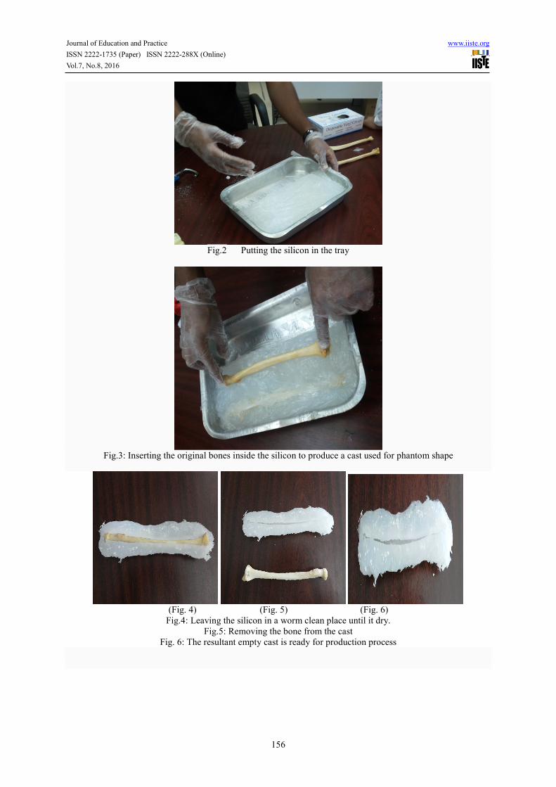
Putting the silicon in the tray as shown in (Fig. 2) insert the original bones inside the silicon to produce a cast
used for phantom shape (Fig. 3), leave the silicon in worm, dry and clean place until it dry. After that, the bone
was removed and the result is an empty cast is ready for production process (Figs. 4, 5, 6).
First experiment with water (Gypsum experiment):
The Gypsum put in the plastic box, and then 300 ml of water added. Therefore, it turns into the liquid form; it
filled into the silicon cast and left it until it dry. It is then be shaped like a bone. It required polishing for final
shaping but extra care was taken not to break the phantom model.
The second experiment (Eggshells mixture with gypsum experiment):
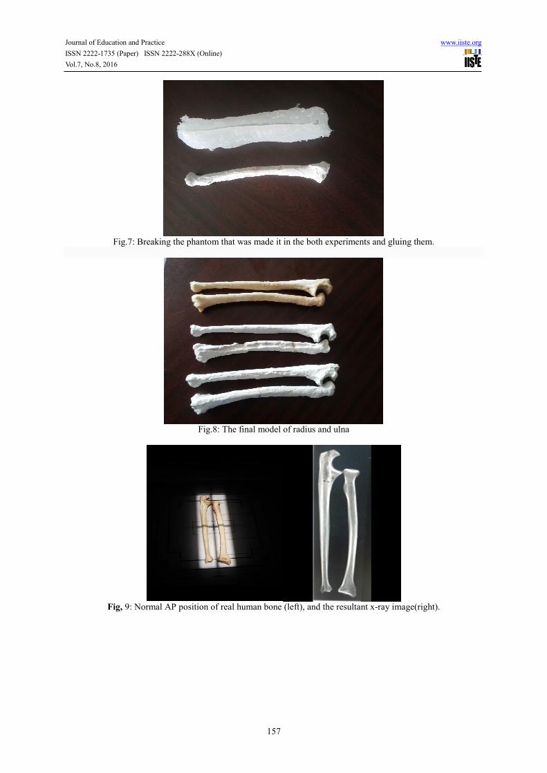
Mixing 25 grams of eggshells and 140 grams of Gypsum with 300 ml of water and removed very carefully
because the mixture is weaker than gypsum in first experiment. Induced fractures made in the phantom to
demonstrate real situations then glued to be hold in its position still when in use (Fig. 7).
Results
The final model of radius and ulna is shown in (Fig. 8). First, the Normal AP forearm of the real human bone
was scanned showing the positioning arrangement and the resultant image (Fig.9). Normal AP position of
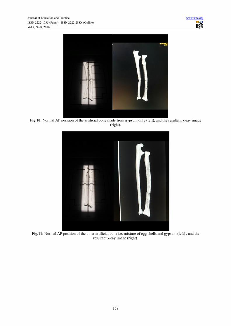
artificial bone and the resultant x-ray image of the developed model produced by Gypsum resulted in
comparable image to that obtained by the above human bone with high degree of similarity (Fig.10). Normal AP
position of artificial bone and the resultant x-ray image of the developed model produced by mixture of egg
shells and gypsum also resulted in comparable image to that obtained by the above human bone (Fig.11). The
models in these figures contains artificially induced fractures clearly observed in the middle of the forearm bone
(transverse and oblique). Additionally, in order to confirm the above results and to ensure the professionalism of
this work by evaluating, assessing and reporting the clinical finding of the abnormality (fractures types), views
and feedback from four independents radiologist from two different health institutes was provided to ensure the
quality and possibility of obtaining diagnostic information from the phantom. Radiologist were involved
voluntary in this research as shown in (Table 1) for gypsum and (Table 2) for egg shells.
Table 1: Radiologist views and feedback for the model using gypsum.
Radiologist
list
Hospital/Health
Institute
Fractue
Type
Image
quality
Can this model
used in teaching
Radiologist Additional
comments
Rad 1 King Abdulaziz
University Hospital
Transverse Fair yes No additional comments
Rad2 King Abdulaziz
University Hospital
Transverse Fair yes Great ideas
Rad3 Faculty of
Medicine
Transverse Fair Yes helpful tool for training
needs little improvement in
shaping
Rad4 Faculty of
Medicine
transverse POOR NO no bone marrow
the material used is non
homogenous which may
give false impression of an
underlying abnormality
Journal of Education and Practice www.iiste.org
ISSN 2222-1735 (Paper) ISSN 2222-288X (Online)
Vol.7, No.8, 2016
154
Table: 2: Radiologist views and feedback for the model using eggshells.
Radiologist
list
Hospital/Health
Institute
Fracture
type
Image
quality
Can this model
used for teaching?
General remark
Rad1 King Abdulaziz
University Hospital
Transverse
and oblique
Fair Yes The other model was better
Rad2 King Abdulaziz
University Hospital
Transverse
and oblique
Fair Yes No additional comments
Rad3 Faculty of
Medicine
Transverse
and oblique
Good Yes I encourage more bone
fractures to be produced it is
very helpful tool for
teaching
Rad4 Faculty of
Medicine
To
transverse
fractures
Fair Yes bone marrow of radius but
not ulna
Cortical irregularity of
radius? infection
Discussion
Radiologists are responsible for introduction new resources for the teaching and educational purposes. Virtual
models are used to obtain radiology images by ability to create materials locally (Tam 2010). The bone material
selection process settled on using gypsum was selected because its electron density was the most similar to that
of bone, ability to be shaped into the bone structures that they would be representing in the final model design
and is a durable material and it's cheap. Also it gives us +1000 HU in the computed tomography. The model for
forearm was used to detect various bone position for different types of fractures. The more important advantage
to use level set is the ability to change topology (Boutiche 2011).
This work results were in agreement with that of (Innocent et al. 2000) that an application of models
was used to aid in the medical pattern application task of image applications by an expert clinician. This work
presents a detailed description of the context and use of this model and showed the affinity of the model and its
importance in education and understanding the performance of different posture of diagnosing fractures of the
radius and ulna.
Many different types of fractures can occur but the author focused on two of them a transverse and
oblique fractures. The modality of choice in the suspected fracture cases is the X-ray that showed clearly the
broken bone or if there is any displacement. Also it provides important information on the type and location of
the fracture. The obtained result revealed that it is possible to produce abnormal forearm model produced from
several local material such as gypsum which give a brighter bone density. The fracture model induced bone
prototype also can be seen clearly in both modes of imaging. The radiologist participating in the image findings
have confirmed the results achieved by this study and 70% of them said it was a very good image quality as
reported (Edmund and Chao 2003).
For different reasons, new models for studying radiological imaging was used in the teaching of
human radiological study of normal and for radius and ulna fracture. The authors briefly review and explore the
advantages and drawbacks of model. The author showed that X-Ray part model gives the unique opportunity to
take x-ray images of single body parts again and again. Radiological models are used in the evaluation of bone
of the fore arm. The model scan will allow evaluating the images produced.
The model allows students of radiographing arm in routine and special circumstances and projections
which may be a useful tool for both training as well as conducting student projects. Imaging Fracture models,
students will be able to practice positioning skills, technical skills, and critical thinking skills as they produce
radiographic images. This model will impact the learning of functional anatomy; such capability will certainly
reach the scope of fracture diagnosis without the need of animals or specimens of cadaver (Buford et al.1990).
Finally, the designed model will offer help to both radiology educators, as well as students. Educators has the
ability to enhance hands-on training of identifying radiographic pathology with a tool that will encourage
students to utilize their critical thinking skills. This has been previously interesting points for researches
(Kauczor et al. 2010)
Conclusion
The development of fractured forearm model was achieved and could be used for educational purposes.
Fortunately, the student feedback for this product was positive in terms of quality of fracture imaging. It improve
skills relevant courses such as radiographic technique and pathology clinical practice. Further studies should be
conducted as recommended by colleagues at the radiology department to enhance the edges (sides of the
developed model) and reduce the density in the bone marrow and finally author recommend to explore
possibility of developing other musculoskeletal system parts.
Journal of Education and Practice www.iiste.org
ISSN 2222-1735 (Paper) ISSN 2222-288X (Online)
Vol.7, No.8, 2016
155
References
Boutiche Y. Detection of Defects in Weld Radiographic Images by Using Chan-Vese Model and Level Set
FormulationCommunications in Computer and Information Science Volume 166, 2011, pp 173-183
Buford, W.L. Myers, L.M. Hollister. A.M. 1990. A modeling and simulation system for the human hand. J Clin
Engin, 15 : 445–451.
Edmund Y.S. Chao. 2003. Graphic-based musculoskeletal model for biomechanical analyses and animation.
Medical Engineering & Physics 25: 201–212.
Innocent P. R., John R. I., Barnes M. 2000. Neuro-Fuzzy Models of Radiographic Image Classification Studies
in Fuzziness and Soft Computing , 41: 361-393.
Inoue, H. Ohgushi, T. Yoshikawa, M. Okumura, T. Sempuku, S. Tamai, Y. Dohi, 1997. The Effect of Aging on
Bone Formation in Porous Hydroxyapatite: Biochemical and Histological Analysis; Journal of Bone
and Mineral Research, 12: 989–994
Kauczor, H.-U. Giesel. F. L. 2010. 3D printing based on imaging data: review of medical applications.
International Journal of Computer Assisted Radiology and Surgery, 5: 335-341
Ohgushi H, Okumura M, Tamai S, Shors EC, Caplan AI Marrow cell induced osteogenesis in porous
hydroxyapatite and tricalcium phosphate: A comparative histomorphometric study of ectopic bone
formation J Biomed Mater Res 24:1563–1570, 1990
Ohgushi H, Okumura M, Yoshikawa T, Inoue K, Sempuku T, Tamai S, Shors EC, Bone formation process in
porous calcium carbonate and hydroxyapatite J Biomed Mater Res 26:885–895, 1992.
Ohgushi, I. H. Yoshikawa, T. Okumura, M. Sempuku, T. Tamai S., Dohi, Y. 1997The Effect of Aging on Bone
Formation in Porous Hydroxyapatite: Biochemical and Histological Analysis; Journal of Bone and
Mineral Research, 32,12: 989–994.
Othman NS, Suhaimi M, Abdul Rahman A, Othman Sazlinayati E, Rozlan Afirah A. 2011 Ultrasound Speed of
Polymer Gel Mimicked Human Soft Tissue within Three Weeks. Int J Biosci Biochem Bioinforma,
223-5.
Richard B.G, Philip K.W. 2005. Exploring the Human Interior: The Roles of Cadaver Dissection and Radiologic
Imaging in Teaching Anatomy. Academic Medicine A 80 - 8: 745-749.
Tam M.D. Building virtual models by postprocessing radiology images: A guide for anatomy faculty. Anat Sci
Educ. 2010 Sep-Oct; 3(5):261-6. doi: 10.1002/ase.175.
Tillack GR. 1999. Simulation of Radiographic Techniques. Review of Progress in Quantitative Nondestructive
Evaluation , 18 : 663-670.
List of figures:
Fig.1 Standard Parameters adjusted for the procedures
Journal of Education and Practice www.iiste.org
ISSN 2222-1735 (Paper) ISSN 2222-288X (Online)
Vol.7, No.8, 2016
156
Fig.2 Putting the silicon in the tray
Fig.3: Inserting the original bones inside the silicon to produce a cast used for phantom shape
(Fig. 4) (Fig. 5) (Fig. 6)
Fig.4: Leaving the silicon in a worm clean place until it dry.
Fig.5: Removing the bone from the cast
Fig. 6: The resultant empty cast is ready for production process
Journal of Education and Practice www.iiste.org
ISSN 2222-1735 (Paper) ISSN 2222-288X (Online)
Vol.7, No.8, 2016
157
Fig.7: Breaking the phantom that was made it in the both experiments and gluing them.
Fig.8: The final model of radius and ulna
Fig, 9: Normal AP position of real human bone (left), and the resultant x-ray image(right).
Journal of Education and Practice www.iiste.org
ISSN 2222-1735 (Paper) ISSN 2222-288X (Online)
Vol.7, No.8, 2016
158
Fig.10: Normal AP position of the artificial bone made from gypsum only (left), and the resultant x-ray image
(right).
Fig.11: Normal AP position of the other artificial bone i.e. mixture of egg shells and gypsum (left) , and the
resultant x-ray image (right).
Related Documents