Journal of Architectural and Planning Research 31:4 (Winter, 2014) 322 Copyright © 2014, Locke Science Publishing Company, Inc. Chicago, IL, USA All Rights Reserved DESIGN AND SOMATIC EXPERIENCE: PRELIMINARY FINDINGS REGARDING DRAWING THROUGH EXPERIENTIAL ANATOMY Galen Cranz Leonardo Chiesi Humans can deliberately activate different parts of the brain to stimulate different kinds of creativity in drawing and design. While most pedagogy has focused on right brain / left brain differences, for this research, we stimulated different levels of the brain — the cortical and the subcortical. Experiential anatomy, somatics, and neuroscience provided the theoretical framework for our study. This quasi-experimental research compares and contrasts sets of drawings of handles and lamps produced by 136 subjects in eight trials. Each set included one drawing produced after stimulating the neocortex and one drawing produced after stimulating subcortical parts of the brain. The two different cognitive states produced design differences as predicted: small, straight, two-dimensional drawings morphed into large, curvilinear, three- dimensional drawings of the same objects. Implications for design pedagogy, “organic architecture,” and creativity more broadly are discussed.

Welcome message from author
This document is posted to help you gain knowledge. Please leave a comment to let me know what you think about it! Share it to your friends and learn new things together.
Transcript
Journal of Architectural and Planning Research31:4 (Winter, 2014) 322
Copyright © 2014, Locke Science Publishing Company, Inc.Chicago, IL, USA All Rights Reserved
DESIGN AND SOMATIC EXPERIENCE: PRELIMINARYFINDINGS REGARDING DRAWING THROUGH
EXPERIENTIAL ANATOMY
Galen CranzLeonardo Chiesi
Humans can deliberately activate different parts of the brain to stimulate different kinds ofcreativity in drawing and design. While most pedagogy has focused on right brain / left braindifferences, for this research, we stimulated different levels of the brain — the cortical and thesubcortical. Experiential anatomy, somatics, and neuroscience provided the theoreticalframework for our study. This quasi-experimental research compares and contrasts sets ofdrawings of handles and lamps produced by 136 subjects in eight trials. Each set included onedrawing produced after stimulating the neocortex and one drawing produced after stimulatingsubcortical parts of the brain. The two different cognitive states produced design differences aspredicted: small, straight, two-dimensional drawings morphed into large, curvilinear, three-dimensional drawings of the same objects. Implications for design pedagogy, “organicarchitecture,” and creativity more broadly are discussed.
Journal of Architectural and Planning Research31:4 (Winter, 2014) 323
SOMATIC THEORY AND DESIGN
Introduction
This paper1 introduces the relatively new field of somatics to the design disciplines in general andto design methods specifically, describing its potential benefits for understanding the interactionbetween designers’ subjective experiences and their objective output. The field of design methodshas focused on designers’ mental processes, especially creativity. Human-factor specialists careabout the effect of products on user experience, and ergonomics focuses on the physical effect ofdesigned environments on users. Each of these specializations is distinctly relevant to architectureand design. In this paper, we propose that the field of somatics introduces new aspects of designthat are relevant to design professionals and educators and that can add to the contributions of theother specializations. We introduce the potential benefits of the somatic perspective in the contextof a specific, empirical example of a somatic approach to design pedagogy and production that hasthe potential to increase the designer’s artistic range and self awareness of how internal processesplay a role in design output.
Somatic Theory and Experiential Anatomy
Somatics is a field of study and practice that emerged in the last 40 years to acknowledge the mutualinteraction of body, mind, and culture.2 According to somatics theory, the body is constituted notonly of physical structures and sensations but also of emotions and culture. According to thisview, the physical body is shaped, both anatomically and physiologically, by emotional habits,cultural ideals, and ideas.3 Likewise, the body shapes culture and experience. Therefore, somaticsfalls within “embodied cognition” theory, in which all aspects of human experience and cognition— from high-level concepts and categories to human performance of various tasks, includingdesign thinking — are shaped by qualities of the body (Gallagher, 2006; Lakoff and Johnson, 1999;Pfeifer and Bongard, 2006). Within somatics, “experiential anatomy” is a tool that helps individualsexperience the ways in which personal concepts, cultural ideas, and the physical body influenceone another.4 The first premise of experiential anatomy is that one can move beyond an intellectualunderstanding of the functioning of the body to direct phenomenological experience — that is, aninternal, subjective appreciation of anatomy. The second premise is that this ability to experiencedifferent parts of one’s anatomy directly can be taught, as discussed further in the “Method” and“Implications for Design Education” sections of this paper.
Anatomy of the Brain and Design Thinking
The human brain has three basic layers, called the triune brain, which evolved successively (Mac-Lean, 1990). In The Accidental Mind, neuroscientist David Linden (2007) described the triune brainusing the metaphor of an ice cream cone with three scoops, each new scoop piled on top of theprevious one without restructuring the prior layer. The first layer, which consists of the brain stemand cerebellum, regulates much of our involuntary activity, like breathing, digestion, and circula-tion. Often called the reptilian brain, it guarantees individual survival; individuals are relativelyindependent when they are born because of this layer. The second layer, called the mammaliancortex or limbic system, processes emotions and memories. Mammals suckle their young, so thislayer of the brain establishes social relations and produces emotions. The final layer, called theneocortex or cerebral cortex, makes language and logical thinking possible and handles informa-tion about the past and the future. The neocortex is where planning and higher cognitive functionstake place. The frontal lobes in particular are where rational, means-ends thinking occurs. Neuro-scientists still acknowledge the basic soundness and usefulness of the idea that the brain evolveddistinct layers, even if they have since acknowledged there is greater complexity in the system,namely that the three basic layers have substructures and interact with one another.5
Journal of Architectural and Planning Research31:4 (Winter, 2014) 324
For designers and planners, recalling the past and anticipating the future is obviously an importantkind of creativity, but a different kind of mental activity emerges from the lower two brains that alsohas value for designers. The first two layers of the brain, the reptilian brain and the limbic system,do not operate using verbal language; instead, these subcortical layers (i.e., the layers locatedbelow the neocortex) use images to communicate with the neocortex, often in the form of dreams or,for example, with shapes, color, spatial relations, motor-related actions, or kinesthesia (along withthe emotional responses attached to them). People have widely varying views on the nature ofintuition, but for those who define it as being outside of reason, these subcortical layers could beviewed as the seat of intuition and are considered a rich source of design inspiration. However, wewould like to examine their power empirically and systematically.6 As designers, we may want to tapinto the subcortical parts of the brain intentionally. Knowing how to design with the subcorticallayers activated changes the design experience and products (or at least their representation) andincreases the involvement of the user, audience, or customer. Earlier design teachers have taughtdesign according to this premise.7
The research in this paper tests the idea that creativity, perception, and design productivity,including drawing, can be influenced by deliberately priming the subcortical layers of the brain.Heretofore, no one has brought these insights about the levels of the brain into the world ofteaching creativity in design and drawing.8 At most, pedagogy has focused on right brain / leftbrain differences, but in this research, we stimulated different levels of the brain — cortical andsubcortical.9 Nevertheless, our work shares the premise that humans can activate different parts ofthe brain to stimulate generativity in drawing and design.
Organ Consciousness and Design
The British architectural theorist Geoffrey Scott (1924:216) wrote in The Architecture of Humanismthat architecture is “the transcription of the body’s states into forms of building.” Somatic scholar-educators like his idea that architecture relates so directly to the human body. However, Scott didnot indicate anything about the mechanism through which that transformation from internal bodilystate to architectural production takes place (Leigh, 1999; Mumford, 1989). Thus, we seek to furtherour collective understanding of how internal experience and design relate to one another.
Bodily organs are governed primarily by the first layer of the brain (i.e., the reptilian brain) and arehence usually outside of our conscious control. However, with deliberate attention and cultivatedpractice, physiological functions can be influenced by the neocortex. For example, an individual’sheartbeat can be regulated by yogic and Taoist practices (see, e.g., Das, 1987; Telles and Viswes-waraiah, 2010). By becoming aware of his or her own organs through specific routines, which wewill describe in the next section, an individual can tune in to his or her physiological functions andstart to perceive them; for lack of a better term, we might call this “organ consciousness.” In thisstate, the qualities of the organs acquire a strong presence, and the individual starts to appreciatethe fact that the organs are more yielding than bones, ligaments, tendons, or even muscles. Theyflow and expand in all directions rather than orient in one direction. Newborns’ organs are moredeveloped than the rest of their anatomy; babies can roll from side to side on their relatively largeorgans, which are covered by relatively soft and pliable muscles and ribs, before their arms and legsdevelop sufficient strength and motor control for crawling and walking.10
If organ consciousness differs from rational consciousness, then one would expect to see differ-ences in what is produced in each state. Organ consciousness would likely reflect its own forms —round, curvilinear, volumetric, flowing, undifferentiated — while the consciousness of the rationalneocortex would be more likely to reflect its own predominantly means-ends schema — that is,efficient, graphic, two-dimensional representations characterized by straight lines, right angles,and well-defined edges. Jader Tolja, the somatically educated doctor of medicine who originallyconceived the experimental hypothesis and procedure of this study, predicted there would bedifferences in the artistic output created in cortical versus subcortical states (Tolja and Speciani,
Journal of Architectural and Planning Research31:4 (Winter, 2014) 325
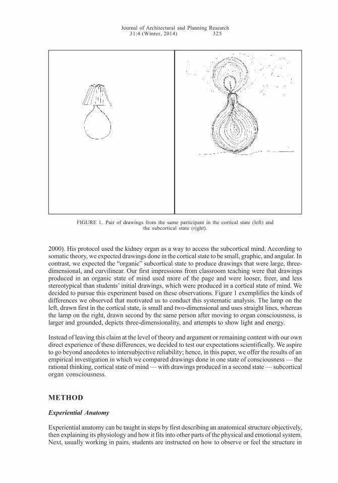
2000). His protocol used the kidney organ as a way to access the subcortical mind. According tosomatic theory, we expected drawings done in the cortical state to be small, graphic, and angular. Incontrast, we expected the “organic” subcortical state to produce drawings that were large, three-dimensional, and curvilinear. Our first impressions from classroom teaching were that drawingsproduced in an organic state of mind used more of the page and were looser, freer, and lessstereotypical than students’ initial drawings, which were produced in a cortical state of mind. Wedecided to pursue this experiment based on these observations. Figure 1 exemplifies the kinds ofdifferences we observed that motivated us to conduct this systematic analysis. The lamp on theleft, drawn first in the cortical state, is small and two-dimensional and uses straight lines, whereasthe lamp on the right, drawn second by the same person after moving to organ consciousness, islarger and grounded, depicts three-dimensionality, and attempts to show light and energy.
Instead of leaving this claim at the level of theory and argument or remaining content with our owndirect experience of these differences, we decided to test our expectations scientifically. We aspireto go beyond anecdotes to intersubjective reliability; hence, in this paper, we offer the results of anempirical investigation in which we compared drawings done in one state of consciousness — therational thinking, cortical state of mind — with drawings produced in a second state — subcorticalorgan consciousness.
METHOD
Experiential Anatomy
Experiential anatomy can be taught in steps by first describing an anatomical structure objectively,then explaining its physiology and how it fits into other parts of the physical and emotional system.Next, usually working in pairs, students are instructed on how to observe or feel the structure in
FIGURE 1. Pair of drawings from the same participant in the cortical state (left) andthe subcortical state (right).
Journal of Architectural and Planning Research31:4 (Winter, 2014) 324
For designers and planners, recalling the past and anticipating the future is obviously an importantkind of creativity, but a different kind of mental activity emerges from the lower two brains that alsohas value for designers. The first two layers of the brain, the reptilian brain and the limbic system,do not operate using verbal language; instead, these subcortical layers (i.e., the layers locatedbelow the neocortex) use images to communicate with the neocortex, often in the form of dreams or,for example, with shapes, color, spatial relations, motor-related actions, or kinesthesia (along withthe emotional responses attached to them). People have widely varying views on the nature ofintuition, but for those who define it as being outside of reason, these subcortical layers could beviewed as the seat of intuition and are considered a rich source of design inspiration. However, wewould like to examine their power empirically and systematically.6 As designers, we may want to tapinto the subcortical parts of the brain intentionally. Knowing how to design with the subcorticallayers activated changes the design experience and products (or at least their representation) andincreases the involvement of the user, audience, or customer. Earlier design teachers have taughtdesign according to this premise.7
The research in this paper tests the idea that creativity, perception, and design productivity,including drawing, can be influenced by deliberately priming the subcortical layers of the brain.Heretofore, no one has brought these insights about the levels of the brain into the world ofteaching creativity in design and drawing.8 At most, pedagogy has focused on right brain / leftbrain differences, but in this research, we stimulated different levels of the brain — cortical andsubcortical.9 Nevertheless, our work shares the premise that humans can activate different parts ofthe brain to stimulate generativity in drawing and design.
Organ Consciousness and Design
The British architectural theorist Geoffrey Scott (1924:216) wrote in The Architecture of Humanismthat architecture is “the transcription of the body’s states into forms of building.” Somatic scholar-educators like his idea that architecture relates so directly to the human body. However, Scott didnot indicate anything about the mechanism through which that transformation from internal bodilystate to architectural production takes place (Leigh, 1999; Mumford, 1989). Thus, we seek to furtherour collective understanding of how internal experience and design relate to one another.
Bodily organs are governed primarily by the first layer of the brain (i.e., the reptilian brain) and arehence usually outside of our conscious control. However, with deliberate attention and cultivatedpractice, physiological functions can be influenced by the neocortex. For example, an individual’sheartbeat can be regulated by yogic and Taoist practices (see, e.g., Das, 1987; Telles and Viswes-waraiah, 2010). By becoming aware of his or her own organs through specific routines, which wewill describe in the next section, an individual can tune in to his or her physiological functions andstart to perceive them; for lack of a better term, we might call this “organ consciousness.” In thisstate, the qualities of the organs acquire a strong presence, and the individual starts to appreciatethe fact that the organs are more yielding than bones, ligaments, tendons, or even muscles. Theyflow and expand in all directions rather than orient in one direction. Newborns’ organs are moredeveloped than the rest of their anatomy; babies can roll from side to side on their relatively largeorgans, which are covered by relatively soft and pliable muscles and ribs, before their arms and legsdevelop sufficient strength and motor control for crawling and walking.10
If organ consciousness differs from rational consciousness, then one would expect to see differ-ences in what is produced in each state. Organ consciousness would likely reflect its own forms —round, curvilinear, volumetric, flowing, undifferentiated — while the consciousness of the rationalneocortex would be more likely to reflect its own predominantly means-ends schema — that is,efficient, graphic, two-dimensional representations characterized by straight lines, right angles,and well-defined edges. Jader Tolja, the somatically educated doctor of medicine who originallyconceived the experimental hypothesis and procedure of this study, predicted there would bedifferences in the artistic output created in cortical versus subcortical states (Tolja and Speciani,
Journal of Architectural and Planning Research31:4 (Winter, 2014) 325
2000). His protocol used the kidney organ as a way to access the subcortical mind. According tosomatic theory, we expected drawings done in the cortical state to be small, graphic, and angular. Incontrast, we expected the “organic” subcortical state to produce drawings that were large, three-dimensional, and curvilinear. Our first impressions from classroom teaching were that drawingsproduced in an organic state of mind used more of the page and were looser, freer, and lessstereotypical than students’ initial drawings, which were produced in a cortical state of mind. Wedecided to pursue this experiment based on these observations. Figure 1 exemplifies the kinds ofdifferences we observed that motivated us to conduct this systematic analysis. The lamp on theleft, drawn first in the cortical state, is small and two-dimensional and uses straight lines, whereasthe lamp on the right, drawn second by the same person after moving to organ consciousness, islarger and grounded, depicts three-dimensionality, and attempts to show light and energy.
Instead of leaving this claim at the level of theory and argument or remaining content with our owndirect experience of these differences, we decided to test our expectations scientifically. We aspireto go beyond anecdotes to intersubjective reliability; hence, in this paper, we offer the results of anempirical investigation in which we compared drawings done in one state of consciousness — therational thinking, cortical state of mind — with drawings produced in a second state — subcorticalorgan consciousness.
METHOD
Experiential Anatomy
Experiential anatomy can be taught in steps by first describing an anatomical structure objectively,then explaining its physiology and how it fits into other parts of the physical and emotional system.Next, usually working in pairs, students are instructed on how to observe or feel the structure in
FIGURE 1. Pair of drawings from the same participant in the cortical state (left) andthe subcortical state (right).
Figures 1-2
Cortical Subcortical Cortical Subcortical

Journal of Architectural and Planning Research31:4 (Winter, 2014) 326
someone else. Palpating a structure and feeling it at work in someone else’s body helps buildconfidence that this experience is “real” — that is, accessible to direct perception. Then, it becomeseasier to sense the same structure or process in oneself. This, in turn, helps the student identifywith that structure or function by taking on its agency, enabling a sort of dialogue with that part ofhis or her own anatomy. This interaction impacts the structure and function of those body partsand leads to a transformation of the body. Thus, these interactions have therapeutic implications,though these implications are outside the scope of the present research. Instead, as design educa-tors, we are interested in the effects of experiential anatomy on generativity and the qualities ofproducts (or outputs) that flow from the state of mind that emerges from a perceptually basedinteraction with one’s own body.
Experimental Design and Sample
In order to test the hypothesis that deep proprioceptive states and the internal organizationassociated with them lead to different perceptions of space and therefore to different designoutputs, Jader Tolja, a doctor of medicine who was also trained in the Feldenkrais Method, Gestalttherapy, and Body-Mind Centering (BMC),11 decided to compare drawings produced in each ofthe states. On visual inspection, the results of his first two samples from Italy looked promising toGalen Cranz, a designer, professor of architecture, and teacher of the Alexander Technique whowas also educated in BMC.12 Her dual training in design and somatics allowed her to understandthe potential significance of the differences in the drawings, so she invited him to conduct addi-tional tests with her in Kolding, Denmark (along with Leonardo Chiesi), and later Bratislava, Slova-kia, and the United States (Berkeley and San Francisco, California). She also conducted two sam-ples on her own, one in Berkeley classrooms and one at an international design research confer-ence in Veracruz, Mexico.
In total, we gathered data13 from eight experimental sessions that took place between 2003 and2008 in Italy (two sessions), Denmark, Slovakia, the U.S. (three sessions, two in Berkeley and onein San Francisco), and Mexico. Our final combined sample consisted of 136 participants (94 female,28 male, and 14 from whom we did not receive gender data) from many different countries on fivecontinents: 37 were from the U.S., 22 were from Slovakia, 16 were from Denmark, 10 were from Italy,and the remaining 51 were from various other countries. Note that some participants did notcomplete each drawing task as assigned; thus, the totals presented in the tables vary and do notmatch the 136 total.
Protocol
The experimental protocol followed a carefully crafted sequence in each trial, as follows:(1) We gathered participants in a suitable environment (classrooms) where they would not
be disturbed for the entire length of the experiment (from two to three hours).(2) We briefed participants on the purpose of the experiment, which was described as part of
a class or special seminar on “the relationship between body and design.” Nothing morewas said about the true objectives of the experiment.
(3) We asked participants to complete a series of simple mathematical exercises (addition,subtraction, multiplication, and division) for five to 10 minutes to generate a state ofcortical mental organization in which the frontal lobes would be activated and dominate.
(4) We then asked participants to draw two sketches, one of a handle of any kind and anotherof a floor or table lamp, each on a different sheet of paper. (The paper was either 8.5'' x 11''letterhead or A4 format.) These drawings constituted batch 1.
(5) Participants were then led through a proprioception routine, which was designed byDr. Tolja to elicit a subcortical mental organization. This routine was carefully replicatedby Dr. Cranz for the two samples taken when Dr. Tolja was not present. The routine lastsapproximately 25-30 minutes. Participants were paired, so that one could place a hand onthe back of the other over one of the kidneys, which are located half under the rib cage
Journal of Architectural and Planning Research31:4 (Winter, 2014) 327
and half extended below it. Those with their hand on another’s back were asked to sensethat individual’s clothing, then to sense the skin underneath, just as one can sense theroad surface when driving a car even though one’s hands are only on the steering wheel.Next, they were asked to sense the muscles below the skin, then the bony ribs, and thenbehind that “something warm,” “perhaps a mass, a little squirrel-like entity that can movea bit.” The experimenter would then explain that, because the kidneys process a hugevolume of blood, they are a site of movement of the blood and are thus extra warm.14 Theexperimenter then asked, “Perhaps you can sense that? Can you invite the kidney intoyour hand? Can you invite it to move to the right or left? Can you invite it to slide up ordown?” The experimenter spoke slowly and deliberately, having taken him or herself intoa mental state of contact with his or her own kidneys. The intention is to help participantsmove from a cortical state of consciousness to a subcortical one. The individuals whosekidneys were being sensed were, of course, listening to these instructions and were takendeeper into their own bodily sensations, so they too were affected by the guided imageryand could sense their kidneys moving subtly in and out, side to side, up and down.
(6) The experimenter would then explain in a soft voice that he or she was about to make aloud noise. He or she issued this warning gently three times before dropping a set ofbooks to the floor or clapping his or her hands sharply. The group was then invited todiscuss what happened to their kidneys when they heard the loud noise. Most reportedthat their kidneys retracted inward or moved upward under the ribs. The experimenterreminded everyone that they were warned about the noise intellectually (cortically), butthat emotionally and instinctually (subcortically), they could not be prepared, underscor-ing the independence, vulnerability, and power of the subcortical brain. This phase of theexperiment established confidence in the participants that they could sense internalorgans because both partners — the sensor and the sensed — experienced how thenoise startled the kidney.
(7) After a 10-20 minute break, the group reassembled, and the partners switched roles. Theywere once again guided through the same steps to make contact with the kidney and tofeel it and its movement. This time, instead of making a loud noise, the experimenter askedparticipants to stay in the mental state they were experiencing right then, quietly movewithout speaking to their drawing materials, and draw another handle and lamp. Thesedrawings constituted batch 2.
(8) We asked participants to pair their before-and-after drawings and then lay them all out ontables to be viewed by the entire group. We then talked about the differences between thetwo batches and made lists of the qualities observed in both batches. Finally, at the endof the workshop, we disclosed the real intent of the experiment to participants, namely, toacknowledge the differences between creativity generated by two different parts of thebrain and to reveal the power of designing from the subcortical level. We closed bydiscussing the experience from both the facilitators’ and the participants’ perspectives.
Limits of the Research Design
First, we should note that our data were gathered in a quasi-experimental fashion. Unlike in a fullexperiment, we were not able to (1) create a control group; (2) control all of the environmentalvariables that may have, at least in theory, had an impact on the design output; or (3) control for theeffect of a reversed intervention, in which participants would be stimulated with the subcorticalroutine before the cortical one. With regard to this last point, we must consider that the results ofthe experiment are probably not indifferent to the order in which the stimuli were presented: whatgets drawn for a second time within a short time frame is probably affected by the very fact that itcame second. More versions of this study are needed to ascertain whether this effect does exist.Another implication of the order of the stimuli might be a stimulus-specific carry-over effect of thefirst intervention on the second. Similarly, we were not able to test the effect of repeatedly switch-ing from one stimulation to the other. More diversified versions of the study might shed light onhow easily participants are able to switch from one state to the other and back again. Moreover, we
Journal of Architectural and Planning Research31:4 (Winter, 2014) 328
were not able to consider the possibility of a dose-response effect (i.e., how the length of exposureto the stimuli affected participants) and therefore develop a dose-response model for our tworoutines. Further work could determine the exposure threshold (in both duration and intensity)needed for the stimuli to be effective; a version of the experiment using a delayed interventiondesign could seek to determine the stimuli response time curve. Finally, we acknowledge that weintended to induce two different mind-sets, and we assume that we did; however, we have noindependent measure of what mind-set participants were in at any given time. We would like tocorrect this in the future with an external measure of brain function.
Moreover, we cannot entirely exclude the so-called “experimenter’s effect” (Rosenthal, 1966).Participants may have responded to our expectations, even though we took the utmost care neverto express them either implicitly or explicitly. Seminar and workshop participants were told only thatthey were participating in a design exercise and were instructed in a step-by-step fashion with nofurther explanation. Although we were always careful not to disclose the purpose of our experimentbefore participants actually sketched their drawings, it is nonetheless possible, though unlikely,that they may have responded unconsciously to our unspoken anticipation of the results (ibid.).Further, we have no independent measure of their state of consciousness, so it is theoreticallypossible that the observed changes were due to something other than a change in consciousness.
The sample we selected could also suffer from a selection bias, as it was not the result of arandomized procedure. Rather, the subjects self-selected themselves as participants in a designclass or workshop (and as such were mostly graduate students and scholars), which limits theexternal validity of the study. Regarding the use of students and workshop participants, there iswidespread consensus in the scientific community that such a sample procedure is acceptablepractice for preliminary studies testing a new hypothesis. Further versions of the experiment couldclarify the extent of the results for different cultures, age groups, socioeconomic cohorts, occupa-tional and interest groups, and so forth.
Despite these limitations, the consistency of the patterns shown by participants in different exper-imental situations and locations worldwide and the strength of the relationships among the vari-ables, as shown in the quantitative data analysis, make us confident that the experimenter’s effectand the methodological shortcomings of the quasi-experiment were kept to a reasonable minimumand do not invalidate our findings. At the very least, our findings suggest there is something atwork here that merits further exploration and testing. We may have only just begun to scratch thesurface of this important topic as it relates to creativity and innovation, but it is a provocativebeginning with the potential to have an impact on design practice.
FINDINGS
Qualitative Analysis of the Drawings
Looking at the sketches immediately shows that the proprioception routine we used had preciseand determinate effects on participants’ design output (Figures 1-2). The sketches shown in thefirst and third columns of Figure 2 belong to batch 1 (in which the neocortex, i.e., the cortical state,prevailed), and those in the second and fourth columns belong to batch 2 (in which the subcorticalstate prevailed). One immediately notices a high level of internal consistency from the point ofview of the forms or shapes of the drawings within each set but a high degree of outside differen-tiation between the two sets. Straight lines predominate in batch 1, while curved lines prevail inbatch 2. There are more details in batch 1, as if the drawers were more attentive to the particulars oftheir drawings, while in batch 2, the attention of the drawers seems to have focused more on theoverall form of the objects they were drawing. The drawings develop more in the vertical plane inbatch 1, while the horizontal plane is dominant in batch 2. In batch 1, there is largely an absence ofthree-dimensionality; conversely, three-dimensionality is more evident in batch 2, in which all of
Journal of Architectural and Planning Research31:4 (Winter, 2014) 329
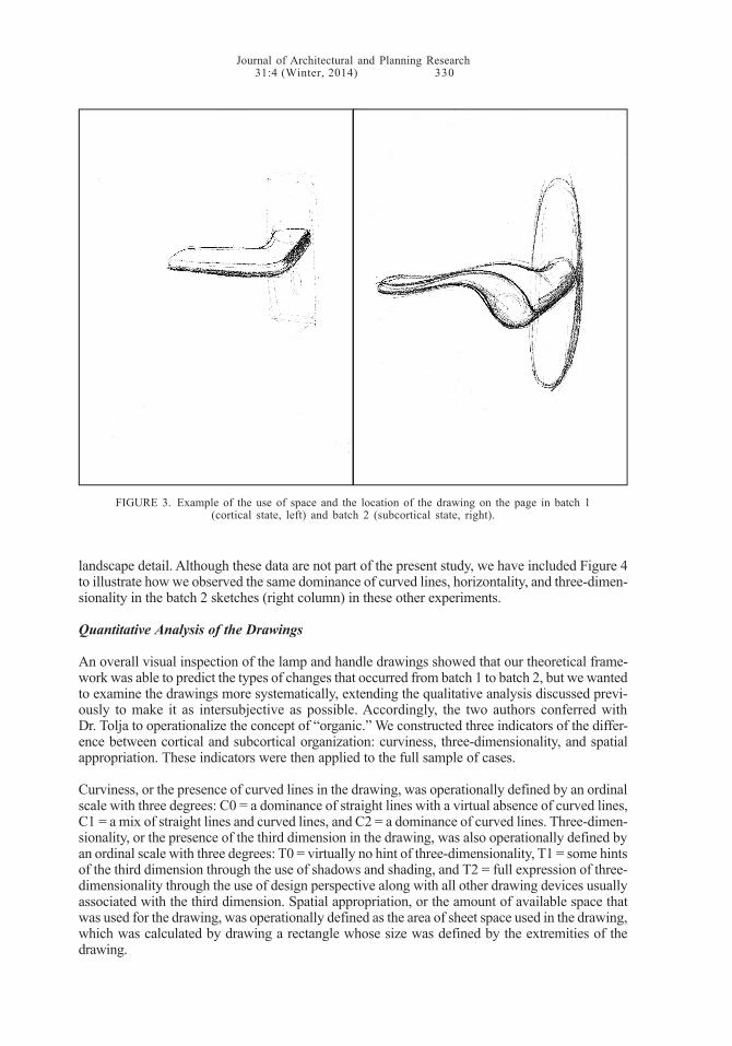
the conventional drawing devices needed to present the third dimension — shading, shadow, andperspective — can be seen. Furthermore, the drawings in batch 2 tend to use a larger proportion ofthe available space. In the discussion that follows, we turned this into an indicator of the differencebetween cortical and subcortical organization. Figure 3 provides one example of this use of theentire page. As one can see in Figures 2-3, the drawings in batch 1 tend to be located toward thecenter of the page, while those in batch 2 move toward the bottom, which we interpret as movingtoward the “heavier” center of gravity.
We observed this same pattern of differences in another set of experimental situations in whichparticipants were assigned various design tasks — to design a room, an urban square, or a
FIGURE 2. Examples of pairs of drawings from the same participant in the cortical state (first and thirdcolumns) and the subcortical state (second and fourth columns). Ratio has been preserved so that
spatial appropriation can be appreciated.

Journal of Architectural and Planning Research31:4 (Winter, 2014) 328
were not able to consider the possibility of a dose-response effect (i.e., how the length of exposureto the stimuli affected participants) and therefore develop a dose-response model for our tworoutines. Further work could determine the exposure threshold (in both duration and intensity)needed for the stimuli to be effective; a version of the experiment using a delayed interventiondesign could seek to determine the stimuli response time curve. Finally, we acknowledge that weintended to induce two different mind-sets, and we assume that we did; however, we have noindependent measure of what mind-set participants were in at any given time. We would like tocorrect this in the future with an external measure of brain function.
Moreover, we cannot entirely exclude the so-called “experimenter’s effect” (Rosenthal, 1966).Participants may have responded to our expectations, even though we took the utmost care neverto express them either implicitly or explicitly. Seminar and workshop participants were told only thatthey were participating in a design exercise and were instructed in a step-by-step fashion with nofurther explanation. Although we were always careful not to disclose the purpose of our experimentbefore participants actually sketched their drawings, it is nonetheless possible, though unlikely,that they may have responded unconsciously to our unspoken anticipation of the results (ibid.).Further, we have no independent measure of their state of consciousness, so it is theoreticallypossible that the observed changes were due to something other than a change in consciousness.
The sample we selected could also suffer from a selection bias, as it was not the result of arandomized procedure. Rather, the subjects self-selected themselves as participants in a designclass or workshop (and as such were mostly graduate students and scholars), which limits theexternal validity of the study. Regarding the use of students and workshop participants, there iswidespread consensus in the scientific community that such a sample procedure is acceptablepractice for preliminary studies testing a new hypothesis. Further versions of the experiment couldclarify the extent of the results for different cultures, age groups, socioeconomic cohorts, occupa-tional and interest groups, and so forth.
Despite these limitations, the consistency of the patterns shown by participants in different exper-imental situations and locations worldwide and the strength of the relationships among the vari-ables, as shown in the quantitative data analysis, make us confident that the experimenter’s effectand the methodological shortcomings of the quasi-experiment were kept to a reasonable minimumand do not invalidate our findings. At the very least, our findings suggest there is something atwork here that merits further exploration and testing. We may have only just begun to scratch thesurface of this important topic as it relates to creativity and innovation, but it is a provocativebeginning with the potential to have an impact on design practice.
FINDINGS
Qualitative Analysis of the Drawings
Looking at the sketches immediately shows that the proprioception routine we used had preciseand determinate effects on participants’ design output (Figures 1-2). The sketches shown in thefirst and third columns of Figure 2 belong to batch 1 (in which the neocortex, i.e., the cortical state,prevailed), and those in the second and fourth columns belong to batch 2 (in which the subcorticalstate prevailed). One immediately notices a high level of internal consistency from the point ofview of the forms or shapes of the drawings within each set but a high degree of outside differen-tiation between the two sets. Straight lines predominate in batch 1, while curved lines prevail inbatch 2. There are more details in batch 1, as if the drawers were more attentive to the particulars oftheir drawings, while in batch 2, the attention of the drawers seems to have focused more on theoverall form of the objects they were drawing. The drawings develop more in the vertical plane inbatch 1, while the horizontal plane is dominant in batch 2. In batch 1, there is largely an absence ofthree-dimensionality; conversely, three-dimensionality is more evident in batch 2, in which all of
Journal of Architectural and Planning Research31:4 (Winter, 2014) 329
the conventional drawing devices needed to present the third dimension — shading, shadow, andperspective — can be seen. Furthermore, the drawings in batch 2 tend to use a larger proportion ofthe available space. In the discussion that follows, we turned this into an indicator of the differencebetween cortical and subcortical organization. Figure 3 provides one example of this use of theentire page. As one can see in Figures 2-3, the drawings in batch 1 tend to be located toward thecenter of the page, while those in batch 2 move toward the bottom, which we interpret as movingtoward the “heavier” center of gravity.
We observed this same pattern of differences in another set of experimental situations in whichparticipants were assigned various design tasks — to design a room, an urban square, or a
FIGURE 2. Examples of pairs of drawings from the same participant in the cortical state (first and thirdcolumns) and the subcortical state (second and fourth columns). Ratio has been preserved so that
spatial appropriation can be appreciated.
Figures 1-2
Cortical Subcortical Cortical Subcortical
Journal of Architectural and Planning Research31:4 (Winter, 2014) 330
landscape detail. Although these data are not part of the present study, we have included Figure 4to illustrate how we observed the same dominance of curved lines, horizontality, and three-dimen-sionality in the batch 2 sketches (right column) in these other experiments.
Quantitative Analysis of the Drawings
An overall visual inspection of the lamp and handle drawings showed that our theoretical frame-work was able to predict the types of changes that occurred from batch 1 to batch 2, but we wantedto examine the drawings more systematically, extending the qualitative analysis discussed previ-ously to make it as intersubjective as possible. Accordingly, the two authors conferred withDr. Tolja to operationalize the concept of “organic.” We constructed three indicators of the differ-ence between cortical and subcortical organization: curviness, three-dimensionality, and spatialappropriation. These indicators were then applied to the full sample of cases.
Curviness, or the presence of curved lines in the drawing, was operationally defined by an ordinalscale with three degrees: C0 = a dominance of straight lines with a virtual absence of curved lines,C1 = a mix of straight lines and curved lines, and C2 = a dominance of curved lines. Three-dimen-sionality, or the presence of the third dimension in the drawing, was also operationally defined byan ordinal scale with three degrees: T0 = virtually no hint of three-dimensionality, T1 = some hintsof the third dimension through the use of shadows and shading, and T2 = full expression of three-dimensionality through the use of design perspective along with all other drawing devices usuallyassociated with the third dimension. Spatial appropriation, or the amount of available space thatwas used for the drawing, was operationally defined as the area of sheet space used in the drawing,which was calculated by drawing a rectangle whose size was defined by the extremities of thedrawing.
FIGURE 3. Example of the use of space and the location of the drawing on the page in batch 1(cortical state, left) and batch 2 (subcortical state, right).
Journal of Architectural and Planning Research31:4 (Winter, 2014) 331
To pretest the reliability of the operational definitions of the indicators, we gave the sketches to anindependent observer, Lusi Morhayim, along with the descriptions of the indicators. She was ableto apply our criteria to the drawings on her own and made the same assessments we did, so weconcluded that our indicators had a very high level of internal consistency and therefore could beused independently of the original researchers.15 This high level of consistency is due to thetwofold simplicity of the indicators. First, the indicators have an intrinsic simplicity derived fromtheir closeness to concrete, mundane notions such as curviness and perspective. Second, the taskof comparing two sketches relatively instead of absolutely is a simple one. The indicators show therelative change or transformation of the drawings from batch 1 to batch 2.
We present the analysis in order of descending support of our hypothesis. The pair comparisonsrelated to curviness (Table 1) showed that changes in favor of the hypothesis outnumberedchanges against the hypothesis by 3.8 times for the lamp drawings and 3.4 times for the handledrawings. Changes in favor of the hypothesis also significantly outnumbered cases with nochange at all: almost twice as many for the lamp drawings (1.9 times) and 1.5 times as many for thehandle drawings. These data therefore support our main hypothesis, with a slightly higher signif-icance for the lamp drawings than the handle drawings. This slight difference between the handle
FIGURE 4. Sketches of urban squares and landscapes drawn in the cortical state (left) andthe subcortical state (right).
Figures 3-4

Journal of Architectural and Planning Research31:4 (Winter, 2014) 330
landscape detail. Although these data are not part of the present study, we have included Figure 4to illustrate how we observed the same dominance of curved lines, horizontality, and three-dimen-sionality in the batch 2 sketches (right column) in these other experiments.
Quantitative Analysis of the Drawings
An overall visual inspection of the lamp and handle drawings showed that our theoretical frame-work was able to predict the types of changes that occurred from batch 1 to batch 2, but we wantedto examine the drawings more systematically, extending the qualitative analysis discussed previ-ously to make it as intersubjective as possible. Accordingly, the two authors conferred withDr. Tolja to operationalize the concept of “organic.” We constructed three indicators of the differ-ence between cortical and subcortical organization: curviness, three-dimensionality, and spatialappropriation. These indicators were then applied to the full sample of cases.
Curviness, or the presence of curved lines in the drawing, was operationally defined by an ordinalscale with three degrees: C0 = a dominance of straight lines with a virtual absence of curved lines,C1 = a mix of straight lines and curved lines, and C2 = a dominance of curved lines. Three-dimen-sionality, or the presence of the third dimension in the drawing, was also operationally defined byan ordinal scale with three degrees: T0 = virtually no hint of three-dimensionality, T1 = some hintsof the third dimension through the use of shadows and shading, and T2 = full expression of three-dimensionality through the use of design perspective along with all other drawing devices usuallyassociated with the third dimension. Spatial appropriation, or the amount of available space thatwas used for the drawing, was operationally defined as the area of sheet space used in the drawing,which was calculated by drawing a rectangle whose size was defined by the extremities of thedrawing.
FIGURE 3. Example of use of space and location of drawing on page in batch 1 (cortical state, left)and batch 2 (subcortical state, right).
Journal of Architectural and Planning Research31:4 (Winter, 2014) 331
To pretest the reliability of the operational definitions of the indicators, we gave the sketches to anindependent observer, Lusi Morhayim, along with the descriptions of the indicators. She was ableto apply our criteria to the drawings on her own and made the same assessments we did, so weconcluded that our indicators had a very high level of internal consistency and therefore could beused independently of the original researchers.15 This high level of consistency is due to thetwofold simplicity of the indicators. First, the indicators have an intrinsic simplicity derived fromtheir closeness to concrete, mundane notions such as curviness and perspective. Second, the taskof comparing two sketches relatively instead of absolutely is a simple one. The indicators show therelative change or transformation of the drawings from batch 1 to batch 2.
We present the analysis in order of descending support of our hypothesis. The pair comparisonsrelated to curviness (Table 1) showed that changes in favor of the hypothesis outnumberedchanges against the hypothesis by 3.8 times for the lamp drawings and 3.4 times for the handledrawings. Changes in favor of the hypothesis also significantly outnumbered cases with nochange at all: almost twice as many for the lamp drawings (1.9 times) and 1.5 times as many for thehandle drawings. These data therefore support our main hypothesis, with a slightly higher signif-icance for the lamp drawings than the handle drawings. This slight difference between the handle
FIGURE 4. Sketches of urban squares and landscapes drawn in the cortical state (left) andthe subcortical state (right).Figures 3-4

Journal of Architectural and Planning Research31:4 (Winter, 2014) 332
and lamp drawings in support of our hypothesis was a recurring pattern. This might be explainedby the nature of the design problem associated with the two objects — that is, more functionalconstraints are associated with handles than with lamps.
The three-dimensionality indicator presented less compelling support for our hypothesis, yet thedata shown in Table 1 are still fully compatible with it. Changes to the lamp drawings in favor of thehypothesis outnumbered changes against the hypothesis by two times. However, we also ob-served a large number of cases (70) in which the routine did not yield any changes. In the instanceof the handle drawings, we observed that cases in favor of the hypothesis outnumbered thoseagainst it by only 1.3 times, and 57 cases showed no effect. As with the curviness indicator, wefound lamp drawings were more susceptible to changes in the direction of our hypothesis thanwere handle drawings.
The spatial-appropriation indicator showed a mild accordance with our hypothesis (Table 1). Thecases in favor of the hypothesis outnumbered those against it by 1.5 times for the lamp drawingsand only 1.2 times for the handle drawings, with a very small number of no-change cases (sevenand 13 cases respectively). Drawings labeled as “no change” increased or decreased the use ofspace within ±10% of the area of the original sketch. As with the other two indicators, the handledrawings showed a weaker response to the proprioception routine.
Quantitative Indexes
In order to synthesize all of the available data, we created a single index, “direction of change,” thatwould tell us how many participants drew a batch 2 sketch that indicated support for our mainhypothesis. We assigned a score to each of the three indicators for each pair of drawings and thentotalled the scores. We assigned a score of -1 to an indicator if the drawings in a pair showed achange against the hypothesis, a score of 0 if there was no change, and a score of +1 if there wasa change in favor of the hypothesis. Total negative scores (ranging from -3 to -1) on the final indexreflected a net change of the drawing pair away from the hypothesis; a score of zero indicated nonet change, as if the drawing pair was indifferent to the hypothesis; and positive scores (rangingfrom +1 to +3) reflected a change aligned with the hypothesis.
The data in Table 2 show that the hypothesized changes were born out. The pairs of lamp sketchesthat showed a net change compatible with the main hypothesis outnumbered the pairs that wentagainst the hypothesis or that did not show any change by three times (63 cases versus 21 cases
TABLE 1. Curviness, three-dimensionality, and spatial appropriation: pair comparisons of drawings before andafter the proprioception routine._______________________________________________________________________________________________________________________________________________________________________________________________________________________________________________________________________________________________________________________________________________________________
Curviness Three-Dimensionality Spatial AppropriationFrequency Valid % Frequency Valid % Frequency Valid %
_______________________________________________________________________________________________________________________________________________________________________________________________________________________________________________________________________________________________________________________________________________________________LampUnhypothesized change 18 14.9 17 14.0 39 37.1No change 35 28.9 70 57.9 7 6.7Hypothesized change 68 56.2 34 28.1 59 56.2Total 121 100.0 121 100.0 105 100.0_______________________________________________________________________________________________________________________________________________________________________________________________________________________________________________________________________________________________________________________________________________________________HandleUnhypothesized change 17 15.0 24 21.2 38 39.2No change 38 33.6 57 50.4 13 13.4Hypothesized change 58 51.3 32 28.3 46 47.4Total 113 100.0 113 100.0 97 100.0_______________________________________________________________________________________________________________________________________________________________________________________________________________________________________________________________________________________________________________________________________________________________Note. Percentages shown may not add up to 100 due to rounding. The totals reflect the number of drawing pairsthat met the criteria for the given indicator. In addition, some participants did not complete each drawing task asassigned._______________________________________________________________________________________________________________________________________________________________________________________________________________________________________________________________________________________________________________________________________________________________
Journal of Architectural and Planning Research31:4 (Winter, 2014) 333
for both). In the case of the handle sketches, pairs that showed a net change in favor of thehypothesis outnumbered those that went against it by almost two times (51 cases versus 26 cas-es); the number of pairs (20) that were seemingly not affected by the experiment was almost thesame as with the lamp sketches.
Adding the two indexes together led to a final overall measure of how our sample fared with regardto our hypothesis. As Table 2 shows, the number of drawing pairs that aligned with the hypothesisoutnumbered the number of pairs that went against the hypothesis by 2.4 times (114 cases versus47 cases); the number of pairs in favor of the hypothesis also outnumbered the number of pairs thatshowed no effect by 2.8 times (114 cases versus 41 cases). Thus, the analysis fully supported ourhypothesis. According to the null hypothesis, we would have expected a more or less equaldistribution of cases among the three values of this final index (202 / 3 = ~67 cases each), which wasobviously not the case.
Summary and Discussion
The organic subcortical state that we assumed we induced in participants produced curvier, bigger,more three-dimensional drawings than the rational cortical state, which produced straighter, moreangular, smaller, more two-dimensional drawings.16 Further, more drawing pairs showed evidenceof such changes than showed no change or changes opposed to the hypothesis. Finally, ourfindings suggest that an organic mind-set could be deliberately induced or cultivated for use in thedesign process.
We recognize there are alternative explanations for our findings. For instance, participants mighthave simply become more relaxed during the routine (which still suggests a mental shift wasresponsible for the changes in production). Participants might also have searched for somethingdifferent just for the sake of not repeating themselves. However, if this was the case, why were thedifferences so similar, pointing in the same direction toward fuller, curvier shapes? It could alsohave been that our preferred or expected outcome was unconsciously communicated non-verballyto participants, and they could sense it in the experimenter’s tone of voice, which was deliberatelyslow and soft, as if speaking from the point of view of an organ, in order to take participants into anawareness of the internal organ. The verbal directions themselves were part of the stimulus tochange participants’ mind-sets, and in this sense, they “contaminated” the outcome. Conceivably,we could try to use the same tone of voice to give the directions regarding the arithmetic in roundone and the kidney in round two or even use a recorder or digital voice to see how that might affectthe outcomes. Far more important, however, would be to have an independent measure of whichmental state each participant was in. This would require monitoring with biofeedback equipment,to which we did not have access financially or professionally at the time of this study. Futureresearch would benefit from collaboration with colleagues in neuroscience or psychology to meetthis goal.
TABLE 2. Individual and overall indexes of direction of change._______________________________________________________________________________________________________________________________________________________________________________________________________________________________________________________________________________________________________________________________________________________________
Lamp Handle Handle + LampFrequency Valid % Frequency Valid % Frequency Valid %
_______________________________________________________________________________________________________________________________________________________________________________________________________________________________________________________________________________________________________________________________________________________________Unhypothesized change 21 20.0 26 26.8 47 23.3No change 21 20.0 20 20.6 41 20.3Hypothesized change 63 60.0 51 52.6 114 56.4Total 105 100.0 97 100.0 202 100.0_______________________________________________________________________________________________________________________________________________________________________________________________________________________________________________________________________________________________________________________________________________________________Note. The totals reflect the number of drawing pairs for which we had values for all three indicators. In addition,some participants did not complete each drawing task as assigned._______________________________________________________________________________________________________________________________________________________________________________________________________________________________________________________________________________________________________________________________________________________________
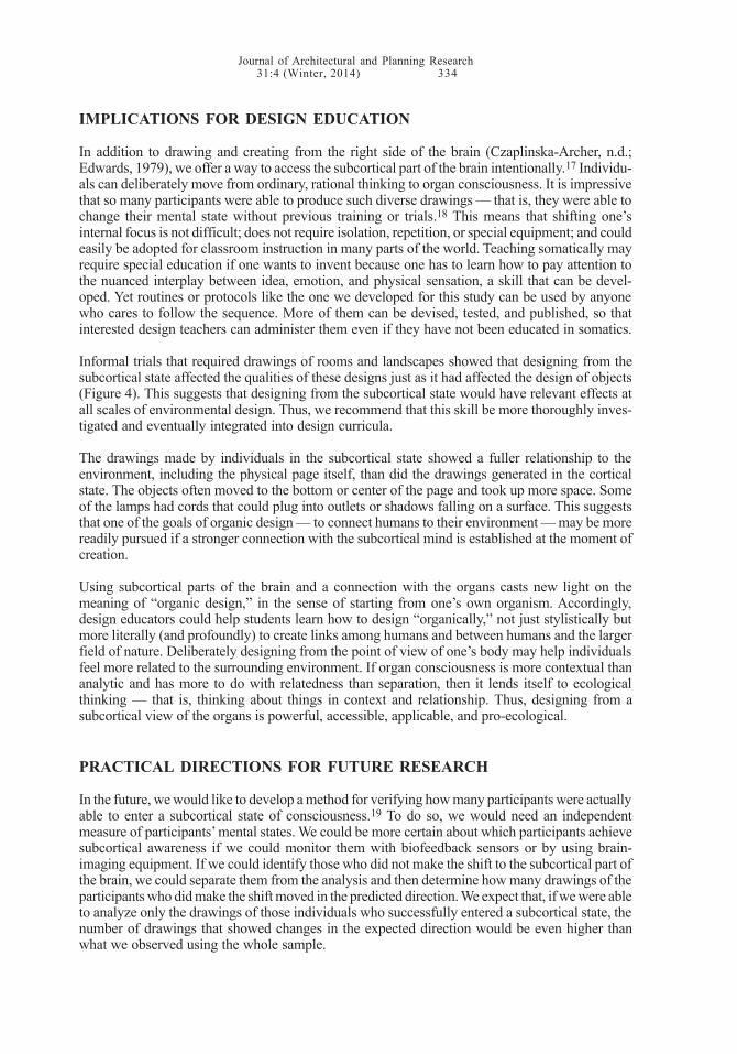
Journal of Architectural and Planning Research31:4 (Winter, 2014) 334
IMPLICATIONS FOR DESIGN EDUCATION
In addition to drawing and creating from the right side of the brain (Czaplinska-Archer, n.d.;Edwards, 1979), we offer a way to access the subcortical part of the brain intentionally.17 Individu-als can deliberately move from ordinary, rational thinking to organ consciousness. It is impressivethat so many participants were able to produce such diverse drawings — that is, they were able tochange their mental state without previous training or trials.18 This means that shifting one’sinternal focus is not difficult; does not require isolation, repetition, or special equipment; and couldeasily be adopted for classroom instruction in many parts of the world. Teaching somatically mayrequire special education if one wants to invent because one has to learn how to pay attention tothe nuanced interplay between idea, emotion, and physical sensation, a skill that can be devel-oped. Yet routines or protocols like the one we developed for this study can be used by anyonewho cares to follow the sequence. More of them can be devised, tested, and published, so thatinterested design teachers can administer them even if they have not been educated in somatics.
Informal trials that required drawings of rooms and landscapes showed that designing from thesubcortical state affected the qualities of these designs just as it had affected the design of objects(Figure 4). This suggests that designing from the subcortical state would have relevant effects atall scales of environmental design. Thus, we recommend that this skill be more thoroughly inves-tigated and eventually integrated into design curricula.
The drawings made by individuals in the subcortical state showed a fuller relationship to theenvironment, including the physical page itself, than did the drawings generated in the corticalstate. The objects often moved to the bottom or center of the page and took up more space. Someof the lamps had cords that could plug into outlets or shadows falling on a surface. This suggeststhat one of the goals of organic design — to connect humans to their environment — may be morereadily pursued if a stronger connection with the subcortical mind is established at the moment ofcreation.
Using subcortical parts of the brain and a connection with the organs casts new light on themeaning of “organic design,” in the sense of starting from one’s own organism. Accordingly,design educators could help students learn how to design “organically,” not just stylistically butmore literally (and profoundly) to create links among humans and between humans and the largerfield of nature. Deliberately designing from the point of view of one’s body may help individualsfeel more related to the surrounding environment. If organ consciousness is more contextual thananalytic and has more to do with relatedness than separation, then it lends itself to ecologicalthinking — that is, thinking about things in context and relationship. Thus, designing from asubcortical view of the organs is powerful, accessible, applicable, and pro-ecological.
PRACTICAL DIRECTIONS FOR FUTURE RESEARCH
In the future, we would like to develop a method for verifying how many participants were actuallyable to enter a subcortical state of consciousness.19 To do so, we would need an independentmeasure of participants’ mental states. We could be more certain about which participants achievesubcortical awareness if we could monitor them with biofeedback sensors or by using brain-imaging equipment. If we could identify those who did not make the shift to the subcortical part ofthe brain, we could separate them from the analysis and then determine how many drawings of theparticipants who did make the shift moved in the predicted direction. We expect that, if we were ableto analyze only the drawings of those individuals who successfully entered a subcortical state, thenumber of drawings that showed changes in the expected direction would be even higher thanwhat we observed using the whole sample.
Journal of Architectural and Planning Research31:4 (Winter, 2014) 335
Although we offered some visual evidence that the demonstrated relationship between organconsciousness and quality of creative production at the scale of the object is the same for otherscales, we would like to replicate this study for rooms, buildings, and exterior landscapes. Wewould also like to develop and test additional protocols for use by design educators to guidestudents into subcortical sources of creativity, for example, by focusing on organs other than thekidney.
Moreover, if organ consciousness can influence design output, this raises questions about thepossible effects of the reverse. Can the design of an object, a room, or a place affect a user’s mentalstate? Does a low, round room elicit a subcortical mental state? Would such a mental state, in turn,elicit an organic, egalitarian social feeling? Does a tall, rectilinear room stimulate our capacity forthinking and, perhaps, high performance in math or creativity (Meyers-Levy and Zhu, 2007)? Whatabout the Internet — how might the shape of a place online affect a user’s internal state (Kalay andMarx, 2003)? If the relationship goes both ways, then continuing to learn more about it would beprofoundly important for environmental-design professionals. For example, hospital designersmight choose to emphasize the horizontal plane in hospital rooms if we learned that patients healfaster in a subcortical state of consciousness, or library designers might choose to emphasize thevertical plane if we confirmed that vertical spaces stimulate cortical functions like reading and note-taking. Courtrooms might benefit from a balance of all planes in order to promote wise, ethicaldecisions that acknowledge the primal (behavioral), socio-emotional (social), and intellectual (ra-tional) aspects of legal cases. Bedroom designs could emphasize the horizontal to support thenonverbal, subcortical activities of rest, sleep, and sex, while study areas could be more uprightand vertical to encourage verbal, rational, and technical thinking in the cortex. Every place could beplanned and designed to cultivate a particular aspect of the consciousness. Accordingly, we seevalue in developing a theoretical explanation of how and why the design of the physical worldtriggers different mental states. These hypothesized relationships appear to be sufficiently impor-tant to merit further empirical testing.
If confirmed, a close relationship between mental states and the environment would mean thedesign of the environment is extremely significant because of its effect on people practically,behaviorally, socially, mentally, and emotionally in a way that still needs to be investigated fully.20
If space influences the deep mental state of inhabitants, then it is doubly important to train design-ers to work from the full range of consciousness. We are just beginning to understand this topic,which would allow us to understand how better to encourage creativity and innovation. Of course,the current application is focused on design and promises to have an impact on design practice,but this type of work has broader implications regarding creativity in all fields. That is, learning touse parts of the brain deliberately might produce new perspectives in psychotherapy, creativewriting, and the performing arts, for example, and might be used to gain insight and generatehypotheses in the sciences, including the biological sciences.
NOTES1. This paper is the result of a collaboration between the two authors: Galen Cranz wrote the sections entitled“Somatic Theory and Design” and “Implications for Design Education,” Leonardo Chiesi wrote the “Method”and “Findings” sections, and both authors wrote the final section, “Practical Directions for Future Research.” Inaddition, Jader Tolja conceived the original experimental hypothesis and procedure, and Lusi Morhayim codedthe drawings. The division of labor is discussed further in the “Method” section.
2. The field was first named “somatics” by Thomas Hanna in 1976 (Hanna, 1979).
3. In contrast, ergonomics and physical therapy are more concerned with the body as a biomechanical structure,usually independent of subjective experience, and psychological and sociological approaches usually do notconsider the physical correlates of mental states.

Journal of Architectural and Planning Research31:4 (Winter, 2014) 336
4. The concept of experiential anatomy comes from somatic-movement educator Bonnie Bainbridge Cohenand has been applied to design research and pedagogy at all scales (Cohen, 1993; Cranz, 2000; Tolja and Speciani,2000). The concept has also been used in other creative and academic fields (e.g., Saunders, 2011).
5. Some of those who use or have used the triune model include Greenfield (2001), Koch (2004), Linden (2007),and the late neuroanatomist Professor Jeffery Winer (2008).
6. Anthropologist E. T. Hall (1959, 1966) was the first to bring the triune brain into spatial and architecturalthinking in his seminal books The Silent Language and The Hidden Dimension. He applied the idea of the threedistinct layers — reptilian, limbic, and neocortex — to analyze culture and communication. The reptilian part ofthe brain is the source of what he called “formal” knowledge, which is deeply ingrained because we are socializedpre-verbally by watching the forms of how “things are done” at a very early age. The limbic part of the brainholds knowledge that we learn verbally from our primary caregivers at home before going to school; hence, it is“informal” and socio-emotional in nature. The neocortex holds the rational and “technical” knowledge that weare taught explicitly, usually in school. John P. Eberhard (2007) reopened the discourse about architecture and thebrain.
7. There is at least one historical antecedent for this practice in the world of design teaching: Johannes Itten andothers at the Bauhaus used exercises to relax students and create the conditions for creativity in painting, design,and color, thus showing that body practices can change design outcomes (Droste, 2002:24-32). Physical educa-tion at the Bauhaus was not used to strengthen the body in order to work more efficiently (as in factory sport)but rather to experience space, rhythm, and movement in order to integrate bodily sensations into the artisticexpression of the students. The organs were explicitly mentioned in Itten’s work (Scharenberg, 2003).
8. Though one might assume that indirect or implicit references to the “unconscious” or “intuition” in designrefer to levels of the brain, these references usually are not tied to locations in the brain structures.
9. This research bears some relation to Betty Edwards’s (1979) Drawing on the Right Side of the Brain, whichestablished techniques for activating the right hemisphere of the brain in order to enhance creativity and artisticconfidence. Edwards applied the scientific discoveries of Nobel-Prize-winning psychobiologist Roger W. Sperryabout the dual nature of human thinking to teaching drawing. Her text has become a classic for teaching drawingin art and design schools in the U.S. and internationally. Since its publication, “left brain / right brain” has becomea common phrase in design discourse. Our perspective differs in that we explore different layers of the brainrather than its different hemispheres.
10. This lateral rotation is movement on the horizontal plane, which makes experience more equal thanhierarchical, according to Cohen (1993). Returning to organ consciousness is a way to return to equality,harmony, and flow without borders. When perceiving their organs deliberately, people are more alike thandifferent. Hence, another potential benefit of this state of mind is a stronger possibility for conflict resolutionand integration of opposites. By finding common organ experiences, we might be able to link with “others” weview as different. This was the premise of a presentation by Galen Cranz (2008) entitled “Linking Differences byFinding Common Somatic Experience: Designing from the Consciousness of the Organs.” (The conferencetheme was “Linking Differences.”) Further development of this possibility and specific techniques for achievingit would be the subject of another paper for a different audience.
11. After studying medicine, Dr. Tolja eventually decided to give up surgery to teach environmental design, basedon the observation that people’s problems remained after surgery and his conviction that the entire environment— physical and social — needs to be transformed in order to heal people.
12. The Alexander Technique, Feldenkrais Method, and BMC are all systems of kinesthetic education; Gestalttherapy is a form of psychotherapy that includes the whole person, including the body. Training in these practiceshelped Dr. Tolja and the authors conceive of the linkages between organs and the nervous system.
13. Because our data (drawings) are no longer individually identifiable in our analysis, we did not need institution-al review or approval, according to the Office for the Protection of Human Subjects at the University ofCalifornia-Berkeley in an email dated November 12, 2013.
14. Dr. Tolja chose the kidney because it is located at the back of the body, and it is less invasive to touchsomeone’s back than to touch his or her front, where the liver, stomach, intestines, and heart are located. Forbeginners, this is a safe place to begin, but advanced students in BMC can make contact with any of the organs.
15. We conducted a cross-check of Ms. Morhayim’s scoring with ours by checking every 10th drawing to see ifher scores matched ours. The results were identical.
16. If we did not in fact create mental changes in participants through the proprioception routine, what elsemight explain the observed changes in their sketches? As mentioned previously in this paper, we would like toaddress this issue in future research using an external measure of brain function to assess whether a mental changeactually occurred.
Journal of Architectural and Planning Research31:4 (Winter, 2014) 337
17. Edward’s (1979) book offered techniques for moving into the more analogical part of the brain’s righthemisphere. Our research adds to that body of pedagogical techniques, even though it comes from a differenttheoretical formulation of cortical versus subcortical. The experiences of Dr. Tolja (1983) and Peter Grunwald(2008), another somatic expert, led them to view the difference between the cortical and the subcortical layers(namely, between the neocortex and the mammalian and reptilian layers) as equally if not more significant thanlateralization (i.e., the two hemispheres of the brain, right and left).
18. Edwards (1979) was also able to help participants change their mental state without hands-on guidance oreven a physical presence.
19. Participants generally reported that the proprioception routine was a pleasurable experience. However,several things could interfere with a successful shift to a subcortical state. For example, if participants wereconcerned about what they were “supposed” to feel, they might experience anxiety rather than sensory aware-ness. Some might have tried to second guess the experiment to figure out what it was about, while others mighthave been skeptical or unwilling to cooperate except by going through the motions. Perhaps others were simplynot successful in shifting their consciousness on this occasion, despite their best efforts.
20. We invite others to join us in this larger research agenda. This future research fits within an even moreencompassing plan of action. We want to encourage others to learn how to teach somatically and also to teachothers as needed. The first step is to advance this point of view and then to find collaborators and funding toconduct further research. We invite interested collaborators to contact us directly via email, take courses insomatic disciplines, and review the material being offered by the Association for Body Conscious Design (https://www.facebook.com/groups/bodyconsciousdesign/), an international group. Eventually, we would like to developa continuing education certification program in body-conscious design.
REFERENCES
Cohen BB (1993) Sensing, feeling, and action: The experiential anatomy of body-mind centering.Northampton, MA: Contact Editions.
Cranz G (2000) The chair: Rethinking culture, body, and design. New York: W. W. Norton &Company, Inc.
Cranz G (2008) Linking differences by finding common somatic experience: Designing from theconsciousness of the organs. Paper presented at the 39th annual conference of theEnvironmental Design Research Association (EDRA 39). Veracruz, Mexico (28 May-1 June).
Czaplinska-Archer R (n.d.) Time for drawing: On coming to our senses. Unpublished manuscript;available by contacting author via email at [email protected].
Das AK (1987) Indigenous models of therapy in traditional Asian societies. Journal of Multicul-tural Counseling and Development 15(1):25-37.
Droste M (2002) Bauhaus: 1919-1933. Berlin: Taschen.
Eberhard JP (2007) Architecture and the brain: A new knowledge base from neuroscience. Atlan-ta, GA: Greenway Communications.
Edwards B (1979) Drawing on the right side of the brain. New York: Jeremy P. Tarcher/Penguin.
Gallagher S (2006) How the body shapes the mind, 1st edition. New York: Oxford University Press.
Greenfield S (2001) Brain story: Unlocking our inner world of emotions, memories, ideas anddesires. New York: Dorling Kindersley.
Grunwald P (2008) EyeBody: The art of integrating eye, brain and body. Auckland, New Zealand:Condevis.
Journal of Architectural and Planning Research31:4 (Winter, 2014) 338
Hall ET (1959) The silent language. New York: Anchor Books.
Hall ET (1966) The hidden dimension. New York: Anchor Books.
Hanna T (1979) The body of life: Creating new pathways for sensory awareness and fluid move-ment. Rochester, VT: Healing Arts Press.
Kalay Y, Marx J (2003) Changing the metaphor: Cyberspace as a place. In ML Chiu, JY Tsou,T Kvan, M Morozumi, and TS Jeng (Eds.), Digital design: Research and practice(Proceedings of the 10th international conference on computer aided architectur-al design futures). Dordrecht, The Netherlands: Kluwer Academic Publishers,pp. 19-28.
Koch C (2004) The quest for consciousness: A neurobiological approach. Denver, CO: Roberts &Company Publishers.
Lakoff G, Johnson M (1999) Philosophy in the flesh: The embodied mind and its challenge towestern thought. New York: Basic Books.
Leigh C (1999) Goethe and the mess we’re in: A natural history of the organic fallacy in architecture,art, and culture. Classicist 5:19-36.
Linden DJ (2007) The accidental mind: How brain evolution has given us love, memory, dreams,and God. Cambridge, MA: Harvard University Press.
MacLean PD (1990) The triune brain in evolution: Role in paleocerebral functions. New York:Plenum Press.
Meyers-Levy J, Zhu RJ (2007) The influence of ceiling height: The effect of priming on the type ofprocessing that people use. Journal of Consumer Research 34(2):174-186.
Mumford M (1989) Form follows nature: The origins of American organic architecture. Journal ofArchitectural Education 42(3)(Spring):26-37.
Pfeifer R, Bongard J (2006) How the body shapes the way we think: A new view of intelligence.Cambridge: The MIT Press.
Rosenthal R (1966) Experimenter effects in behavioral research. New York: Appleton-Century-Crofts.
Saunders L (2011) Simon, Jennifer, and me: A foot in a sock in a shoe. In GW Miller, P Ethridge, andKT Morgan (Eds.), Exploring body-mind centering: An anthology of experienceand method. Berkeley, CA: North Atlantic Books, pp. 97-120.
Scharenberg S (2003) Physical education at the Bauhaus, 1919-33. The International Journal of theHistory of Sport 20(3):115-127.
Scott G (1924) The architecture of humanism: A study in the history of taste. London: Constableand Company Ltd.
Telles S, Visweswaraiah NK (2010) The status of yoga research in India. In PC Leung (Ed.), Healthyaging (Annals of traditional Chinese medicine, Vol. 4). Hackensack, NJ: WorldScientific, pp. 245-257.
Journal of Architectural and Planning Research31:4 (Winter, 2014) 339
Tolja J (1983) Il cervello destro e i giardini Zen (Riza Scienze no. 2) (Italian). Milan: Edizioni RizaS.p.a.
Tolja J, Speciani F (2000) Pensare col corpo (Italian). Milan: Zelig.
Winer J (2008) Personal conversation. Fall.
Additional information may be obtained by writing directly to Dr. Chiesi at Scuola di Architettura,Università di Firenze, Piazza Ghiberti 27, 50126 Firenze, Italy; email: [email protected].
ACKNOWLEDGMENTSThe authors would like to thank Koranjali Alfonseca for her help in editing the images for publication.
AUTOBIOGRAPHICAL SKETCHES
Dr. Galen Cranz is a professor of architecture, sociologist, designer, and certified teacher of the AlexanderTechnique (a system of body/mind postural education). She is interested in the social and cultural components ofenvironmental design and specifically how the body meets the environment (the new subject called BMC).Dr. Cranz (2000) provided a critique of the practice of sitting in chairs from a somatic point of view in The Chair:Rethinking Culture, Body, and Design. She also studies the post-occupancy effects of designs on users, the roleof urban parks, and taste as a communication process in design. Dr. Cranz’s email is [email protected].
Dr. Leonardo Chiesi teaches sociology in the School of Architecture at the University of Florence, Italy. He isinterested in social research methods for architecture and planning, and he studies how sociology and design canbe mutually engaged. He has been involved in several architectural design and city-planning projects and isinterested in the subjects of local identity, community processes, and participation methods. He has written bookson the construction of scientific discourse (Retorica Nella Scienza: Come la Scienza Costruisce i Suoi ArgomentiAnche al di là Della Logica; Bonano, 2009) and the relationship between the social sciences, architecture, andplanning (Il Doppio Spazio dell’Architettura: Ricerca Sociologica e Progettazione; Liguori, 2010).
Manuscript revisions completed 22 September 2014.
Related Documents