Colloids and Surfaces B: Biointerfaces 88 (2011) 477–482 Contents lists available at ScienceDirect Colloids and Surfaces B: Biointerfaces jou rn al h om epage: www.elsevier.com/locate/colsurfb Design and characterization of bi-soft segmented polyurethane microparticles for biomedical application Elisa Campos a,∗,1 , Rosemeyre Cordeiro a,1 , Ana Cristina Santos b , Cláudia Matos b , M.H. Gil a a Research Centre for Chemical Processes Engineering and Forest Products (CIEPQPF), Department of Chemical Engineering, Faculty of Sciences and Technology, University of Coimbra, Rua Sílvio Lima, 3030-790 Coimbra, Portugal b Institute of Biophysics and Biomathematics, Biomedical Institute of Research in Light and Image (IBILI), Faculty of Medicine, University of Coimbra, Azinhaga de Santa Comba, Celas, 3000-548 Coimbra, Portugal a r t i c l e i n f o Article history: Received 10 June 2011 Received in revised form 13 July 2011 Accepted 13 July 2011 Available online 23 July 2011 Keywords: Bi-soft segmented polyurethane Microparticles ATR-FTIR Hydrolytic degradation Cytotoxicity a b s t r a c t Bi-soft segmented poly(ester urethane urea) microparticles were prepared and characterized aiming a biomedical application. Two different formulations were developed, using poly(propylene glycol), toly- lene 2,4-diisocyanate terminated pre-polymer (TDI) and poly(propylene oxide)-based tri-isocyanated terminated pre-polymer (TI). A second soft segment was included due to poly(-caprolactone) diol (PCL). Infrared spectroscopy, used to study the polymeric structure, namely its H-bonding properties, revealed a slightly higher degree of phase separation in TDI-microparticles. TI-microparticles presented slower rate of hydrolytic degradation, and, accordingly, fairly low toxic effect against macrophages. These new formulations are good candidates as non-biodegradable biomedical systems. © 2011 Elsevier B.V. All rights reserved. 1. Introduction Polyurethanes form a class of thermoplastic elastomers that have been used extensively in biomedical applications due to their excellent biocompatibility and mechanical properties [1,2]. They are clinically used in central venous catheters, vascular grafts, cardiac valves, mammary prostheses, ocular implants and drug delivery systems [3]. Their interesting properties are due to their microphase-separated morphology, comprising “soft” and “hard” segments. The soft segments, consisting of polyol, typically polyester, polyether or polycarbonate diol, provide the elastomeric character to the polymer backbone, whereas the polyurethane/urea hard segments, consisting of diisocyanate and/or chain extender, provide mechanical strength [2,4]. If the isocyanate contains per se a soft segment, bi-soft segment polyurethane can be prepared by adding an extra soft segment. The presence of two soft segments can show different extents of phase separation, offering new possi- bilities on tuning polymeric matrix properties [5]. Phase separation ∗ Corresponding author. Present address: Instituto de Tecnologia Química e Biológica, Universidade Nova de Lisboa, Av. da República, 2780-157 Oeiras, Portugal. Tel.: +351 21 446 97 22; fax: +351 21 441 12 77. E-mail addresses: [email protected] (E. Campos), [email protected] (R. Cordeiro), [email protected] (A.C. Santos), [email protected] (C. Matos), [email protected] (M.H. Gil). 1 These authors contributed equally to this work. is related to the presence and the extent of hydrogen bonding in these segmented polyurethanes, which can be determined qualita- tively by infrared spectroscopy [6]. Concerning medical and biochemical applications, microparti- cles are largely used, namely in bioseparation, immunoassay and affinity diagnosis, blood flow determinations, carriers and con- trolled drug delivery systems [7]. Polymeric microparticles can be designed aiming a variety of size, surface chemistry, compo- sition, morphology and topography. These systems are indeed quite interesting since their small size and large surface area favor absorption/release of any compound in comparison with larger car- riers [8]. In the present work, the previous concepts were put together: the development of a new and promising polymeric matrix as a widely used biomedical system. Here bi-soft segmented poly(ester urethane urea) microparticles were prepared and characterized aiming a biomedical application. TDI and TI were used as iso- cyanates, containing poly(propylene glycol) and poly(propylene oxide), as soft segment, respectively. The second soft segment was included by adding PCL, a biodegradable, semi-crystalline and aliphatic polyester. Structural analysis was made by atten- uated total reflectance-Fourier transform infrared (ATR-FTIR) spectroscopy. Besides brief morphologic and size analysis, both in vitro degradation and toxicological approaches were made. In fact, when developing a polymeric system aiming a biomedical appli- cation, particular attention is given to the interaction between the 0927-7765/$ – see front matter © 2011 Elsevier B.V. All rights reserved. doi:10.1016/j.colsurfb.2011.07.037

Welcome message from author
This document is posted to help you gain knowledge. Please leave a comment to let me know what you think about it! Share it to your friends and learn new things together.
Transcript

Df
Ea
Ub
U
a
ARRAA
KBMAHC
1
htTgdt“pchpaacb
BT
rc
0d
Colloids and Surfaces B: Biointerfaces 88 (2011) 477– 482
Contents lists available at ScienceDirect
Colloids and Surfaces B: Biointerfaces
jou rn al h om epage: www.elsev ier .com/ locate /co lsur fb
esign and characterization of bi-soft segmented polyurethane microparticlesor biomedical application
lisa Camposa,∗,1, Rosemeyre Cordeiroa,1, Ana Cristina Santosb, Cláudia Matosb, M.H. Gila
Research Centre for Chemical Processes Engineering and Forest Products (CIEPQPF), Department of Chemical Engineering, Faculty of Sciences and Technology,niversity of Coimbra, Rua Sílvio Lima, 3030-790 Coimbra, PortugalInstitute of Biophysics and Biomathematics, Biomedical Institute of Research in Light and Image (IBILI), Faculty of Medicine,niversity of Coimbra, Azinhaga de Santa Comba, Celas, 3000-548 Coimbra, Portugal
r t i c l e i n f o
rticle history:eceived 10 June 2011eceived in revised form 13 July 2011ccepted 13 July 2011vailable online 23 July 2011
a b s t r a c t
Bi-soft segmented poly(ester urethane urea) microparticles were prepared and characterized aiming abiomedical application. Two different formulations were developed, using poly(propylene glycol), toly-lene 2,4-diisocyanate terminated pre-polymer (TDI) and poly(propylene oxide)-based tri-isocyanatedterminated pre-polymer (TI). A second soft segment was included due to poly(�-caprolactone) diol (PCL).Infrared spectroscopy, used to study the polymeric structure, namely its H-bonding properties, revealed
eywords:i-soft segmented polyurethaneicroparticles
TR-FTIRydrolytic degradationytotoxicity
a slightly higher degree of phase separation in TDI-microparticles. TI-microparticles presented slowerrate of hydrolytic degradation, and, accordingly, fairly low toxic effect against macrophages. These newformulations are good candidates as non-biodegradable biomedical systems.
© 2011 Elsevier B.V. All rights reserved.
. Introduction
Polyurethanes form a class of thermoplastic elastomers thatave been used extensively in biomedical applications due toheir excellent biocompatibility and mechanical properties [1,2].hey are clinically used in central venous catheters, vascularrafts, cardiac valves, mammary prostheses, ocular implants andrug delivery systems [3]. Their interesting properties are dueo their microphase-separated morphology, comprising “soft” andhard” segments. The soft segments, consisting of polyol, typicallyolyester, polyether or polycarbonate diol, provide the elastomericharacter to the polymer backbone, whereas the polyurethane/ureaard segments, consisting of diisocyanate and/or chain extender,rovide mechanical strength [2,4]. If the isocyanate contains per se
soft segment, bi-soft segment polyurethane can be prepared by
dding an extra soft segment. The presence of two soft segmentsan show different extents of phase separation, offering new possi-ilities on tuning polymeric matrix properties [5]. Phase separation∗ Corresponding author. Present address: Instituto de Tecnologia Química eiológica, Universidade Nova de Lisboa, Av. da República, 2780-157 Oeiras, Portugal.el.: +351 21 446 97 22; fax: +351 21 441 12 77.
E-mail addresses: [email protected] (E. Campos),[email protected] (R. Cordeiro), [email protected] (A.C. Santos),[email protected] (C. Matos), [email protected] (M.H. Gil).
1 These authors contributed equally to this work.
927-7765/$ – see front matter © 2011 Elsevier B.V. All rights reserved.oi:10.1016/j.colsurfb.2011.07.037
is related to the presence and the extent of hydrogen bonding inthese segmented polyurethanes, which can be determined qualita-tively by infrared spectroscopy [6].
Concerning medical and biochemical applications, microparti-cles are largely used, namely in bioseparation, immunoassay andaffinity diagnosis, blood flow determinations, carriers and con-trolled drug delivery systems [7]. Polymeric microparticles canbe designed aiming a variety of size, surface chemistry, compo-sition, morphology and topography. These systems are indeedquite interesting since their small size and large surface area favorabsorption/release of any compound in comparison with larger car-riers [8].
In the present work, the previous concepts were put together:the development of a new and promising polymeric matrix as awidely used biomedical system. Here bi-soft segmented poly(esterurethane urea) microparticles were prepared and characterizedaiming a biomedical application. TDI and TI were used as iso-cyanates, containing poly(propylene glycol) and poly(propyleneoxide), as soft segment, respectively. The second soft segmentwas included by adding PCL, a biodegradable, semi-crystallineand aliphatic polyester. Structural analysis was made by atten-uated total reflectance-Fourier transform infrared (ATR-FTIR)
spectroscopy. Besides brief morphologic and size analysis, both invitro degradation and toxicological approaches were made. In fact,when developing a polymeric system aiming a biomedical appli-cation, particular attention is given to the interaction between the
4 aces B: Biointerfaces 88 (2011) 477– 482
mifamCptmdctacip
2
2
p((scptfw1p(MC3swaaCfl
2
mPuccaTr
2
wmw(
78 E. Campos et al. / Colloids and Surf
atrix and neighboring cells. It is well known that biocompatibil-ty depends, namely, on the nature and concentration of interfacialunctional groups, surface hydrophilicity, degree of crystallinitynd polymer surface topography [9]. In this work, the assess-ent of microparticles’ biocompatibility was carried out in vitro.
ytotoxicity methods are simpler, faster, more cost effective andose no ethical problems compared to in vivo approaches. Hence,he cytotoxity of bi-soft segmented poly(ester urethane urea)
icroparticles was assessed by colorimetric MTT assay [3-(4,5-imethylthyazol-2-y)-2,5-diphenyl tetrazolium bromide]. This is aolorimetric non-radioactive method, extensively used for in vitrooxicity evaluation, since it allows rapid assessment of cell viabilitynd proliferation, and gives good reproducibility [10–12]. As modelells, mouse peritoneal macrophages were used due to their rolen inducing and regulating the body immune response to foreignarticles [13].
. Materials and methods
.1. Materials
PCL (number average molecular weight (Mn) ∼530),oly(propylene glycol) tolylene 2,4-diisocyanate terminatedTDI) (Mn ∼ 2300, isocyanate ∼ 3.6 wt%), poly(vinyl alcohol) (PVA)Mn = 9000–10,000, 80% hydrolyzed) and phosphate bufferedaline (PBS) tablets (0.01 M phosphate buffer, 0.0027 M potassiumhloride and 0.137 M sodium chloride, pH 7.4, at 25 ◦C) wereurchased from Aldrich (Germany). Poly(propylene oxide)-basedri-isocyanated pre-polymer (TI) (Mn ∼ 3500) was purchasedrom Companhia Petroquímica do Barreiro (Portugal). Tolueneas bought from J. Vaz Pereira (Portugal). Complete RPMI
640 was used as culture medium and was prepared withenicillin–streptomycin solution (1%), l-glutamine (1%) and FBS10%) purchased from Aldrich (Germany). Trypan Blue (0.4%) and
TT (5 mg/mL in sterile PBS) were supplied by Aldrich (Germany).hloroform, 75◦ alcohol, isopropanol and chloridric acid (fumant7%) were bought from Merck (Germany). Animals (mice) wereupplied by Charles River (Spain). Sterile needles (19 and 25G)ere purchased to TerumoTM (Belgium) and sterile syringes (1, 2, 5
nd 10 mL) from BD PlasticK (Spain). Sterile Falcons (15 and 30 mL)nd sterile 96 well flat bottom culture plates were supplied byorning Inc., Costar® (USA). All these chemicals were used withouturther purification. The distilled water used was prepared in theaboratory.
.2. Microparticles preparation
Microparticles were prepared by emulsion polymerizationethod. The 50% wt organic solution, composed by TDI or TI and
CL (mass ratio 80/20) in toluene was added, drop-wise, at a vol-me ratio 1/100, to the 1% w/v PVA aqueous solution. Emulsion wasontinuously stirred, at 1400 rpm, 60 ◦C for 2 h. Microparticles wereentrifuged at 3000 rpm for 10 min, washed with distilled waternd dried at room temperature. The microparticles prepared withDI or TI will be denoted by TDI-microparticles or TI-microparticles,espectively.
.3. Morphology and size
Morphological analysis of dried microparticles in suspension
as performed by optical microscopy using a BH2 (Olympus, Japan)icroscope. Particle size and size distribution of microparticlesere evaluated by laser light scattering using a Coulter LS 130Coulter Co., USA) particle analyzer. Dried microparticles were
Fig. 1. PU-based microparticles obtained by emulsification solvent evaporationmethod using two pre-polymers isocyanate-terminated, TDI and TI, and PCL, as asecond soft segment. (A) TDI-microparticles and (B) TI-microparticles.
suspended in distilled water and sonicated before analysis. Frauen-hofer’s method was used to determine the particle size distribution.
2.4. ATR-FTIR analysis
The chemical composition of microparticles’ matrix was studiedby FTIR spectroscopy in the ATR mode, using a Magma-IR Spectrom-eter 750 (Nicolet Instrument Corp., USA) with an ATR accessory(golden gate Mk II with diamond top-plate and ZnSe lenses, Specac,UK). Microparticles, in a dry form, were sprayed on a ATR crystal andall spectra were recorded at room temperature with a resolution of4 cm−1, over the frequency range 4000–500 cm−1 and scanned 32times.
2.5. Hydrolytic degradation of microparticles
Microparticles were weighed (W0), placed in sealed glass vialscontaining PBS (pH 7.4) and incubated at 37 ◦C over 28 days. Aftera certain period of time (t), microparticles were removed fromthe solution, washed with distilled water, dried and weighed to

E. Campos et al. / Colloids and Surfaces B: Biointerfaces 88 (2011) 477– 482 479
Fig. 2. Chemical structure of PU-based microparticles. (A) TDI-microparticles and (B) TI-microparticles [5,15,16]. The soft and hard segments are marked by light gray anddark gray rectangles, respectively. The structures were obtained with ChemBioDraw Ultra 12.0 (CambridgeSoft).

4 aces B: Biointerfaces 88 (2011) 477– 482
ct
2
buaRopmscAccsimadmsgaar5ltttia
3
ficumps
3
oi2iTsww
80 E. Campos et al. / Colloids and Surf
onstant weight (Wt). The extent of degradation was quantified ashe weight loss of samples according to [(W0 − Wt)/W0] × 100.
.6. Cytotoxicity assay
To quantitatively assess cell cytotoxicity, proliferation or via-ility of microparticles, the colorimetric MTT assay was generallysed. Peritoneal macrophages were harvested from Balb C mice2
nd cultured at a density of 200–230 × 104 cells/mL in completePMI 1640 medium, at 37 ◦C, in a 5% CO2 atmosphere, for 5 h, inrder to get a pure macrophage adherent population, according to areviously established protocol [11], and optimized from the Weir’sethod [14]. Culture medium was then replaced by microparticle
uspensions containing 0.08 �g/mL, 0.8 �g/mL and 5 �g/mL (finaloncentration) of microparticles in new complete RPMI medium.s negative control, the peritoneal macrophages were incubated inomplete culture medium without microparticles. All microparti-le concentrations and negative control were each seeded in threeeparate wells in order to provide statistically reliable results. Afterncubation for 3 days at 37 ◦C and in a 5% CO2 atmosphere, the
edium was removed, replaced by 270 �L of medium and 30 �L of 5 mg/mL MTT solution, and incubated for 3 h, in the same con-itions, in order to allow viable cells to take up MTT into theiritochondria and metabolize it into blue formazan crystals. The
upernatant in each well was aspirated and 300 �L of a hydro-en chloride–2-propanol solution 4% was added to lyse the cellsnd to solubilise the MTT crystals. After 15 min at room temper-ture (to dissolve all crystals), the absorbance of each well wasead on a microplate reader (Spectra SLT, Tecan, Switzerland) at40 nm, with a reference filter of 620 nm. Cell viability was calcu-
ated by comparing the sample absorbance (Asample) to the one ofhe control cells (Acontrol), which was by definition 100%, accordingo (Asample/Acontrol) × 100. Moreover, the effect of incubation withhe PU-based microparticles on the morphology of cells was exam-ned by optical microscopy (Nikon, Japan) and photographed with
digital camera (Nikon, Japan).
. Results and discussion
Although polyurethanes are extensively used in the biomedicaleld, there are few works reporting microparticles preparation andharacterization. In the present work, bi-soft segmented poly(esterrethane urea) microparticles (hereafter designated by PU-basedicroparticles, for simplicity) were prepared starting with two pre-olymers isocyanate-terminated, TDI and TI, and PCL, as a secondoft segment.
.1. Morphology and size of PU-based microparticles
Optical microscopy observation (Fig. 1) revealed the formationf spherical polymeric microparticles. TDI-microparticles rangedn size from approximately 3 �m–57 �m with a mean size of4 �m, whereas TI-microparticles ranged in size from approx-
mately 2 �m–42 �m with a mean size of 19 �m. Accordingly,I-microparticles presented size range narrower and were slightly
maller than TDI-microparticles. In fact, since TI is a pre-polymerith higher content of hard segments, a matrix more crystallineas expected in TI-microparticles (Fig. 2).2 According to national and international regulations on animal welfare.
Fig. 3. ATR-FTIR spectra of PU-based microparticles.
3.2. Structural analysis
The molecular structure of PU-based microparticles was char-acterized by ATR-FTIR spectroscopy. The spectra of both polymericmatrices (Fig. 3) were quite similar. The absence of the peak at2270 cm−1 (asymmetric isocyanate stretching vibration) indicatedthat the isocyanate reaction was complete. In fact, isocyanategroups were expect to react with the hydroxyl groups of PCL, form-ing urethane linkages, and with water (in aqueous phase) or witheach other (when in excess), forming urea linkages. Characteris-tic polyurethane bonds were detected at 3150–3500 cm−1 (N–Hstretching vibrations), at 2935 and 2855 cm−1 (CH2 asymmetric andsymmetric stretching vibrations, respectively), at 1600–1800 cm−1
(C O stretching vibrations), and at 1525 cm−1 (urethane N–H bend-ing + C–N stretching). Also typical peaks of soft segments weredetected at 1260 cm−1 (symmetric CH3 bending), at 1080 cm−1
(C–O–C stretching), and at 810 cm−1 (CH3 rocking) [9,17,18].Phase morphology in polyurethanes was assessed through
analysis of the type of hydrogen bonds (H-bonds) involving ure-thane/urea carbonyl groups, since the occurrence of H-bondsbetween soft and hard segments is an indication of phase mix-ing, while H-bonds between hard segments predominate in phaseseparation [5]. Thus two particular regions were analyzed in moredetail: the C O stretching vibration (1600–1800 cm−1) (Fig. 4A)and the N–H stretching vibration (3150–3500 cm−1) (Fig. 4B). Thesevibrations are strongly perturbed by the formation of H-bonds. Bothfrequency shifts and intensities are characteristics of the specificityor magnitude of the H-bonds formed [17]. Therefore, hydrogenbonded N–H and C O peaks shift to lower frequencies relativelyto free N–H and C O, due to the weakening of these bonds as aresult of hydrogen bonding [6].
In poly(ester urethane urea) matrices, bands assigned to ure-thane/urea C O stretching vibrations are overlapped in some wayto ester C O stretching vibrations [5]. Accordingly, the major bandobserved at about 1730 cm−1, would have the contribution of bothfree C O (i.e., not involved in H-bonds) in urethane and estergroups. In Fig. 4A, it can also be observed a weak shoulder atabout 1710 cm−1, more evident in TI-microparticles, attributed toH-bonded C O in urethane linkages. These results indicate that H-bonding between N–H and C O is less extensive in both matrices.It is likely that in polyether–urethanes N–H groups form H-bondswith C O of the urethane hard segments, and with oxygen in thepolyether soft segments. Because ether groups are quite abundantin these matrices, C O will have less chance to form H-bond [6].
Concerning C O stretching vibrations in urea groups, the follow-ing bands were detected: at about 1700 cm−1, assigned to freeC O but also to H-bonded C O in ester groups; at 1660 cm−1,
E. Campos et al. / Colloids and Surfaces B: Biointerfaces 88 (2011) 477– 482 481
Fs
an1tHsm
cifb3mim
3
mtTTmeettottttmt
ig. 4. (A) C O stretching region and (B) N–H stretching region of the ATR-FTIRpectra of PU-based microparticles.
ttributed to disordered H-bonded C O (i.e., involved in H-bondsot belonging to ordered, three-dimensional hydrogen bonding); at640 cm−1, assigned to ordered H-bonded (i.e., involved in ordered,hree-dimensional hydrogen bonding). From Fig. 4A, it is clear that-bonded urea carbonyl groups decreased in TDI-microparticles,
uggesting a larger phase separation between hard and soft seg-ents.In N–H stretching region (Fig. 4B), one observes a single band
entered at around 3300 cm−1, assigned to H-bonded N–H stretch-ng vibration, significantly more intense in TI-microparticles. Theree (non H-bonded) N–H stretching band appears, more visi-ly in TI-microparticles, as a weak shoulder, absorbing at about440 cm−1, indicating that almost all N–H groups in the obtainedatrices were completely H-bonded. Moreover, this 140 cm−1 shift
ndicates a very strong N–H hydrogen bonding in both polymericatrices [5,6,17].
.3. In vitro hydrolytic degradation of TDI- and TI-microparticles
The hydrolytic degradation behaviour of TDI- and TI-icroparticles in PBS, pH 7.4 was monitored at 37 ◦C, in order
o mimic physiologic conditions. Fig. 5 shows the weight loss ofDI- and TI-microparticles in these conditions, throughout 28 days.here was a minor and slow decrease of weight of TDI- and TI-icroparticles, meaning that hydrolysis occurred at a very low
xtent, as usually observed in polyurethane matrices. However, thextent of degradation of TI-microparticles was slightly lower thanhe TDI-microparticles (6.9% and 5.9%, respectively). The degrada-ion of TDI- and TI-microparticles in PBS depends on susceptibilityf ester and urethane bonds to hydrolytic degradation. It is knownhat hard segments (consisting of urethane linkage) degrade slowerhan soft segments (consisting of ester linkage), since the ure-
hane bond is much less susceptible to hydrolytic degradationhan an ester bond [19–21]. Besides, in vitro degradation of poly-ers in aqueous solutions is facilitated by the hydrophilicity ofhe polymer structure, since water can more efficiently penetrate
Fig. 5. Hydrolytic degradation of PU-microparticles incubated in PBS (pH 7.4), at37 ◦C.
into the polymers and hydrolyze the esters bond [22]. Indeed, thegreater the hydrophilicity of the polymer, the faster the degrada-tion. Accordingly, the lower degradation rate of TI-microparticlesis apparently due to the high content of hard segments and, thus,high crystallinity. The hard segment is hydrophobic and difficult todegrade, thus inhibiting the penetration of water into the material.On the other hand, throughout the incubation time, an increase inmicroparticles matrix’s degradation is expected to take place dueto the increase of ester bond cleavage, since its inferior crystallinityallows water permeation into each polymer more easily [23].
3.4. Cytotoxicity assays
In the literature, the colorimetric MTT assay is an outstandingmethod, among other in vitro toxicity methods, for probing the tox-icity of materials. This test is based on enzymatic (dehydrogenase)activity, namely mitochondrial. In fact, mitochondria represent avulnerable target for toxic injury due to their crucial role in main-taining cellular structure and function via aerobic ATP production[24].
To assess microparticles’ toxicity, mouse peritonealmacrophages were “cultured” up to 3 days in the presence of theTDI- and TI-microparticles. Macrophage cultures were observeddaily, just prior and immediately after MTT addition. Fig. 6 showscell viability with the MTT assay method after 3 days for cellscultured with increasing amounts of TDI- and TI-microparticles(ranging from 0.08 to 5 �g/mL). According to the MTT assayresults, TI-microparticles exhibited lower toxicity for peritonealmacrophages than TDI-microparticles. Generally, materials withcell viability higher than 80% are considered as biocompatible [25].In this case, TDI- and TI-microparticles showed low toxic effectonly at the lowest applied concentration (0.08 �g/mL). For thatreason, since microparticles’ concentration is determinant, regard-ing cytotoxic effect, for a possible biomedical use, a 0.08 �g/mLmicroparticles suspension is indicated. However, microscopicobservations of peritoneal macrophages after incubation withdifferent concentrations of TDI- and TI-microparticles for 3 daysrevealed that cells were metabolically active, since formazan
crystals inside the cells could clearly be observed (data not shown).Moreover, no significant modification in cell morphology andproliferation relative to control was observed (data not shown).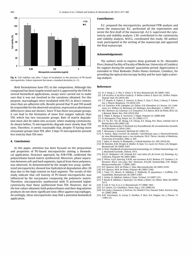
482 E. Campos et al. / Colloids and Surfaces B
Fm
csspsbdiTtAoil
4
acptwsdsiTctpAa
[[
[[
[
[
[
[[[
[[
[
ig. 6. Cell viability rate after 3 days of incubation in the presence of PU-basedicroparticles. Values represent the mean ± standard deviation (n = 3).
Both formulations have PCL in the composition. Although thisompound has been largely tested and it is approved by the FDA foreveral biomedical applications, assays were carried out to makeure that it was not involved in the variations obtained. For thisurpose, macrophages were incubated with PCL in direct contact,ince they are adherent cells. Results proved that TI and TDI woulde responsible for the observed variations, expressed as absorbanceifferences (data not shown). Since TI has three isocyanate groups,
t can lead to the formation of more free oxygen radicals thanDI, which has two isocyanate groups. Rate of matrix degrada-ion must also be taken into account, when studying cytotoxicity.s shown before, TI-microparticles degrade more slowly than TDInes. Therefore, it seems reasonable that, despite TI having moresocyanate groups than TDI, after 3 days TI-microparticles presentess toxicity than TDI ones.
. Conclusions
In this paper, attention has been focused on the preparationnd properties of PU-based microparticles aiming a biomedi-al application. Structure approach, by ATR-FTIR, confirmed theolyurethane-based matrix synthesized. Moreover, phase separa-ion between soft and hard segments, typical from these polymers,as observed. As demonstrated by the weight loss assay, synthe-
ized microparticles showed low hydrolytical degradation after 28ays due to the high content on hard segment. The results of thistudy indicate that cell toxicity of PU-based microparticles wasnfluenced by the isocyanates composing the polymeric matrix.herefore, microparticles synthesized with TI presented higherytotoxicity than those synthesized from TDI. However, due to
he low values obtained, both polyurethanes and their degradationroducts do not show significant toxic effect against macrophages.ccordingly, these microparticles may find a potential biomedicalpplication.[[
[
: Biointerfaces 88 (2011) 477– 482
Contributors
E.C. prepared the microparticles, performed FTIR analysis andwrote the manuscript. R.C. performed all the experiments andwrote the first draft of the manuscript. A.C.S. supervised the cyto-toxicty and viability analysis. C.M. contributed to the cytotoxicityand viability analysis. M.H.G. coordinated this study. All authorsread, participated in the writing of the manuscript and approvedthe final manuscript.
Acknowledgements
The authors wish to express deep gratitude to Dr. AlexandrePires (Animal Facility of Faculty of Medicine, University of Coimbra)for support during the animals’ quarantine period. Appreciation isextended to Vítor Redondo (Pedro Nunes Institute, Coimbra), forproviding the optical microscopy facility and for laser light scatter-ing analysis.
References
[1] H.-S. Hung, C.-C. Wu, S. Chien, S.-H. Hsu, Biomaterials 30 (2009) 1502.[2] G.R. da Silva, A. da Silva-Cunha Jr., F. Behar-Cohen, E. Ayres, R.L. Oréfice, Polym.
Degrad. Stab. 95 (2010) 491.[3] T.T. Huynh, K. Padois, F. Sonvico, A. Rossi, F. Zani, F. Pirot, J. Doury, F. Falson,
Eur. J. Pharm. Biopharm. 74 (2010) 255.[4] S.A. Guelcher, K.M. Gallagher, J.E. Didier, D.B. Klinedinst, J.S. Doctor, A.S. Gold-
stein, G.L. Wilkes, E.J. Beckman, J.O. Hollinger, Acta Biomater. 1 (2005) 471.[5] M.C. Besteiro, A.J. Guiomar, C.A. Gonc alves, V.A. Bairos, M.N. de Pinho, M.H. Gil,
J. Biomed. Mater. Res. 93A (2010) 954.[6] E. Yilgör, E. Burgaz, E. Yurtsever, I. Yilgör, Polymer 41 (2000) 849.[7] H. Kawaguchi, Prog. Polym. Sci. 25 (2000) 1171.[8] C.Y. Yu, B.C. Yin, W. Zhang, S.X. Cheng, X.Z. Zhang, R.X. Zhuo, Colloids Surf. B
Biointerfaces 68 (2009) 245.[9] M. Bil, J. Ryszkowska, P. Wozniak, K.J. Kurzydłowski, M. Lewandowska-Szumieł,
Acta Biomater. 6 (2010) 2501.10] T. Mosmann, J. Immunol. Methods 65 (1983) 55.11] A. Santos, Água trocável do pulmão: Contribuic ão para o Desenvolvimento
de uma Metodologia para a sua Avaliac ão, Ph.D. Thesis, Faculty of Medicine,University of Coimbra, 2002, p. 305.
12] T. Jafari, A. Simchi, N. Khakpash, J. Colloid Interface Sci. 345 (2010) 64.13] M. Motskin, D.M. Wright, K. Muller, N. Kyle, T.G. Gard, A.E. Porter, J.N. Skepper,
Biomaterials 30 (2009) 3307.14] D. Weir, Handbook of experimental immunology, in: Cellular Immunology, vol.
2, Blackwell Scientific, Oxford, 1973.15] R.G.J.C. Heijkants, L.W. Schwab, R.V. van Calck, J.H. de Groot, A.J. Pennings, A.J.
Schouten, Polymer 46 (2005) 8981.16] E. Wisse, A.J.H. Spiering, E.N.M. van Leeuwen, R.A.E. Renken, P.Y. Dankers, L.A.
Brouwer, M.J.A. van Luyn, M.C. Harmsen, N.A.J.M. Sommerdijk, E.W. Meijer,Biomacromolecules 7 (2006) 3385.
17] D.P. Queiroz, M.N. de Pinho, C. Dias, Macromolecules 36 (2003) 4195.18] D.P. Queiroz, M.N. de Pinho, Polymer 46 (2005) 2346.19] L. Tatai, T.G. Moore, R. Adhikari, F. Malherbe, R. Jayasekara, I. Griffiths, P.A.
Gunatillake, Biomaterials 28 (2007) 5407.20] S. Sarkar, B. Adhikari, Indian J. Chem. Technol. 14 (2007) 221.21] P. Basak, B. Adhikari, I. Banerjee, T.K. Maiti, J. Mater. Sci. Mater. Med. 20 (2009)
S213.22] X. Loh, K. Tan, X. Li, J. Li, Biomaterials 27 (2006) 1841.
23] S.S. Umare, A.S. Chandure, Chem. Eng. J. 142 (2008) 65.24] H.A. Jeng, J. Swanson, J. Environ. Sci. Health A Tox. Hazard. Subst. Environ. Eng.41 (2006) 2699.25] R.W. Korsmeyer, R. Gurny, E. Doelker, P. Buri, N.A. Peppas, Int. J. Pharm. 15
(1983) 25.
Related Documents