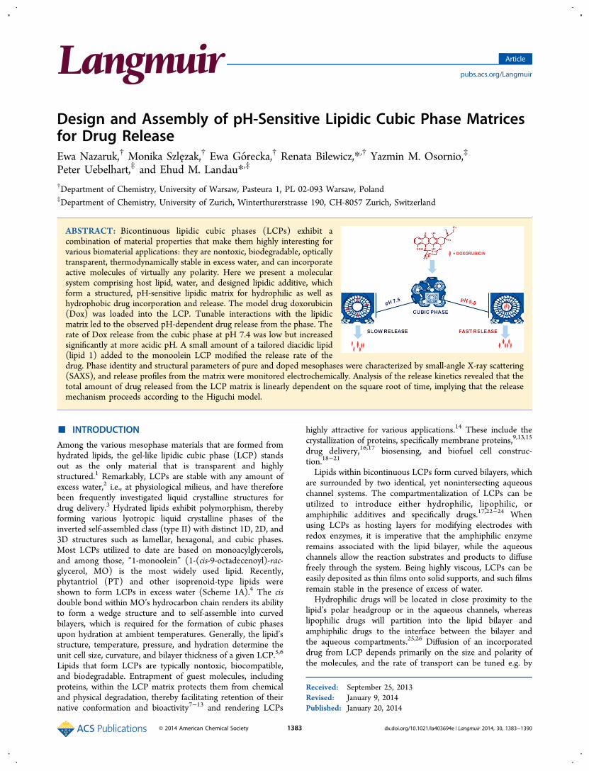
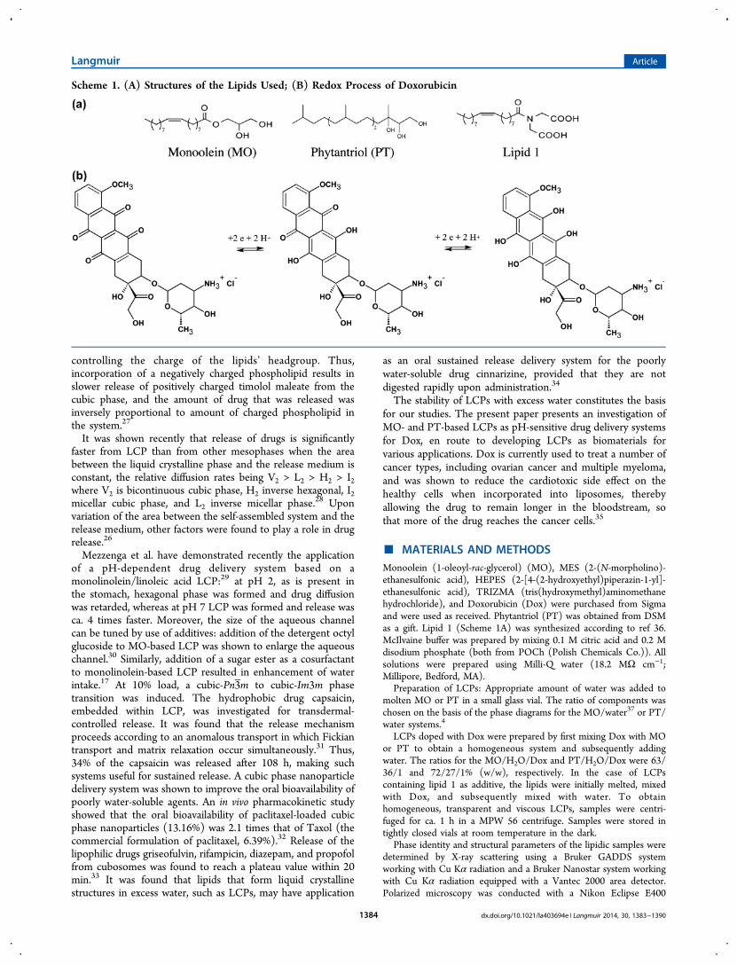
Design and Assembly of pH-Sensitive Lipidic Cubic Phase Matrices for Drug Release Ewa Nazaruk, † Monika Szlęzak, † Ewa Gó recka, † Renata Bilewicz,* ,† Yazmin M. Osornio, ‡ Peter Uebelhart, ‡ and Ehud M. Landau* ,‡ † Department of Chemistry, University of Warsaw, Pasteura 1, PL 02-093 Warsaw, Poland ‡ Department of Chemistry, University of Zurich, Winterthurerstrasse 190, CH-8057 Zurich, Switzerland ABSTRACT: Bicontinuous lipidic cubic phases (LCPs) exhibit a combination of material properties that make them highly interesting for various biomaterial applications: they are nontoxic, biodegradable, optically transparent, thermodynamically stable in excess water, and can incorporate active molecules of virtually any polarity. Here we present a molecular system comprising host lipid, water, and designed lipidic additive, which form a structured, pH-sensitive lipidic matrix for hydrophilic as well as hydrophobic drug incorporation and release. The model drug doxorubicin (Dox) was loaded into the LCP. Tunable interactions with the lipidic matrix led to the observed pH-dependent drug release from the phase. The rate of Dox release from the cubic phase at pH 7.4 was low but increased significantly at more acidic pH. A small amount of a tailored diacidic lipid (lipid 1) added to the monoolein LCP modified the release rate of the drug. Phase identity and structural parameters of pure and doped mesophases were characterized by small-angle X-ray scattering (SAXS), and release profiles from the matrix were monitored electrochemically. Analysis of the release kinetics revealed that the total amount of drug released from the LCP matrix is linearly dependent on the square root of time, implying that the release mechanism proceeds according to the Higuchi model. ■ INTRODUCTION Among the various mesophase materials that are formed from hydrated lipids, the gel-like lipidic cubic phase (LCP) stands out as the only material that is transparent and highly structured. 1 Remarkably, LCPs are stable with any amount of excess water, 2 i.e., at physiological milieus, and have therefore been frequently investigated liquid crystalline structures for drug delivery. 3 Hydrated lipids exhibit polymorphism, thereby forming various lyotropic liquid crystalline phases of the inverted self-assembled class (type II) with distinct 1D, 2D, and 3D structures such as lamellar, hexagonal, and cubic phases. Most LCPs utilized to date are based on monoacylglycerols, and among those, “1-monoolein” (1-(cis-9-octadecenoyl)-rac- glycerol, MO) is the most widely used lipid. Recently, phytantriol (PT) and other isoprenoid-type lipids were shown to form LCPs in excess water (Scheme 1A). 4 The cis double bond within MO’s hydrocarbon chain renders its ability to form a wedge structure and to self-assemble into curved bilayers, which is required for the formation of cubic phases upon hydration at ambient temperatures. Generally, the lipid’s structure, temperature, pressure, and hydration determine the unit cell size, curvature, and bilayer thickness of a given LCP. 5,6 Lipids that form LCPs are typically nontoxic, biocompatible, and biodegradable. Entrapment of guest molecules, including proteins, within the LCP matrix protects them from chemical and physical degradation, thereby facilitating retention of their native conformation and bioactivity 7−13 and rendering LCPs highly attractive for various applications. 14 These include the crystallization of proteins, specifically membrane proteins, 9,13,15 drug delivery, 16,17 biosensing, and biofuel cell construc- tion. 18−21 Lipids within bicontinuous LCPs form curved bilayers, which are surrounded by two identical, yet nonintersecting aqueous channel systems. The compartmentalization of LCPs can be utilized to introduce either hydrophilic, lipophilic, or amphiphilic additives and specifically drugs. 17,22−24 When using LCPs as hosting layers for modifying electrodes with redox enzymes, it is imperative that the amphiphilic enzyme remains associated with the lipid bilayer, while the aqueous channels allow the reaction substrates and products to diffuse freely through the system. Being highly viscous, LCPs can be easily deposited as thin films onto solid supports, and such films remain stable in the presence of excess of water. Hydrophilic drugs will be located in close proximity to the lipid’s polar headgroup or in the aqueous channels, whereas lipophilic drugs will partition into the lipid bilayer and amphiphilic drugs to the interface between the bilayer and the aqueous compartments. 25,26 Diffusion of an incorporated drug from LCP depends primarily on the size and polarity of the molecules, and the rate of transport can be tuned e.g. by Received: September 25, 2013 Revised: January 9, 2014 Published: January 20, 2014 Article pubs.acs.org/Langmuir © 2014 American Chemical Society 1383 dx.doi.org/10.1021/la403694e | Langmuir 2014, 30, 1383−1390

Welcome message from author
This document is posted to help you gain knowledge. Please leave a comment to let me know what you think about it! Share it to your friends and learn new things together.
Transcript
Design and Assembly of pH-Sensitive Lipidic Cubic Phase Matricesfor Drug ReleaseEwa Nazaruk,† Monika Szlęzak,† Ewa Gorecka,† Renata Bilewicz,*,† Yazmin M. Osornio,‡
Peter Uebelhart,‡ and Ehud M. Landau*,‡
†Department of Chemistry, University of Warsaw, Pasteura 1, PL 02-093 Warsaw, Poland‡Department of Chemistry, University of Zurich, Winterthurerstrasse 190, CH-8057 Zurich, Switzerland
ABSTRACT: Bicontinuous lipidic cubic phases (LCPs) exhibit acombination of material properties that make them highly interesting forvarious biomaterial applications: they are nontoxic, biodegradable, opticallytransparent, thermodynamically stable in excess water, and can incorporateactive molecules of virtually any polarity. Here we present a molecularsystem comprising host lipid, water, and designed lipidic additive, whichform a structured, pH-sensitive lipidic matrix for hydrophilic as well ashydrophobic drug incorporation and release. The model drug doxorubicin(Dox) was loaded into the LCP. Tunable interactions with the lipidicmatrix led to the observed pH-dependent drug release from the phase. Therate of Dox release from the cubic phase at pH 7.4 was low but increasedsignificantly at more acidic pH. A small amount of a tailored diacidic lipid(lipid 1) added to the monoolein LCP modified the release rate of thedrug. Phase identity and structural parameters of pure and doped mesophases were characterized by small-angle X-ray scattering(SAXS), and release profiles from the matrix were monitored electrochemically. Analysis of the release kinetics revealed that thetotal amount of drug released from the LCP matrix is linearly dependent on the square root of time, implying that the releasemechanism proceeds according to the Higuchi model.
■ INTRODUCTION
Among the various mesophase materials that are formed fromhydrated lipids, the gel-like lipidic cubic phase (LCP) standsout as the only material that is transparent and highlystructured.1 Remarkably, LCPs are stable with any amount ofexcess water,2 i.e., at physiological milieus, and have thereforebeen frequently investigated liquid crystalline structures fordrug delivery.3 Hydrated lipids exhibit polymorphism, therebyforming various lyotropic liquid crystalline phases of theinverted self-assembled class (type II) with distinct 1D, 2D, and3D structures such as lamellar, hexagonal, and cubic phases.Most LCPs utilized to date are based on monoacylglycerols,and among those, “1-monoolein” (1-(cis-9-octadecenoyl)-rac-glycerol, MO) is the most widely used lipid. Recently,phytantriol (PT) and other isoprenoid-type lipids wereshown to form LCPs in excess water (Scheme 1A).4 The cisdouble bond within MO’s hydrocarbon chain renders its abilityto form a wedge structure and to self-assemble into curvedbilayers, which is required for the formation of cubic phasesupon hydration at ambient temperatures. Generally, the lipid’sstructure, temperature, pressure, and hydration determine theunit cell size, curvature, and bilayer thickness of a given LCP.5,6
Lipids that form LCPs are typically nontoxic, biocompatible,and biodegradable. Entrapment of guest molecules, includingproteins, within the LCP matrix protects them from chemicaland physical degradation, thereby facilitating retention of theirnative conformation and bioactivity7−13 and rendering LCPs
highly attractive for various applications.14 These include thecrystallization of proteins, specifically membrane proteins,9,13,15
drug delivery,16,17 biosensing, and biofuel cell construc-tion.18−21
Lipids within bicontinuous LCPs form curved bilayers, whichare surrounded by two identical, yet nonintersecting aqueouschannel systems. The compartmentalization of LCPs can beutilized to introduce either hydrophilic, lipophilic, oramphiphilic additives and specifically drugs.17,22−24 Whenusing LCPs as hosting layers for modifying electrodes withredox enzymes, it is imperative that the amphiphilic enzymeremains associated with the lipid bilayer, while the aqueouschannels allow the reaction substrates and products to diffusefreely through the system. Being highly viscous, LCPs can beeasily deposited as thin films onto solid supports, and such filmsremain stable in the presence of excess of water.Hydrophilic drugs will be located in close proximity to the
lipid’s polar headgroup or in the aqueous channels, whereaslipophilic drugs will partition into the lipid bilayer andamphiphilic drugs to the interface between the bilayer andthe aqueous compartments.25,26 Diffusion of an incorporateddrug from LCP depends primarily on the size and polarity ofthe molecules, and the rate of transport can be tuned e.g. by
Received: September 25, 2013Revised: January 9, 2014Published: January 20, 2014
Article
pubs.acs.org/Langmuir
© 2014 American Chemical Society 1383 dx.doi.org/10.1021/la403694e | Langmuir 2014, 30, 1383−1390
controlling the charge of the lipids’ headgroup. Thus,incorporation of a negatively charged phospholipid results inslower release of positively charged timolol maleate from thecubic phase, and the amount of drug that was released wasinversely proportional to amount of charged phospholipid inthe system.27
It was shown recently that release of drugs is significantlyfaster from LCP than from other mesophases when the areabetween the liquid crystalline phase and the release medium isconstant, the relative diffusion rates being V2 > L2 > H2 > I2where V2 is bicontinuous cubic phase, H2 inverse hexagonal, I2micellar cubic phase, and L2 inverse micellar phase.28 Uponvariation of the area between the self-assembled system and therelease medium, other factors were found to play a role in drugrelease.26
Mezzenga et al. have demonstrated recently the applicationof a pH-dependent drug delivery system based on amonolinolein/linoleic acid LCP:29 at pH 2, as is present inthe stomach, hexagonal phase was formed and drug diffusionwas retarded, whereas at pH 7 LCP was formed and release wasca. 4 times faster. Moreover, the size of the aqueous channelcan be tuned by use of additives: addition of the detergent octylglucoside to MO-based LCP was shown to enlarge the aqueouschannel.30 Similarly, addition of a sugar ester as a cosurfactantto monolinolein-based LCP resulted in enhancement of waterintake.17 At 10% load, a cubic-Pn3m to cubic-Im3m phasetransition was induced. The hydrophobic drug capsaicin,embedded within LCP, was investigated for transdermal-controlled release. It was found that the release mechanismproceeds according to an anomalous transport in which Fickiantransport and matrix relaxation occur simultaneously.31 Thus,34% of the capsaicin was released after 108 h, making suchsystems useful for sustained release. A cubic phase nanoparticledelivery system was shown to improve the oral bioavailability ofpoorly water-soluble agents. An in vivo pharmacokinetic studyshowed that the oral bioavailability of paclitaxel-loaded cubicphase nanoparticles (13.16%) was 2.1 times that of Taxol (thecommercial formulation of paclitaxel, 6.39%).32 Release of thelipophilic drugs griseofulvin, rifampicin, diazepam, and propofolfrom cubosomes was found to reach a plateau value within 20min.33 It was found that lipids that form liquid crystallinestructures in excess water, such as LCPs, may have application
as an oral sustained release delivery system for the poorlywater-soluble drug cinnarizine, provided that they are notdigested rapidly upon administration.34
The stability of LCPs with excess water constitutes the basisfor our studies. The present paper presents an investigation ofMO- and PT-based LCPs as pH-sensitive drug delivery systemsfor Dox, en route to developing LCPs as biomaterials forvarious applications. Dox is currently used to treat a number ofcancer types, including ovarian cancer and multiple myeloma,and was shown to reduce the cardiotoxic side effect on thehealthy cells when incorporated into liposomes, therebyallowing the drug to remain longer in the bloodstream, sothat more of the drug reaches the cancer cells.35
■ MATERIALS AND METHODSMonoolein (1-oleoyl-rac-glycerol) (MO), MES (2-(N-morpholino)-ethanesulfonic acid), HEPES (2-[4-(2-hydroxyethyl)piperazin-1-yl]-ethanesulfonic acid), TRIZMA (tris(hydroxymethyl)aminomethanehydrochloride), and Doxorubicin (Dox) were purchased from Sigmaand were used as received. Phytantriol (PT) was obtained from DSMas a gift. Lipid 1 (Scheme 1A) was synthesized according to ref 36.McIlvaine buffer was prepared by mixing 0.1 M citric acid and 0.2 Mdisodium phosphate (both from POCh (Polish Chemicals Co.)). Allsolutions were prepared using Milli-Q water (18.2 MΩ cm−1;Millipore, Bedford, MA).
Preparation of LCPs: Appropriate amount of water was added tomolten MO or PT in a small glass vial. The ratio of components waschosen on the basis of the phase diagrams for the MO/water37 or PT/water systems.4
LCPs doped with Dox were prepared by first mixing Dox with MOor PT to obtain a homogeneous system and subsequently addingwater. The ratios for the MO/H2O/Dox and PT/H2O/Dox were 63/36/1 and 72/27/1% (w/w), respectively. In the case of LCPscontaining lipid 1 as additive, the lipids were initially melted, mixedwith Dox, and subsequently mixed with water. To obtainhomogeneous, transparent and viscous LCPs, samples were centri-fuged for ca. 1 h in a MPW 56 centrifuge. Samples were stored intightly closed vials at room temperature in the dark.
Phase identity and structural parameters of the lipidic samples weredetermined by X-ray scattering using a Bruker GADDS systemworking with Cu Kα radiation and a Bruker Nanostar system workingwith Cu Kα radiation equipped with a Vantec 2000 area detector.Polarized microscopy was conducted with a Nikon Eclipse E400
Scheme 1. (A) Structures of the Lipids Used; (B) Redox Process of Doxorubicin
Langmuir Article
dx.doi.org/10.1021/la403694e | Langmuir 2014, 30, 1383−13901384

microscope equipped with a LINKAM THMS 600 heating/coolingstage.Electrochemical measurements were performed using a CHI
bipotentiostat in a three-electrode arrangement with a SCE referenceelectrode and a platinum foil as the counter electrode. The workingelectrode was glassy carbon (GCE) modified with the MO or PT cubicphase film. Prior to the measurements, the electrode was polished onalumina (0.1 and 0.05 μm) with a polishing cloth. The electrodes werethen rinsed with water in the ultrasonic bath and left to dry. For eachtype of cubic phase triplicate experiments were performed.
■ RESULTS AND DISCUSSIONThe cubic phase is unique among all hydrated lipidicmesophases, as it is highly viscous and transparent, due tothe lack of birefringence. LCPs can therefore be detected byvisual inspection using direct and cross-polarized microscopy.To confirm and evaluate the effect of addition of Dox and ofthe amphiphilic diacidic compound lipid 1 on the cubic phasetype and structure, X-ray scattering and cross-polarizedmicroscopy were conducted.X-ray Data for Doped and Nondoped Samples. X-ray
scattering measurements were conducted to identify the typeand structural parameters of the liquid crystalline phases. In thewide angle regime (WAXS) all the mesophases exhibited thediffused signal positioned at the angle that corresponds to 4.5Å. This value is characteristic for phases with liquid-like orderof the alkyl chains. In the small-angle regime (SAXS) sharpBragg reflections characteristic of the long-range positionalorder were detected. The SAXS data are shown in Table 1. Thephase identity and structural parameters for reference MO/H2O and PT/H2O systems are in accord with publishedvalues.4,38,39
Sample MO/H2O/Dox (62.9/36/1.1% w/w) exhibited acubic-Pn3 m phase in the temperature range studied, 25−90 °C,above which a hexagonal phase was observed. As expected, thecrystallographic unit cell shows a negative thermal expansion(Figure 1B). Upon addition of 2% (w/w) of the diacidic lipid 1the Pn3m structure is preserved (Figure 1A), but the latticeparameter increases (by 1−2 Å) in a temperature range aboveto 45 °C (Figure 1B). In both samples below 45 °C the thermalexpansion of the lattice parameters becomes smaller, which maybe due to a glass transition in which the Pn3 m structure ispreserved but the molecular motions are frozen. However, since
the intensity ratios of the X-ray signals for the sample dopedwith lipid 1 and the nondoped one are similar (Figure 1A), itcan be concluded that the electron density, and thus themolecular distribution, is not significantly altered by incorpo-rating lipid 1 into the matrix.X-ray data collected for the nondoped PT/H2O system
(containing 27% w/w water) show the formation of a cubic-Pn3 m structure at 25 °C, with lattice parameter a of 64.5 Å. Thecubic-Pn3m phase transforms into the reverse hexagonal phaseHII with lattice parameter a of 45.8 Å at 45 °C. At 63 °C, theHII phase transforms into the isophase. When doped with 1%(w/w) of Dox, the phase behavior is altered: A cubic-Ia3dphase with lattice parameter a of 100.5 Å is formed at roomtemperature. Above 29 °C, the cubic-Ia3d undergoes a phasetransformations to the cubic-Pn3 m phase, which in turntransforms into an HII phase above 45 °C and to the isophaseat 62 °C. At a level of 1% (w/w) Dox the unit cell parametersfor both the Pn3 m and HII phases are comparable to thenondoped systems. When the Dox content is increased to 6%(w/w), the cubic-Ia3d phase exists in the temperature rangefrom room temperature to 38 °C.Thus, we conclude that addition of a small amount of Dox
(∼1% w/w) to both MO- and PT-based lipidic cubic phasesystems does not alter significantly the properties of the system;both systems preserve the cubic structures at room temper-ature. Moreover, incorporating the diacidic lipid 1 into theMO/H2O/Dox system results in only a small increase of thecrystallographic unit cell parameter at temperatures above 45°C (Table 1 and Figure 1).
Electrochemical Behavior of Doxorubicin Incorpo-rated in the Cubic Phase. Because of the presence of thequinone and hydroquinone group, Dox is electroactive and itsbehavior in LCP can be monitored electrochemically (Scheme1B). At pH 4.5 a pair of reversible peaks can be observed at ca.−0.5 V, which are related to the reduction and oxidation ofquinone redox group undergoing 2e/2H+ processes.40 Theelectrode processes of Dox in aqueous solution wereinvestigated at room temperature using glassy carbon electrodesmodified with nondoped MO-based LCPs. The electrode wasimmersed in McIlvaine buffer solution at pH 4.5 containingDox at 10−5 M concentration. Full saturation of the cubic phasewith Dox was achieved after 500 min (Figure 2A). The cyclicvoltammogram recorded for the electrode modified with theLCP and saturated with Dox is presented in Figure 2B. On thebasis of the dependence of peak current on the square root ofscan rate, it was found that the process is diffusion controlled.Dox was incorporated into the MO matrix at concentrations
of 0.3, 0.6, and 1.1% (w/w). Peak current increases withamount of Dox in the cubic phase, and at 1.1% (w/w) thecurrent reaches a plateau. This Dox concentration was thereforeapplied in all subsequent experiments. Significantly, current wasnot dependent on the mass of cubic phase placed on theelectrode surface up to ca. 150 mg cm−2 (Figure 2C). At higherloads the electrode was blocked and signal was irreversible.A Dox release profile was evaluated with electrochemical
methods (DPV and CV). The fraction of Dox released as afunction of time for all concentrations was of the same order ofmagnitude, leading to the conclusion that drug release underthese conditions is independent of the initial drug loading atthe investigated Dox concentration range. Location of the drugis an important parameter affecting the release rate. In the caseof a hydrophobic drug that can be embedded in the lipid bilayer
Table 1. Composition, Phase Identity and CrystallographicUnit Cell Parameters for the Observed Liquid CrystallinePhases Investigated
phase composition (% w/w)temperature (°C) and phase
symmetryunit cell[Å]
PT/H2O (73/27) 25; Pn3 m a = 64.545; HII a = 45.8
MO/H2O (64/36) 25; Ia3d a = 145.060; Pn3 m a = 68.0
PT/H2O/Dox (72/27/1) 25; Ia3d a = 100.529; Pn3 m a = 64.045; HII a = 46.5
MO/H2O/Dox (63/36/1) 25; Pn3 m a = 95.945; Pn3 m a = 90.250; Pn3 m a = 85.0
MO/H2O/Dox/lipid 1 25; Pn3 m a = 94.0(61/36/1/2) 45; Pn3 m a = 91.5
50; Pn3 m a = 88.8
Langmuir Article
dx.doi.org/10.1021/la403694e | Langmuir 2014, 30, 1383−13901385

compartment of the cubic phase matrix, the release depends onthe partitioning between the lipidic and hydrophilic domains.Electrochemical methods were also employed to compare
the behavior of Dox in MO- and PT-based cubic phases.Dividing the peak current by the amount of Dox deposited onthe electrode yields a normalized Dox diffusion rate, which wasfound to be lower in the PT-based LCP than in the MO-basedphase under identical conditions (Figure 3). This may be dueto the different cubic phase type, which at 25 °C is Pn3m andIa3d for the MO and PT systems, respectively (Table 1), and tothe higher viscosity of PT-based LCP. On the basis ofdependence of peak current on the square root of scan rate, itwas found that in both cases the process was diffusioncontrolled (Figure 3B,C).
The method for determining the diffusion coefficient inLCPs using electrochemical means was described earlier.41 Thediffusion coefficients for Dox at pH 5.8 determined from thevoltammetric curves were found to be (4.56 ± 0.31) × 10−9
and (2.25 ± 0.35) × 10−8 cm2 s−1 for PT/H2O/Dox and MO/H2O/Dox, respectively. Increasing the pH to 7.5 results insignificant decrease of diffusion coefficient of Dox embedded inthe MO-based LCP, which was found to be (4.36 ± 0.42) ×10−9 cm2 s−1.
Doxorubicin Release Profile as a Function of pH. Theeffect of pH and the affinity of the solubilized drug to lipidbilayers were recently studied.27,42,43 In our investigation thepH dependence of Dox diffusion from the PT- and MO-basedLCPs was established by electrochemical methods. DPV onGCE electrode modified with 1.1% (w/w) Dox-doped MO
Figure 1. (A) 2D X-ray scattering pattern of the cubic-Pn3 m phase observed for the MO/H2O/Dox/Lipid 1 system and X-ray signals vs 2θ obtainedby azimuthal integration of the X-ray intensities from 2D X-ray scattering patterns of the MO/H2O/Dox/Lipid 1 and MO/H2O/Dox systems. (B)Temperature dependence of the lattice parameter a of the cubic Pn3m phase for the MO/H2O/Dox/Lipid 1 and MO/H2O/Dox systems.
Figure 2. (A) Saturation of the MO-based cubic phase with Dox (10−5 M). (B) Cyclic voltammogram recorded for the electrode modified with cubicphase and saturated with Dox. (C) Dependence of measured peak current on the LCP mass per area of the electrode.
Figure 3. (A) Cyclic voltammogram on GC electrode modified with MO-based (black trace) and PT-based (red trace) LCPs doped with 1.1% (w/w) Dox. The y-axis depicts the peak current per mole of Dox deposited on the electrode. (B, C) Dependence of the peak current on the square rootof scan rate for the MO- and PT-based cubic phases, respectively.
Langmuir Article
dx.doi.org/10.1021/la403694e | Langmuir 2014, 30, 1383−13901386

LCP at pH 9.0, 7.5, and pH 5.8 is depicted in Figure 4. Thepotential of maximum peak is shifted toward positive potentials
as the pH of electrolyte is decreased indicating that protonswere involved in the electrochemical process. The formalpotential of Dox was plotted versus pH values; the slope was−60 mV/pH, as expected for a 2e/2H+ electrode process.Figure 5A depicts the time dependence of the peak current.
The initial current at pH 5.8 is ca. 5 times higher than those atpH 7.5 and 9.0, indicating that at the low pH Dox residesmainly in the aqueous channels, where diffusion is faster than inthe lipid bilayer domains. Because the pKa value of Dox is 8.2,its charge is strongly pH dependent in the investigated pHrange: 99.6% of Dox is protonated at pH 5.8, whereas thefraction of uncharged Dox increases to 16.6% at pH 7.5 and to87% at pH 9.0. Uncharged Dox is expected to reside mainly inthe lipidic domain of the cubic phase, while the charged speciespartitions predominantly into the aqueous channel compart-ment. Figure 5B clearly demonstrates a faster release from thecubic phase at pH 5.8 than at pH 7.5 and 9.0. At pH 9.0 Doxexists mainly in the unprotonated state, having higher affinity tothe lipidic domain and hence its release is much slower. SuchpH dependence of the rate of Dox delivery from the cubicphase may be utilized for controlling drug release into tumorcells, since the pH in cancer cells is lower than that of healthycells. In buffer solution of pH 5.8, the time required for releaseof half of the total amount of drug incorporated in the LCP(T50 value) is 45 min. At ca. 100 min the Dox current reaches a
plateau. Because at pH 7.5 and 9.0 Dox molecules arepredominantly incorporated into the lipid bilayer, penetrationinto the aqueous channels may become the rate-limiting step.Thus, at pH 7.5 less than 10% of Dox is released after 45 min.Dox release from the PT-based cubic phase was much slowerthan that from the MO-based cubic phase, as can be seen inFigure 5C: T50 was as large as 5000 min. PT-based LCP cantherefore be considered as a potential matrix for slow sustainedrelease of incorporated Dox.
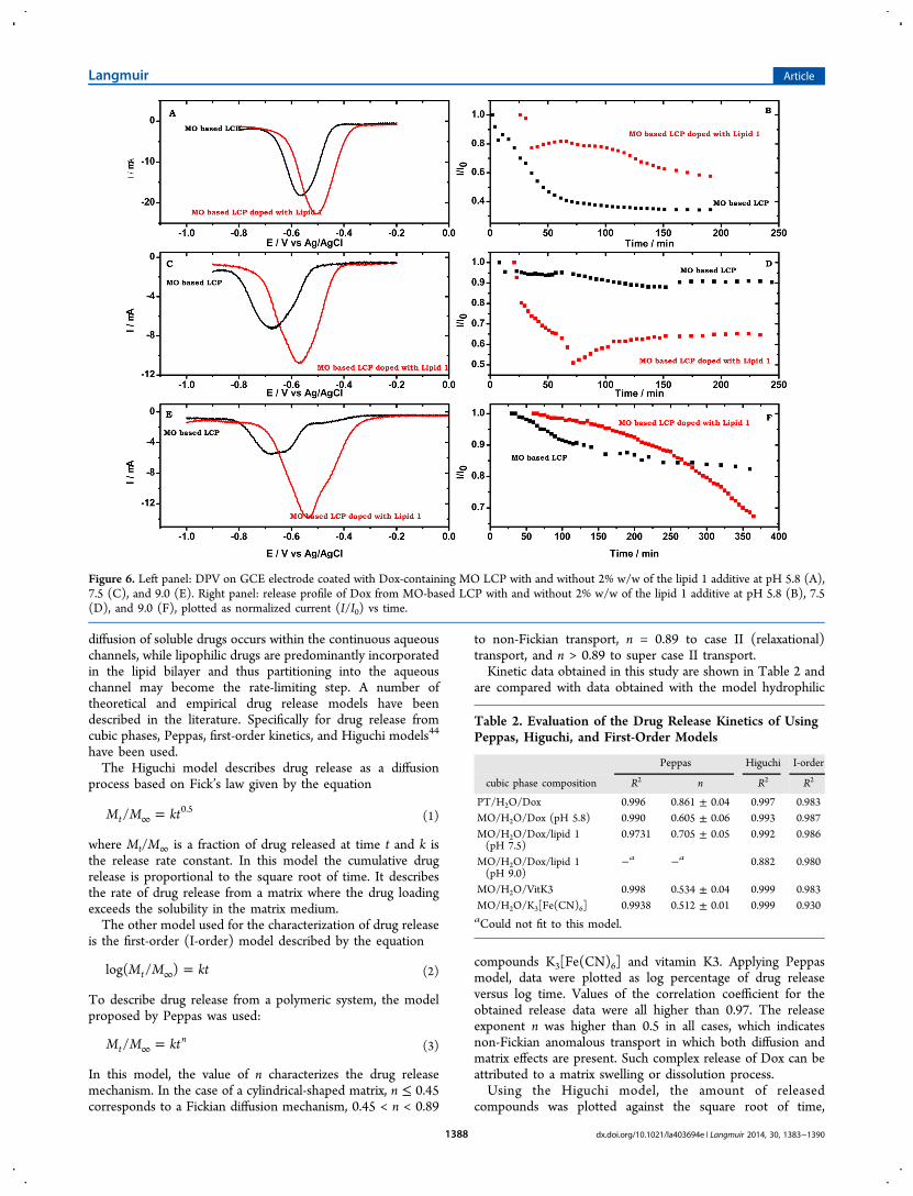
Formation of pH-Sensitive Cubic Phase. Up to 10% (w/w) of lipid 1 can be added to a MO-based LCP withoutnoticeable perturbation of the cubic phase texture, as evidencedby optical microscopy [Osornio, Y. M., personal communica-tion]. Dox-containing MO-based LCP was doped with 2% w/wof the diacidic lipid 1 in order to prepare a cubic phase matrixthat is pH-sensitive (Figure 6). The two carboxylate groups inthe headgroup region of lipid 1 are exposed to the LCP’saqueous channel, resulting in charging of the hydrophilic/hydrophobic interface. As a consequence, Dox release is fasterat pH 7.5 than at pH 5.8 (Figure 6B,D), presumably due toelectrostatic attraction between the negatively charged head-group of lipid 1 and the positively charged Dox at the lower pH.In contrast, in the absence of lipid 1 Dox release from theuncharged LCP is faster at pH 5.8 than at pH 7.5. At pH 9 themajority of Dox (87%) is unprotonated and hence located inthe lipidic bilayer domain. Charging of the interface by additionof lipid 1 to the cubic phase at this pH results in slower Doxrelease from the LCP as compared to the release in the absenceof lipid 1 up to ca. 260 min. This may be due to slow transfer ofDox from the lipidic to aqueous LCP domains (Figure 6 F). Atlonger times, an increase in the release rate of Dox in thepresence of lipid 1 occurs, whereas in the uncharged LCPrelease is slowed down, indicating that lipid 1 can control thespeed of removal of the drug from the film. This change in thekinetics of release over time makes it impossible to fit the plotof current vs time for this case to any of the mechanismsdescribing kinetics of drug release from the phase to thesolution.At the three pH values investigated Dox potential is shifted
to more positive values, and peak current is larger upon dopingthe LCP with lipid 1, as evidenced by differential pulsevoltammetry (Figure 6A,C,E). This may suggest that in thepresence of lipid 1 at the hydrophobic−hydrophilic interface ofthe LCP, a higher fraction of Dox partitions into the aqueouschannel domain.
Kinetics. An ideal profile of drug release from a prolongedrelease carrier obeys zero-order kinetics. In the case of LCPs,
Figure 4. DPV recorded on GCE electrode modified with Dox-dopedMO LCP at pH 9.0, 7.5, and 5.8.
Figure 5. (A) Release profiles of Dox from a MO/H2O/Dox (63/36/1% w/w) cubic phase, in buffers, pH 9.0, 7.4, and 5.8. (B) Release profiles ofDox from a MO/H2O/Dox (63/36/1% w/w) cubic phase, in buffers, pH 9.0, 7.4, and 5.8, plotted as normalized current I/I0 vs time. (C) Releaseprofile of Dox from a PT/H2O/Dox (72/27/1% w/w) cubic phase at pH 5.8, plotted as normalized current (I/I0) vs time.
Langmuir Article
dx.doi.org/10.1021/la403694e | Langmuir 2014, 30, 1383−13901387
diffusion of soluble drugs occurs within the continuous aqueouschannels, while lipophilic drugs are predominantly incorporatedin the lipid bilayer and thus partitioning into the aqueouschannel may become the rate-limiting step. A number oftheoretical and empirical drug release models have beendescribed in the literature. Specifically for drug release fromcubic phases, Peppas, first-order kinetics, and Higuchi models44
have been used.The Higuchi model describes drug release as a diffusion
process based on Fick’s law given by the equation
=∞M M kt/t0.5
(1)
where Mt/M∞ is a fraction of drug released at time t and k isthe release rate constant. In this model the cumulative drugrelease is proportional to the square root of time. It describesthe rate of drug release from a matrix where the drug loadingexceeds the solubility in the matrix medium.The other model used for the characterization of drug release
is the first-order (I-order) model described by the equation
=∞M M ktlog( / )t (2)
To describe drug release from a polymeric system, the modelproposed by Peppas was used:
=∞M M kt/tn
(3)
In this model, the value of n characterizes the drug releasemechanism. In the case of a cylindrical-shaped matrix, n ≤ 0.45corresponds to a Fickian diffusion mechanism, 0.45 < n < 0.89
to non-Fickian transport, n = 0.89 to case II (relaxational)transport, and n > 0.89 to super case II transport.Kinetic data obtained in this study are shown in Table 2 and
are compared with data obtained with the model hydrophilic
compounds K3[Fe(CN)6] and vitamin K3. Applying Peppasmodel, data were plotted as log percentage of drug releaseversus log time. Values of the correlation coefficient for theobtained release data were all higher than 0.97. The releaseexponent n was higher than 0.5 in all cases, which indicatesnon-Fickian anomalous transport in which both diffusion andmatrix effects are present. Such complex release of Dox can beattributed to a matrix swelling or dissolution process.Using the Higuchi model, the amount of released
compounds was plotted against the square root of time,
Figure 6. Left panel: DPV on GCE electrode coated with Dox-containing MO LCP with and without 2% w/w of the lipid 1 additive at pH 5.8 (A),7.5 (C), and 9.0 (E). Right panel: release profile of Dox from MO-based LCP with and without 2% w/w of the lipid 1 additive at pH 5.8 (B), 7.5(D), and 9.0 (F), plotted as normalized current (I/I0) vs time.
Table 2. Evaluation of the Drug Release Kinetics of UsingPeppas, Higuchi, and First-Order Models
Peppas Higuchi I-order
cubic phase composition R2 n R2 R2
PT/H2O/Dox 0.996 0.861 ± 0.04 0.997 0.983MO/H2O/Dox (pH 5.8) 0.990 0.605 ± 0.06 0.993 0.987MO/H2O/Dox/lipid 1(pH 7.5)
0.9731 0.705 ± 0.05 0.992 0.986
MO/H2O/Dox/lipid 1(pH 9.0)
−a −a 0.882 0.980
MO/H2O/VitK3 0.998 0.534 ± 0.04 0.999 0.983MO/H2O/K3[Fe(CN)6] 0.9938 0.512 ± 0.01 0.999 0.930aCould not fit to this model.
Langmuir Article
dx.doi.org/10.1021/la403694e | Langmuir 2014, 30, 1383−13901388

yielding linear relationships with very high correlationcoefficients (R2 = 0.999) for diffusion of the model hydrophiliccompounds from LCPs, indicating that release is diffusioncontrolled. Such data are in agreement with the Peppas model,where the n value was close to 0.5, indicating that the diffusionprocess is based on Fick’s law. Lower R2 correlation coefficientswere obtained for Dox embedded into MO- or PT-based cubicphases, which can be explained by the pH-dependent nature ofDox. At pH 5.8 99.6% of the Dox molecules are protonated andare predominantly located in the LCP’s aqueous channels.When fitted to the Higuchi model, these data result in a lowercorrelation coefficient of 0.993, suggesting that both diffusionand matrix effect influence the release mechanism. Theexponent value n of 0.605 ± 0.06 extracted from Peppasmodel also suggests non-Fickian transport. At pH 7.5, 16.6% ofDox is unprotonated and partitions into the lipidic domain. Then value obtained for this system (0.705 ± 0.05) and the lowcorrelation coefficient point to deviation from simple diffusivetransport. In case of Dox embedded into PT-based cubic phase,the drug release proceeds according to a zero-order mechanism.Release data could be fitted to the I-order model. The plotswere close to linear, but the correlation coefficient was poor(Figure 7B).Data obtained for the hydrophilic compounds and Dox
embedded into the MO-based cubic phase at pH 5.8 suggestthat the release mechanism proceeds according to the Higuchimodel, whereas in case where Dox can be incorporated into thelipidic domain non-Fickian transport dominates.
■ CONCLUSIONS
MO- and PT-based LCPs were investigated as potentialmatrices for pH-dependent drug delivery. Dox, used in cancerchemotherapy, is a hydrophobic molecule containing an aminegroup whose pKa is 8.2. Incorporation of up to 1.1% (w/w) ofthis model drug into the LCP systems does not significantlyaffect the liquid crystalline structure at room temperature, asevidenced by SAXS measurements. Further addition of 2% (w/w) of the diacidic additive lipid 1 to the PT/H2O/Dox systemdoes not affect the LCP symmetry, which remains Pn3m. Onthe basis of the voltammetric reduction peak of Dox, it wasfound that release kinetics is independent of the initial drugloading. Partitioning of the guest drug molecule between theLCPs lipidic and aqueous domains is crucial for the release rate,which is furthermore pH-dependent. The release rate of loadedDox was slow at pH 7.5 and increased significantly at pH 5.8.At pH 9.0 Dox is expected to partition preferentially into thelipidic compartment of the LCP, and its release rate is evenslower. Dox release was further modulated by the addition of
the pH responsive lipid 1 to the LCP, resulting in higherreduction peak currents compared to the correspondingnondoped LCPs at all pH values studied. This may suggestthat the carboxylate headgroup of lipid 1 affects the charging ofthe LCP’s hydrophobic−hydrophilic interface, enabling higherloading of Dox in the aqueous channels, where diffusion ismuch faster than in the lipidic domains. Kinetics of the drugrelease is enhanced accordingly. Kinetic data obtained forhydrophilic probes ([Fe(CN)6]
3− and vitamin K3) as well asfor positively charged Dox at pH 5.8 suggest that releasemechanism is in agreement with the Higuchi model. At higherpH values, at which Dox is uncharged and readily incorporatedinto the lipidic domain, non-Fickian transport dominates.The stability of LCP in excess water, its gel consistence, the
ability to incorporate active moleculesincluding membraneproteinsof various sizes and polarities, and to stabilize theirnative conformation are of great potential en route todeveloping LCPs as biomaterials for various applications. Thecurrent investigation demonstrates that designed lipid additivesmay be implemented to render LCPs pH active, therebymodulating the partitioning of active molecules between theLCP compartments and influencing their release kinetics.Further SAXS studies will reveal the effect of varying pH on themesophase structure and should be helpful in understandingthe drug release behavior as a function of pH. Additionalmodifications of the host matrix structure and its phasebehavior, as well as the guest active compound, will result infine-tuning of such systems and improved biomaterials for drugdelivery and release.
■ AUTHOR INFORMATIONCorresponding Authors*E-mail: [email protected] (R.B.).*E-mail [email protected] (E.M.L.).NotesThe authors declare no competing financial interest.
■ ACKNOWLEDGMENTSThis work was supported by grant PSPB 079/2010 to R.B. andE.M.L. through the Swiss Contribution to the enlargedEuropean Union.
■ REFERENCES(1) Lynch, M. L., Spicer, P. T., Eds.; Bicontinuous Liquid Crystals;Surfactant Science Series; Taylor and Francis: New York, 2005; Vol.127.(2) Tardieu, A.; Luzzati, V. Polymorphism of lipids. A novel cubicphase a cage-like network of rods with enclosed spherical micelles.Biochim. Biophys. Acta 1970, 219, 11−17.
Figure 7. Kinetics of Dox release from the PT, MO, and lipid 1-doped MO LCP at pH 5.8. Y-axis: the fraction of doxorubicin molecules released tothe solution. (A) Higuchi model; (B, C) I-order model.
Langmuir Article
dx.doi.org/10.1021/la403694e | Langmuir 2014, 30, 1383−13901389

(3) Guo, C.; Wang, J.; Cao, F.; Lee, R. J.; Zhai, G. Lyotropic liquidcrystal systems in drug delivery. Drug Discovery Today 2010, 15, 1032−1040.(4) Barauskas, J.; Landh, T. Phase behavior of the phytantriol/watersystem. Langmuir 2003, 19, 9562−9565.(5) Kulkarni, C. V.; Tang, T. Y.; Seddon, A. M.; Seddon, J. M.; Ces,O.; Templer, R. H. Engineering bicontinuous cubic structures at thenanoscale - the role of chain splay. Soft Matter 2010, 6, 3191−3194.(6) Fong, C.; Le, T.; Drummond, C. J. Lyotropic liquid crystalengineering-ordered nanostructured small molecule amphiphile self-assembly materials by design. Chem. Soc. Rev. 2012, 41, 1297−1322.(7) Portmann, M.; Landau, E. M.; Luisi, P. L. Spectroscopic andrheological studies of enzymes in rigid lipidic matrices: the case of α-chymotrypsin in a lysolecithin/water cubic phase. J. Phys. Chem. 1991,95, 8437−8440.(8) Landau, E. M.; Luisi, P. L. Lipidic cubic phases as transparent,rigid matrices for the direct spectroscopic study of immobilizedmembrane proteins. J. Am. Chem. Soc. 1993, 115, 2102−2106.(9) Landau, E. M.; Rosenbusch, J. P. Lipidic cubic phases: A novelconcept for the crystallization of membrane proteins. Proc. Natl. Acad.Sci. U. S. A. 1996, 93, 14532−14535.(10) Kraineva, J.; Nicolini, C.; Thiyagarajan, P.; Kondrashkina, E.;Winter, R. Incorporation of α-chymotrypsin into the 3D channels ofbicontinuous cubic lipid mesophases. Biochim. Biophys. Acta, ProteinsProteomics 2006, 1764, 424−433.(11) Razumas, V.; Larsson, K.; Miezis, Y.; Nylander, T. A cubicmonoolein-cytochrome c-water phase: X-ray diffraction, FT-IR,Differential Scanning Calorimetric, and electrochemical studies. J.Phys. Chem. 1996, 100, 11766−11774.(12) Angelova, A.; Ollivon, M.; Campitelli, A.; Bourgaux, C. Lipidcubic phases as stable nanochannel network structures for proteinbiochip development: X-ray diffraction study. Langmuir 2003, 19,6928−6935.(13) Chiu, M. L.; Nollert, P.; Loewen, M. C.; Belrhali, H.; Pebay-Peyroula, E.; Rosenbusch, J. P.; Landau, E. M. Crystallization in cubo:General applicability to membrane proteins. Acta Crystallogr., Sect. D2000, 56, 781−784.(14) Landau, E. M. Applications of lipidic cubic phases in structuralbiology. In Bicontinuous Structured Liquid Crystals; Lynch, M. L.,Spicer, P. T., Eds.; Marcel Dekker, Inc.: New York, 2005; pp 422−445.(15) Cherezov, V.; Rosenbaum, D. M.; Hanson, M. A.; Rasmussen, S.G.; Thian, F. S.; Kobilka, T. S.; Choi, H. J.; Kuhn, P.; Weis, W. I.;Kobilka, B. K.; Stevens, R. C. High-resolution crystal structure of anengineered human beta2-adrenergic G protein-coupled receptor.Science 2007, 318, 1258−1265.(16) Conn, C. E.; Drummond, C. J. Nanostructured bicontinuouscubic lipid self-assembly materials as matrices for protein encapsula-tion. Soft Matter 2013, 9, 3449−3464.(17) Negrini, R.; Mezzenga, R. Diffusion, molecular separation, anddrug delivery from lipid mesophases with tunable water channels.Langmuir 2012, 28, 16455−16462.(18) Nazaruk, E.; Smolin ski, S.; Swatko-Ossor, M.; Ginalska, G.;Fiedurek, J.; Rogalski, J.; Bilewicz, R. Enzymatic biofuel cell based onelectrodes modified with lipid liquid-crystalline cubic phases. J. PowerSources 2008, 183, 533−538.(19) Nazaruk, E.; Sadowska, K.; Madrak, K.; Biernat, J.; Rogalski, J.;Bilewicz, R. Composite bioelectrodes based on lipidic cubic phase withcarbon nanotube network. Electroanalysis 2009, 21, 507−511.(20) Nazaruk, E.; Gorecka, E.; Bilewicz, R. Enzymes and mediatorshosted together in lipidic mesophases for the construction ofbiodevices. J. Colloid Interface Sci. 2012, 385, 130−136.(21) Komisarski, M.; Osornio, Y. M.; Siegel, J. S.; Landau, E. M.Tailored host-guest lipidic cubic phases: A protocell model exhibitingnucleic acid recognition. Chem.Eur. J. 2013, 19, 1262−1267.(22) Engstrom, S. Cubic phases as drug delivery systems. Am. Chem.Soc., Div. Polym. Chem. 1990, 31, 157−158.(23) Wyatt, D. M.; Dorschel, D. A cubic phase delivery systemcomposed of glyceryl monooleate and water for sustained release ofwater-soluble drugs. Pharm. Technol. 1992, 16, 116−130.
(24) Engstrom, S.; Norden, T. P.; Nyquist, H. Cubic phases forstudies of drug partition into lipid bilayers. Eur. J. Pharm. Sci. 1999, 8,243−254.(25) Chang, C. M.; Bodmeier, R. Binding of drugs to monoglyceride-based drug delivery systems. Int. J. Pharm. 1997, 147, 135−142.(26) Yaghmur, A.; Rappolt, M.; Østergaard, J.; Larsen, C.; Larsen, S.W. Characterization of bupivacaine-loaded formulations based onliquid crystalline phases and microemulsions: The effect of lipidcomposition. Langmuir 2012, 28, 2881−2889.(27) Lindell, K.; Engblom, J.; Jonstromer, M.; Carlsson, A.;Engstrom, S. Influence of a charged phospholipid on the releasepattern of timolol maleate from cubic liquid crystalline phases. Prog.Colloid Polym. Sci. 1998, 108, 111−118.(28) Phan, S.; Fong, W.-K.; Kirby, N.; Hanley, T.; Boyd, B. J.Evaluating the link between self-assembled mesophase structure anddrug release. Int. J. Pharm. 2011, 421, 176−182.(29) Negrini, R.; Mezzenga, R. pH-responsive lyotropic liquidcrystals for controlled drug delivery. Langmuir 2011, 27, 5296−5303.(30) Angelov, B.; Angelova, A.; Ollivon, M.; Bourgaux, C.;Campitelli, A. Diamond-type lipid cubic phase with large waterchannels. J. Am. Chem. Soc. 2003, 125, 7188−7189.(31) Peng, X.; Wen, X.; Pan, X.; Wang, R.; Chen, B.; Wu, C. Designand in vitro evaluation of capsaicin transdermal controlled releasecubic phase gels. AAPS PharmSciTech 2010, 11, 1405−1410.(32) Zeng, N.; Gao, X.; Hu, Q.; Song, Q.; Xia, H.; Liu, Z.; Fang, L.Lipid-based liquid crystalline nanoparticles as oral drug deliveryvehicles for poorly water-soluble drugs: Cellular interaction and in vivoabsorption. Int. J. Nanomed. 2012, 7, 3703−3718.(33) Boyd, B. J. Characterisation of drug release from cubosomesusing the pressure ultrafiltration method. Int. J. Pharm. 2003, 260,239−247.(34) Boyd, B. J.; Khoo, S. M.; Whittaker, D. V.; Davey, G.; Porter, C.J. A lipid-based liquid crystalline matrix that provides sustained releaseand enhanced oral bioavailability for a model poorly water soluble drugin rats. Int. J. Pharm. 2007, 340, 52−60.(35) Addeo, R.; Faiola, V.; Guarrasi, R. Liposomal pegylateddoxorubicin plus vinorelbine combination as first-line chemotherapyfor metastatic breast cancer in elderly women ≥65 years of age. CancerChemother. Pharmacol. 2008, 62, 285−292.(36) Osornio, Y. M.; Uebelhart, P.; Bosshard, S.; Konrad, F.; Siegel, J.S.; Landau, E. M. Design and synthesis of lipids for the fabrication offunctional lipidic cubic-phase biomaterials. J. Org. Chem. 2012, 77,10583−10595.(37) Briggs, J.; Chung, H.; Caffrey, M. The temperature-compositionphase diagram and mesophase structure characterization of themonoolein/water system. J. Phys. II 1996, 6, 723−752.(38) Qiu, H.; Caffrey, M. The phase diagram of the monoolein/watersystem: Metastability and equilibrium aspects. Biomaterials 2000, 21,223−234.(39) Caboi, F.; Nylander, T.; Razumas, V.; Talaikyte, Z.; Monduzzi,M.; Larsson, K. Structural effects, mobility, and redox behavior ofvitamin K1 hosted in the monoolein/water liquid crystalline phases.Langmuir 1997, 13, 5476−5483.(40) Komorsky-Lovric, S. Redox kinetics of adriamycin adsorbed onthe surface of graphite and mercury electrodes. Bioelectrochemistry2006, 69, 82−87.(41) Rowin ski, P.; Korytkowska, A.; Bilewicz, R. Diffusion ofhydrophilic probes in bicontinuous lipidic cubic phase. Chem. Phys.Lipids 2003, 124, 147−156.(42) Yaghmur, A.; Larsen, S. W.; Schmitt, M.; Østergaard, J.; Larsen,C.; Jensen, H.; Urtti, A.; Rappolt, M. In situ characterization of lipidicbupivacaine-loaded formulations. Soft Matter 2011, 7, 8291−8295.(43) Shah, J. C.; Sadhale, Y.; Chilukuri, D. M. Cubic phase gels asdrug delivery systems. Adv. Drug Delivery Rev. 2001, 47, 229−250.(44) Costa, P.; Sousa Lobo, J. M. Modeling and comparison ofdissolution profiles. Eur. J. Pharm. Sci. 2001, 13, 123−133.
Langmuir Article
dx.doi.org/10.1021/la403694e | Langmuir 2014, 30, 1383−13901390
Related Documents