Inhalation Toxicology, 20:245–256, 2008 Copyright c Informa Healthcare USA, Inc. ISSN: 0895-8378 print / 1091-7691 online DOI: 10.1080/08958370701864250 Derivation of an Inhalation Reference Concentration Based upon Olfactory Neuronal Loss in Male Rats following Subchronic Acetaldehyde Inhalation David C. Dorman, Melanie F. Struve, Brian A. Wong, Elizabeth A. Gross, and Carl Parkinson CIIT at The Hamner Institutes for Health Sciences, Research Triangle Park, North Carolina, USA Gabrielle A. Willson EPL, Inc., Research Triangle Park, North Carolina, USA Yu-Mei Tan and Jerry L. Campbell CIIT at The Hamner Institutes for Health Sciences, Research Triangle Park, North Carolina, USA Justin G.Teeguarden Pacific Northwest National Laboratory, Richland, Washington, USA Harvey J. Clewell III and Melvin E. Andersen CIIT at The Hamner Institutes for Health Sciences, Research Triangle Park, North Carolina, USA Acetaldehyde inhalation induces neoplastic and nonneoplastic responses in the rodent nasal cavity. This experiment further characterizes the dose-response relationship for nasal pathol- ogy, nasal epithelial cell proliferation, and DNA–protein cross-link formation in F-344 rats exposed subchronically to acetaldehyde. Animals underwent whole-body exposure to 0, 50, 150, 500, or 1500 ppm acetaldehyde for 6 h/day, 5 days/wk for up to 65 exposure days. Respiratory tract histopathology was evaluated after 4, 9, 14, 30, and 65 exposure days. Acetaldehyde expo- sure was not associated with reduced body weight gain or other evidence of systemic toxicity. Histologic evaluation of the nasal cavity showed an increased incidence of olfactory neuronal loss (ONL) following acute to subchronic exposure to ≥150 ppm acetaldehyde and increased olfactory epithelial cell proliferation following exposure to 1500 ppm acetaldehyde. The sever- ity of the ONL demonstrated dose- and temporal-dependent behaviors, with minimal effects noted at 150–500 ppm acetaldehyde and moderately severe lesions seen in the highest exposure group, with increased lesion severity and extent as the exposure duration increased. Acetalde- hyde exposure was also associated with inflammation, hyperplasia, and squamous metaplasia of the respiratory epithelium. These responses were seen in animals exposed to ≥500 ppm ac- etaldehyde. Acetaldehyde exposure was not associated with increased DNA–protein cross-link formation in the respiratory or olfactory epithelium. A model of acetaldehyde pharmacokinetics in the nose was used to derive an inhalation reference concentration (RfC) of 0.4 ppm, based on the no-observed-adverse-effect level (NOAEL) of 50 ppm for the nasal pathology seen in this study. Received 10 July 2007; accepted 2 November 2007. The authors thank Iris Decker, Shane Cantu, Corrie Dunn, R. Arden James, Otis Lyght, Paul Ross, and the staff of the CIIT inhalation, animal care facility, and necropsy and histology support groups for their contributions. We also thank Dr. Mel Andersen for his critical review of this article. This study was sponsored and funded by the American Forest and Paper Association. Current address for David C. Dorman and Melanie F. Struve is College of Veterinary Medicine, North Carolina State University, Raleigh, NC 27606, USA. Address correspondence to David C. Dorman, DVM, PhD, College of Veterinary Medicine, North Carolina State University, Raleigh, NC 27606, USA. E-mail: david [email protected] 245 Inhalation Toxicology Downloaded from informahealthcare.com by University Library Utrecht on 05/03/13 For personal use only.

Welcome message from author
This document is posted to help you gain knowledge. Please leave a comment to let me know what you think about it! Share it to your friends and learn new things together.
Transcript
Inhalation Toxicology, 20:245–256, 2008Copyright c© Informa Healthcare USA, Inc.ISSN: 0895-8378 print / 1091-7691 onlineDOI: 10.1080/08958370701864250
Derivation of an Inhalation Reference ConcentrationBased upon Olfactory Neuronal Loss in Male Ratsfollowing Subchronic Acetaldehyde Inhalation
David C. Dorman, Melanie F. Struve, Brian A. Wong,Elizabeth A. Gross, and Carl ParkinsonCIIT at The Hamner Institutes for Health Sciences, Research Triangle Park, North Carolina, USA
Gabrielle A. WillsonEPL, Inc., Research Triangle Park, North Carolina, USA
Yu-Mei Tan and Jerry L. CampbellCIIT at The Hamner Institutes for Health Sciences, Research Triangle Park, North Carolina, USA
Justin G.TeeguardenPacific Northwest National Laboratory, Richland, Washington, USA
Harvey J. Clewell III and Melvin E. AndersenCIIT at The Hamner Institutes for Health Sciences, Research Triangle Park, North Carolina, USA
Acetaldehyde inhalation induces neoplastic and nonneoplastic responses in the rodent nasalcavity. This experiment further characterizes the dose-response relationship for nasal pathol-ogy, nasal epithelial cell proliferation, and DNA–protein cross-link formation in F-344 ratsexposed subchronically to acetaldehyde. Animals underwent whole-body exposure to 0, 50, 150,500, or 1500 ppm acetaldehyde for 6 h/day, 5 days/wk for up to 65 exposure days. Respiratorytract histopathology was evaluated after 4, 9, 14, 30, and 65 exposure days. Acetaldehyde expo-sure was not associated with reduced body weight gain or other evidence of systemic toxicity.Histologic evaluation of the nasal cavity showed an increased incidence of olfactory neuronalloss (ONL) following acute to subchronic exposure to ≥150 ppm acetaldehyde and increasedolfactory epithelial cell proliferation following exposure to 1500 ppm acetaldehyde. The sever-ity of the ONL demonstrated dose- and temporal-dependent behaviors, with minimal effectsnoted at 150–500 ppm acetaldehyde and moderately severe lesions seen in the highest exposuregroup, with increased lesion severity and extent as the exposure duration increased. Acetalde-hyde exposure was also associated with inflammation, hyperplasia, and squamous metaplasiaof the respiratory epithelium. These responses were seen in animals exposed to ≥500 ppm ac-etaldehyde. Acetaldehyde exposure was not associated with increased DNA–protein cross-linkformation in the respiratory or olfactory epithelium. A model of acetaldehyde pharmacokineticsin the nose was used to derive an inhalation reference concentration (RfC) of 0.4 ppm, basedon the no-observed-adverse-effect level (NOAEL) of 50 ppm for the nasal pathology seen in thisstudy.
Received 10 July 2007; accepted 2 November 2007.The authors thank Iris Decker, Shane Cantu, Corrie Dunn, R. Arden James, Otis Lyght, Paul Ross, and the staff of the CIIT inhalation, animal
care facility, and necropsy and histology support groups for their contributions. We also thank Dr. Mel Andersen for his critical review of thisarticle. This study was sponsored and funded by the American Forest and Paper Association.
Current address for David C. Dorman and Melanie F. Struve is College of Veterinary Medicine, North Carolina State University, Raleigh, NC27606, USA.
Address correspondence to David C. Dorman, DVM, PhD, College of Veterinary Medicine, North Carolina State University, Raleigh, NC27606, USA. E-mail: david [email protected]
245
Inha
latio
n T
oxic
olog
y D
ownl
oade
d fr
om in
form
ahea
lthca
re.c
om b
y U
nive
rsity
Lib
rary
Utr
echt
on
05/0
3/13
For
pers
onal
use
onl
y.
246 D. C. DORMAN ET AL.
Acetaldehyde is a widely used chemical intermediate and isalso a by-product of certain metabolic oxidation reactions. Ac-etaldehyde concentrations in water and most beverages are gen-erally ≤0.1 µg/L and exposure from drinking water is consid-ered negligible (WHO IPCS 1995). The main source of humanexposure to acetaldehyde is through the metabolism of alcohol.Acetaldehyde derives from ethanol through aerobic oxidation byalcohol dehydrogenases, and typically is rapidly metabolizedto acetate by aldehyde dehydrogenases. Even without drink-ing alcohol, concentrations of acetaldehyde expelled in breathrange from 4 to 12 ppb, with higher levels observed in smok-ers and abstinent alcoholics (Jones, 1995). Acetaldehyde is alsofound in trace amounts in ambient air. For example, acetalde-hyde has been measured in Los Angeles air at approximately 32ppb (U.S. EPA, 1987). Acetaldehyde is listed by the U.S. En-vironmental Protection Agency (U.S. EPA) as a hazardous airpollutant (HAP). Occupational exposure standards have beenset for inhaled acetaldehyde including a permissible exposurelimit (PEL) of 200 ppm (360 mg/m3) and a threshold limit valueceiling (TLV-C) of 25 ppm (ACGIH, 2001).
The nasal cavity is an important target for inhaled acetalde-hyde. Appleman and colleagues (1982, 1986) showed that re-peated exposure of male Wistar rats (6 h/day, 5 days/wk for 4wk) to ≥500 ppm acetaldehyde was associated with degenera-tion of the olfactory epithelium. Thinning of the olfactory ep-ithelium with loss of sensory and sustentacular cells is likewiseseen following subchronic inhalation (Woutersen et al., 1984).Replacement of the olfactory epithelium by respiratory epithe-lium can occur during subchronic exposure (Woutersen et al.,1984). Woutersen and coworkers (1984) also reported the pres-ence of hyperplasia and metaplasia of the respiratory (nasal), la-ryngeal, and tracheal epithelium in rats subchronically exposedto 3000 ppm acetaldehyde. Metaplasia of the nasal mucosa andtrachea also occurs in male Syrian golden hamsters exposedchronically (7 h/day, 5 days/wk for 52 wk) to 1500 ppm ac-etaldehyde (Feron, 1979).
Acetaldehyde-induced olfactory degeneration in rodents hasbeen used by the U.S. EPA to establish an inhalation referenceconcentration (RfC) for this chemical. The RfC is an estimateof a continuous inhalation exposure to humans that is likely tobe without appreciable risk of adverse effects over a person’slifetime (U.S. EPA, 1994). The current RfC for acetaldehyde is0.009 mg/m3 or 0.005 ppm (Integrated Risk Information Sys-tem, http://www.epa.gov/iris, accessed May 2007), based on thestudies performed by Appleman and coworkers (1982, 1986).
Acetaldehyde is also classified as a probable human carcino-gen by the U.S. EPA based on the increased incidence of nasalsquamous-cell carcinomas and adenocarcinomas in rats and la-ryngeal and nasal carcinomas in hamsters after chronic inhala-tion exposure to ≥750 (rats) or ≥1500 (hamsters) ppm acetalde-hyde (Feron, 1979; Feron et al., 1982; Woutersen et al., 1984,1986). The mechanism of acetaldehyde-induced nasal tumorsis incompletely understood. Acetaldehyde is a weak mutagen(Feron et al., 1991). Repeated, but not a single, exposure to
≥1000 ppm acetaldehyde results in decreased extractability ofDNA from proteins isolated from the respiratory and olfactoryepithelium (Lam et al., 1986). Decreased extractability of DNAfrom proteins is consistent with the formation of DNA–proteincross-links (DPX). Formation of DPX can impair mammalianDNA replication and transcription and may contribute to the ob-served mutagenic and carcinogenic effects of these chemicals(Kurtz & Lloyd, 2003). An increased rate of cell proliferationhas long been recognized as an important factor in nasal carcino-genesis (Bogdanffy & Valentine, 2003; Monticello & Morgan,1997), and increased cell proliferation in the nasal mucosa hasbeen reported following acetaldehyde inhalation (Cassee et al.,1996).
The overall goal of this experiment is to further characterizethe dose-response relationship for nasal pathology, nasal epithe-lial cell proliferation, and DPX formation in rats exposed sub-chronically to acetaldehyde. The results of this study are usedto estimate a human inhalation reference concentration (RfC),with cross-species extrapolation performed by a pharmacoki-netic model of the rat and human nose (Teeguarden et al., 2008).
MATERIALS AND METHODS
Study DesignThis study had four acetaldehyde exposure groups (50, 150,
500, and 1500 ppm) and an air-exposed (0 ppm acetaldehyde)control group. Rats were exposed whole-body to acetaldehydeby inhalation for 6 h/day, 5 days/wk for up to 65 exposure days(13 exposure weeks). Respiratory-tract histopathology and cellproliferation were evaluated after 4, 9, 14, 30, and 65 exposuredays (n = 12 rats/exposure concentration/time point). An addi-tional cohort of rats (n = 12 rats/exposure concentration/timepoint) were evaluated for DNA–protein cross-link formation af-ter 4 and 65 exposure days.
ChemicalsAcetaldehyde (CAS number 75-07-0) in aluminum cylinders
was purchased from Aldrich Chemical Company (Milwaukee,WI). A certificate of analysis for each cylinder was provided bythe supplier. Unless otherwise noted, all other chemicals werepurchased from Sigma-Aldrich (St. Louis, MO).
Animals and Their HusbandryThis study was conducted under federal guidelines for the
care and use of laboratory animals (National Research Council,1996) and was approved by the CIIT Centers for Health Re-search Institutional Animal Care and Use Committee. A totalof 420 male F344 rats, 6 wk of age at purchase, obtained fromCharles River Laboratory (Kingston, NY), were used for thisstudy. Animals were acclimated to the CIIT animal facility for2 wk prior to the start of exposures. Animals were individuallyhoused in stainless-steel cages (R-24, Lab Products, Seaford,DE) and given certified NIH-07 rodent diet (Zeigler Brothers,Gardners, PA) and reverse-osmosis water (HydroService and
Inha
latio
n T
oxic
olog
y D
ownl
oade
d fr
om in
form
ahea
lthca
re.c
om b
y U
nive
rsity
Lib
rary
Utr
echt
on
05/0
3/13
For
pers
onal
use
onl
y.
ACETALDEHYDE NASAL TOXICITY 247
Supplies, Research Triangle Park, NC) ad libitum. Animal roomswere ventilated with HEPA-filtered air and maintained at 18–26◦C with relative humidity of 30–70% on a 12-h light–darkcycle.
Test Atmosphere Generation and CharacterizationAnimals were exposed in 1-m3 whole-body exposure cham-
bers (H1000, Lab Products, Seaford, DE). Separate 1-m3 cham-bers were used for each concentration, including a separatecontrol chamber. Test atmospheres were generated by passingnitrogen through the head space of a pressure vessel containingliquid acetaldehyde. The mixture was metered through a massflow controller (MKS Instruments, Andover, MA) and injectedinto the chamber air supply. The acetaldehyde and HEPA-filteredair supply mixture was adjusted to achieve the target concen-trations. Animal positions within the exposure chamber wererotated weekly to randomize for experimental error due to anyundetected differences in the environment or test atmosphere.Each chamber was checked once during the prestudy trials foruniformity of distribution of the test compound by measuringthe acetaldehyde concentration at nine locations in each cham-ber. Acetaldehyde concentrations were measured with cagingand catch pans in place, but without the animals.
Acetaldehyde exposure atmospheres in the chambers wereanalyzed by a Hewlett Packard 5890 (Agilent Technologies, Inc.,Palo Alto, CA) gas chromatograph (GC) equipped with a flameionization detector (FID), an 8-port gas sampling valve, and an1.8 m × 0.3 cm packed stainless steel column containing 10%free fatty acid phase (FFAP) on Chromosorb W-AW 80/100(Alltech Associates, Deerfield, IL). The GC was calibrated byvalve injections from samples prepared by delivering a measuredvolume of acetaldehyde in nitrogen from a certified cylinderand further diluting with nitrogen/house air to attain desiredcalibration concentrations in Tedlar gas sampling bags (SKC,Inc., Eighty-Four, PA). The air and acetaldehyde mixture wasdrawn from the bags through the GC gas-sampling valve foranalysis. Area counts were converted to concentrations in partsper million of acetaldehyde.
Clinical AssessmentsThe general condition of all animals (based on cage side
examinations) was checked and recorded daily. Cage-side ob-servations included changes in skin and fur, eyes and mucousmembranes, respiratory system, circulatory system, autonomicand central nervous system, somatomotor activity, and behaviorpattern. Body weights and detailed clinical observations weremeasured and recorded within 1–2 days of arrival and weeklythroughout the duration of the study.
Necropsy and HistopathologyOn the day of each necropsy, rats were deeply anesthetized
with sodium pentobarbital (intraperitoneal injection, approxi-mately 30 mg/kg) and exsanguinated by transection of the ab-dominal aorta. The trachea was exposed and the lungs were
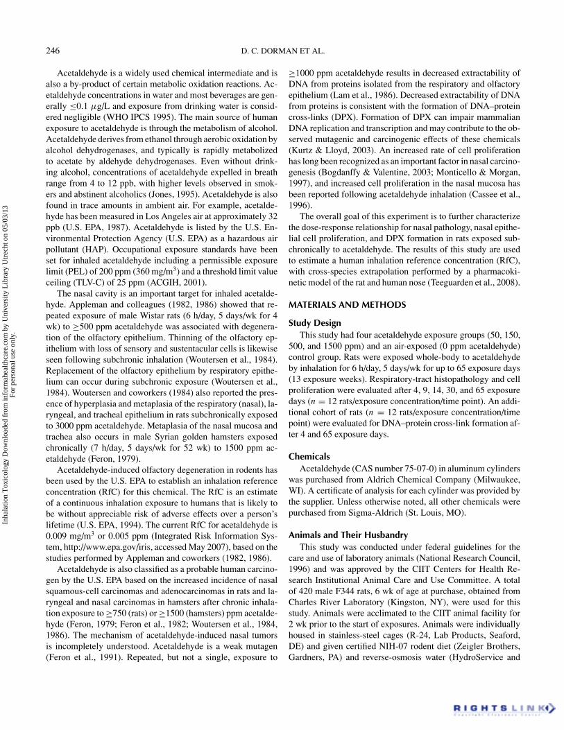
filled with 10% neutral buffered formalin (NBF) at approxi-mately 30 cm H2O pressure. The trachea was ligated to keepthe lungs inflated during fixation. Following this procedure thenasal cavities were flushed (retrograde through the trachea) withNBF. Lungs with trachea and larynx attached were excised andplaced in NBF. The heads were removed, skinned, trimmed ofexcess tissue, and placed in NBF. Respiratory-tract tissues werefixed in NBF for approximately 48 h. Following fixation theheads were rinsed in running tap water for at least 30 min anddecalcified in Immunocal formic acid bone decalcifier (DecalChemical Corp., Tallman, NY) for 7 to 14 days. Noses weresectioned transversely to provide sections of the nasal cav-ity at six standard levels (Morgan, 1991; Figure 1), routinelyprocessed and embedded in paraffin wax, sectioned (5 µm),deparaffinized, and stained with hematoxylin and eosin (H&E).Following fixation the larynx, trachea, and lung were changedto 70% ethanol, gross trimmed, routinely processed to paraffinblock, sectioned (5 µm), and stained with H&E. H&E sectionsfrom all animals were examined histologically. Additional nasalsections were immunostained for proliferating cell nuclear anti-gen (PCNA) following high-heat antigen retrieval with mousemonoclonal anti-proliferating cell nuclear antigen Clone PC10diluted 1:1000 (Dako, Carpinteria, CA) and an aminoethyl car-bazole (AEC, red) chromagen substrate kit (Zymed Laborato-ries, Inc., South San Francisco, CA) according to the manufac-turer’s instructions and standard procedures in place at CIIT.PCNA-positive cells stained red.
Histological findings were recorded at each level. The ma-jority of the findings were graded on a scale of 1–5 where 1 =minimal, 2 = slight or mild, 3 = moderate, 4 = moderately se-vere, and 5 = severe/high. The entire area of the nasal epitheliumfor each of the six levels was examined. To aid in mapping the
FIG. 1. Parasagittal view of the rodent nasal cavity showing theapproximate location of transverse sections used for microscopicexamination, squamous epithelium (S), transitional/respiratoryepithelium (T/R), and olfactory epithelium (O). Figure alsoshows the approximate location of tissue samples used for thedetermination of whether DNA–protein cross-links occurred.
Inha
latio
n T
oxic
olog
y D
ownl
oade
d fr
om in
form
ahea
lthca
re.c
om b
y U
nive
rsity
Lib
rary
Utr
echt
on
05/0
3/13
For
pers
onal
use
onl
y.
248 D. C. DORMAN ET AL.
lesions the histological findings were recorded according to thesite in which they occurred. Some sites for which histologicalfindings were recorded included:
• Nose tip. Dorsal meatus: area extending from the sep-tum adjacent to the nasoturbinate (NT), up around thedorsal meatus and along the medial aspect of the NTto the tip of the NT. Lateral wall: area extending fromthe tip of the NT around the lateral wall to the tip ofthe maxilloturbinate.
• Nose, level I. Dorsal meatus: extended from the sep-tum adjacent to the NT, up around the dorsal meatus anddown the medial aspect of the NT to the tip of the NT.Lateral wall: extended from the tip of the NT around thelateral wall to the tip of the maxilloturbinate. Maxillo-turbinate: extended from the tip of the maxilloturbinatealong its medial aspect.
• Nose, level II. Dorsal meatus: extended from the sep-tum adjacent to the NT, up around the dorsal meatusand down the medial aspect of the NT to the tip of theNT. Lateral wall: began from midventral midpoint ofthe NT and extended along the lateral aspect of the NT,continued along the lateral wall ventrally and extendedup to the lateral aspect of the maxilloturbinate where itended at the dorsal midpoint of the maxilloturbinate.
• Nose levels III, IV, and V. These levels were evaluatedwith respect to histological changes in the olfactoryepithelium that is found at these levels.
• Larynx, trachea, and lung. These regions were evalu-ated with respect to histological changes in the airwayepithelium.
Cell ProliferationPrevious nasal cell replication studies have shown that the
labeling index (LI) provides a valuable endpoint for assessmentof toxic and potentially carcinogenic responses in rats (Mon-ticello et al., 1990). Cells in S-phase were distinguished fromother cells by the intense red nuclei of the PCNA stain (palered nuclei represent other phases of the cell cycle; blue/purplenuclei represent unlabeled cells). Attention was focused on thedorsal meatus because this was a sensitive site for acetaldehyde-induced olfactory neuronal loss. Epithelial cells of the dorsalmeatus at level I were counted from the tip of the nasoturbinatemedially along the nasoturbinate through the dorsal meatus anddown the nasal septum to a point approximately opposite the tipof the nasoturbinate. All cells in the nasal epithelium betweenthese landmarks were counted as “+” for S-phase or “–” fornot S-phase. Olfactory epithelial basal cells of the dorsal mea-tus, which are generally believed to be a replicating population(Beites et al., 2005), were counted at Level II. Basal cells in theolfactory epithelium of the dorsal meatus were counted as “+”for S-phase or “–” for not S-phase.
For the labeling index (LI), the number of PCNA-labeledepithelial cell nuclei and the total number of epithelial cells
(labeled and unlabeled) were determined and expressed as thepercentage of labeled cells (labeled divided by total times 100).For unit length labeling index (ULLI), the number of PCNA-labeled epithelial cell nuclei is divided by the length of basementmembrane over which the labeled cells were counted, measuredon electronic images of sections using ImagePro Plus (MediaCybernetics, Bethesda, MD). ULLI data is expressed as numberof cells per millimeter.
DNA–Protein Cross-Link AssessmentAfter euthanasia, the head was sectioned sagitally on the
bridge of the nose. Representative samples of the nasal respira-tory and olfactory mucosa and nasopharyngeal duct (Figure 1)were removed with the supporting turbinates and flash-frozenin liquid nitrogen. Three samples (nasoturbinate, ethmoidturbinate, and nasopharyngeal duct) were collected from ani-mals exposed to acetaldehyde for 4 days. Two additional sam-ples, from the nasoturbinate and dorsal meatus, were collectedfrom rats that underwent 65 exposure days. These samples wereused to determine whether DNA–protein cross-links occurredusing methods described by Sutherland and Costa (1999). TheDNA–protein cross-link method detects the amount of proteinscovalently linked to DNA. The procedure is based on the fact thatsodium dodecyl sulfate (SDS) binds to proteins but not to DNA.Addition of potassium chloride to SDS solution results in theformation of a potassium–SDS precipitate that is easily recov-ered by low-speed centrifugation. Binding of SDS to proteinscross-linked to DNA leads to selective precipitation of DNAcontaining a cross-linked protein upon addition of potassiumchloride.
Briefly, frozen tissues (∼50 to 100 mg) were rinsed with750 µl ice-cold PBS and then digested at room temperature withtwo 2-ml, 10-min washes with 0.01 M HEPES buffer supple-mented with hyaluronidase, type IV collagenase, and protease.The pooled digestion solutions were centrifuged at 600 × g for10 min at 4◦C. The resulting pellet was resuspended in 1 ml PBSand transferred into a 1.5-ml centrifuge tube and recentrifugedat 600 × g for 5 min at 4◦C. The resulting pellet was lysed byresuspending the pellet with 750 µl PBS and adding 250 µl of10% SDS. The sample DNA was sheared by passing the lysedsample through a 21-gauge needle 5 times using a 3-ml syringe.The sample was then stored overnight at −70◦C. On the follow-ing day the samples were thawed at 37◦C for ∼10–15 min. Next,100 µl of 2.5 M KCl was added, and samples were placed on icefor 5 min, followed by a 5-min centrifugation step (13,000 rpm at4◦C). The supernatant containing the unbound DNA (“free”) wascollected in a 15-ml centrifuge tube and kept on ice. The resultingpellet was resuspended and washed 3 times in a pH 7.5 buffer (1ml) containing 100 mM KCl and 20 mM Tris-Cl (wash buffer)followed by a 5-min centrifugation step (13,000 rpm at 4◦C). Thesupernatant from this step was collected and stored at −70◦Cuntil further analysis. The protein-bound DNA (DPX) was iso-lated by resuspending the final pellet with 1 ml proteinase K inwash buffer (0.1 mg/ml) followed by a 4-h incubation at 37◦C.
Inha
latio
n T
oxic
olog
y D
ownl
oade
d fr
om in
form
ahea
lthca
re.c
om b
y U
nive
rsity
Lib
rary
Utr
echt
on
05/0
3/13
For
pers
onal
use
onl
y.
ACETALDEHYDE NASAL TOXICITY 249
The resulting digested sample was refrigerated overnight. Onthe following day the DPX samples were centrifuged for 20 minat 3300 × g at 4◦C and the supernatant was collected for DNAanalysis. DNA concentrations in reference standards, DPX sam-ples, and free samples were determined using PicoGreen reagentin TE buffer followed by fluorescence spectrometry (∼520 nm).
Statistical EvaluationThe data for quantitative, continuous variables were in-
tercompared for the exposure and control groups by testsfor homogeneity of variance, analysis of variance (ANOVA),and Dunnett’s multiple comparison procedure for significantANOVAs. When the ANOVA indicated statistical significanceamong experimental groups, Dunnett’s test was used to delineatewhich groups differ from the control group. When appropriate,Williams’s test was also used to determine the lowest dose groupthat significantly differed from the control group. If the assump-tions for a parametric ANOVA were not met, nonparametricprocedures were used. Incidence data were compared using theappropriate statistical test, generally Pearson’s chi-square test.Statistical analyses were performed using JMP Statistical Soft-ware (SAS, Cary, NC). Tests of homogeneity used a signifi-cance level of .01. A probability value of less than .05 was usedas the critical level of significance for all other statistical tests.Unless otherwise noted, data presented represent mean value(± SD).
Risk AssessmentA physiologically based pharmacokinetic (PBPK) model of
acetaldehyde in the upper respiratory tract was developed toevaluate interspecies differences in tissue acetaldehyde dosime-try (Teeguarden et al., 2008). The model is structurally the sameas the inhalation-route vinyl acetate model (Bogdanffy et al.,1999), but the primary focus of the acetaldehyde model is thenasal cavity, which is the target site for acetaldehyde-inducedlesions. The rat nasal cavity model has five compartments, onerepresenting the dorsal respiratory region, two representing thedorsal olfactory tissue, and two representing the ventral respira-tory tissue. The human model reduces the two dorsal olfactorycompartments into one, since the size of human dorsal olfac-tory tissue is not large enough to expect a significant anterior-posterior concentration gradient. For both the rat and humanmodels, each compartment is divided into a three-layered sub-structure consisting of a lumen, an epithelial cell layer, and asubmucosal tissue layer (Teeguarden et al., 2007). Metabolismin the model occurs only in the epithelial cell layer; this layer isthe target site for acetaldehyde toxicity.
The rat and human nasal cavity models (Teeguarden et al.,2008) allowed the tissue concentrations, rather than inhaled con-centrations, to be used in estimating a human RfC. This risk as-sessment was accomplished by initially employing the rat modelto simulate an inhaled exposure to the lowest NOAEL obtainedfrom the in vivo study for 6 h/day, 5 days/wk for 65 exposuredays to estimate the area under the curve (AUC) of the tissue con-
centration (in nmol-h/ml). The AUC was then divided by time(2136 h) to obtain an average acetaldehyde tissue concentrationin rats exposed to the lowest NOAEL. Next, an uncertainty fac-tor of 30 (for human variability and possible species differencesin pharmacodynamics) was applied at the level of average tissueconcentration to obtain a target human tissue concentration. Thehuman model was then exercised under the condition of contin-uous exposure until steady state was reached in order to identifythe human exposure concentration that generated the target hu-man tissue concentration. The resulting exposure concentrationis taken to be the RfC. Since the exposure-tissue concentrationrelationship is nonlinear, it is possible that the relationship oftissue dosimetry across species might be different at concen-trations near the NOAEL as compared to those near the RfC.Therefore, the RfC was also estimated by calculating the hu-man equivalent concentration for the NOAEL itself, and thendividing the resulting human exposure by the uncertainty factorto obtain the RfC. This approach is more similar to the defaultapproach (U.S. EPA, 1984).
RESULTS
Exposure AtmospheresActual (grand mean) chamber concentrations were 0.0 ± 0.0,
48.9 ± 3.5, 147.8 ± 17.0, 497.9 ± 19.7, and 1486.0 ± 53.7for the target concentrations of 0, 50, 150, 500, and 1500 ppmacetaldehyde, respectively.
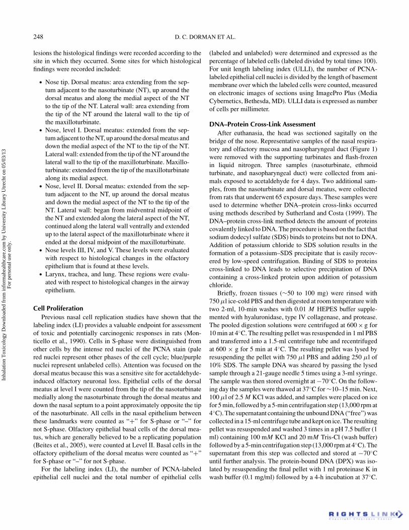
Clinical SignsThere was no mortality observed in rats during the study. Ac-
etaldehyde exposure was not associated with an effect on eitherbody weight gain (Figure 2) or terminal body weight (data not
FIG. 2. Mean (± SD) weekly body weights in male F344 ratsfollowing subchronic inhalation of acetaldehyde. Data are onlyshown for the air (0 ppm) and 1500 ppm acetaldehyde exposuregroups. No treatment-related effect on body weight gain wasobserved in animals exposed to acetaldehyde when comparedto air-exposed controls. Missing error bars are obscured by thesymbol.
Inha
latio
n T
oxic
olog
y D
ownl
oade
d fr
om in
form
ahea
lthca
re.c
om b
y U
nive
rsity
Lib
rary
Utr
echt
on
05/0
3/13
For
pers
onal
use
onl
y.
250 D. C. DORMAN ET AL.
shown). All animals exposed to ≥500 ppm acetaldehyde devel-oped rough and red-pigment-stained hair coats after exposureday 14. Although this coat change was not observed in con-trol animals at this time, all control animals developed a similarcoat appearance by the termination of the study. This changein coat appearance is therefore unlikely to be of toxicologicalsignificance.
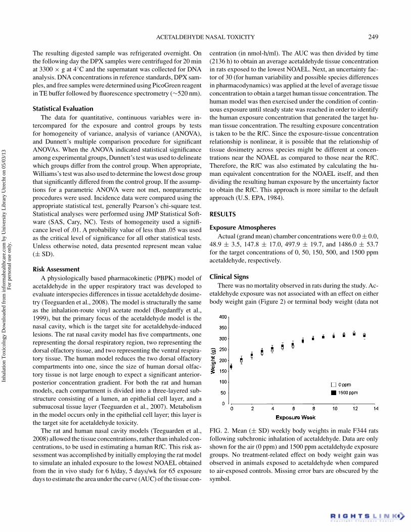
PathologyAcetaldehyde exposure was associated with inflammation,
hyperplasia, and squamous metaplasia of the respiratory epithe-lium (Tables 1–3, Figure 3). Affected sites included the dorsalmeatus at the tip of the nose; the dorsal meatus, lateral wall,and maxilloturbinate at level I; and the lateral wall at level II.Minimal inflammatory response was seen in the lateral wall atlevel I following 14 days of exposure to 1500 ppm acetalde-hyde. Minimal inflammation of the respiratory epithelium lin-ing the dorsal meatus at level I was also noted following sub-chronic exposure to 1500 ppm acetaldehyde. Minimal to mildrespiratory epithelial hyperplasia was also observed in the res-piratory epithelium lining the dorsal meatus at the tip of thenose (Table 2). This response was seen in all groups exposed to1500 ppm acetaldehyde and after 14 days of exposure to 500 ppmacetaldehyde. Animals exposed to 500 ppm acetaldehyde for
FIG. 3. Photomicrographs of the nasal epithelium from F344 rats following exposure to either air (left) or 1500 ppm acetaldehyde(right) for 30 exposure days. Insets show higher magnification (×40) of the affected mucosa. (A) Normal respiratory mucosa liningthe anterior nose at level I (×4). Epithelial hyperplasia (left inset) and inflammation (right inset) are present in the respiratoryepithelium lining the anterior nose following acetaldehyde exposure (B). (C) Normal olfactory mucosa lining the dorsal medialmeatus at level II (×2). Severe olfactory neuronal loss occurred following exposure to acetaldehyde (D).
≥14 days also developed minimal hyperplasia of the respiratoryepithelium lining the dorsal meatus at level I. The lateral wallat level II was also affected; however, hyperplasia was more in-termittent (present in rats exposed to 500 ppm acetaldehyde for14 days and in rats exposed to 1500 ppm for 4 or 9 exposuredays). Minimal to mild squamous metaplasia of the respiratoryepithelium also occurred (Table 3). Squamous metaplasia de-veloped at level I along the dorsal meatus and maxilloturbinatefollowing exposure to 1500 ppm acetaldehyde. Squamous meta-plasia of the respiratory epithelium lining the maxilloturbinateat level I was also present following ≥4 exposure days to 500ppm acetaldehyde.
Histologic evaluation of the nasal cavity also showed injuryto the olfactory epithelium. One of the earliest changes wasincreased intercellular space with disruption of the olfactory ep-ithelium (Table 4, Figure 3). An exposure-related incidence ofolfactory neuronal loss also occurred (Table 4, Figure 4). Mini-mal olfactory neuronal loss was present in the dorsal meatus (atlevel I) following ≥4 days of exposure to 150 ppm acetaldehyde.The severity and distribution of this lesion increased with moreprolonged exposure to higher exposure concentrations. Mild tomoderately severe olfactory neuronal loss developed after 14days of exposure to 1500 ppm acetaldehyde. Mild to moder-ately severe olfactory neuronal loss developed more posteriorly
Inha
latio
n T
oxic
olog
y D
ownl
oade
d fr
om in
form
ahea
lthca
re.c
om b
y U
nive
rsity
Lib
rary
Utr
echt
on
05/0
3/13
For
pers
onal
use
onl
y.
ACETALDEHYDE NASAL TOXICITY 251
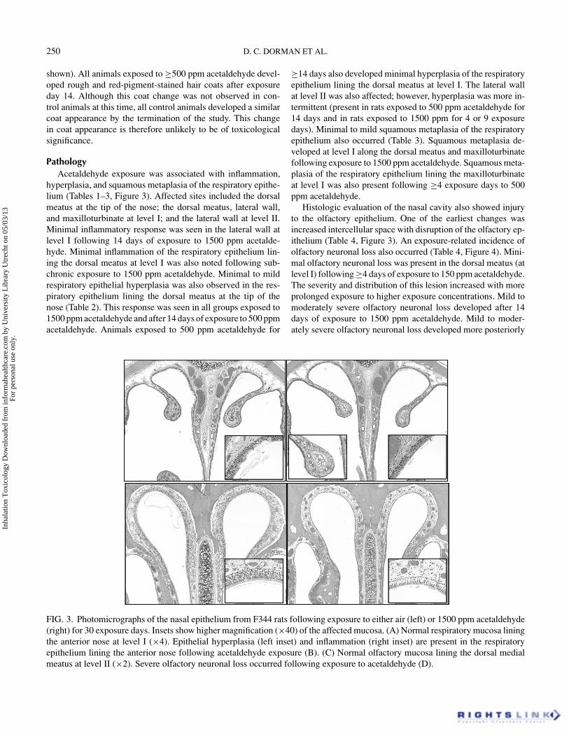
FIG. 4. Photomicrographs (×40) of the olfactory mucosa liningthe dorsal meatus at level III from F344 rats following exposureto either air (A) or 150 ppm acetaldehyde (B) for 9 days, or 1500ppm acetaldehyde (C) for 65 days. (A) Normal olfactory mucosalining the dorsal meatus at level III. Mild olfactory vacuolationoccurred following exposure to acetaldehyde (B). Olfactory ep-ithelial atrophy occurred following 65 days of exposure to 1500ppm acetaldehyde (C).
affecting the dorsal meatus and ethmoid turbinate at level III inrats subchronically exposed to 1500 ppm acetaldehyde. The no-observed-adverse-effect level (NOAEL) for olfactory neuronalloss seen in this study was 50 ppm acetaldehyde.
Minimal to mild squamous metaplasia at the base of theepiglottis was also observed in rats exposed to 1500 ppm ac-etaldehyde (Table 3). Mild laryngeal epithelial squamous meta-plasia was also evident in rats exposed to 500 ppm acetaldehydefor 65 days of exposure (Table 3). No apparent treatment-relatedeffects were observed in either the lung or trachea.
Cell ProliferationExposure to acetaldehyde was associated with minimal ef-
fects on cell proliferation in the upper respiratory tract (Figure 5).Statistically significant increases in both the LI and the ULLI ver-sus air-exposed controls were seen in the respiratory epitheliumat day 14 following exposure to 150 and 500 ppm acetaldehyde.Statistically significant increases in both LI and ULLI were seenin the olfactory epithelium at exposure days 4, 14, and 65 in ratsexposed to1500 ppm acetaldehyde.
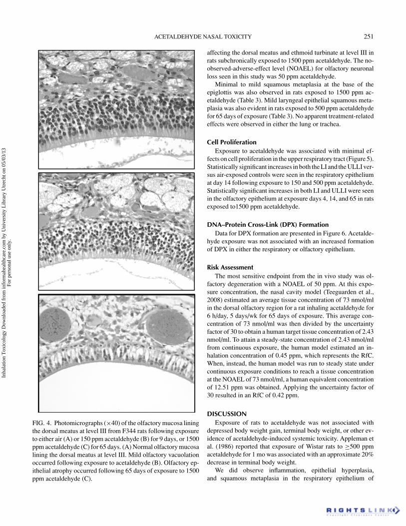
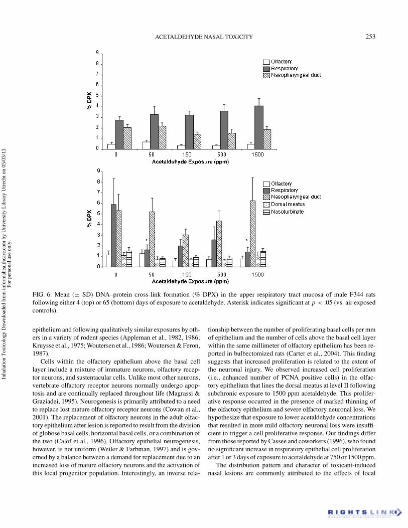
DNA–Protein Cross-Link (DPX) FormationData for DPX formation are presented in Figure 6. Acetalde-
hyde exposure was not associated with an increased formationof DPX in either the respiratory or olfactory epithelium.
Risk AssessmentThe most sensitive endpoint from the in vivo study was ol-
factory degeneration with a NOAEL of 50 ppm. At this expo-sure concentration, the nasal cavity model (Teeguarden et al.,2008) estimated an average tissue concentration of 73 nmol/mlin the dorsal olfactory region for a rat inhaling acetaldehyde for6 h/day, 5 days/wk for 65 days of exposure. This average con-centration of 73 nmol/ml was then divided by the uncertaintyfactor of 30 to obtain a human target tissue concentration of 2.43nmol/ml. To attain a steady-state concentration of 2.43 nmol/mlfrom continuous exposure, the human model estimated an in-halation concentration of 0.45 ppm, which represents the RfC.When, instead, the human model was run to steady state undercontinuous exposure conditions to reach a tissue concentrationat the NOAEL of 73 nmol/ml, a human equivalent concentrationof 12.51 ppm was obtained. Applying the uncertainty factor of30 resulted in an RfC of 0.42 ppm.
DISCUSSIONExposure of rats to acetaldehyde was not associated with
depressed body weight gain, terminal body weight, or other ev-idence of acetaldehyde-induced systemic toxicity. Appleman etal. (1986) reported that exposure of Wistar rats to ≥500 ppmacetaldehyde for 1 mo was associated with an approximate 20%decrease in terminal body weight.
We did observe inflammation, epithelial hyperplasia,and squamous metaplasia in the respiratory epithelium of
Inha
latio
n T
oxic
olog
y D
ownl
oade
d fr
om in
form
ahea
lthca
re.c
om b
y U
nive
rsity
Lib
rary
Utr
echt
on
05/0
3/13
For
pers
onal
use
onl
y.

252 D. C. DORMAN ET AL.
FIG. 5. Mean (± SD) labeling index (%) and unit length labeling index (ULLI) in male F344 rats following acetaldehyde inhalation.(A and B) Data for the respiratory epithelium. (C and D) Data for the olfactory epithelium. Asterisk indicates significant at p < .05(vs. air-exposed controls). Cell proliferation was assessed using PCNA immunohistochemistry, where cells in S-phase weredistinguished from other cells by the intense red nuclei of the PCNA stain (pale red nuclei represent other phases of the cell cycle;blue/purple nuclei represent unlabeled cells).
acetaldehyde-exposed rats. The extent of the lesion (both dis-tribution and severity) demonstrated dose, time, and site depen-dency. We found that the dorsal meatus, especially that foundat the tip and level I, was a sensitive site for respiratory ep-ithelial injury. Respiratory epithelial lesions at this site weremore prevalent than those found elsewhere in the rat nasal cav-ity and developed earlier and at lower exposure concentrationsthan elsewhere. Mild hyperplasia of the dorsal meatus respira-tory epithelium occurred after 14 days of exposure to 500 ppmand after 4 or more days of exposure to 1500 ppm acetaldehyde.Squamous metaplasia of the dorsal meatus respiratory epithe-lium was also present in rats exposed to the highest exposureconcentration. Another affected site was the maxilloturbinateat level I. Minimal squamous metaplasia of this epithelium oc-curred after 4 days of exposure to ≥500 ppm acetaldehyde. In-terestingly, squamous metaplasia of the respiratory epitheliumlining the maxilloturbinate was not associated with either in-flammation or hyperplasia. Other investigators have reportedqualitatively similar acetaldehyde-induced respiratory epithe-lial lesions (Appleman et al., 1982, 1986; Kruysse et al., 1975;Woutersen et al., 1986; Woutersen & Feron, 1987). One possi-
ble mode of action for these responses is upregulation of in-terleukin (IL)-6, IL-8, IL-1β, monocyte chemotactic protein(MCP)-1, tumor necrosis factor (TNF)-α, and other genes asso-ciated with inflammation (Gosepath et al., 2006). Transient (<24h) upregulation of these inflammatory response elements wereobserved in an in vitro system that examined acute (overnight)human nasal respiratory mucosal cell responses to gas atmo-spheres containing 50 or 500 ppm acetaldehyde.
Our study also showed that high-dose acetaldehyde expo-sure was associated with olfactory epithelial degeneration andatrophy with marked olfactory neuronal loss. Histologic eval-uation of the nasal cavity showed an increased incidence ofolfactory neuronal loss following acute to subchronic expo-sure to ≥150 ppm acetaldehyde. The severity of the olfactorydegeneration and atrophy demonstrated dose- and temporal-dependent behaviors, with minimal effects noted at 150–500 ppm acetaldehyde and moderately severe lesions seen in thehighest exposure group. Olfactory neuronal loss in the 1500 ppmacetaldehyde group became more severe as the duration of theexposure increased. Acetaldehyde-induced olfactory degener-ation has been reported at similar sites within the olfactory
Inha
latio
n T
oxic
olog
y D
ownl
oade
d fr
om in
form
ahea
lthca
re.c
om b
y U
nive
rsity
Lib
rary
Utr
echt
on
05/0
3/13
For
pers
onal
use
onl
y.
ACETALDEHYDE NASAL TOXICITY 253
FIG. 6. Mean (± SD) DNA–protein cross-link formation (% DPX) in the upper respiratory tract mucosa of male F344 ratsfollowing either 4 (top) or 65 (bottom) days of exposure to acetaldehyde. Asterisk indicates significant at p < .05 (vs. air exposedcontrols).
epithelium and following qualitatively similar exposures by oth-ers in a variety of rodent species (Appleman et al., 1982, 1986;Kruysse et al., 1975; Woutersen et al., 1986; Woutersen & Feron,1987).
Cells within the olfactory epithelium above the basal celllayer include a mixture of immature neurons, olfactory recep-tor neurons, and sustentacular cells. Unlike most other neurons,vertebrate olfactory receptor neurons normally undergo apop-tosis and are continually replaced throughout life (Magrassi &Graziadei, 1995). Neurogenesis is primarily attributed to a needto replace lost mature olfactory receptor neurons (Cowan et al.,2001). The replacement of olfactory neurons in the adult olfac-tory epithelium after lesion is reported to result from the divisionof globose basal cells, horizontal basal cells, or a combination ofthe two (Calof et al., 1996). Olfactory epithelial neurogenesis,however, is not uniform (Weiler & Farbman, 1997) and is gov-erned by a balance between a demand for replacement due to anincreased loss of mature olfactory neurons and the activation ofthis local progenitor population. Interestingly, an inverse rela-
tionship between the number of proliferating basal cells per mmof epithelium and the number of cells above the basal cell layerwithin the same millimeter of olfactory epithelium has been re-ported in bulbectomized rats (Carter et al., 2004). This findingsuggests that increased proliferation is related to the extent ofthe neuronal injury. We observed increased cell proliferation(i.e., enhanced number of PCNA positive cells) in the olfac-tory epithelium that lines the dorsal meatus at level II followingsubchronic exposure to 1500 ppm acetaldehyde. This prolifer-ative response occurred in the presence of marked thinning ofthe olfactory epithelium and severe olfactory neuronal loss. Wehypothesize that exposure to lower acetaldehyde concentrationsthat resulted in more mild olfactory neuronal loss were insuffi-cient to trigger a cell proliferative response. Our findings differfrom those reported by Cassee and coworkers (1996), who foundno significant increase in respiratory epithelial cell proliferationafter 1 or 3 days of exposure to acetaldehyde at 750 or 1500 ppm.
The distribution pattern and character of toxicant-inducednasal lesions are commonly attributed to the effects of local
Inha
latio
n T
oxic
olog
y D
ownl
oade
d fr
om in
form
ahea
lthca
re.c
om b
y U
nive
rsity
Lib
rary
Utr
echt
on
05/0
3/13
For
pers
onal
use
onl
y.
254 D. C. DORMAN ET AL.
tissue dose and the susceptibility to injury of the exposed tissue(Morgan & Monticello, 1990; Harkema et al., 2006). The extentof the olfactory lesions following acute exposure to 1500 ppmacetaldehyde was influenced by the number of exposure eventssuggesting that local tissue dose is an important determinant ofthis response. Olfactory lesions associated with 4 days of ex-posure were primarily localized to the olfactory mucosa liningthe dorsal meatus in the middorsal portion of the nose (level II).The distribution of olfactory lesions following 65 days of expo-sure, however, extended from the dorsal meatus to the ethmoidturbinate at level III. The increase in extent of the olfactory le-sions seen with repeated exposure to acetaldehyde suggests thatrepeated exposure results in an increased delivered dose to, andhence an extension of pathology into, the more caudal regions ofthe nose. Within the olfactory epithelium, the sites commonlyaffected by acetaldehyde are likely exposed to a higher localtissue dose of this aldehyde since they border high-velocity airstreams emanating from the dorsal meatus (Kimbell et al., 1997).
Formaldehyde, acrolein, benzaldehyde, and certain other re-active aldehydes are associated with in vivo or in vitro forma-tion of DNA–protein cross-links (DPX) in the rodent and non-human primate nasal cavity (Casanova-Schmitz & Heck, 1983;Casanova et al., 1991) or other mammalian tissues or cells (Kurtz& Lloyd, 2003; Kuykendall et al., 2007). Formaldehyde-inducedDPX has been used as a dose metric in quantitative cancer risk as-sessments for this chemical (Conolly et al., 2004; Schlosser et al.,2003). Costa and coworkers (1997) showed that increased lev-els of DPX can be formed in vitro following exposure of humanBurkitt’s lymphoma cells to acetaldehyde; however, enhancedDPX formation only occurred at cytotoxic concentrations. Previ-ous investigators have examined whether acetaldehyde exposureis associated with increased DPX formation in the nasal cavity.Stanek and Morris (1999) did not observe an increase in DPXformation in the respiratory epithelium of F344 rats followingexposure to 1500 ppm acetaldehyde for 6 h. Lam and coworkers(1986) reported increased DPX formation in respiratory epithe-lial cells after a single 6-h exposure to 1000 ppm acetaldehyde.Increased DPX formation occurred in the olfactory epitheliumfollowing repeated (6 h/day for 5 days), but not single, exposureto 1000 ppm acetaldehyde. Our study, however, failed to confirmthis observation.
Although acetaldehyde is chemically related to formalde-hyde, several similarities and differences in nasal response occurwith these two chemicals. Both formaldehyde and acetaldehydeinduce squamous-cell carcinomas in the rat respiratory epithe-lium (Kerns et al., 1983; Woutersen et al., 1984, 1986). Acetalde-hyde, on the other hand, is also associated with a low incidenceof olfactory epithelial adenocarcinomas in rats (Woutersen et al.,1984, 1986; Appelman et al., 1982). The finding of tumors in theolfactory epithelium is consistent with nonneoplastic responsesshowing that the rodent olfactory epithelium is uniquely sen-sitive to the effect of inhaled acetaldehyde. This difference inresponse may be explained in part by the decreased efficiency(vs. formaldehyde) of absorption of acetaldehyde by the rodent
nasal mucosa, leading to increased delivery of this chemical tothe more caudal portion of the nose lined by the olfactory epithe-lium (Morris & Blanchard, 1992). These differences in dosime-try and tissue responses should not be ignored in the noncancerand cancer risk assessments for this class of chemicals.
The current RfC for acetaldehyde in the U.S. Environmen-tal Protection Agency (EPA) Integrated Risk Information Sys-tem (IRIS) is based on an HEC derived using the default cross-species dosimetry approach for category 1 (reactive) gases (U.S.EPA 1994). This categorical default approach calculates a re-gional gas dose ratio (RGDR) between the human and the exper-imental animal on the basis of ventilation rate and extrathoracicsurface area. In the case of the acetaldehyde RfC, the RGDR wascalculated to be 0.18, resulting in an HEC that is a factor of 5.6lower than the duration-adjusted NOAEL in the rat. However,more recent developments in the area of computational fluid dy-namic (CFD) and PBPK modeling of the nose have made it pos-sible to provide chemical-specific RGDRs for many category 1gases (Morris et al., 1993; Kimbell et al., 1997; Plowchalk et al.,1997; Frederick et al., 1998). In some cases, chemical-specificanalyses for category 1 gases have resulted in RGDRs closerto unity than the default approach (Andersen et al., 1999). Inthe case of acetaldehyde the RGDR calculated with the modelwas 1.4. This result is consistent with the results obtained withother models (formaldehyde, acrylic acid), where the RGDR isnear unity and the default approach is highly conservative. Italso made little difference in this case whether the uncertaintyfactor was applied at the level of the target tissue concentrationor after the human equivalent inhaled concentration had beendetermined.
In conclusion, the most significant lesion observed in thisstudy was olfactory neuronal loss. Evaluation of the relevanceof this olfactory lesion to a human health risk assessment ofacetaldehyde exposure must take into account the significantdifferences between the breathing styles and nasal anatomy ofrodents and humans (Harkema et al., 2006). First, rats and micemay be more predisposed to nasal lesions than humans becausethey are obligatory nasal breathers. Second, a larger portion ofthe rodent nasal cavity is lined by olfactory mucosa (50%) rela-tive to humans (10%). Third, the olfactory mucosa in the caudalregion of the rat nasal cavity lines a complex network of eth-moid turbinates that greatly expands the surface area availablefor chemical uptake and slows the speed of airflow, enhancingthe deposition efficiency of inhaled toxicants. These factors in-crease the likelihood that an inhaled chemical in the rodent nosewill reach the olfactory mucosa at a local concentration sufficientto cause toxicity and, with time, a histologic lesion.
REFERENCESAmerican Conference of Governmental Industrial Hygienists. 2001.
Acetaldehyde, pp. 1–5. In Documentation of threshold limit valuesand biological exposure indices, 7th ed. Cincinnati, OH: ACGIH.
Andersen, M. E., Sarangapani, R., Frederick, C. B., and Kimbell, J. S.1999. Dosimetric adjustment factors for methylmethacrylate derived
Inha
latio
n T
oxic
olog
y D
ownl
oade
d fr
om in
form
ahea
lthca
re.c
om b
y U
nive
rsity
Lib
rary
Utr
echt
on
05/0
3/13
For
pers
onal
use
onl
y.
ACETALDEHYDE NASAL TOXICITY 255
from a steady-state analysis of a physiologically based clearance-extraction model. Inhal. Toxicol. 11:899–9236
Appelman, L. M., Woutersen, R. A., and Feron, V. J. 1982. Inhala-tion toxicity of acetaldehyde in rats. I. Acute and subacute studies.Toxicology 23(4):293–307.
Appelman, L. M., Woutersen, R. A., Feron, V. J., Hooftman, R. N., andNotten, W. R. 1986. Effect of variable versus fixed exposure levels onthe toxicity of acetaldehyde in rats. J. Appl. Toxicol. 6(5):331–336.
Beites, C. L., Kawauchi, S., Crocker, C. E., and Calof, A. L. 2005.Identification and molecular regulation of neural stem cells in theolfactory epithelium. Exp. Cell Res. 306:309–316.
Bogdanffy, M., Sarangapani, R., Andersen, M., Jarabek, A., andDellarco, V. 1999. Inhalation hazard identification and dose-responsecharacterization for vinyl acetate. Newark, DE: DuPont Hasekll Lab-oratory.
Bogdanffy, M. S., and Valentine, R. 2003. Differentiating between localcytotoxicity, mitogenesis, and genotoxicity in carcinogen risk assess-ments: The case of vinyl acetate. Toxicol. Lett. 140–141:83–98.
Calof, A. L., Hagiwara, N., Holcomb, J. D., Mumm, J. S., and Shou, J.1996. Neurogenesis and cell death in olfactory epithelium. J. Neu-robiol. 30(1):67–81.
Carter, L. A., MacDonald, J. L., and Roskams, A. J. 2004. Olfactoryhorizontal basal cells demonstrate a conserved multipotent progeni-tor phenotype. J. Neurosci. 24(25):5670–5683.
Casanova, M., Morgan, K. T., Steinhagen, W. H., Everitt, J. I., Popp,J. A., and Heck, H. D. 1991. Covalent binding of inhaled formalde-hyde to DNA in the respiratory tract of rhesus monkeys: Pharma-cokinetics, rat-to-monkey interspecies scaling, and extrapolation toman. Fundam. Appl. Toxicol. 17(2):409–428.
Casanova-Schmitz, M., and Heck, H. d’A. 1983. Effects of formalde-hyde exposure on the extractability of DNA from proteins in the ratnasal mucosa. Toxicol. Appl. Pharmacol. 70(1):121–132.
Cassee, F. R., Groten, J. P., and Feron, V. J. 1996. Changes in the nasalepithelium of rats exposed by inhalation to mixtures of formalde-hyde, acetaldehyde, and acrolein. Fundam. Appl. Toxicol. 29(2):208–218.
Conolly, R. B., Kimbell, J. S., Janszen, D., Schlosser, P. M., Kalisak,D., Preston, J., and Miller, F. J. 2004. Human respiratory tract cancerrisks of inhaled formaldehyde: dose-response predictions derivedfrom biologically-motivated computational modeling of a combinedrodent and human dataset. Toxicol. Sci. 82(1):279–296.
Costa, M., Zhitkovich, A., Harris, M., Paustenbach, D., and Gargas,M. 1997. DNA–protein cross-links produced by various chemi-cals in cultured human lymphoma cells. J. Toxicol. Environ. Health50(5):433–449.
Cowan, C. M., Thai, J., Krajewski, S., Reed, J. C., Nicholson, D. W.,Kaufmann, S. H., and Roskams, A. J. 2001. Caspases 3 and 9 send apro-apoptotic signal from synapse to cell body in olfactory receptorneurons. J. Neurosci. 21:7099–7109.
Feron, V. J. 1979. Effects of exposure to acetaldehyde in syrian hamsterssimultaneously treated with benzo(a)pyrene or diethylnitrosamine.Prog. Exp. Tumor Res. 24:162–176.
Feron, V. J., Kruysse, A., and Woutersen, R. A. 1982. Respiratory tracttumours in hamsters exposed to acetaldehyde vapour alone or simul-taneously to benzo(a)pyrene or diethylnitrosamine. Eur. J. CancerClin. Oncol. 18:13–31.
Feron, V. J., Til, H. P., de Vrijer, F., Woutersen, R. A., Cassee, F. R., andvan Bladeren, P. J. 1991. Aldehydes: Occurrence, carcinogenic po-
tential, mechanism of action and risk assessment. Mutat. Res. 259(3–4):363–385.
Frederick, C. B., Bush, M. L., Lomax, L. G., Black, K. A., Finch, L.,Kimbell, J. S., Morgan, K. T., Subramaniam, R. P., Morris, J. B., andUltman, J. S. 1998. Application of a hybrid computational fluid dy-namics and physiologically based inhalation model for interspeciesdosimetry extrapolation of acidic vapors in the upper airways. Toxi-col. Appl. Pharmacol. 152:211–231.
Gosepath, J., Brieger, J., Muttray, A., Best, S., Pourianfar, M., Jung,D., Letzel, S., and Mann, W. J. 2006. mRNA induction and cy-tokine release of inflammatory mediators during in vitro exposureof human nasal respiratory epithelia to acetaldehyde. Inhal. Toxicol.18(14):1083–1090.
Harkema, J. R., Carey, S. A., and Wagner, J. G. 2006. The nose revisited:a brief review of the comparative structure, function, and toxicologicpathology of the nasal epithelium. Toxicol. Pathol. 34(3):252–269.
Jones, A. W. 1995. Measuring and reporting the concentration of ac-etaldehyde in human breath. Alcohol Alcohol. 30(3):271–285.
Kerns, W. D., Pavkov, K. L., Donofrio, D. J., Gralla, E. J., and Swenberg,J. A. 1983. Carcinogenicity of formaldehyde in rats and mice afterlong-term inhalation exposure. Cancer Res. 43(9):4382–4392.
Kimbell, J. S., Godo, M. N., Gross, E. A., Joyner, D. R., Richardson,R. B., and Morgan, K. T. 1997. Computer simulation of inspiratoryairflow in all regions of the F344 rat nasal passages. Toxicol. Appl.Pharmacol. 145(2):388–398.
Kruysse, A., Feron, V. J., and Til, H. P.V. 1975. Repeated exposure to ac-etaldehyde vapor. Studies in Syrian golden hamsters. Arch. Environ.Health 30(9):449–452.
Kurtz, A. J., and Lloyd, R. S. 2003. 1,N 2-Deoxyguanosine adducts ofacrolein, crotonaldehyde, and trans-4-hydroxynonenal cross-link topeptides via Schiff base linkage. J. Biol. Chem. 278(8):5970–5976.
Kuykendall, J. R., Jarvi, E. J., Finley, B. L., and Paustenbach, D. J. 2007.DNA–protein cross-link formation in Burkitt lymphoma cells cul-tured with benzaldehyde and the sedative paraldehyde. Drug Chem.Toxicol. 30(1):1–16.
Lam, C. W., Casanova, M., and Heck, H. D. 1986. Decreased ex-tractability of DNA from proteins in the rat nasal mucosa after ac-etaldehyde exposure. Fundam. Appl. Toxicol. 6(3):541–550.
Magrassi, L., and Graziadei, P. P. 1995. Cell death in the olfactoryepithelium. Anat. Embryol. (Berl). 192(1):77–87.
Monticello, T. M., and Morgan, K. T. 1997. Chemically-induced nasalcarcinogenesis and epithelial cell proliferation: A brief review. Mutat.Res. 380(1–2):33–41.
Monticello, T. M., Morgan, K. T., and Hurtt, M. E. 1990. Unit lengthas the denominator for quantitation of cell proliferation in nasal ep-ithelia. Toxicol. Pathol. 18:24–31.
Morgan, K. T. 1991. Approaches to the identification and recording ofnasal lesions in toxicology studies. Toxicol. Pathol. 19(4 Pt. 1):337–351.
Morgan, K. T., and Monticello, T. M. 1990. Airflow, gas deposition, andlesion distribution in the nasal passages. Environ. Health Perspect.85:209–218.
Morris, J. B., and Blanchard, K. T. 1992. Upper respiratory tract deposi-tion of inspired acetaldehyde. Toxicol. Appl. Pharmacol. 114(1):140–146.
Morris, J. B., Hassett, D. N., and Blanchard, K. T. 1993. A physiologi-cally based pharmacokinetic model for nasal uptake and metabolismof nonreactive vapors. Toxicol. Appl. Pharmacol. 123:120–129.
Inha
latio
n T
oxic
olog
y D
ownl
oade
d fr
om in
form
ahea
lthca
re.c
om b
y U
nive
rsity
Lib
rary
Utr
echt
on
05/0
3/13
For
pers
onal
use
onl
y.

256 D. C. DORMAN ET AL.
National Research Council. 1996. Guide for the care and use of labo-ratory animals. Washington, DC: National Academy Press.
Plowchalk, D. R., Andersen, M. E., and Bogdanffy, M. S. 1997. Physi-ologically based modeling of vinyl acetate uptake, metabolism, andintracellular pH changes in the rat nasal cavity. Toxicol. Appl. Phar-macol. 142:386–400.
Schlosser, P. M., Lilly, P. D., Conolly, R. B., Janszen, D. B., and Kimbell,J. S. 2003. Benchmark dose risk assessment for formaldehyde usingairflow modeling and a single-compartment, DNA–protein cross-link dosimetry model to estimate human equivalent doses. Risk Anal.23(3):473–487.
Stanek, J. J., and Morris, J. B. 1999. The effect of inhibition of aldehydedehydrogenase on nasal uptake of inspired acetaldehyde. Toxicol Sci.49:225–231.
Sutherland, J. E., and Costa, M. 1999. Assays for DNA damage. InCurrent protocols in toxicology, eds. M. D. Maines, G. Costa, D. J.Reed, S. Sassa, and I. G. Sipes, pp. 3.5.1–3.5.36. New York: JohnWiley & Sons.
Teeguarden, J. G., Bogdanffy, M. S., Covington, T. R., Jarabek, A.M., and Tan, Y.-M. 2008. A PBPK model for evaluating the impactof aldehyde dehydrogenase polymorphisms on comparative rat andhuman nasal tissue acetaldehyde dosimetry. Inhal. Toxicol. 20:375–390.
U.S. Environmental Protection Agency.1987. Health Assessment Docu-ment for Acetaldehyde. EPA/600/8-86-015A. Research Triangle Park,NC: U.S. EPA.
U.S. Environmental Protection Agency. 1994. Methods for the deriva-tion of inhalation reference concentrations and application of inhala-tion dosimetry. EPA/600/8-90/066F. Washington, DC: U.S. EPA.
Weiler, E., and Farbman, A. I. 1997. Proliferation in the rat olfac-tory epithelium: Age-dependent changes. J. Neurosci. 17:3610–3622.
World Health Organization.1995. International Programme on Chem-ical Safety (IPCS), Safety environmental health criteria 167. Ac-etaldehyde. Geneva: WHO.
Woutersen, R. A., and Feron, V. J. 1987. Inhalation toxicity of ac-etaldehyde in rats. IV. Progression and regression of nasal le-sions after discontinuation of exposure. Toxicology 47(3):295–305.
Woutersen, R. A., Appelman, L. M., Feron, V. J., and Van der Heijden,C. A. 1984. Inhalation toxicity of acetaldehyde in rats. II. Car-cinogenicity study: Interim results after 15 months. Toxicology31(2):123–133.
Woutersen, R. A., Appelman, L. M., Van Garderen-Hoetmer, A., andFeron, V. J. 1986. Inhalation toxicity of acetaldehyde in rats. III.Carcinogenicity study. Toxicology 41(2):213–231.
Inha
latio
n T
oxic
olog
y D
ownl
oade
d fr
om in
form
ahea
lthca
re.c
om b
y U
nive
rsity
Lib
rary
Utr
echt
on
05/0
3/13
For
pers
onal
use
onl
y.
Related Documents