Derailed Regulates Development of the Drosophila Neuromuscular Junction Faith L.W. Liebl 1 , Yuping Wu 1 , David E. Featherstone 2 , Jasprina N. Noordermeer 3 , Lee Fradkin 3 , and Huey Hing 1,4 1 Cell and Developmental Biology Program, University of Illinois at Urbana-Champaign, Urbana, Illinois 61801 2 Department of Biological Sciences, University of Illinois at Chicago, Chicago, Illinois 60607 3 Laboratory of Developmental Neurobiology, Department of Molecular Cell Biology, Leiden University Medical Center, 2300 RC Leiden, The Netherlands 4 Neuroscience Program, University of Illinois at Urbana- Champaign, Urbana, Illinois 61801 Abstract Neural function is dependent upon the proper formation and development of synapses. We show here that Wnt5 regulates the growth of the Drosophila neuromuscular junction (NMJ) by signaling through the Derailed receptor. Mutations in both wnt5 and drl result in a significant reduction in the number of synaptic boutons. Cell-type specific rescue experiments show that wnt5 functions in the presynaptic motor neuron while drl likely functions in the postsynaptic muscle cell. Epistatic analyses indicate that drl acts downstream of wnt5 to promote synaptic growth. Structure–function analyses of the Drl protein indicate that normal synaptic growth requires the extracellular Wnt inhibitory factor domain and the intracellular domain, which includes an atypical kinase. Our findings reveal a novel signaling mechanism that regulates morphology of the Drosophila NMJ. Keywords Wnt; Drl; Ryk; NMJ; synapse; boutons Introduction The development and maintenance of synapses is of critical importance for nervous system development and higher order processes such as learning and memory. Once established, synapses undergo continuous remodeling in response to genetic and environmental cues. The molecular machinery that regulates synaptic growth and synaptic remodeling is not well understood. The Drosophila larval neuromuscular junction (NMJ) provides an excellent system for dissecting the molecular basis of synapse formation, growth, and remodeling. These synapses are similar to mammalian central synapses in that they are glutamatergic and remodel in response to activity (Gramates and Budnik, 1999; Collins and Diantonio, in press). The terminals of the presynaptic motor neuron arrive at its target muscle midway through embryogenesis. After the initial contact, the motor neuron growth cone collapses and forms nascent presynaptic processes, termed prevaricosities. Seventeen to 22 h after egg laying Correspondence to: H. Hing ([email protected]). This article contains supplementary material available via the Internet at http://www.mrw.interscience.wiley.com/suppmat/1932-8451/suppmat/ NIH Public Access Author Manuscript Dev Neurobiol. Author manuscript; available in PMC 2009 February 1. Published in final edited form as: Dev Neurobiol. 2008 February 1; 68(2): 152–165. NIH-PA Author Manuscript NIH-PA Author Manuscript NIH-PA Author Manuscript

Welcome message from author
This document is posted to help you gain knowledge. Please leave a comment to let me know what you think about it! Share it to your friends and learn new things together.
Transcript
Derailed Regulates Development of the DrosophilaNeuromuscular Junction
Faith L.W. Liebl1, Yuping Wu1, David E. Featherstone2, Jasprina N. Noordermeer3, LeeFradkin3, and Huey Hing1,4
1Cell and Developmental Biology Program, University of Illinois at Urbana-Champaign, Urbana, Illinois61801 2Department of Biological Sciences, University of Illinois at Chicago, Chicago, Illinois 606073Laboratory of Developmental Neurobiology, Department of Molecular Cell Biology, Leiden UniversityMedical Center, 2300 RC Leiden, The Netherlands 4Neuroscience Program, University of Illinois at Urbana-Champaign, Urbana, Illinois 61801
AbstractNeural function is dependent upon the proper formation and development of synapses. We show herethat Wnt5 regulates the growth of the Drosophila neuromuscular junction (NMJ) by signalingthrough the Derailed receptor. Mutations in both wnt5 and drl result in a significant reduction in thenumber of synaptic boutons. Cell-type specific rescue experiments show that wnt5 functions in thepresynaptic motor neuron while drl likely functions in the postsynaptic muscle cell. Epistatic analysesindicate that drl acts downstream of wnt5 to promote synaptic growth. Structure–function analysesof the Drl protein indicate that normal synaptic growth requires the extracellular Wnt inhibitory factordomain and the intracellular domain, which includes an atypical kinase. Our findings reveal a novelsignaling mechanism that regulates morphology of the Drosophila NMJ.
KeywordsWnt; Drl; Ryk; NMJ; synapse; boutons
IntroductionThe development and maintenance of synapses is of critical importance for nervous systemdevelopment and higher order processes such as learning and memory. Once established,synapses undergo continuous remodeling in response to genetic and environmental cues. Themolecular machinery that regulates synaptic growth and synaptic remodeling is not wellunderstood.
The Drosophila larval neuromuscular junction (NMJ) provides an excellent system fordissecting the molecular basis of synapse formation, growth, and remodeling. These synapsesare similar to mammalian central synapses in that they are glutamatergic and remodel inresponse to activity (Gramates and Budnik, 1999; Collins and Diantonio, in press). Theterminals of the presynaptic motor neuron arrive at its target muscle midway throughembryogenesis. After the initial contact, the motor neuron growth cone collapses and formsnascent presynaptic processes, termed prevaricosities. Seventeen to 22 h after egg laying
Correspondence to: H. Hing ([email protected]).This article contains supplementary material available via the Internet athttp://www.mrw.interscience.wiley.com/suppmat/1932-8451/suppmat/
NIH Public AccessAuthor ManuscriptDev Neurobiol. Author manuscript; available in PMC 2009 February 1.
Published in final edited form as:Dev Neurobiol. 2008 February 1; 68(2): 152–165.
NIH
-PA Author Manuscript
NIH
-PA Author Manuscript
NIH
-PA Author Manuscript
(AEL), sections of the prevaricosities begin to swell and/or constrict such that presynapticterminals (known as synaptic boutons) are formed (Johansen et al., 1989; Halpern et al.,1991; Sink and Whitington, 1991; Yoshihara et al., 1997; Rose and Chiba, 1999). During larvaldevelopment (24–120 h AEL), the presynaptic arborization grows dramatically toaccommodate the rapidly growing larval muscles. This growth includes increasing the numberof boutons, branches, and active zones per bouton (Schuster et al., 1996a,b; Zito et al., 1999).The coordinated growth of the presynaptic motor neuron and postsynaptic muscle requirestranssynaptic signaling between cells (for reviews see Koh et al., 2000; Budnik and Ruiz-Canada, 2006).
A number of molecular regulators of synaptic growth and plasticity have been identified.Mutations in ion channels proteins (Cacophony, GluRIIA, and Shaker), cytoplasmic signalingproteins (Nervous Wreck), or a cell adhesion molecule (FasII) lead to perturbations in boutonnumbers (Budnik et al., 1990; Schuster et al., 1996a,b; Rieckhof et al., 2003; Schmid et al.,2006). Of particular interest are the discoveries that the secreted signaling proteins, Wingless(Wg) and Glass Bottom Boat (Gbb), reduces the size of the NMJ with mutations in Wg,reducing the number of synaptic boutons by ∼25% (Packard et al., 2002; McCabe et al.,2003). Wg and Gbb are evolutionary conserved proteins involved in a number of developmentalprocesses including cell fate specification, axis patterning, and recently, neural development(for reviews see Logan and Nusse, 2004; Zou, 2004; Ille and Sommer, 2005; Marques,2005).
We now report a novel anterograde signaling pathway that regulates the development of theDrosophila NMJ. We show that Wnt5 regulates growth of the NMJ by signaling throughDerailed (Drl), a transmembrane receptor of the atypical receptor tyrosine kinase (RYK)family. Mutations in either wnt5 or drl result in a significant reduction in the number of synapticboutons. Epistatic analyses indicate that wnt5, which is expressed by the presynaptic motorneuron, signals through drl, which is expressed by the postsynaptic muscle, to regulate synapticgrowth. Our study is the first to demonstrate that a RYK protein regulates synapse development.
MethodsAntibodies and Immunocytochemistry
For staining and microscopy, animals were dissected and fixed for 30–60 min in either Bouin'sfixative (when GluRIIA or nc82 antibodies were used) or 4% paraformaldehyde (for all otherstaining). First and second instar larvae were dissected and fillet preparations were glued downusing Sylgard-coated coverslips. Third instar larvae were dissected and fillet preparations werepinned down in Sylgard-lined Petri dishes. All dissections were done in Drosophila standardsaline (135 mM NaCl, 5 mM KCl, 4 mM MgCl, 1.8 mM CaCl, 5 mM TES, 72 mM sucrose)with 2 mM glutamate to preserve neuronal morphology (Augustin et al., 2007) at RT. Mousemonoclonal anti-GluRIIA and nc82 (Iowa Developmental Studies Hybridoma Bank, IowaCity, IA) were used at 1:100 and 1:50, respectively. Rabbit polyclonal anti-Drl and anti-Wnt5(Fradkin et al., 2004) were used at 1:100. Fluorescently conjugated anti-HRP (JacksonImmunoresearch Labs, West Grove, PA) was used at 1:100. Goat anti-rabbit or goat anti-mousefluorescent (FITC or TRITC) secondary antibodies (Jackson Immunoresearch Labs) were usedat 1:400. The 6/7 NMJ of abdominal hemisegments A3 or A4 were used for all studies. Confocalimages were obtained using a Zeiss LSM 510 laser-scanning confocal microscope. Imageanalysis and quantification was performed using ImageJ and Adobe Photoshop software.
The anti-Drl antiserum was raised in rabbits against a GST fusion protein, including Drl aminoacids 123–222, and was affinity-purified against the same protein coupled to a column.
Liebl et al. Page 2
Dev Neurobiol. Author manuscript; available in PMC 2009 February 1.
NIH
-PA Author Manuscript
NIH
-PA Author Manuscript
NIH
-PA Author Manuscript
Molecular BiologyFull-length drl cDNA was obtained from Open Biosystems (Huntville, AL) and subcloned intothe pUAST vector to generate the UAS-drl transgene. To generate the UAS-drlΔWIF construct,nucleotides 472–1830 were amplified by PCR from drl cDNA and ligated to the first 60nucleotides of drl cDNA. This construct was then subcloned into pUAST. All constructs weresubcloned into pUAST using the EcoRI and XbaI sites. The sequence was verified for allconstructs. Fly transformation was performed by DNA microinjection of embryos usingstandard methods. To verify transcription of these constructs, total RNA was isolated usingtrizol extraction, and reverse-transcribed using cDNA-specific primers.
ElectrophysiologyAll electrophysiology was performed on the ventral body wall muscle 6. Larval recordingswere performed on third instar larvae 110–120 h AEL. Muscle 6 was voltage-clamped at −60mV. Standard two-electrode voltage clamp techniques were used, as previously described(Liebl et al., 2005). Data were acquired and analyzed using an Axopatch amplifier and pClamp9(Axon Instruments, Union City, CA). All dissections and recordings were done in standardDrosophila saline at 19°C.
Data Acquisition and StatisticsAll animals were raised at 25°C in standard fly vials with corn meal molasses medium. Thetotal number of boutons was acquired from 6/7 NMJs of hemisegments A3 or A4. The densityof nc82 labeling was quantified by counting the total number of nc82 puncta and dividing bythe total NMJ area using ImageJ (NIH) software. Statistics were performed using GraphPadPrism (v. 4.01). All statistical comparisons were made using unpaired students t-tests. Controlanimals used were WT-Berlin, elav-Gal4, 24B-Gal4, Actin-Gal4, UAS-wnt5, and UAS-drl.There was no significant difference in bouton numbers between groups (WT-Berlin = 56.36 ±3.609 boutons, n = 11; elav-Gal4 = 57.50 ± 4.586 boutons, n = 8, p = 0.8459; 24B-Gal4 =52.11 ± 3.080 boutons, n = 9, p = 0.4157; actin-Gal4 = 57.38 ± 4.496 boutons, n = 8, p = 0.8614;UAS-wnt5 = 55.11 ± 3.438 boutons, n = 9, p = 0.8263; UAS-drl = 55.20 ± 3.286 boutons, n =10, p = 0.8178). Therefore, the data were combined into one control group. Statisticalsignificance in figures is represented as follows: *p < 0.05, **p < 0.01, and ***p < 0.001. Allerror bars represent SEM.
ResultsWnt5 and Drl Are Expressed at the 6/7 NMJ
We first investigated whether Drl and Wnt5 were expressed at the NMJ usingimmunolocalization. Drl [Fig. 1(B,D)] and Wnt5 [Fig. 1(A,C)] are each localized to the areaof boutons in third instar larvae (Fig. 1). Wnt5 and Drl antibody immunoreactivity was almostundetectable in homozygous wnt5400 null mutants and dramatically reduced in drl2 nullmutants (Fig. 1, right panels), indicating that these antibodies are specific. We were unable todistinguish whether the Wnt5 and Drl immunoreactivity were pre- or postsynaptic bycolocalization experiments with the presynaptic marker CSP and the postsynaptic marker DLG(data not shown). The pre- and postsynaptic membranes of the NMJ are closely apposed(Atwood et al., 1993; Prokop et al., 1996) making it difficult to determine the cellular locationsof proteins using confocal microscopy. The Drl labeling is consistent with previous reports,which showed that Drl is expressed in subsets of CNS axons (Yoshikawa et al., 2003) andventral body wall muscles (Callahan et al., 1996). Wnt5 is expressed in posterior commissuralaxons of the CNS (Fradkin et al., 2004) but has not been localized to the NMJ. Ourimmunlocalization experiments suggest that Drl and Wnt5 may function at the NMJ.
Liebl et al. Page 3
Dev Neurobiol. Author manuscript; available in PMC 2009 February 1.
NIH
-PA Author Manuscript
NIH
-PA Author Manuscript
NIH
-PA Author Manuscript
Wnt5 Is a Positive Regulator of NMJ Size and Active Zone FormationTo investigate the role of wnt5 at the NMJ, we examined the NMJs of the null mutant,wnt5400. Homozygous wnt5400 mutants are adult-viable as previously described (Fradkin etal., 2004). Mutant larvae exhibit no visible defects in muscle patterning or muscle size (muscle6: WT = 26,490 ± 1526 μm2, n = 6; wnt5400 = 26,430 ± 1077 μm2, n = 7, p = 0.9750; muscle7: WT = 20,020 ± 764.6 μm2, n = 6; wnt5400 = 20,370 ± 1313 μm2, n = 7, p = 0.8214). Toexamine NMJ morphology, we visualized presynaptic motor neurons with HRP to label theneuronal membranes. Our analysis revealed that wnt5 mutants have significantly fewer boutonscompared with control animals [Fig. 2(A,C), control = 55.85 ± 3.059 boutons, n = 13;wnt5400 = 45.36 ± 3.459 boutons, n = 14, p = 0.0329]. This reduction correlated with a reductionin NMJ size (WT = 526.2 ± 42.66 μm2, n = 10; wnt5400 = 382.0 ± 37.80 μm2, n = 10, p =0.0209). There was no significant difference in the number of branches at the 6/7 NMJ (control= 4.938 ± 0.4784 branches, n = 16; wnt5400 = 4.688 ± 0.6240 branches, n = 16, p = 0.7527).
We examined the synaptic expression and localization of the presynaptic protein, Bruchpilot/nc82 (Brp), and the postsynaptic glutamate receptor subunit, GluRIIA, by immunolabeling.Expression of Brp, which promotes the assembly of presynaptic active zones (Kittel et al.,2006; Wagh et al., 2006), is significantly reduced in wnt5 mutants compared with controls [Fig.2(B,C); WT = 0.5585 ± 0.03462 puncta/μm2, n = 10; wnt5400 = 0.3904 ± 0.02506 puncta/μm2, n = 7, p = 0.0026]. The expression and localization of GluRIIA appeared normal inwnt5 mutant animals (data not shown, normalized fluorescence: WT = 1.000 ± 0.1006, n = 11;wnt5400 = 0.8977 ± 0.06632, n = 9, p = 0.4288). We placed wnt5400 in trans with the deficiency,Df (1) N19, which uncovers the wnt5 gene to exclude the possibility that the NMJ defects arecaused by extraneous mutations in the wnt5 mutant background. wnt5400/Df (1) N19 animalsexhibited a significant reduction in both the number of boutons and density of Brp (boutons:WT = 55.85 ± 3.059, n = 13; wnt5400/Df (1) N19 = 42.50 ± 2.141, n = 10, p = 0.003; Brp:WT = 0.5585 ± 0.0346 puncta/μm2, n = 10; wnt5400/Df (1) N19 = 0.3766 ± 0.0246 puncta/μm2, n = 9, p = 0.0006). These data indicate that wnt5 mutant animals exhibit a significantreduction in the number of boutons and suggest that the density of active zones in thepresynaptic terminals is also significantly reduced.
We performed electrophysiology to determine whether the reduction in the number of activezones affected synaptic function. Muscle 6 was voltage clamped at −60 mV and the presynapticsegmental nerve was stimulated (1 Hz, 5 V) to induce synaptic activity. The amplitude ofevoked excitatory junctional currents (EJCs) was reduced 40% in wnt5 mutant animals [Fig.2(D), WT = 207.2 ± 18.51 nA, n = 8; wnt5400 = 114.7 ± 17.94 nA, n = 6, p = 0.0044]. Similarly,the frequency of spontaneous miniature EJCs (mEJCs) was significantly reduced in wnt5mutants [Fig. 2(E), WT = 2.030 ± 0.3035 s, n = 9; wnt5400 = 1.231 ± 0.1985 s, n = 8, p = 0.0491].These data suggest that presynaptic function is impaired in wnt5 mutants and is consistent withthe reduction in the number of boutons and active zones observed immunocytochemically.Postsynaptic function, however, appeared normal, as there was no significant difference inmEJC amplitudes [Fig. 2(E), WT = 0.8677 ± 0.05136 nA, n = 9; wnt5400 = 0.8034 ± 0.04789nA, n = 8, p = 0.3781], an indicator of postsynaptic glutamate receptor function. Taken together,our results suggest that wnt5 promotes synaptic growth and the assembly of presynaptic activezones.
The Reduction in NMJ Size Is the Result of UnderdevelopmentGrowth of the NMJ involves the addition of new boutons during larval development tocorrespond with the increase in muscle size (Schuster et al., 1996a,b; Zito et al., 1999). Toascertain whether the deficit in bouton number resulted from a failure to coordinate growth ofthe presynaptic motor neuron and postsynaptic muscle, we also examined wnt5 mutant NMJsduring both first and second instar larval stages (24- and 48-h AEL, respectively). There was
Liebl et al. Page 4
Dev Neurobiol. Author manuscript; available in PMC 2009 February 1.
NIH
-PA Author Manuscript
NIH
-PA Author Manuscript
NIH
-PA Author Manuscript
no significant difference in the number of boutons between mutant and control first instar larvae[Supp. Fig. (A,B), WT = 11.13 ± 1.093 boutons, n = 8; wnt5400 = 9.429 ± 1.232 boutons, n =7, p = 0.3199]. However by second instar, there was a significant reduction in the number ofboutons in wnt5 mutants compared with controls [Fig. Supp. Fig. (C,D), WT = 19.44 ± 0.9146boutons, n = 9; wnt5400 = 15.13 ± 1.217 boutons, n = 8, p = 0.0115]. There was no significantdifference in the density of active zones during first or second instar larval stages (Fig. Supp.Fig., 1st instar: WT = 0.6530 ± 0.0534 puncta/μm2, n = 8; wnt5400 = 0.6084 ± 0.0516 puncta/μm2, n = 7, p = 0.5613; 2nd instar: WT = 0.5846 ± 0.0716 puncta/μm2, n = 7; wnt5400 = 0.4614± 0.0378 puncta/μm2, n = 7, p = 0.1383). These data suggest that mutant NMJs fail to keeppace with postsynaptic muscle development.
Restoration of wnt5 in Neurons But Not Muscle Rescues the Null PhenotypeTo identify the cell type in which wnt5 functions, we performed cell-type specific gene rescueexperiments by expressing the UAS-wnt5 transgene specifically in neurons (using the elav-Gal4 driver) or muscle (using the 24B-Gal4 driver) in the wnt5400 mutant background.Expression of wnt5 in neurons but not in muscles restored the number of boutons to slightlygreater than control levels [Fig. 3(A,B), wnt5400 = 45.36 ± 3.459 boutons, n = 14; neuronexpression = 59.64 ± 3.123 boutons, n = 11, p = 0.007, muscle expression = 50.31 ± 4.531boutons, n = 13, p = 0.3893; control; compared with control animals the p values were 0.3978and 0.3211 for neuronal and muscle expression, respectively]. Similarly, expression of wnt5in neurons but not muscles restored the NMJ size to slightly greater than control levels(wnt5400 = 382.0 ± 37.80 μm2, n = 10, neuron expression = 595.8 ± 29.72 μm2, n = 10, p =0.0003; muscle expression = 437.3 ± 28.30 μm2, n = 8, p = 0.2794; compared with controlanimals the p values were 0.6837 and 0.0415 for neuronal and muscle expression, respectively).Expression of the transgene in either neurons or muscle, however, restored the number of activezones/μm2 to near control levels [Fig. 3(A,C), wnt5400 = 0.3904 ± 0.02506, n = 7; neuronexpression = 0.5140 ± 0.03132, n = 8, p = 0.0099, muscle expression = 0.5364 ± 0.03560, n =8, p = 0.0062]. These data indicate that wnt5 functions in the presynaptic motor neuron topromote synaptic growth, but may function in either the presynaptic neuron or the postsynapticmuscle cell to regulate active zone formation. The differential requirement for wnt5 suggeststhat synaptic growth and active zone development may be governed by distinct wnt5-dependentsignaling mechanisms.
Overexpression of wnt5 in Neurons Increases Synaptic GrowthIf wnt5 promotes NMJ development, we would expect that increasing the dose of wnt5 in wild-type animals would alter NMJ development. We therefore overexpressed wnt5 by driving theUAS-wnt5 transgene with cell-type specific Gal4 drivers in wild-type animals. Driving UAS-wnt5 using either a ubiquitous (Actin-Gal4) or neuron-specific drivers (elav-Gal4 and OK6-Gal4) significantly increased the number of boutons of the 6/7 NMJ [Fig. 4(A,B), control =55.85 ± 3.059 boutons, n = 13; Act>wnt5 = 78.07 ± 5.274 boutons, n = 14, p = 0.0015;elav>wnt5 = 77.75 ± 4.254 boutons, n = 8, p = 0.0004; OK6>wnt5 = 68.50 ± 4.323 boutons,n = 12, p = 0.0239]. Driving UAS-wnt5 in postsynaptic muscle cells, using the 24B-Gal4 driver,significantly reduced the number of boutons (control = 55.85 ± 3.059 boutons, n = 13;24B>wnt5 = 42.92 ± 4.087 boutons, n = 12, p = 0.0176). Similar results were obtained whenwe overexpressed wnt5 using the GSV line, wnt51192, and used the Actin-Gal4, elav-Gal4, and24B-Gal4 drivers (data not shown). The overexpression results support our rescue experiments,which show that wnt5 functions in the presynaptic neuron to positively regulate NMJ size. Theinhibition of NMJ growth by postsynaptic wnt5 overexpression might be due to a deleteriouseffect of high levels of wnt5 on muscle physiology. Indeed, muscle defects are observed inwnt5-overexpressing muscle (R.W. Wouda, L.G.F. and J.N.N., in preparation). Ouroverexpression results support the idea that wnt5 functions in motor neurons to promote NMJgrowth.
Liebl et al. Page 5
Dev Neurobiol. Author manuscript; available in PMC 2009 February 1.
NIH
-PA Author Manuscript
NIH
-PA Author Manuscript
NIH
-PA Author Manuscript
drl Mutant Animals Exhibit a Similar Morphology as wnt5 MutantsThe observation that wnt5 acts presynaptically to regulate NMJ morphology raises the questionas to the downstream signaling mechanism involved. Our immnunolocalization experimentsshow that similar to Wnt5, the Drl receptor is also localized to the boutons of the NMJ [Fig. 1(B)]. To test whether Drl also functions in NMJ development, we examined the phenotype ofdrl2 mutant animals, which are protein nulls and homozygous-viable (Dura et al., 1995).Although the lateral ventral body wall muscles (specifically, muscles 21, 22, and 23) areabnormal and sometimes missing in mutant animals as previously described (Callahan et al.,1996), the medial ventral body wall muscles exhibited no visible defects in muscle size (muscle6: WT = 26,490 ± 1526 μm2, n = 6; drl2 = 26,300 ± 1392 μm2, n = 6, p = 0.9257; muscle 7:WT = 20,020 ± 764.6 μm2, n = 6; drl2 = 20,860 ± 1020 μm2, n = 6, p = 0.5188). Similar townt5 mutants, drl mutant animals exhibited a significant reduction in the number of boutons[Fig. 5(A,B) and data not shown, control = 55.85 ± 3.059 boutons, n = 13; drl2 = 43.17 ± 2.458boutons, n = 12, p = 0.0040] and this correlated with a reduction in the size of the 6/7 NMJ(WT = 526.2 ± 42.66 μm2, n = 10; drl2 = 382.6 ± 49.34 μm2, n = 10, p = 0.0410). Also similarto the wnt5 mutant phenotype, there was no significant difference in the number of branchesat the 6/7 NMJ (control = 4.938 ± 0.4784 branches, n = 16; drl2 = 4.167 ± 0.8424, n = 12branches, n = 12, p = 0.4888) and the synaptic growth defect became apparent only during thesecond larval instar (Supp. Fig., first instar: WT = 11.13 ± 1.093 boutons, n = 8; drl2 = 9.000± 0.9512 boutons, n = 7, p = 0.1720; second instar: WT = 19.44 ± 0.9146 boutons, n = 9;drl2 = 13.29 ± 1.539 boutons, n = 7, p = 0.0028).
We examined the synaptic expression and localization of Brp and the postsynaptic glutamatereceptor subunit, GluRIIA, by immunolabeling third instar mutant animals. Unlike the wnt5mutant, the expression and localization of both Brp and GluRIIA were not significantlydifferent than controls in drl mutants [Fig. 5(A,B), Brp: WT = 0.5585 ± 0.03462 puncta/μm2,n = 10; drl2 = 0.4739 ± 0.03352 puncta/μm2, n = 8, p = 0.1035; GluRIIA normalizedfluorescence: WT = 1.000 ± 0.1006, n = 11; drl2 = 1.155 ± 0.1140, n = 12, p = 0.3240]. Inaddition, drl mutant animals exhibited small statistically insignificant reductions in evokedamplitude and mini frequency [Fig. 5(C–F), evoked amplitude: WT = 207.2 ± 18.51 nA, n =8; drl2 = 166.4 ± 9.015 nA, n = 7, p = 0.3080; mini amplitude: WT = 0.8677 ± 0.05136 nA, n= 9; drl2 = 0.8665 ± 0.02951 nA, n = 10, p = 0.9832; mini frequency: WT = 2.030 ± 0.3035Hz, n = 9; drl2 = 1.605 ± 0.3264 Hz, n = 11, p = 0.2677]. To exclude the possibility of secondarymutations in the drl2 mutant background, we placed the drl2 mutation in trans to the deficiency,Df(2R) lio2, pigeon2, drl2. These animals also exhibited a significant reduction in the numberof NMJ boutons, with no significant difference in the density of active zones (control = 55.85± 3.059 boutons, n = 13; drl2/Df(2R) lio2, pigeon2, drl2 = 43.78 ± 4.390 boutons, n = 9, p =0.030; Brp: WT = 0.5585 ± 0.03462 puncta/μm2, n = 10; drl2/Df(2R) lio2, pigeon2, drl2 = 0.5240± 0.0336 puncta/μm2, n = 9, p = 0.486). Taken together, these results indicate that drl regulatespresynaptic neuronal morphology, but is not likely to regulate the formation of active zones.
drl Likely Functions in Postsynaptic MuscleTo delineate the cell type in which drl functions, we sought to restore drl functions in specificcell types in the drl2 mutant background. Neither the presence of the UAS-drl transgene alonenor the expression of the transgene under the control of the pan-glial driver, Repo-Gal4, rescuedthe growth phenotype [Fig. 6(A,C), drl2 = 43.17 ± 2.458 boutons, n = 12, drl2; UAS-drl = 40.80± 2.476 boutons, n = 10, p = 0.509; glia expression = 41.67 ± 3.322 boutons, n =12, p = 0.7201].Expression of the transgene in muscle strongly rescued the drl2 mutant phenotype [Fig. 6(A,C),drl2 = 43.17 ± 2.458 boutons, n = 12, muscle expression = 56.83 ± 3.303 boutons, n = 12, p =0.0031] as the number of boutons is restored to slightly greater than control levels. Interestingly,drl expression in neurons produced an increase in the number of NMJ branches [Fig. 6(C),control = 5.846 ± 0.5644 branches, n = 13; neuron expression = 8.500 ± 0.7638 branches, n =
Liebl et al. Page 6
Dev Neurobiol. Author manuscript; available in PMC 2009 February 1.
NIH
-PA Author Manuscript
NIH
-PA Author Manuscript
NIH
-PA Author Manuscript
12, p = 0.0096], a phenotype not seen in the wild type. The increased arborization produces anincrease in bouton number [Fig. 6(A,C), drl2 = 43.17 ± 2.458 boutons, n = 12, neuronexpression = 59.55 ± 4.288 boutons, n = 11, p = 0.0028], leading to ambiguity about whetherdrl functions in the presynaptic motor neuron or postsynaptic muscle cell. Examination of NMJsize produced similar results (drl2 = 382.6 ± 49.34 μm2, n = 10, neuron expression = 564.8 ±49.61 μm2, n = 9, p = 0.019; muscle expression = 559.6 ± 37.99 μm2, n = 9, p = 0.012, glialexpression = 465.3 ± 36.04 μm2, n = 9, p = 0.202).
We also overexpressed drl in an otherwise wild-type background. Overexpressing drl usingvarious Gal4 drivers did not significantly affect NMJ morphology [Fig. 6(B), control = 55.85± 3.059, n = 13; Act>drl = 60.50 ± 6.425, n = 12, p = 0.5091; elav>drl = 64.93 ± 5.195, n =14, p = 0.1521; 24B>drl = 52.50 ± 5.704, n = 12, p = 0.6023]. We conclude from theaforementioned rescue experiments that drl likely functions in the postsynaptic muscle toregulate NMJ growth, but may also function in the presynaptic motor neuron for otherdevelopmental processes.
The NMJ Overgrowth Produced by Overexpression of wnt5 in Neurons Is Suppressed in thedrl Mutant Background
The reduction in bouton numbers in both wnt5 and drl mutants raised the possibility that thetwo genes may function in a signaling pathway to regulate synaptic growth. To test thispossibility, we constructed animals bearing simultaneously the wnt5 and drl mutations. TheNMJ morphology of wnt5400; drl2 double mutants is not statistically different than that of eachof the single mutants [Fig. 7(A,B), drl2 = 43.17 ± 2.458 boutons, n = 12; wnt5400 = 45.36 ±3.459 boutons, n = 14; wnt5400; drl2 = 49.55 ± 4.345, n = 11, p = 0.2056], supporting ourhypothesis that wnt5 and drl act in the same signaling pathway to regulate synaptic growth.
We postulate that wnt5 acts upstream of drl. This hypothesis makes the prediction thatmutations in the drl gene would block the effects of wnt5. To test this possibility, weoverexpressed the UAS-wnt5 transgene in the drl mutant background. Neuronal overexpressionof wnt5 in the wild-type background leads to overgrowth of the NMJ with a significant increasein the number of boutons [Figs. 4(A,B) and 7(C,D)]. Removal of a single copy of the drl gene(drl2/CyO) in the wnt5 overexpressing animal partially, but significantly suppressed the NMJovergrowth phenotype [Fig. 7(C,D) elav>wnt5 = 77.75 ± 4.254 boutons, n = 8; drl2/Cyo;elav>wnt5 = 60.33 ± 4.311 boutons, n = 9, p = 0.0119]. Removal of both copies of the drl genein the wnt5 overexpressing animal results in a phenotype indistinguishable from that of thedrl2 mutant [Fig. 7(C,D), drl2 = 43.17 ± 2.458 boutons, n = 12; drl2; elav>wnt5 = 47.55 ±2.855 boutons, n = 11, p = 0.2560]. These data indicate that the effect of neuronaloverexpression of wnt5 is highly sensitive to the dose of the drl gene and that drl is epistaticto wnt5. This result supports the idea that wnt5 functions through drl to promote the growth ofthe NMJ.
The Intracellular and WIF Domains of Drl Are Required for Normal Synaptic GrowthThe observation that Drl acts downstream of Wnt5 during NMJ development raises intriguingquestions about the mechanisms by which Drl transduces the Wnt5 signal. The Drl protein isa member of the evolutionarily conserved RYK receptor tyrosine kinase-related family.Members of this family possess a WIF motif in the extracellular domain and an intracellulardomain that includes an “atypical” kinase. Our ability to rescue the drl mutant phenotype usingtransgenes afforded us the opportunity to probe the requirement of these domains of the Drlprotein in synapse development. We expressed mutant drl constructs in the muscle of drl2mutant animals to assess their ability to rescue the drl2 mutant phenotype. Deletion of the WIFmotif completely abolished Drl's capacity to rescue the mutant phenotype, indicating that theWIF motif is necessary for Drl function in NMJ development (Fig. 8, drl2 = 43.17 ± 2.458
Liebl et al. Page 7
Dev Neurobiol. Author manuscript; available in PMC 2009 February 1.
NIH
-PA Author Manuscript
NIH
-PA Author Manuscript
NIH
-PA Author Manuscript
boutons, n = 12; drl2, 24B>drlΔWIF = 49.85 ± 3.123 boutons, n = 13, p = 0.1102; p-value whencompared with control = 0.280). Expression of the truncated DrlΔcyt protein containing onlythe extracellular and transmembrane domains failed to rescue the drl2 mutant phenotype,indicating that the cytoplasmic domain is necessary for Drl function in NMJ development (Fig.8, drl2 = 43.17 ± 2.458 boutons, n = 12, drl2, 24B>drlΔcyt = 49.09 ± 2.246 boutons, n = 11,p = 0.0915; p value when compared to control = 0.171). Examination of NMJ size yieldedsimilar results (drl2 = 382.6 ± 49.34 μm2, n = 10, 24B>drlΔWIF = 361.6 ± 44.12 μm2, n = 10,p = 0.906; 24B>drlΔcyt = 361.9 ± 39.10 μm2, n = 11, p = 0.889). We conclude that thecytoplasmic and WIF domains of drl are required for normal growth of the presynaptic motorneuron.
DiscussionAfter its establishment, the Drosophila NMJ exhibits as much as a 10-fold increase in growthbetween the first and third instar larval stages by adding additional boutons and branches tocorrespond with the increase in surface area of the postsynaptic muscle (Schuster et al.,1996a,1996b; Zito et al., 1999). This growth requires coordination between the presynapticmotoneuron and postsynaptic muscle. This process is likely mediated by a number of signalingmechanisms. Here we provide evidence that Wnt5 positively regulates NMJ growth bysignaling through the Drl RYK receptor. This is the first study to demonstrate that a memberof the RYK family plays a role in synapse formation.
Wnt5 Is an Anterograde Signal That Regulates NMJ DevelopmentSeveral pieces of evidence support our conclusion that the wnt5 gene acts as a positive regulatorof NMJ growth and synaptic transmission. Mutation of wnt5 leads to stunted NMJs withdecreased number of boutons. In addition to the structural defects, the NMJ shows reducednumber of active zones. Electrophysiological recordings showed that the amplitudes of evokedreleases and frequency of spontaneous releases are both significantly decreased. Examinationof the mutant NMJs early in development showed that they are normal, indicating that wnt5 isneeded for NMJ growth to maintain pace with muscle growth. Cell-type specific cDNAexpression showed that wnt5 functions in the motoneurons. In contrast to the reduced boutonnumbers in the loss-of-function wnt5 mutant, animals overexpressing wnt5 in the motoneuronsexhibit significant overgrowth of the NMJ, indicating that NMJ development is highly sensitiveto the dose of wnt5. Our results therefore indicate that Wnt5 is an anterograde signal thatpromotes the development of the NMJ.
Our finding that Wnt5 apparently acts as a transsynaptic signal adds it to a growing list of Wntproteins and Wnt signaling pathway components that play key roles in synaptogenesis (forreview see Salinas, 2005). Most notably, Packard et al. (2002) observed that at theDrosophila NMJ, Wg is expressed by the presynaptic motor neuron where it governs thedevelopment of both pre- and postsynaptic structures (Packard et al., 2002; Mathew et al.,2005). In the mouse, Wnt3 and Wnt7a are necessary for the cessation of axon growth and theremodeling of the axons into presynaptic terminals, characterized by growth cone spreadingand clustering of the synapsin I protein (Hall et al., 2000; Krylova et al., 2002). Dissection ofthe mechanisms by which these Wnt proteins regulate synapse development showed that theyact through the Frizzled receptor (Packard et al., 2002; Mathew et al., 2005). However, ourobservations indicate that Wnt5 functions through the Drl RYK receptor to govern NMJgrowth.
Drl Functions downstream of Wnt5 to Regulate Synapse GrowthSeveral lines of evidence indicate drl is necessary for the growth of the Drosophila NMJ.Mutation of the drl gene results in a significant reduction in NMJ size, a phenotype that
Liebl et al. Page 8
Dev Neurobiol. Author manuscript; available in PMC 2009 February 1.
NIH
-PA Author Manuscript
NIH
-PA Author Manuscript
NIH
-PA Author Manuscript
resembles that of the wnt5 mutant. As with the wnt5 mutant, the NMJs of the drl mutant appearnormal during the first instar larval stage, indicating that drl regulates NMJ growth. Cell-typespecific gene rescue showed that drl likely functions in the postsynaptic muscle cell.Interestingly, despite the significant reduction in NMJ size in the drl mutant, this synapsedisplayed wild-type electrophysiology, and thus differs from wnt5 mutation in this respect.The density of active zones, the amplitudes of both evoked and spontaneous releases, and thefrequency of miniature releases are normal compared with wild-type animals, suggesting thatdrl does not regulate the formation of active zones. Instead, drl appears to function specificallyto regulate presynaptic growth. This observation raised the possibility that drl and wnt5 mayfunction in the same pathway to regulate NMJ growth.
That drl functions downstream to mediate wnt5 signaling is reinforced by the followingobservations. First, the wnt5; drl double mutants exhibit reduced NMJ sizes, a phenotype thatresembles both the single wnt5 and drl mutants. Second, the overgrowth phenotype producedby neuronal overexpression of wnt5 is completely suppressed in the drl mutant background,indicating that drl acts downstream of wnt5. We conclude that wnt5 signals through the drlreceptor to regulate NMJ growth. Collectively, our results suggest a novel anterogradesignaling pathway [Fig. 8(C)] where Wnt5, expressed in the presynaptic cell, acts through Drl,likely in the postsynaptic cell, to promote synaptic growth.
The Drl Cytoplasmic and WIF Domains Are Necessary for NMJ GrowthThe capacity of the Drl RYK to mediate Wnt5 function in NMJ growth raises the question ofthe mechanism by which Drl transduces the Wnt5 signal. The Drl protein contains anextracellular WIF motif with homology to the Wnt-binding domain of the Wnt inhibitoryfactor-1 (WIF-1) protein (Yoshikawa et al., 2003) and an intracellular atypical kinase domain(Yoshikawa et al., 2001). To assess the requirements of these domains during NMJdevelopment, we engineered Drl proteins lacking these domains and expressed them in musclecells. Deletion of the extracellular WIF domain completely abolished NMJ function in NMJdevelopment. Since the extracellular domain of Drl has been shown to bind to Wnt5(Yoshikawa et al., 2003), our result suggests that Wnt5 may regulate growth of the NMJ bybinding to the WIF motif of Drl. Deletion of the cytoplasmic domain of Drl also abolished itsfunction in NMJ development. This result is consistent with observations in the embryonicventral nerve cord, where the Drl cytoplasmic domain is needed for commissural axons tonavigate properly in response to the Wnt5 signal (Yoshikawa et al., 2001). This domain islikely to play a role in signal transduction, although the mechanism by which it functions isunknown. Recently, the cytoplasmic tail of the mammalian RYK protein was shown to bindto the Dishevelled protein, raising the possibility that RYK proteins may act through thecanonical pathway (Lu et al., 2004).
Wnt5 Signals Via Different Mechanisms to Regulate Structure and FunctionWe found that the Wnt5-Drl signaling pathway mediates a signal that passes from motorneurons to muscle cells. This raises the question as to how the pathway regulates NMJdevelopment. One intriguing possibility is that the signaling pathway stimulates the productionof a signal in the muscle cell, which acts in a retrograde fashion to influence synaptic growth.One known retrograde signal is the BMP signaling pathway (for reviews see Marques, 2005).Intriguingly, mutations in components of this pathway (Gbb, Saxophone, Thick Veins, Wishfulthinking, and Mad) lead to a reduction in NMJ size with fewer boutons (Aberle et al., 2002;Marques et al., 2002; McCabe et al., 2003, 2004; Rawson et al., 2003), a phenotype thatresembles that of the wnt5 and drl mutants. We are currently exploring the possibility that BMPsignaling is downstream of drl.
Liebl et al. Page 9
Dev Neurobiol. Author manuscript; available in PMC 2009 February 1.
NIH
-PA Author Manuscript
NIH
-PA Author Manuscript
NIH
-PA Author Manuscript
Besides regulating NMJ structure, Wnt5 also regulates the function of the synapse. In this role,however, it does not appear to act via the Drl receptor. Wnts have been previously implicatedin the development of presynaptic active zones (Hall et al., 2000; Krylova et al., 2002; Packardet al., 2002; Ahmad-Annuar et al., 2006). We show that in the absence of wnt5, mutant NMJshave fewer active zones and impaired presynaptic function. Both the amplitude of evokedcurrents and the frequency of spontaneous currents are significantly reduced. It is unclear howWnt5, which is produced by the presynaptic motor neuron, acts on the same cell that producesit. However, our data demonstrate that Wnt5 does not signal through Drl to regulate activezone formation. Drl mutant animals are functionally normal with no detectable loss of Brplabeling. Further studies will hopefully identify the mechanisms by which Wnt5 and Drl governNMJ structure and function.
Acknowledgements
We thank the Iowa Developmental Hybridoma Bank for antibodies and the Bloomington Stock Center for fly stocks.
Contract grant sponsor: NIH Institutional Training Grant (University of Illinois); contract grant number: HD07333.
Contract grant sponsor: NIH Individual NRSA (NIH/NIDCD); contract grant number: 1 F32 DC08443-01.
Contract grant sponsor: NIH-NINDS Grant; contract grant number: R01NS045628.
Contract grant sponsor: Pionier Grant from the NOW.
Contract grant sponsor: NIH/NIDCD; contract grant number: DC5408-01.
Contract grant sponsor: Roy J. Carver Charitable Trust; contract grant number: 03-27.
ReferencesAberle H, Haghighi AP, Fetter RD, McCabe BD, Magalhaes TR, Goodman CS. Wishful thinking encodes
a BMP type II receptor that regulates synaptic growth in Drosophila. Neuron 2002;33:545–558.[PubMed: 11856529]
Ahmad-Annuar A, Ciani L, Simeonidis I, Herreros J, Fredj NB, Rosso SB, Hall A, et al. Signaling acrossthe synapse: A role for Wnt and dishevelled in presynaptic assembly and neurotransmitter release. JCell Biol 2006;174:127–139. [PubMed: 16818724]
Atwood HL, Govind CK, Wu CF. Differential ultrastructure of synaptic terminals on ventral longitudinalabdominal muscles in Drosophila larvae. J Neurobiol 1993;24:1008–1024. [PubMed: 8409966]
Augustin H, Grosjean Y, Chen K, Sheng Q, Featherstone DE. Nonvesicular release of glutamate by glialxCT transporters suppresses glutamate receptor clustering in vivo. J Neurosci 2007;27:111–123.[PubMed: 17202478]
Budnik V, Ruiz-Canada. The fly neuromuscular junction: Structure and Function. Int Rev Neurobiol2006;75
Budnik V, Zhong Y, Wu CF. Morphological plasticity of motor axons in Drosophila mutants with alteredexcitability. J Neurosci 1990;10:3754–3768. [PubMed: 1700086]
Callahan CA, Bonkovsky JL, Scully AL, Thomas JB. Derailed is required for muscle attachment siteselection in Drosophila. Development 1996;122:2761–2767. [PubMed: 8787750]
Collins CA, Diantonio A. Synaptic development: Insights from Drosophila. Curr Opin Neurobiol 17:35–42. [PubMed: 17229568]
Dura JM, Taillebourg E, Preat T. The Drosophila learning and memory gene linotte encodes a putativereceptor tyrosine kinase homologous to the human RYK gene product. FEBS Lett 1995;370:250–254.[PubMed: 7656987]
Fradkin LG, van Schie M, Wouda RR, de Jong A, Kamphorst JT, Radjkoemar-Bansraj M, NoordermeerJN. The Drosophila Wnt5 protein mediates selective axon fasciculation in the embryonic centralnervous system. Dev Biol 2004;272:362–375. [PubMed: 15282154]
Liebl et al. Page 10
Dev Neurobiol. Author manuscript; available in PMC 2009 February 1.
NIH
-PA Author Manuscript
NIH
-PA Author Manuscript
NIH
-PA Author Manuscript

Gramates LS, Budnik V. Assembly and maturation of the Drosophila larval neuromuscular junction. IntRev Neurobiol 1999;43:93–117. [PubMed: 10218156]
Hall AC, Lucas FR, Salinas PC. Axonal remodeling and synaptic differentiation in the cerebellum isregulated by WNT-7a signaling. Cell 2000;100:525–535. [PubMed: 10721990]
Halpern ME, Chiba A, Johansen J, Keshishian H. Growth cone behavior underlying the development ofstereotypic synaptic connections in Drosophila embryos. J Neurosci 1991;11:3227–3238. [PubMed:1658247]
Ille F, Sommer L. Wnt signaling: Multiple functions in neural development. Cell Mol Life Sci2005;62:1100–1108. [PubMed: 15928805]
Johansen J, Halpern ME, Johansen KM, Keshishian H. Stereotypic morphology of glutamatergicsynapses on identified muscle cells of Drosophila larvae. J Neurosci 1989;9:710–725. [PubMed:2563766]
Kittel RJ, Wichmann C, Rasse TM, Fouquet W, Schmidt M, Schmid A, Wagh DA, et al. Bruchpilotpromotes active zone assembly, Ca2+ channel clustering, and vesicle release. Science2006;312:1051–1054. [PubMed: 16614170]
Koh YH, Gramates LS, Budnik V. Drosophila larval neuromuscular junction: Molecular componentsand mechanisms underlying synaptic plasticity. Microsc Res Tech 2000;49:14–25. [PubMed:10757875]
Krylova O, Herreros J, Cleverley KE, Ehler E, Henriquez JP, Hughes SM, Salinas PC. WNT-3, expressedby motoneurons, regulates terminal arborization of neurotrophin-3-responsive spinal sensoryneurons. Neuron 2002;35:1043–1056. [PubMed: 12354395]
Liebl FL, Chen K, Karr J, Sheng Q, Featherstone DE. Increased synaptic microtubules and altered synapsedevelopment in Drosophila sec8 mutants. BMC Biol 2005;3:27. [PubMed: 16351720]
Logan CY, Nusse R. The Wnt signaling pathway in development and disease. Annu Rev Cell Dev Biol2004;20:781–810. [PubMed: 15473860]
Lu W, Yamamoto V, Ortega B, Baltimore D. Mammalian Ryk is a Wnt coreceptor required for stimulationof neurite outgrowth. Cell 2004;119:97–108. [PubMed: 15454084]
Marques G. Morphogens and synaptogenesis in Drosophila. J Neurobiol 2005;64:417–434. [PubMed:16041756]
Marques G, Bao H, Haerry TE, Shimell MJ, Duchek P, Zhang B, O'Connor MB. The Drosophila BMPtype II receptor wishful thinking regulates neuromuscular synapse morphology and function. Neuron2002;33:529–543. [PubMed: 11856528]
Mathew D, Ataman B, Chen J, Zhang Y, Cumberledge S, Budnik V. Wingless signaling at synapses isthrough cleavage and nuclear import of receptor DFrizzled2. Science 2005;310:1344–1347.[PubMed: 16311339]
McCabe BD, Hom S, Aberle H, Fetter RD, Marques G, Haerry TE, Wan H, et al. Highwire regulatespresynaptic BMP signaling essential for synaptic growth. Neuron 2004;41:891–905. [PubMed:15046722]
McCabe BD, Marques G, Haghighi AP, Fetter RD, Crotty ML, Haerry TE, Goodman CS, et al. The BMPhomolog Gbb provides a retrograde signal that regulates synaptic growth at the Drosophilaneuromuscular junction. Neuron 2003;39:241–254. [PubMed: 12873382]
Packard M, Koo ES, Gorczyca M, Sharpe J, Cumberledge S, Budnik V. The Drosophila Wnt, wingless,provides an essential signal for pre- and postsynaptic differentiation. Cell 2002;111:319–330.[PubMed: 12419243]
Prokop A, Landgraf M, Rushton E, Broadie K, Bate M. Presynaptic development at the Drosophilaneuromuscular junction: Assembly and localization of presynaptic active zones. Neuron1996;17:617–626. [PubMed: 8893020]
Rawson JM, Lee M, Kennedy EL, Selleck SB. Drosophila neuromuscular synapse assembly and functionrequire the TGF-β type I receptor saxophone and the transcription factor Mad. J Neurobiol2003;55:134–150. [PubMed: 12672013]
Rieckhof GE, Yoshihara M, Guan Z, Littleton JT. Presynaptic N-type calcium channels regulate synapticgrowth. J Biol Chem 2003;278:41099–41108. [PubMed: 12896973]
Liebl et al. Page 11
Dev Neurobiol. Author manuscript; available in PMC 2009 February 1.
NIH
-PA Author Manuscript
NIH
-PA Author Manuscript
NIH
-PA Author Manuscript
Rose D, Chiba A. A single growth cone is capable of integrating simultaneously presented andfunctionally distinct molecular cues during target recognition. J Neurosci 1999;19:4899–4906.[PubMed: 10366624]
Salinas PC. Retrograde signalling at the synapse: A role for Wnt proteins. Biochem Soc Trans2005;33:1295–1298. [PubMed: 16246102]
Schmid A, Qin G, Wichmann C, Kittel RJ, Mertel S, Fouquet W, Schmidt M, et al. Non-NMDA-typeglutamate receptors are essential for maturation but not for initial assembly of synapses at Drosophilaneuromuscular junctions. J Neurosci 2006;26:11267–11277. [PubMed: 17079654]
Schuster CM, Davis GW, Fetter RD, Goodman CS. Genetic dissection of structural and functionalcomponents of synaptic plasticity. I. Fasciclin II controls synaptic stabilization and growth. Neuron1996a;17:641–654. [PubMed: 8893022]
Schuster CM, Davis GW, Fetter RD, Goodman CS. Genetic dissection of structural and functionalcomponents of synaptic plasticity. II. Fasciclin II controls presynaptic structural plasticity. Neuron1996b;17:655–667. [PubMed: 8893023]
Sink H, Whitington PM. Pathfinding in the central nervous system and periphery by identified embryonicDrosophila motor axons. Development 1991;112:307–316. [PubMed: 1769336]
Wagh DA, Rasse TM, Asan E, Hofbauer A, Schwenkert I, Durrbeck H, Buchner S, et al. Bruchpilot, aprotein with homology to ELKS/CAST, is required for structural integrity and function of synapticactive zones in Drosophila. Neuron 2006;49:833–844. [PubMed: 16543132]
Yoshihara M, Rheuben MB, Kidokoro Y. Transition from growth cone to functional motor nerve terminalin Drosophila embryos. J Neurosci 1997;17:8408–8426. [PubMed: 9334414]
Yoshikawa S, Bonkowsky JL, Kokel M, Shyn S, Thomas JB. The Derailed guidance receptor does notrequire kinase activity in vivo. J Neurosci 2001;21:RC119. [PubMed: 11150355]
Yoshikawa S, McKinnon RD, Kokel M, Thomas JB. Wnt-mediated axon guidance via the DrosophilaDerailed receptor. Nature 2003;422:583–588. [PubMed: 12660735]
Zito K, Parnas D, Fetter RD, Isacoff EY, Goodman CS. Watching a synapse grow: Noninvasive confocalimaging of synaptic growth in Drosophila. Neuron 1999;22:719–729. [PubMed: 10230792]
Zou Y. Wnt signaling in axon guidance. Trends Neurosci 2004;27:528–532. [PubMed: 15331234]
Liebl et al. Page 12
Dev Neurobiol. Author manuscript; available in PMC 2009 February 1.
NIH
-PA Author Manuscript
NIH
-PA Author Manuscript
NIH
-PA Author Manuscript
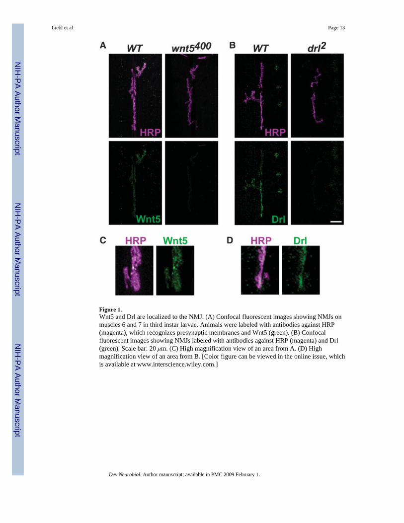
Figure 1.Wnt5 and Drl are localized to the NMJ. (A) Confocal fluorescent images showing NMJs onmuscles 6 and 7 in third instar larvae. Animals were labeled with antibodies against HRP(magenta), which recognizes presynaptic membranes and Wnt5 (green). (B) Confocalfluorescent images showing NMJs labeled with antibodies against HRP (magenta) and Drl(green). Scale bar: 20 μm. (C) High magnification view of an area from A. (D) Highmagnification view of an area from B. [Color figure can be viewed in the online issue, whichis available at www.interscience.wiley.com.]
Liebl et al. Page 13
Dev Neurobiol. Author manuscript; available in PMC 2009 February 1.
NIH
-PA Author Manuscript
NIH
-PA Author Manuscript
NIH
-PA Author Manuscript
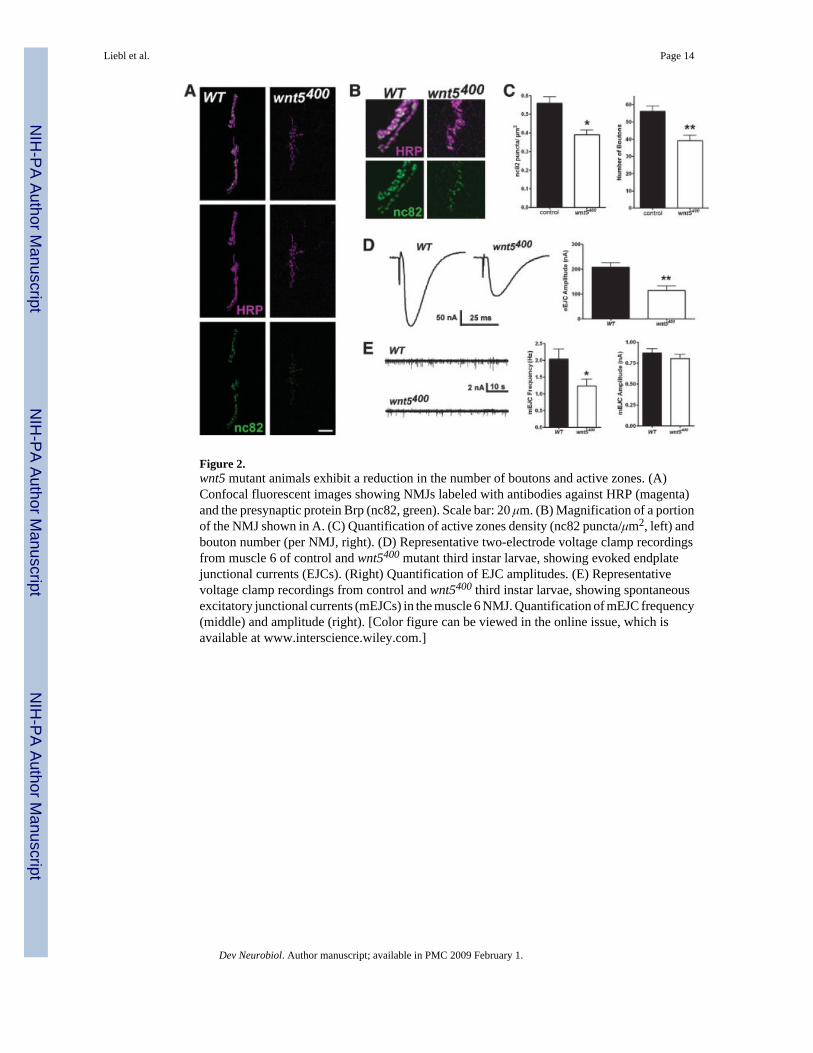
Figure 2.wnt5 mutant animals exhibit a reduction in the number of boutons and active zones. (A)Confocal fluorescent images showing NMJs labeled with antibodies against HRP (magenta)and the presynaptic protein Brp (nc82, green). Scale bar: 20 μm. (B) Magnification of a portionof the NMJ shown in A. (C) Quantification of active zones density (nc82 puncta/μm2, left) andbouton number (per NMJ, right). (D) Representative two-electrode voltage clamp recordingsfrom muscle 6 of control and wnt5400 mutant third instar larvae, showing evoked endplatejunctional currents (EJCs). (Right) Quantification of EJC amplitudes. (E) Representativevoltage clamp recordings from control and wnt5400 third instar larvae, showing spontaneousexcitatory junctional currents (mEJCs) in the muscle 6 NMJ. Quantification of mEJC frequency(middle) and amplitude (right). [Color figure can be viewed in the online issue, which isavailable at www.interscience.wiley.com.]
Liebl et al. Page 14
Dev Neurobiol. Author manuscript; available in PMC 2009 February 1.
NIH
-PA Author Manuscript
NIH
-PA Author Manuscript
NIH
-PA Author Manuscript
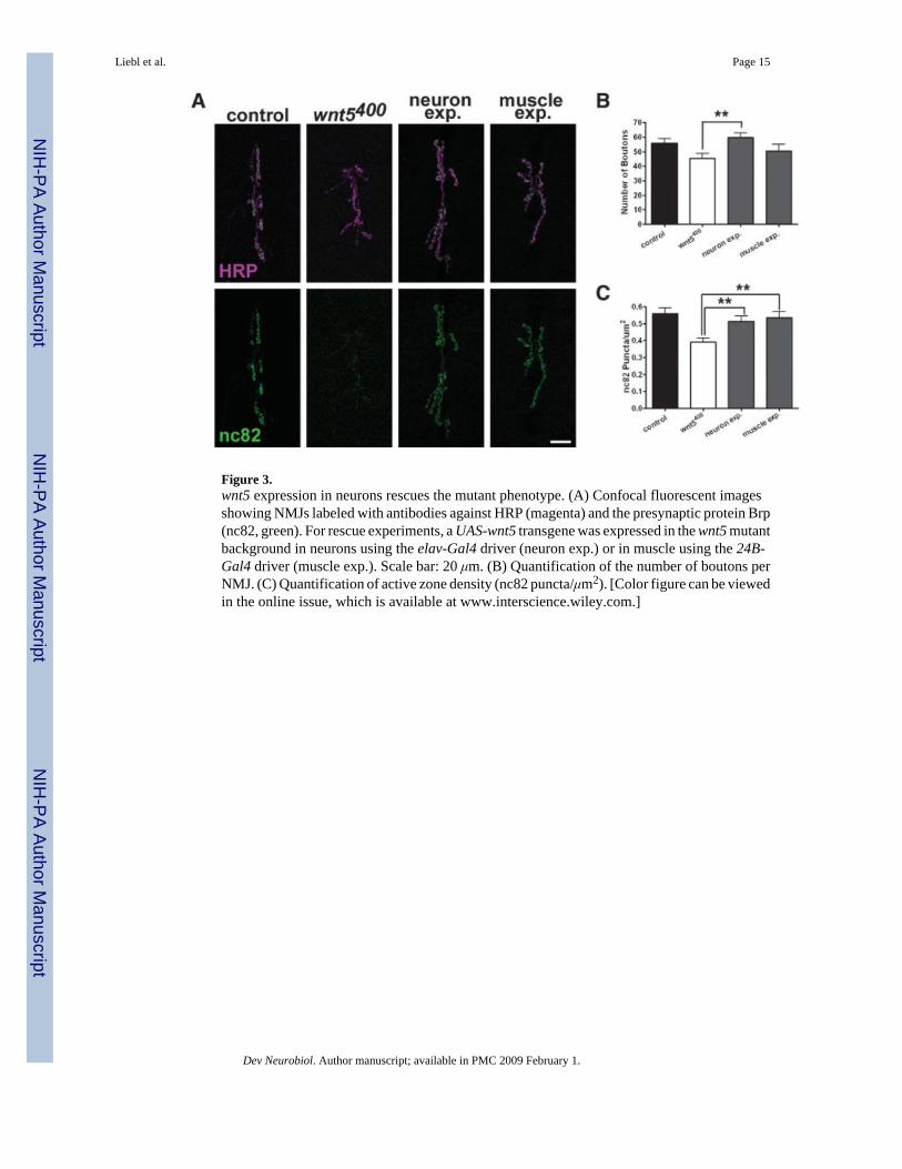
Figure 3.wnt5 expression in neurons rescues the mutant phenotype. (A) Confocal fluorescent imagesshowing NMJs labeled with antibodies against HRP (magenta) and the presynaptic protein Brp(nc82, green). For rescue experiments, a UAS-wnt5 transgene was expressed in the wnt5 mutantbackground in neurons using the elav-Gal4 driver (neuron exp.) or in muscle using the 24B-Gal4 driver (muscle exp.). Scale bar: 20 μm. (B) Quantification of the number of boutons perNMJ. (C) Quantification of active zone density (nc82 puncta/μm2). [Color figure can be viewedin the online issue, which is available at www.interscience.wiley.com.]
Liebl et al. Page 15
Dev Neurobiol. Author manuscript; available in PMC 2009 February 1.
NIH
-PA Author Manuscript
NIH
-PA Author Manuscript
NIH
-PA Author Manuscript
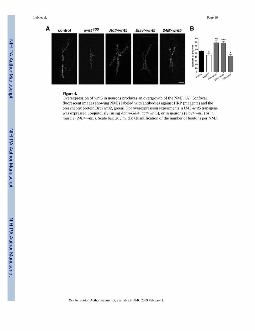
Figure 4.Overexpression of wnt5 in neurons produces an overgrowth of the NMJ. (A) Confocalfluorescent images showing NMJs labeled with antibodies against HRP (magenta) and thepresynaptic protein Brp (nc82, green). For overexpression experiments, a UAS-wnt5 transgenewas expressed ubiquitously (using Actin-Gal4, act>wnt5), or in neurons (elav>wnt5) or inmuscle (24B>wnt5). Scale bar: 20 μm. (B) Quantification of the number of boutons per NMJ.
Liebl et al. Page 16
Dev Neurobiol. Author manuscript; available in PMC 2009 February 1.
NIH
-PA Author Manuscript
NIH
-PA Author Manuscript
NIH
-PA Author Manuscript
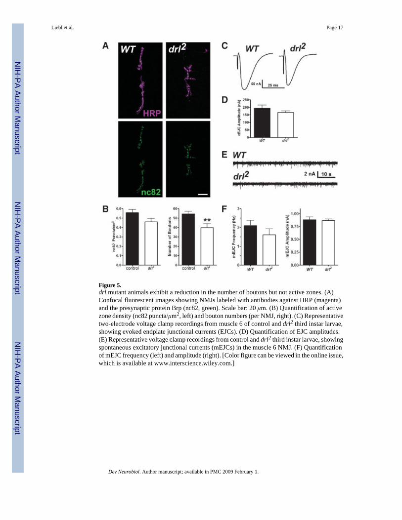
Figure 5.drl mutant animals exhibit a reduction in the number of boutons but not active zones. (A)Confocal fluorescent images showing NMJs labeled with antibodies against HRP (magenta)and the presynaptic protein Brp (nc82, green). Scale bar: 20 μm. (B) Quantification of activezone density (nc82 puncta/μm2, left) and bouton numbers (per NMJ, right). (C) Representativetwo-electrode voltage clamp recordings from muscle 6 of control and drl2 third instar larvae,showing evoked endplate junctional currents (EJCs). (D) Quantification of EJC amplitudes.(E) Representative voltage clamp recordings from control and drl2 third instar larvae, showingspontaneous excitatory junctional currents (mEJCs) in the muscle 6 NMJ. (F) Quantificationof mEJC frequency (left) and amplitude (right). [Color figure can be viewed in the online issue,which is available at www.interscience.wiley.com.]
Liebl et al. Page 17
Dev Neurobiol. Author manuscript; available in PMC 2009 February 1.
NIH
-PA Author Manuscript
NIH
-PA Author Manuscript
NIH
-PA Author Manuscript
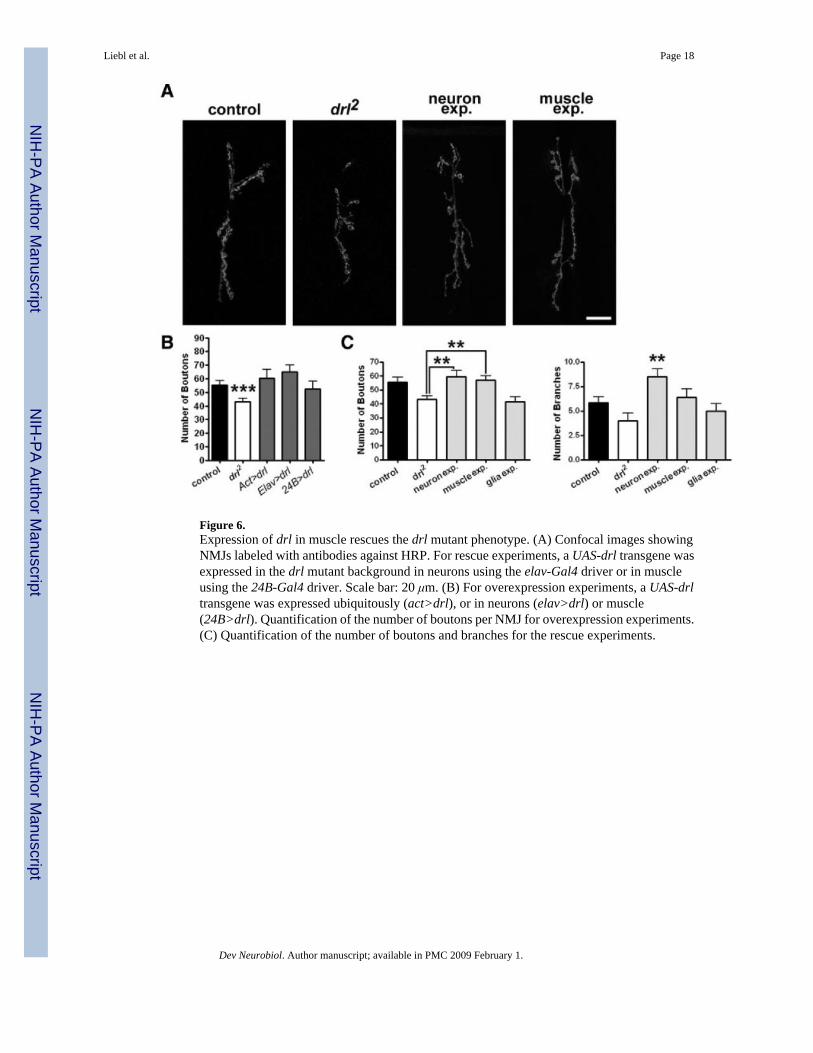
Figure 6.Expression of drl in muscle rescues the drl mutant phenotype. (A) Confocal images showingNMJs labeled with antibodies against HRP. For rescue experiments, a UAS-drl transgene wasexpressed in the drl mutant background in neurons using the elav-Gal4 driver or in muscleusing the 24B-Gal4 driver. Scale bar: 20 μm. (B) For overexpression experiments, a UAS-drltransgene was expressed ubiquitously (act>drl), or in neurons (elav>drl) or muscle(24B>drl). Quantification of the number of boutons per NMJ for overexpression experiments.(C) Quantification of the number of boutons and branches for the rescue experiments.
Liebl et al. Page 18
Dev Neurobiol. Author manuscript; available in PMC 2009 February 1.
NIH
-PA Author Manuscript
NIH
-PA Author Manuscript
NIH
-PA Author Manuscript
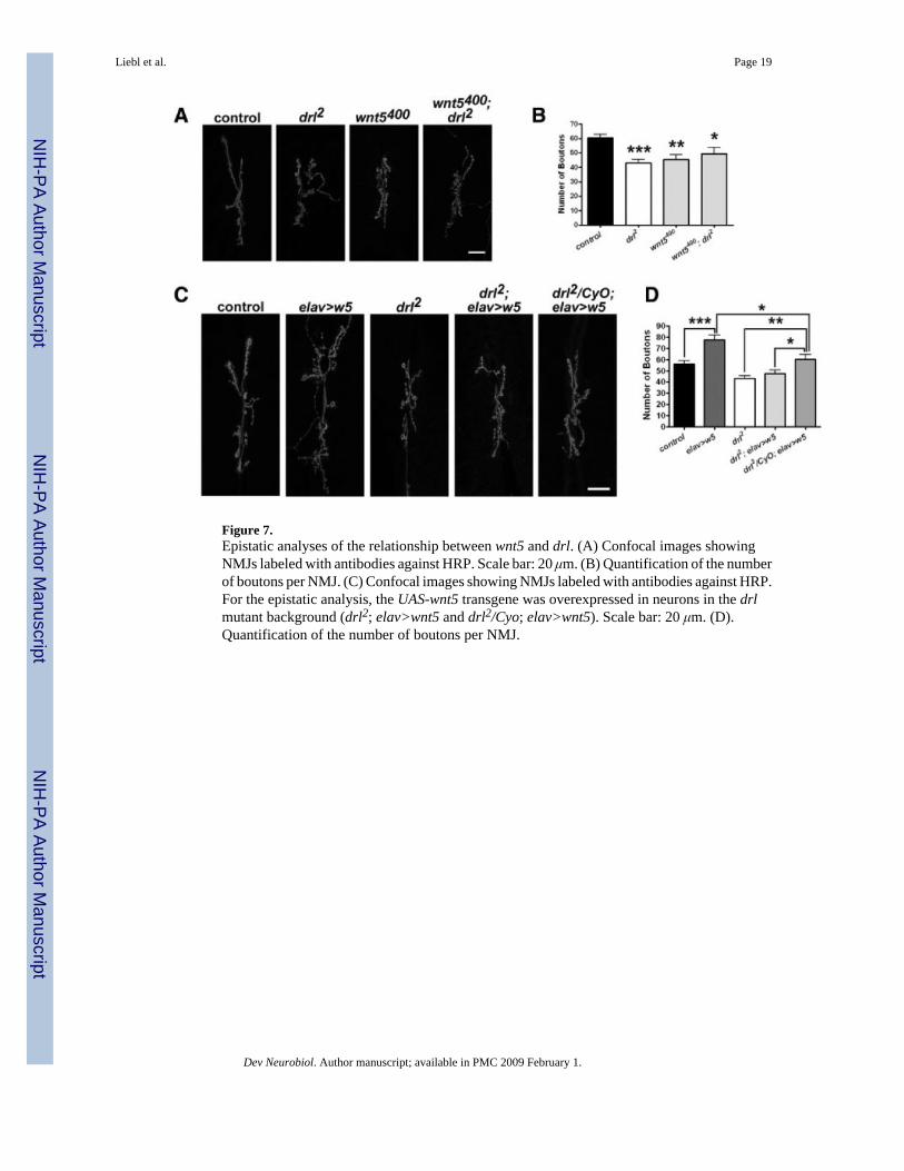
Figure 7.Epistatic analyses of the relationship between wnt5 and drl. (A) Confocal images showingNMJs labeled with antibodies against HRP. Scale bar: 20 μm. (B) Quantification of the numberof boutons per NMJ. (C) Confocal images showing NMJs labeled with antibodies against HRP.For the epistatic analysis, the UAS-wnt5 transgene was overexpressed in neurons in the drlmutant background (drl2; elav>wnt5 and drl2/Cyo; elav>wnt5). Scale bar: 20 μm. (D).Quantification of the number of boutons per NMJ.
Liebl et al. Page 19
Dev Neurobiol. Author manuscript; available in PMC 2009 February 1.
NIH
-PA Author Manuscript
NIH
-PA Author Manuscript
NIH
-PA Author Manuscript
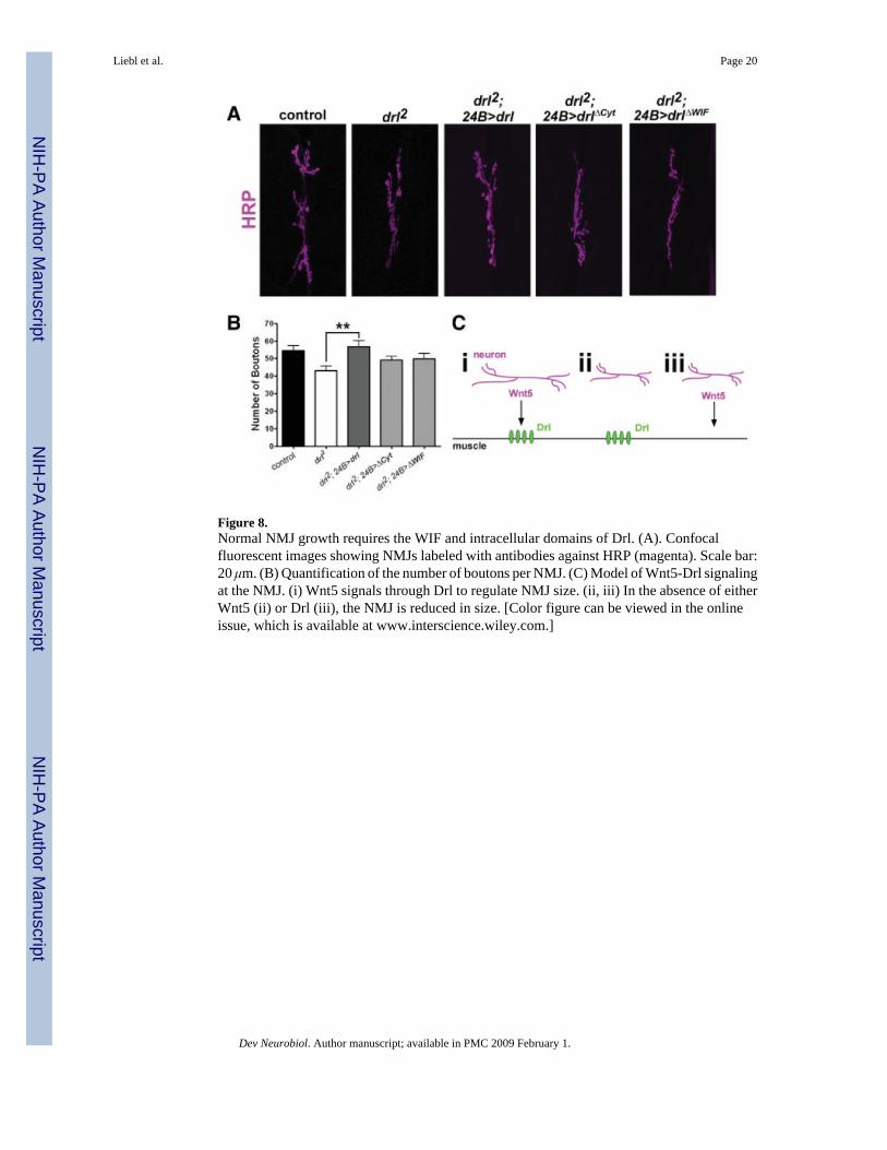
Figure 8.Normal NMJ growth requires the WIF and intracellular domains of Drl. (A). Confocalfluorescent images showing NMJs labeled with antibodies against HRP (magenta). Scale bar:20 μm. (B) Quantification of the number of boutons per NMJ. (C) Model of Wnt5-Drl signalingat the NMJ. (i) Wnt5 signals through Drl to regulate NMJ size. (ii, iii) In the absence of eitherWnt5 (ii) or Drl (iii), the NMJ is reduced in size. [Color figure can be viewed in the onlineissue, which is available at www.interscience.wiley.com.]
Liebl et al. Page 20
Dev Neurobiol. Author manuscript; available in PMC 2009 February 1.
NIH
-PA Author Manuscript
NIH
-PA Author Manuscript
NIH
-PA Author Manuscript
Related Documents