Depression and Anxious Apprehension Distinguish Frontocingulate Cortical Activity During Top-Down Attentional Control Rebecca Levin Silton, Wendy Heller, Anna S. Engels, David N. Towers, Jeffrey M. Spielberg, J. Christopher Edgar, Sarah M. Sass, Jennifer L. Stewart, and Bradley P. Sutton University of Illinois at Urbana-Champaign Marie T. Banich University of Colorado at Boulder Gregory A. Miller University of Illinois at Urbana-Champaign A network consisting of left dorsolateral prefrontal cortex (LDLPFC) and dorsal anterior cingulate cortex (dACC) has been implicated in top-down attentional control. Few studies have systematically investi- gated how this network is altered in psychopathology, despite evidence that depression and anxiety are associated with attentional control impairments. Functional MRI and dense-array event-related brain potential (ERP) data were collected in separate sessions from 100 participants during a color–word Stroop task. Functional MRI results guided ERP source modeling to characterize the time course of activity in LDLPFC (300 – 440 ms) and dACC (520 – 680 ms). At low levels of depression, LDLPFC activity was indirectly related to Stroop interference and only via dACC activity. In contrast, at high levels of depression, dACC did not play an intervening role, and increased LDLPFC activity was directly related to decreased Stroop interference. Specific to high levels of anxious apprehension, higher dACC activity was related to more Stroop interference. Results indicate that depression and anxious apprehen- sion modulate temporally and functionally distinct aspects of the frontocingulate network involved in top-down attention control. Keywords: depression, anxious apprehension, dACC, LDLPFC, attention Attentional difficulties are highlighted as key diagnostic criteria for both depression and anxiety in the Diagnostic and Statistical Manual of Mental Disorders (4th ed., text rev.; DSM–IV–TR; American Psychiatric Association, 2000). Contributing to cogni- tive misattributions, individuals with depression often demonstrate an attentional bias that favors negative information, and neutral information is interpreted in a negative manner (Gotlib & Kras- noperova, 1998; Gotlib, Krasnoperova, Yue, & Joorman, 2004). These attentional abnormalities may intensify and prolong symp- toms of sadness and worry due to the negatively biased misinter- pretation of events and information that is commonly observed in depression and anxiety (Gotlib et al., 2004), leading to a downward Rebecca Levin Silton, Anna S. Engels, David N. Towers, Jeffrey M. Spielberg, Sarah M. Sass, and Jennifer L. Stewart, Department of Psychol- ogy, University of Illinois at Urbana-Champaign; Wendy Heller, J. Chris- topher Edgar, and Bradley P. Sutton, Department of Psychology and Beckman Institute Biomedical Imaging Center, University of Illinois at Urbana-Champaign; Marie T. Banich, Department of Psychology, Univer- sity of Colorado; Gregory A. Miller, Departments of Psychology and Psychiatry and Beckman Institute Biomedical Imaging Center, University of Illinois at Urbana-Champaign. Rebecca Levin Silton is now at Department of Psychology, Loyola University Chicago; Anna S. Engels is now at Department of Psychology and Social, Life, and Engineering Sciences Imaging Center, Pennsylvania State University; J. Christopher Edgar is now at Department of Radiology, Children’s Hospital of Philadelphia; Sarah M. Sass is now at Department of Psychology, University of Texas at Tyler; Jennifer L. Stewart is now at Psychiatry Department, University of California, San Diego. This research was supported by National Institute of Mental Health Grants P50 MH079485, R01 MH61358, and T32 MH19554; National Institute on Drug Abuse Grant R21 DA14111); and the University of Illinois Beckman Institute, Department of Psychology, and Intercampus Research Initiative in Biotechnology. This article is based on data that were included in Rebecca Levin Silton’s doctoral dissertation. A portion of this research was presented at the 2009 meeting of the Society for Research in Psychopathology. A different article that involves a subset of the partic- ipants included in this article was published in NeuroImage. That article evaluated the temporal course of the dorsal anterior cingulate cortex and the left dorsolateral prefrontal cortex proposed in the cascade-of-control model and did not address the role of psychopathology. We thank Adrienne Abramowitz, Kirstin Aschbacher, Patrick Berg, Keith Bredemeier, Amanda Bull, Emily Cahill, Laura Crocker, Monica Fabiani, Kara Federmeier, Joscelyn Fisher, Christian Hendershot, Brenda Hernandez, Karsten Hoechstetter, An- gela Lawson, Renee Thompson, Edelyn Verona, and Stacie Warren for their contributions to this project. Correspondence concerning this article should be addressed to Rebecca Levin Silton, Department of Psychology, Loyola University Chicago, 1032 West Sheridan Road, Chicago, IL 60660. E-mail: [email protected] Journal of Abnormal Psychology © 2011 American Psychological Association 2011, Vol. 120, No. 2, 272–285 0021-843X/11/$12.00 DOI: 10.1037/a0023204 272

Welcome message from author
This document is posted to help you gain knowledge. Please leave a comment to let me know what you think about it! Share it to your friends and learn new things together.
Transcript
Depression and Anxious Apprehension Distinguish Frontocingulate CorticalActivity During Top-Down Attentional Control
Rebecca Levin Silton, Wendy Heller,Anna S. Engels, David N. Towers,
Jeffrey M. Spielberg, J. Christopher Edgar,Sarah M. Sass, Jennifer L. Stewart, and
Bradley P. SuttonUniversity of Illinois at Urbana-Champaign
Marie T. BanichUniversity of Colorado at Boulder
Gregory A. MillerUniversity of Illinois at Urbana-Champaign
A network consisting of left dorsolateral prefrontal cortex (LDLPFC) and dorsal anterior cingulate cortex(dACC) has been implicated in top-down attentional control. Few studies have systematically investi-gated how this network is altered in psychopathology, despite evidence that depression and anxiety areassociated with attentional control impairments. Functional MRI and dense-array event-related brainpotential (ERP) data were collected in separate sessions from 100 participants during a color–wordStroop task. Functional MRI results guided ERP source modeling to characterize the time course ofactivity in LDLPFC (300–440 ms) and dACC (520–680 ms). At low levels of depression, LDLPFCactivity was indirectly related to Stroop interference and only via dACC activity. In contrast, at highlevels of depression, dACC did not play an intervening role, and increased LDLPFC activity was directlyrelated to decreased Stroop interference. Specific to high levels of anxious apprehension, higher dACCactivity was related to more Stroop interference. Results indicate that depression and anxious apprehen-sion modulate temporally and functionally distinct aspects of the frontocingulate network involved intop-down attention control.
Keywords: depression, anxious apprehension, dACC, LDLPFC, attention
Attentional difficulties are highlighted as key diagnostic criteriafor both depression and anxiety in the Diagnostic and StatisticalManual of Mental Disorders (4th ed., text rev.; DSM–IV–TR;American Psychiatric Association, 2000). Contributing to cogni-tive misattributions, individuals with depression often demonstratean attentional bias that favors negative information, and neutral
information is interpreted in a negative manner (Gotlib & Kras-noperova, 1998; Gotlib, Krasnoperova, Yue, & Joorman, 2004).These attentional abnormalities may intensify and prolong symp-toms of sadness and worry due to the negatively biased misinter-pretation of events and information that is commonly observed indepression and anxiety (Gotlib et al., 2004), leading to a downward
Rebecca Levin Silton, Anna S. Engels, David N. Towers, Jeffrey M.Spielberg, Sarah M. Sass, and Jennifer L. Stewart, Department of Psychol-ogy, University of Illinois at Urbana-Champaign; Wendy Heller, J. Chris-topher Edgar, and Bradley P. Sutton, Department of Psychology andBeckman Institute Biomedical Imaging Center, University of Illinois atUrbana-Champaign; Marie T. Banich, Department of Psychology, Univer-sity of Colorado; Gregory A. Miller, Departments of Psychology andPsychiatry and Beckman Institute Biomedical Imaging Center, Universityof Illinois at Urbana-Champaign.
Rebecca Levin Silton is now at Department of Psychology, LoyolaUniversity Chicago; Anna S. Engels is now at Department of Psychologyand Social, Life, and Engineering Sciences Imaging Center, PennsylvaniaState University; J. Christopher Edgar is now at Department of Radiology,Children’s Hospital of Philadelphia; Sarah M. Sass is now at Departmentof Psychology, University of Texas at Tyler; Jennifer L. Stewart is now atPsychiatry Department, University of California, San Diego.
This research was supported by National Institute of Mental HealthGrants P50 MH079485, R01 MH61358, and T32 MH19554; National
Institute on Drug Abuse Grant R21 DA14111); and the University ofIllinois Beckman Institute, Department of Psychology, and IntercampusResearch Initiative in Biotechnology. This article is based on data that wereincluded in Rebecca Levin Silton’s doctoral dissertation. A portion of thisresearch was presented at the 2009 meeting of the Society for Researchin Psychopathology. A different article that involves a subset of the partic-ipants included in this article was published in NeuroImage. That articleevaluated the temporal course of the dorsal anterior cingulate cortex and theleft dorsolateral prefrontal cortex proposed in the cascade-of-control modeland did not address the role of psychopathology. We thank AdrienneAbramowitz, Kirstin Aschbacher, Patrick Berg, Keith Bredemeier, AmandaBull, Emily Cahill, Laura Crocker, Monica Fabiani, Kara Federmeier, JoscelynFisher, Christian Hendershot, Brenda Hernandez, Karsten Hoechstetter, An-gela Lawson, Renee Thompson, Edelyn Verona, and Stacie Warren for theircontributions to this project.
Correspondence concerning this article should be addressed to RebeccaLevin Silton, Department of Psychology, Loyola University Chicago, 1032West Sheridan Road, Chicago, IL 60660. E-mail: [email protected]
Journal of Abnormal Psychology © 2011 American Psychological Association2011, Vol. 120, No. 2, 272–285 0021-843X/11/$12.00 DOI: 10.1037/a0023204
272

spiral of maladaptive thoughts. Individuals with anxiety demon-strate an attentional bias to threat-related information (Compton,Heller, Banich, Palmieri, & Miller, 2000; Nitschke & Heller,2002). Once threatening stimuli are attended to, it is difficult forindividuals with anxiety to disengage their attention and shift toless anxiety-provoking thoughts. These attentional control diffi-culties often affect daily life function. In clinical settings, it istypical to hear clients with depression and/or anxiety complain of“difficulties attending to a conversation or lecture” or “problemsfocusing on reading or homework.” Attentional control problemsand related executive function deficits can greatly interfere withinterpersonal relationships and daily life activities such as jobperformance (Jaeger, Berns, Uzelac, & Davis-Conway, 2006). Inturn, these difficulties fuel cycles of self-deprecation, sadness, andworry. Cognitive and neural mechanisms associated with the at-tentional problems that accompany symptoms of depression andanxiety are not well understood.
Complicating the characterization of these phenomena, depres-sion and anxiety frequently co-occur (Engels et al., 2010; Kessler,DuPont, Berglund, & Wittchen, 1999). On the basis of DSM–IV–TR criteria, it can often be difficult, or even impossible, todistinguish whether an individual’s attentional problems are re-lated to depression, anxiety, or both. Further developing clinicalassessment methods that have high diagnostic sensitivity and spec-ificity is crucial to advancing treatment for these debilitatingdisorders. If differential patterns of attentional control difficultiescan be identified in depression and anxiety, it will informevidence-based treatments for depression and anxiety that involvetraining individuals to use attentional control methods, such ascognitive control therapy (Siegle, Ghinassi, & Thase, 2007) andmindfulness-based cognitive behavioral therapy (Segal, Williams,& Teasdale, 2002).
The effectiveness of attentional control or cognitive controltraining for individuals with depression has been demonstrated intreatment outcome studies (Siegle et al., 2007) as well as inexperimental research. Hertel (1994) showed that individuals withdepression who were coached to use attentional control strategiesachieved performance on an attention task that was comparable tothat of individuals without depression. These findings indicatedthat individuals with depression have sufficient attentional re-sources; their attentional problems arise due to a failure to controlthese resources (Hertel, 1994). Fundamental attentional controlfunctions that are involved with maintaining focus on the task athand, rather than getting distracted by threatening or negativetask-irrelevant information, or getting caught up in a ruminativeloop, may be interrupted in depression (Hertel, 2007) and anxiety(Eysenck, Derakshan, Santos, & Calvo, 2007).
Several dimensions of psychopathology, including anhedonicdepression, anxious apprehension (worry), and anxious arousal(panic or autonomic arousal), are accompanied by unique patternsof abnormal activity in regions of the brain involved in attentionalcontrol (e.g., Engels et al., 2007, 2010; Nitschke, Heller, & Miller,2000). In order to examine associated neural mechanisms of at-tentional disruption that accompany depression and anxiety symp-toms, it is strategic to partition anxiety according to these theoret-ical and methodological distinctions. This is particularly relevantbecause worry or anxious apprehension is a key feature of gener-alized anxiety disorder (GAD), and GAD is the most common
anxiety disorder to precede and co-occur with depression (Kessleret al., 1996; Kessler, Zhao, Blazer, & Swartz, 1997),
Banich (2009) identified a network of brain regions involved intop-down attentional control, including left dorsolateral prefrontalcortex (LDLPFC) and dorsal anterior cingulate cortex (dACC).Present analyses addressed the possibility that depression andanxious apprehension differentially influence this network. Ac-cording to Banich’s (2009) cascade-of-control model, duringattentionally demanding tasks LDLPFC imposes a top-down at-tentional set for task-relevant processing while late-stage orresponse-related aspects of selection are implemented by dACC.Hence, a temporal course in which LDLPFC is activated first,followed by dACC, is a key component of Banich’s cascade-of-control model. Furthermore, the model posits that the less top-down control exerted by LDLPFC, the more activity should beobserved in dACC, as it will need to resolve any remaining aspectsof selection before a response can be emitted.
A recent source analysis study investigating the time course ofactivity in LDLPFC and dACC during an attentional control task(color–word Stroop) in a nonclinical, undergraduate sample (Siltonet al., 2010) provided support for this model.1 Results indicatedthat LDLPFC activity preceded dACC activity. Moreover, mea-sures of performance (Stroop interference) were directly related tolater dACC activity but not LDLPFC activity. The Stroop inter-ference effect refers to a typical response pattern involving longerreaction time (RT) following incongruent stimuli (the word red inblue ink) than congruent (the word red in red ink) or neutralstimuli (a nonword or a noncolor word, such as XXXX or bond, inred ink). The extent to which dACC activation influenced Stroopperformance depended on the degree of earlier LDLPFC activity,showing an interdependent relationship among these brain regions.Consistent with the cascade-of-control model, when LDLPFCactivity was high, dACC activity did not affect performance. Thispattern of activity was attributed to adequate top-down controlimposed by LDLPFC. When LDLPFC activity was low, highdACC activity was associated with better performance and longerRT, suggesting that, as predicted, dACC was compensating for thelack of top-down LDLPFC control. When LDLPFC and dACCactivity were both low, a higher error rate and shorter RT wereobserved, indicating a lack of dACC compensatory action.
These findings are relevant for psychopathology, as the Stroop(1935) task has been used to investigate cognitive impairments indepression in top-down control. A number of studies examiningperformance on the color–word Stroop task in depressed individ-uals have shown a range of attentional difficulties evidenced byincreased RT, increased errors, and greater interference (Biringeret al., 2005; Duncan & Owen, 2000; Dunkin et al., 2000; Holmes& Pizzagalli, 2008; Paradiso, Lamberty, Garvey, & Robinson,1997; Ravnkilde et al., 2002; Stordal et al., 2004; Videbech et al.,2004). Impaired Stroop performance has been reported for indi-
1 Silton et al. (2010) did not address the relationship between psycho-pathology and the temporal course of LDLPFC and dACC. The presentstudy was designed to follow up questions raised about psychopathology inthe Silton et al. study. As a follow-up study, a superset of the Silton et al.sample and similar methodology were used in the present study. Resultsfrom Silton et al. guided present hypotheses regarding how depression andanxiety influence the temporal course of the frontocingulate network.
273DEPRESSION, APPREHENSION, AND CORTICAL ACTIVITY

viduals with major depressive disorder (Videbech et al., 2004),recurrent major depressive episodes (Stordal et al., 2004), remitteddepression (Biringer et al., 2005; Paradiso et al., 1997), and failureto respond to antidepressant medication (Dunkin et al., 2000).Recent research has revealed that abnormal focal LDLPFC anddACC activity is related to depression during Stroop performance(Holmes & Pizzagalli, 2008; Killgore, Gruber, & Yurgelun-Todd,2007; Wagner et al., 2006), but precisely how depression influ-ences the timing and relations among relevant brain regions re-mains an open question.
Research investigating the relationship between anxiety andcolor–word Stroop performance has used diverse definitions andtypes of anxiety, as well as various paradigms, perhaps contribut-ing to a lack of consistency in results. In an early study, a statemanipulation of anxiety adversely affected performance accuracyin an incongruent but not a congruent condition (Hochman, 1967).Fox (1993) compared behavioral performance for high- and low-trait-anxious participants on incongruent, neutral, and threateningwords in a spatially “separated” Stroop task (attend to a centralcolor patch and ignore the word in the periphery). High-trait-anxious participants showed interference effects for both incon-gruent and threat words presented in the periphery, suggesting ageneral disruption in the ability to maintain attentional focus thatwas not limited to threatening information. Other studies involvingthe color–word Stroop task have not found RT condition differ-ences as a function of anxiety (Gehring, Himle, & Nisenson, 2000;Hajcak, McDonald, & Simons, 2003).
The brain regions that were shown to work in conjunction toexert attentional control in a nonclinical sample, specificallyLDLPFC and dACC (Silton et al., 2010), appear to be differen-tially affected in depression and anxiety. Depression has beenmore commonly linked to reduced LDLPFC activity than hasanxiety (Fitzgerald et al., 2006; Herrington et al., 2010; Holmes &Pizzagalli, 2008; Rogers, Bradshaw, Pantelis, & Phillips, 1998).Both depression and anxiety have been associated with altereddACC activity (Engels et al., 2010; Hajcak et al., 2003). Anxietyhas typically been associated with increased dACC activity (Olvet& Hajcak, 2008; Paulus, Feinstein, Simmons, & Stein, 2004),whereas both dACC hyperactivity and hypoactivity have beenreported in depression (George et al., 1997; Holmes & Pizzagalli,2008; Killgore et al., 2007). These mixed findings may be in partdue to unassessed, comorbid anxiety.
Limited research has focused on how psychopathology affectsfrontocingulate networks. Mayberg’s (1997) proposed limbic-cortical network model of depression specifies “ventral” and “dor-sal” components. Relevant to the present study, Mayberg proposedthat the attentional impairments observed in depression are relatedto abnormalities in the dorsal components, which include DLPFC,dACC, inferior parietal cortex, and striatum. In line with thismodel, Holmes and Pizzagalli (2008) showed a reduction in bothLDLPFC and dACC activity as measured by event-related poten-tial (ERP) source analysis during a color–word Stroop task at620-ms poststimulus presentation in individuals with depressioncompared with controls. Reporting different findings using func-tional magnetic resonance imaging (fMRI) methods, Wagner et al.(2006) observed that individuals with depression had hyperactiveLDLPFC but no changes in dACC activity relative to healthycontrols during Stroop performance. With implications for trans-lational research, an fMRI study showed posttreatment changes in
LDLPFC function after individuals with depression received cog-nitive control therapy (Siegle et al., 2007). Decreased LDLPFCactivity was observed for an easy cognitive control task condition,and increased LDLPFC activity was observed for a more difficultcondition. These depressed individuals demonstrated improvedperformance on this cognitive control task following treatment.These findings support other studies in pointing to changes inpatterns of DLPFC and ACC activation as a function of depres-sion, although the precise nature and direction of these changesremains to be determined.
The present study used ERP source analysis to evaluate how thefrontocingulate network described in Banich’s (2009) cascade-of-control model of top-down attentional control is affected by de-pression and anxiety. Source analysis techniques are an idealmethod to study the timing of network function, as they provideinformation regarding the time course of regional brain activity.Moderated mediation analyses were used to evaluate the hypoth-esis that depression and anxiety would influence the time course ofearly LDLPFC activity and later dACC activity in different waysduring a task that requires top-down attentional control (the color–word Stroop task). It was predicted that depression would berelated to reduced earlier LDLPFC activity and that later dACCactivity would be related to either compensatory behavior, asevidenced by increased activity and normal Stroop performance, ora lack of compensatory behavior, as indicated by decreased activ-ity and poor Stroop performance. Anxious apprehension was hy-pothesized to be associated with increased dACC activity only. Itwas uncertain whether this pattern of network activity would affectperformance. Increased dACC activity was shown to mediateStroop performance only when LDLPFC activity was low andassociated top-down attentional control was poor (Silton et al.,2010). Because anxious apprehension was not expected to affectLDLPFC activity, it was uncertain whether potential changes tosubsequent downstream dACC activity (but not earlier LDLPFCactivity) would affect performance.
Method
Participants and Selection Procedures
Participants (N � 100) were recruited from introductory psychol-ogy classes via group questionnaire screening sessions as well as fromthe community via advertisements placed in local newspapers andthrough recruitment efforts at the campus-run community psychologyclinic. Participants (45% female, 80% Caucasian) were paid volun-teers ages 18–35 years (M � 20.2, SD � 3.6). Participants wereright-handed as determined by the Edinburgh Handedness Inventory(Oldfield, 1971) and were native English speakers. Because psycho-active medications are known to affect cognitive function and relatedregional brain activity (Brody et al., 2001; Mayberg et al., 2000),participants were screened by self-report to be free of such medica-tions. Participants were also screened for abnormal color vision, lossof consciousness greater than 10 min, claustrophobia, recent drug oralcohol use, excessive caffeine intake, and lack of sleep. Participantswere given a laboratory tour, were informed of the study procedures,and provided written consent. DSM–IV–TR diagnoses were used toselect participants from a larger project in order to ensure that indi-viduals who had a lifetime history of clinically defined depression
274 LEVIN SILTON ET AL.

were included in present analyses. The participant selection methoddescribed here was employed prior to running subsequent analyses.
The Structured Clinical Interview for Axis I Disorders, Non-Patientedition (SCID–NP, First, Spitzer, Gibbon, & Williams, 1997), wasadministered to all participants to assess Axis I disorders. LifetimeDSM–IV–TR diagnoses were determined by the interviewer and re-viewed by a consensus team consisting of a second interviewer and aclinical faculty supervisor (Gregory A. Miller) reviewing a writtencase summary detailing each criterion symptom on the scale: 1 �absent, 2 � features (at least two symptoms), 3 � provisional (oneshort of full DSM–IV–TR criteria), and 4 � definite. SCID–NP datawere used to select 34 participants who had a lifetime history of aprovisional or definite depressive disorder, 18 of whom had a lifetimehistory of one or more provisional or definite anxiety disorders.Participants with anxiety disorders had diagnoses primarily of GAD(n � 7), as well as of obsessive-compulsive disorder (n � 5), socialphobia (n � 5), specific phobia (n � 4), posttraumatic stress disorder(n � 4), panic (n � 1), and anxiety not otherwise specified (n � 1).The SCID–NP does not provide information regarding current anxietydisorders, only information regarding lifetime history of anxiety dis-orders. Sixty-six participants were free of any depressive or anxietydisorders. None of the participants was in a current major depressiveepisode. Although participants were screened for all Axis I disorders,other disorders were not used as criteria to select participants for thepresent study. Participants’ diagnostic status was not revealed to theresearch team until after the participants had completed the entirestudy protocol. These participant selection methods were used toensure that a range of depression- and anxiety-related psychopathol-ogy was represented in the sample, as dimensional analyses of self-reported depression and anxious apprehension were used to examinetheir moderating effects (described below).
To provide dimensional measures of depression, anxious appre-hension, and anxious arousal, participants completed the Moodand Anxiety Symptom Questionnaire Anhedonic Depressioneight-item depressed mood subscale (MASQ–AD-8; Nitschke,Heller, Imig, McDonald, & Miller, 2001; Watson, Clark, et al.,1995; Watson, Weber, et al., 1995) and the Penn State WorryQuestionnaire (PSWQ; Meyer, Miller, Metzger, & Borkovec,1990; Molina & Borkovec, 1994). These measures have beenshown to provide effective discrimination among these dimensions(for review, see Nitschke et al., 2001) and to distinguish brain
regions involved in each (e.g., Engels et al., 2010). Althoughparticipants completed other questionnaires as part of a largerstudy, only the MASQ–AD-8 and PSWQ were analyzed in thepresent study. With regard to construct validity, the MASQ–AD-8predicts current major depressive episode and lifetime major de-pressive disorder (Bredemeier et al., 2010). Similarly, the PSWQis an excellent predictor of GAD (Behar, Alcaine, Zuellig, &Borkovec, 2003). See Table 1 for further information about de-mographics, and questionnaire scores for the depression, anxiety,and comorbid diagnostic categories for the present sample.
Three analyses of variance were conducted to examine (a) whetherparticipants with diagnosed depression scored higher than those with-out diagnoses on the MASQ–AD-8, (b) whether participants withcomorbid diagnoses scored higher than those without diagnoses onboth questionnaire measures, and (c) whether participants with co-morbid diagnoses scored higher than the participants with only de-pression diagnoses on the PSWQ but not the MASQ–AD-8. Theparticipants with comorbid and pure depression were expected to havecomparable levels of depression as measured by the MASQ–AD-8and to vary only on anxiety levels as measured by the PSWQ. Theresults were as expected. The participants with depression diagnosesscored higher than those without diagnoses on the MASQ–AD-8, F(1,80) � 5.41, p � .02. The participants with comorbid diagnoses scoredhigher than those without diagnoses on the PSWQ, F(1, 82) � 17.22,p � .00, and MASQ–AD-8, F(1, 82) � 7.60, p � .01. The partici-pants with comorbid diagnoses did not differ from those with onlydepression diagnoses on the MASQ–AD-8, F(1, 32) � 0.12, p � .74,but they scored higher than those with only depression diagnoses onthe PSWQ, F(1, 32) � 9.33, p � .01. Given that the diagnosis-basedcategorical groups were formed via a different measure (the SCID–NP) than the dimensional questionnaires, these analyses providedevidence for convergent validity.
Stimuli and Experimental Design
In brief overview, ERP and fMRI data were collected from allparticipants during a task requiring top-down attentional control(color–word Stroop task). The fMRI data were used to guideplacement of ERP sources, and information regarding the timecourse of LDLPFC and dACC activity was extracted for neural
Table 1Demographics and Questionnaire Scores by Diagnostic Group
Variable
Comorbid(n � 18)
Depression(n � 16)
No diagnosis(n � 66)
Full sample(N � 100)
M SD M SD M SD M SD
Age 23.72 5.15 21.12 5.44 19 0.89 20.19 3.60Gender (n)a 12/6 7/9 26/30 45/55PSWQ 57.44 18.92 39.38 15.05 40.55 14.22 43.40 16.51MASQ–AA 27.72 7.01 24.00 6.55 24.18 6.90 24.79 6.93MASQ–AD-8 17.72 6.24 17.06 4.80 14.39 3.95 15.42 4.74GAF 67.89 10.64 81.44 6.50 87.76 5.02 83.17 9.97
Note. PSWQ � Penn State Worry Questionnaire; MASQ–AA � Mood and Anxiety Symptom Questionnaire–Anxious Arousal; MASQ–AD-8 � Mood and Anxiety Symptom Questionnaire–Anhedonic Depression eight-item depressed mood subscale; GAF � Global Assessment of Function.a Women/men.
275DEPRESSION, APPREHENSION, AND CORTICAL ACTIVITY

network analyses involving dimensional depression and anxiousapprehension variables.
Participants completed a color–word Stroop task and anemotion–word Stroop task. Both tasks were administered duringan fMRI session and again during an electroencephalogram (EEG)session. The order of presentation of the two Stroop tasks within asession was counterbalanced across participants, as was the orderof the EEG and fMRI sessions, with the SCID session in betweenfor most participants. The emotion–word Stroop data do not ad-dress present goals and will not be considered further here. Thecolor–word Stroop task consisted of blocks of color-congruent orcolor-incongruent words alternating with blocks of neutral words,with 256 trials in 16 blocks (four color congruent, four colorincongruent, eight neutral). Half the trials in congruent and incon-gruent blocks were neutral, to prevent the development of word-reading strategies. There were eight orders of stimulus presentationfor each Stroop task, designed specifically to control stimulusorder effects. Each participant received one of the eight orders.
Each trial consisted of one word presented in one of four inkcolors (red, yellow, green, blue). Trials began with the presentationof a word for 1,500 ms, followed by a fixation cross for 275–725ms (onset-to-onset intertrial interval � 2,000 � 225 ms). Wordpresentation and response recording were controlled by STIMsoftware (James Long Company, Caroga Lake, NY). In the fMRIsession, words were presented in capital letters with Tahoma72-point font via back projection onto a screen outside the scannerbore and a mirror fixed to the head coil, providing a vertical spanof 2.9° and a horizontal span of 6.1°–16.4°. In the ERP session, thesame words were presented in Tahoma 72-point font on a CRTmonitor 1.35 m from the participants’ eyes, providing a verticalspan of 1.5° and a horizontal span of 3.2°–8.7°. Participantsresponded with the middle and index fingers of both hands, witheach task using the same mapping of color to button. There was acolor-to-key-mapping acquisition phase of 32 practice trials. Inaddition to the 16 word blocks, there were four fixation blocks: oneat the beginning, one at the end, and two in the middle of thesession. In the fixation condition, a brightened fixation cross waspresented for 1,500 ms.
MRI Recording, Data Reduction, and Analysis
Functional MRI data were analyzed from a subset of 30 of the66 participants without a psychopathology history, providing guid-ance for the ERP source analysis that was carried out for all 100participants. Two-tailed t tests showed that the 30 participants whowere used in fMRI analyses did not differ from the other 36participants without anxiety or depression diagnoses in terms ofage, t(65) � 1.11, p � .27; gender balance, �2(1, N � 66) � 2.03,p � .15; Global Assessment of Function, t(65) � 0.26, p � .80; orStroop interference effect, t(65) � �0.78, p � .44. Participantswithout lifetime depression and/or anxiety diagnoses were used forfMRI analyses because the purpose of the study was to understandhow network activity in depression and anxiety differs from typ-ical network activity observed in healthy individuals.
The magnetic resonance technologist and experimenter assistedthe participant in correct placement of earplugs and protectiveheadphones. Magnetic resonance data were collected using aresearch-dedicated 3T Siemens Allegra. Three hundred and sev-enty functional images were acquired via a gradient-echo echo-
planar imaging sequence (repetition time � 2,000 ms, echo time �25 ms, flip angle � 80°, field of view � 22 cm). Thirty-eightoblique axial slices (slice thickness � 3 mm, in-plane resolution �3.4375 � 3.4375 mm, 0.3-mm gap between slices) were acquiredparallel to the anterior and posterior commissures. After the echo-planar imaging sequence, a 160-slice MPRAGE structural se-quence was acquired (slice thickness � 1 mm, in-plane resolu-tion � 1 � 1 mm) for registering each participant’s functional datato standard space.
Image processing and analyses relied primarily on tools fromthe FMRIB Software Library analysis package (http://www.fmri-b.ox.ac.uk/fsl). Each fMRI time series was first motion-correctedwith FMRIB’s Linear Image Registration Tool (Jenkinson, Ban-nister, Brady, & Smith, 2002), and spikes (artifactual suddenintensity shifts) were corrected with the AFNI tool 3dDespike(http://afni.nimh.nih.gov/afni). Participants demonstrated less than3.3-mm absolute motion or 2-mm relative motion (participantswith motion exceeding this threshold were excluded from analysis,beyond the 30 control participants relied on in the present analy-sis). After motion correction and despiking, each time series wascorrected for geometric distortions caused by magnetic field inho-mogeneity. Remaining preprocessing steps, single-subject statis-tics, and group statistics were implemented by FMRIB’s ExpertAnalysis Tool. The first three volumes of each data set werediscarded to allow the magnetic resonance signal to reach a steadystate. Each time series was then temporally filtered with a nonlin-ear high-pass filter (to remove drift in signal intensity), mean-based intensity-normalized by the same single scaling factor, andspatially smoothed via a third-dimensional Gaussian kernel (full-width half maximum � 5 mm) prior to analysis.
Regression analyses were performed on each participant’s timeseries with FMRIB’s Improved Linear Model. Statistical mapswere generated via multiple regression computed for each intra-cerebral voxel (Woolrich, Ripley, Brady, & Smith, 2001). Anexplanatory variable (EV) was created for each trial block type(color congruent, color incongruent, neutral, rest), with the fixationcondition the unmodeled baseline. Each EV was convolved with agamma function to better approximate the temporal course of theblood-oxygen-level-dependent hemodynamic response (e.g., Agu-irre, Zarahn, & D’Esposito, 1998; Miezin, Maccotta, Ollinger,Petersen, & Buckner, 2000). Each EV yielded a per-voxel effect-size parameter estimate map representing the magnitude of activityassociated with that EV. The beta values for the incongruent wordcondition were contrasted with those for the congruent wordcondition, resulting in a per-voxel contrast parameter estimate mapfor each subject. These functional activation maps as well as thecorresponding structural MRI map were registered into MontrealNeurological Institute stereotaxic space with FMRIB’s NonlinearImage Registration Tool via FMRIB Software Library’s defaultconfiguration file and a warp resolution of 10 mm.
Inferential statistical analyses were carried out with FMRIB’sLocal Analysis of Mixed Effects. In order to identify regionsassociated with the Stroop interference effect, significantly acti-vated voxels were identified for the incongruent minus congruentcontrast via a one-sample t test, yielding a three-dimensionalfunctional z map image. Monte Carlo simulations via AFNI’sAlphaSim program (Ward, 2000) estimated the overall signifi-cance level (probability of a false detection) for thresholding, usinga gray-matter mask to limit the number of voxels under consider-
276 LEVIN SILTON ET AL.

ation. These simulations provided a z value (z � 3.0902, p � .01)and cluster size (34) combination for thresholding that resulted inan overall familywise error rate of .05. Clusters that survived thisthresholding are reported in Table 2. Center of mass coordinatesfor clusters in hypothesized regions of interest were used to placeregional sources in the ERP source model.
Electrophysiological Recording, Data Reduction, andAnalysis
Participants were seated in a comfortable chair in a quiet roomconnected to the adjacent equipment room by intercom. EEG wasrecorded with a custom-designed Falk Minow 64-channel cap withequidistantly spaced Ag and AgCl electrodes. After placementof the electrode cap, electrode positions were digitized forlater topographic and source localization analyses. An additionalelectrode was placed below each eye; these and nearby electrodesin the cap provided a basis for offline eye-blink artifact correctionof the EEG data implemented in BESA (Version 5.1.8; Berg &Scherg, 1994). The left mastoid served as the online reference forall other sites, including electrooculogram. Impedances were be-low 20 �, appropriate given the high input impedance of theamplifiers. Half-power amplifier bandpass was 0.1–100 Hz, withdigitization at 250 Hz.
The following steps were done separately for each participant.Muscle and other artifact was manually removed with BESA. Aseries of steps were taken to remove and/or correct eye blinks andmovements. Electrodes above and below the right and left eyes andnear the left and right external canthi were used to measure verticaland horizontal eye movements. Pairs of channels were used tocompute bipolar derivations, to identify epochs that included eithera horizontal or vertical saccade. The saccades were marked asartifact periods and removed from the data. A typical blink wasidentified in the data. With the pattern search function in BESA,the data were scanned to identify other blink periods. Stimulus-locked averages were calculated for the experimental conditions(congruent, incongruent, and neutral) for each participant. Onlytrials with correct responses that occurred 350–1,400 ms after
stimulus onset were included in the individual participant aver-ages. All participants included in the sample had a minimum of 16trials for each condition average. Following these steps, the sur-rogate multiple source eye correction algorithm was used to cor-rect blink artifacts for each participant (Berg & Scherg, 1994). Inthe multiple source eye correction method, using all EEG chan-nels, sources of brain and artifact activity (e.g., blink) aresimultaneously modeled, and only the modeled blink activity isremoved from each EEG channel. The Berg and Scherg (1994)method reduces distortion of brain activity by accounting forthe EEG signal during the estimation of eye activity (see Siltonet al., 2010, for additional details about the blink removalprocess and application of the multiple source eye correctionmethod).
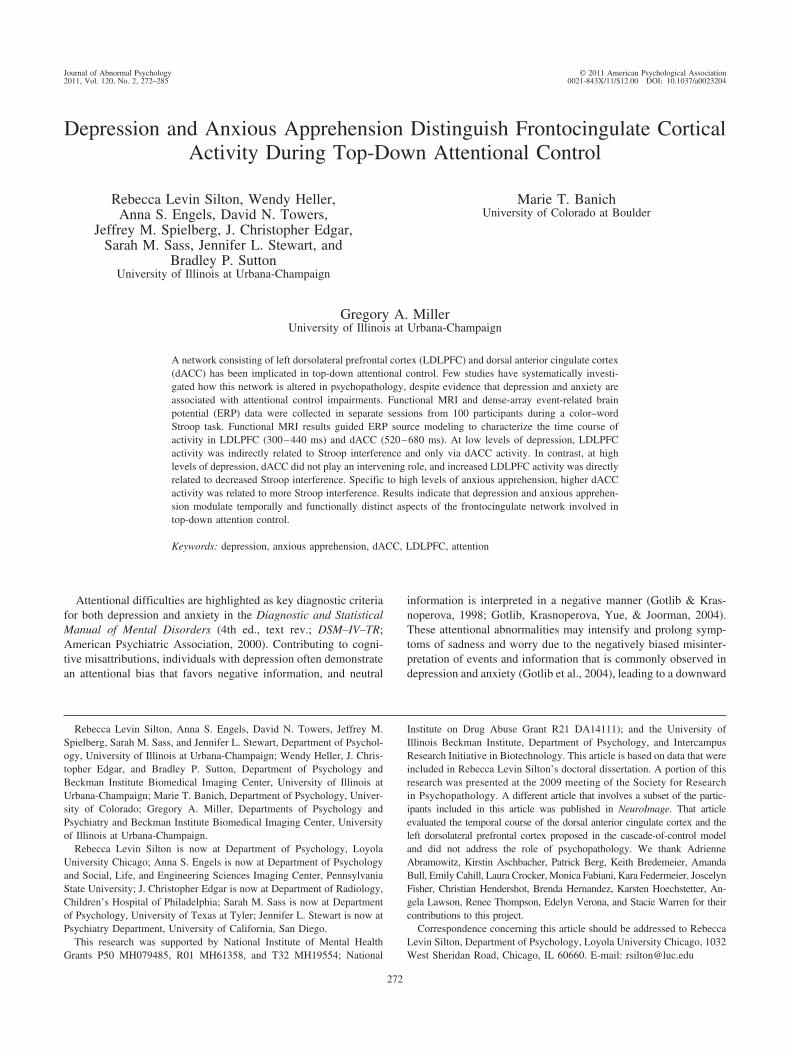
Source modeling was carried out with BESA. The source model(see Figure 1B for full model) was created by placing a prioriregional sources based on center of mass coordinates for fMRIactivation clusters obtained from the 30 psychopathology-freeparticipants as discussed above. Fourteen candidate locations sur-vived thresholding (see Table 2). If all 14 clusters were placed assources in the model, the model would have overfit the data.Rather, the selection of sources from among these clusters wasbased on relevant color–word Stroop fMRI research (Michel etal., 2004). Four of these 14 clusters (LDLPFC, dACC, rightinferior gyrus, left parietal cortex) were used in the sourcemodel (see Figure 1A for fMRI images). Although analyses forthe present study primarily involved LDLPFC and dACC, thefull source model included right inferior gyrus, left parietalcortex, and right parietal cortex to account for variance that isthought to be contributed by these sources based on availableliterature. The LDLPFC and dACC locations were very close tothe locations proposed by the cascade-of-control model(Banich, 2009). Similar studies that have used nonverbal stim-uli have also implicated LDLPFC and dACC, suggesting thattasks that involve top-down attentional control recruit thesebrain regions across stimulus types (Fan, Flombaum,
Table 2Functional MRI Center of Mass Coordinates (Montreal Neurological Institute)
Region Cluster size Mean z x y z
Left frontal orbital cortex 545 3.76 �31 19 �8Right frontal orbital cortex 749 3.61 36 21 �6Left inferior temporal gyrus 36 3.36 �53 �56 �15Left intracalcarine cortex 111 3.39 �8 �79 2Right thalamus 1,126 3.61 0 �16 7Right caudate 67 3.28 11 11 7Left putamen 37 3.31 �23 �2 9Right inferior frontal gyrus 42 3.35 41 35 13Left precentral gyrus 1,195 3.72 �40 11 34Right anterior cingulate gyrus 182 3.50 7 21 27Left lateral occipital cortex 1,376 3.77 �35 �57 44Left precuneus cortex 443 3.61 �5 �63 45Paracingulate gyrus 335 3.54 0 14 51Right superior parietal lobule 132 3.49 39 �50 48
Note. Table data from “The Time Course of Activity in Dorsolateral Prefrontal Cortex and Anterior CingulateCortex During Top-Down Attentional Control,” by R. L. Silton, W. Heller, D. N. Towers, A. S. Engels, J. M.Spielberg, J. C. Edgar, . . . G. A. Miller, 2010, NeuroImage, 50, p. 1295. Copyright 2010 by Elsevier.
277DEPRESSION, APPREHENSION, AND CORTICAL ACTIVITY
McCandliss, Thomas, & Posner, 2003; Liu, Banich, Jacobson,& Tanabe, 2006).
Prior to placing these sources in the model, blink activity wasmodeled as described above. Next, bilateral visual cortex sources(left occipital cortex and right occipital cortex) were localizedbased on ERP data from correct trials in the neutral condition. Theneutral condition involved the largest number of trials and wasselected to maximize the signal-to-noise ratio for localization. Agrand average computed from per-subject waveform averages for
neutral-trial blocks from all psychopathology-free participants(n � 66) was used for localizing the visual sources. The epochused for the localization was 100–188 ms, spanning primary andsecondary visual cortex responses. The left and right occipitalcortex sources were constrained to be symmetrical (see Figure 1Bfor left and right occipital cortex coordinates). Finally, theLDLPFC, dACC, right inferior frontal gyrus, and left parietalcortex sources were placed in the model along with a contralateralright parietal cortex source. Because magnitude of source activity,
Figure 1. (A) Left dorsolateral prefrontal cortex (LDLPFC) and dorsal anterior cingulate cortex (dACC)activation for incongruent versus congruent stimuli (z � 3.0902, p � .01, cluster size � 34; corrected p � .05).Crosshairs placed at center of intensity. (B) Functional MRI Montreal Neurological Institute (MNI) coordinatesfor source model containing seven regional sources used in brain electrical source analysis source modeling. L �left; R � right; RIFG � right inferior frontal gyrus; LPC � left parietal cortex; RPC � right parietal cortex;LOc � left occipital cortex; ROc � right occipital cortex. Adapted from “The Time Course of Activity inDorsolateral Prefrontal Cortex and Anterior Cingulate Cortex During Top-Down Attentional Control,” by R. L.Silton, W. Heller, D. N. Towers, A. S. Engels, J. M. Spielberg, J. C. Edgar, . . . G. A. Miller, 2010, NeuroImage,50, p. 1296. Copyright 2010 by Elsevier.
278 LEVIN SILTON ET AL.

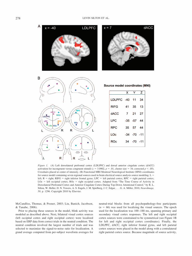
rather than orientation of source activity, was the primary variableof interest, all dipoles were converted to regional sources. TheERP data were digitally filtered 0.1–12 Hz, and the sourcemodel was applied separately in each Stroop condition (con-gruent, incongruent) for each participant. Prestimulus baselineactivity (�200 ms to 0 ms) was removed from the sourcewaveforms after the model was fit to each participant. Scoringwindows were based on visual inspection of the source wave-forms as well as taking into consideration findings from rele-vant scalp- and source-ERP color–word Stroop research. Onewindow for LDLPFC (300 – 440 ms) and two windows fordACC (220 –340 ms, 520 – 680 ms) were identified. Sourcecomponent amplitude was calculated by averaging data points24 ms before and 24 ms after peak latency. With the exceptionof determining the location and temporal scoring window of thesources, all source analysis steps described above were per-formed separately for each of the 100 participants.
Moderated Mediation Analyses
Moderated mediation analyses (Preacher, Rucker, & Hayes,2007) were used to evaluate the hypothesis that high levels ofdepression would interfere with the relationship between LDLPFCand dACC previously observed in the nonclinical sample (Silton etal., 2010). In the context of a mediation model, a moderatorvariable is an additional variable that is not part of the causalsequence that modifies the relationship between two variables(e.g., independent and dependent variables). Moderator effects arealso referred to as interactions. Continuous psychopathology vari-ables (MASQ–AD-8 and PSWQ scores) were assigned as moder-ators to evaluate whether the relationships between LDLPFC anddACC and between dACC and Stroop interference depended onlevels of psychopathology (Figure 2 provides a graphic represen-
tation of these moderated mediation models). A series of linearregressions were used to test the moderated mediation models.Five participants were considered outliers on source measures (3standard deviations from the mean for at least one component) andwere omitted from subsequent analyses (resulting n � 95). TheSPSS macro (MODMED) described in Preacher et al. (2007;http://www.comm.ohio-state.edu/ahayes) was used to conduct themoderation analyses.
Results
Behavioral Performance
RT analyses were conducted to confirm that the Stroop inter-ference effect was obtained in the present sample. A multivariateanalysis of variance with condition (congruent RT, incongruentRT) and gender confirmed slower RT for incongruent than forcongruent trials: for condition, F(1, 94) � 214.10, p � .001(congruent: M � 631 ms, SD � 95; incongruent: M � 791 ms,SD � 138). The Stroop effect did not vary by gender. Participantsmade more errors during the incongruent than the congruent con-dition, F(1, 94) � 48.66, p � .001 (congruent: M � 0.68 errors,SD � 0.95; incongruent: M � 2.37 errors, SD � 2.22). Thedepression and anxiety measures were not significantly correlatedwith congruent or incongruent RT, errors, or Stroop interference.
Source-Waveform ERP Moderated Mediation Analysis
The ERP source-waveform data analyses (see Figure 3 forwaveforms) employed scores from incongruent trials only, toexamine the effects of psychopathology within the context ofcognitive control mechanisms prompted by Stroop conflict.
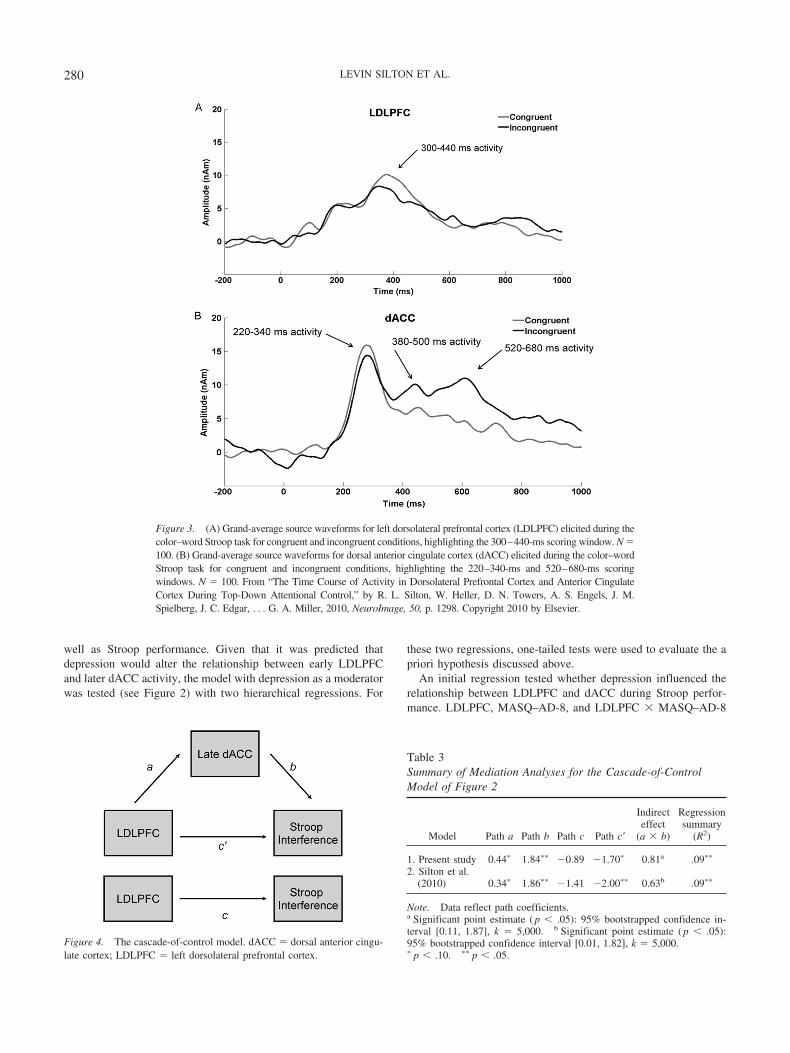
Replication of cascade-of-control model mediation analysis.Prior to proceeding with moderation analyses, the mediation anal-yses were repeated (see Figure 4), as the sample selection proce-dures varied from Silton et al. (2010). Figure 4 depicts the medi-ation model that was tested. The present sample includedparticipants recruited from the community, which broadened thesample, increased the sample size, and included more psychopa-thology. The mediation analyses for the cascade-of-control modelwere replicated and are presented in Table 3 (see Model 1). Theindirect effect was used to test directly the overall significance ofthe cascade-of-control model (Preacher & Hayes, 2008). As be-fore, now with a sample expanded to include participants recruitedfrom the community, the indirect effect was significant, and thecascade-of-control model was supported, with relevant LDLPFCactivity preceding rather than following relevant dACC activity.Similar to the findings in Silton et al. (2010), the total varianceaccounted for was 9%, F(2, 92) � 4.29, p � .02, which representsa medium effect size (Cohen, 1992; 9% corresponds to r � .30,which is standard for a medium effect size).
The influence of psychopathology on the frontocingulatenetwork: Moderated mediation analyses. It was predicted thatdepression would influence the frontocingulate network that isactivated during Stroop performance. Depression was expected tobe associated with reduced LDLPFC activity, which in turn wouldinfluence subsequent dACC activity and related Stroop perfor-mance. Specifically, the interaction of depression with earlyLDLPFC activity was expected to predict later dACC activity as
Figure 2. Moderation models for cascade-of-control model: depression(Mood and Anxiety Symptom Questionnaire–Anhedonic Depression eight-item depressed mood subscale) as moderator (A) and anxiety (Penn StateWorry Questionnaire) as moderator (B). LDLPFC � left dorsolateralprefrontal cortex; dACC � dorsal anterior cingulate cortex.
279DEPRESSION, APPREHENSION, AND CORTICAL ACTIVITY
well as Stroop performance. Given that it was predicted thatdepression would alter the relationship between early LDLPFCand later dACC activity, the model with depression as a moderatorwas tested (see Figure 2) with two hierarchical regressions. For
these two regressions, one-tailed tests were used to evaluate the apriori hypothesis discussed above.
An initial regression tested whether depression influenced therelationship between LDLPFC and dACC during Stroop perfor-mance. LDLPFC, MASQ–AD-8, and LDLPFC � MASQ–AD-8
Figure 3. (A) Grand-average source waveforms for left dorsolateral prefrontal cortex (LDLPFC) elicited during thecolor–word Stroop task for congruent and incongruent conditions, highlighting the 300–440-ms scoring window. N �100. (B) Grand-average source waveforms for dorsal anterior cingulate cortex (dACC) elicited during the color–wordStroop task for congruent and incongruent conditions, highlighting the 220–340-ms and 520–680-ms scoringwindows. N � 100. From “The Time Course of Activity in Dorsolateral Prefrontal Cortex and Anterior CingulateCortex During Top-Down Attentional Control,” by R. L. Silton, W. Heller, D. N. Towers, A. S. Engels, J. M.Spielberg, J. C. Edgar, . . . G. A. Miller, 2010, NeuroImage, 50, p. 1298. Copyright 2010 by Elsevier.
Figure 4. The cascade-of-control model. dACC � dorsal anterior cingu-late cortex; LDLPFC � left dorsolateral prefrontal cortex.
Table 3Summary of Mediation Analyses for the Cascade-of-ControlModel of Figure 2
Model Path a Path b Path c Path c�
Indirecteffect
(a � b)
Regressionsummary
(R2)
1. Present study 0.44� 1.84�� �0.89 �1.70� 0.81a .09��
2. Silton et al.(2010) 0.34� 1.86�� �1.41 �2.00�� 0.63b .09��
Note. Data reflect path coefficients.a Significant point estimate ( p � .05): 95% bootstrapped confidence in-terval [0.11, 1.87], k � 5,000. b Significant point estimate ( p � .05):95% bootstrapped confidence interval [0.01, 1.82], k � 5,000.� p � .10. �� p � .05.
280 LEVIN SILTON ET AL.

were predictors, and late dACC activity was the DV. Added last,LDLPFC � MASQ–AD-8 was significant, b � �0.06, t(91) ��1.99, p � .035 (one-tailed). The results of this analysis showedthat the interaction of LDLPFC and depression predicted dACCactivity. The omnibus model accounted for 14% of the variance,F(3, 91) � 4.82, p � .01, which corresponds to r � .37, repre-senting a medium effect size (Cohen, 1992). The LDLPFC �MASQ–AD-8 interaction accounted for 4% of the total variancefor this model. Interactions observed in psychological researchtypically account for a few percentage points of variance beyondfirst-order effects (Cohen, Cohen, West, & Aiken, 2003).
Next, a regression evaluated whether the interaction of depressionand LDLPFC predicted Stroop performance when variance related todACC was accounted for. LDLPFC, MASQ–AD-8, LDLPFC �MASQ–AD-8, and dACC were predictors, and Stroop interferencewas the DV. Added last, the interaction was significant, b � �0.32,t(90) � �1.64, p � .05 (one-tailed), a finding sufficient for adirectional a priori hypothesis. The omnibus model accounted for15% of the variance, F(4, 90) � 3.86, p � .01, which corresponds tor � .39, representing a medium effect size (Cohen, 1992). TheLDLPFC � MASQ–AD-8 interaction contributed 3% of the totalvariance for this model. Overall, these findings indicate that depres-sion and LDLPFC interact to predict Stroop performance.
In order to better understand the moderating effects of depres-sion, the interactions for both regressions were plotted (see Figures5A and 5B), and the significance of the slopes was tested (Aiken& West, 1991). In Figures 5A–5C, “low” and “high” refer to �1standard deviation (Aiken & West, 1991). Importantly, the test ofsimple slopes is not a test of an interaction effect (the interactionsare tested in the regression above). Rather, it is a method ofdescribing the nature of the interactive relationship (Aiken &West, 1991). High and low are defined relative to the presentsample, and these terms are used to represent two portions of adimension rather than classification categories used to distinguishthe absence or presence of clinical diagnoses. The t test forwhether a simple slope differed from zero was calculated bydividing the value of the simple slope by its standard error with(n � k � 1) degrees of freedom (where n is the number of casesand k is the number of predictors). The standard error was calcu-lated from the variance–covariance matrix of the regression coef-ficients. As shown in Figure 5A, LDLPFC activity predicteddACC activity at low levels of depression, t(91) � 3.69, p � .001,but not at higher levels of depression, t(91) � 0.54, p � .59.Furthermore, Figure 5B shows that LDLPFC activity was associ-ated with less Stroop interference at higher levels of depression,t(90) � �2.28, p � .03, but not at low levels of depression,t(90) � �0.05, p � .96. That is, at low levels of depression, Stroopinterference did not vary as a function of LDLPFC activity. Forindividuals higher in depression, LDLPFC and dACC were lesswell coupled, and LDLPFC activity was more tightly linked di-rectly to RT performance.
Moderated mediation analyses were conducted to evaluatewhether anxious apprehension also influenced the frontocingulatenetwork during Stroop performance (see Figure 2B). Similar instructure to the regression analyses conducted with MASQ–AD-8,two hierarchical regressions were used to test the influence ofanxious apprehension on the mechanisms articulated in thecascade-of-control model. Two-tailed significance tests were usedfor these regressions, because these analyses were implemented to
evaluate exploratory hypotheses. First, a regression was conductedto assess whether anxious apprehension influenced the relationshipbetween LDLPFC and dACC. LDLPFC, PSWQ, and LDLPFC �PSWQ were predictors, and late dACC activity was the DV. Aspredicted, the interaction was not significant. Second, a regressionwas conducted to evaluate whether anxiety interacted with either
A
B
C
0
5
10
15
20
25
30
Low LDLPFC High LDLPFC
dAC
C A
ctiv
ity (n
Am)
LDLPFC x MASQ-AD-8
Low MASQ-AD-8High MASQ-AD-8
0
50
100
150
200
250
Low LDLPFC High LDLPFC
Stro
op In
terfe
renc
e
LDLPFC x MASQ-AD-8
Low MASQ-AD-8
High MASQ-AD-8
0
50
100
150
200
250
Low dACC High dACC
Stro
op In
terfe
renc
e
dACC x PSWQ
Low PSWQHigh PSWQ
Figure 5. (A) Left dorsolateral prefrontal cortex (LDLPFC) � Mood andAnxiety Symptom Questionnaire–Anhedonic Depression Eight-Item De-pressed Mood Subscale (MASQ–AD-8) interaction and tests of simpleslopes show that at low levels of depression, LDLPFC predicts dorsalanterior cingulate cortex (dACC). (B) LDLPFC � MASQ–AD-8 interac-tion, with Stroop interference as the predictor and tests of simple slopes,shows that at high levels of depression, increased LDLPFC activity isrelated to less interference. (C) dACC � Penn State Worry Questionnaire(PSWQ) interaction and tests of simple slopes show that at high levels ofanxiety, increased dACC is related to greater Stroop interference.
281DEPRESSION, APPREHENSION, AND CORTICAL ACTIVITY

LDLPFC or dACC activity to predict Stroop performance.LDLPFC, dACC, PSWQ, LDLPFC � PSWQ, and dACC �PSWQ were predictors, and Stroop interference was the DV. ThedACC � PSWQ interaction was significant, b � �0.11, t(89) �2.37, p � .02, but LDLPFC � PSWQ was not, b � �0.04, t(89) ��0.70, p � .48, indicating that anxious apprehension influencedthe relationship between dACC and Stroop interference effect butnot the relationship between LDLPFC and dACC. The modelincluding only the significant dACC � PSWQ interaction (theLDLPFC � PSWQ interaction was not included) accounted for14% of the variance, F(4, 90) � 3.79, p � .01, which correspondsto r � .37, a medium effect size (Cohen, 1992). The dACC �PSWQ interaction accounted for 5% of the total variance in thismodel. In order to interpret the moderating effects of anxiety, theinteraction was plotted (see Figure 5C). The slope was significantfor higher levels of anxiety, t(89) � 3.59, p � .001, but not for lowlevels of anxiety t(89) � 0.39, p � .67. At higher levels of anxiousapprehension, increased dACC activity was related to greaterStroop interference.
In order to ascertain whether the influence of anxiety on dACCfunction was specific to anxiety, a final moderation analysis wasconducted to evaluate whether depression also modified dACCfunction during Stroop performance, when variance related toLDLPFC was accounted for. This regression included LDLPFC,dACC, MASQ–AD-8, and dACC � MASQ–AD-8 (onlyLDLPFC � MASQ–AD-8 was tested previously) as predictors,and Stroop interference was the DV. The interaction was notsignificant, b � �0.06, t(90) � 0.17, p � .74, indicating thatdepression and anxious apprehension influence distinct aspects ofthe frontocingulate network in distinct ways.
Discussion
The present study examined how depression and anxiety influ-ence frontocingulate activity under conditions of high attentionaldemand. A previous study showed that, ignoring psychopathology,the extent to which dACC activation influenced Stroop perfor-mance depended on the degree of earlier LDLPFC activity (Siltonet al., 2010). When earlier LDLPFC activity was high, later dACCactivity did not influence the degree of Stroop interference,whereas when earlier LDLPFC activity was low, higher laterdACC activity was associated with reduced Stroop interference.On the basis of this pattern of activity, it was predicted in thepresent study that depression would be related to reduced earlyLDLPFC activity, which in turn was expected to influence subse-quent later dACC activity and related Stroop interference. Anxiousapprehension was expected to influence dACC activity but notLDLPFC activity. It was unclear how this pattern of networkactivity might affect performance. Results showed that both de-pression and anxiety affected this frontocingulate network in-volved in attentional control and did so in different ways.
LDLPFC activity predicted dACC activity only at low levels ofdepression during Stroop performance, indicating a functionalrelationship similar to the one observed in Silton et al. (2010), suchthat earlier LDLPFC activity predicted later dACC activity. Athigher levels of depression, however, LDLPFC and dACC activitywere less related. As the relationship between LDLPFC and dACCactivity weakened with increasing depression, a direct relationshipbetween LDLPFC and performance emerged. In the context of this
weakened neural coupling associated with depression, increasedLDLPFC activity was associated with reduced Stroop interference(better performance). Although the degree of depression alonedoes not directly predict performance on the Stroop task, it appearsthat it does alter the neural circuitry that is employed to meet taskdemands. This pattern of activity is consistent with the predictionsof the cascade-of-control model, such that increased LDLPFCactivity is indicative of an increased need for compensatory top-down control.
At higher levels of anxious apprehension, increased dACCactivity was related to greater Stroop interference (worse perfor-mance), suggesting that as anxious apprehension increases, cogni-tive control is implemented increasingly via dACC. Conceivably,anxious apprehension is associated with worries about aspects ofperformance, which in turn interfere with adaptive conflict reso-lution, leading to increased recruitment of dACC to aid in responseselection.
Very few studies have addressed the relationship between anx-iety and dACC function during top-down attentional control. In-stead, most research has focused on rostral anterior cingulatecortex (rACC; called the “affective” region by Bush, Luu, &Posner, 2000) and its role in processing affective information (e.g.,Bishop, Duncan, Brett, & Lawrence, 2004; Engels et al., 2007;Mohanty et al., 2007). Evidence suggests that dACC and rACC aredistinct regions that contribute to different cortical and subcorticalpathways. Whereas rACC has been implicated in the evaluation ofemotional information and the regulation of emotional responses,dACC is often associated with cognitive function, particularlyduring tasks that involve conflict resolution (Bush et al., 2000;Mohanty et al., 2007).
Given different roles, it is not surprising that inverse patterns ofrACC activity and dACC activity have been associated with anx-iety. Lower rACC activity has been related to higher anxiety,possibly indicating less control in the presence of threateningstimuli (Bishop et al., 2004; Engels et al., 2007). In contrast torACC, present results showed that higher dACC activity wasassociated with higher anxious apprehension levels. Similar find-ings have been reported in other studies (Breiter et al., 1996;Bystritsky et al., 2001; Eisenberger, Liberman, & Satpute, 2005;Ursu, Stenger, Shear, Jones, & Carter, 2003). Eisenberger et al.(2005) showed that neuroticism, a personality factor that is con-sistently related to anxiety, was positively correlated with dACCactivity but negatively correlated with rACC. Moreover, the Eisen-berger et al. study showed that individuals high in neuroticismdemonstrated increased dACC activity during conflict trials. Thesefindings suggested that individuals high in anxiety have an abnor-mal conflict system that is reflected in higher dACC activity,consistent with many studies showing that dACC is engaged inconflict resolution and later aspects of response selection (e.g.,Banich, 2009; Botvinick, Cohen, & Carter, 2004; Silton et al.,2010). Similarly, a study that involved individuals with obsessive-compulsive disorder (an anxiety disorder that commonly involveshigh levels of worry) showed more dACC activity during high-conflict trials (Ursu et al., 2003), and Krug and Carter (2010)found that individuals high on trait anxiety had more dACCactivity than individuals low on trait anxiety during conflict trialsin a facial Stroop task. However, another study that directlyinvestigated the impact of trait and state anxiety on dACC functionfound that anxiety did not influence dACC during attentional
282 LEVIN SILTON ET AL.

control (Bishop, 2009). Possibly, the letter search task used in thatstudy did not involve the level of conflict resolution demanded bythe Stroop task, which has repeatedly been shown to involvedACC (Botvinick et al., 2004). Although more research is neededto elucidate the various ways that anxiety types may differentiallyaffect dACC and rACC activity and related cognitive function, thebulk of the evidence favors the conclusion that anxious apprehen-sion or worry is associated with more dACC activity.
Present data suggest that performance impairments in anxiousindividuals during conflict resolution tasks are related to inade-quate dACC-mediated cognitive control mechanisms that wouldtypically suppress attentional disruption caused by worries orruminations. Inadequate control mechanisms may lead to furtherdifficulties shifting attention away from such concerns. Inadequatecompensatory dACC activity and related difficulties resolvingconflict may accentuate problems resolving issues of daily life andthus contribute to a ruminative cycle due to a lack of moreeffective and efficient problem-solving options.
The present study is apparently the first to explicitly evaluatethe influence of depression and anxious apprehension on afrontocingulate network (not solely focal cortical activity) dur-ing top-down attentional control. Results showed that depres-sion and anxiety affect the network in different ways, and thesedifferent patterns of network activity were generally consistentwith the predictions of the cascade-of-control model. This studyprovides support for models that posit that depression influ-ences a network rather than individual brain regions in isolation(e.g., Heller, 1993; Mayberg, 1997). Unlike previous depressionneuroimaging studies that have used nondirectional correlationmethods such as functional connectivity, the present studyprovides unique information regarding how depression andanxiety modify specific temporal relationships between net-work segments involved in attentional control. Medium effectsizes were obtained for the models that were evaluated, and thepresent study was adequately powered to detect a mediumeffect size (per Cohen, 1992). Effect sizes for interactions inpsychological research are typically within the small to mediumrange (Cohen et al., 2003). It is rare for studies that have usedconnectivity methods to explicitly report effect size, so it isdifficult to estimate how the effect size obtained in the presentstudy compares with that of other studies. Because distinctpatterns of network activity were related to behavioral out-comes, the medium effect size in the present study suffices todemonstrate functional significance.
Future studies should continue to address how psychopathologyinfluences network activity during cognitive function, as multiplenetworks may be recruited based on specific task demands, anddifferent types of psychopathology will likely differentiate thesevarious networks and related function. Furthermore, evidence-based treatment outcome research that incorporates pre- and post-treatment neuroimaging measures may benefit from studyinghow treatment changes network activity rather than focusing onhow treatment influences individual brain regions. Research in thisvein may help inform future diagnostic categories and methodsthat aim to provide reliable identification of psychological disor-ders, along with furthering the development of effective evidence-based treatments for depression and anxiety.
References
Aguirre, G. K., Zarahn, E., & D’Esposito, M. (1998). The variability ofhuman, BOLD hemodynamic responses. NeuroImage, 8, 360–369. doi:10.1006/nimg.1998.0369
Aiken, L. S., & West, S. G. (1991). Multiple regression: Testing andinterpreting interactions. Thousand Oaks, CA: Sage.
American Psychiatric Association. (2000). Diagnostic and statistical man-ual of mental disorders (4th ed., text rev.). Washington, DC: Author.
Banich, M. T. (2009). Executive function: The search for an integratedaccount. Current Directions in Psychological Science, 18, 89–94. doi:10.1111/j.1467-8721.2009.01615.x
Behar, E., Alcaine, O., Zuellig, A. R., & Borkovec, T. D. (2003). Screeningfor generalized anxiety disorder using the Penn State Worry Question-naire: A receiver operating characteristic analysis. Journal of BehaviorTherapy and Experimental Psychiatry, 34, 25–43. doi:10.1016/S0005-7916(03)00004-1
Berg, P., & Scherg, M. (1994). A multiple source approach to the correc-tion of eye artifacts. Electroencephalography and Clinical Neurophysi-ology, 90, 229–241. doi:10.1016/0013-4694(94)90094-9
Biringer, E., Lundervold, A., Stordal, K., Mykletun, A., Egeland, J.,Bottlender, R., & Lund, A. (2005). Executive function improvementupon remission of recurrent unipolar depression. European Archives ofPsychiatry and Clinical Neuroscience, 255, 373–380. doi:10.1007/s00406-005-0577-7
Bishop, S. J. (2009). Trait anxiety and impoverished prefrontal control ofattention. Nature Neuroscience, 12, 92–98. doi:10.1038/nn.2242
Bishop, S. J., Duncan, J., Brett, M., & Lawrence, A. D. (2004). Prefrontalcortical function and anxiety: Controlling attention to threat-relatedstimuli. Nature Neuroscience, 7, 184–188. doi:10.1038/nn1173
Botvinick, M. M., Cohen, J. D., & Carter, C. S. (2004). Conflict monitoringand anterior cingulate cortex: An update. Trends in Cognitive Sciences,8, 539–546. doi:10.1016/j.tics.2004.10.003
Bredemeier, K., Spielberg, J. M., Silton, R. L., Berenbaum, H., Heller, W.,& Miller, G. A. (2010). Screening for depressive disorders using theMood Anxiety Symptoms Questionnaire Anhedonic Depression Scale:A receiver-operating characteristic analysis. Psychological Assessment,22, 702–710. doi:10.1037/a0019915
Breiter, H. C., Rauch, S. L., Kwong, K. K., Baker, J. R., Weisskoff, R. M.,Kennedy, D. N., . . . Rosen, B. R. (1996). Functional magnetic resonanceimaging of symptom provocation in obsessive-compulsive disorder.Archives of General Psychiatry, 53, 595–606.
Brody, A. L., Saxena, S., Stoessel, P., Gillies, L. A., Fairbanks, L. A.,Alborzian, S., . . . Baxter, L. R., Jr. (2001). Regional brain metabolicchanges in patients with major depression treated with either paroxetineor interpersonal therapy: Preliminary findings. Archives of GeneralPsychiatry, 58, 631–640. doi:10.1001/archpsyc.58.7.631
Bush, G., Luu, P., & Posner, M. I. (2000). Cognitive and emotionalinfluences in anterior cingulate cortex. Trends in Cognitive Sciences, 4,215–222. doi:10.1016/S1364-6613(00)01483-2
Bystritsky, A., Pontillo, D., Powers, M., Sabb, F. W., Craske, M. G., &Bookheimer, S. Y. (2001). Functional MRI changes during panic antic-ipation and imagery exposure. NeuroReport, 12, 3953–3957. doi:10.1097/00001756-200112210-00020
Cohen, J. (1992). A power primer. Psychological Bulletin, 112, 155–159.doi:10.1037/0033-2909.112.1.155
Cohen, J., Cohen, P., West, S. G., & Aiken, L. S. (2003). Applied multipleregression/correlation analysis for the behavioral sciences (3rd ed.).Mahwah, NJ: Erlbaum.
Compton, R. J., Heller, W., Banich, M. T., Palmieri, P. A., & Miller, G. A.(2000). Responding to threat: Hemispheric asymmetries and interhemi-spheric division of input. Neuropsychology, 14, 254–264. doi:10.1037/0894-4105.14.2.254
Duncan, J., & Owen, A. M. (2000). Common regions of the human frontal
283DEPRESSION, APPREHENSION, AND CORTICAL ACTIVITY

lobe recruited by diverse cognitive demands. Trends in Neurosciences,23, 475–483. doi:10.1016/S0166-2236(00)01633-7
Dunkin, J. J., Leuchter, A. F., Cook, I. A., Kasl-Godley, J. E., Abrams, M.,& Rosenberg-Thompson, S. (2000). Executive dysfunction predicts non-response to fluoxetine in major depression. Journal of Affective Disor-ders, 60, 13–23. doi:10.1016/S0165-0327(99)00157-3
Eisenberger, N. I., Liberman, M. D., & Satpute, A. J. (2005). Personalityfrom a controlled processing perspective: An fMRI study of neuroticism,extraversion, and self-consciousness. Cognitive, Affective & BehavioralNeuroscience, 5, 169–181. doi:10.3758/CABN.5.2.169
Engels, A. S., Heller, W., Mohanty, A., Herrington, J. D., Banich, M. T.,Webb, A. G., & Miller, G. A. (2007). Specificity of regional brainactivity in anxiety types during emotion processing. Psychophysiology,44, 352–363. doi:10.1111/j.1469-8986.2007.00518.x
Engels, A. S., Heller, W., Spielberg, J. M., Warren, S. L., Sutton, B. P.,Banich, M. T., & Miller, G. A. (2010). Co-occurring anxiety influencespatterns of brain activity in depression. Cognitive, Affective & Behav-ioral Neuroscience, 10, 141–156. doi:10.3758/CABN.10.1.141
Eysenck, M. W., Derakshan, N., Santos, R., & Calvo, M. G. (2007).Anxiety and cognitive performance: Attentional control theory. Emo-tion, 7, 336–353. doi:10.1037/1528-3542.7.2.336
Fan, J., Flombaum, J. I., McCandliss, B. D., Thomas, K. M., & Posner,M. I. (2003). Cognitive and brain consequences of conflict. NeuroImage,18, 42–57. doi:10.1006/nimg.2002.1319
First, M. B., Spitzer, R. L., Gibbon, M., & Williams, J. B. W. (1997).Structured Clinical Interview for DSM–IV Axis I Disorders: Non-Patient edition (SCID–I/NP, Version 2.0, 4/97 revision). New York, NY:Biometrics Research, New York State Psychiatric Institute.
Fitzgerald, P. B., Oxley, T. J., Laird, A. R., Kulkarni, J., Egan, G. F., &Daskalakis, Z. J. (2006). An analysis of functional neuroimaging studiesof dorsolateral prefrontal cortical activity in depression. PsychiatryResearch: Neuroimaging, 148, 33– 45. doi:10.1016/j.pscychresns.2006.04.006
Fox, E. (1993). Attentional bias in anxiety: Selective or not? BehaviourResearch and Therapy, 31, 487– 493. doi:10.1016/0005-7967(93)90129-I
Gehring, W. J., Himle, J., & Nisenson, L. G. (2000). Action monitoringdysfunction in obsessive-compulsive disorder. Psychological Science,11, 1–6. doi:10.1111/1467-9280.00206
George, M. S., Ketter, T. A., Parekh, P. I., Rosinsky, N., Ring, H. A.,Pazzaglia, P. J., . . . Post, R. M. (1997). Blunted left cingulate activationin mood disorder subjects during a response interference task (theStroop). Journal of Neuropsychiatry and Clinical Neurosciences, 9,55–63.
Gotlib, I. H., & Krasnoperova, E. (1998). Information processing as avulnerability factor for depression. Behavior Therapy, 29, 603–617.doi:10.1016/S0005-7894(98)80020-8
Gotlib, I. H., Krasnoperova, E., Yue, D. N., & Joorman, J. (2004). Atten-tional biases for negative interpersonal stimuli in clinical depression.Journal of Abnormal Psychology, 113, 127–135. doi:10.1037/0021-843X.113.1.121
Hajcak, G., McDonald, N., & Simons, R. F. (2003). Anxiety and error-related brain activity. Biological Psychology, 64, 77–90. doi:10.1016/S0301-0511(03)00103-0
Heller, W. (1993). Neuropsychological mechanisms of individual differ-ences in emotion, personality, and arousal. Neuropsychology, 7, 476–489. doi:10.1037/0894-4105.7.4.476
Herrington, J. D., Heller, W., Mohanty, A., Engels, A. S., Banich, M. T.,Webb, A. G., & Miller, G. A. (2010). Localization of asymmetric brainfunction in emotion and depression. Psychophysiology, 47, 442–454.
Hertel, P. T. (1994). Depression and memory: Are impairments remediablethrough attentional control? Current Direction in Psychological Science,3, 190–193. doi:10.1111/1467-8721.ep10770707
Hertel, P. T. (2007). Impairments in inhibition or cognitive control in
psychological disorders. Applied and Preventive Psychology, 12, 149–153. doi:10.1016/j.appsy.2007.09.006
Hochman, S. H. (1967). The effects of stress on Stroop color–wordperformance. Psychonomic Science, 9, 475–476.
Holmes, A. J., & Pizzagalli, D. A. (2008). Response conflict and fronto-cingulate dysfunction in unmedicated participants with major depression.Neuropsychologia, 46, 2904 –2913. doi:10.1016/j.neuropsychologia.2008.05.028
Jaeger, J., Berns, S., Uzelac, S., & Davis-Conway, S. (2006). Neurocog-nitive deficits and disability in major depressive disorder. PsychiatryResearch, 145, 39–48. doi:10.1016/j.psychres.2005.11.011
Jenkinson, M., Bannister, P., Brady, M., & Smith, S. (2002). Improvedoptimization for the robust and accurate linear registration and motioncorrection of brain images. NeuroImage, 17, 825–841. doi:10.1006/nimg.2002.1132
Kessler, R. C., DuPont, R. L., Berglund, P., & Wittchen, H.-U. (1999).Impairment in pure and comorbid generalized anxiety disorder andmajor depression at 12 months in two national surveys. AmericanJournal of Psychiatry, 156, 1915–1923.
Kessler, R. C., Nelson, C. B., McGonagle, K. A., Liu, J., Swartz, M., &Blazer, D. G. (1996). Comorbidity of DSM–III–R major depressivedisorder in the general population: Results from the US National Co-morbidity Survey. British Journal of Psychiatry, 168, 17–30.
Kessler, R. C., Zhao, S., Blazer, D. G., & Swartz, M. (1997). Prevalence,correlates, and course of minor depression and major depression in theNational Comorbidity Survey. Journal of Affective Disorders, 45, 19–30. doi:10.1016/S0165-0327(97)00056-6
Killgore, W. D. S., Gruber, S. A., & Yurgelun-Todd, D. A. (2007).Depressed mood and lateralized prefrontal activity during a Stroop taskin adolescent children. Neuroscience Letters, 416, 43–48. doi:10.1016/j.neulet.2007.01.081
Krug, M. K., & Carter, C. S. (2010). Adding fear to conflict: A generalpurpose cognitive control network is modulated by trait anxiety. Cog-nitive, Affective & Behavioral Neuroscience, 10, 357–371.
Liu, X., Banich, M. T., Jacobson, B. L., & Tanabe, J. L. (2006). Functionaldissociation of attentional selection within PFC: Response and non-response related aspects of attentional selection as ascertained by fMRI.Cerebral Cortex, 16, 827–834. doi:10.1093/cercor/bhj026
Mayberg, H. S. (1997). Limbic-cortical dysregulation: A proposed modelof depression. Journal of Neuropsychiatry and Clinical Neurosciences,9, 471–481.
Mayberg, H. S., Brannan, S. K., Tekell, J. L., Silva, J. A., Mahurin, R. K.,McGinnis, S., & Jerabek, P. A. (2000). Regional metabolic effects offluoxetine in major depression: Serial changes and relationship to clin-ical response. Biological Psychiatry, 48, 830–843. doi:10.1016/S0006-3223(00)01036-2
Meyer, T. J., Miller, M. L., Metzger, R. L., & Borkovec, T. D. (1990).Development and validation of the Penn State Worry Questionnaire.Behaviour Research and Therapy, 28, 487–495. doi:10.1016/0005-7967(90)90135-6
Michel, C. M., Murray, M. M., Lantz, G., Gonzalez, S., Spinelli, L., &Grave de Peralta, R. (2004). EEG source imaging. Clinical Neurophys-iology, 115, 2195–2222. doi:10.1016/j.clinph.2004.06.001
Miezin, F. M., Maccotta, L., Ollinger, J. M., Petersen, S. E., & Buckner,R. L. (2000). Characterizing the hemodynamic response: Effects ofpresentation rate, sampling procedure, and the possibility of orderingbrain activity based on relative timing. NeuroImage, 11, 735–739. doi:10.1006/nimg.2000.0568
Mohanty, A., Engels, A. S., Herrington, J. D., Heller, W., Ringo Ho,M.-H., Banich, M. T., . . . Miller, G. A. (2007). Differential engagementof anterior cingulate cortex subdivisions for cognitive and emotionalfunction. Psychophysiology, 44, 343–351. doi:10.1111/j.1469-8986.2007.00515.x
Molina, S., & Borkovec, T. D. (1994). The Penn State Worry Question-
284 LEVIN SILTON ET AL.

naire: Psychometric properties and associated characteristics. In G. C. L.Davey & F. Tallis (Eds.), Worrying: Perspectives on theory, assessmentand treatment (pp. 265–283). Chichester, England: Wiley.
Nitschke, J. B., & Heller, W. (2002). The neuropsychology of anxietydisorders: Affect, cognition, and neural circuitry. In H. A. H. D’haenen,J. A. den Boer, & P. Willner (Eds.), Biological psychiatry (Vol. 2, pp.975–988). Chichester, England: Wiley.
Nitschke, J. B., Heller, W., Imig, J. C., McDonald, R. P., & Miller, G. A.(2001). Distinguishing dimensions of anxiety and depression. CognitiveTherapy and Research, 25, 1–22. doi:10.1023/A:1026485530405
Nitschke, J. B., Heller, W., & Miller, G. A. (2000). Anxiety, stress, andcortical brain function. In J. C. Borod (Ed.), The neuropsychology ofemotion (pp. 298–319). New York, NY: Oxford University Press.
Oldfield, R. C. (1971). The assessment and analysis of handedness: TheEdinburgh Inventory. Neuropsychologia, 9, 97–113. doi:10.1016/0028-3932(71)90067-4
Olvet, D. M., & Hajcak, G. (2008). The error-related negativity (ERN) andpsychopathology: Toward an endophenotype. Clinical Psychology Re-view, 28, 1343–1354. doi:10.1016/j.cpr.2008.07.003
Paradiso, S., Lamberty, G. J., Garvey, M. J., & Robinson, R. G. (1997).Cognitive impairment in the euthymic phase of chronic unipolar depres-sion. Journal of Nervous and Mental Disease, 185, 748–754. doi:10.1097/00005053-199712000-00005
Paulus, M. P., Feinstein, J. S., Simmons, A., & Stein, M. B. (2004).Anterior cingulate activation in high trait anxious subjects is related toaltered error processing during decision making. Biological Psychiatry,55, 1179–1187. doi:10.1016/j.biopsych.2004.02.023
Preacher, K. J., & Hayes, A. F. (2008). Asymptotic and resamplingstrategies for assessing and comparing indirect effects in multiple me-diator models. Behavior Research Methods, 40, 879–891. doi:10.3758/BRM.40.3.879
Preacher, K. J., Rucker, D. D., & Hayes, A. F. (2007). Addressing mod-erated mediation hypotheses: Theory, methods, and prescriptions. Mul-tivariate Behavioral Research, 42, 185–227.
Ravnkilde, B., Videbech, P., Clemmensen, K., Egander, A., Rasmussen,N. A., & Rosenberg, R. (2002). Cognitive deficits in major depression.Scandinavian Journal of Psychology, 43, 239–251. doi:10.1111/1467-9450.00292
Rogers, M. A., Bradshaw, J. L., Pantelis, C., & Phillips, J. G. (1998).Frontostriatal deficits in unipolar major depression. Brain ResearchBulletin, 47, 297–310. doi:10.1016/S0361-9230(98)00126-9
Segal, Z. V., Williams, J. M. G., & Teasdale, J. D. (2002). Mindfulness-based cognitive therapy for depression: A new approach to preventingrelapse. New York, NY: Guilford Press.
Siegle, G. J., Ghinassi, F., & Thase, M. E. (2007). Neurobehavioraltherapies in the 21st century: Summary of an emerging field and an
extended example of cognitive control training for depression. CognitiveTherapy and Research, 31, 235–262. doi:10.1007/s10608-006-9118-6
Silton, R. L., Miller, G. A., Towers, D. N., Engels, A. S., Edgar, J. C.,Spielberg, J. M., . . . Heller, W. (2010). The time course of activity indorsolateral prefrontal cortex and anterior cingulate cortex during top-down attentional control. NeuroImage, 50, 1292–1302. doi:10.1016/j.neuroimage.2009.12.061
Stordal, K. I., Lundervold, A. J., Egeland, J., Mykletun, A., Asbjørnsen, A.,Landrø, N. I., . . . Lund, A. (2004). Impairment across executive func-tions in recurrent major depression. Nordic Journal of Psychiatry, 58,41–47. doi:10.1080/08039480310000789
Stroop, J. R. (1935). Studies of interference in serial verbal reactions.Journal of Experimental Psychology, 18, 643– 662. doi:10.1037/h0054651
Ursu, S., Stenger, V. A., Shear, M. K., Jones, M. R., & Carter, C. S. (2003).Overactive action monitoring in obsessive-compulsive disorder: Evi-dence from functional magnetic resonance imaging. Psychological Sci-ence, 14, 347–353. doi:10.1111/1467-9280.24411
Videbech, P., Ravnkilde, B., Gammelgaard, L., Egander, A., Clemmensen,K., Rasmussen, N. A., . . . Rosenberg, R. (2004). The Danish PET/depression project: Performance on Stroop’s test linked to white matterlesions in the brain. Psychiatry Research: Neuroimaging, 130, 117–130.doi:10.1016/j.pscychresns.2003.10.002
Wagner, G., Sinsel, E., Sobanski, T., Köhler, S., Marinou, V., Mentzel,H.-J., . . . Schlösser, R. G. M. (2006). Cortical inefficiency in patientswith unipolar depression: An event-related fMRI study with the Strooptask. Biological Psychiatry, 59, 958 –965. doi:10.1016/j.biopsych.2005.10.025
Ward, B. D. (2000). Simultaneous inference for fMRI data. Retrieved fromhttp://afni.nimh.nih.gov/pub/dist/doc/manual/AlphaSim.pdf
Watson, D., Clark, L. A., Weber, K., Assenheimer, J. S., Strauss, M. E., &McCormick, R. A. (1995). Testing a tripartite model: II. Exploring thesymptom structure of anxiety and depression in student, adult, andpatient samples. Journal of Abnormal Psychology, 104, 15–25. doi:10.1037/0021-843X.104.1.15
Watson, D., Weber, K., Assenheimer, J. S., Clark, L. A., Strauss, M. E., &McCormick, R. A. (1995). Testing a tripartite model: I. Evaluating theconvergent and discriminant validity of anxiety and depression symptomscales. Journal of Abnormal Psychology, 104, 3–14. doi:10.1037/0021-843X.104.1.3
Woolrich, M. W., Ripley, B. D., Brady, M., & Smith, S. M. (2001).Temporal autocorrelation in univariate linear modeling of fMRI data.NeuroImage, 14, 1370–1386. doi:10.1006/nimg.2001.0931
Received September 7, 2009Revision received September 27, 2010
Accepted September 30, 2010 �
285DEPRESSION, APPREHENSION, AND CORTICAL ACTIVITY
Related Documents