Jay Kelley Department of Oral Biology, College of Dentistry, University of Illinois, 801 S. Paulina, Chicago, Illinois 60612, U.S.A. E-mail: [email protected] Steve Ward Department of Anatomy, Northeastern Ohio Universities College of Medicine, P.O. Box 95, Rootstown, Ohio 44272, U.S.A. E-mail: [email protected] Barbara Brown Department of Orthopaedic Surgery, Northeastern Ohio Universities College of Medicine, P.O. Box 95, Rootstown, Ohio 44272, U.S.A. E-mail: [email protected] Andrew Hill Department of Anthropology, Yale University, P.O. Box 208277, Yale Station, New Haven, Connecticut 06520-8277, U.S.A. E-mail: [email protected] Dana L. Duren School of Biomedical Sciences, Kent State University, 110 Cunningham Hall, Kent, Ohio 44242, U.S.A. E-mail: [email protected] Received 28 February 2001 Revision received 6 June 2001 and accepted 20 June 2001 Keywords: Equatorius, Kenyapithecus, Miocene, Kipsaramon, Baringo, Muruyur, hominoids, dentition. Dental remains of Equatorius africanus from Kipsaramon, Tugen Hills, Baringo District, Kenya Forty-one isolated large hominoid teeth, as well as most of the mandibular and three maxillary teeth associated with a partial skeleton, were recovered from middle Miocene Muruyur sediments near Kipsaramon in the Tugen Hills, Baringo District, Kenya. The isolated teeth were collected as surface finds and the skeleton was excavated in situ at locality BPRP#122 dated between 15·58 Ma and 15·36 Ma. The majority of the teeth recovered at BPRP#122 are referable to a minimum of five individuals of the hominoid Equatorius africanus. Three of the teeth, however, are provisionally assigned to Nyanzapithecus sp. The new hominoids from Kipsaramon add to an increasing inventory of specimens that suggest greater large hominoid taxonomic diversity from the middle Miocene of Kenya than was previously recognized. It is suggested that there are two large-bodied hominoid species present at Mabako, only one of which is assignable to Equatorius. 2002 Academic Press Journal of Human Evolution (2002) 42, 39–62 doi:10.1006/jhev.2001.0504 Available online at http://www.idealibrary.com on 0047–2484/02/010039+24$35.00/0 2002 Academic Press

Welcome message from author
This document is posted to help you gain knowledge. Please leave a comment to let me know what you think about it! Share it to your friends and learn new things together.
Transcript
Jay KelleyDepartment of Oral Biology,College of Dentistry,University of Illinois,801 S. Paulina, Chicago,Illinois 60612, U.S.A.E-mail: [email protected]
Steve WardDepartment of Anatomy,Northeastern Ohio UniversitiesCollege of Medicine,P.O. Box 95, Rootstown,Ohio 44272, U.S.A.E-mail: [email protected]
Barbara BrownDepartment of OrthopaedicSurgery, Northeastern OhioUniversities College ofMedicine, P.O. Box 95,Rootstown, Ohio 44272,U.S.A. E-mail:[email protected]
Andrew HillDepartment of Anthropology,Yale University, P.O. Box208277, Yale Station,New Haven, Connecticut06520-8277, U.S.A.E-mail: [email protected]
Dana L. DurenSchool of Biomedical Sciences,Kent State University,110 Cunningham Hall, Kent,Ohio 44242, U.S.A. E-mail:[email protected]
Received 28 February 2001Revision received6 June 2001 andaccepted 20 June 2001
Keywords: Equatorius,Kenyapithecus, Miocene,Kipsaramon, Baringo,Muruyur, hominoids,dentition.
Dental remains of Equatorius africanusfrom Kipsaramon, Tugen Hills, BaringoDistrict, Kenya
Forty-one isolated large hominoid teeth, as well as most of themandibular and three maxillary teeth associated with a partialskeleton, were recovered from middle Miocene Muruyur sedimentsnear Kipsaramon in the Tugen Hills, Baringo District, Kenya. Theisolated teeth were collected as surface finds and the skeleton wasexcavated in situ at locality BPRP#122 dated between 15·58 Ma and15·36 Ma. The majority of the teeth recovered at BPRP#122 arereferable to a minimum of five individuals of the hominoid Equatoriusafricanus. Three of the teeth, however, are provisionally assigned toNyanzapithecus sp. The new hominoids from Kipsaramon add to anincreasing inventory of specimens that suggest greater large hominoidtaxonomic diversity from the middle Miocene of Kenya than waspreviously recognized. It is suggested that there are two large-bodiedhominoid species present at Mabako, only one of which is assignableto Equatorius.
� 2002 Academic Press
Journal of Human Evolution (2002) 42, 39–62doi:10.1006/jhev.2001.0504Available online at http://www.idealibrary.com on
0047–2484/02/010039+24$35.00/0 � 2002 Academic Press

40 . ET AL.
Figure 1. Location of BPRP#122 in Kenya.
Introduction
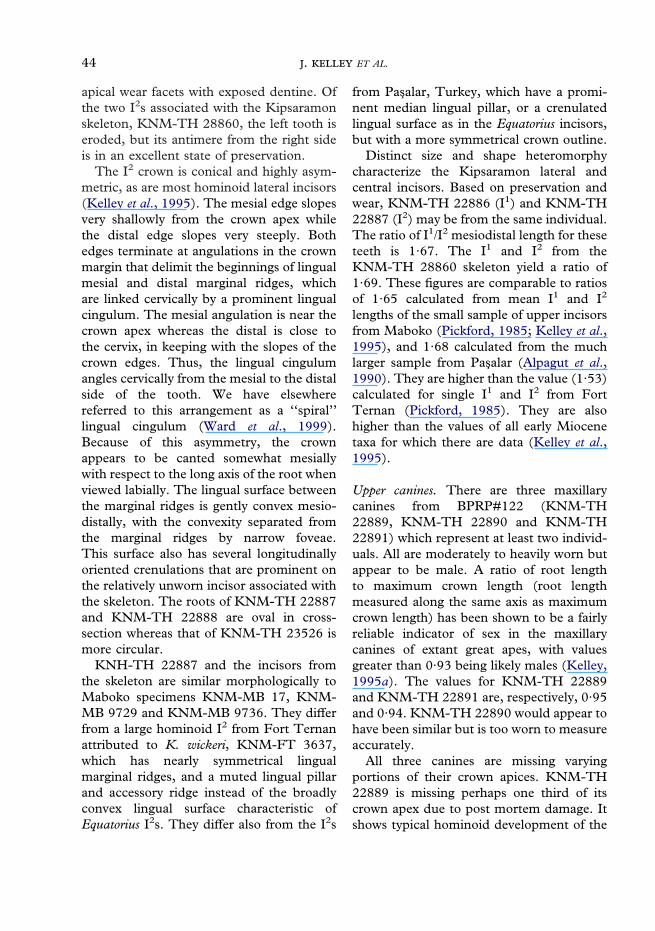
Forty-one isolated teeth representing twomedium to large-bodied hominoid specieswere recovered from site BPRP#122 inthe Kipsaramon site complex, MuruyurBeds, at the northern end of the TugenHills, west of Lake Baringo, Kenya (Figure1). The Baringo Paleontological ResearchProject (BPRP) began to find hominoidteeth at this location in June, 1990, and aprogram of systematic excavation andscreening of the surface sediments was car-ried out over the next few seasons. Themajority of the isolated teeth were surfacefinds, from a more or less horizontal surfaceabout 12 m�6 m in extent, enclosed on thenortheast side by a short section of Muruyursediment. Numerous fragments were recov-ered in the screens, most of which proved tobe missing portions of previously recoveredincomplete teeth or bits of root. During thecourse of this work we had been unable toestablish the stratigraphic level from whichthe teeth derived. However, it seemed theyhad not been transported far since we
were retrieving teeth apparently belonging tosingle individuals, and fragments of teeththat joined together.
At the end of the 1993 season, BonifaceKimeu discovered a mandible just weather-ing out from a horizon about 7 m up-sectionfrom the surface accumulation. The con-dition of the bone provided an explanationfor the nature of the tooth accumulation.The bone becomes soft when wet, and as itdries the expansion of clay minerals causes itto expand and fragment into small pieces.Had the mandible remained undiscovered,the bone would have disintegrated leavingonly the tooth crowns to drift down slope tothe horizontal surface below. We are confi-dent that the isolated teeth also derive fromthis same level. Since first finding teeth at thesite we have monitored the whole site, in-cluding the level from which the mandibleeventually came, for in situ material. Hence,we are also confident that we have notmissed the gnathic elements from whichthe isolated teeth derive, which appear tohave been destroyed before the site wasdiscovered. We examined the site again in1997 and 1999, but no further material wasfound.
Quarrying operations at BPRP#122 at theend of 1993 season and again in 1994revealed that the mandible was associatedwith a partial skeleton of the same individual(Ward et al., 1999; Sherwood et al., 2002).Given the fragility and fragmented natureof the bone, the blocks of sediment con-taining the mandible and the rest of theskeleton were jacketed in plaster andremoved to the National Museums ofKenya, where they were prepared over thenext few years, mainly by B.B. and WillDowns. The skeleton, KNM-TH 28860,includes the fragmented mandible withportions of both corpora and most of thedentition preserved in excellent condition.Three maxillary incisors were also associ-ated with the skeleton, one central and bothlaterals.

41EQUATORIUS
Morphology and metrics
Morphological observations and measure-ments on the Kipsaramon dental series
were made from the original specimens.Comparative observations were made onother middle Miocene hominoid teeth fromboth Kenya and Turkey. Following theformat of Alpagut et al. (1990) we presentdescriptions by tooth class in cases wheremore than one tooth is represented in a class.
Maxillary teeth
Upper central incisors (Figure 2). There arethree upper central incisors in the sample,one of which, KNM-TH 22919, is a weath-ered root. KNM-TH 22886, is a moderatelyworn left I1 with a thin dentine bandexposed along the entire incisal margin. TheI1 associated with the partial skeleton,KNM-TH 28860, is well preserved andminimally worn. A small chip of the distalincisal edge is missing.
Lingually, KNM-TH 22886 has thinmesial and distal marginal ridges, which arecontinuous with a clear but weakly devel-oped lingual cingulum. There is a smalllingual tubercle. The tubercle is triangular inoutline, with a broad and prominent basethat gradually tapers and decreases in reliefapically to finally merge into the lingual faceat a point that would have been approxi-mately half the unworn lingual crownheight. The tubercle is set off from themarginal ridges by mesial and distal foveaethat broaden apically as the tubercle nar-rows. Numerous approximately parallel,longitudinally oriented crenulations, mutedby wear, especially over the basal tubercle,dissect the part of the lingual surfacebounded by the marginal ridges and cingu-lum. All of these features are betterpreserved on the less worn specimen,KNM-TH 28860. The presence of a lingualtubercle and crenulations on the lingualsurface is common among Miocene homi-noid taxa (Kelley et al., 1995; McCrossin &Benefit, 1997).
The I1s from BPRP#122 are virtuallyidentical to other Equatorius I1s from
All but three of the isolated teeth, as wellas the skeleton and its associated dentition,have been assigned to Equatorius africanus(Ward et al., 1999). The hypodigm ofEquatorius includes all middle Miocenelarge hominoids from Kenya previouslyattributed to Kenyapithecus africanus, withthe exception of material now assigned toNacholapithecus kerioi (Ishida et al., 1999).The remaining three teeth from BPRP#122are referable to Nyanzapithecus.
Few other vertebrates occur at the site; wefound scraps of crocodiles, turtles, fish, anda piece of rib, probably proboscidean. Thissite is slightly higher in the KipsaramonMuruyur sequence than the level of theKipsaramon bone bed (Behrensmeyer et al.,2002). The bone bed in Member 1(BPRP#89), dated between 15·83 Ma and15·59 Ma, has previously produced evi-dence of Proconsul major, Kalepithecus andVictoriapithecus (Hill et al., 1991). BPRP#122 is in Member 3, and constrained inage to between 15·58 Ma and 15·36 Ma(Behrensmeyer et al., 2002). This super-cedes our initial age estimate for the site ofabout 15·2 Ma based on local correlations(Brown et al., 1991).
At least one specimen of each permanenttooth of E. africanus is represented in thecollections from BPRP#122. Based on theirpreservation, state of wear, size, and whetherfrom the left or right side, the isolated teethappear to represent a minimum of four adultindividuals, with a fifth individual repre-sented by the partial skeleton. The size andmorphology of the canines indicate the pres-ence of at least three males. A deciduouscanine, a fairly complete molar germ, andthree unerupted maxillary molars indicatethe presence of at least one and perhaps twojuvenile individuals.

42 . ET AL.
1KNM-RU 1681 is almost surely from Mabokorather than Rusinga (Kelley, 1986), as are several other‘‘Rusinga’’ specimens including the type specimen ofE. africanus BMNH-M 16649 (Andrews & Molleson,1979; Pickford, 1985; Harrison, 1992).
Figure 2. Maxillary incisors, lingual view. (a), (b), KNM-TH 28860, E. africanus (I1); (c), KNM-FT 49,K. wickeri (I1); (d), (e), KNM-TH 28860, E. africanus, (I2); (f), KNM-FT 3637, K. wickeri (I2).Scale 1 cm.
western Kenya (KNM-MB 104, KNM-MB11834, KNM-MJ 9734 and KNM-RU16811). The Equatorius I1s differ in severalimportant respects from KNM-FT 49, anisolated I1 from Fort Ternan referable toK. wickeri (Leakey, 1967; Pickford, 1985;Kelley, 1986; Harrison, 1992; McCrossin &Benefit, 1997). The latter has a very distinc-tive morphology characterized by massivelyinflated mesial and distal marginal ridgesthat turn out abruptly from the lingualsurface. Viewed either mesially or distally,the margins of the crown turn lingually
at a nearly 90� angle, initiating the marginalridges. This contrasts with the moregradual marginal curvature and progressiveexpansion of the marginal ridges, withgradual labiolingual thickening of the crowncervically, seen in KNM-TH 22886,KNM-TH 28860 and the other EquatoriusI1s. In KNM-FT 49, the marginal ridges,especially the mesial ridge, turn inward toconverge on and nearly enfold the basaltubercle, thereby eliminating the foveae thatseparate the tubercle from the marginalridges in the Equatorius incisors. Viewedfrom the lingual aspect, the marginal ridgesand cingulum form a very constricted ‘‘U’’in KNM-FT 49, in contrast to the relativelyquite broad ‘‘U’’ or ‘‘V’’ evident on

43EQUATORIUS
KNM-TH 22886 and the Equatorius I1sfrom Maboko and Majiwa (Figure 3). As aconsequence, the tubercle of KNM-FT 49extends between the marginal ridges form-ing a ‘‘shelf’’ where it angles sharply into thelingual surface to merge with the incisalportion of the tooth. The lingual surface ofKNM-FT 49 is finely dissected by numer-ous longitudinal crenulations as in the other
middle Miocene large hominoid incisorsfrom East Africa.
Equatorius I1s differ from both I1 mor-phologies described from the middleMiocene site of Pasalar in Turkey (Alpagutet al., 1990; Martin & Andrews, 1993). Theless common of the two, ‘‘Morphology I’’ ofAlpagut et al. (1990), is similar in manyrespects to KNM-FT 49, with inflated mar-ginal ridges that form abruptly out of thelingual tooth margins and a crenulated lin-gual tubercle that is broad and shelf-likeapically. ‘‘Morphology II’’ incisors are dis-tinguished by a prominent, relatively narrowlingual pillar that stands out in sharp relieffrom the lingual surface. The pillar broadensbasally and is continuous with the cervicalenamel extension of the crown, interruptingthe continuity of the lingual cingulumbetween the mesial and distal marginalridges. Equatorius I1s have more graduallyemergent and less prominent marginalridges, and a much more muted lingualtubercle that does not interrupt the lingualcingulum and that tapers and pinches outapically.
The isolated I1 root, KNM-TH 22919, isstout, tapers toward its apex, and presentsa rounded triangle in cross-section. Thisshape is similar to I1 root anatomy fromother middle Miocene hominoids fromwestern Kenya.
Figure 3. Maxillary central and lateral incisor cingulumtopography. (a) KNM-TH 22886, E. africanus (I1); (b)KNM-FT 49, K. wickeri (I1); (c) KNM-TH 28860, E.africanus (I2); (d) KNM-FT-3637, K.wickeri (I2). Seetext for discussion. Scale 1 cm.
Upper lateral incisors (Figure 2). Five isolatedI2s were found at locality BPRP#122. Three(KNM-TH 22887, KNM-TH 22888 andKNM-TH 23526) were recovered as surfacefinds, and two were associated with thepartial skeleton, KNM-TH 28860. Thethree specimens collected on the surface aremoderately to heavily worn, KNM-TH22887 the least so, with extensive wear alongthe distolingual aspect of the crown pro-duced by contact with the mandibularcanine. In KNM-TH 22888 and KNM-TH23526, wear extends onto the root as well.All three I2s also have lingually inclined
44 . ET AL.
apical wear facets with exposed dentine. Ofthe two I2s associated with the Kipsaramonskeleton, KNM-TH 28860, the left tooth iseroded, but its antimere from the right sideis in an excellent state of preservation.
The I2 crown is conical and highly asym-metric, as are most hominoid lateral incisors(Kelley et al., 1995). The mesial edge slopesvery shallowly from the crown apex whilethe distal edge slopes very steeply. Bothedges terminate at angulations in the crownmargin that delimit the beginnings of lingualmesial and distal marginal ridges, whichare linked cervically by a prominent lingualcingulum. The mesial angulation is near thecrown apex whereas the distal is close tothe cervix, in keeping with the slopes of thecrown edges. Thus, the lingual cingulumangles cervically from the mesial to the distalside of the tooth. We have elsewherereferred to this arrangement as a ‘‘spiral’’lingual cingulum (Ward et al., 1999).Because of this asymmetry, the crownappears to be canted somewhat mesiallywith respect to the long axis of the root whenviewed labially. The lingual surface betweenthe marginal ridges is gently convex mesio-distally, with the convexity separated fromthe marginal ridges by narrow foveae.This surface also has several longitudinallyoriented crenulations that are prominent onthe relatively unworn incisor associated withthe skeleton. The roots of KNM-TH 22887and KNM-TH 22888 are oval in cross-section whereas that of KNM-TH 23526 ismore circular.
KNH-TH 22887 and the incisors fromthe skeleton are similar morphologically toMaboko specimens KNM-MB 17, KNM-MB 9729 and KNM-MB 9736. They differfrom a large hominoid I2 from Fort Ternanattributed to K. wickeri, KNM-FT 3637,which has nearly symmetrical lingualmarginal ridges, and a muted lingual pillarand accessory ridge instead of the broadlyconvex lingual surface characteristic ofEquatorius I2s. They differ also from the I2s
from Pasalar, Turkey, which have a promi-nent median lingual pillar, or a crenulatedlingual surface as in the Equatorius incisors,but with a more symmetrical crown outline.
Distinct size and shape heteromorphycharacterize the Kipsaramon lateral andcentral incisors. Based on preservation andwear, KNM-TH 22886 (I1) and KNM-TH22887 (I2) may be from the same individual.The ratio of I1/I2 mesiodistal length for theseteeth is 1·67. The I1 and I2 from theKNM-TH 28860 skeleton yield a ratio of1·69. These figures are comparable to ratiosof 1·65 calculated from mean I1 and I2
lengths of the small sample of upper incisorsfrom Maboko (Pickford, 1985; Kelley et al.,1995), and 1·68 calculated from the muchlarger sample from Pasalar (Alpagut et al.,1990). They are higher than the value (1·53)calculated for single I1 and I2 from FortTernan (Pickford, 1985). They are alsohigher than the values of all early Miocenetaxa for which there are data (Kelley et al.,1995).
Upper canines. There are three maxillarycanines from BPRP#122 (KNM-TH22889, KNM-TH 22890 and KNM-TH22891) which represent at least two individ-uals. All are moderately to heavily worn butappear to be male. A ratio of root lengthto maximum crown length (root lengthmeasured along the same axis as maximumcrown length) has been shown to be a fairlyreliable indicator of sex in the maxillarycanines of extant great apes, with valuesgreater than 0·93 being likely males (Kelley,1995a). The values for KNM-TH 22889and KNM-TH 22891 are, respectively, 0·95and 0·94. KNM-TH 22890 would appear tohave been similar but is too worn to measureaccurately.
All three canines are missing varyingportions of their crown apices. KNM-TH22889 is missing perhaps one third of itscrown apex due to post mortem damage. Itshows typical hominoid development of the

45EQUATORIUS
mesiolingual and distolingual wear facetsresulting from contact with the lowercanine/P3 complex. KNM-TH 22891 ismissing about one half of its crown apex as aresult of ante mortem breakage and subse-quent wear to what was a recently eruptedtooth (indicated by the virtual absence ofeither mesial or distal wear facets).KNM-TH 22890 is missing most of thecrown through some combination of wearand breakage, the latter having seeminglyoccurred just prior to death judging by theminimal wear polishing that is evident. Theremains of an extensive distal wear facetreveal that this tooth was from an olderanimal. While original crown height is diffi-cult to judge on the Kipsaramon maxillarycanines because of the wear and breakage,it would appear to have been relativelylow in relation to crown length. Based onrelative crown height in males of otherMiocene catarrhine species, this would notbe unusual (Kelley, 1995b).
Despite being relatively low crowned, themorphology of the Kipsaramon uppercanines is typical of that of most hominoidmale upper canines. The buccal surface isconvexly curved and is separated from thelingual surface by sharp mesial and distalridges. There is a deep mesial groove on themesiolingual face, bounded by the mesialridge on one side and separated from theremaining, gently concave lingual surface bya sharp angulation in the crown contourdistal to the groove. There is a weak butclearly expressed lingual cingulum thatcourses from the base of the mesial groove toterminate by merging in a continuous arcwith the distal ridge. The entire crown isscored by numerous longitudinally orientedcrenulations.
KNM-TH 22890 and KNM-TH 22891have nearly complete roots, missing only theapices. The root is robust and quadrilateral/ovoid in cross-section, unlike the more lat-erally compressed roots in small to medium-sized species of Proconsul (Ward & Pilbeam,
1983). Based on crown/root relationships,the Kipsaramon canines may have beenmore externally rotated in the alveolar pro-cess, rather than antero-posteriorly alignedas in Proconsul (Ward & Pilbeam, 1983).
The only two published Equatorius uppercanines from western Kenya (KNM-MB 70and KNM-MJ 4) appear to be female, thuslimiting comparison with the Kipsaramonspecimens. There is a single male uppercanine from Fort Ternan (KNM-FT 39),attributed to K. wickeri (Pickford, 1985;Harrison, 1992; Kelley, 1995b). Althoughsubstantially larger and heavily worndistally, it is similar in most respects to theKipsaramon canines. Based on the portionof the crown preserved at the base of themesial groove, it does not appear to havehad a distinct lingual cingulum. Presumedmale canines from Pasalar (Kelley, 1995b)also have a similar morphology, including aweakly developed lingual cingulum (Alpagutet al., 1990).
Upper premolars (Figure 4). There are sixmaxillary premolars in the Kipsaramonsample. Four of the premolars are P3s andtwo are P4s. Each specimen consists of acrown broken transversely at or just belowthe cervix. Based on what is preserved at thelevel of the cervix, three of the premolars(KNM-TH 22892, KNM-TH 22893 andKNM-TH 22896/22912) had three roots.Crown morphology and preservation sug-gests that there are two sets of antimeres inthe sample. P3 paracones are moderatelytaller than the protocones. A central crestconnecting the two cusps is indented by amesiodistally-oriented fissure, which bisectsthe crown asymmetrically so that the buccalportion of the crown constitutes approxi-mately two-thirds of the occlusal surface.The mesial fovea is small and compressed,while the distal fovea is somewhat moreexpanded. Two styles are present on thebuccal surface of the crown, the mesialstyle being larger and connected to the

46 . ET AL.
mesial marginal ridge. A lingual cingulum isminimally expressed or, more frequently,completely absent.
The Kipsaramon maxillary premolars dif-fer considerably from those of Proconsul andAfropithecus, the latter including BMNHM35145 from Ad Dabtiyah (Andrews &Martin, 1987). Differences with respect toProconsul involve less projecting, less pointedbuccal cusps, and a greater expansion of thedistal fovea. Overall, the premolar proto-cones and paracones are much less hetero-morphic in the Kipsaramon specimens thanthey are in the premolars of Afropithecus andProconsul (Leakey & Walker, 1997). How-ever, the morphology of the Kipsaramon
upper premolars is similar to that describedfor Equatorius premolars from westernKenya by Pickford (1986b), and to that ofpremolars assigned to Griphopithecus fromPasalar (Alpagut et al., 1990).
Compared to most earlier Miocene taxa,the maxillary premolars from Kipsaramontend to be relatively large in relation tothe molars. This is true as well forGriphopithecus from Pasalar, Equatorius fromwestern Kenya, and, in its most extremeform, it is considered diagnostic of Afro-pithecus (Andrews & Martin, 1987).
Figure 4. E. africanus: Maxillary premolars and molars, occlusal view. (a) KNM-TH 22894 (P3); (b) 93-1(P4); (c) KNM-TH 22897 (M1); (d) KNM-TH 22903 (M2); (e) KNM-TH 22904 (M3). Scale 10 mm.
Upper molars (Figure 4). Like the maxillarypremolars, the majority of molar specimens

47EQUATORIUS
were either unerupted or in functionalocclusion for only a short period. Fracturedteeth show that the enamel is fairly thick andthat there is minimal penetrance of dentinehorns into the cusps. This is confirmed inmore heavily worn specimens, in which thecrowns are worn flat before there is muchdentine exposure.
The M1 and M2 have four distinct cusps,while the M3 shows a fairly pronouncedreduction of the hypocone and talon basin.Buccal flare on all the molars is moderateand both lingual and buccal cingula areeither much reduced or absent. When alingual cingulum is present it is confined tothe mesio-lingual corner of the protocone. Asmall tubercle is connected to the mesio-buccal angle of the protocone by a small butdistinct crest. The M1s are square in outlinedue to the prominence of the hypocone. TheM2s are more elongate and rectangular andthe M3s are rhomboidal in shape. Based onaverage measurements, M2 is the largesttooth in the series, while the M1 and M3 areapproximately equal in size (Table 1).Coarse crenulations are present on theocclusal surface of all molars, but these aremore prominent on the M2 and M3. TheM3s are extensively crenulated, which tendsto obscure occlusal contours. The distalmarginal ridge of the M3s is distinguished bythe presence of multiple small cuspules.
As noted by Pickford (1986b), Equatoriusmolars are generally characterized by simpli-fication of accessory occlusal elements,such as accessory cusps and course ridges onthe external surfaces of the main cusps,which clearly distinguishes them fromearlier large hominoids (Morotopithecus,Afropithecus, Proconsul, Rangwapithecus). Themorphology of the Kipsaramon maxillarymolars is essentially identical to thatdescribed by Pickford (1982, 1985) for thesmaller maxillary molars of E. africanus fromwestern Kenya. There are also similarities tothe molars of Griphopithecus from Pasalardescribed by Alpagut et al. (1990), except
for M3; in Griphopithecus M3s, all four cuspstend to be well developed. The relative sizesof the maxillary molars are also similar in thethree samples, with the M2 being the largestof the three and some of the M3s beingslightly smaller than the M1s.
Mandibular teeth
Figure 5. E. africanus: Mandibular incisors, lingualview. (a) KNM-TH 28860, symphyseal fragment withLI2, LI1, RI2. (b) KNM-MB 15 (RI2). Scale 2 cm.
Lower incisors (Figure 5). There are fivemandibular incisors from Kipsaramon. Twoare isolated specimens and are very heavilyworn; KNM-TH 22907 is possibly an I1 andKNM-TH 22908 is a right I2. The others,one central and two lateral incisors, areassociated with the mandible of KNM-TH 28860 and are in an excellent state ofpreservation. The Kipsaramon incisors arecharacterized by tall and narrow, but labio-lingually thick crowns. The lingual surfaceof the I1 is nearly featureless, which con-trasts with I1s from western Kenya(KNM-MJ 6, KNM-MB 11830 andKNM-MB 11833), which have a centralrib and mesial and distal marginal ridges

48 . ET AL.
(Pickford, 1985). The Kipsaramon I2s areidentical in all anatomical details to thosepreviously described from western Kenya,KNM-MJ 5, KNM-MB 15, and KNM-MB20573 (Pickford, 1982, 1985; McCrossin& Benefit, 1993) (Figure 5). In theKipsaramon teeth, the lingual cingulum iscontinuous with a faint distal marginal ridge.The lingual crown surface is flexed about alongitudinal ridge that divides the surfaceinto approximately mesial two-thirds anddistal one-third areas. The distal margin ofthe I2 curves, with a slight flexure in thecurvature about a third of the distancefrom the incisal edge to the cervix. TheKipsaramon lower incisors are also similar inmost respects to those from Pasalar assignedto Griphopithecus (Alpagut et al., 1990).
The lower incisor roots are long and,according to McCrossin & Benefit (1993),procumbently implanted in the symphysealalveolar process. However, this conclusion isbased on a juvenile mandible, KNM-MB20573, and two damaged jaws, KNM-MJ 5and KNM-FT 45, and thus merits furtherscrutiny as additional material becomesavailable.
Figure 6. Mandibular canines, lingual view. (a) Afropithecus turkanensis (KNM-WK 17010); (b) E.africanus (KNM-TH 28860); (c) K. wickeri (KNM-FT 28).
Lower canines (Figure 6). Three mandibularcanines have been recovered from
BPRP#122. Two are isolated specimens(KNM-TH 22909 and KNM-TH 22911)while the other is associated with themandible of the partial skeleton, KNM-TH28860. KNM-TH 22909 preserves the baseof a heavily worn crown and also much ofthe damaged root. KNM-TH 22911 is anearly complete root missing its crown.None of the sex-specific features describedby Kelley (1995a) is preserved onKNM-TH 22909, but it can be judged to bemale based on size concordance with thesexed upper canines from BPRP#122(Table 1). The wear surfaces on KNM-TH22909, produced by occlusion with themaxillary canine and lateral incisor, havecoalesced into a flattened, undulating plat-form which gives the appearance of apicalwear (see Plavcan & Kelley, 1996). Aremaining small portion of the mesio-labialface of the crown has a series of fine verticalcrenulations just above the cervix, likethose on the upper canines. Based on thecervical proportions of KNM-TH 22909and on the morphology of KNM-TH22911, the lower canine root is relativelyshort but robust.
The canine associated with the partialskeleton (KNM-TH 28860) is preservedintact and is minimally worn. Although it is

49EQUATORIUS
relatively low crowned (Ward et al., 1999),its morphology is clearly male based on thecriteria developed by Kelley (1995a); theheight/length index is 1·35 and the mesialridge/length index is 0·71. Both the buccaland lingual surfaces bear numerous verti-cally oriented crenulations. There is aprominent heel at the base of the mesialridge and a distinct cingulum distally. Thetwo are linked by a faint lingual cingulum.The distal ridge is gently convexly curvedlingually and terminates about two-thirds ofthe way down from the crown apex to thecervix. The mesial ridge is offset from thelingual surface of the crown by a shallowfurrow.
The only Equatorius mandibular caninesavailable for study from western Kenyasites, KNM-MB 5 and KNM-MB 11820,both appear to be female. KNM-FT 3636from Fort Ternan, attributed to K. wickeri(Pickford, 1985; Harrison, 1992), is like-wise probably female. KNM-FT 28, alsoattributed to K. wickeri (Pickford, 1985;Harrison, 1992), is a male canine that has arelatively taller and more gracile crown thanthat of KNM-TH 28860 (Ward et al., 1999)(Figure 6). Benefit & McCrossin (2000a)and Gitau et al. (2000) recently described amale lower canine from Maboko which theyattribute to ‘‘K.’’ africanus and which theycharacterize as being virtually identical toKNM-FT 28. However, Kelley et al. (2000)expressed the opinion that this canine in factbelongs to a species of Kenyapithecus sensustricto, suggested also to be present atMaboko. This suggestion was based on thepresence at Maboko of a number of post-cranial elements that are more derived thanthose attributed to Equatorius from eitherMaboko or Kipsaramon (see below).
Lower premolars (Figure 7). P3s are preservedin the mandible of KNM-TH 28860. Theypossess tall protoconids and small, but dis-tinct metaconids. A low, disto-linguallyoriented crest connects the two cusps and
then turns distally to continue into a fairlyexpansive talonid basin. A prominent mesio-buccal extension of the crown envelops thebase of the mesial root. There is minimallingual relief on the crowns, with a shortcingular remnant confined to the base of themesial ridge.
There is one P3 available from Mabokoassigned to E. africanus, KNM-MB 9737(Pickford, 1985). However, McCrossin &Benefit (1997) removed this tooth from theE. africanus hypodigm based on the size andmorphology of further P3s recovered fromthe site, which have not yet been described.The only other available western KenyaP3, that in the Kaloma mandible, KNM-MJ5, is missing most of its enamel. TheKipsaramon P3s are nearly identical to thePasalar P3s assigned to G. alpani; thosebelonging to the second Pasalar species havea somewhat different morphology, includ-ing a continuous lingual cingulum (Alpagutet al., 1990; Kelley, in prep.). Two P3s of K.wickeri from Fort Ternan, KNM-FT 35 andKNM-FT 45, are similar to those fromKipsaramon in having distinct metaconidsand expansive talonids, but differ in havingprominent lingual cingula.
Both P4s are preserved in the mandibleof KNM-TH 28860; there is also one iso-lated P4 (KNM-TH 22910). In occlusalview their crowns are rectangular with fourdistinct cusps, two prominent cusps mesiallyand two much smaller cusps distally. Thetalonid basin represents nearly two-thirds ofthe occlusal surface area. The buccal surfaceof the protoconid bulges slightly, and thereis a small mesio-buccal extension onto themesial root. Otherwise, there is minimalbuccal flare and no evidence of a buccalcingulum. A small accessory tubercleappears on the buccal surface in the area ofthe hypoconid. The crown of the isolatedpremolar has been flattened by wear butthere is only modest exposure of the under-lying dentine, suggesting relatively thickocclusal enamel.

50 . ET AL.
Comparison with Maboko specimensKNM-MB 9735 and KNM-MB 9728reveals a similar morphology, although theMaboko premolars are somewhat com-pressed mesiodistally and do not haveclearly expressed distal cusps. Both speci-mens possess distinct protoconids andmetaconids, a mesial fovea, and anexpanded talonid basin. A moderatelyexpressed buccal cingulum is present onboth teeth. The Pasalar P4 sample encom-passes considerable variation in crown
shape. However, two mesial cusps are con-sistently present. The Pasalar P4s also tendto have less expansive talonid basins andmore buccal flare than is the case for theKipsaramon P4s. Finally, they generallyhave distinct mesial and distal tubercles onthe buccal surface of the crown, with vari-ably expressed continuations into partialbuccal cingula.
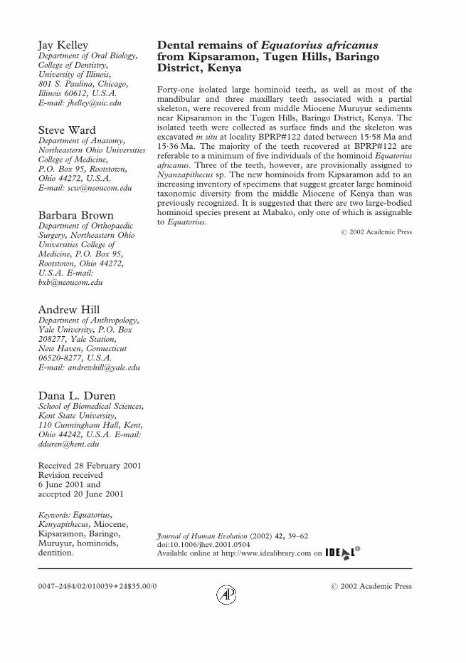
Figure 7. E. africanus: Mandibular premolars and molars. (a) KNM-TH 28860, left mandible with C-M3.Scale 3 cm. (b) Composite mandibular dentition from at least two individuals to show approximatecomparative sizes of teeth; KNM-TH 22914 (LM1), KNM-TH 22917 (LM2), KNM-TH 22908 (RI2),KNM-TH 22910 (RP4), KNM-TH 22931 (RM1), KNM-TH 22915 (RM2). Scale 3 cm.
Lower molars (Figure 7). The M1 is repre-sented by two pairs of antimeres (KNM-TH

51EQUATORIUS
22914 and KNM-TH 22931; KNM-TH28860 L and R). The two isolated M1s aremoderately worn, while those associatedwith the mandible are only lightly worn. Thecrowns are approximately rectangular inocclusal outline. Five relatively low andblunt cusps are present and the trigonidsegment of the crown represents about onethird of the total occlusal surface area. Thehypoconulid is positioned on the buccal sideof the crown and a small distal fovea isinterposed between it and the entoconid.There is a well-defined buccal shelf occupy-ing the notch between the protoconid andhypoconid, which on some specimens iscontinuous with a nearly complete cingulumaround the mesio-buccal margin of theprotoconid.
Two of the four isolated M2s, KNM-TH22915 and KNM-TH 22917, are antimeresand may be from the same individual asthe two isolated M1s just described. An M2
is also associated with the partial skeleton.All M2s are rectangular in outline. Themetaconid appears to be the largest cuspand the protoconid and hypoconid are sub-equal in size. The trigonid basin is com-pressed, buccal flare is minimal, and theonly sign of a buccal cingulum is a slightswelling on the buccal surface of theprotoconid.
Like the isolated M1s, most of the isolatedM2s are moderately worn. The occlusal sur-faces of these teeth have been flattened bywear, but the dentine exposures on themajor cusps have not yet coalesced. Judgingfrom the extent of occlusal wear on boththe M1s and M2s and the limited dentineexposure, it can be concluded that molarenamel is relatively thick.
The only M3s in the Kipsaramon sampleare those associated with the partial skel-eton, KNM-TH 28860. The crowns taperdistally, resulting in a somewhat triangularocclusal outline. There are no major acces-sory cusps but there are tiny, barely discern-ible cuspules between both the metaconid
and entoconid, and the entoconid andhypoconulid. As with the M2s, the onlyevidence of a cingulum is a faint remnanton the mesio-buccal quadrant of theprotoconid.
The Kipsaramon M1s are morphologi-cally similar to their counterparts fromwestern Kenya. Four western Kenya speci-mens (KNM-MB 11812, KNM-MB 11818,KNM-MB 11825 and KNM-NC 9740)identified as M1s by Pickford (1985) areavailable for comparison. Two of theMaboko specimens (KNM-MB 11818 andKNM-MB 11825) are similar in size andshape to the Kipsaramon M1s, while thepreserved right M1 in the Kaloma mandible(KNM-MJ 5) is too heavily worn forcomparative purposes.
The morphology of western Kenya M2s isknown from fragmentary and, for the mostpart, heavily worn specimens (KNM-MB108, KNM-MB 11816 and KNM-MB11824). Their crowns are also rectangular inshape but few other details can be discerned.One, KNM-MB 11816, has a fairly promi-nent buccal cingulum, which is not presenton most lower molars from Maboko(McCrossin & Benefit, 1997).
The Kipsaramon lower molars are alsomorphologically similar to those fromPasalar (Alpagut et al., 1990). In bothsamples, the protoconids and metaconidslie in a transverse axis and are joined by alow transverse crest. A large talonid basindominates the crown and a small distalfovea is compressed between the hypo-conulid and the entoconid. However, mostlower molars from Pasalar have a distinct,either complete or partial buccal cingulum.The pattern of dentine exposure with wearappears quite similar, with crowns wearingflat prior to significant coalescence ofdentine islands. Enamel thickness hasbeen measured directly on several Pasalarlower molars and the enamel is relativelythick (Martin, 1985; Andrews & Martin,1991).

52 . ET AL.
Deciduous dentitionKNM-TH 23529 is a left upper deciduouscanine. Most of the root is preserved but thefractured crown is lightly worn with a smallpoint exposure of dentine at the apex. Asmall but distinct tubercle is present at thedistal cervix. Two crests extend toward thecrown apex from this tubercle, which unitejust below the apical wear facet. A lingualcingulum extends mesially from the tubercleto a mesial ridge that runs to the apex. Thereis also a small buccal cingulum that extendsfrom the distal tubercle for about 50% of thedistance between the distal and mesial mar-gins. KNM-TH 23529 is similar to a maxil-lary dC1 from Pasalar. However, the PasalardC1 is slightly larger and taller with a morepronounced mesial tuberosity.
MetricsLength and breadth dimensions of theKipsaramon hominoid teeth are presentedin Table 1. Published measurements of E.africanus teeth from western Kenya (pri-marily Maboko) and teeth of K. wickeri fromFort Ternan are shown in Table 2.
The larger of the two measurable I1s fromKipsaramon, that associated with the partialskeleton KNM-TH 28860, is approximatelythe same size as the larger I1s from Mabokoand KNM-FT 49 from Fort Ternan. Theother is substantially smaller and couldbe from a female. KNM-MB 9734 fromMaboko may have been about the same sizeas the latter but can only be measuredlabiolingually. The only measurable I2 fromMaboko falls within the range of those fromKipsaramon. KNM-FT 3637 is relativelyquite narrow labiolingually compared to theI2s from either Kipsaramon or Maboko.
Metric comparison of maxillary canines iscomplicated by the fact that the Kipsaramoncanines are morphologically male whereasthe only E. africanus specimens presentlyavailable from western Kenya are morpho-logically female. A male canine from Fort
Ternan (KNM-FT 39), referable to K.wickeri, is substantially larger than all theE. africanus canines from Kipsaramon.
Both measurable P3s from Kipsaramonare much smaller than the one availableMaboko specimen in the E. africanus typemaxilla, which is almost certainly a male.The P4s are approximately the same sizeor slightly larger than two isolated P4sfrom Maboko. A large P4 (KNM-RU 1732),purportedly from Rusinga but suggested tobe from Maboko based on preservation andmorphology, was assigned to E. africanus byPickford (1985). This tooth and the simi-larly sized P4 in the E. africanus type maxillaare both much larger than the KipsaramonP4s or the others from Maboko.
Five of six M1s from Kipsaramon areuniformly smaller than the four M1s avail-able from Maboko; the sixth falls in themiddle of the range of the Maboko teeth.This general pattern holds for the M2sas well, and is even more exaggerated forthe M3s. The three measurable M3s fromKipsaramon are substantially smaller thanthe two from Maboko. The mean length/breadth ratios (�100) for the maxillarymolars from Kipsaramon are: M1: 91, M2:93, and M3: 99. Comparable ratios for E.africanus molars from western Kenyalocalities published by Pickford (1985) are:M1: 92; M2: 92, and M3: 101.
The I1s from Kipsaramon are larger over-all than three E. africanus I1s from westernKenya, especially labiolingually. The two I2sfrom the Kipsaramon partial skeleton aremuch larger than a third, isolated I2 fromthe site. Two isolated I2s from Maboko areslightly larger than the smaller tooth fromKipsaramon. A lateral incisor in the juvenilemandible KNM-MB 20573 from Maboko isapproximately the size of the two largerincisors belonging to the skeleton.
The mandibular canines from Kipsara-mon are larger than two Equatorius caninesfrom Maboko described by Pickford (1985),but, as noted above, both lower canines

53EQUATORIUS
Table 1 Measurements of E. africanus and Nyanzapithecus sp. teeth from BPRP#122
Maxillary teeth Side MD BL Mandibular teeth Side MD BL
E. africanusI1 I1
22886 L 8·0 6·4 22907 ? 4·3 6·722919 ? — — 28860 L 5·0 6·128860 R 10·2 7·5
I2 I2
22887 R 4·8 5·5 22908 R 3·5 5·522888 R 4·8 6·3 28860 L 5·5 8·323526 L (5·9) — 28860 R 4·9 8·228860 L 6·5 7·1
dC1
23529 L 5·1 3·7C1 C1
22889 R 10·1 8·6 22909 ? 8·3 9·322890 R 11·8 9·2 22911 ? — —22891 L 11·6 9·4 28860 L 12·4 9·5
P3 P3
22893 R 6·1 9·2 28860 L 8·8 12·922895 ? — — 28860 R 8·8 12·722896/22912 L 6·3 9·1
P4 P4
22892 R 5·9 9·0 22910 R 8·4 6·622894 L 6·5 9·1 28860 L 8·0 8·293-1 L 6·2 8·8 28860 R 7·5 8·5
M1 M1
22897 R 8·9 9·7 22914 L 9·7 7·822898 R 8·7 9·6 22931 R 9·8 7·822899 L 8·5 9·8 28860 L — 8·922900 L 9·8 10·3 28860 R 9·9 8·523525 L 8·8 9·823180 L 8·8 9·6
M2 M2
22901/23528 L 10·1 11·7 22915 R 11·2 9·822902 L 9·8 10·3 22917 L 11·2 9·822903 R 10·7 11·0 93-53 R 11·0 10·122916 R (11·0) (11·3) 28860 L 11·3 10·522918 L (10·5) (11·3)
M3 M3
22904 R 9·6 9·7 28860 L 13·9 11·322906 ? — — 28860 R 14·4 11·223527 R 10·0 10·393-52 L 9·3 9·2
Nyanzapithecus sp.M2 M1
93-2 L 8·7 7·8 22913 R 9·6 7·5M3
22905 R 8·8 7·8
?Teeth too worn and/or broken for side to be reliably determined. Values in parentheses are estimates.
from Maboko are probably femalewhereas those from Kipsaramon are male.Comparison is further complicated by whatappears to be a transposition by Pickford of
the length and breadth measurements forthe two Maboko specimens. The K. wickerimale lower canine from Fort Ternan,KNM-FT 28, has approximately the same

54 . ET AL.
Table 2 Measurements of Equatorius africanus teeth from western Kenya localities (Maboko, Majiwa,Kaloma and Nyakach Fm) and Kenyapithecus wickeri teeth from Fort Ternan
Maxillary teeth Side MD BL Mandibular teeth Side MD BL
I1 I1
MB 104 L 10·1 7·4 MB 11830 L 4·4 5·0MJ 9734 L — 6·7 MB 11833 ? 4·4 5·0RU 1681 R 10·0 7·3 MJ 6 R 4·1 5·1FT 49 L 9·8 7·8
I2 I2
MB 17 L (5·0) (5·0) MB 15 L 4·0 5·5MB 9729 R 6·0 6·4 MB 20573‡ L 5·6 7·8FT 3637 R 6·4 5·5 MJ 5 R (4·0) (6·5)
C1 C1
MJ 4* R 6·9 8·6 MB 11820* R 5·6 8·1MB 70* R 6·8 8·5 MB 32348§ ? 12·0 8·5FT 46b* L 7·2 9·4 MB 5* R 5·8 9·0FT 39* R 11·8 15·2 FT 28* R 8·7 12·4
FT 3636* L 6·5 9·2P3 P3
M 16649 L 8·5 12·5 MJ 5 L (8·3) (6·8)FT 35 R 10·9 10·2FT 45 L 10·9 6·6
P4 P4
MB 155 L 5·0 7·9 MB 9728 L 7·5 8·9MB 9725 L — (11·0) MB 9735 R 8·0 8·2MB 11817 R 6·2 9·4 MJ 2 R 7·7 9·2M 16649† L 7·5 11·8 MJ 5 L 7·5 6·5RU 1732 R 7·7 12·0 FT 7 R 8·3 8·2FT 46 L 6·7 10·3 FT 45 L 7·6 8·3
M1 M1
MB 107 R 10·6 11·2 MB 11812 L 9·6 8·6MB 9738 L 9·1 10·2 MB 11818 R 10·2 8·6MB 11821 R (9·7) 10·0 MB 11823 R — 7·1M 16649 L 10·2 11·6 MB 11825 R 10·0 8·1FT 46 L 10·2 10·9 MB 20573‡ L 10·3 9·3FT 47 R 10·2 10·8 MJ 5 R 8·0 7·5
NC 9740 R 9·2 7·4FT 7 R (10·0) (9·2)FT 48 R 11·0 9·1
M2 M2
MB 11819 R 11·2 12·1 MB 9727 R (12·8) (11·0)MJ 8 R 10·7 11·9 MB 11816 R — 10·8FT 46 L 12·2 11·9 MB 11824 L — 10·5FT 47 R 11·5 11·9 MB 20573‡ R 12·6 11·5
MJ 5 L 10·3 9·6M3 M3
MJ 1 R 12·1 12·0 MB 108 L (12·0) 10·9RU 1692 R 12·1 12·0 MB 9726 R 13·7 11·1
MB 9731 L (13·4) 12·0MJ 5 L/R 11·0 9·2FT 34 L 12·3 10·1
All measurements from Pickford (1985) except as noted below; parentheses indicate estimates. Abbreviations:FT: Fort Ternan; MB: Maboko; MJ: Majiwa and Kaloma; M: Natural History Museum, London (specimens fromMaboko); NC: Nyakach Formation; RU: Rusinga (but specimens considered to be from Maboko).
*MD and BL measurements transposed with respect to canine measurements in Table 1.†Measurements from Clark & Leakey (1951).‡Measurements from McCrossin & Benefit (1993).§Measurements from Benefit & McCrossin (2000a).

55EQUATORIUS
cervical dimensions as KNM-TH 28860 butis much higher crowned (Ward et al., 1999).Likewise, the male mandibular canine fromMaboko (KNM-MB 32348) attributed to‘‘K.’’ africanus by McCrossin & Benefit(2000) and Gitau et al. (2000), but sug-gested to belong to Kenyapithecus sensu strictoby Kelley et al. (2000), has cervical dimen-sions close to those of KNM-TH 28860, butis nearly identical to KNM-FT 28 in crownheight.
The P3s of KNM-TH 28860 are substan-tially larger than either of those fromwestern Kenya listed by Pickford (1985).Given the apparent sex representation inthe two samples based on the canines, thisis not unexpected. The three P4s fromKipsaramon are longer on average than thefour P4s from western Kenya but fall withintheir range for breadth.
The M1s from Kipsaramon are slightlylarger on average than those of E. africanusfrom western Kenya, but mostly fall withinthe ranges of the latter for both lengthand breadth dimensions. The situationis reversed for the M2s; those fromKipsaramon are smaller on average thanthose from western Kenya and the size dis-parity between the two samples is greaterthan for M1, but the Kipsaramon teeth againlie within the ranges of the western Kenyateeth for both dimensions. The M3s ofKNM-TH 28860 are slightly larger than thelargest of four M3s of E. africanus fromwestern Kenya. The breadth/length ratios(�100) for the Kipsaramon molars areM1: 82 and M2: 90. Equivalent ratios forwestern Kenya E. africanus are M1: 87 andM2: 90.
Summary comparison of Equatorius teethfrom Kipsaramon and western KenyaThere are few nonmetric morphological dif-ferences between the Kipsaramon teeth andthe available dental samples from the west-ern Kenyan sites of Maboko, Majiwa andKaloma. Only in the lower central incisors
and lower fourth premolars are there con-sistent, discernible differences. However,these are mostly minor in character andcould well disappear with larger samplesizes. I1s from Kipsaramon are featurelesslingually, whereas those from Mabokohave a distinct central rib and marginalridges. P4s from Kipsaramon have atendency to express a pair of small distalcusps and lack a buccal cingulum. MabokoP4s do not express distal cusps but do have amoderately developed buccal cingulum.
There is no clear pattern to the metriccomparisons between Equatorius teeth fromKipsaramon and those from the westernKenya localities. This is not surprising giventhe vagaries of small sample sizes, particu-larly with the likelihood of differing sexrepresentation at different tooth positions inwhat appears to be a fairly highly dimorphictaxon. Nevertheless, there are indicationsthat, dentally, the local population repre-sented by the Kipsaramon sample mighthave been somewhat smaller on averagethan that represented at the western Kenyalocalities, at least in the postcanine den-tition. As noted above, it appears that theKipsaramon sample represents relativelyfew individuals. Moreover, judging by thecanines and P3s, most of the individuals aremale. In spite of this, at many postcaninetooth positions Kipsaramon teeth aresmaller on average than those from westernKenya. Incisors in the two samples tend tobe more or less equal in size.
Presently, nothing more can be con-cluded concerning dental variation withinEquatorius. When additional data fromrecently collected, but as yet unpublished,material from Maboko become available,and hopefully with additions to the samplefrom the vicinity of BPRP#122, a clearerpicture of dental size and morphologicalvariation within Equatorius will emerge. Thiswill facilitate alpha-taxonomic assessmentwithin Equatorius and promote an improvedunderstanding of its relationships to

56 . ET AL.
contemporary and later large hominoidtaxa.
Other hominoid dental remains
Three teeth recovered during surface collec-tion at locality BPRP#122 are morphologi-cally distinct from the majority of the homi-noid teeth collected. A right M2 (KNM-TH37368) has four well-defined cusps (Figure8). This tooth is slightly longer than it isbroad, and the crown narrows distally. Thefour primary cusps are low but quiteinflated. The two mesial cusps are sig-nificantly larger than the distal cusps. Theinflation of all cusps has the effectof restricting the size of the trigon fovea aswell as the mesial and distal foveae. Thelingual cingulum is well developed andextends from the mesial base of the proto-cone distally to the hypocone. A small crestlinks the hypocone to the crista obliqua.
A left M3 (KNM-TH 22905) is somewhatreminiscent of the M3s of Proconsul heseloni.It has a prominent protocone at the mid-point of the lingual side overlying a relativelysmall cingulum. The paracone is almost
equal in size. A metaconule that is approxi-mately the same size as the hypocone isinterposed between the metacone and pro-tocone. A cingulum expressed as a smallledge is present along the buccal surfacebetween the paracone and metacone andextends distally. The cusps are virtuallyunworn, but there is a small mesial wearfacet.
KNM-TH 22913 is a right M1 crownbroken along the cervix. It is weathered butminimally worn and rectangular in shape.The crown is narrow relative to length. Allcusps are low and rounded, and there isevidence of enamel crenulations on theocclusal surface. The metaconid is promi-nent, both in height and overall size. Theprotoconid is somewhat smaller. Due toinflation of the cusps, the mesial and distalfoveae are quite restricted and all crestsare poorly expressed. The talonid basin islong and compressed bucco-lingually. Thereis also a small buccal cingulum in theinterval between the hypoconid andprotoconid.
Based on the diagnosis and descriptionsby Harrison (1986), we provisionally assignthese specimens to Nyanzapithecus sp.Key diagnostic criteria for the uppermolars include a relatively long crown, lowbut strongly inflated cusps and crestswith the mesial cusps larger than thedistal cusps—producing what Harrison(1986) describes as a ‘‘crowded occlusaltopography’’—and a prominent lingual cin-gulum. The M1 is diagnosed by being longand narrow, with low, rounded and inflatedcusps and crests consequently much dimin-ished in size, and by having restricted mesialand distal foveae, a long but compressedtalonid basin, and a small buccal cingulum.
Figure 8. Nyanzapithecus sp. Maxillary and mandibularmolars. (a) KNM-TH 37368 (RM2); (b) KNM-TH22905 (LM3); (c) KNM-TH 22913 (RM1).
Discussion
Recent discoveries of new primate fossilsfrom Maboko Island, the Samburu region,and Kipsaramon in the Tugen Hills have

57EQUATORIUS
done much to provide a sense of the truerange of taxonomic diversity of middleMiocene hominoids (Benefit et al., 1998;Nakatsukasa et al., 1998, 2000; Ishida et al.1999; Ward et al., 1999). However, becausemuch of the dental material from Mabokoand the Samburu Hills is still undescribed, acomplete inventory of dental similarities anddifferences amongst the various samples isnot yet possible. Nevertheless, enough isknown of each of the samples to makecertain comparisons and to make sometentative judgments concerning taxonomyand phylogeny.
Taxonomy of the Kipsaramon and Mabokolarge hominoid samplesBased primarily on dental and gnathic char-acters, Ward et al. (1999) erected the newnomen E. africanus from the hypodigm of K.africanus, known from several localities inwestern Kenya. Kenyapithecus was thusmade monotypic and was restricted to thelarge hominoid species from Fort Ternan,K. wickeri. However, Benefit and McCrossinhave repeatedly asserted that the mor-phology expressed in the K. wickeri teethfrom Fort Ternan is in every case encom-passed by the variation present in thedental sample from Maboko (McCrossin& Benefit, 1997; Benefit, 1999; Benefit &McCrossin, 2000a,b; Gitau et al., 2000).Unfortunately, these claims cannot be sub-stantiated for the most part since there areno published data or detailed descriptions ofthe relevant material, nor are the specimensyet available for comparative examination.Incisors and canines were particularlyimportant in differentiating Equatorius fromKenyapithecus (Ward et al., 1999 and above)and the one exception to the paucity ofinformation concerning new specimensfrom Maboko is in male mandibular caninemorphology.
Canine morphology is important in thetaxonomy and phylogeny of early andmiddle Miocene hominoids. Nearly all
species from this time span have robust,relatively low-crowned male lower canines(Kelley, 1995b). The only known exceptionsare Rangwapithecus gordoni from the earlyMiocene (Kelley, 1995b), and K. wickeriand an unnamed species from Pasalar fromthe middle Miocene (Ward et al., 1999), allof which have derived, relatively high-crowned male lower canines. The malelower canine of E. africanus associated withthe Tugen Hills skeleton, KNM-TH 28860,is relatively low crowned (crown height/maximum length=1·35). This was one ofthe features used to justify the transferof ‘‘Kenyapithecus’’ africanus to Equatorius(Ward et al., 1999). No male lower canineswere then known from Maboko, fromwhence comes the type specimen of E.africanus.
Benefit & McCrossin (2000a) and Gitauet al. (2000) subsequently assigned to ‘‘K.’’africanus a male lower canine from Maboko,KNM-MB 32348, which is nearly identicalin size and shape to the derived, relativelyhigh-crowned canine of K. wickeri fromFort Ternan, KNM-FT 28 (crown height/maximum length: KNM-FT 28=1·62;KNM-MB 32348=1·67). Accordingly, theyargued, ‘‘K.’’ africanus cannot be character-ized as having retained primitive, relativelylow-crowned canines as claimed by Wardet al. (1999) and this feature cannot, there-fore, be used to support the transfer ofthe Maboko material to the new genus,Equatorius (Ward et al., 1999). In response,Kelley et al. (2000) suggested that what wasin fact in error was the taxonomic assign-ment of KNM-MB 32348, which theyregarded as belonging to a second, unrecog-nized species at Maboko, perhaps belongingto Kenyapithecus as defined by the FortTernan material. Questions regardinghominoid taxonomy at Maboko mostly con-cern postcranial remains, but KNM-MB32348 is a key element in the argumentsof Kelley et al. (2000) so these will beelaborated upon here.

58 . ET AL.
Most of the primate fossils at Mabokoderive from two superposed units, Beds 3and 5 (Gitau et al., 1998). According toBlue & McCrossin (2001), Bed 5 could beas much as 1 Ma younger than Bed 3, whichis reflected in numerous faunal differencesbetween the two beds (Gitau et al., 1998).In particular, there are differences in theprimate faunas from Beds 3 and 5 (Gitauet al., 1998). While the early cercopithecidVictoriapithecus and the primitive catarrhineSimiolus are present and occur in compar-able frequency in both beds, the oreopithe-cids Mabokopithecus and Nyanzapithecus arelargely or entirely confined to the later Bed5. In addition, the two beds contain differ-ent species of the bushbaby Komba. Gitauet al. (1998) also state that ‘‘Kenyapithecus’’is present and similarly abundant in bothbeds. These authors have long maintainedthat there is only one large hominoidspecies present at Maboko, ‘‘K.’’ africanus(McCrossin & Benefit, 1997).
There are, however, some striking differ-ences in remains assigned to ‘‘K.’’ africanusfrom Beds 3 and 5, and some interestingpatterns of association among differentelements. Each bed has produced a partialhumerus of a large-bodied hominoid,both of which have been assigned to ‘‘K.’’africanus (McCrossin, 1997; McCrossin &Benefit, 1997; McCrossin et al., 1998a), buttheir morphology is strikingly different. Thatfrom Bed 3 (Pickford, 1984) resembleshumeri of cercopithecoid monkeys in havinga retroflexed shaft in association with aprominent deltopectoral crest, a posteriorlyoriented head, and a weakly developedlateral supracondyler (supinator) crest(Clark & Leakey, 1951; McCrossin &Benefit, 1997; McCrossin et al., 1998a). Incontrast, that from Bed 5 (McCrossin &Benefit, 2000) resembles the humeri ofgreat apes in having a straight shaft anda more developed lateral supracondylarcrest (McCrossin, 1997; McCrossin et al.,1998a). In the latter feature and in its
postero-medially oriented medial epicondyleand deep olecranon fossa, the Bed 5humerus also resembles the K. wickeri distalhumerus from Fort Ternan (McCrossin,1997). No satisfactory explanation has beengiven as to why a single species shoulddisplay a degree of humeral variation thatis never found within a single genus ofextant primates, and that in fact distin-guishes living apes and monkeys. Thederived K. wickeri-like canine, KNM-MB32348, is also from Bed 5.
While most of the E. africanus postcranialremains from Maboko express a more primi-tive catarrhine morphology, like the Bed 3humerus (McCrossin & Benefit, 1997;McCrossin et al., 1998a), others have beenrecovered that instead, like the Bed 5humerus, resemble those of African greatapes. Included among the latter are a distalradius, a cuboid and a proximal femur(McCrossin et al., 1998a,b). The bed orbeds from which the latter were recoveredhave not been reported. However, like thehigh-crowned canine and humerus fromBed 5, most were collected in 1996(McCrossin, 1997); the radius was collectedin 1997 (McCrossin et al., 1998a). Sincethe 1996 collections also include nearly150 craniodental specimens of oreopithecids(Benefit & McCrossin, 1997; Benefit et al.,1998), which have a ‘‘highly restrictedspatial distribution in the deposit’’ (Benefitet al., 1998) mostly or entirely restricted toBed 5 (Gitau et al., 1998; Palmer et al.,2001), it would appear that the 1996 exca-vation was confined to, or at least concen-trated in, Bed 5. Thus, most or all of thepostcrania with resemblances to those ofextant great apes may come from Bed 5.
It is important to note here that, in spite ofthe above argument, it is not necessary forthere to be temporal segregation to recog-nize two species; two or more species canobviously come from the same stratigraphichorizon and could even have been sym-patric in life. Species are defined by their

59EQUATORIUS
morphology regardless of their associationsor where they occur. However, the temporalsegregation of the two humeri, the suggestedconcentration of the great ape-like post-crania in Bed 5, and the presence of thederived canine in Bed 5 as well, can all beconsidered as additional circumstantial evi-dence for the presence of two possibly time-successive species at Maboko. In light ofthis, it is critical that variation in thenew dental remains from Maboko also beevaluated with respect to stratigraphiclevel.
Since the type specimen of E. africanus,BMNH 16649, is almost surely from Bed3, having been recovered by W. E. Owenduring his 1933 excavation at Maboko(Andrews & Molleson, 1979; Andrews et al.,1981; Pickford, 1984), this is the largehominoid species present in Bed 3. Thespecies proposed to be present in Bed 5 isunnamed but, based on available infor-mation, would be most reasonably assignedto Kenyapithecus sp.
There are a number of features that theKipsaramon teeth share with the E. africanusdental specimens from western Kenya.Upper central incisors are relatively broadboth mesiodistally and labiolingually inrelation to height and have a stout lingualtubercle. Upper lateral incisors have aspiraled lingual morphology. Premolars andmolars mostly have reduced cingula (moreso in the mandibular teeth) and low,rounded cusps with diminished relief at theenamel–dentine interface. Based on thesesimilarities, Ward et al. (1999) assigned theKipsaramon specimens to E. africanus aswell. The Kipsaramon and western Kenyateeth do appear to differ, however, in anumber of details as described above, mostlyrelating to tooth size and proportions(Kipsaramon with a generally smaller post-canine dentition—particularly if mostly rep-resented by males—but with similarly sizedincisors as Maboko). For the present, weregard these as reflecting population differ-
ences, either temporal or geographic, withina single species. When the new materialfrom Maboko is more fully described, itmay become necessary to recognize theKipsaramon sample as a distinct species ofEquatorius.
Comparisons of Equatorius to other middleMiocene taxaCiting the diagnosis of Equatorius (Wardet al., 1999), Begun (2000) has suggestedthat Equatorius is congeneric with Gripho-pithecus Abel, 1902 as represented at themiddle Miocene sites of Pasalar and Cq andirin Turkey. Begun’s opinion was basedmostly on dental comparisons betweensamples from the various Kenyan andTurkish sites. However, as described above,there are a number of differences in thedental remains of Equatorius and those fromthe Turkish sites assigned to Griphopithecus.The latter have labiolingually thick I1s with aprominent, high-relief lingual pillar, identi-cal to that on the I1s of a number of lateMiocene Eurasian species. Pasalar I2s lackthe distinctive lingual morphology expressedby Equatorius. Griphopithecus lower caninesare relatively low crowned (Kelley &Alpagut, 1999) but have a distinctive mor-phology, with a prominent longitudinal cleftadjacent to the distal ridge. This feature isagain shared with some late Miocenespecies. Most importantly, Griphopithecuslower molars almost without exception havea prominent partial or complete buccal cin-gulum, a feature shared with early Miocenespecies but one that is rarely expressed inEquatorius lower molars. We therefore con-sider it prudent to retain separate genera forthe Kenyan and Turkish samples until thelatter has been fully described and morecompletely characterized.
Ishida et al. (1999) allocated all themiddle Miocene large hominoids from theAiteputh and Nachola Formations in theSamburu Hills to another new taxon,Nacholapithecus kerioi. Previously, it had

60 . ET AL.
been suggested that the Nacholapithecusmaterial was referable to ‘‘K.’’ africanus,based on a number of dental similaritiesbetween the western Kenya and SamburuHills samples (Pickford 1986a,b). Perhapsfor this reason, Benefit & McCrossin(2000a,b) and McCrossin & Benefit (2000)asserted that Equatorius as described byWard et al. (1999) was in fact a chimera oftwo taxa, K. africanus and Nacholapithecus.In response, Kelley et al. (2000) acknowl-edged the distinctiveness of Nacholapithecus,both dentally and postcranially (seeNakatsukasa et al., 1999, 2000; Senut, et al.,1999), and noted that no Nacholapithecusspecimens had contributed to the diagnosisof Equatorius.
Shared features of the dentition of E.africanus and N. kerioi include robust, low-crowned canines, enlarged upper premolarswith respect to the first molar, reducedlingual cingula on maxillary molars andpremolars, and relatively thick molar enamelwith low relief at the dentine–enamel junc-tion (Ishida et al., 1984; Pickford, 1986a).More detailed dental comparisons betweenEquatorius and Nacholapithecus cannot yet bemade, although Kunimatsu et al. (1999)describe Nacholapithecus as having highercrowned and more robust upper centralincisors, more robust canines, relativelybroader upper premolars, a high frequencyof a distal transverse crest on the uppermolars, and an overall smaller dentition.
With the exception of canine morphology,in all of the ennumerated charactersEquatorius and Nacholapithecus express thederived condition with respect to the earlyMiocene stem catarrhines. K. wickeriexpresses the derived condition vis-a-visEquatorius and Nacholapithecus in details ofmaxillary anatomy (Pickford, 1985, 1986b;Harrison, 1992; Begun, 1994; Ward et al.,1999) and in dental anatomy, includingmore robust upper central incisors withinflated marginal ridges, upper lateralincisors possessing a more symmetrical
crown and lacking a primitive ‘‘spiral’’ lin-gual cingulum, and lower canines with arelatively high and narrow crown in relationto basal dimensions. Thus, we have tenta-tively allied Equatorius with the Afro-pithecinae (Andrews, 1992), but as aderived member judging by the noted fea-tures of dental anatomy, as well as aspects ofpostcranial anatomy (McCrossin & Benefit,1997; Ward et al., 1999; Sherwood et al.,2002). As with issues of taxonomy, however,larger samples might require reevaluatingthis position.
Acknowledgements
This research forms part of the work of theBaringo Paleontological Research Project(BPRP), based at Yale University, andcarried out in collaboration with theNational Museums of Kenya. We thank theGovernment of the Republic of Kenya forpermission to carry out research in Kenya(Permit OP/13/001/C 1391/ issued to A.H.),and permission to excavate from theMinister for Home Affairs and NationalHeritage. BPRP has been supported bygrants to A.H. from the NationalScience Foundation (most recently SBR-9208903), the Louise H. and David S.Ingalls Foundation, the Louise BrownFoundation, Clayton Stephenson, and YaleUniversity. J.K. was supported by theNational Science Foundation (No. SBR9408664). We thank Boniface Kimeu for hisconsiderable help in the field and with sub-sequent laboratory work. Among othermembers of BPRP who worked atBPRP#122 are Kay Behrensmeyer, AlDeino, Will Downs, Bonnie Fine Jacobs,John Kingston, Daria Lucas, Jeff Saundersand Richard Sherwood. The NationalMuseums of Kenya provided valuable logis-tic support, and we thank particularlyMeave Leakey, Head of the PalaeontologyDivision, and Emma Mbua, CollectionsManager of Palaeoanthropology. For

61EQUATORIUS
comments and suggestions on variousaspects of the work, we thank PeterAndrews, David Begun, Lawrence Martin,Terry Harrison, Alan Walker and an anony-mous reviewer.
References
Alpagut, B., Andrews, P. & Martin, L. (1990). Newhominoid specimens from the Middle Miocene site atPasalar, Turkey. J. hum. Evol. 19, 397–422.
Andrews, P. (1992). Evolution and environment in theHominoidea. Nature 360, 641–646.
Andrews, P. & Martin, L. (1987). The phyletic positionof the Ad Dabtiyah hominoid. Bull. Br. Mus. nat.Hist. (Geol.) 41, 383–393.
Andrews, P. & Martin, L. (1991). Hominoid dietaryevolution. Phil. Trans. R. Soc. Lond. 334, 199–209.
Andrews, P., Meyer, G. E., Pilbeam, D. R., VanCouvering, J. A. & Van Couvering, J. A. H. (1981).The Miocene fossil beds of Maboko Island, Kenya:Geology, age, taphonomy and palaeontology. J. hum.Evol. 10, 35–48.
Andrews, P. & Molleson, T. (1979). The provenance ofSivapithecus africanus. Bull. Br. Mus. nat. Hist.(Geol.) 32, 19–23.
Begun, D. R. (1994). Relations among the great apesand humans: New interpretations based on the fossilgreat ape Dryopithecus. Yearb. phys. Anthrop. 37,11–63.
Begun, D. R. (2000). Technical Comments: MiddleMiocene hominoid origins. Science 287, http://www.sciencemag.org/cgi/content/full/287-5462/2375a.
Behrensmeyer, A. K., Deino, A. L., Hill, A.,Kingston, J. D. & Saunders, J. J. (2002). Geologyand geochronology of the middle MioceneKipsaramon site complex, Muruyur Beds, TugenHills, Kenya. J. hum Evol. 1/2, 11–38.
Benefit, B. R. (1999). The dentition of Kenyapithecusafricanus. International Symposium: Evolution ofMiddle-to-Late Miocene Hominoids in Africa, July11–13, 1999, Takaragaike, Kyoto, Abstracts.
Benefit, B. R. & McCrossin, M. L. (1997). Newfossil evidence bearing on the relationships ofNyanzapithecus and Oreopithecus. amer. J. phys.Anthrop. 24(Suppl.), 74.
Benefit, B. R. & McCrossin, M. L. (2000a). TechnicalComments: Middle Miocene hominoid origins.Science 287, http://www.sciencemag.org/cgi/content/full/287-5462/2375a.
Benefit, B. R. & McCrossin, M. L. (2000b). Newperspectives on the taxonomy and phylogeny oflarge-bodied hominoids from the middle Miocene ofKenya. Am. J. phys. Anthrop. 30(Suppl.), 106.
Benefit, B. R., Gitau, S. N., McCrossin, M. L.& Palmer, A. K. (1998). A mandible ofMobokopithecus clarki sheds new light on oreo-pithecid evolution. Am. J. phys. Anthrop. 26(Suppl.),109.
Blue, K. T. & McCrossin, M. L. (2001). Are-investigation into the number of cercopithecoidtaxa at the middle Miocene site of Maboko, Kenya.Am. J. phys. Anthrop. 32(Suppl.), 41.
Brown, B., Hill, A. & Ward, S. (1991). New Miocenelarge hominoids from the Tugen Hills, BaringoDistrict, Kenya. Am. J. phys. Anthrop. 12(Suppl.), 55.
Clark, W. E. Le Gros & Leakey, L. S. B. (1951). TheMiocene Hominoidea of East Africa. In FossilMammals of Africa, No. 1, pp. 1–117. London:British Museum (Natural History).
Gitau, S. N., Benefit, B. R., McCrossin, M. L. &Roedl, T. (1998). Fossil primates and associatedfauna from 1997 excavations at the middle Miocenesite of Maboko Island, Kenya. Am. J. phys. Anthrop.26(Suppl.), 87.
Gitau, S. N., Benefit, B. R., Johnson, K. B. &McCrossin, M. L. (2000). New dental remains ofKenyapithecus africanus from Maboko Island, Kenyasupport the congeneric status of Kenyapithecus wickeriand K. africanus. Am. J. phys. Anthrop. 30(Suppl.),159.
Harrison, T. (1986). New fossil anthropoids from theMiddle Miocene of East Africa and their bearing onthe origin of the Oreopithecideae. Am J. phys.Anthrop. 71, 265–284.
Harrison, T. (1992). A reassessment of the taxonomicand phylogenetic affinities of the fossil catarrhinesfrom Fort Ternan, Kenya. Primates 33, 501–522.
Hill, A., Behrensmeyer, K., Brown, B., Deino, A.,Rose, M., Saunders, J., Ward, S. & Winkler, A.(1991). Kipsaramon: a lower Miocene hominoid sitein the Tugen Hills, Baringo District, Kenya. J. hum.Evol. 20, 67–75.
Ishida, H., Pickford, M., Nakaya, H. & Nakano, Y.(1984). Fossil anthropoids from Nachola andSamburu Hills, Samburu District, Kenya. Afr. Stud.Monogr. Kyoto Univ. 2(Suppl. Issue), 73–85.
Ishida, H., Kunimatsu, Y., Nakatsukasa, M. &Nakano, Y. (1999). New hominoid genus from themiddle Miocene of Nachola, Kenya. Anthrop. Sci.107, 189–191.
Kelley, J. (1986). Species recognition and sexualdimorphism in Proconsul and Rangwapithecus. J. hum.Evol. 15, 461–495.
Kelley, J. (1995a). Sexual dimorphism in canine shapeamong extant great apes. Am. J. phys. Anthrop. 96,365–389.
Kelley, J. (1995b). Sex determination in Miocene catar-rhine primates. Am. J. phys. Anthrop. 96, 390–417.
Kelley, J. & Alpagut, B. (1999). Canine sexing andspecies number in the Pasalar large hominoidsample. J. hum. Evol. 36, 335–341.
Kelley, J., Anwar, M., McCollum, M. & Ward, S. C.(1995). The anterior dentition of Sivapithecus par-vada with comments on the phylogenetic significanceof incisor heteromorphy in hominoids. J. hum. Evol.28, 503–517.
Kelley, J., Ward, S., Brown, B., Hill, A. & Downs, W.(2000). Technical Comments: Middle Miocenehominoid origins. Science 287, http://www.sciencemag.org/cgi/content/full/287-5462/2375a.

62 . ET AL.
Kunimatsu, Y., Nakatsukasa, M., Nakano, Y. & Ishida,H. (1999). The jaws and dentition of the largeMiocene hominoid from Nachola, Northern Kenya.International Symposium: Evolution of Middle-to-LateMiocene Hominoids in Africa, July 11–13, 1999,Takaragaike, Kyoto, Abstracts.
Leakey, L S. B. (1967). An early Miocene member ofHominidae. Nature 213, 155–163.
Leakey, M. & Walker, A. (1997). Afropithecus functionand phylogeny. In (D. R. Begun, C. V. Ward &M. D. Rose, Eds) Function, Phylogeny and Fossils:Miocene Hominoid Evolution and Adaptations,pp. 225–239. New York: Plenum.
Martin, L. (1985). Significance of enamel thickness inhominoid evolution. Nature 314, 260–263.
Martin, L. B. & Andrews, P. (1993). Species recog-nition in middle Miocene hominoids. In (W. H.Kimbel & L. B. Martin, Eds) Species, Species Conceptsand Primate Evolution, pp. 393–427. New York:Plenum.
McCrossin, M. L. (1997). New postcranial remains ofKenyapithecus and their implications for understand-ing the origins of hominoid terrestriality. Am. J. phys.Anthrop. 24(Suppl.), 164.
McCrossin, M. L. & Benefit, B. R. (1993). Recentlyrecovered Kenyapithecus mandible and its impli-cations for great ape and human origins. Proc. nat.Acad. Sci., U.S.A. 90, 1962–1966.
McCrossin, M. L. & Benefit, B. R. (1997). On therelationships and adaptations of Kenyapithecus, alarge bodied hominoid from the middle Miocene ofEast Africa. In (D. R. Begun, C. V. Ward & M. D.Rose, Eds) Function, Phylogeny and Fossils: MioceneHominoid Evolution and Adaptations, pp. 241–267.New York: Plenum.
McCrossin, M. L. & Benefit, B. R. (2000). Locomotordiversity among large-bodied hominoids from themiddle Miocene of Africa. Am. J. phys. Anthrop.30(Suppl.), 224.
McCrossin, M. L., Benefit, B. R., Gitau, S. N., Palmer,A. K. & Blue, K. T. (1998a). Fossil evidence for theorigins of terrestriality among Old World higherprimates. In (E. Strasser, J. Fleagle, A. Rosenberger& H. McHenry, Eds) Primate Locomotion: RecentAdvances, pp. 353–396. New York: Plenum.
McCrossin, M. L., Benefit, B. R. & Gitau, S. N.(1998b). Functional and phylogenetic analysis of thedistal radius of Kenyapithecus, with comments on theorigin of the African great ape and human clade. Am.J. phys. Anthrop. 26(Suppl.), 158.
Nakatsukasa, M., Yamanka, A., Kunimatsu, Y,Shimizu, D. & Ishida, H. (1998). A newly discoveredKenyapithecus skeleton and its implications forthe evolution of positional behavior in MioceneEast African hominoids. J. hum. Evol. 34, 657–664.
Nakatsukasa, M., Kunimatsu, Y., Nakano, Y. & Ishida,H. (1999). Postcranial features of the large hominoidfrom Nachola in comparison with Kenyapithecusafricanus and K. wickeri. International Symposium:Evolution of Middle-to-Late Miocene Hominoids inAfrica, July 11–13, 1999, Takaragaike, Kyoto,Abstracts.
Nakatsukasa, M., Kunimatsu, Y., Nakano, Y. & Ishida,H. (2000). A new skeleton of the large hominoidfrom Nachola, Northern Kenya. Am. J. phys.Anthrop. 30(Suppl.), 235.
Palmer, A. K., Benefit, B. R. & McCrossin, M. L.(2001). Lessons from the study of microwear vari-ation within and between populations of middleMiocene primate species at Maboko Island. Am. J.phys. Anthrop. 32(Suppl.), 116–117.
Pickford, M. (1982). New higher primate fossilsfrom the middle Miocene deposits at Majiwa andKaloma, western Kenya. Am. J. phys. Anthrop. 58,1–19.
Pickford, M. (1984). Kenya Palaeontology Gazetteer,Vol. 1, Western Kenya. Nairobi: National Museums ofKenya.
Pickford, M. (1985). A new look at Kenyapithecus basedon recent discoveries in western Kenya. J. hum. Evol.14, 113–143.
Pickford, M. (1986a). Hominoids from the Mioceneof East Africa and the phyletic position ofKenyapithecus. Z. Morph. Anthrop. 76, 117–130.
Pickford, M. (1986b). A reappraisal of Kenyapithecus.In (J. G. Else & P. C. Lee, Eds) Primate Evolution,pp. 163–172. Cambridge: Cambridge UniversityPress.
Plavcan, J. M. & Kelley, J. (1996). Evaluating the‘‘Dual selection’’ hypothesis of canine reduction.Am. J. phys. Anthrop. 99, 379–387.
Senut, B., Nakatsukasa, M. & Ishida, H. (1999).Preliminary analysis of Kenyapithecus shoulder joint.International Symposium: Evolution of Middle-to-LateMiocene Hominoids in Africa, July 11–13, 1999,Takaragaike, Kyoto, Abstracts.
Sherwood, R. J., Ward, S. C., Hill, A., Duren, D.,Brown, B. & Downs, W. (2002). Preliminarydescription.of the Equatorius africanus partial skeleton(KNM-TH 28860) from Kipsaramon, TugenHills, Baringo District, Kenya. J. hum. Evol. 1/2,63–73.
Ward, S. C. & Pilbeam, D. (1983). Maxillofacial mor-phology of Miocene hominoids from Africa andIndo-Pakistan. In (R. L. Ciochon & R. S. Corruccini,Eds) New Interpretations of Ape and Human Ancestry,pp. 211–238. New York: Plenum.
Ward, S., Brown, B., Hill, A., Kelley, J. & Downs, W.(1999). Equatorius: A new hominoid genus fromthe middle Miocene of Kenya. Science 285, 1382–1386.
Related Documents