170 Stom Glas S, vol. 55, 2008 UDC: 616.314.18-036-07 ISSN 0039-1743 COBISS. SR-ID 8417026 Matične ćelije zubne pulpe i njihov potencijalni značaj u regenerativnoj medicini DOI: 10.2298/SGS0803170T Dental pulp stem cells - potential significance in regenerative medicine Vera Todorović 1 , Dejan Marković 2 , Nadežda Milošević-Jovčić 1 , Marijana Petakov 1 , Bela Balint 3 , Miodrag Čolić 4 , Ana Milenković 1,2 , Ivana Čolak 1 , Vukoman Jokanović, Nebojša Nikolić 2 1 Institut za medicinska istraživanja, Univerzitet u Beogradu 2 Klinika za dečju i preventivnu stomatologiju, Stomatološki fakultet, Univerzitet u Beogradu 3 Institut za transfuziju, Vojnomedicinska akademija u Beogradu 4 Institut za medicinska istraživanja, Vojnomedicinska akademija u Beogradu 5 Institut za nuklearna istraživanja Vinča, Univerzitet u Beogradu 1 Institute for Medical Research, University of Belgrade 2 Department of Preventive and Paediatric Dentistry, Faculty of Dentistry, University of Belgrade 3 Institute for Tranfusion, Military Medical Academy in Belgrade 4 Institute for Medical Research, Military Medical Academy in Belgrade 5 Institute of Nuclear Scinces – Vinca, University of Belgrade INFORMATIVNI RAD (IR) INFORMATIVE ARTICLE KRATAK SADRŽAJ Iz zubne pulpe su do danas izolovane tri populacije matičnih ćelija koje su označene kao matične ćelije zubne pulpe (engl. Dental Pulp Stem Cells, DPSC), matične ćelije iz eksfoliranih mlečnih zuba (engl. Stem Cells From Human Exfoliated Decidual Teeth, SHED) i nezrele matične ćelije zubne pulpe (engl. Immature Dental Pulp Stem Cells, IDPC). Sve matične ćelije zubne pulpe su ektomezenhimalnog porekla i lokalizovane su u perivaskularnoj niši. One se lako i efikasno izoluju, visoko su proliferativne, klonogene, multipotentne, ispoljavaju visok stepen plasticiteta i i slične su mezenhimalnim matičnim ćelijama koštane srži (BMSC). U njima je pokazana visoka ekspresija gena alkalne fosfataze, proteina 1 matriksa dentina i dentin-sijalofosfoproteina. Takođe, istaknuta je važnost u ovim ćelijama ekspresije više gena koji kodiraju sintezu komponenti ekstracelularnog matriksa, molekula ćelijske adhezije, faktora rasta, transkripcionih faktora, gena prenosa ćelijskih signala, ćelijske komunikacije i metabolizma. U uslovima in vitro ili in vivo ove ćelije mogu da se diferenciraju, s određenim međusobnim razlikama, u pravcu odontoblasta, hondrocita, osteoblasta, adipocita, neurona/glije, glatkih i skeletnih mišićnih ćelija, endotelnih ćelija i melanocita. U uslovima in vivo, nakon implantacije, pokazuju različit potencijal za formiranje dentina, ali i koštanog, masnog i nervnog tkiva. SUMMARY To date, three types of dental stem cells have been isolated: Dental Pulp Stem Cells (DPSC), Stem Cells From Human Exfoliated Deciduous Teeth (SHED) and Immature Dental Pulp Stem Cells (IDPC). These dental stem cells are considered as mesenchymal stem cells. They reside within the perivascular niche of dental pulp. They are highly proliferative, clonogenic, multipotent and are similar to mesenchymal Bone Marrow Stem Cells (BMSC). Also, they have high plasticity and can be easy isolated. The expressions of the alkaline phosphatase gene, dentin matrix protein 1 and dentinsialophosphoprotein are verified in these cells. Analyses of gene expression patterns indicated several genes which encode extracellular matrix components, cell adhesion molecules, growth factors and transcription regulators, cell signaling, cell communication or cell metabolism. In both conditions, in vivo and in vitro, these cells have the ability to differentiate into odontoblasts, chondrocytes, osteoblasts, adipocytes, neurons, melanocytes, smooth and skeletal muscles and endothelial cells. In vivo, after implantation, they have shown potential to differentiate into dentin but also into tissues like bone, adipose or neural tissue. In general, DPSCs are considered to have antiinflammatory and immunomodulatory abilities.

Welcome message from author
This document is posted to help you gain knowledge. Please leave a comment to let me know what you think about it! Share it to your friends and learn new things together.
Transcript
170 Stom Glas S, vol. 55, 2008
UDC: 616.314.18-036-07 ISSN 0039-1743 COBISS. SR-ID 8417026
Matične ćelije zubne pulpe i njihov potencijalni značaj u regenerativnoj medicini
DOI: 10.2298/SGS0803170T
Dental pulp stem cells - potential significance in regenerative medicine
Vera Todorović1, Dejan Marković2, Nadežda Milošević-Jovčić1, Marijana Petakov1, Bela Balint3, Miodrag Čolić4, Ana Milenković1,2, Ivana Čolak1, Vukoman Jokanović, Nebojša Nikolić2 1Institut za medicinska istraživanja, Univerzitet u Beogradu 2Klinika za dečju i preventivnu stomatologiju, Stomatološki fakultet, Univerzitet u Beogradu 3Institut za transfuziju, Vojnomedicinska akademija u Beogradu 4Institut za medicinska istraživanja, Vojnomedicinska akademija u Beogradu 5Institut za nuklearna istraživanja Vinča, Univerzitet u Beogradu 1 Institute for Medical Research, University of Belgrade 2Department of Preventive and Paediatric Dentistry, Faculty of Dentistry, University of Belgrade 3Institute for Tranfusion, Military Medical Academy in Belgrade 4Institute for Medical Research, Military Medical Academy in Belgrade 5Institute of Nuclear Scinces – Vinca, University of Belgrade INFORMATIVNI RAD (IR) INFORMATIVE ARTICLE
KRATAK SADRŽAJ
Iz zubne pulpe su do danas izolovane tri populacije matičnih ćelija koje su označene kao matične ćelije zubne pulpe (engl. Dental Pulp Stem Cells, DPSC), matične ćelije iz eksfoliranih mlečnih zuba (engl. Stem Cells From Human Exfoliated Decidual Teeth, SHED) i nezrele matične ćelije zubne pulpe (engl. Immature Dental Pulp Stem Cells, IDPC). Sve matične ćelije zubne pulpe su ektomezenhimalnog porekla i lokalizovane su u perivaskularnoj niši. One se lako i efikasno izoluju, visoko su proliferativne, klonogene, multipotentne, ispoljavaju visok stepen plasticiteta i i slične su mezenhimalnim matičnim ćelijama koštane srži (BMSC). U njima je pokazana visoka ekspresija gena alkalne fosfataze, proteina 1 matriksa dentina i dentin-sijalofosfoproteina. Takođe, istaknuta je važnost u ovim ćelijama ekspresije više gena koji kodiraju sintezu komponenti ekstracelularnog matriksa, molekula ćelijske adhezije, faktora rasta, transkripcionih faktora, gena prenosa ćelijskih signala, ćelijske komunikacije i metabolizma. U uslovima in vitro ili in vivo ove ćelije mogu da se diferenciraju, s određenim međusobnim razlikama, u pravcu odontoblasta, hondrocita, osteoblasta, adipocita, neurona/glije, glatkih i skeletnih mišićnih ćelija, endotelnih ćelija i melanocita. U uslovima in vivo, nakon implantacije, pokazuju različit potencijal za formiranje dentina, ali i koštanog, masnog i nervnog tkiva.
SUMMARY
To date, three types of dental stem cells have been isolated: Dental Pulp Stem Cells (DPSC), Stem Cells From Human Exfoliated Deciduous Teeth (SHED) and Immature Dental Pulp Stem Cells (IDPC). These dental stem cells are considered as mesenchymal stem cells. They reside within the perivascular niche of dental pulp. They are highly proliferative, clonogenic, multipotent and are similar to mesenchymal Bone Marrow Stem Cells (BMSC). Also, they have high plasticity and can be easy isolated. The expressions of the alkaline phosphatase gene, dentin matrix protein 1 and dentinsialophosphoprotein are verified in these cells. Analyses of gene expression patterns indicated several genes which encode extracellular matrix components, cell adhesion molecules, growth factors and transcription regulators, cell signaling, cell communication or cell metabolism. In both conditions, in vivo and in vitro, these cells have the ability to differentiate into odontoblasts, chondrocytes, osteoblasts, adipocytes, neurons, melanocytes, smooth and skeletal muscles and endothelial cells. In vivo, after implantation, they have shown potential to differentiate into dentin but also into tissues like bone, adipose or neural tissue. In general, DPSCs are considered to have antiinflammatory and immunomodulatory abilities.
Serbian Dental J, 2008, 55 171
Generalno se smatra da DPSC imaju anti-inflamatorno dejstvo i ispoljavaju imunom-odulatorni efekat. Takođe, dovode do imunološke tolerancije ukoliko se implantiraju u alogena tkiva. Sposobnost inhibicije proliferacije T limfocita ukazuje na njihovo imunosupresivno dejstvo. Matične ćelije zubne pulpe otvorile su nove perspektive u terapijskoj primeni ovih ćelija ne samo u regeneraciji dentina, tkiva periodoncijuma i koštano-zglobnog tkiva kraniofacijalne regije, već i u lečenju neurotraume, autoimunskih oboljenja, infarkta miokarda, mišićne distrofije i oštećenja vezivnog tkiva. Ključne reči: matične ćelije, zubna pulpa, regeneracija tkiva
Uvod
Matične ćelije su ključna podgrupa nespecijalizovanih ćelija organizma u vrlo ranom stadijumu razvića, koje u normalnim uslovima u datom tkivu mogu da se diferenciraju u različite tipove funkcionalno specijalizovanih zrelih ćelija. One igraju važnu ulogu u embrionalnom razviću i organogenezi (embrionalne i fetalne matične ćelije), kao i u tkivnoj homeostazi i regeneraciji (adultne matične ćelije) 1, 2. Matične ćelije se klasifikuju u odnosu na njihovu funkciju (normalne i kancerske matične ćelije) ili izvor izolacije (embrionalne, fetalne, matične ćelije iz krvi pupčanika i adultne matične ćelije). Postojanje humanih adultnih matičnih ćelija do danas je dokazano u koštanoj srži, perifernoj krvi, folikulu dlake, epitelu digestivnog trakta, skeletnom i srčanom mišiću, plućima, retini, mozgu, jetri, pankreasu, masnom tkivu, sinovijumu, periostijumu i zubu2, 3, 4, 5. Iako su u relativnom stanju mirovanja, ove ćelije su sposobne da snažno odgovore na tkivno oštećenje.
Najnovija istraživanja pokazuju da embrionalne i neke adultne matične ćelije mogu biti upotrebljene za regeneraciju dentina, tkiva periodoncijuma i rekonstrukciju kraniofacijalnih struktura. Ne-hematopoetske mezenhimalne matične ćelije koštane srži (engl. Bone Marow Stromal Stem Cells, BMSC/MAPC), koje poseduju multipotentni potencijal i veliki plasticitet, mogu da se diferenciraju u osteoblaste, cementoblaste i ameloblaste, što otvara nove mogućnosti u regenerativnoj stomatologiji6,7. Potrebno je istaći pionirski pokušaj i uspeh u kliničkoj upotrebi BMSC i plazme obogaćene trombocitima, upotrebom tehnologije tkivnog inženjeringa, u regeneraciji periodontalnog tkiva ljudi 8. Takođe, dokazano je da kultivisane matične ćelije masnog tkiva pacova pomešane sa plazmom obogaćenom trombocitima i aplikovane lokalno životinjama sa periodontalnim defektom, indukuju duž alveolarne kosti stvaranje tkiva sličnog periodontalnom ligamentu9, pa se smatra da će u budućnosti zbog lake dostupnosti masnog tkiva dobijenog liposukcijom, matične ćelije masnog tkiva biti korisne u ćelijskoj terapiji oboljenja periodoncijuma.
After being grafted into allogenic tissues these cells are ableto induce immunological tolerance. Immunosuppressive effect is shown through the ability to inhibit proliferation of T lymphocytes. Dental pulp stem cells open new perspectives in therapeutic use not only in dentin regeneration, periodontal tissues and skeletoarticular, tissues of craniofacial region but also in treatment of neurotrauma, autoimmune diseases, myocardial infarction, muscular dystrophy and connective tissue damages. Keywords: stem cells, dental pulp, tissue regeneration
Introduction
Stem cells are unspecialized cells in early stage of development which have, in normal condititions, the ability to differentiate into a diverse range of specialized mature cells. They play important role in embryonic development and organogenesis (embryonic and fetal stem cells) as well as in homeostasis and tissue regeneration (adult stem cells)1, 2 Stem cells can be classified due to their function (normal and cancer stem cells) or potential source of isolation (embryonic, fetal, umbilical cord, adult stem cells). To date, adult stem cells have been isolated from many tissues including bone marrow, periferal blood, hair follicle, digestive tract epithelium, skeletal and cord muscle, lungs, retina, brain, liver, pancreas, adipose tissue, synovium, periost and tooth2, 3, 4, 5. Although, these cells are in relatively inactive stage, they are capable of responding to tissue damage. Recent advances in stem cell biology and gene therapy technology have provided the great potential of embryonic and some adult stem cells for use in dentin regeneration, periodontal tissue regeneration and reconstruction of cranifacial structures. Non-hematopoietic mesenchymal BMSC posses multilineage potential and plasticity so they are able to differentiate into osteoblasts, cementoblasts and ameloblasts6, 7. These findings promise significant implications for regenerative dentistry. Also, recent study has demonstrated successful periodontal tissue regeneration with mesenchymal stem cells and platelet-rich plasma using tissue engineering technology8. Adipose Stem Cells (ASCs) were isolated from a rat, mixed with platelet-rich plasma obtained from inbred rats, and implanted into the periodontal tissue defect that had been generated in the test rats. Few weeks after implantation, a periodontal ligament-like structure was observed along with alveolar bone9. These observations suggest that ASCs can promote periodontal tissue regeneration in vivo. Because large amounts of human lipoaspirates are readily available, ASCs may be useful in future clinical cell-based therapy for periodontal diseases.
172 Stom Glas S, vol. 55, 2008
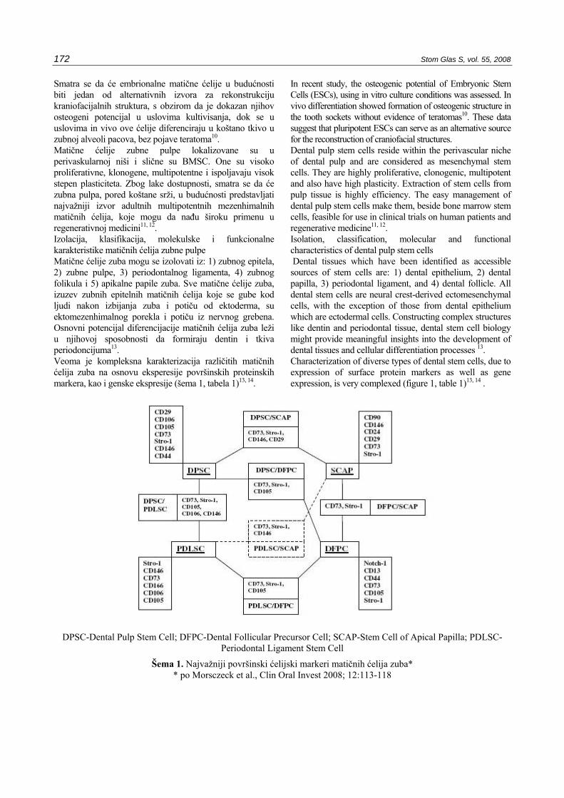
Smatra se da će embrionalne matične ćelije u budućnosti biti jedan od alternativnih izvora za rekonstrukciju kraniofacijalnih struktura, s obzirom da je dokazan njihov osteogeni potencijal u uslovima kultivisanja, dok se u uslovima in vivo ove ćelije diferenciraju u koštano tkivo u zubnoj alveoli pacova, bez pojave teratoma10. Matične ćelije zubne pulpe lokalizovane su u perivaskularnoj niši i slične su BMSC. One su visoko proliferativne, klonogene, multipotentne i ispoljavaju visok stepen plasticiteta. Zbog lake dostupnosti, smatra se da će zubna pulpa, pored koštane srži, u budućnosti predstavljati najvažniji izvor adultnih multipotentnih mezenhimalnih matičnih ćelija, koje mogu da nađu široku primenu u regenerativnoj medicini11, 12. Izolacija, klasifikacija, molekulske i funkcionalne karakteristike matičnih ćelija zubne pulpe Matične ćelije zuba mogu se izolovati iz: 1) zubnog epitela, 2) zubne pulpe, 3) periodontalnog ligamenta, 4) zubnog folikula i 5) apikalne papile zuba. Sve matične ćelije zuba, izuzev zubnih epitelnih matičnih ćelija koje se gube kod ljudi nakon izbijanja zuba i potiču od ektoderma, su ektomezenhimalnog porekla i potiču iz nervnog grebena. Osnovni potencijal diferencijacije matičnih ćelija zuba leži u njihovoj sposobnosti da formiraju dentin i tkiva periodoncijuma13. Veoma je kompleksna karakterizacija različitih matičnih ćelija zuba na osnovu eksperesije površinskih proteinskih markera, kao i genske ekspresije (šema 1, tabela 1)13, 14.
In recent study, the osteogenic potential of Embryonic Stem Cells (ESCs), using in vitro culture conditions was assessed. In vivo differentiation showed formation of osteogenic structure in the tooth sockets without evidence of teratomas10. These data suggest that pluripotent ESCs can serve as an alternative source for the reconstruction of craniofacial structures. Dental pulp stem cells reside within the perivascular niche of dental pulp and are considered as mesenchymal stem cells. They are highly proliferative, clonogenic, multipotent and also have high plasticity. Extraction of stem cells from pulp tissue is highly efficiency. The easy management of dental pulp stem cells make them, beside bone marrow stem cells, feasible for use in clinical trials on human patients and regenerative medicine11, 12. Isolation, classification, molecular and functional characteristics of dental pulp stem cells Dental tissues which have been identified as accessible sources of stem cells are: 1) dental epithelium, 2) dental papilla, 3) periodontal ligament, and 4) dental follicle. All dental stem cells are neural crest-derived ectomesenchymal cells, with the exception of those from dental epithelium which are ectodermal cells. Constructing complex structures like dentin and periodontal tissue, dental stem cell biology might provide meaningful insights into the development of dental tissues and cellular differentiation processes 13. Characterization of diverse types of dental stem cells, due to expression of surface protein markers as well as gene expression, is very complexed (figure 1, table 1)13, 14 .
DPSC-Dental Pulp Stem Cell; DFPC-Dental Follicular Precursor Cell; SCAP-Stem Cell of Apical Papilla; PDLSC-Periodontal Ligament Stem Cell
Šema 1. Najvažniji površinski ćelijski markeri matičnih ćelija zuba* * po Morsczeck et al., Clin Oral Invest 2008; 12:113-118
Serbian Dental J, 2008, 55 173
*DPSC-Dental Pulp Stem Cells; SHED-Stem Cells From Human Exfoliated Decidual Teeth; PDLSC-Periodontal Ligament Stem Cells; (++) jaka ekspresija; (+) slaba ekspresija; (-) negativno; (/) subpopulacija
Tabela 1. Profil ekspresije proteina ili genske ekspresije u nekim matičnim ćelijama zuba u uslovima kultivisanja in
vitro i odnos prema BMSC
Međutim, neki površinski proteinski markeri, kao što su Stro-1 i CD73 su ubikvitarno eksprimirani na svim matičnim i prekursorskim ćelijama zuba13. Posebna populacija matičnih ćelija zubne pulpe označena kao nezrele matične ćelije zubne pulpe (engl. Immature Dental Pulp Stem Cells, IDPC) eksprimira markere embrionalnih matičnih ćelija Oct-4, Nanog, SSEA-3, SSEA-4, TRA-1-60 i TRA-1-81. Pretpostavlja se da su ove ćelije u stvari pluripotentni prekursori druge dve populacije matičnih ćelija zubne pulpe označenih kao matične ćelije zubne pulpe u užem smislu reči (engl. Dental Pulp Stem Cells, DPSC) i matičnih ćelija zubne pulpe izolovanih iz eksfoliranih mlečnih zuba (engl. Stem Cells From Human Exfoliated Decidual Teeth, SHED)15. Regulacija homeostaze adultnih matičnih ćelija rezultat je suptilnog balansa između genetičkih i molekularnih ćelijskih mehanizama, spoljašnjih faktora iz lokalnih i sistemskih niša tela, kao i različitih puteva prenosa ćelijskih signala. Određivanjem profila genske ekspresije DPSC i funkcionalnom klasifikacijom tih gena ustanovljena je, pre svega, visoka ekspresija gena alkalne fosfataze, proteina 1 matriksa dentina (DMP1) i dentin-sijalofosfoproteina. Takođe, ukazano je na važnost u ovim ćelijama ekspresije više gena koji kodiraju sintezu komponenti ekstracelularnog matriksa, molekula ćelijske adhezije, faktora rasta i transkripcionih faktora. Funkcionalna analiza i analiza klasteriranja ukazala je važnost ekspresije gena prenosa ćelijskih signala, ćelijske komunikacije i ćelijskog metabolizma (tabela 2)14.
Some surface proteins, like Stro-1 and CD73, are ubiquitously expressed by all dental stem or precursor cells13. Population of Immature Dental Pulp Stem Cells (IDPSC) express embryonic stem cell markers Oct-4, Nanog, SSEA-3, SSEA-4, TRA-1-60 and TRA-1-81. It is assumed that these cells are pluripotent precursors to the other two stem cell populations known as Dental Pulp Stem Cells (DPSC) and Stem Cells From Human Exfoliated Deciduous Teeth (SHED)15. To maintain homeostasis, a balance between genetic and molecular cell mechanism, extrinsic signals from the microenvironment (called niche) in which stem cells reside and signaling pathway in regulation of stem cell properties, is essential. Determining the profile of gene expression of dental pulp stem cells and functional classification of these genes, the expressions of the alkaline phosphatase gene, dentin matrix protein 1, and dentinsialophosphoprotein are verified and consequently the high expressions of these genes are discovered. Analyses of gene expression patterns indicated several genes which encode extracellular matrix components, cell adhesion molecules, growth factors, and transcription regulators. Functional and clustering analyses of differences in gene expression levels revealed cell signaling, cell communication, or cell metabolism (table 2)14.
Antigen DPSC* SHED PDLSC BMSC CD14 - - - - CD34 - - - - CD44 ++ ++ ++ ++ CD45 - - - - CD106 + +/- +/- ++ CD146 ++/+/- ++/+/- ++/+/- ++/+/- 3G5 +/- +/- +/- +/- Stro-1 ++/+/- ++/+/- ++/+/- ++/+/- α-glatkomišićni aktin ++/- ++/- ++/- ++/+/- kolagen tip-I ++ ++ ++ ++ kolagen tip-III ++/+ ++/+/- ++/+/- ++/+ alkalna fosfataza ++/+/- ++/+/- ++/+/ ++/+/- osteokalcin ++/+ ++/+/- ++/- +/- osteonektin ++/+ ++/+ ++/+ ++/+ osteopontin +/- +/- +/- +/- sialoprotein kosti - - - - skleraksis + + ++ + sialofosfoprotein dentina - - - -

174 Stom Glas S, vol. 55, 2008
hBMSC (I)* hDPSC hDPSC (I) hOMC hDPSC (I) (18–24) Glavna
kategorija gena
Podkategorija gena
DNK sinteza/replikacija 8 7 4 9 7 6 12 2 2
apoptoza 15 6 39 20 31 16 39 12 11 6 ćelijski ciklus 25 8 27 31 39 15 68 7 11 14 hromozom 6 5 4 9 5 3 9 1 1 3 ikupno 54 26 74 69 82 40 128 22 25 23
deoba ćelije
ćelijska athezija 16 14 33 22 23 24 32 26 10 4 kanali/transport proteina 6 9 13 11 18 14 18 8 7 6
faktori rasta/citokini 15 9 13 8 23 6 30 7 16 3 metabolizam 18 12 17 20 28 14 42 8 10 4 modifkacija proteina 5 8 20 18 23 3 12 10 14 9 receptori 59 32 78 53 65 44 81 62 28 16
signalni putevi ćelije ili ćelijska komunikacija
Ukupno 119 84 174 132 180 105 215 121 85 42
citoskelet 4 3 5 7 6 5 11 2 2 5 ekstracelularni matriks 30 11 17 25 15 31 15 31 5 7
protein vezan za mikrotubule 1 7 5 14 3
struktura ćelije / motilitet
ukupno 35 14 29 32 26 36 40 33 7 15
homeostaza (odgovor na stres) 41 14 40 45 61 25 56 39 28 16
imunologija (imuni odgovor) 46 10 41 30 53 17 51 46 24 11
ćelija / odbrana organizma
ukupno 87 24 81 75 114 42 107 85 52 27
RNK sinteza (transkripcioni faktor)
31 16 62 49 52 21 55 31 35 13
sinteza proteina (vezivanje proteina) 4 3 3 7 6 2 5 2 2 5
gen / ekspresija proteina
ukupno 35 19 65 56 58 23 60 33 37 18
aminokiselina 5 1 7 4 9 5 3 5 7 1 energija/TCA ciklus 1 1 3 3 4 lipid 21 4 27 7 33 6 21 6 13 1 nukleotid 2 2 4 7 6 4 21 8 4 8 modifkacija proteina 13 12 41 32 20 3 13 21 19 11 transport 45 19 50 39 83 32 62 35 39 12
metabolizam
ukupno 87 39 132 89 154 50 124 75 82 33
UKUPNO 417 206 555 453 614 296 674 369 288 158
*hBMSC (I)-humane mezenhimalne matične ćelije koštane srži u osteoidnuktivnoj sredini 18. dana kultivacije; hDPSC (I)-humane matične ćelije zubne pulpe u osteoinsuktivnoj sredini 18. dana kultivacije; hOMC-humane ćelije oralne mukoze; hDPSC (I) (18-24)- humane matične ćelije zubne pulpe u osteoinsuktivnoj sredini 18-24 dana kultivacije
Tabela 2. Funkcionalna klasifikacija gena hDPSC
Serbian Dental J, 2008, 55 175
Što se tiče razlika u genskoj ekspresiji između MAPC/BMSC i DPSC, one se ogledaju pre svega u povećanoj ekspresiji kod DPSC gena koji kontrolišu ćelijski ciklus, naročito gena ciklin-zavisne kinaze-6 koja je aktivator ćelijskog ciklusa, što objašnjava višu stopu proliferacije DPSC u odnosu na MAPC/BMSC14, 16. Put prenosa signala Wnt/beta-katenin17, koji je ključan za samoobnavljanje matičnih ćelija, kao i put Noch18, negativno regulišu diferencijaciju in vitro DPSC u odontoblaste, dok induktivni efekat imaju FGF2 i TGFbeta119. U poslednje vreme poznato je da je hipoksija jedan od ključnih faktora u održavanju nediferenciranog stanja i plasticiteta adultnih matičnih ćelija20. Međutim, uslovi kultivisanja koji imitiraju ishemiju (hipoksiju i hipoglikemiju) nepovoljno utiču na preživljavanje i diferencijaciju DPSC21. Bolje poznavanje ekspresije površinskih markera i gena matičnih ćelija zubne pulpe, puteva prenosa signala, i njihov odnos prema ćelijskoj diferencijaciji i plasticitetu, svakako će u budućnosti doprineti razvoju terapije bazirane na primeni ovih ćelija u humanoj medicini i stomatologiji. Matične ćelije zubne pulpe imaju sve osobine potrebne za uspešnu terapijsku primenu: 1) lako su dostupne za izolaciju; 2) izolacija je veoma efikasna; 3) imaju multipotentni potencijal; 4) pokazuju interakciju sa biomaterijalima koji upotrebljeni kao matrice indukuju intenzivnu proliferaciju ovih ćelija; 5) dugovečne su i 6) podležu uspešnoj krioprotekciji, slično drugim matičnim ćelijama11, 22. Podatak da vijabilne matične ćelije zubne pulpe mogu da se izoluju i nakon 5 dana od ekstrakcije zuba, kao i da ih je moguće izolovati i kultivisati iz intaktnih zuba koji su prethodno krioprezervirani, ukazuje da su potrebni minimalni zahtevi za stvaranje banke ovih ćelija u cilju buduće terapijske primene12. Studijama in vitro je dokazano da matične ćelije zubne pulpe imaju izrazit plasticitet, tj. sposobnost da se diferenciraju u ćelije različite od ćelija prisutnih na mestu odakle potiču, a najnoviji radovi pružaju dokaze o njihovom plasticitetu i u uslovima in vivo. U uslovima in vitro ili in vivo ove ćelije mogu da se diferenciraju, s određenim međusobnim razlikama, u pravcu odontoblasta, hondrocita, osteoblasta, adipocita, neurona/glije, glatkih i skeletnih mišićnih ćelija, endotelnih ćelija i melanocita15, 16, 23-29. U uslovima in vivo, nakon implantacije, pokazuju različit potencijal za formiranje dentina, ali i koštanog i masnog tkiva30. Generalno se smatra da DPSC, slično drugim mezenhimalnim matičnim ćelijama, imaju anti-inflamatorno dejstvo. One ispoljavaju imunomodulatorni efekat jer mogu biti uključene u imunski odgovor u toku infekcije zubne pulpe, a verovatno i periodoncijuma, koji je veoma kompleksan31, 32, putem aktivacije NF-�B33. Takođe, dovode do imunološke tolerancije ukoliko se implantiraju u alogena tkiva11.
Comparing the gene expression profiles of human dental pulp stem cells to bone marrow stromal stem cells a few differentially expressed genes, including cell cyclus activator cyclin-dependent kinase 6, were highly expressed in DPSCs. This explains the higher proliferation rate of dental pulp stem cells than bone marrow stromal stem cells14, 16. Wnt/beta-katenin, important for stem cells self-renewal17, and Notch signaling18 can inhibit the odontoblastic differentiation, while two crucial growth factors, FGF2 and TGFbeta119, have inductive effects on the odontoblastic differentiation of human dental pulp stem cells in vitro. Although differentiated and undifferentiated cells can be exposed to ischemic conditions in cases of injury or inflammation, the effects of ischemia on cell survival and differentiation have not been well characterized20. These data showed that the ischemic conditions have similar detrimental influence on both undifferentiated and differentiated porcine Dental Pulp-derived Cells (pDPCs), and affect differentiation status of pDPCs21. Dental pulp stem cells have all characteristics for successful therapeutic use: 1) access to the collection site of these cells is easy, 2) extraction of stem cells from pulp tissue is highly efficiency; 3) they have an extensive differentiation ability, 4) demonstrated interactivity with biomaterials makes them ideal for tissue reconstruction, 5) they have a long lifespan, 6) they can be safely cryopreserved11, 22. Recent studies indicate that DPSC isolation is feasible for at least 5 days after tooth extraction. Further, the recovery of viable DPSC after cryopreservation of intact teeth suggests that minimal processing may be needed for the banking of samples with no immediate plans for expansion and use12. An increasing number of investigations supports that stem cells have the potential to differentiate into matured cell types beyond their origin, a property defined as plasticity. Previously, the plasticity of DPSCs has been confirmed by culturing cells in lineage-specific media in vitro. In recent studies, it has been confirmed in vivo conditions as well. In both conditions, in vivo and in vitro, these cells have the ability to differentiate into odontoblasts, chondrocytes, osteoblasts, adipocytes, neurons, melanocytes, smooth and skeletal muscles and endothelial cells15, 16, 23-29. In vivo, after implantation, they have shown potential to differentiate into dentin but also into bone or adipose tissue30. In general, DPSCs are considered to have anti-inflammatory and immunomodulatory abilities because they may be involved in immune responses during pulpal infection, and probably periodontium as well31,32, through activating NF- B 33. After being grafted into allogenic tissues these cells are able to induce immunological tolerance11.
176 Stom Glas S, vol. 55, 2008
Sposobnost inhibicije proliferacije T limfocita ukazuje na njihovo imunosupresivno dejstvo i moguću primenu kod autoimunskih bolesti34. Različite vrste matičnih ćelija zubne pulpe i njihov potencijalni značaj u regenerativnoj medicini i stomatologiji Sredinom devedesetih godina XX. veka došlo je do uspešne izolacije prekursorskih ćelija zubne pulpe35. Kasnije su matične ćelije izolovane iz zubne pulpe kutnjaka i nazvane DPSC23, kao i iz eksfoliranih mlečnih zuba–SHED36 i IDPC15. DPSC Ove ćelije su do sada izdvojene iz umnjaka, mlečnih sekutića i prekobrojnih zuba16, 23, 37. Kao što je već iustaknuto, ispoljavaju slične karakteristike kao BMSC38. Prva istraživanja na DPSC izolovanim iz umnjaka su pokazala da se ove ćelije diferenciraju u odontoblaste, adipocite i u neuronima-slične ćelije i da ta diferencijacija podseća na embrionalnu ontologiju kranijalnih ćelija nervnog grebena. Istovremeno je pokazano i da imaju neke karakteristike identične sa osteoblastima24. Međutim, najnovija istraživanja pokazuju da humane (h) DPSC imaju daleko veći potencijal diferencijacije, s obzirom da mogu da se diferenciraju u ćelije mezenhimalnog porekla (odontoblaste, osteoblaste, adipocite, hondrocite i poprečnoprugaste mišićne ćelije) ali i u melanocite (ćelije koje nisu porekla mezenhima, već potiču od ćelija nervnog grebena)26. Stoga se pretpostavlja da se u kulturama hDPSC nalaze i ćelije koje bi mogle biti multipotentne matične ćelije nervnog grebena. Pokazano je da humane DPSC izolovane iz mlečnih zuba, koje u uslovima in vitro pokazuju osteogeni, adipogeni i miogeni potencijal diferencijacije, transplantirane pacovima sa velikim defektom parijetalne regije, dovode do stvaranja nove kosti nakon 1 meseca od načinjene lezije, bez upotrebe imunosupresivne terapije25. To daje nadu da će se ove ćelije u budućnosti moći upotrebiti u rekonstruktivnoj hirurgiji kraniofacijalne regije. Matične ćelije označene kao SBP-DPSC su multipotentna subpopulacija DPSC, koja je sposobna da se diferencira u osteoblaste i da sintetiše trodimenzionalnu lamelarnu kost in vitro. Ukoliko se SBP-DPSC ili koštani model dobijen iz njih u uslovimsa in vitro transplantira pacovu sa deficitom imunološkog sistema stvoriće se tkivna struktura sa integralnim krvnim sudovima, slična pravoj kosti odraslih ljudi28,29. Dokazano je da čak i nakon 2 godine od krioprezervacije SBP-DPSC zadržavaju sposobnost diferencijacije, proliferacije i sinteze koštanog tkiva29.
Immunosuppressive effect and possible therapeutic use in autoimmune diseases is shown through the ability to inhibit proliferation of T lymphocytes34. Different types of dental pulp stem cells - potential significance in regenerative medicine and dentistry In the last decade of the twentieth century, precursor cells of dental pulp were successfully isolated35. Furthermore, DPSCs23 were isolated from dental pulp of human adult third molar as well as from human exfoliated deciduous teeth36 and immature dental pulp 15. DPSC To date, these cells have been isolated from third molars, decidual incisors and supernumerary teeth16, 23, 37. DPSC have similar characteristics to Bone Marrow Stem Cells (BMSC)38. DPSCs derived from third molars were also found to be capable of differentiating into odontoblasts, adipocytes and neural-like cells and that differentiation reminiscent of cranial neural crest (CNC) cells embryonic ontology. At the same time, it is found that DPSCs have some characteristics identical to osteoblasts24. It is demonstrated that human dental pulp contains self renewing human Dental Pulp Stem Cells (hDPSCs) capable of differentiating into mesenchymal-derived odontoblasts, osteoblasts, adipocytes, chondrocytes and striated muscle, and interestingly, also into non-mesenchymal, neural crest- derived melanocytes 26. Furthermore, this study showed that hDPSC cultures include cells with traits attributed to multipotent stem cells, and provide evidence that these might be multipotent neural crest stem cells. Human dental pulp stem cells, were isolated from deciduous teeth, to reconstruct large-sized cranial bone defects in nonimmunosuppressed rats. They showed osteogenic, adipogenic, and myogenic in vitro differentiation25. These findings suggest that hDPSC is an additional cell resource for correcting large cranial defects in rats and constitutes a promising model for reconstruction of human large cranial defects in craniofacial surgery. Stromal stem cells from human dental pulp (SBP-DPSCs) are multipotent stem cells able to differentiate into osteoblasts, which synthesize three-dimensional woven bone tissue chips in vitro. When either SBP-DPSCs or bone chips obtained in vitro were transplanted into immunocompromised rats, they generated a tissue structure with an integral blood supply similar to that of human adult bone28,29. After storage for 2 years, it is found that stem cells are still capable of differentiation, and that their differentiated cytotypes proliferate and produce woven bone tissue29.
Serbian Dental J, 2008, 55 177
Subpopulaciju DPSC, označenu kao SP-DPSC imaju veliki angiogenetski potencijal. Ukoliko se miševima sa ishemijom prednje šape lokalno transplantiraju ove ćelije doći će do njihovog uspešnog kalemljenja, povećanja gustine kapilarne mreže i bolje prokrvljenosti ishemijske zone, a kondicionirani medijum od ovih ćelija pokazuje mitogenu i anti-apoptotsku aktivnost na endotelne ćelije humane umbilikalne vene39. Stvaranje in vivo tkiva sličnog zubnoj pulpi, nakon subkutane aplikacije mišu trijade DPSC, matrice od kolagena i DMP1, ukazuje na veliki potencijalni značaj ovih ćelija u tkivnom inženjeringu 40. SHED Matične ćelije iz eksfoliranih mlečnih zuba su sposobne za diferencijaciju u odontoblaste, adipocite i nervne ćelije. U uslovima in vivo one indukuju formiranje kosti i produkciju dentina; nakon transplantacije u miša sa kompromitovanim imunološkim sistemom migriraju u mozak i sposobne su za preživljavanje 36. Studije in vivo su takođe pokazale da se humane SHED diferenciraju u odontoblaste i endotelne ćelije i da stoga eksfolirani mlečni zubi predstavljaju vijabilni izvor matičnih ćelija za tkivni inženjering zubne pulpe. Naime, ukoliko se hSHED oblože biodegradibilnim materijalom pripremljenim sa presecima normalnog zuba i kao takve implantiraju mišu sa imunološkim deficitom, oformiće se tkivo koje u histološkom pogledu i u pogledu ćelijskog sastava veoma podseća na zubnu pulpu 41. IDPC Radi se o najprimitivnijoj populaciji matičnih ćelija zubne pulpe, s obzirom da je pokazano da eksprimiraju markere embrionalnih matičnih ćelija. U toku kultivisanja kroz 25 pasaža, u toku 4 meseca, one zadržavaju normalni kariotip i stopu ekspanzije karakterističnu za matične ćelije. U hemijski definisanim uslovima kultivisanja in vitro, IDPC se diferenciraju u glatke i poprečnoprugaste mišićne ćelije, neurone, hondrocite i osteoblaste. U uslovima in vivo, ukoliko se transplantiraju miševima sa imunološkim deficitom, dobro se kaleme u različitim tkivima 15. Aplikacija hIDPC bez primene imunosupresivne terapije, pokazala se uspešnom u kontroli mišićne distrofije kod zlatnih retrivera (najboljeg animalnog model za Duchenne-ovu mišićnu distrofijiju kod ljudi), pri čemu je sistemska aplikacija ovih matičnih ćelija bila bolji izbor od lokalne aplikacije 42. Zaključak Matične ćelije zubne pulpe otvorile su nove perspektive u terapijskoj primeni ovih ćelija ne samo u regeneraciji dentina, tkiva periodoncijuma i koštano-zglobnog tkiva kraniofacijalne regije, već i u lečenju neurotraume, infarkta miokarda, mišićne distrofije i oštećenja vezivnog tkiva.
Side population (SP) dental pulp stem cells, SP-DPSC, has high angiogenetic potential. In models of mouse hindlimb ischemia, local transplantation of one subfraction of SP cells resulted in successful engraftment and an increase in the blood flow including high density of capillary formation. Conditioned medium from this subfraction showed the mitogenic and anti-apoptotic activity on human umbilical vein endothelial cells 39. In vivo generation of dental pulp-like tissue by using the triad of dental pulp stem cells, a collagen scaffold, and dentin matrix protein 1 after subcutaneous transplantation in mice, might lead to hard tissue formation. This finding indicate that these cells might have high potential significance in tissue engineering 40. SHED SHED were identified to be a population capable of differentiating into a variety of cell types including neural cells, adipocytes, and odontoblasts. After in vivo transplantation, SHED were found to be able to induce bone formation, generate dentin and survive in mouse brain along with expression of neural markers 36. SHED differentiated into odontoblast-like cells in vivo and in endothelial-like cells. SHED seeded in biodegradable scaffolds prepared within human tooth slices and transplanted into immunodeficient mice result in tissue with architecture and cellularity that closely resemble those of a physiologic dental pulp. This work suggests that exfoliated deciduous teeth constitute a viable source of stem cells for dental pulp tissue engineering 41. IDPC The population of immature dental pulp stem cells is dental pulp stem cell population which expresses embryonic stem cell markers during at least 25 passages while maintaining the normal karyotype and the rate of expansion characteristic of stem cells. Moreover, in vitro these cells can be induced to undergo uniform differentiation into smooth and skeletal muscles, neurons, cartilage, and bone under chemically defined culture conditions. After in vivo transplantation of these cells into immunocompromised mice, they showed dense engraftment in various tissues 15. Human Immature Dental Pulp Stem Cells (hIDPSC) were transplanted, without any immunosuppression, into golden retriever muscular dystrophy dogs, who represent the best available animal model for therapeutic trials aiming at the future treatment of human Duchenne muscular dystrophy. Data from this trial suggested that systemic multiple deliveries seemed more effective than local injections. These findings open important avenues for further researches 42.
Conclusion
Dental pulp stem cells open new perspectives in therapeutic use not only in dentin regeneration, periodontal tissues and skeletoarticular tissues of craniofacial region but also in treatment of neurotrauma, autoimmune diseases, myocardial infarction, muscular dystrophy and connective tissue damages.
178 Stom Glas S, vol. 55, 2008
Literatura / References
1. Todorović V, Nikolić I, Glibetić M, Balint B. Humane embrionalne matične ćelije – dosadašnja saznanja. Anestezija, reanimacija, transfuzija 2006; 34:109-28
2. Todorović V, Nikolić RI. Stem ćelije, kloniranje sisara. U: Ivan R. Nikolić, urednik i ilustrator. Embriologija čoveka, tekst i atlas (treća izdanje). Beograd: Data Status, 2007: 45-70
3. Malanchi I, Peinado H, Kassen D, et al.Cutaneus cancer stem cell maintenance is dependent on catenin signalling. Nature 2008; 452:650-3
4. Brašanac D, Boričić I, Todorović V, Tomanović N, Radojević S. Cyclin A and �-catenin expression in actinic keratosis, Bowen’s disease and invasive squampus cell carcinoma of the skin. Br J Dermatology 2005; 153:1166-75
5. Vats A, Bielby RC, Tolley NS, Nerem R, Polak JM. Stem cells. Lancet 2005; 366:592-602
6. Maria OM, Khosravi R, Mezey E, Tran SD. Cells from bone marrow that evolve into oral tissues and their clinical applications. Oral Dis 2007; 13:11-16
7. Ueda M, Yamada Y, Ozawa R, Okazaki Y. Clinical case reports of injectable tissue-engineered bone for alveolar augmentation with simultaneous implant placement. Int J Periodontics Restorative Dent 2005; 25:129-37
8. Yamada Y, Ueda M, Hibi H, Baba S. A novel approach to periodontal tissue regeneration with mesenchymal stem cells and platelet-rich plasma using tissue engineering technology: A clinical case report. Int J Periodontics Restorative Dent 2006; 26:363-9
9. Tobita M, Uysal AC, Ogawa R, Hyakusoku H, Mizuno H. Periodontal tissue regeneration with adipose-derived stem cells. Tissue Eng Part A 2008; 14:945-53
10. Kang HK, Roh S, Lee G, Hong SD, Kang H, Min BM. Osteogenic potential of embryonic stem cells in tooth sockets. Int J Mol Med. 2008;21:539-44
11. d'Aquino R, Papaccio G, Laino G, Graziano A. Dental pulp stem cells: a promising tool for bone regeneration. Stem Cell Rev 2008;4:21-6
12. Perry BC, Zhou D, Wu X, et al. Collection, cryopreservation, and characterization of human dental pulp-derived mesenchymal stem cells for banking and clinical use. Tissue Eng Part C Methods 2008;14:149-56
13. Morsczeck C, Schmalz G, Reichert TE, Volner F, Galler K, Driemel O. Somatic stem cells for regenerative dentisty. Clin Oral Invest 2008; 12:113-8
14. Yamada Y, Fujimoto A, Ito A, Yoshimi R, Ueda M. Cluster analysis and gene expression profiles: a cDNA microarray system-based comparison between human dental pulp stem cells (hDPSCs) and human mesenchymal stem cells (hMSCs) for tissue engineering cell therapy. Biomaterials 2006; 27:3766-81
15. Kerkis I, Kerkis A, Dozortsev D, et al. Isolation and characterization of a population of immature dental pulp stem cells expressing OCT-4 and other embryonic stem cell markers. Cells Tissues Organs 2006; 184:105-16
16. Shi S, Gronthos S. Perivascular niche of postnatal mesenchimal stem cells in human bone marrow and dental pulp. J Bone Miner Res 2003; 18:696-704
17. Scheller EL, Chang J, Wang CY. Wnt/beta-catenin inhibits dental pulp stem cell differentiation. J Dent Res 2008;87:126-30
18. Zhang C, Chang J, Sonoyama W, Shi S, Wang CY. Inhibition of human dental pulp stem cell differentiation by Notch signaling. J Dent Res 2008;87:250-5
19. He H, Yu J, Liu Y, et al. Effects of FGF2 and TGFbeta1 on the differentiation of human dental pulp stem cells in vitro. Cell Biol Int 2008;32:827-34
20. Kovacevic-Filipovic M, Petakov M, Hermitte F, et al. Interleukin-6 (IL-6) and low O-2 concentration (1%) synergize to improve the mainteance of hematopoietic stem cells (Pre-CSF). J Cell Physiol 2007; 212:68-75
21. Agata H, Kagami H, Watanabe N, Ueda M. Effect of ishemic culture contitions on the survival and differentiation of porcine dental pulp-derived cells. Differentiation 2008; DOI: 10.111/j.1432-0436.2008.00282.x
22. Škorić D, Balint B, Petakov M, Sindjić M, Rodić P. Collection strategies and cryopreservation of umbilical cord blood. Transfusion Med 2007; 17:107-13
23. Gronthos S, Mancani M, Brahim J, Robey PG, Shi S. Postnatal human dental pulp stem cells (DPSCs) in vitro and in vivo. Proc Natl Acad Sci 2000; 97:13625-30
24. Gronthos S, Brahim J, Li W, et al. Stem cell properties of human pulp stem cells. J Dent Res 2002; 81:531-5
25. de Mendonça Costa A, Bueno DF, Martins MT, et al. Reconstruction of large cranial defects in nonimmunosuppressed experimental design with human dental pulp stem cells. J Craniofac Surg 2008; 19:204-10
Serbian Dental J, 2008, 55 179
26. Stevens A, Zuliani T, Olejnik C, et al. Human
dental pulp stem cells differentiate into neural crest-derived melanocytes and have label-retaining and sphere-forming abilities. Stem Cells Dev 2008; Mart 25. [Epub ahead of print]
27. Arthur A, Rychkov G, Shi S, Koblar SA, Gronthos S. Adult human dental pulp stem cells differentiate toward functionally active neurons under appropriate environmental cues. Stem Cells 2008; 26:1787-95
28. d'Aquino R, Graziano A, Sampaolesi M, et al. Human postnatal dental pulp cells co-differentiate into osteoblasts and endotheliocytes: a pivotal synergy leading to adult bone tissue formation. Cell Death Differ 2007; 14:1162-71
29. Papaccio G, Graziano A, d'Aquino R, et al. Long-term cryopreservation of dental pulp stem cells (SBP-DPSCs) and their differentiated osteoblasts: a cell source for tissue repair. J Cell Physiol 2006; 208:319-25
30. Zhang W, Walboomers XF, Van Kuppevelt TH, et al. In vivo evaluation of human dental pulp stem cells differentiated towards multiple lineages. J Tissue Eng Regen Med 2008; 2:117-25
31. Colic M, Lukic A, Vucevic D, et al. Correlation between phenotypic characteristic of mononuclear cells isolated from human periapical lesions and their in vitro production of Th1 and Th2 cytokines. Arch Oral Biol 2006; 51:1120-30
32. Stefanović G, Marković D, Ilić V, Brajović G, Petrović S, Milošević-Jovčić N. Hypogalactosylation of salivary and gingival fluid IgG in patients with advanced periodontitis. J Periodontol 2006; 77:1887-93
33. Chang J, Zhang C, Tani-Ishii N, Shi S, Wang C-Y. NF-�B activation in human dental pulp stem cells by TNF and LPS. J Dent Res 2005; 84:994-8
Adresa za korespondenciju dr sci. med. Vera Todorović, naučni savetnik Institut za medicinska istraživanja, Univerzitet u Beogradu Dr Subotića 4, 11129 Beograd, PF 129 e-mail: [email protected] Tel:011/2685-788/L-111 ili 113
34. Pierdomenico L, Bonsi L, Calvitti M, et al.
Multipotent mesenchymal stem cells with immunosupresive acticity can be easily isolated from dental pulp. Transplantation 2005; 80:836-42
35. Stanislawski L, Carreau JP, Pouchelet M, Chen ZN, Goldberg M. In vitro culture of human dental pulp cells: some aspects of cells emerging early from the explant. Clin Oral Investig 1997; 1:131-40
36. Miura M, Gronthos S, Zhao M, et al. SHED: Stem cells from human exfoliated decidual teeth. Proc Natl Acad Sci USA 2003; 100:5807-12
37. Huang AH, Chen YK, Lin LM, Shieh TY, Chan AW. Isolation and characterization of dental pulp stem cells from a supernumerary tooth. J Oral Pathol Med 2008; Mart 5. [Epub ahead of print]
38. Shi S, Rhobey PG, Gronthos S. Comparison of human dental pulp and bone marrow stromal cells by cDNA microarray analysis. Bone 2001; 29:532-9
39. Iohara K, Zheng L, Wake H, et al. A novel stem cell source for vasculogenesis in ischemia: subfraction of side population cells from dental pulp. Stem Cells 2008; Jun 26. [Epub ahead of print]
40. Prescott RS, Alsanea R, Fayad MI, et al. In vivo generation of dental pulp-like tissue by using dental pulp stem cells, a collagen scaffold, and dentin matrix protein 1 after subcutaneous transplantation in mice. J Endod 2008; 34:421-6
41. Cordeiro MM, Dong Z, Kaneko T, et al. Dental pulp tissue engineering with stem cells from exfoliated deciduous teeth. J Endod 2008; 34:962-9
42. Kerkis I, Ambrosio CE, Kerkis A, et al Early transplantation of human immature dental pulp stem cells from baby teeth to golden retriever muscular dystrophy (GRMD) dogs: local or systemic? J Transl Med 2008; 6:35
Address for correspondence Vera Todorovic, research professor Institute for medical research University of Belgrade Dr Subotica 4 11000 Belgrade, PO BOX 129 e-mail: [email protected] Tel:+381 11/2685-788/ L-111 ili 113
Related Documents