International Journal of Molecular Sciences Article Density Functional Theory-Based Calculation Shed New Light on the Bizarre Addition of Cysteine Thiol to Dopaquinone Ryo Kishida 1, * , Shosuke Ito 2 , Manickam Sugumaran 3 , Ryan Lacdao Arevalo 4 , Hiroshi Nakanishi 5,6 and Hideaki Kasai 5,7 Citation: Kishida, R.; Ito, S.; Sugumaran, M.; Arevalo, R.L.; Nakanishi, H.; Kasai, H. Density Functional Theory-Based Calculation Shed New Light on the Bizarre Addition of Cysteine Thiol to Dopaquinone. Int. J. Mol. Sci. 2021, 22, 1373. https://doi.org/ 10.3390/ijms22031373 Academic Editor: Michele Navarra Received: 30 December 2020 Accepted: 27 January 2021 Published: 29 January 2021 Publisher’s Note: MDPI stays neutral with regard to jurisdictional claims in published maps and institutional affil- iations. Copyright: © 2021 by the authors. Licensee MDPI, Basel, Switzerland. This article is an open access article distributed under the terms and conditions of the Creative Commons Attribution (CC BY) license (https:// creativecommons.org/licenses/by/ 4.0/). 1 Department of Biomaterials, Faculty of Dental Science, Kyushu University, Maidashi, Fukuoka 812-8582, Japan 2 Institute for Melanin Chemistry, Fujita Health University, Toyoake, Aichi 470-1192, Japan; [email protected] 3 Department of Biology, University of Massachusetts Boston, 100 Morrissey Boulevard, Boston, MA 02125-3393, USA; [email protected] 4 Department of Physics, Talamban Campus, University of San Carlos, Cebu City 6000, Philippines; [email protected] 5 National Institute of Technology, Akashi College, Akashi, Hyogo 674-8501, Japan; [email protected] (H.N.); [email protected] (H.K.) 6 Institute of Industrial Science, The University of Tokyo, Meguro, Tokyo 153-8505, Japan 7 Graduate School of Engineering, Osaka University, Suita, Osaka 565-0871, Japan * Correspondence: [email protected] Abstract: Two types of melanin pigments, brown to black eumelanin and yellow to reddish brown pheomelanin, are biosynthesized through a branched reaction, which is associated with the key in- termediate dopaquinone (DQ). In the presence of L-cysteine, DQ immediately binds to the –SH group, resulting in the formation of cysteinyldopa necessary for the pheomelanin production. L-Cysteine prefers to bond with aromatic carbons adjacent to the carbonyl groups, namely C5 and C2. Surprisingly, this Michael addition takes place at 1,6-position of the C5 (and to some extent at C2) rather than usually expected 1,4-position. Such an anomaly on the reactivity necessitates an atomic-scale understanding of the binding mechanism. Using density functional theory-based calculations, we investigated the binding of L-cysteine thiolate (Cys–S - ) to DQ. Interestingly, the C2–S bonded intermediate was less energetically stable than the C6–S bonded case. Furthermore, the most preferred Cys–S - -attacked intermediate is at the carbon-carbon bridge between the two carbonyls (C3–C4 bridge site) but not on the C5 site. This structure allows the Cys–S - to migrate onto the adjacent C5 or C2 with small activation energies. Further simulation demonstrated a possible con- version pathway of the C5–S (and C2–S) intermediate into 5-S-cysteinyldopa (and 2-S-cysteinyldopa), which is the experimentally identified major (and minor) product. Based on the results, we propose that the binding of Cys–S - to DQ proceeds via the following path: (i) coordination of Cys–S - to C3–C4 bridge, (ii) migration of Cys–S - to C5 (C2), (iii) proton rearrangement from cysteinyl –NH 3 + to O4 (O3), and (iv) proton rearrangement from C5 (C2) to O3 (O4). Keywords: dopaquinone; cysteine; melanin; density functional theory; quinone reactions; thiol addition to quinone 1. Introduction Melanin, the polyphenolic pigment found throughout living organisms, is an im- portant biopolymer that provides protection against damaging solar radiation [1–9]. In animals, specialized cells called melanocytes produce melanin pigments and transport them to the skin, hair, and eyes, where it provides external coloration. Understanding the biochemically distinct nature of melanocytes and melanocyte-related tissue reactions is cru- cial to treat diseases associated with melanogenic processes such as albinism, leukoderma, melanoma cancer, and other related skin disorders. Int. J. Mol. Sci. 2021, 22, 1373. https://doi.org/10.3390/ijms22031373 https://www.mdpi.com/journal/ijms

Welcome message from author
This document is posted to help you gain knowledge. Please leave a comment to let me know what you think about it! Share it to your friends and learn new things together.
Transcript
International Journal of
Molecular Sciences
Article
Density Functional Theory-Based Calculation Shed New Lighton the Bizarre Addition of Cysteine Thiol to Dopaquinone
Ryo Kishida 1,* , Shosuke Ito 2 , Manickam Sugumaran 3 , Ryan Lacdao Arevalo 4, Hiroshi Nakanishi 5,6
and Hideaki Kasai 5,7
�����������������
Citation: Kishida, R.; Ito, S.;
Sugumaran, M.; Arevalo, R.L.;
Nakanishi, H.; Kasai, H. Density
Functional Theory-Based Calculation
Shed New Light on the Bizarre
Addition of Cysteine Thiol to
Dopaquinone. Int. J. Mol. Sci. 2021,
22, 1373. https://doi.org/
10.3390/ijms22031373
Academic Editor: Michele Navarra
Received: 30 December 2020
Accepted: 27 January 2021
Published: 29 January 2021
Publisher’s Note: MDPI stays neutral
with regard to jurisdictional claims in
published maps and institutional affil-
iations.
Copyright: © 2021 by the authors.
Licensee MDPI, Basel, Switzerland.
This article is an open access article
distributed under the terms and
conditions of the Creative Commons
Attribution (CC BY) license (https://
creativecommons.org/licenses/by/
4.0/).
1 Department of Biomaterials, Faculty of Dental Science, Kyushu University,Maidashi, Fukuoka 812-8582, Japan
2 Institute for Melanin Chemistry, Fujita Health University, Toyoake, Aichi 470-1192, Japan; [email protected] Department of Biology, University of Massachusetts Boston, 100 Morrissey Boulevard,
Boston, MA 02125-3393, USA; [email protected] Department of Physics, Talamban Campus, University of San Carlos, Cebu City 6000, Philippines;
[email protected] National Institute of Technology, Akashi College, Akashi, Hyogo 674-8501, Japan;
[email protected] (H.N.); [email protected] (H.K.)6 Institute of Industrial Science, The University of Tokyo, Meguro, Tokyo 153-8505, Japan7 Graduate School of Engineering, Osaka University, Suita, Osaka 565-0871, Japan* Correspondence: [email protected]
Abstract: Two types of melanin pigments, brown to black eumelanin and yellow to reddish brownpheomelanin, are biosynthesized through a branched reaction, which is associated with the key in-termediate dopaquinone (DQ). In the presence of L-cysteine, DQ immediately binds to the –SHgroup, resulting in the formation of cysteinyldopa necessary for the pheomelanin production.L-Cysteine prefers to bond with aromatic carbons adjacent to the carbonyl groups, namely C5and C2. Surprisingly, this Michael addition takes place at 1,6-position of the C5 (and to some extentat C2) rather than usually expected 1,4-position. Such an anomaly on the reactivity necessitatesan atomic-scale understanding of the binding mechanism. Using density functional theory-basedcalculations, we investigated the binding of L-cysteine thiolate (Cys–S−) to DQ. Interestingly, theC2–S bonded intermediate was less energetically stable than the C6–S bonded case. Furthermore,the most preferred Cys–S−-attacked intermediate is at the carbon-carbon bridge between the twocarbonyls (C3–C4 bridge site) but not on the C5 site. This structure allows the Cys–S− to migrate ontothe adjacent C5 or C2 with small activation energies. Further simulation demonstrated a possible con-version pathway of the C5–S (and C2–S) intermediate into 5-S-cysteinyldopa (and 2-S-cysteinyldopa),which is the experimentally identified major (and minor) product. Based on the results, we proposethat the binding of Cys–S− to DQ proceeds via the following path: (i) coordination of Cys–S− toC3–C4 bridge, (ii) migration of Cys–S− to C5 (C2), (iii) proton rearrangement from cysteinyl –NH3
+
to O4 (O3), and (iv) proton rearrangement from C5 (C2) to O3 (O4).
Keywords: dopaquinone; cysteine; melanin; density functional theory; quinone reactions; thioladdition to quinone
1. Introduction
Melanin, the polyphenolic pigment found throughout living organisms, is an im-portant biopolymer that provides protection against damaging solar radiation [1–9]. Inanimals, specialized cells called melanocytes produce melanin pigments and transportthem to the skin, hair, and eyes, where it provides external coloration. Understanding thebiochemically distinct nature of melanocytes and melanocyte-related tissue reactions is cru-cial to treat diseases associated with melanogenic processes such as albinism, leukoderma,melanoma cancer, and other related skin disorders.
Int. J. Mol. Sci. 2021, 22, 1373. https://doi.org/10.3390/ijms22031373 https://www.mdpi.com/journal/ijms
Int. J. Mol. Sci. 2021, 22, 1373 2 of 15
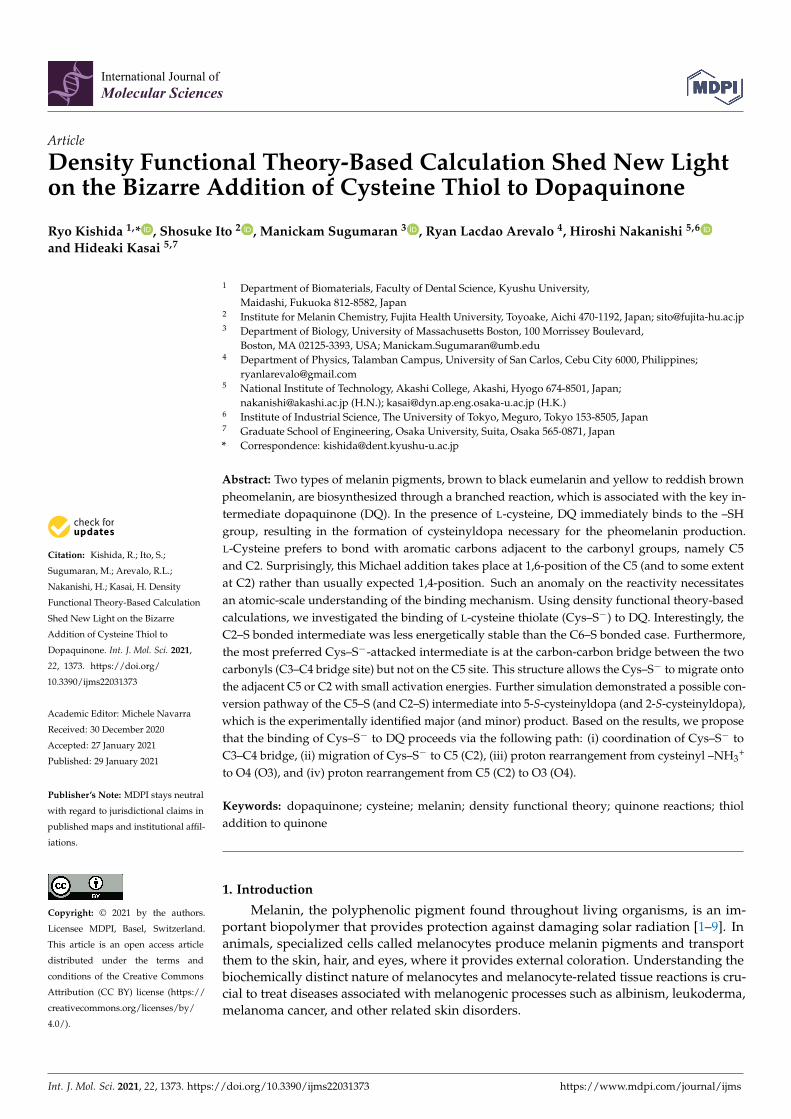
Biosynthesis of melanin (melanogenesis) is initiated by tyrosinase-catalyzed oxidationof the amino acid, tyrosine and its hydroxylated dopa to dopaquinone (DQ). At DQ level,two important reactions determine the nature of melanin formed (Figure 1).
Figure 1. Initial stages of melanin biosynthesis. Tyrosine and its hydroxylated product dopa areconverted to dopaquinone (DQ) by the enzyme, tyrosinase. Further reactivity of DQ determines thenature of melanin product formed. Non-enzymatic addition of Cys−SH results in the formation of5-S-cysteinyldopa (or 2-S-cysteinyldopa) as the major (or minor) product. Oxidative polymerizationof cysteinyldopa generates yellow to reddish brown pheomelanin product. On the other hand,intramolecular cyclization of DQ produces cyclodopa which leads to brown to black eumelanin.
Interestingly, both reactions occur without any enzymatic assistance and are consid-ered key examples of novel biological reactions that are of a non-enzymatic nature. In thepresence of cellular thiols such as L-cysteine (Cys–SH), DQ exhibits rapid non-enzymaticaddition. Thus, DQ and similar o-quinones bind to cellular thiols, including Cys–SH,glutathione, and protein thiols [1–4,9–12]. The binding of cellular Cys–SH to DQ pro-duces cysteinyldopa which further transforms into yellow to reddish brown pheomelanin(pheomelanogenesis). After cellular Cys–SH is consumed enough (<1 µmol/L), DQ under-goes the competing reaction, i.e., intramolecular cyclization of the alanyl side chain [13].This cyclization produces cyclodopa which after further transformations leads to brownto black eumelanin production (eumelanogenesis). Thus, melanin is a mixed pigmentwhich consists of eumelanin and pheomelanin. The presence of pheomelanin influencesthe property of melanin in various contexts, including the color [14,15] and the bindingability for metals and/or drugs [16,17]. Furthermore, pheomelanogenesis can also causecytotoxic and/or carcinogenic biochemical reactions [18–22].
Such non-enzymatic reactions are not limited to DQ alone as a number of otherrelated catecholamine-derived quinones have also been shown to participate in a varietyof biological processes. For example, neuromelanin production is associated with thenon-enzymatic reactions of quinonoid products derived from dopamine [5]. Insects andother arthropods generate N-acyldopamine quinones as key components of sclerotizedcuticle during the hardening of their exoskeleton [23]. Peptidyl dopa-derived quinonoidproducts are also established to play crucial role in mussel glue protein [24,25], as well astunic formation and defense reaction in tunicates [26]. As a result, extensive studies havebeen carried out on the reactions of quinones with biological nucleophiles [4,7].
One of the most intriguing aspects of the reactivity of o-quinones with different nucle-ophiles is the fact that while most nucleophiles including amines exhibit normal Michael1,4-addition reaction to quinones, thiols uniquely exhibit abnormal Michael 1,6-addition

Int. J. Mol. Sci. 2021, 22, 1373 3 of 15
reactions. Moreover, in spite of the fact that proximity effects play a crucial role in tremen-dously accelerating the course of any chemical as well as biological reaction, the reaction ofsuitably situated internal amine group with the quinone ring in the same molecule is in factslower than the reaction of externally present thiol group with a quinone. Such unusualreactivities of thiols have puzzled several organic chemists for decades.
Atomic-scale understanding of thiol binding to quinones could shed light on pheome-lanogenesis and related processes. Effectiveness of density functional theory- (DFT-) basedcalculation for obtaining the potential energy hypersurface of amino acid motions as wellas their electronic structures has been widely validated [27–36]. Previous computationalstudies revealed that thiolates undergo a charge transfer to o-quinones during the additionreaction [31,32]. Thus, thiols prefer lower levels of lowest unoccupied molecular orbital(LUMO) of o-quinones, thereby the electrons occupy the vacant orbitals with higher affinity.Furthermore, the cyclization of the alanyl side chain of DQ increases the LUMO level [31].Therefore, the binding of thiols would become unfavorable for the cyclized products. Thisexplains the reported competing behavior of o-quinones from the electronic point of view.
The addition reaction of Cys–SH derivatives to DQ derivatives has been investigatedusing several analysis techniques including high performance liquid chromatography(HPLC) [8,10,37,38], pulse radiolysis [9], and stopped-flow spectrophotometry [39] un-der enzymatic or electrochemical oxidation. This reaction proceeds through the bind-ing of sulfhydryl (–SH) sulfur to an aromatic carbon [1–4,10]. The yield of 6-adduct(6-S-cysteinyldopa) has been reported to be only 1%, together with a relatively high amountof 5-adduct (74%) and 2-adduct (14%) [1–4,10], indicating the importance of the aromaticC5 and C2 carbon atoms. As a possible mechanism, the 1,6-Michael addition mechanismof Cys–SH has been proposed [37–39]. On the other hand, the competing nucleophilicreaction, i.e., cyclization of the alanyl side chain, occurs at C6 atom, corresponding to1,4-Michael addition [40]. The reported positive correlations between pH and the additionrate indicate a base-catalyzed character of the reaction [38,39]. Thus, the binding of thiolswould be initiated by deprotonation from the –SH group.
While these experimental results provided insights into the Cys–SH reaction withDQ, it is imperative to establish a robust atomic-scale understanding of the mechanismof this reaction. In this current study, we investigated the binding mechanism of Cys–S−
to DQ using density functional theory-based first principles calculations. Briefly, resultsshow quasi-stable Cys–S−-attacked intermediates with their binding sites at the C5, C2,C6, C3–C4 (bridge), and C1. Interestingly, the C2–S bonded intermediate was found to beless energetically stable than the C6–S bonded case. The most preferred Cys–S−-attackedintermediate is at the carbon-carbon bridge between the two carbonyls (C3–C4 bridge) butnot at C5. This structure allows the Cys–S− to migrate onto the adjacent C5 or C2 withsmall activation energies. Further simulation demonstrated a possible conversion pathwayof the C5–S (and C2–S) intermediate into 5-S-cysteinyldopa (and 2-S-cysteinyldopa), whichis the experimentally identified major (and minor) product.
2. Results2.1. Initial Binding Sites for Cysteine Thiolate (Cys−S−) on Dopaquinone (DQ)
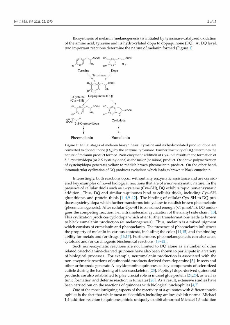
To identify the initial process of cysteine binding, we investigated the energetic pref-erence of thiolate-attacked intermediates. We performed geometrical optimization of DQwith Cys−S− located around the benzene ring. We found five binding sites, namely C5,C2, C6, C3–C4 (bridge), and C1. Conformational rotation of DQ and Cys−S− gave variousisomers with slightly different binding energies. Moreover, hydrogen bonding betweenDQ and Cys−S− also resulted in the formation of various bound states. For simplicity, wepresent here the bound structures based on the energetically favorable conformation in theisolated state, and focus on the most stable hydrogen-bonded structures. The optimizedstructures, the binding energies, and the Gibbs binding energies (based on vibrationalanalyses) are shown in Figure 2a–c, respectively.
Int. J. Mol. Sci. 2021, 22, 1373 4 of 15
Figure 2. Properties of Cys–S−-attacked dopaquinone (DQ) intermediates. (a) Optimized ge-ometries resulting from (C5) 5-addition, (C2) 2-addition, (C6) 6-addition, (C3–C4) bound on3,4-bridge, and (C1) 1-addition. (b) Binding energies Eb of Cys–S− on DQ, which are defined asEb = (ECys–S
− + EDQ) − ECys–S−
–DQ, where ECys–S−, EDQ, and ECys–S
−–DQ are the total energy of
Cys–S−, DQ, and Cys–S−-attacked DQ, respectively. (c) Gibbs binding energies of Cys–S− on DQcomputed based on the vibrational analysis at 37.0 ◦C.
The vibrational analysis for the Cys–S−-attacked intermediates confirmed no imag-inary frequencies. As shown in Figure 2, the C2-bound structure was less energeticallyfavorable than the C6-bound case. Furthermore, Cys−S− preferred the C3−C4 bridgesite more than C5 site as an immediate active site. The negative values of the Gibbsbinding energies indicate that the Cys–S−-attacked intermediates themselves cannot bethe thermodynamically stable products. Therefore, further conversion pathway mustbe explored.
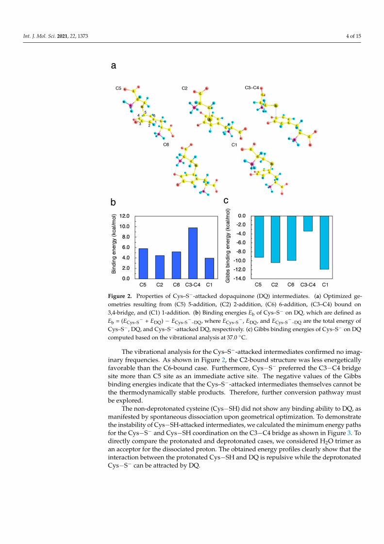
The non-deprotonated cysteine (Cys−SH) did not show any binding ability to DQ, asmanifested by spontaneous dissociation upon geometrical optimization. To demonstratethe instability of Cys−SH-attacked intermediates, we calculated the minimum energy pathsfor the Cys−S− and Cys−SH coordination on the C3−C4 bridge as shown in Figure 3. Todirectly compare the protonated and deprotonated cases, we considered H2O trimer asan acceptor for the dissociated proton. The obtained energy profiles clearly show that theinteraction between the protonated Cys−SH and DQ is repulsive while the deprotonatedCys−S− can be attracted by DQ.
Int. J. Mol. Sci. 2021, 22, 1373 5 of 15
Figure 3. (Left) Minimum energy path for Cys–S− (S–H distance: 1.97 Å) andCys–SH (S–H distance: 1.37 Å) coordination on C3−C4 bridge plotted as squares and triangles,respectively. H2O trimer was used as an acceptor for dissociating proton. Energies were ref-erenced to the total energy of the protonated Cys–SH-DQ complex with 2.92 Å of C3−S dis-tance. All geometrical parameters except for the C3−S and S−H distance were allowed to relax.(Right) Cys–S−- (#) and Cys–SH- (*) coordinated structure.
2.2. Migration of Cysteine Thiolate (Cys−S−)
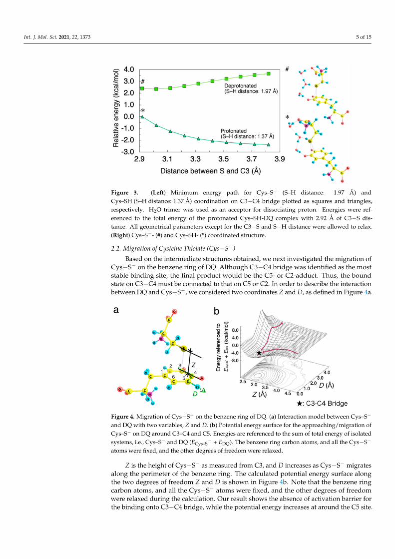
Based on the intermediate structures obtained, we next investigated the migration ofCys−S− on the benzene ring of DQ. Although C3−C4 bridge was identified as the moststable binding site, the final product would be the C5- or C2-adduct. Thus, the boundstate on C3−C4 must be connected to that on C5 or C2. In order to describe the interactionbetween DQ and Cys−S−, we considered two coordinates Z and D, as defined in Figure 4a.
Figure 4. Migration of Cys−S− on the benzene ring of DQ. (a) Interaction model between Cys–S−
and DQ with two variables, Z and D. (b) Potential energy surface for the approaching/migration ofCys–S− on DQ around C3–C4 and C5. Energies are referenced to the sum of total energy of isolatedsystems, i.e., Cys–S− and DQ (ECys–S
− + EDQ). The benzene ring carbon atoms, and all the Cys−S−
atoms were fixed, and the other degrees of freedom were relaxed.
Z is the height of Cys−S− as measured from C3, and D increases as Cys−S− migratesalong the perimeter of the benzene ring. The calculated potential energy surface alongthe two degrees of freedom Z and D is shown in Figure 4b. Note that the benzene ringcarbon atoms, and all the Cys−S− atoms were fixed, and the other degrees of freedomwere relaxed during the calculation. Our result shows the absence of activation barrier forthe binding onto C3−C4 bridge, while the potential energy increases at around the C5 site.
Int. J. Mol. Sci. 2021, 22, 1373 6 of 15
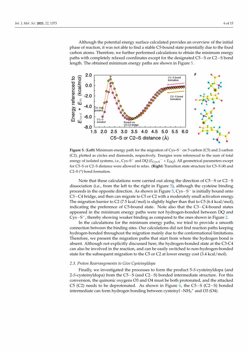
Although the potential energy surface calculated provides an overview of the initialphase of reaction, it was not able to find a stable C5-bound state potentially due to the fixedcarbon atoms. Therefore, we further performed calculations to obtain the minimum energypaths with completely relaxed coordinates except for the designated C5−S or C2−S bondlength. The obtained minimum energy paths are shown in Figure 5.
Figure 5. (Left) Minimum energy path for the migration of Cys–S− on 5-carbon (C5) and 2-carbon(C2), plotted as circles and diamonds, respectively. Energies were referenced to the sum of totalenergy of isolated systems, i.e., Cys–S− and DQ (ECys-S
− + EDQ). All geometrical parameters exceptfor C5–S or C2–S distance were allowed to relax. (Right) Transition state structure for C5–S (#) andC2–S (*) bond formation.
Note that these calculations were carried out along the direction of C5−S or C2−Sdissociation (i.e., from the left to the right in Figure 5), although the cysteine bindingproceeds in the opposite direction. As shown in Figure 5, Cys−S− is initially bound ontoC3−C4 bridge, and then can migrate to C5 or C2 with a moderately small activation energy.The migration barrier to C2 (7.5 kcal/mol) is slightly higher than that to C5 (6.4 kcal/mol),indicating the preference of C5-bound state. Note also that the C3−C4-bound statesappeared in the minimum energy paths were not hydrogen-bonded between DQ andCys−S−, thereby showing weaker binding as compared to the ones shown in Figure 2.
In the calculations for the minimum energy paths, we tried to provide a smoothconnection between the binding sites. Our calculations did not find reaction paths keepinghydrogen-bonded throughout the migration mainly due to the conformational limitations.Therefore, we present the migration paths that start from where the hydrogen bond isabsent. Although not explicitly discussed here, the hydrogen-bonded state at the C3-C4can also be involved in the reaction, and can be easily switched to non-hydrogen-bondedstate for the subsequent migration to the C5 or C2 at lower energy cost (3.4 kcal/mol).
2.3. Proton Rearrangements to Give Cysteinyldopa
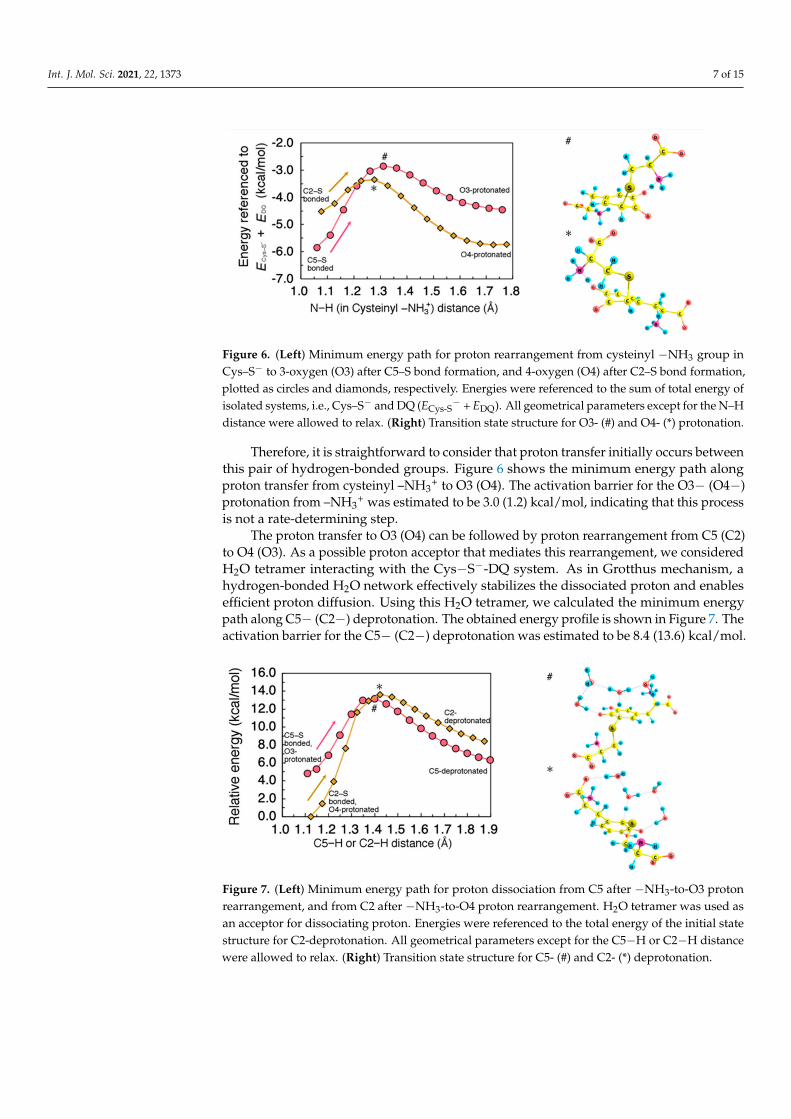
Finally, we investigated the processes to form the product 5-S-cysteinyldopa (and2-S-cysteinyldopa) from the C5−S (and C2−S) bonded intermediate structure. For thisconversion, the quinonic oxygens O3 and O4 must be both protonated, and the attackedC5 (C2) needs to be deprotonated. As shown in Figure 6, the C5−S (C2−S) bondedintermediate can form hydrogen bonding between cysteinyl –NH3
+ and O3 (O4).
Int. J. Mol. Sci. 2021, 22, 1373 7 of 15
Figure 6. (Left) Minimum energy path for proton rearrangement from cysteinyl −NH3 group inCys–S− to 3-oxygen (O3) after C5–S bond formation, and 4-oxygen (O4) after C2–S bond formation,plotted as circles and diamonds, respectively. Energies were referenced to the sum of total energy ofisolated systems, i.e., Cys–S− and DQ (ECys-S
− + EDQ). All geometrical parameters except for the N–Hdistance were allowed to relax. (Right) Transition state structure for O3- (#) and O4- (*) protonation.
Therefore, it is straightforward to consider that proton transfer initially occurs betweenthis pair of hydrogen-bonded groups. Figure 6 shows the minimum energy path alongproton transfer from cysteinyl –NH3
+ to O3 (O4). The activation barrier for the O3− (O4−)protonation from –NH3
+ was estimated to be 3.0 (1.2) kcal/mol, indicating that this processis not a rate-determining step.
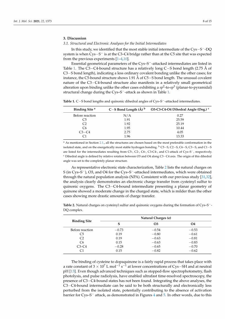
The proton transfer to O3 (O4) can be followed by proton rearrangement from C5 (C2)to O4 (O3). As a possible proton acceptor that mediates this rearrangement, we consideredH2O tetramer interacting with the Cys−S−-DQ system. As in Grotthus mechanism, ahydrogen-bonded H2O network effectively stabilizes the dissociated proton and enablesefficient proton diffusion. Using this H2O tetramer, we calculated the minimum energypath along C5− (C2−) deprotonation. The obtained energy profile is shown in Figure 7. Theactivation barrier for the C5− (C2−) deprotonation was estimated to be 8.4 (13.6) kcal/mol.
Figure 7. (Left) Minimum energy path for proton dissociation from C5 after −NH3-to-O3 protonrearrangement, and from C2 after −NH3-to-O4 proton rearrangement. H2O tetramer was used asan acceptor for dissociating proton. Energies were referenced to the total energy of the initial statestructure for C2-deprotonation. All geometrical parameters except for the C5−H or C2−H distancewere allowed to relax. (Right) Transition state structure for C5- (#) and C2- (*) deprotonation.
Int. J. Mol. Sci. 2021, 22, 1373 8 of 15
3. Discussion3.1. Structural and Electronic Analyses for the Initial Intermediates
In this study, we identified that the most stable initial intermediate of the Cys−S−-DQsystem is when Cys−S− is at the C3–C4 bridge rather than at the C5 site that was expectedfrom the previous experiments [1–4,10].
Essential geometrical parameters of the Cys–S−-attacked intermediates are listed inTable 1. The C3−C4-bound structure has a relatively long C−S bond length (2.75 Å ofC3−S bond length), indicating a less ordinary covalent bonding unlike the other cases; forinstance, the C5-bound structure shows 1.91 Å of C5−S bond length. The unusual covalentnature of the C3−C4-bound structure also manifests in a relatively small geometricalalteration upon binding unlike the other cases exhibiting a sp2-to-sp3 (planar-to-pyramidal)structural change during the Cys–S−-attack as shown in Table 1.
Table 1. C−S bond lengths and quinonic dihedral angles of Cys–S−-attacked intermediates.
Binding Site a C−S Bond Length (Å) b O3-C3-C4-O4 Dihedral Angle (Deg.) c
Before reaction N/A 0.27C5 1.91 25.59C2 1.92 25.19C6 1.95 10.44
C3−C4 2.75 4.05C1 1.96 13.33
a As mentioned in Section 2.1., all the structures are chosen based on the most preferable conformation in theisolated state, and on the energetically most stable hydrogen bonding. b C5−S, C2−S, C6−S, C3−S, and C1−Sare listed for the intermediates resulting from C5-, C2-, C6-, C3-C4-, and C1-attack of Cys–S−, respectively.c Dihedral angle is defined by relative rotation between O3 and O4 along C3−C4 axis. The origin of this dihedralangle was set to the completely planar structure.
As representative electronic state characterization, Table 2 lists the natural charges onS (in Cys–S−), O3, and O4 for the Cys–S−-attacked intermediates, which were obtainedthrough the natural population analysis (NPA). Consistent with our previous study [31,32],the analysis clearly demonstrates an electronic charge transfer from cysteinyl sulfur toquinonic oxygens. The C3−C4-bound intermediate presenting a planar geometry ofquinone showed a moderate change in the charged state, which is milder than the othercases showing more drastic amounts of charge transfer.
Table 2. Natural charges on cysteinyl sulfur and quinonic oxygens during the formation of Cys–S−-DQ complex.
Binding SiteNatural Charges (e)
S O3 O4
Before reaction −0.73 −0.54 −0.53C5 0.19 −0.80 −0.61C2 0.19 −0.63 −0.81C6 0.15 −0.63 −0.83
C3–C4 −0.28 −0.65 −0.70C1 0.15 −0.82 −0.62
The binding of cysteine to dopaquinone is a fairly rapid process that takes place witha rate constant of 3 × 10⁷ L mol−1 s−1 at lower concentrations of Cys−SH and at neutralpH [13]. Even though advanced techniques such as stopped-flow spectrophotometry, flashphotolysis, and pulse radiolysis, have enabled ultrafast time-resolved spectroscopy, thepresence of C3−C4-bound states has not been found. Integrating the above analyses, theC3−C4-bound intermediate can be said to be both structurally and electronically lessperturbed from the isolated state, potentially contributing to the absence of activationbarrier for Cys–S− attack, as demonstrated in Figures 4 and 5. In other words, due to this
Int. J. Mol. Sci. 2021, 22, 1373 9 of 15
nature, the C3−C4-bound intermediate might not give easy detectable responses upontypical spectroscopic perturbations, making experimental identifications difficult. Thepresence of such an energy landscape, that enables reactions to occur at nearly zero energycost, may explain the reason why Cys−SH but not the other non-thiolic cellular compoundscan compete the rapid cyclization of DQ.
Throughout this study, we used the Becke’s three-parameters hybrid functional [41]combined with the Lee-Yang-Parr correlation functionals [42] (B3LYP) as the exchange-correlation potential. This exchange correlation functional has been widely successful indescribing typical organic chemical reactions as well as electron densities. Nevertheless,in several cases B3LYP fails to accurately account for non-covalent interactions due toLondon dispersion force. Considering its non-local nature, the accurate calculation of suchnon-covalent interactions may be improved by increasing the weight of exact exchangepotential and including meta-GGA functionals. As mentioned above, we found a non-covalent nature of the C3−C4-bound intermediate, which is energetically more stable thanthe covalent-bonded C5-bound intermediate. In order to assess the validity of B3LYPfunctional, we compared the binding energies at the C3−C4 bridge and C5 site usingdifferent exchange correlation functionals as representative results. For this comparison, weused mPW1PW91 [43], M06-2X [44], and CAM-B3LYP [45] for the structural optimizationas well as the total energy calculation. The increased weight of exact exchange potentialand the inclusion of a meta-GGA functional resulted in higher binding energies regardlessof the binding sites (Table S1). However, the choice of exchange correlation functional didnot remarkably affect the energetic preference. In other words, B3LYP functional can beregarded as sufficient for elucidating the cysteine addition reaction.
As pointed out by previous studies [38,39], cysteine binding is a base-catalyzednucleophilic addition reaction. This is consistent with our results where protonated Cys–SH(but not Cys–S−) did not show any binding ability to DQ as demonstrated in Figure 3.As the initial step of the reaction, a previous study assumed an equilibrium of cysteineprotonation/deprotonation in the presence of DQ to propose a kinetic model [39]. However,it was not clarified whether the deprotonation occurs before or during the binding to DQ.Based on the potential energy curves shown in Figure 3, it is more plausible that cysteineundergoes deprotonation prior to the binding with DQ.
Although we assumed cysteinyl deprotonation as the initial step, an alternate freeradical-mediated addition was also proposed to account for the abnormal addition ofthiols to o-quinones [46]. This mechanism exploits the reducing property of thiols toaccount for the observed Michael 1,6-addition product. According to this mechanism(Table 3, Figure S1), thiols initially reduce the o-quinone to semiquinone by one electrontransfer reaction.
The rapid coupling of the resultant semiquinone with the thiyl radical would producethe unconventional 1,6-adduct and not the typical Michael 1,4-adduct. Correspondingly, intriplet state, there were no bound states for the C3–C4- and the C6-bound structure, whilethe C5-bound structure exhibited a quasi-stable bound state, as shown in Table 3. However,all the radical species calculated were less stable than the corresponding spin-singlet system.Further detailed studies for possible redox reactions between Cys–SH and DQ are neededto unveil the reaction to the generalized extent of more oxidative conditions.

Int. J. Mol. Sci. 2021, 22, 1373 10 of 15
Table 3. Reaction energy for radical coupling reaction.
Binding Site Reaction a Relative Energy(kcal/mol) b
C5Cys−SH + DQ→ Cys−S· + DQ−H· (at O3) 12.9
Cys−S· + DQ−H· → Cys−S·/DQ−H· 20.9Cys−S·/DQ−H· → Cys−S/DQ−H −5.0
C3−C4Cys−SH + DQ→ Cys−S· + DQ−H· (at O3) 12.9
Cys−S· + DQ−H· → Cys−S·/DQ−H· Not bonded c
Cys−S·/DQ−H· → Cys−S/DQ−H −9.2
C6Cys−SH + DQ→ Cys−S· + DQ−H· (at O4) 13.4
Cys−S· + DQ−H· → Cys−S·/DQ−H· Not bonded c
Cys−S·/DQ−H· → Cys−S/DQ−H −12.9a Cys−S and DQ−H· respectively denote the thiyl radical and the semiquinone radical resulting from hydrogenatom transfer. The radical species were calculated by specifying doublet spin multiplicity. O3 was chosen as abinding site for hydrogen atom. Cys−S·/DQ−H·and Cys−S/DQ−H respectively denotes the cysteine-attackedintermediate in triplet and singlet state. b Relative energies were referenced to the total energy of the isolatedsystem of Cys−SH and DQ. c Spontaneous dissociation upon structural optimization was observed. Initially,the corresponding singlet structure was optimized, and then re-optimized for the triplet state, resulting in thespontaneous dissociation.
3.2. Effects of Cysteinyl Amino Group on Binding Sites and Reaction Rate
The preference of 1,6-Michael addition over 1,4-Michael addition can be explained byconsidering the presence of the C3–C4-bound state, which is formed with high affinity andwithout activation energy. As mentioned, the C3–C4 bridge acts as an immediate bindingsite for selective C5- and C2-binding by allowing Cys–S− to migrate to the adjacent sites.Therefore, the energetic stability of the C3–C4-bound state is one of the most importantfactors contributing to the selective formation of 5-adduct and 2-adduct.
The stability of the C3–C4-bound state is also partially derived from the hydrogenbond between cysteinyl –NH3
+ and DQ. In fact, this hydrogen bonding on C3–C4 wasstronger than that on C6, as shown in Table 4. In other words, the relative energetic stabilityon C3–C4 with respect to that on C6 could become less significant without hydrogen bond-ing. Therefore, a thiol lacking primary amino groups, such as glutathione, N-acetylcysteine,and thioglycolic acid, would exhibit an increased yield of 6-adduct due to the absenceof hydrogen bonds. In fact, glutathione was reported to react with DQ to give 76, 12and 5% yields of 5-S-glutathionyldopa, 2-S-glutathionyldopa, and 6-S-glutathionyldopa,respectively [47]. The yield of 6-adduct for glutathione addition (5%) is higher than thatfor cysteine addition (1%) [1–4,10]. Thus, although still minor, an amino-free thiol wouldpossess slightly reactive C6, thereby affecting the structure of pheomelanin produced.
Table 4. Effect of hydrogen bonding between cysteinyl –NH3+ and DQ on binding energy.
Binding Site ∆Eb = Eb, Not HB − Eb, HB (kcal/mol) a
C5 2.8C2 4.6C6 1.0
C3–C4 3.4C1 4.5
a The binding energy of non-hydrogen-bonded intermediate (Eb, Not HB) was subtracted by that of hydrogen-bonded intermediate (Eb, HB).
Pheomelanin is a pigment consisting of benzothiazines and benzothiazoles as buildingmonomers [48]. After the formation of 5-S-cysteinyldopa and 2-S-cysteinyldopa, theyundergo redox exchange with unreacted DQ, and then cyclize to form quinone iminesthrough the cysteinyl –NH3
+ and the quinonic carbonyl [48]. This cyclization is a necessaryprocess for the further conversion to benzothiazines and benzothiazoles. On the other hand,

Int. J. Mol. Sci. 2021, 22, 1373 11 of 15
6-S-cysteinyldopa cannot produce such cyclized benzothiazine chromophores, consideringthe distance between the cysteinyl –NH3
+ and the quinonic carbonyl. Therefore, thepreference of 1,6-Michael addition over 1,4-Michael addition is of great significance indetermining the color of pheomelanin.
Cysteinyl –NH3+ also acts as a proton donor that enables rapid quinonic protonation
with the very small activation energy. Therefore, we assumed that C5- (C2-) deprotonationoccurs after O3- (O4-) protonation. On the other hand, our mechanism might not be directlyextrapolated to thiols lacking proton donors such as primary amino groups. If a thiol doesnot have amino groups, the quinonic protonation becomes a diffusion-controlled process,thus deprotonation from the thiolate-attacked carbon atom may occur even before thequinonic protonation. As shown in Figure S2, we confirmed that the activation barrier forC5- (C2-) deprotonation becomes higher when O3 (O4) is not protonated, indicating slowerreaction rate of amino-free thiols.
This view is consistent with a previous kinetic study on the thiol binding reactions,where the effect of the presence of cysteinyl –NH3
+ was discussed by comparing withthe amino-free analogue thioglycolic acid [39]. According to the kinetic analysis, thethioglycolic acid-attacked intermediate lacking amino groups is more stable, leading to aslower reaction rate because of the rate-limiting proton rearrangement.
3.3. Energy Diagram for Cysteine Binding to Form Cysteinyldopa
To see the whole picture of cysteine binding, we show an energy diagram for the reac-tion to form 5-S-cysteinyldopa and 2-S-cysteinyldopa along with a hypothetical reactionscheme. Based on the geometrical optimization for each step and the potential energycurves, we estimated the energy change as shown in Figure 8a. In the final proton rear-rangement, the Cys−S−-DQ system undergoes a significant stabilization, that makes thisbinding irreversible. A remarkable difference between 5-adduct and 2-adduct formationcan be seen in the activation barrier for the final proton rearrangement [denoted as (iv)].Therefore, the experimentally observed preference toward 5-S-cysteinyldopa would haveoriginated from this elementary process. The corresponding hypothetical reaction schemeis shown in Figure 8b. Briefly, we propose that the binding of Cys–S− to DQ proceeds with(i) coordination of Cys–S− to C3–C4 bridge, (ii) migration of Cys–S− to C5 (C2), (iii) protonrearrangement from cysteinyl –NH3
+ to O4 (O3), and (iv) proton rearrangement from C5(C2) to O3 (O4).
Figure 8. Overview of the reaction between Cys–S− and DQ. (a) Energy diagram for the formation of5-S-cysteinyldopa and 2-S-cysteinyldopa plotted as circles and diamonds, respectively. (b) Proposedbinding mechanism.
Int. J. Mol. Sci. 2021, 22, 1373 12 of 15
4. Materials and Methods4.1. Electronic State Calculation Methods
Throughout this work, we performed first-principles calculations based on DFT [49,50]using the Gaussian 09 computational package [51]. We used B3LYP functional as theexchange-correlation potential [41,42]. The calculations employed 6-31++G(d,p) basis setto expand the Kohn-Sham orbitals.
We estimated the atomic charges through the natural population analysis (NPA) [52,53].As a solvation model, we considered dielectric response of surrounding water moleculesby the integral equation formalism polarizable continuum model (IEF-PCM) [54]. We usedthe IEF-PCM for both the single point calculations and the structural optimizations. Wecarried out vibrational frequency analyses on the same level of theory for all the opti-mized structures to confirm their stability and to calculate the Gibbs free energies. For theGibbs free energy calculations, we considered the degrees of freedom for the molecularvibration, rotation, and translation. The temperature and pressure were set to 37.0 ◦C and1.0 atm, respectively. This thermodynamic model assumes a non-interacting ideal gas ofDQ and Cys−S− (with PCM correction), and that the pressure–volume product is uniquelydetermined by the temperature.
4.2. Structures for Calculations
Total energy changes during the reaction can be slightly affected by the choice ofisomer. DQ and Cys−S− includes a saturated hydrocarbon chain, that can rotate at arelatively lower energy cost. Furthermore, the presence of electrically polarized hydrogen-containing groups such as amino and carboxyl groups can easily be a cause of hydrogenbonding. Thus, conformational rotation and hydrogen bonding give various isomers withslightly different binding energies. For simplicity, here we constructed bound structuresbased on the energetically favorable conformation in the isolated state, and focused on themost stable hydrogen-bonded structures. Throughout the calculation, we used a consistentconformation of DQ and Cys−S−, although the hydrogen bonding site can switch atcysteinyl migration on DQ.
4.3. Reaction Analyses
The binding energy (Eb) for Cys–S− was defined as Eb = (ECys-S− + EDQ) − ECys-S-DQ,
where ECys-S−, EDQ, and ECys-S-DQ are the total energy of Cys–S−, DQ, and Cys–S−-attacked
DQ, respectively. A positive value of binding energy means an energetically favorablebinding. The Gibbs binding energy was defined in the same manner.
Potential energy surfaces were calculated at specified points of geometry. For eachpoint, partial geometrical optimization was conducted with some degrees of freedom heldfixed (The detailed specification of the active and the frozen coordinates are given in thetext). The potential energy surfaces were calculated so as to provide smooth motions ofmolecules. However, to precisely determine the transition state energy and structure, it isnecessary to be optimized so that the structure exhibits only one imaginary frequency. Inthis study, our attempts to obtain the true transition states based on the vibration analysiswere unsuccessful potentially due to the relatively flat nature of potential energy surface aswell as the complexity of the internal motion. In other words, the reaction paths shown inthis study are not fully parallel with the true intrinsic reaction coordinates. Therefore, theactivation barriers shown in this study must be slightly overestimated.
Deprotonation reactions were described using H2O trimer or tetramer as a protonacceptor. The H2O trimer and tetramer were constructed based on a tetragonal hydrogen-bonded network structure, and then placed around the proton to be dissociated.
5. Conclusions
In the present study, we investigated the binding mechanism of L-cysteine to DQusing density functional theory-based calculation. We calculated the binding energies ofCys−S−-attacked intermediates and the minimum energy paths for the approach/migration
Int. J. Mol. Sci. 2021, 22, 1373 13 of 15
of Cys−S− on the aromatic carbons. We identified the C3−C4 bridge of DQ as the mostpreferable site for Cys−S−, while the protonated Cys−SH did not show binding abilityat any binding sites of DQ. We found that the calculated minimum energy paths for theC5−S and C2−S bond formation involve a precursor Cys−S−-bound state on C3−C4bridge. Therefore, the C5− and C2−S bond formation can be affected by this precursorstate, causing moderately small activation barriers. The C5− and C2−S bond formationare followed by further proton rearrangement to form 5- and 2-S-cysteinyldopa, which arethe major and minor products, respectively.
Based on our results, we propose that the binding of Cys−S- to DQ proceeds in thefollowing sequence: (i) coordination of Cys−S- to C3−C4 bridge and (ii) migration ofCys−S- to C5 (or C2), (iii) proton rearrangement from cysteinyl –NH3+ to O3 (O4), and(iv) proton rearrangement from C5 (C2) to O4 (O3). Throughout the reaction, a significantstabilization occurs at the final step (iv), making the binding of cysteine irreversible.
The obtained findings in this study provide a foundation for understanding themechanism of cysteine binding, and can be a basis for pheomelanogenesis.
Supplementary Materials: The following are available online at https://www.mdpi.com/1422-0067/22/3/1373/s1, Figure S1: Alternate mechanism to account for the abnormal addition of thiolsto quinone, Table S1: Comparison of binding energies at C5 and C3−C4 using different exchangecorrelation functionals.
Author Contributions: The individual contributions of this research articles are listed here. Con-ceptualization, R.K., S.I., and M.S.; methodology, R.K., R.L.A., H.N., and H.K.; validation, M.S. andR.L.A.; investigation, R.K.; original draft preparation, R.K.; review and editing, S.I., M.S., R.L.A.;supervision, H.K.; project administration, H.N. All authors have read and agreed to the publishedversion of the manuscript.
Funding: This work is supported in part by: MEXT Grant-in-Aid for Scientific Research (15H05736,24246013, 15KT0062, 26248006); Grant-in-Aid for JSPS Research Fellow (17J01276); JST ACCELProgram “Creation of the Functional Materials on the Basis of the Inter-Element-Fusion Strategyand their Innovative Applications” (JPMJAC1501); JST CREST Innovative Catalysts and CreationTechnologies for the Utilization of Diverse Natural Carbon Resources: In-situ Atomic Characterizationof Catalytic Reactions for the Development of Innovative Catalysts (No. 17942262); and NEDOProject “R&D Towards Realizing an Innovative Energy Saving Hydrogen Society based on QuantumDynamics Applications”.
Institutional Review Board Statement: Not applicable.
Informed Consent Statement: Not applicable.
Data Availability Statement: The data presented in this study are available in this article and itsaccompanying supplementary materials.
Acknowledgments: Some of the numerical calculations presented here were done using the computerfacilities at the following institutes: National Institute of Technology (NIT), Akashi College, Japan;CMC (Osaka University); ISSP; KEK; NIFS; and YITP. An author (RK) acknowledges Qdai-jumpResearch Program of Kyushu University, Japan. An author (RLA) acknowledges the Balik ScientistProgram of the Department of Science and Technology, Philippines.
Conflicts of Interest: The authors declare no conflict of interest.
Abbreviations
Cys–SH L-cysteineDFT density functional theoryDQ dopaquinoneHPLC high performance liquid chromatographyIEF-PCM integral equation formalism polarizable continuum modelLUMO lowest unoccupied molecular orbitalNPA natural population analysis

Int. J. Mol. Sci. 2021, 22, 1373 14 of 15
References1. Ito, S. A chemist’s view of melanogenesis. Pigment Cell Res. 2003, 16, 230–236. [CrossRef] [PubMed]2. Ito, S.; Wakamatsu, K. Chemistry of mixed melanogenesis—Pivotal roles of dopaquinone. Photochem. Photobiol. 2008, 84, 582–592.
[CrossRef] [PubMed]3. Prota, G. Recent advances in the chemistry of melanogenesis in mammals. J. Investig. Dermatol. 1980, 75, 122–127. [CrossRef]
[PubMed]4. Ito, S.; Sugumaran, M.; Wakamatsu, K. Chemical reactivities of ortho-quinones produced in living organisms: Fate of quinonoid
products formed by tyrosinase and phenoloxidase action on phenols and catechols. Int. J. Mol. Sci. 2020, 21, 6080. [CrossRef]5. Wakamatsu, K.; Murase, T.; Zucca, F.A.; Zecca, L.; Ito, S. Biosynthetic pathway to neuromelanin and its aging process. Pigment
Cell Melanoma Res. 2012, 25, 792–803. [CrossRef]6. Sugumaran, M. Molecular mechanisms for mammalian melanogenesis. FEBS Lett. 1991, 293, 4–10. [CrossRef]7. Sugumaran, M. Reactivities of quinone methides versus o-quinones in catecholamine metabolism and eumelanin biosynthesis.
Int. J. Mol. Sci. 2016, 17, 1576. [CrossRef]8. Tse, D.C.S.; McCreery, R.L.; Adams, R.N. Potential oxidative pathways of brain catecholamines. J. Med. Chem. 1976, 19, 37–40.
[CrossRef]9. Cooksey, C.J.; Land, E.J.; Rushton, F.A.P.; Ramsden, C.A.; Riley, P.A. Tyrosinase-mediated cytotoxicity of 4-substituted phenols:
Use of QSAR to forecast reactivities of thiols towards the derived ortho-quinones. Quant. Struct. Act. Relat. 1996, 15, 498–503.[CrossRef]
10. Ito, S.; Prota, G. A facile one-step synthesis of cysteinyldopas using mushroom tyrosinase. Experientia 1977, 33, 1118–1119.[CrossRef]
11. Kato, T.; Ito, S.; Fujita, K. Tyrosinase-catalyzed binding of 3,4-dihydroxyphenylalanine with proteins through the sulfhydrylgroup. Biochim. Biophys. Acta 1986, 881, 415–421. [CrossRef]
12. Ito, S.; Kato, T.; Fujita, K. Covalent binding of catechols to proteins through the sulphydryl group. Biochem. Pharmacol. 1988, 37,1707–1710. [CrossRef]
13. Thompson, A.; Land, E.J.; Chedekel, M.R.; Subbarao, K.V.; Truscott, T.G. A pulse radiolysis investigation of the oxidation ofthe melanin precursors 3,4-dihydroxyphenylalanine (dopa) and the cysteinyldopas. Biochim. Biophys. Acta 1985, 843, 49–57.[CrossRef]
14. Ozeki, H.; Ito, S.; Wakamatsu, K.; Hirobe, T. Chemical characterization of hair melanins in various coat-color mutants of mice. J.Investig. Dermatol. 1995, 105, 361–365. [CrossRef]
15. Ozeki, H.; Ito, S.; Wakamatsu, K.; Thody, A.J. Spectrophtometric characterization of eumelanin and pheomelanin in hair. PigmentCell Res. 1996, 9, 265–270. [CrossRef]
16. Liu, Y.; Hong, L.; Wakamatsu, K.; Ito, S.; Adhyaru, B.; Cheng, C.Y.; Bowers, C.R.; Simon, J.D. Comparison of structural andchemical properties of black and red human hair melanosomes. Photochem. Photobiol. 2005, 81, 135–144. [CrossRef]
17. Mårs, U.; Larsson, B.S. Pheomelanin as a binding site for drugs and chemicals. Pigment Cell Res. 1999, 12, 266–274. [CrossRef]18. Chedekel, M.R.; Post, P.W.; Deibel, R.M.; Kalus, M. Photodestruction of phaeomelanin. Photochem. Photobiol. 1977, 26, 651–653.
[CrossRef]19. Chedekel, M.R.; Smith, S.K.; Post, P.W.; Pokora, A.; Vessell, D.L. Photodestruction of pheomelanin: Role of oxygen. Proc. Natl.
Acad. Sci. USA 1978, 75, 5395–5399. [CrossRef]20. Mitra, D.; Luo, X.; Morgan, A.; Wang, J.; Hoang, M.P.; Lo, J.; Guerrero, C.R.; Lennerz, J.K.; Mihm, M.C.; Wargo, J.A.; et al. An
ultraviolet-independent pathway to melanoma carcinogenesis in the red hair/fair skin background. Nature 2012, 491, 449–453.[CrossRef]
21. Panzella, L.; Leone, L.; Greco, G.; Vitiello, G.; D’Errico, G.; Napolitano, A. Red human hair pheomelanin is a potent pro-oxidantmediating UV-independent contriburoty mechanisms of melanogenesis. Pigment Cell Melanoma Res. 2014, 27, 244–252. [CrossRef][PubMed]
22. Napolitano, A.; Panzella, L.; Monfrecola, G.; D’Ischia, M. Pheomelanin-induced oxidative stress: Bright and dark chemistrybridging red hair phenotype and melanoma. Pigment Cell Melanoma Res. 2014, 27, 721–733. [CrossRef] [PubMed]
23. Rubin, D.J.; Miserez, A.; Waite, H.J. Diverse strategies of protein sclerotization in marine invertebrates: Structure-propertyrelationships in natural biomaterials. Adv. Insect Physiol. 2010, 38, 75–132.
24. Sugumaran, M. Chemistry of cuticular sclerotization. Adv. Insect Physiol. 2010, 39, 151–209.25. Abebe, A.; Zheng, D.; Evans, J.; Sugumaran, M. Novel post-translational oligomerization of peptidyl dehydrodopa model
compound, 1,2-dehydro-N-acetyldopa methyl ester. Bioorg. Chem. 2016, 66, 33–40. [CrossRef]26. Sugumaran, M.; Robinson, W. Structure, biosynthesis and possible function of tunichromes and related compounds. Comp.
Biochem. Physiol. B Biochem. Mol. Biol. 2012, 163, 1–25. [CrossRef]27. Roman, T.; Diño, W.A.; Nakanishi, H.; Kasai, H. Amino acid adsorption effects on nanotube electronics. J. Vac. Soc. Jpn. 2006, 49,
440–442. [CrossRef]28. Roman, T.; Diño, W.A.; Nakanishi, H.; Kasai, H. Amino acid adsorption on single-walled carbon nanotubes. Eur. Phys. J. D 2006,
38, 117–120. [CrossRef]29. Kishida, R.; Ushijima, Y.; Saputro, A.G.; Kasai, H. Effect of pH on elementary steps of dopachrome conversion from first-principles
calculation. Pigment Cell Melanoma Res. 2014, 27, 734–743. [CrossRef]
Int. J. Mol. Sci. 2021, 22, 1373 15 of 15
30. Kishida, R.; Saputro, A.G.; Kasai, H. Mechanism of dopachrome tautomerization into 5,6-dihydroxyindole-2-carboxylic acidcatalyzed by Cu(II) based on quantum chemical calculations. Biochim. Biophys. Acta 2015, 1850, 281–286. [CrossRef]
31. Kishida, R.; Kasai, H.; Aspera, S.M.; Arevalo, R.L.; Nakanishi, H. Branching reaction in melanogenesis: The effect of intramolecularcyclization on thiol binding. J. Electron. Mater. 2017, 46, 3784–3788. [CrossRef]
32. Kishida, R.; Kasai, H.; Aspera, S.M.; Arevalo, R.L.; Nakanishi, H. Density functional theory-based first principles calculations ofrhododendrol-quinone reactions: Preference to thiol binding over cyclization. J. Phys. Soc. Jpn. 2017, 86, 024804. [CrossRef]
33. Kishida, R.; Saputro, A.G.; Arevalo, R.L.; Kasai, H. Effects of introduction of α-carboxylate, N-methyl, and N-formyl groups onintramolecular cyclization of o-quinone amines: Density functional theory-based study. Int. J. Quant. Chem. 2017, 117, e25445.[CrossRef]
34. Kishida, R.; Kasai, H. Cyclic bond formation of rhododendrol-quinone and dopamine-quinone: Effects of proton rearrangement.J. Phys. Soc. Jpn. 2018, 87, 084802. [CrossRef]
35. Kishida, R.; Kasai, H. Computational Materials Design Case Study III: Biosynthesis of Melanin Pigment; Osaka University Press: Osaka,Japan, 2019. (In Japanese)
36. Kishida, R.; Aspera, S.M.; Kasai, H. Melanin chemistry explored by quantum mechanics. Springer-Nature. (in preparation).37. Huang, X.; Xu, R.; Hawley, M.D.; Hopkins, T.L.; Kramer, K.J. Electrochemical oxidation of N-acyldopamines and regioselective
reactions of their quinones with N-acetylcysteine and thiourea. Arch. Biochem. Biophys. 1988, 352, 19–30. [CrossRef]38. Xu, R.; Huang, X.; Kramer, K.J.; Hawley, M.D. Characterization of products from the reactions of dopamine quinone with
N-acetylcysteine. Bioorg. Chem. 1996, 24, 110–126. [CrossRef]39. Jameson, G.N.L.; Zhang, J.; Jameson, R.F.; Linert, W. Kinetic evidence that cysteine reacts with dopaminoquinone via reversible
adduct formation to yield 5-cysteinyl-dopamine: An important precursor of neuromelanin. Org. Biomol. Chem. 2004, 2, 777–782.[CrossRef]
40. Land, E.J.; Ramsden, C.A.; Riley, P.A. ortho-Quinone amines and derivatives: The influence of structure on the rates and modes ofintramolecular reaction. Arkivoc 2007, xi, 23–36.
41. Becke, A.D. Density-functional thermocheimstry. J. Chem. Phys. 1993, 98, 5648–5652. [CrossRef]42. Lee, C.; Yang, W.; Parr, R.G. Development of the Colle-Salvetti correlation-energy formula into a functional of the electron density.
Phys. Rev. B 1988, 37, 785–789. [CrossRef] [PubMed]43. Adamo, C.; Barone, V. Exchange functionals with improved long-range behavior and adiabatic connection methods without
adjustable parameters: The mPW and mPW1PW models. J. Chem. Phys. 1998, 108, 664. [CrossRef]44. Zhao, Y.; Truhlar, D.G. The M06 suite of density functionals for main group thermochemistry, thermochemical kinetics, noncova-
lent interactions, excited states, and transition elements: Two new functionals and systematic testing of four M06-class functionalsand 12 other functionals. Theor. Chem. Account. 2008, 120, 215–241.
45. Yanai, T.; Tew, D.P.; Handy, N.C. A new hybrid exchange-correlation functional using the Coulomb-attenuating method (CAM-B3LYP). Chem. Phys. Lett. 2004, 393, 51–57. [CrossRef]
46. Sugumaran, M.; Dali, H.; Semensi, V. Chemical- and cuticular phenoloxidase-mediated synthesis of cysteinyl-catechol adducts.Arch. Insect Biochem. Physiol. 1989, 11, 127–137. [CrossRef]
47. Ito, S.; Palumbo, A.; Prota, G. Tyrosinase-catalyzed conjugation of dopa with glutathione. Experientia 1985, 41, 960–961. [CrossRef]48. Wakamatsu, K.; Ohtara, K.; Ito, S. Chemical analysis of late stages of pheomelanogenesis: Conversion of dihydrobenzothiazine to
a benzothiazole structure. Pigment Cell Melanoma Res. 2009, 22, 474–486. [CrossRef]49. Hohenberg, P.; Kohn, W. Inhomogeneous electron gas. Phys. Rev. 1964, 136, B864–B871. [CrossRef]50. Kohn, W.; Sham, L.J. Self-consistent equations including exchange and correlation effects. Phys. Rev. 1965, 140, A1133–A1138.
[CrossRef]51. Frisch, M.J.; Trucks, G.W.; Schlegel, H.B.; Scuseria, G.E.; Robb, M.A.; Cheeseman, J.R.; Scalmani, G.; Barone, V.; Mennucci, B.;
Petersson, G.A.; et al. Gaussian 09 (Revision C. 01); Gaussian, Inc.: Wallingford, CT, USA, 2009.52. Foster, J.P.; Weinhold, F. Natural hybrid orbitals. J. Am. Chem. Soc. 1980, 102, 7211–7218. [CrossRef]53. Reed, A.E.; Curtiss, L.A.; Weinhold, F. Intermolecular interactions from a natural bond orbital, donor-acceptor viewpoint. Chem.
Rev. 1988, 88, 899–926. [CrossRef]54. Tomasi, J.; Mennucci, B.; Cammi, R. Quantum mechanical continuum solvation models. Chem. Rev. 2005, 105, 2999–3094.
[CrossRef] [PubMed]
Related Documents