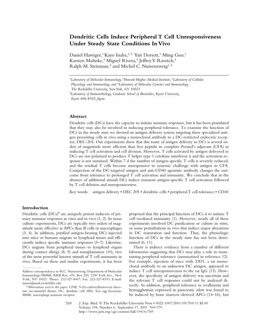
J. Exp. Med. The Rockefeller University Press • 0022-1007/2001/09/769/11 $5.00 Volume 194, Number 6, September 17, 2001 769–779 http://www.jem.org/cgi/content/full/194/6/769 769 Dendritic Cells Induce Peripheral T Cell Unresponsiveness Under Steady State Conditions In Vivo Daniel Hawiger, 1 Kayo Inaba, 3, 5 Yair Dorsett, 1 Ming Guo, 1 Karsten Mahnke, 3 Miguel Rivera, 3 Jeffrey V. Ravetch, 4 Ralph M. Steinman, 3 and Michel C. Nussenzweig 1, 2 1 Laboratory of Molecular Immunology, 2 Howard Hughes Medical Institute, 3 Laboratory of Cellular Physiology and Immunology, and 4 Laboratory of Molecular Genetics and Immunology, The Rockefeller University, New York, NY 10021 5 Laboratory of Immunobiology, Graduate School of Biostudies, Kyoto University, Kyoto 606-8502, Japan Abstract Dendritic cells (DCs) have the capacity to initiate immune responses, but it has been postulated that they may also be involved in inducing peripheral tolerance. To examine the function of DCs in the steady state we devised an antigen delivery system targeting these specialized anti- gen presenting cells in vivo using a monoclonal antibody to a DC-restricted endocytic recep- tor, DEC-205. Our experiments show that this route of antigen delivery to DCs is several or- ders of magnitude more efficient than free peptide in complete Freund’s adjuvant (CFA) in inducing T cell activation and cell division. However, T cells activated by antigen delivered to DCs are not polarized to produce T helper type 1 cytokine interferon and the activation re- sponse is not sustained. Within 7 d the number of antigen-specific T cells is severely reduced, and the residual T cells become unresponsive to systemic challenge with antigen in CFA. Coinjection of the DC-targeted antigen and anti-CD40 agonistic antibody changes the out- come from tolerance to prolonged T cell activation and immunity. We conclude that in the absence of additional stimuli DCs induce transient antigen-specific T cell activation followed by T cell deletion and unresponsiveness. Key words: antigen delivery • DEC 205 • dendritic cells • peripheral T cell tolerance • CD40 Introduction Dendritic cells (DCs)* are uniquely potent inducers of pri- mary immune responses in vitro and in vivo (1, 2). In tissue culture experiments, DCs are typically two orders of mag- nitude more effective as APCs than B cells or macrophages (3, 4). In addition, purified antigen-bearing DCs injected into mice or humans migrate to lymphoid tissues and effi- ciently induce specific immune responses (5–7). Likewise, DCs migrate from peripheral tissues to lymphoid organs during contact allergy (8, 9) and transplantation (10), two of the most powerful known stimuli of T cell immunity in vivo. Based on these and similar experiments, it has been proposed that the principal function of DCs is to initiate T cell–mediated immunity (1). However, nearly all of these experiments involved DC purification or culture in vitro, or some perturbations in vivo that induce major alterations in DC maturation and function. Thus, the physiologic function of DCs in the steady state has not been deter- mined (6, 11). There is indirect evidence from a number of different laboratories suggesting that DCs may play a role in main- taining peripheral tolerance (summarized in reference 12). For example, injection of mice with 33D1, a rat mono- clonal antibody to an unknown DC antigen, appeared to induce T cell unresponsiveness to the rat IgG (13). How- ever, the specificity of antigen delivery was uncertain and the relevant T cell responses could not be analyzed di- rectly. In addition, peripheral tolerance to ovalbumin and hemagglutinin expressed in pancreatic islets was found to be induced by bone marrow–derived APCs (14–16), but Address correspondence to M.C. Nussenzweig, Department of Molecular Immunology/HHMI, RRB Rm. 470, Box 220, 1230 York Ave., New York, NY 10021. Phone: 212-327-8067; Fax: 212-327-8370; E-mail: [email protected] *Abbreviations used in this paper: CFSE, 5-(6)-carboxyfluorescein diace- tate succinimidyl diester; DC, dendritic cell; HEL, hen egg lysozyme; MMR, macrophage mannose receptor.

Welcome message from author
This document is posted to help you gain knowledge. Please leave a comment to let me know what you think about it! Share it to your friends and learn new things together.
Transcript
J. Exp. Med.
The Rockefeller University Press • 0022-1007/2001/09/769/11 $5.00Volume 194, Number 6, September 17, 2001 769–779http://www.jem.org/cgi/content/full/194/6/769
769
Dendritic Cells Induce Peripheral T Cell Unresponsiveness Under Steady State Conditions In Vivo
Daniel Hawiger,
1
Kayo Inaba,
3, 5
Yair Dorsett,
1
Ming Guo,
1
Karsten Mahnke,
3
Miguel Rivera,
3
Jeffrey V. Ravetch,
4
Ralph M. Steinman,
3
and Michel C. Nussenzweig
1, 2
1
Laboratory of Molecular Immunology,
2
Howard Hughes Medical Institute,
3
Laboratory of Cellular Physiology and Immunology, and
4
Laboratory of Molecular Genetics and Immunology, The Rockefeller University, New York, NY 10021
5
Laboratory of Immunobiology, Graduate School of Biostudies, Kyoto University,Kyoto 606-8502, Japan
Abstract
Dendritic cells (DCs) have the capacity to initiate immune responses, but it has been postulatedthat they may also be involved in inducing peripheral tolerance. To examine the function ofDCs in the steady state we devised an antigen delivery system targeting these specialized anti-gen presenting cells
in vivo using a monoclonal antibody to a DC-restricted endocytic recep-tor, DEC-205. Our experiments show that this route of antigen delivery to DCs is several or-ders of magnitude more efficient than free peptide in complete Freund’s adjuvant (CFA) ininducing T cell activation and cell division. However, T cells activated by antigen delivered toDCs are not polarized to produce T helper type 1 cytokine interferon
�
and the activation re-sponse is not sustained. Within 7 d the number of antigen-specific T cells is severely reduced,and the residual T cells become unresponsive to systemic challenge with antigen in CFA.Coinjection of the DC-targeted antigen and anti-CD40 agonistic antibody changes the out-come from tolerance to prolonged T cell activation and immunity. We conclude that in theabsence of additional stimuli DCs induce transient antigen-specific T cell activation followedby T cell deletion and unresponsiveness.
Key words: antigen delivery • DEC 205 • dendritic cells • peripheral T cell tolerance • CD40
Introduction
Dendritic cells (DCs)
*
are uniquely potent inducers of pri-mary immune responses in vitro and in vivo (1, 2). In tissueculture experiments, DCs are typically two orders of mag-nitude more effective as APCs than B cells or macrophages
(3, 4). In addition, purified antigen-bearing
DCs injectedinto mice or humans migrate to lymphoid tissues and effi-ciently induce specific immune responses (5–7). Likewise,DCs migrate from peripheral tissues to lymphoid organsduring contact allergy (8, 9) and transplantation (10), twoof the most powerful known stimuli of T cell immunity invivo. Based on these and similar experiments, it has been
proposed that the principal function of DCs is to initiate Tcell–mediated immunity (1). However, nearly all of theseexperiments involved DC purification or culture in vitro
,
or some perturbations in vivo that induce major alterationsin DC maturation and function. Thus, the physiologicfunction of DCs in the steady state has not been deter-mined (6, 11).
There is indirect evidence from a number of differentlaboratories suggesting that DCs may play a role in main-taining peripheral tolerance (summarized in reference 12).For example, injection of mice with 33D1, a rat mono-clonal antibody to an unknown DC antigen, appeared toinduce T cell unresponsiveness to the rat IgG (13). How-ever, the specificity of antigen delivery was uncertain andthe relevant T cell responses could not be analyzed di-rectly. In addition, peripheral tolerance to ovalbumin andhemagglutinin expressed in pancreatic islets was found tobe induced by bone marrow–derived APCs (14–16), but
Address correspondence to M.C. Nussenzweig, Department of MolecularImmunology/HHMI, RRB Rm. 470, Box 220, 1230 York Ave., NewYork, NY 10021. Phone: 212-327-8067; Fax: 212-327-8370; E-mail:[email protected]
*
Abbreviations used in this paper:
CFSE, 5-(6)-carboxyfluorescein diace-tate succinimidyl diester; DC, dendritic cell; HEL, hen egg lysozyme;MMR, macrophage mannose receptor.
770
Dendritic Cells Induce Peripheral T Cell Tolerance in the Steady State
the identity of these antigen presenting cells has not beendetermined (17).
Materials and Methods
Mice.
6–8-wk-old females were used in all experiments andwere maintained under specific pathogen free conditions.B10.BR, B6.SJL (CD45.1), and B6/MRL (Fas lpr) mice werepurchased from The Jackson Laboratory. 3A9 transgenic micewere maintained by crossing with B10.BR mice. To obtainCD45.1 3A9 or 3A9/lpr T cells, B6.SJL or B6/MRL mice werecrossed extensively with 3A9 mice and tested for CD45.1 and I-A
k
,by flow cytometry. Fas lpr mutation was tested by PCR. Micewere injected subcutaneously with peptide in CFA and subcuta-neously or intravenously with chimeric antibodies. All experi-ments with mice were performed in accordance with NationalInstitutes of Health guidelines.
Flow Cytometry and Antibodies Used for Staining.
CD4- (L3T4),MHC II- (10-3.6), CD11c- (HL3), CD11c- (HL3), B220-(RA3-6B2), or CD3- (145-2C11), CD80(B7-1)-(16-10A1) I-A
k
-(10-3.6) CD45.1- (A20), Il-2- (JES6-5H4), IFN-
�
- (XMG1.2),CD40- (HM40-3-FITC), CD86(B7-2)- (GL1) specific antibod-ies were from BD PharMingen. Rat IgG-PE
(goat
anti–rat IgG)specific antibody was from Serotec. 3A9 T cell receptor (1G12)–specific antibody was a gift from Dr. Emil Unanue, WashingtonUniversity, St. Louis, MO (18).
For visualization of rat IgGs on surface of mononuclear cells,lymphoid cells were purified from peripheral LNs 14 h after anti-body injection and stained with anti–rat IgG-RPE
(goat
anti–ratIgG-RPE; Serotec) to visualize surface bound NLDC145 andGL117 antibodies. The cells were then incubated in mouse serumto block nonspecific binding and stained with FITC anti-CD11c(HL3), or -B220 (RA3-6B2), or -CD3 (145-2C11).
For intracellular cytokine staining, lymphocytes were stimu-lated in vitro for 4 h with leukocyte activation cocktail (BDPharMingen) according to the manufacturer’s manual. Cellswere fixed and permeabilized using cytofix/cytoperm bufferfrom BD PharMingen.
Immunohistology.
Popliteal LNs were removed from antibodyinjected mice and 5-
�
m cryosections (Microm; ZEISS) wereprepared. Tissue specimens were fixed in acetone (5 min, roomtemperature [RT]) air dried, and stained in a moist chamber.The injected antibodies were detected by incubating the sectionswith streptavidin Cy3 or streptavidin-FITC (Jackson Immuno-tech). In double labeling experiments, the PE-conjugated anti-bodies were added for additional 30 min. Specimens were exam-ined using a fluorescence microscope and confocal opticalsections of
�
0.3-
�
m thickness were generated using deconvolu-tion software (Metamorph).
Constructing and Production of Hybrid Antibodies.
Total RNAwas prepared from NLDC-145 (19) and GLII7 (gift of R.J. Hodes,National Institutes of Health, Bethesda, MD) hybridomas (bothrat IgG2a) using Trizol (GIBCO BRL). Full-length IgcDNAs were produced with 5
�
-RACE PCR kit (GIBCO BRL)using primers specific for 3
�
-ends of rat IgG2a and Ig kappa. TheV regions were cloned in frame with mouse Ig kappa constant re-gions and IgG1 constant regions carrying mutations that interferewith FcR binding (20). DNA coding for hen egg lysozyme (HEL)peptide 46–61 with spacing residues on both sides was added tothe C terminus of the heavy chain using synthetic oligonucle-otides. Gene specific primers for cloning of rat IgG2a and Igkappa: 3
�
-ATAGTTTAGCGGCCGCGATATCTCACTAA-CACTCATTCCTGTTGAAGCT; 3
�
-ATAGTTTAGCGGC-
CGCTCACTAGCTAGCTTTACCAGGAGAGTGGGAGAG-ACTCTTCT; HEL peptide fragment construction: 5
�
-CTAGC-GACATGGCCAAGAAGGAGACAGTCTGGAGGCTCGAG-GAGTTCGGTAGGTTCACAAACAGGAAC; 5
�
-ACAGACG-TAGCACAGACTATGGTATTCTCCAGATTAACAGCAG-GTATTATGACGGTAGGACATGATAGGC; 3
�
-GCTGTA-CCGGTTCTTCCTCTGTCAGACCTCCGAGCTCCTCAA-GCCATCCAAGTGTTTGTCCTTGTGTCTG; 3
�
-CCATC-GTGTCTGATACCATAAGAGGTCTAATTGTCGTCCATAATACTGCCATCCTGTACTATCCGCCGG.
Hybrid antibodies were transiently expressed in 293 cells aftertransfection using calcium-phosphate. Cells were grown in se-rum-free DMEM supplemented with Nutridoma SP (Boeh-ringer). Antibodies were purified on Protein G columns (Amer-sham Pharmacia Biotech). The concentrations of purifiedantibodies were determined by ELISA using goat anti–mouseIgG1 (Jackson Immunotech).
Cell Culture and Proliferation Assays.
Pooled axillary, brachial,inguinal, and popliteal LNs were dissociated in 5% FCS RPMIand incubated in presence of collagenase (Boehringer) and EDTAas described (21). For antigen presentation CD19
�
and CD11c
�
cells were purified using microbeads coupled to anti-mouseCD11c or CD19 IgG (Miltenyi Biotec) and irradiated with 1,500rad. CD4 T cells were purified by depletion using rat antibodiessupernatants specific for mouse: CD8 (TIB 211), B220 (RA3-6B2), MHC II (M5/114, TIB 120), F4/80 (F4/80), and magneticbeads coupled to anti–rat IgG (Dynal). In antigen loading experi-ments the isolated presenting cells from each experimental groupwere cultured in 96-well plates with 2
�
10
5
purified 3A9 CD4
�
T cells. Cultures were maintained for 48 h with [
3
H]thymidine(1
�
Ci) added for the last 6 h. The results were calculated as a ra-tio of proliferation in experimental groups to a PBS controlgroup. The proliferation in PBS controls ranged from 500 to2,000 cpm.
For T cell proliferation assays in adoptive transfer recipients,9
�
10
4
of the same irradiated CD11c
�
cells isolated from spleensof wild-type B10.BR mice were cultured in 96-well plates with3
�
10
5
T cells from each experimental group. Synthetic HEL pep-tide, at final concentration of 100
�
g/ml, was added to half of thecultures. Cultures were maintained for 24 h with [
3
H]thymidine(1
�
Ci/ml) added for the last 6 h. Response to HEL peptide wasdetermined by subtracting background (no HEL peptide added)proliferation from proliferation in the presence of HEL peptide.Proliferation index was calculated as the ratio of the response toHEL peptide in a given experimental group to the response toHEL of T cells from a PBS-injected control. Proliferation in PBSgroups ranged from 4,000–8,000 cpm in the presence of peptideand the response to HEL peptide in these PBS controls was1,000–3,000 counts above the background. Synthetic HEL 46-61peptide was provided by the Howard Hughes Medical InstituteKeck Biotechnology Resource Center.
Adoptive Transfer.
CD4 cells from 3A9 mice were enrichedby depletion as described above, washed 3
�
with PBS, and 5
�
10
6
cells injected intravenously per mouse. Alternatively, beforedepletion total cells were labeled with 2
�
M 5-(6)-carboxyfluo-rescein diacetate succinimidyl diester (CFSE) in 5% FCS RPMI(Molecular Probes) at 37
�
C for 20 min and washed twice.
Results
To examine the function of DCs in vivo, we devised ameans of delivering antigens to DCs in situ. We usedNLDC145 (19), a monoclonal antibody specific for DEC-
771
Hawiger et al.
205, an endocytic receptor that is a member of a family ofmultilectin receptors including the macrophage mannosereceptor (MMR) (22, 23). Like MMR, DEC-205 displaysan NH
2
-terminal cysteine-rich domain, a fibronectin typeII domain, and multiple C-type lectin domains (22). How-ever, the tissue distribution of DEC-205 and the MMRdiffer in that DEC-205 is highly expressed by DCs withinthe T cell areas of lymphoid tissues, particularly on CD8
�
DCs that have been implicated in cross-priming (24),whereas the MMR is expressed by some tissue macro-phages (25, 26). We chose DEC-205 for targeting antigensto DCs because the cytoplasmic domain of DEC-205 or-chestrates a distinct endocytic pathway that enhances anti-gen presentation (23). DEC-205 recycles through late en-dosomes or lysosomes rich in MHC II, and antigensdelivered to these compartments by DEC-205 are effi-ciently processed and presented to T cells (23).
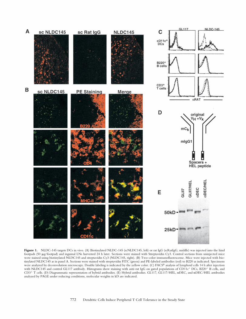
To determine whether the NLDC145 antibody targetsDCs in vivo
,
we injected mice subcutaneously with puri-fied NLDC145 or GL117, a nonspecific isotype-matchedrat monoclonal antibody control, and visualized the in-jected antibody in tissue sections 24 h after injection,NLDC145 was found localized to scattered large dendriticprofiles in the T cell areas of LNs and spleen while uptakeof control GL117 was undetectable (Fig. 1 A, left and mid-dle). This pattern was similar to the pattern found when theantibody was applied to sections directly (Fig. 1 A, right).The NLDC145-targeted cells were negative for B220 andCD4, markers for B cells and T cells, respectively, but pos-itive for characteristic DC markers including MHC IIand CD11c (Fig. 1 B). Thus, subcutaneously injectedNLDC145 targets specifically to CD11c
�
MHC II
�
DCs inlymphoid tissues in vivo.
To further characterize the lymphoid cells that were tar-geted by NLDC145 in vivo
,
we stained lymphoid cell sus-pensions from antibody injected mice with anti–rat Ig andexamined the cells by multiparameter flow cytometry (Fig.1 C). High levels of injected NLDC145 were found on thesurface of most CD11c
�
DCs but not on the surface ofB220
�
B cells or CD3
�
T cells (Fig. 1 C). We concludethat when NLDC145 is injected into mice it binds effi-ciently and directly to DCs but not to other lymphoid cells.
To deliver antigens to DCs in vivo
,
we produced fusionproteins with amino acids 46–61 of HEL added to theCOOH terminus of cloned NLDC145 (
�
DEC/HEL) andGL117 (GL117/HEL) control antibody (Fig. 1 D). Tominimize antibody binding to Fc (FcR) receptors and fur-ther ensure the specificity of antigen targeting,
the ratIgG2a constant regions of the original antibodies were re-placed with mouse IgG1 constant regions that carry pointmutations interfering with FcR binding (20). The hybridantibodies and control Igs without the terminal HEL pep-tide (
�
DEC and GL117) were produced by transient trans-fection in 293 cells (Fig. 1 E).
To determine whether antigens delivered by
�
DEC/HEL were processed by DCs in vivo
,
we injected micewith the hybrid antibodies and controls and testedCD11c
�
DCs, CD19
�
B cells and CD11c
CD19
mono-
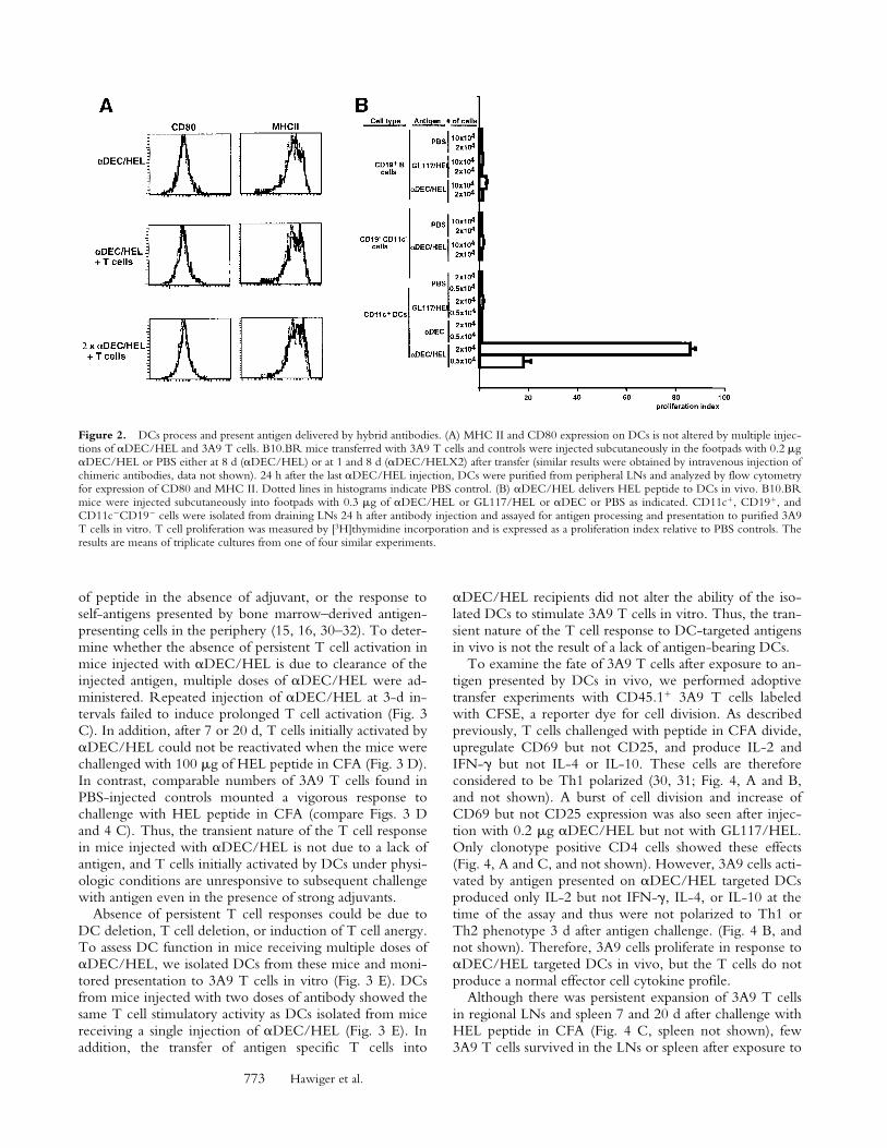
nuclear cells for their capacity to present HEL peptide tonaive HEL-specific T cells from 3A9 TCR transgenicmice (27). DCs isolated from antibody-injected mice ex-pressed levels of CD80 and MHC II similar to those foundon PBS controls and thus showed no signs of increasedmaturation, in contrast to what occurs when DCs are stim-ulated with microbial products like bacterial LPS and CpGdeoxyoligonucleotides (28, 29; Fig. 2 A). NeverthelessDCs from mice injected with
�
DEC/HEL induced strongT cell proliferative responses, whereas DCs isolated fromPBS-injected mice or mice injected with the control anti-bodies had no effect (Fig. 2 B). DC isolated 3 d after
�
DEC/HEL injection showed reduced antigen-presentingactivity (data not shown). In contrast to DCs, B cells andbulk CD11c
CD19
mononuclear cells purified from thesame mice showed little antigen-presenting activity (Fig. 2B). We conclude that antigens can be selectively and effi-ciently delivered to DC by
�
DEC/HEL in vivo
,
and thetargeted DCs successfully process and load the peptidesonto MHC II.
As DC isolation leads to activation, we performedadoptive transfer experiments with HEL-specific trans-genic T cells to follow the response of these T cells to oth-erwise unmanipulated, antigen-targeted DCs in vivo.CD4
�
3A9 T cells were transferred into B10.BR recipi-ents and 24 h later hybrid antibodies were injected subcu-taneously. To measure T cell responses, CD4
�
cells wereisolated from the draining LNs of the injected mice andcultured in vitro in the presence or absence of added HELpeptide. T cell responses were measured by [
3
H]thymidineincorporation and are shown as proliferation
indices nor-malized to the PBS control (this index facilitates compari-son between experiments, see Materials and Methods).In addition to
�
DEC/HEL, GL117/HEL,
�
DEC, andGL117 antibodies, we included 100
�
g of HEL peptide inCFA as a positive control.
As described in previous reports (30, 31), CD4
�
T cellsisolated 2 d after challenge with 100
�
g of HEL peptide inCFA showed strong proliferative responses to antigenwhen compared with PBS controls (Fig. 3 A). Similar re-sponses were obtained from mice injected with as little as0.2
�
g of
�
DEC/HEL (i.e.,
�
4 ng peptide per mouse)
butnot from mice injected with up to 1
�
g of
�
DEC, GL117,or GL117/HEL controls (Fig. 3 A, and not shown). Weconclude that antigen delivered to DCs in vivo by
�
DEC/HEL efficiently induces activation of specific T cells.
To determine whether antigen delivered to DCs in vivoinduces persistent T cell activation, we measured T cell re-sponses to antigen 7 d after the administration of
�
DEC/HEL. CD4 T cells continued to show heightened re-sponses to antigen
when purified from LNs 7 d after injec-tion with 100
�
g of HEL peptide in CFA (30, 31; Fig. 3B). In contrast, T cells isolated from mice 7 d after injec-tion with
�DEC/HEL were no longer activated whencompared with PBS controls (Fig. 3 B). Thus, T cell acti-vation by antigen delivered to DCs by �DEC/HEL in vivois transient, readily detected at 2 but not 7 d. This transientactivation resembles the CD4 T cell response to large doses
772 Dendritic Cells Induce Peripheral T Cell Tolerance in the Steady State
Figure 1. NLDC-145 targets DCs in vivo. (A) Biotinylated NLDC-145 (scNLDC145, left) or rat IgG (scRatIgG, middle) was injected into the hindfootpads (50 �g/footpad) and inguinal LNs harvested 24 h later. Sections were stained with Streptavidin Cy3. Control sections from uninjected micewere stained using biotinylated NLDC145 and streptavidin Cy3 (NLDC145, right). (B) Two-color immunofluorescense. Mice were injected with bio-tinylated NLDC145 as in panel A. Sections were stained with streptavidin FITC (green) and PE-labeled antibodies (red) to B220 as indicated. Specimenswere analyzed by deconvolution microscopy. Double labeling is indicated by the yellow color. (C) FACS® analysis of lymphoid cells 14 h after injectionwith NLDC145 and control GL117 antibody. Histograms show staining with anti–rat IgG on gated populations of CD11c� DCs, B220� B cells, andCD3� T cells. (D) Diagrammatic representation of hybrid antibodies. (E) Hybrid antibodies. GL117, GL117/HEL, �DEC, and �DEC/HEL antibodiesanalyzed by PAGE under reducing conditions, molecular weights in kD are indicated.
773 Hawiger et al.
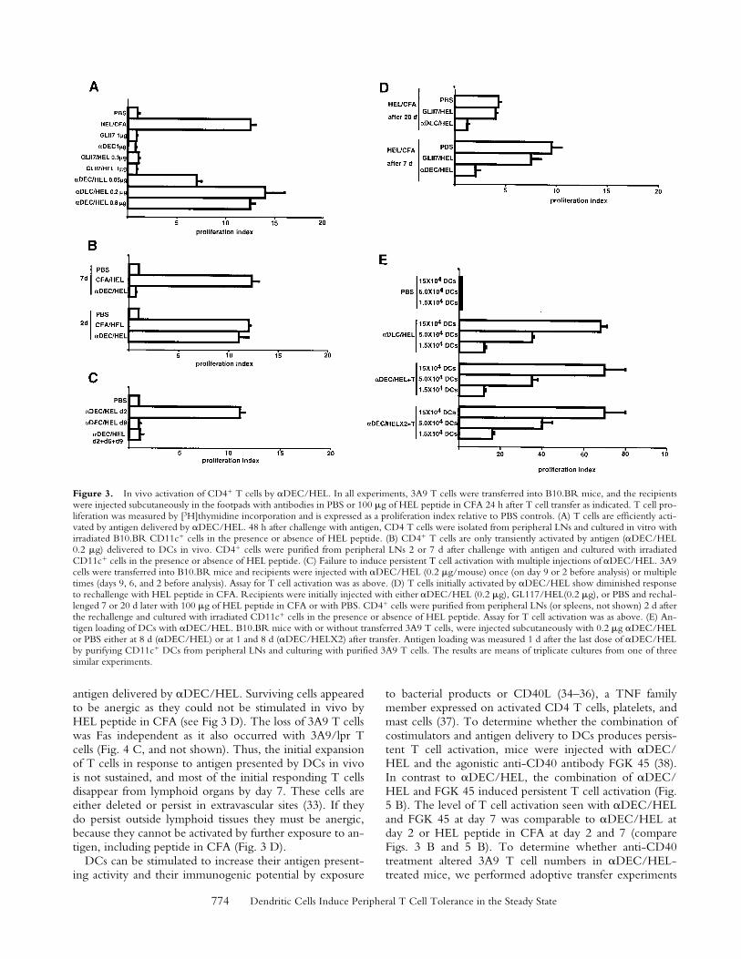
of peptide in the absence of adjuvant, or the response toself-antigens presented by bone marrow–derived antigen-presenting cells in the periphery (15, 16, 30–32). To deter-mine whether the absence of persistent T cell activation inmice injected with �DEC/HEL is due to clearance of theinjected antigen, multiple doses of �DEC/HEL were ad-ministered. Repeated injection of �DEC/HEL at 3-d in-tervals failed to induce prolonged T cell activation (Fig. 3C). In addition, after 7 or 20 d, T cells initially activated by�DEC/HEL could not be reactivated when the mice werechallenged with 100 �g of HEL peptide in CFA (Fig. 3 D).In contrast, comparable numbers of 3A9 T cells found inPBS-injected controls mounted a vigorous response tochallenge with HEL peptide in CFA (compare Figs. 3 Dand 4 C). Thus, the transient nature of the T cell responsein mice injected with �DEC/HEL is not due to a lack ofantigen, and T cells initially activated by DCs under physi-ologic conditions are unresponsive to subsequent challengewith antigen even in the presence of strong adjuvants.
Absence of persistent T cell responses could be due toDC deletion, T cell deletion, or induction of T cell anergy.To assess DC function in mice receiving multiple doses of�DEC/HEL, we isolated DCs from these mice and moni-tored presentation to 3A9 T cells in vitro (Fig. 3 E). DCsfrom mice injected with two doses of antibody showed thesame T cell stimulatory activity as DCs isolated from micereceiving a single injection of �DEC/HEL (Fig. 3 E). Inaddition, the transfer of antigen specific T cells into
�DEC/HEL recipients did not alter the ability of the iso-lated DCs to stimulate 3A9 T cells in vitro. Thus, the tran-sient nature of the T cell response to DC-targeted antigensin vivo is not the result of a lack of antigen-bearing DCs.
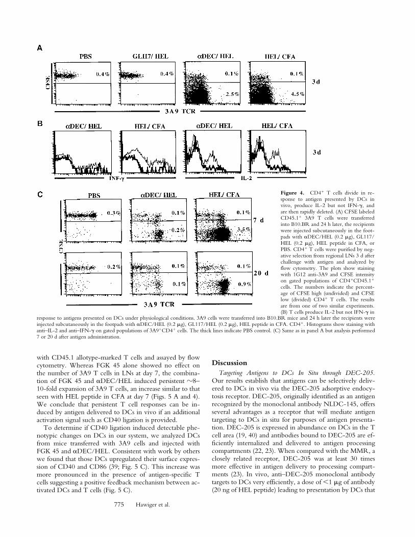
To examine the fate of 3A9 T cells after exposure to an-tigen presented by DCs in vivo, we performed adoptivetransfer experiments with CD45.1� 3A9 T cells labeledwith CFSE, a reporter dye for cell division. As describedpreviously, T cells challenged with peptide in CFA divide,upregulate CD69 but not CD25, and produce IL-2 andIFN-� but not IL-4 or IL-10. These cells are thereforeconsidered to be Th1 polarized (30, 31; Fig. 4, A and B,and not shown). A burst of cell division and increase ofCD69 but not CD25 expression was also seen after injec-tion with 0.2 �g �DEC/HEL but not with GL117/HEL.Only clonotype positive CD4 cells showed these effects(Fig. 4, A and C, and not shown). However, 3A9 cells acti-vated by antigen presented on �DEC/HEL targeted DCsproduced only IL-2 but not IFN-�, IL-4, or IL-10 at thetime of the assay and thus were not polarized to Th1 orTh2 phenotype 3 d after antigen challenge. (Fig. 4 B, andnot shown). Therefore, 3A9 cells proliferate in response to�DEC/HEL targeted DCs in vivo, but the T cells do notproduce a normal effector cell cytokine profile.
Although there was persistent expansion of 3A9 T cellsin regional LNs and spleen 7 and 20 d after challenge withHEL peptide in CFA (Fig. 4 C, spleen not shown), few3A9 T cells survived in the LNs or spleen after exposure to
Figure 2. DCs process and present antigen delivered by hybrid antibodies. (A) MHC II and CD80 expression on DCs is not altered by multiple injec-tions of �DEC/HEL and 3A9 T cells. B10.BR mice transferred with 3A9 T cells and controls were injected subcutaneously in the footpads with 0.2 �g�DEC/HEL or PBS either at 8 d (�DEC/HEL) or at 1 and 8 d (�DEC/HELX2) after transfer (similar results were obtained by intravenous injection ofchimeric antibodies, data not shown). 24 h after the last �DEC/HEL injection, DCs were purified from peripheral LNs and analyzed by flow cytometryfor expression of CD80 and MHC II. Dotted lines in histograms indicate PBS control. (B) �DEC/HEL delivers HEL peptide to DCs in vivo. B10.BRmice were injected subcutaneously into footpads with 0.3 �g of �DEC/HEL or GL117/HEL or �DEC or PBS as indicated. CD11c�, CD19�, andCD11cCD19 cells were isolated from draining LNs 24 h after antibody injection and assayed for antigen processing and presentation to purified 3A9T cells in vitro. T cell proliferation was measured by [3H]thymidine incorporation and is expressed as a proliferation index relative to PBS controls. Theresults are means of triplicate cultures from one of four similar experiments.
774 Dendritic Cells Induce Peripheral T Cell Tolerance in the Steady State
antigen delivered by �DEC/HEL. Surviving cells appearedto be anergic as they could not be stimulated in vivo byHEL peptide in CFA (see Fig 3 D). The loss of 3A9 T cellswas Fas independent as it also occurred with 3A9/lpr Tcells (Fig. 4 C, and not shown). Thus, the initial expansionof T cells in response to antigen presented by DCs in vivois not sustained, and most of the initial responding T cellsdisappear from lymphoid organs by day 7. These cells areeither deleted or persist in extravascular sites (33). If theydo persist outside lymphoid tissues they must be anergic,because they cannot be activated by further exposure to an-tigen, including peptide in CFA (Fig. 3 D).
DCs can be stimulated to increase their antigen present-ing activity and their immunogenic potential by exposure
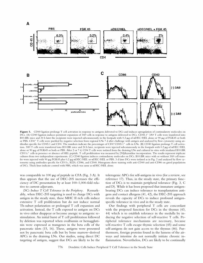
to bacterial products or CD40L (34–36), a TNF familymember expressed on activated CD4 T cells, platelets, andmast cells (37). To determine whether the combination ofcostimulators and antigen delivery to DCs produces persis-tent T cell activation, mice were injected with �DEC/HEL and the agonistic anti-CD40 antibody FGK 45 (38).In contrast to �DEC/HEL, the combination of �DEC/HEL and FGK 45 induced persistent T cell activation (Fig.5 B). The level of T cell activation seen with �DEC/HELand FGK 45 at day 7 was comparable to �DEC/HEL atday 2 or HEL peptide in CFA at day 2 and 7 (compareFigs. 3 B and 5 B). To determine whether anti-CD40treatment altered 3A9 T cell numbers in �DEC/HEL-treated mice, we performed adoptive transfer experiments
Figure 3. In vivo activation of CD4� T cells by �DEC/HEL. In all experiments, 3A9 T cells were transferred into B10.BR mice, and the recipientswere injected subcutaneously in the footpads with antibodies in PBS or 100 �g of HEL peptide in CFA 24 h after T cell transfer as indicated. T cell pro-liferation was measured by [3H]thymidine incorporation and is expressed as a proliferation index relative to PBS controls. (A) T cells are efficiently acti-vated by antigen delivered by �DEC/HEL. 48 h after challenge with antigen, CD4 T cells were isolated from peripheral LNs and cultured in vitro withirradiated B10.BR CD11c� cells in the presence or absence of HEL peptide. (B) CD4� T cells are only transiently activated by antigen (�DEC/HEL0.2 �g) delivered to DCs in vivo. CD4� cells were purified from peripheral LNs 2 or 7 d after challenge with antigen and cultured with irradiatedCD11c� cells in the presence or absence of HEL peptide. (C) Failure to induce persistent T cell activation with multiple injections of �DEC/HEL. 3A9cells were transferred into B10.BR mice and recipients were injected with �DEC/HEL (0.2 �g/mouse) once (on day 9 or 2 before analysis) or multipletimes (days 9, 6, and 2 before analysis). Assay for T cell activation was as above. (D) T cells initially activated by �DEC/HEL show diminished responseto rechallenge with HEL peptide in CFA. Recipients were initially injected with either �DEC/HEL (0.2 �g), GL117/HEL(0.2 �g), or PBS and rechal-lenged 7 or 20 d later with 100 �g of HEL peptide in CFA or with PBS. CD4� cells were purified from peripheral LNs (or spleens, not shown) 2 d afterthe rechallenge and cultured with irradiated CD11c� cells in the presence or absence of HEL peptide. Assay for T cell activation was as above. (E) An-tigen loading of DCs with �DEC/HEL. B10.BR mice with or without transferred 3A9 T cells, were injected subcutaneously with 0.2 �g �DEC/HELor PBS either at 8 d (�DEC/HEL) or at 1 and 8 d (�DEC/HELX2) after transfer. Antigen loading was measured 1 d after the last dose of �DEC/HELby purifying CD11c� DCs from peripheral LNs and culturing with purified 3A9 T cells. The results are means of triplicate cultures from one of threesimilar experiments.
775 Hawiger et al.
with CD45.1 allotype-marked T cells and assayed by flowcytometry. Whereas FGK 45 alone showed no effect onthe number of 3A9 T cells in LNs at day 7, the combina-tion of FGK 45 and �DEC/HEL induced persistent �8–10-fold expansion of 3A9 T cells, an increase similar to thatseen with HEL peptide in CFA at day 7 (Figs. 5 A and 4).We conclude that persistent T cell responses can be in-duced by antigen delivered to DCs in vivo if an additionalactivation signal such as CD40 ligation is provided.
To determine if CD40 ligation induced detectable phe-notypic changes on DCs in our system, we analyzed DCsfrom mice transferred with 3A9 cells and injected withFGK 45 and �DEC/HEL. Consistent with work by otherswe found that those DCs upregulated their surface expres-sion of CD40 and CD86 (39; Fig. 5 C). This increase wasmore pronounced in the presence of antigen-specific Tcells suggesting a positive feedback mechanism between ac-tivated DCs and T cells (Fig. 5 C).
DiscussionTargeting Antigens to DCs In Situ through DEC-205.
Our results establish that antigens can be selectively deliv-ered to DCs in vivo via the DEC-205 adsorptive endocy-tosis receptor. DEC-205, originally identified as an antigenrecognized by the monoclonal antibody NLDC-145, offersseveral advantages as a receptor that will mediate antigentargeting to DCs in situ for purposes of antigen presenta-tion. DEC-205 is expressed in abundance on DCs in the Tcell area (19, 40) and antibodies bound to DEC-205 are ef-ficiently internalized and delivered to antigen processingcompartments (22, 23). When compared with the MMR, aclosely related receptor, DEC-205 was at least 30 timesmore effective in antigen delivery to processing compart-ments (23). In vivo, anti–DEC-205 monoclonal antibodytargets to DCs very efficiently, a dose of 1 �g of antibody(20 ng of HEL peptide) leading to presentation by DCs that
Figure 4. CD4� T cells divide in re-sponse to antigen presented by DCs invivo, produce IL-2 but not IFN-�, andare then rapidly deleted. (A) CFSE labeledCD45.1� 3A9 T cells were transferredinto B10.BR and 24 h later, the recipientswere injected subcutaneously in the foot-pads with �DEC/HEL (0.2 �g), GL117/HEL (0.2 �g), HEL peptide in CFA, orPBS. CD4� T cells were purified by neg-ative selection from regional LNs 3 d afterchallenge with antigen and analyzed byflow cytometry. The plots show stainingwith 1G12 anti-3A9 and CFSE intensityon gated populations of CD4�CD45.1�
cells. The numbers indicate the percent-age of CFSE high (undivided) and CFSElow (divided) CD4� T cells. The resultsare from one of two similar experiments.(B) T cells produce IL-2 but not IFN-� in
response to antigens presented on DCs under physiological conditions. 3A9 cells were transferred into B10.BR mice and 24 h later the recipients wereinjected subcutaneously in the footpads with �DEC/HEL (0.2 �g), GL117/HEL (0.2 �g), HEL peptide in CFA. CD4�. Histograms show staining withanti–IL-2 and anti–IFN-� on gated populations of 3A9�CD4� cells. The thick lines indicate PBS control. (C) Same as in panel A but analysis performed7 or 20 d after antigen administration.
776 Dendritic Cells Induce Peripheral T Cell Tolerance in the Steady State
was comparable to 100 �g of peptide in CFA (Fig. 3 A). Itthus appears that the use of DEC-205 increases the effi-ciency of DC presentation by at least 100–1,000-fold rela-tive to current adjuvants.
DCs Induce T Cell Tolerance in the Periphery. Remark-ably, when DEC-205 targeting is used to charge DCs withantigen in the steady state, these MHC II rich cells induceextensive T cell proliferation but do not induce normalTh-subset polarization or prolonged T cell expansion andactivation. Instead, the T cells exposed to antigen on DCsin vivo either disappear or become anergic to antigenic re-stimulation. An initial burst of T cell proliferation followedby deletion was reported when ovalbumin or hemaggluti-nin were expressed as transgenes in the beta cells of thepancreatic islets (15, 16). These, antigens were presentednot by pancreatic beta cells but by bone marrow–derivedAPCs in the draining LNs. Our studies, using direct DCtargeting of antigen, suggest that DCs are likely to be the
tolerogenic APCs for self-antigens in vivo (for a review, seereference 17). Thus, in the steady state, the primary func-tion of DCs is to maintain peripheral tolerance (Fig. 3, Cand D). While it has been proposed that immature antigen-bearing DCs can induce tolerance to transplantation anti-gens and contact allergens (41, 42), the DEC-205 approachreveals the capacity of DCs to induce profound antigen-specific tolerance in vivo and in the steady state.
Our findings with peripheral T cells are concordantwith the proposed function for DCs in the thymus (43,44) which is to establish tolerance in the medulla by in-ducing the negative selection of self-reactive T cells. Pe-ripheral tolerance mechanisms are necessary, becauseself-reactive T cells escape thymic selection (45) and someself-antigens do not gain access to the thymus (46). Fur-thermore, foreign proteins found in the lumens of the air-ways and intestine do not normally initiate chronic in-flammation. Nevertheless, DCs are likely to be continually
Figure 5. CD40 ligation prolongs T cell activation in response to antigens delivered to DCs and induces upregulation of costimulatory molecules onDCs. (A) CD40 ligation induces persistent expansion of 3A9 cells in response to antigens delivered to DCs. CD45.1� 3A9 T cells were transferred intoB10.BR mice and 24 h later the recipients were injected subcutaneously in the footpads with 0.2 �g of �DEC/HEL alone or 90 �g of FGK45 or bothor PBS. CD4� T cells were purified by negative selection from regional LNs 7 d after challenge with antigen and analyzed by flow cytometry using an-tibodies specific for CD45.1 and CD4. The numbers indicate the percentages of CD4�CD45.1� cells in LNs. (B) CD40 ligation prolongs T cell activa-tion. 3A9 T cells were transferred into B10.BR mice and 24 h later, recipients were injected subcutaneously in the footpads with 0.2 �g of �DEC/HELalone or 90 �g of FGK45 or both or PBS. After 2 or 7 d, CD4 T cells were isolated from the draining LNs and cultured in vitro with irradiated B10.BRCD11c� cells in presence or absence of HEL peptide. T cell proliferation was measured by [3H]thymidine incorporation. The results represent triplicatecultures from two independent experiments. (C) CD40 ligation induces costimulatory molecules on DCs. B10.BR mice with or without 3A9 cell trans-fer were injected with 90 �g FGK45 plus 0.2 �g �DEC/HEL or �DEC/HEL or PBS. 3 d later DCs were isolated as in Fig. 2 and analyzed by flow cy-tometry using antibodies specific for CD11c, B220, CD86, and CD40. Histograms show staining with anti-CD40 and anti-CD86 on gated populationsof DCs. Thick lines indicate control with PBS, which was same as �DEC/HEL alone.
777 Hawiger et al.
internalizing potential self-antigens from tissues (47) andfrom noninfectious environmental proteins (48). We pro-pose that in the steady state this uptake of proteins by DCsleads to peripheral tolerance.
Superficially, the idea that DCs induce tolerance appearsto conflict with abundant evidence that DCs initiate im-mune responses (1). However, all prior work demonstrat-ing the function of DCs as inducers of primary immuneresponses involved adoptive immunization with DCs cul-tured in vitro with antigen and then injected (6, 11), orstrong T cell responses in the setting of contact allergy andtransplantation (8–10). Tissue disruption and inflammationalter DCs, increasing expression of critical costimulatorslike B7 (49, 50) MHC-peptide complexes (35) and chemo-kine receptors (51–53). These altered DCs are referred to asmature (34, 53–55). The critical role of DC maturation inimmunogenicity (1) is consistent with the idea that the im-mune system must focus on antigens delivered in the con-text of danger signals, some of which are registered by pat-tern recognition receptor (46, 56, 57). Indeed, combinedadministration of DC-targeted antigen with an agonisticanti-CD40 antibody that upregulates costimulatory mole-cules like CD86 on the surface of DCs (Fig. 5 C), preventsinduction of peripheral tolerance and leads to prolonged Tcell activation.
Our experiments are consistent with the notion thatself-antigens such as serum components and apoptotic cellscaptured and presented to T cells by DCs under physio-logical conditions induce tolerance. In contrast, antigenstaken up by DCs in the context of activation stimuli suchas those found during inflammation or tissue destructioninduce prolonged T cell activation. These two functionsof DCs, maintaining tolerance to self and inducing immu-nity, are not in conflict because they are elicited under dis-tinct circumstances, the steady state versus inflammationand infection. Moreover, the steady state tolerizing func-tion of DCs may be essential for their subsequent role ineliciting immunity. During inflammation or infection,DCs present self-antigens simultaneously with non-self. Byestablishing tolerance to self and nonpathogenic environ-mental proteins before challenge with pathogens, DCs canfocus the adaptive immune system entirely on the patho-gen, thereby avoiding autoimmunity. The ability to targetantigens to DCs and control their function in vivo has sig-nificant implications for development of vaccines andtherapies for autoimmunity.
The authors thank Dr. Hitoshi Nagaoka for help with surgical pro-cedures, Dr. Mark Davis for 3A9 mice, Dr. Fritz Melchers for FGK45 hybridoma, Dr. Emil Unanue for 1G12 hybridoma, and Dr.Richard J. Hodes for the GL117 hybridoma. The authors alsothank Dr. Eva Besmer for critical review of the manuscript.
This work was supported in part by Human Frontier Science(HFS) and grants to M.C. Nussenzweig and R.M. Steinman fromthe National Institute of Allergy and Infectious Diseases to R.M.Steinman and National Institutes of Health to M.C. Nussenzweigand AI13013, DK program project grant to M.C. Nussenzweig,R.M. Steinman, and J.V. Ravetch. M.C. Nussenzweig is a HowardHughes Medical Institute investigator.
Submitted: 26 June 2001Revised: 2 August 2001Accepted: 10 August 2001
References1. Banchereau, J., and R.M. Steinman. 1998. Dendritic cells
and the control of immunity. Nature. 392:245–252.2. Thery, C., and S. Amigorena. 2001. The cell biology of anti-
gen presentation in dendritic cells. Curr. Opin. Immunol. 13:45–51.
3. Inaba, K., R.M. Steinman, W.C. Van Voorhis, and S. Mura-matsu. 1983. Dendritic cells are critical accessory cells forthymus-dependent antibody responses in mouse and in man.Proc. Natl. Acad. Sci. USA. 80:6041–6045.
4. Steinman, R.M., B. Gutchinov, M.D. Witmer, and M.C.Nussenzweig. 1983. Dendritic cells are the principal stimula-tors of the primary mixed leukocyte reaction in mice. J. Exp.Med. 157:613–627.
5. Dhodapkar, M.V., R.M. Steinman, M. Sapp, H. Desai, C.Fossella, J. Krasovsky, S.M. Donahoe, P.R. Dunbar, V.Cerundolo, D.F. Nixon, and N. Bhardwaj. 1999. Rapid gen-eration of broad T-cell immunity in humans after a single in-jection of mature dendritic cells. J. Clin. Invest. 104:173–180.
6. Inaba, K., J.P. Metlay, M.T. Crowley, and R.M. Steinman.1990. Dendritic cells pulsed with protein antigens in vitro canprime antigen-specific, MHC-restricted T cells in situ. J.Exp. Med. 172:631–640.
7. Lechler, R.I., and J.R. Batchelor. 1982. Restoration of im-munogenicity to passenger cell-depleted kidney allografts bythe addition of donor strain dendritic cells. J. Exp. Med. 155:31–41.
8. Macatonia, S.E., S.C. Knight, A.J. Edwards, S. Griffiths, andP. Fryer. 1987. Localization of antigen on lymph node den-dritic cells after exposure to the contact sensitizer fluoresceinisothiocyanate. Functional and morphological studies. J. Exp.Med. 166:1654–1667.
9. Moodycliffe, A.M., V. Shreedhar, S.E. Ullrich, J. Walter-scheid, C. Bucana, M.L. Kripke, and L. Flores-Romo. 2000.CD40–CD40 ligand interactions in vivo regulate migrationof antigen-bearing dendritic cells from the skin to draininglymph nodes. J. Exp. Med. 191:2011–2020.
10. Larsen, C.P., P.J. Morris, and J.M. Austyn. 1990. Migrationof dendritic leukocytes from cardiac allografts into hostspleens. A novel pathway for initiation of rejection. J. Exp.Med. 171:307–314.
11. Thurner, B., I. Haendle, C. Roder, D. Dieckmann, P.Keikavoussi, H. Jonuleit, A. Bender, C. Maczek, D.Schreiner, P. von den Driesch, et al. 1999. Vaccination withmage-3A1 peptide-pulsed mature, monocyte-derived den-dritic cells expands specific cytotoxic T cells and induces re-gression of some metastases in advanced stage IV melanoma.J. Exp. Med. 190:1669–1678.
12. Steinman, R.M., S. Turley, I. Mellman, and K. Inaba. 2000.The induction of tolerance by dendritic cells that have cap-tured apoptotic cells. J. Exp. Med. 191:411–416.
13. Finkelman, F.D., A. Lees, R. Birnbaum, W.C. Gause, andS.C. Morris. 1996. Dendritic cells can present antigen in vivoin a tolerogenic or immunogenic fashion. J. Immunol. 157:1406–1414.
14. Adler, A.J., D.W. Marsh, G.S. Yochum, J.L. Guzzo, A. Ni-gam, W.G. Nelson, and D.M. Pardoll. 1998. CD4� T celltolerance to parenchymal self-antigens requires presentation
778 Dendritic Cells Induce Peripheral T Cell Tolerance in the Steady State
by bone marrow–derived antigen-presenting cells. J. Exp.Med. 187:1555–1564.
15. Kurts, C., H. Kosaka, F.R. Carbone, J.F. Miller, and W.R.Heath. 1997. Class I–restricted cross-presentation of exoge-nous self-antigens leads to deletion of autoreactive CD8� Tcells. J. Exp. Med. 186:239–245.
16. Morgan, D.J., H.T. Kreuwel, and L.A. Sherman. 1999. Anti-gen concentration and precursor frequency determine therate of CD8� T cell tolerance to peripherally expressed anti-gens. J. Immunol. 163:723–727.
17. Heath, W.R., and F.R. Carbone. 2001. Cross-presentation,dendritic cells, tolerance and immunity. Annu. Rev. Immunol.19:47–64.
18. Peterson, D.A., R.J. DiPaolo, O. Kanagawa, and E.R.Unanue. 1999. Quantitative analysis of the T cell repertoirethat escapes negative selection. Immunity. 11:453–462.
19. Kraal, G., M. Breel, M. Janse, and G. Bruin. 1986. Langer-hans’ cells, veiled cells, and interdigitating cells in the mouserecognized by a monoclonal antibody. J. Exp. Med. 163:981–997.
20. Clynes, R.A., T.L. Towers, L.G. Presta, and J.V. Ravetch.2000. Inhibitory Fc receptors modulate in vivo cytotoxicityagainst tumor targets. Nat. Med. 6:443–446.
21. Inaba, K., M. Pack, M. Inaba, H. Sakuta, F. Isdell, and R.M.Steinman. 1997. High levels of a major histocompatibilitycomplex II–self peptide complex on dendritic cells from theT cell areas of lymph nodes. J. Exp. Med. 186:665–672.
22. Jiang, W., W.J. Swiggard, C. Heufler, M. Peng, A. Mirza,R.M. Steinman, and M.C. Nussenzweig. 1995. The receptorDEC-205 expressed by dendritic cells and thymic epithelialcells is involved in antigen processing. Nature. 375:151–155.
23. Mahnke, K., M. Guo, S. Lee, H. Sepulveda, S.L. Swain, M.Nussenzweig, and R.M. Steinman. 2000. The dendritic cellreceptor for endocytosis, DEC-205, can recycle and enhanceantigen presentation via major histocompatibility complexclass II–positive lysosomal compartments. J. Cell Biol. 151:673–684.
24. den Haan, J.M., S.M. Lehar, and M.J. Bevan. 2000. CD8�
but not CD8 dendritic cells cross-prime cytotoxic T cells invivo. J. Exp. Med. 192:1685–1696.
25. Guo, M., S. Gong, S. Maric, Z. Misulovin, M. Pack, K.Mahnke, M.C. Nussenzweig, and R.M. Steinman. 2000. Amonoclonal antibody to the DEC-205 endocytosis receptoron human dendritic cells. Hum. Immunol. 61:729–738.
26. Linehan, S.A., L. Martinez-Pomares, P.D. Stahl, and S. Gor-don. 1999. Mannose receptor and its putative ligands in nor-mal murine lymphoid and nonlymphoid organs: in situ ex-pression of mannose receptor by selected macrophages,endothelial cells, perivascular microglia, and mesangial cells,but not dendritic cells. J. Exp. Med. 189:1961–1972.
27. Ho, W.Y., M.P. Cooke, C.C. Goodnow, and M.M. Davis.1994. Resting and anergic B cells are defective in CD28-dependent costimulation of naive CD4� T cells. J. Exp. Med.179:1539–1549.
28. De Smedt, T., B. Pajak, E. Muraille, L. Lespagnard, E. Hei-nen, P. De Baetselier, J. Urbain, O. Leo, and M. Moser.1996. Regulation of dendritic cell numbers and maturationby lipopolysaccharide in vivo. J. Exp. Med. 184:1413–1424.
29. Sparwasser, T., R.M. Vabulas, B. Villmow, G.B. Lipford,and H. Wagner. 2000. Bacterial CpG-DNA activates den-dritic cells in vivo: T helper cell-independent cytotoxic T cellresponses to soluble proteins. Eur. J. Immunol. 30:3591–3597.
30. Kearney, E.R., K.A. Pape, D.Y. Loh, and M.K. Jenkins.
1994. Visualization of peptide-specific T cell immunity andperipheral tolerance induction in vivo. Immunity. 1:327–339.
31. Van Parijs, L., D.A. Peterson, and A.K. Abbas. 1998. TheFas/Fas ligand pathway and Bcl-2 regulate T cell responses tomodel self and foreign antigens. Immunity. 8:265–274.
32. Aichele, P., K. Brduscha-Riem, R.M. Zinkernagel, H. Hen-gartner, and H. Pircher. 1995. T cell priming versus T celltolerance induced by synthetic peptides. J. Exp. Med. 182:261–266.
33. Reinhardt, R.L., A. Khoruts, R. Merica, T. Zell, and M.K.Jenkins. 2001. Visualizing the generation of memory CD4 Tcells in the whole body. Nature. 410:101–105.
34. Caux, C., C. Massacrier, B. Vanbervliet, B. Dubois, C. VanKooten, I. Durand, and J. Banchereau. 1994. Activation ofhuman dendritic cells through CD40 cross-linking. J. Exp.Med. 180:1263–1272.
35. Inaba, K., S. Turley, T. Iyoda, F. Yamaide, S. Shimoyama,C. Reis e Sousa, R.N. Germain, I. Mellman, and R.M.Steinman. 2000. The formation of immunogenic major his-tocompatibility complex class II–peptide ligands in lysosomalcompartments of dendritic cells is regulated by inflammatorystimuli. J. Exp. Med. 191:927–936.
36. Sallusto, F., and A. Lanzavecchia. 1994. Efficient presentationof soluble antigen by cultured human dendritic cells is main-tained by granulocyte/macrophage colony–stimulating factorplus interleukin 4 and downregulated by tumor necrosis fac-tor alpha. J. Exp. Med. 179:1109–1118.
37. Foy, T.M., A. Aruffo, J. Bajorath, J.E. Buhlmann, and R.J.Noelle. 1996. Immune regulation by CD40 and its ligandGP39. Annu. Rev. Immunol. 14:591–617.
38. Rolink, A., F. Melchers, and J. Andersson. 1996. The SCIDbut not the RAG-2 gene product is required for S mu-S ep-silon heavy chain class switching. Immunity. 5:319–330.
39. Koch, F., U. Stanzl, P. Jennewein, K. Janke, C. Heufler, E.Kampgen, N. Romani, and G. Schuler. 1996. High level IL-12 production by murine dendritic cells: upregulation viaMHC class II and CD40 molecules and downregulation byIL-4 and IL-10. [published erratum at 184:1590]. J. Exp.Med. 184:741–746.
40. Witmer-Pack, M.D., W.J. Swiggard, A. Mirza, K. Inaba, andR.M. Steinman. 1995. Tissue distribution of the DEC-205protein that is detected by the monoclonal antibody NLDC-145. II. Expression in situ in lymphoid and nonlymphoid tis-sues. Cell. Immunol. 163:157–162.
41. Fu, F., Y. Li, S. Qian, L. Lu, F. Chambers, T.E. Starzl, J.J.Fung, and A.W. Thomson. 1996. Costimulatory molecule-deficient dendritic cell progenitors (MHC class II�,CD80dim, CD86) prolong cardiac allograft survival innonimmunosuppressed recipients. Transplantation. 62:659–665.
42. Steinbrink, K., M. Wolfl, H. Jonuleit, J. Knop, and A.H.Enk. 1997. Induction of tolerance by IL-10-treated dendriticcells. J. Immunol. 159:4772–4780.
43. Matzinger, P., and S. Guerder. 1989. Does T-cell tolerancerequire a dedicated antigen-presenting cell? Nature. 338:74–76.
44. Zal, T., A. Volkmann, and B. Stockinger. 1994. Mechanismsof tolerance induction in major histocompatibility complexclass II-restricted T cells specific for a blood-borne self-anti-gen. J. Exp. Med. 180:2089–2099.
45. Bouneaud, C., P. Kourilsky, and P. Bousso. 2000. Impact ofnegative selection on the T cell repertoire reactive to a self-peptide: a large fraction of T cell clones escapes clonal dele-
779 Hawiger et al.
tion. Immunity. 13:829–840.46. Matzinger, P. 1994. Tolerance, danger, and the extended
family. Annu. Rev. Immunol. 12:991–1045.47. Huang, F.P., N. Platt, M. Wykes, J.R. Major, T.J. Powell,
C.D. Jenkins, and G.G. MacPherson. 2000. A discrete sub-population of dendritic cells transports apoptotic intestinalepithelial cells to T cell areas of mesenteric lymph nodes. J.Exp. Med. 191:435–444.
48. Vermaelen, K.Y., I. Carro-Muino, B.N. Lambrecht, andR.A. Pauwels. 2001. Specific migratory dendritic cells rapidlytransport antigen from the airways to the thoracic lymphnodes. J. Exp. Med. 193:51–60.
49. Caux, C., B. Vanbervliet, C. Massacrier, M. Azuma, K.Okumura, L.L. Lanier, and J. Banchereau. 1994. B70/B7-2 isidentical to CD86 and is the major functional ligand forCD28 expressed on human dendritic cells. J. Exp. Med. 180:1841–1847.
50. Inaba, K., M. Witmer-Pack, M. Inaba, K.S. Hathcock, H.Sakuta, M. Azuma, H. Yagita, K. Okumura, P.S. Linsley, S.Ikehara, et al. 1994. The tissue distribution of the B7-2 co-stimulator in mice: abundant expression on dendritic cells insitu and during maturation in vitro. J. Exp. Med. 180:1849–1860.
51. Ngo, V.N., H.L. Tang, and J.G. Cyster. 1998. Epstein-Barr
virus-induced molecule 1 ligand chemokine is expressed bydendritic cells in lymphoid tissues and strongly attracts naiveT cells and activated B cells. J. Exp. Med. 188:181–191.
52. Schoenberger, S.P., R.E. Toes, E.I. van der Voort, R. Of-fringa, and C.J. Melief. 1998. T-cell help for cytotoxic Tlymphocytes is mediated by CD40-CD40L interactions. Na-ture. 393:480–483.
53. Sallusto, F., P. Schaerli, P. Loetscher, C. Schaniel, D. Lenig,C.R. Mackay, S. Qin, and A. Lanzavecchia. 1998. Rapid andcoordinated switch in chemokine receptor expression duringdendritic cell maturation. Eur. J. Immunol. 28:2760–2769.
54. Pierre, P., S.J. Turley, E. Gatti, M. Hull, J. Meltzer, A.Mirza, K. Inaba, R.M. Steinman, and I. Mellman. 1997. De-velopmental regulation of MHC class II transport in mousedendritic cells. Nature. 388:787–792.
55. Schuler, G., and R.M. Steinman. 1985. Murine epidermalLangerhans cells mature into potent immunostimulatory den-dritic cells in vitro. J. Exp. Med. 161:526–546.
56. Janeway, C. 1989. Immunogenicity signals 1,2,3 ... and 0. Im-munol. Today. 10:283–286.
57. Medzhitov, R., and C. Janeway, Jr. 2000. Innate immunerecognition: mechanisms and pathways. Immunol. Rev. 173:89–97.
Related Documents