RESEARCH ARTICLE Dendrite regeneration in C. elegans is controlled by the RAC GTPase CED-10 and the RhoGEF TIAM-1 Harjot Kaur Brar ID 1 , Swagata Dey ID 1 , Smriti Bhardwaj ID 1 , Devashish Pande 1 , Pallavi SinghID 1 , Shirshendu Dey ID 2 , Anindya Ghosh-Roy ID 1 * 1 Department of Cellular & Molecular Neuroscience, National Brain Research Centre, Manesar, Haryana, India, 2 Fluorescence Microscopy Division, Bruker India Scientific Pvt. Ltd., International Trade Tower, Nehru Place, New Delhi, India * [email protected] Abstract Neurons are vulnerable to physical insults, which compromise the integrity of both dendrites and axons. Although several molecular pathways of axon regeneration are identified, our knowledge of dendrite regeneration is limited. To understand the mechanisms of dendrite regeneration, we used the PVD neurons in C. elegans with stereotyped branched dendrites. Using femtosecond laser, we severed the primary dendrites and axon of this neuron. After severing the primary dendrites near the cell body, we observed sprouting of new branches from the proximal site within 6 hours, which regrew further with time in an unstereotyped manner. This was accompanied by reconnection between the proximal and distal dendrites, and fusion among the higher-order branches as reported before. We quantified the regener- ation pattern into three aspects–territory length, number of branches, and fusion phenom- ena. Axonal injury causes a retraction of the severed end followed by a Dual leucine zipper kinase-1 (DLK-1) dependent regrowth from the severed end. We tested the roles of the major axon regeneration signalling hubs such as DLK-1-RPM-1, cAMP elevation, let-7 miRNA, AKT-1, Phosphatidylserine (PS) exposure/PS in dendrite regeneration. We found that neither dendrite regrowth nor fusion was affected by the axon injury pathway molecules. Surprisingly, we found that the RAC GTPase, CED-10 and its upstream GEF, TIAM-1 play a cell-autonomous role in dendrite regeneration. Additionally, the function of CED-10 in epi- dermal cell is critical for post-dendrotomy fusion phenomena. This work describes a novel regulatory mechanism of dendrite regeneration and provides a framework for understanding the cellular mechanism of dendrite regeneration using PVD neuron as a model system. Author summary The knowledge of the repair of injured neural circuits comes from the study of the regen- eration of injured axons. The information receiving neurites, namely dendrites, are also vulnerable to physical insult during stroke and trauma. However, little knowledge is avail- able on the mechanism of dendrite regeneration since the study of Cajal. In order to get PLOS GENETICS PLOS Genetics | https://doi.org/10.1371/journal.pgen.1010127 March 28, 2022 1 / 25 a1111111111 a1111111111 a1111111111 a1111111111 a1111111111 OPEN ACCESS Citation: Brar HK, Dey S, Bhardwaj S, Pande D, Singh P, Dey S, et al. (2022) Dendrite regeneration in C. elegans is controlled by the RAC GTPase CED- 10 and the RhoGEF TIAM-1. PLoS Genet 18(3): e1010127. https://doi.org/10.1371/journal. pgen.1010127 Editor: Kaveh Ashrafi, University of California San Francisco, UNITED STATES Received: July 29, 2021 Accepted: February 28, 2022 Published: March 28, 2022 Peer Review History: PLOS recognizes the benefits of transparency in the peer review process; therefore, we enable the publication of all of the content of peer review and author responses alongside final, published articles. The editorial history of this article is available here: https://doi.org/10.1371/journal.pgen.1010127 Copyright: © 2022 Brar et al. This is an open access article distributed under the terms of the Creative Commons Attribution License, which permits unrestricted use, distribution, and reproduction in any medium, provided the original author and source are credited. Data Availability Statement: All relevant data are within the manuscript and its Supporting Information files.

Welcome message from author
This document is posted to help you gain knowledge. Please leave a comment to let me know what you think about it! Share it to your friends and learn new things together.
Transcript
RESEARCH ARTICLE
Dendrite regeneration in C. elegans is
controlled by the RAC GTPase CED-10 and the
RhoGEF TIAM-1
Harjot Kaur BrarID1, Swagata DeyID
1, Smriti BhardwajID1, Devashish Pande1,
Pallavi SinghID1, Shirshendu DeyID
2, Anindya Ghosh-RoyID1*
1 Department of Cellular & Molecular Neuroscience, National Brain Research Centre, Manesar, Haryana,
India, 2 Fluorescence Microscopy Division, Bruker India Scientific Pvt. Ltd., International Trade Tower, Nehru
Place, New Delhi, India
Abstract
Neurons are vulnerable to physical insults, which compromise the integrity of both dendrites
and axons. Although several molecular pathways of axon regeneration are identified, our
knowledge of dendrite regeneration is limited. To understand the mechanisms of dendrite
regeneration, we used the PVD neurons in C. elegans with stereotyped branched dendrites.
Using femtosecond laser, we severed the primary dendrites and axon of this neuron. After
severing the primary dendrites near the cell body, we observed sprouting of new branches
from the proximal site within 6 hours, which regrew further with time in an unstereotyped
manner. This was accompanied by reconnection between the proximal and distal dendrites,
and fusion among the higher-order branches as reported before. We quantified the regener-
ation pattern into three aspects–territory length, number of branches, and fusion phenom-
ena. Axonal injury causes a retraction of the severed end followed by a Dual leucine zipper
kinase-1 (DLK-1) dependent regrowth from the severed end. We tested the roles of the
major axon regeneration signalling hubs such as DLK-1-RPM-1, cAMP elevation, let-7
miRNA, AKT-1, Phosphatidylserine (PS) exposure/PS in dendrite regeneration. We found
that neither dendrite regrowth nor fusion was affected by the axon injury pathway molecules.
Surprisingly, we found that the RAC GTPase, CED-10 and its upstream GEF, TIAM-1 play a
cell-autonomous role in dendrite regeneration. Additionally, the function of CED-10 in epi-
dermal cell is critical for post-dendrotomy fusion phenomena. This work describes a novel
regulatory mechanism of dendrite regeneration and provides a framework for understanding
the cellular mechanism of dendrite regeneration using PVD neuron as a model system.
Author summary
The knowledge of the repair of injured neural circuits comes from the study of the regen-
eration of injured axons. The information receiving neurites, namely dendrites, are also
vulnerable to physical insult during stroke and trauma. However, little knowledge is avail-
able on the mechanism of dendrite regeneration since the study of Cajal. In order to get
PLOS GENETICS
PLOS Genetics | https://doi.org/10.1371/journal.pgen.1010127 March 28, 2022 1 / 25
a1111111111
a1111111111
a1111111111
a1111111111
a1111111111
OPEN ACCESS
Citation: Brar HK, Dey S, Bhardwaj S, Pande D,
Singh P, Dey S, et al. (2022) Dendrite regeneration
in C. elegans is controlled by the RAC GTPase CED-
10 and the RhoGEF TIAM-1. PLoS Genet 18(3):
e1010127. https://doi.org/10.1371/journal.
pgen.1010127
Editor: Kaveh Ashrafi, University of California San
Francisco, UNITED STATES
Received: July 29, 2021
Accepted: February 28, 2022
Published: March 28, 2022
Peer Review History: PLOS recognizes the
benefits of transparency in the peer review
process; therefore, we enable the publication of
all of the content of peer review and author
responses alongside final, published articles. The
editorial history of this article is available here:
https://doi.org/10.1371/journal.pgen.1010127
Copyright: © 2022 Brar et al. This is an open
access article distributed under the terms of the
Creative Commons Attribution License, which
permits unrestricted use, distribution, and
reproduction in any medium, provided the original
author and source are credited.
Data Availability Statement: All relevant data are
within the manuscript and its Supporting
Information files.
insight into this process, we severed both axon and dendrites of PVD neuron in C. elegansusing laser. By comparing the roles of axon regeneration pathways in both dendrite and
axon regeneration in this neuron, we found that dendrite regeneration is independent of
molecular mechanisms involving axon regrowth. We discovered that dendrite regenera-
tion is dependent on the RAC GTPase CED-10 and GEF TIAM-1. Moreover, we found
that CED-10 plays roles within both neuron and in the surrounding epithelia for mount-
ing regeneration response to dendrite injury. This work provides mechanistic insight into
the process of dendrite repair after physical injury.
Introduction
The functional nervous system of an organism requires intact neuronal processes and synaptic
connections for proper transmission of electrical signals. A deficit in the structural integrity in
the cognitive areas of brain leads to manifestation of neuropathologies[1–4]. Due to their sen-
sitivity towards excitatory and inhibitory inputs, dendrites are often the sites of neurotoxic
damage leading to severe dendritic dystrophy such as formation of dendritic varicosities, loss
of dendritic spines, mitochondrial swelling and dysfunction and disruption of microtubules
[5–7]. One or more of these hallmarks of dendrite damage have also been observed in focal
stroke or anoxic depolarization[8], mild Traumatic Brain Injury (mTBI)[9], and epilepsy[10].
Though these features may appear neuroprotective and reversible in favorable conditions,
their frequent or chronic occurrence may be devastating or fatal. Unlike axonal damage and
regeneration, dendrite regeneration has not been comprehensively explored.
The knowledge about neurite regeneration has been attained mostly from axonal injury
models. An injury to the axons elicits a local calcium increase [11,12] that triggers elevation in
Cyclic Adenosine monophosphate (cAMP) levels, activation of downstream Protein Kinase A
(PKA), and mitogen-activated protein kinase kinase kinase (MAPKKK) Dual Leucine Zipper
Kinase (DLK-1) [13–15]. DLK-1 initiates local microtubule remodeling [16] and activates Ets-
C/EBP-1 transcription complex promoting axon regeneration [17]. The Dendritic arborization
(da) neurons in Drosophila have been recently established as an efficient model for studying
dendrite regeneration [18,19]. Both intrinsic and extrinsic mechanisms of neurons can regu-
late the efficiency of dendrite regeneration [20]. The dendrite regeneration is independent of
Dual Leucine zipper Kinase (DLK) MAPK pathway [21], which is an essential factor for the
initiation of axon regeneration [13]. However, other kinases like AKT, and Ror have been
implicated in the process [18,22]. Also, Wnt effectors, which regulate the dendritic morphol-
ogy and branching, can also regulate dendrite regeneration process [22,23]. Although some of
the cytoskeleton-based mechanisms controlling the axon regrowth do not affect dendrite
regeneration [24], microtubule minus-end binding protein, Patronin-1 controls both axon
and dendrite regeneration [25–27]. The roles of the axon regeneration machineries have not
been extensively tested for dendrite regeneration.
PVD neurons in C. elegans, which is responsible for proprioception and harsh touch sensa-
tion, have an elaborate dendritic branching pattern [28,29]. Laser-induced small damage to
the dendrites of PVD neurons triggers a regenerative self-fusion process [30,31]. The Fusogen
AFF-1 plays a crucial role in promoting fusion between the proximal and distal dendrites after
injury [30]. However, the early signalling mechanisms initiating dendrite regrowth remain
elusive.
In this report, by combining 2-photon laser neurosurgery and quantitative imaging, we
have established both axon and dendrite injury paradigms using the PVD neurons in worms.
PLOS GENETICS RAC GTPase in dendrite regeneration
PLOS Genetics | https://doi.org/10.1371/journal.pgen.1010127 March 28, 2022 2 / 25
Funding: The Department of Biotechnology,
ministry of science and technology, DBT/Wellcome
Trust India Alliance (Grant # IA/I/13/1/500874) to
AGR, and DBT/Wellcome Trust India Alliance
(Grant # IA/E/18/1/504331) to SD.Caenorhabditis
Genetics Center (CGC) is supported by the NIH
Office of Research Infrastructure Programs (P40
OD010440). The funders had no role in study
design, data collection and analysis, decision to
publish, or preparation of the manuscript.
Competing interests: The authors have declared
that no competing interests exist.
Using both dendrite and axon regeneration assays in the same neuron, we assessed the roles of
axon regeneration pathways in dendrite regeneration. Our results showed that the dendrite
regeneration involves multiple cellular processes comprising regrowth, branching, and fusion
events independent of conventional axon regeneration pathways, including DLK-1/MLK-1.
Our results highlight the neuronal and epidermal roles of Rac GTPase, CED-10 in the initia-
tion of dendrite regrowth and self-fusion processes. We also showed that TIAM-1, a Rho Gua-
nine Exchange Factor (Rho GEF) acts upstream to CED-10 for dendrite regrowth and
branching.
Results
Primary dendrite injury in PVD neuron triggers multiple responses
involving regrowth, branching, and fusion
In C. elegans, PVD neurons are located mediolaterally with a well-defined ventrally targeted
axon and a dendritic structure spanning anterior-posterior direction with their orthogonal
arbors reaching the dorsal and ventral midline (Fig 1A). These dendrites are hierarchically
classified from primary to quaternary based on their order of branching [32]. Previous studies
have elucidated that following injury, primary major dendrites of PVD neurons sprout neur-
ites, which quickly fuse with their distal counterparts with the help of the fusogen, AFF-1 deliv-
ered from the epidermal cells [30]. The mechanisms that initiate regeneration process after
dendrotomy are yet to be investigated. Using GFP, and mCherry::RAB-3 labelled PVD neu-
rons (Fig 1A), we identified the axonal and primary dendritic compartments of PVD and per-
formed dendrotomy with a modified paradigm (Fig 1A). We delayed the self-fusion process by
creating a big gap between the proximal and distal parts of the primary dendrite by using two
successive laser-shots at 10–15 μm apart (Red arrow, Fig 1B). In this paradigm, regrowing pro-
cesses are emanated from the severed end of primary dendrite as well as from the adjacent
proximal tertiary dendrites (3h post-dendrotomy). These regrowing processes subsequently
branched more (6h post-dendrotomy) (Fig 1B) and eventually reconnected with the distal
dendrites at 12h and 24h post-injury (green arrowheads, Fig 1B). Due to a significant overlap
between proximal and distal dendrites, we classified the reconnections based on their relative
depth of the counterparts in a confocal image using a “depth-coded” projection (S1G Fig). The
overlaps with the proximal and distal dendrites on the same depth (same color in the depth
coded image) were quantified as reconnection events as opposed to differently colored proxi-
mal and distal counterparts (S1G Fig). This allowed a correction of false reconnections from
80% to 60% (S1G, S1H and S1I Fig). The dendrite regeneration process was also accompanied
by menorah-menorah fusions [30], in which the tertiary dendrites adjacent to the injury site
merge with each other (red semi-transparent box, Fig 1B) to bypass the gap of injury, and
degeneration of the distal part (Fig 1B, grey traces in schematics). As time progressed (48h),
the regrowing dendrite expanded the territory further with an increased number of branches
(Fig 1B). The longest regrowing neurite (yellow dotted trace, Fig 1B) was used to quantify the
territory covered by these regenerated neurites, termed ‘territory length’. The territory length
increased with respect to time after dendrotomy (Fig 1C). The regenerating branches also
increased in number with time after dendrotomy (Fig 1D). The other parameters, such as
‘reconnection’ with the distal dendrite and ‘menorah-menorah fusion’ also increased with
time after injury (Fig 1E and 1F). As both dendrite regrowth and reconnections happened con-
currently, we asked whether the reconnection or fusion process could prevent the extent of
dendrite regeneration.
To delay the reconnection process further, we severed the primary dendrites using four
consecutive shots, which created a bigger gap ~100μm (S1A Fig). The gap between proximal
PLOS GENETICS RAC GTPase in dendrite regeneration
PLOS Genetics | https://doi.org/10.1371/journal.pgen.1010127 March 28, 2022 3 / 25
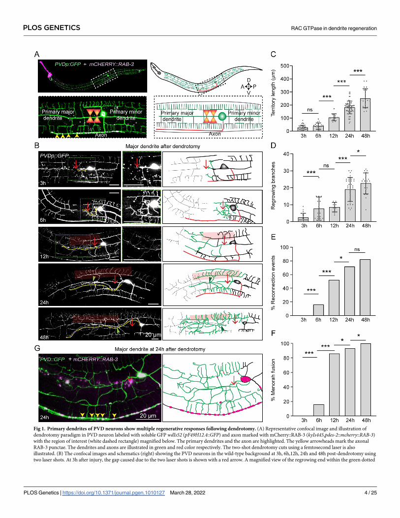
Fig 1. Primary dendrites of PVD neurons show multiple regenerative responses following dendrotomy. (A) Representative confocal image and illustration of
dendrotomy paradigm in PVD neuron labeled with soluble GFP wdIs52 (pF49H12.4::GFP) and axon marked with mCherry::RAB-3 (kyIs445,pdes-2::mcherry::RAB-3)
with the region of interest (white dashed rectangle) magnified below. The primary dendrites and the axon are highlighted. The yellow arrowheads mark the axonal
RAB-3 punctae. The dendrites and axons are illustrated in green and red color respectively. The two-shot dendrotomy cuts using a femtosecond laser is also
illustrated. (B) The confocal images and schematics (right) showing the PVD neurons in the wild-type background at 3h, 6h,12h, 24h and 48h post-dendrotomy using
two laser shots. At 3h after injury, the gap caused due to the two laser shots is shown with a red arrow. A magnified view of the regrowing end within the green dotted
PLOS GENETICS RAC GTPase in dendrite regeneration
PLOS Genetics | https://doi.org/10.1371/journal.pgen.1010127 March 28, 2022 4 / 25
and distal dendritic parts due to multiple shots was significantly bigger as observed at 3-6h
after injury (orange double-headed arrow, S1A Fig). Although the menorah-menorah fusion
and reconnection events were significantly lower in multi-shot experiments (S1B and S1C
Fig), both the territory length and branching in this experiment were comparable to the two-
shot dendrotomy experiments (S1D and S1E Fig). This suggested that the reconnection or
fusion processes do not influence the regenerative growth initiated upon dendrotomy in PVD
neurons. Unlike axon regeneration [33], dendrite regeneration did not disrupt the axon-den-
drite compartmentalization as the synaptic reporter mCherry::RAB-3 mostly remained at the
ventral cord (yellow arrowheads, Fig 1G) and did not invade the regenerated neurites after
dendrotomy.
Additionally, we have performed dendrotomy on the minor dendrite (S1F Fig). We
observed similar regrowth response and fusion events in minor dendrites as well following
dendrotomy. (S1F Fig).
Hence, both major and minor dendrites of PVD neurons can regenerate after dendrotomy
irrespective of the size of the injury. The regrowing dendrites cover up the injury area in a pat-
tern different from the original arbor. In the event of an encounter with the distal remnants,
the regrowing dendrites may fuse or reconnect and integrate into the original arbor. However,
this does not prevent the unfused dendritic tips from growing further. This was indicative of a
molecular mechanism of dendrite regeneration underlying multiple cellular processes such as
regrowth, branching, and cell fusion.
Dendrite regeneration in PVD neurons is independent of DLK/MLK
pathway
The cellular and molecular mechanisms of axon regeneration have been extensively studied
using various model organisms[34]. The conserved Mitogen-activated protein kinase kinase
kinase (MAPKKK) pathway involving DLK-1 is essential for the initiation of regrowth from
the cut stump of axon in multiple model organisms, including mammals [13,14,35], (Fig 2A).
Therefore, the initiation of dendrite regrowth might rely on the DLK-1 mediated injury
response.
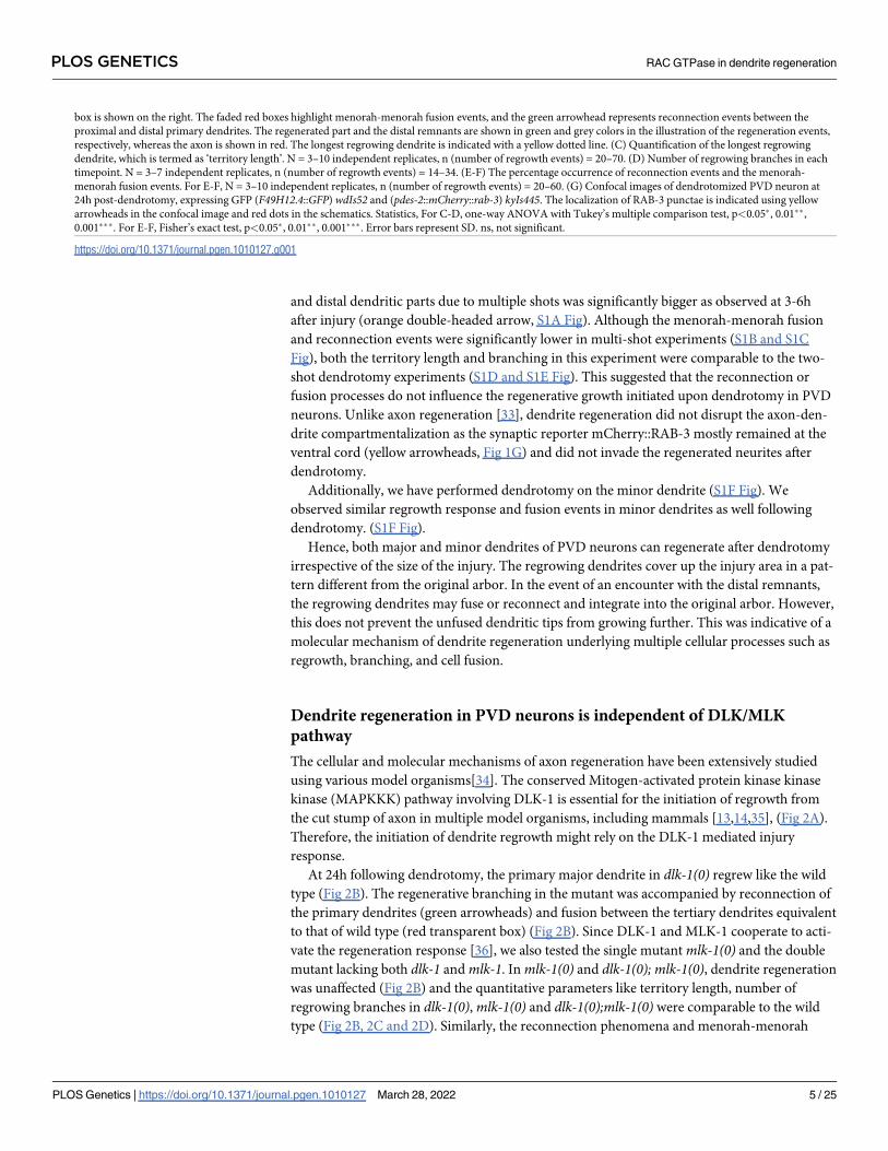
At 24h following dendrotomy, the primary major dendrite in dlk-1(0) regrew like the wild
type (Fig 2B). The regenerative branching in the mutant was accompanied by reconnection of
the primary dendrites (green arrowheads) and fusion between the tertiary dendrites equivalent
to that of wild type (red transparent box) (Fig 2B). Since DLK-1 and MLK-1 cooperate to acti-
vate the regeneration response [36], we also tested the single mutantmlk-1(0) and the double
mutant lacking both dlk-1 andmlk-1. Inmlk-1(0) and dlk-1(0); mlk-1(0), dendrite regeneration
was unaffected (Fig 2B) and the quantitative parameters like territory length, number of
regrowing branches in dlk-1(0),mlk-1(0) and dlk-1(0);mlk-1(0) were comparable to the wild
type (Fig 2B, 2C and 2D). Similarly, the reconnection phenomena and menorah-menorah
box is shown on the right. The faded red boxes highlight menorah-menorah fusion events, and the green arrowhead represents reconnection events between the
proximal and distal primary dendrites. The regenerated part and the distal remnants are shown in green and grey colors in the illustration of the regeneration events,
respectively, whereas the axon is shown in red. The longest regrowing dendrite is indicated with a yellow dotted line. (C) Quantification of the longest regrowing
dendrite, which is termed as ‘territory length’. N = 3–10 independent replicates, n (number of regrowth events) = 20–70. (D) Number of regrowing branches in each
timepoint. N = 3–7 independent replicates, n (number of regrowth events) = 14–34. (E-F) The percentage occurrence of reconnection events and the menorah-
menorah fusion events. For E-F, N = 3–10 independent replicates, n (number of regrowth events) = 20–60. (G) Confocal images of dendrotomized PVD neuron at
24h post-dendrotomy, expressing GFP (F49H12.4::GFP) wdIs52 and (pdes-2::mCherry::rab-3) kyIs445. The localization of RAB-3 punctae is indicated using yellow
arrowheads in the confocal image and red dots in the schematics. Statistics, For C-D, one-way ANOVA with Tukey’s multiple comparison test, p<0.05�, 0.01��,
0.001���. For E-F, Fisher’s exact test, p<0.05�, 0.01��, 0.001���. Error bars represent SD. ns, not significant.
https://doi.org/10.1371/journal.pgen.1010127.g001
PLOS GENETICS RAC GTPase in dendrite regeneration
PLOS Genetics | https://doi.org/10.1371/journal.pgen.1010127 March 28, 2022 5 / 25
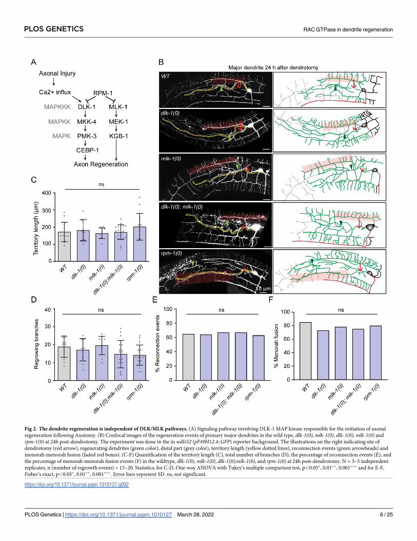
Fig 2. The dendrite regeneration is independent of DLK/MLK pathways. (A) Signaling pathway involving DLK-1 MAP kinase responsible for the initiation of axonal
regeneration following Axotomy. (B) Confocal images of the regeneration events of primary major dendrites in the wild type, dlk-1(0),mlk-1(0), dlk-1(0); mlk-1(0) and
rpm-1(0) at 24h post-dendrotomy. The experiment was done in the in wdIs52 (pF49H12.4::GFP) reporter background. The illustrations on the right indicating site of
dendrotomy (red arrow), regenerating dendrites (green color), distal part (grey color), territory length (yellow dotted lines), reconnection events (green arrowheads) and
menorah-menorah fusion (faded red boxes). (C-F) Quantification of the territory length (C), total number of branches (D), the percentage of reconnection events (E), and
the percentage of menorah-menorah fusion events (F) in the wildtype, dlk-1(0),mlk-1(0), dlk-1(0);mlk-1(0), and rpm-1(0) at 24h post-dendrotomy. N = 3–5 independent
replicates, n (number of regrowth events) = 15–20. Statistics, for C-D, One-way ANOVA with Tukey’s multiple comparison test, p<0.05�, 0.01��, 0.001��� and for E-F,
Fisher’s exact, p<0.05�, 0.01��, 0.001���. Error bars represent SD. ns, not significant.
https://doi.org/10.1371/journal.pgen.1010127.g002
PLOS GENETICS RAC GTPase in dendrite regeneration
PLOS Genetics | https://doi.org/10.1371/journal.pgen.1010127 March 28, 2022 6 / 25
fusion events were equivalent in these mutants as compared to wild type (Fig 2E and 2F). The
dendrite regeneration was also unchanged in the loss of function mutant of E3 ubiquitin ligase,
RPM-1 (Fig 2), which downregulates DLK-1 and downstream kinases in the cascade during
developmental growth of axon(Fig 2A) [37]. The dendrite regrowth and its ability to fuse at
24h after injury in rpm-1(0) was similar to the wild type (Fig 2), suggesting that dlk-1 andmlk-1 are neither necessary nor sufficient for the dendrite regrowth following injury in PVD
neurons.
Furthermore, we checked the dendrite regeneration in the minor dendrite of dlk-1(0);mlk-1(0) double mutant, which was comparable to the wild-type (S2A, S2B and S2C Fig). These
observations corroborated the earlier results in Drosophila da neurons where dendrite regener-
ation was independent of the DLK-1 signalling [21]. Although dlk-1 is expressed in PVD [38],
its role in PVD neuron is unclear. Since a well-known role of E3 Ubiquitin ligase RPM-1 and
downstream MAPKKK DLK-1 is to stabilize synaptic growth along with axon growth during
development (S2D Fig) [37,39–41], we looked at the possible phenotype related to axon devel-
opment in rpm-1mutant. Both the ju23 and ok364 alleles of rpm-1 showed an overgrowth of
axons along the ventral cord (S2E Fig). The length of the axon was significantly higher in rpm-1mutants (S2F Fig), and axon overgrowth phenotype was completely suppressed by loss of
dlk-1 in rpm-1(0) background (S2E and S2F Fig) as seen in other neurons in C. elegans [37]
and other organisms [39,40]. This indicated that rpm-1/dlk-1 cascade is functional in PVD
neurons and strengthened our observation of unaffected dendrite regeneration in dlk-1/mlk-1mutants.
Axon regeneration in PVD neurons depends on DLK-1 and MLK-1
Our finding that dendrite regeneration in PVD is independent of DLK-1 cascade raises the
question of whether the axon regeneration in this neuron would require this MAP Kinase
pathway. We performed axotomy at 50 μm away from the soma (Red arrow, Fig 3A) and
found that the severed end retracted at 3h post-axotomy, and afterwards followed by a
regrowth from the severed end (Fig 3B and 3C, green traces). The punctae of axonal reporter
mCherry::RAB-3 were localized at the tip of this regrowing neurite (Fig 3B, yellow arrow-
heads). These punctae are often relocalized at the adjacent dendrites (Fig 3B, blue traces,
orange arrowheads), suggesting the conversion of some of the adjacent tertiary dendrites into
an axon. This observation was reminiscent of Drosophila da neurons, where the dendrites are
converted to axons following a proximal axotomy [33]. The soma and the proximal part of the
severed axon often emanated some ectopic processes (Fig 3B, orange traces). There was a sig-
nificant extension of the axon from the severed end at 24h and 48h as compared to 3h post-
axotomy (Fig 3B and 3C). Similarly, there was an increase in the conversion of adjacent den-
drites to axon-like branches (Fig 3C) and the number and length of ectopic branches (Fig 3C).
We then carried out the axotomy in loss of function mutants of dlk-1 andmlk-1 (Fig 3D).
At 24h post-axotomy, wildtype worms showed an average regrowth of 26.07±17.4 μm from
the severed end which decreased significantly, due to loss of either dlk-1 (10.17±9.93 μm) or
mlk-1 (9.04±10.59 μm) or both (8.86±11.43 μm) (Fig 3D and 3E), with negligible regrowth in
nearly 50% of the mutant worms. Length of the ectopic neurites during regrowth was also
reduced in the double mutant as compared to wildtype (Fig 3F). This confirmed the require-
ment of dlk-1 andmlk-1 in the PVD axon regeneration.
Thus, the axon regeneration requires DLK/MLK pathway in PVD neurons, but the dendrite
regeneration is not dependent upon this signaling pathway, as seen in Drosophila [21]. How-
ever, dendrite regeneration might rely on other molecular pathways regulating axon
regeneration.
PLOS GENETICS RAC GTPase in dendrite regeneration
PLOS Genetics | https://doi.org/10.1371/journal.pgen.1010127 March 28, 2022 7 / 25
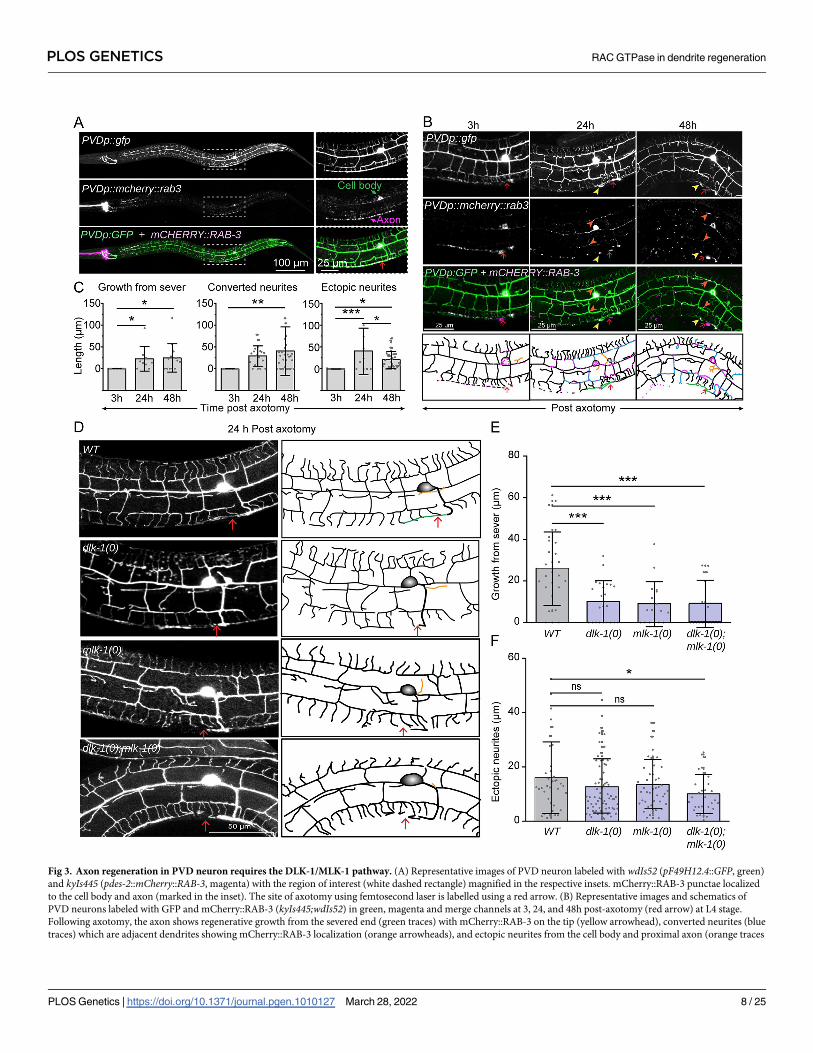
Fig 3. Axon regeneration in PVD neuron requires the DLK-1/MLK-1 pathway. (A) Representative images of PVD neuron labeled with wdIs52 (pF49H12.4::GFP, green)
and kyIs445 (pdes-2::mCherry::RAB-3, magenta) with the region of interest (white dashed rectangle) magnified in the respective insets. mCherry::RAB-3 punctae localized
to the cell body and axon (marked in the inset). The site of axotomy using femtosecond laser is labelled using a red arrow. (B) Representative images and schematics of
PVD neurons labeled with GFP and mCherry::RAB-3 (kyIs445;wdIs52) in green, magenta and merge channels at 3, 24, and 48h post-axotomy (red arrow) at L4 stage.
Following axotomy, the axon shows regenerative growth from the severed end (green traces) with mCherry::RAB-3 on the tip (yellow arrowhead), converted neurites (blue
traces) which are adjacent dendrites showing mCherry::RAB-3 localization (orange arrowheads), and ectopic neurites from the cell body and proximal axon (orange traces
PLOS GENETICS RAC GTPase in dendrite regeneration
PLOS Genetics | https://doi.org/10.1371/journal.pgen.1010127 March 28, 2022 8 / 25
Dendrite regeneration in PVD neurons is independent of conventional
axon regeneration pathways
Axon regeneration is also controlled by pathways other than the DLK-1 pathway [42]. We tested
some of the major genetic regulators implicated in axon regrowth. Axon regeneration is con-
trolled by conserved Calcium and cAMP cascade in many organisms [12,43]. After an axonal
injury, there is a calcium influx [11,12], which triggers a cAMP cascade near the injury site and
activates DLK-1 MAP3K [15]. An elevation of either intracellular calcium using a gain of func-
tion mutation in L-type voltage-gated calcium channel egl-19 or an elevation of cAMP due to
the loss of neuronal phosphodiesterase pde-4 promotes axon regeneration [12]. However, we
observed that neither egl-19(gf) nor pde-4(0) influenced any aspect of dendrite regeneration (Fig
4A, 4B and 4C). After 24h of dendrotomy, the dendrite was able to regenerate to a similar extent
as wild type and the reconnection events were also similar to the wild-type (Fig 4A, 4B and 4C).
The let-7miRNA and its downstream target lin-41 regulate axon regeneration pathway and
fusion phenomena [44–46]. Loss of function mutants of let-7 and lin-41 showed dendrite
regrowth and reconnection comparable to that of the wild type at 24h post-dendrotomy (Fig
4A, 4B and 4C).
PTEN/AKT pathway was previously implicated to play an important role in the regenera-
tion of both axons and dendrites [47,48]. The territory length and reconnection events at 24h
post-dendrotomy were not affected in akt-1mutant (Fig 4A, 4B and 4C), suggesting dendrite
regeneration in PVD neurons is independent of akt-1.
The Phosphatidylserine (PS) exposure pathway has emerged as a critical injury sensing
mechanism during axonal injury and dendrite degeneration [49,50]. Upon injury, the PS signal
activates axon regeneration mechanisms such as DLK/MLK p38 MAPK pathway [51] or fuso-
gen related repair pathway [52]. The PS signal involves exposure of PS to the outer leaflet mem-
brane of the injured neuron through the ABC transporter, CED-7, and further activation of the
downstream effectors such as CED-2/CED-5/CED-12 GEF complex and CED-10 GTPase. This
signal subsequently activates p38 cascade involving MLK-1 [51]. We did not see any effect in
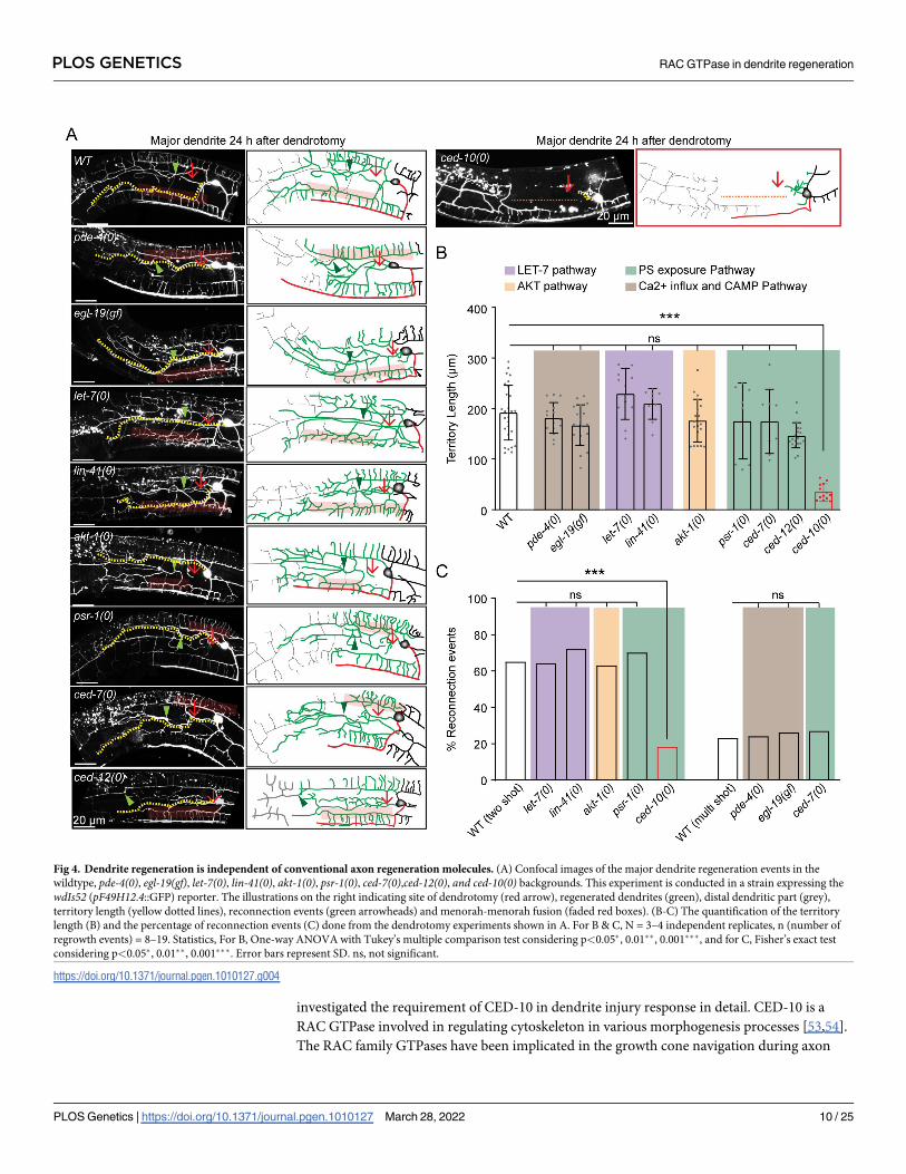
dendrite regeneration parameters in ced-7, psr-1 and ced-12mutants (Fig 4A, 4B and 4C). How-
ever, loss of ced-10 showed a drastic impact on dendrite regeneration, including regrowth and
fusion phenomena (Fig 4A, 4B and 4C). In ced-10mutant, a large gap is seen at 24h post-den-
drotomy since the regrowing branches fail to reach the distal end of the primary dendrites (red
dotted line, Fig 4A). The fusion events were also drastically reduced (Fig 4A and 4C). This indi-
cated that the CED-10/RAC GTPase might have a novel role in the injury response.
The characterization of different axon regeneration pathways in our dendrite regeneration
assay indicated that most known effectors of axon regeneration are not required for dendrite
regeneration in PVD neurons. Nevertheless, the substantial reduction of dendrite regeneration
in ced-10mutant raised exciting questions to explore further.
CED-10 RAC GTPase is required in neuron for dendrite regeneration
Among the candidate genes tested in our dendrite regeneration assay, the ced-10mutant
showed a strong reduction in dendrite regeneration (Fig 4A, 4B and 4C). Therefore, we
in schematics). Axonal injury is marked using red arrow. (C) Quantification of the axon regeneration at 3, 24, and 48h following axotomy in the form of growth from the
severed end, length of the converted neurites, and ectopic neurites. N = 3–5 independent replicates, n (number of regrowth events) = 17–26. (D) The confocal images of
axon regrowth events at 24h post-axotomy in the wildtype, dlk-1(0),mlk-1(0), and dlk-1(0);mlk-1(0)mutants with their representative images and schematics. The
schematics show regenerative growth from the severed end (green traces) and ectopic neurites as orange traces. (E-F) Axon regeneration is quantified as growth from the
severed end (E), and ectopic neurites (F). N = 5–7 independent replicates, n (number of regrowth events) = 17–26. Statistics, For C,E & F, �P<0.05, ��P<0.01, ���P
<0.001; ANOVA with Tukey’s multiple comparison test. Error bars represent SD. ns, not significant.
https://doi.org/10.1371/journal.pgen.1010127.g003
PLOS GENETICS RAC GTPase in dendrite regeneration
PLOS Genetics | https://doi.org/10.1371/journal.pgen.1010127 March 28, 2022 9 / 25
investigated the requirement of CED-10 in dendrite injury response in detail. CED-10 is a
RAC GTPase involved in regulating cytoskeleton in various morphogenesis processes [53,54].
The RAC family GTPases have been implicated in the growth cone navigation during axon
Fig 4. Dendrite regeneration is independent of conventional axon regeneration molecules. (A) Confocal images of the major dendrite regeneration events in the
wildtype, pde-4(0), egl-19(gf), let-7(0), lin-41(0), akt-1(0), psr-1(0), ced-7(0),ced-12(0), and ced-10(0) backgrounds. This experiment is conducted in a strain expressing the
wdIs52 (pF49H12.4::GFP) reporter. The illustrations on the right indicating site of dendrotomy (red arrow), regenerated dendrites (green), distal dendritic part (grey),
territory length (yellow dotted lines), reconnection events (green arrowheads) and menorah-menorah fusion (faded red boxes). (B-C) The quantification of the territory
length (B) and the percentage of reconnection events (C) done from the dendrotomy experiments shown in A. For B & C, N = 3–4 independent replicates, n (number of
regrowth events) = 8–19. Statistics, For B, One-way ANOVA with Tukey’s multiple comparison test considering p<0.05�, 0.01��, 0.001���, and for C, Fisher’s exact test
considering p<0.05�, 0.01��, 0.001���. Error bars represent SD. ns, not significant.
https://doi.org/10.1371/journal.pgen.1010127.g004
PLOS GENETICS RAC GTPase in dendrite regeneration
PLOS Genetics | https://doi.org/10.1371/journal.pgen.1010127 March 28, 2022 10 / 25
development [55]. Therefore, we checked whether ced-10mutant causes any developmental
phenotype in PVD neuron. The length of the axon in PVD neuron remained unaffected in
ced-10mutant (S3A and S3B Fig). Dendrites also seemed normal in ced-10(0) (S3A Fig). The
ced-10mutant did not affect the axon regrowth parameters in PVD neuron (S3C, S3D and S3E
Fig). To understand the requirement of CED-10 in the initiation of dendrite regeneration, we
checked at early time points after dendrotomy (Fig 5A). Both the number of filopodia-like
structures (red arrowheads, Fig 5A) and the territory length showed a significant reduction in
ced-10(0) as compared to the wild type at 6h post-dendrotomy (Fig 5A, 5C and 5D). Con-
versely, when an activated form of CED-10 (G12V) is expressed in the PVD in wild type back-
ground, we found that the number of regrowing branches from proximal dendrite increased at
6h post-dendrotomy (Fig 5A, 5C and 5D). Since the higher concentration transgenic lines
(10ng/ul) led to the formation of ectopic branches around the cell body region without even
performing dendrotomy, we selected a low concentration line (5ng/ul) with a significantly
milder developmental defect for our dendrotomy experiment (S3H and S3I Fig). Similarly, the
territory coverage length was also increased due to CED-10 activation (Fig 5C). Another gene
that codes for RAC GTPase ismig-2, which collaborates with CED-10 during development
[53,56] (S3A Fig). Although the loss ofmig-2 affected the development of PVD axon (S3A and
S3B Fig), the primary major dendrite regrowth was unaffected in this mutant (S3F and S3G
Fig). This indicated that developmental impairment of axons would not necessarily affect the
dendrite regeneration process. This also indicated a specific requirement of CED-10 in the
dendrite regeneration of PVD neurons as axon regeneration was unaffected in the loss of func-
tion of ced-10 (S3C, S3D and S3E Fig).
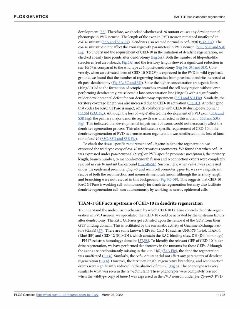
To check the tissue-specific requirement ced-10 gene in dendrite regeneration, we
expressed the wild type copy of ced-10 under various promoters. We found that when ced-10was expressed under pan-neuronal (prgef) or PVD-specific promoter pser2prom3, the territory
length, branch number, % menorah-menorah fusion and reconnection events were completely
rescued in ced-10mutant background (Fig 5B–5F). Surprisingly, when ced-10 was expressed
under the epidermal promoter, pdpy-7 and seam cell promoter, pgrd-10, we saw a significant
rescue of both the reconnection and menorah-menorah fusion, although the territory length
and branching were not rescued in this background (Fig 5C–5F). This suggests that CED-10
RAC GTPase is working cell-autonomously for dendrite regeneration but may also facilitate
dendrite regeneration cell-non autonomously by working in nearby epidermal cells.
TIAM-1 GEF acts upstream of CED-10 in dendrite regeneration
To understand the molecular mechanism by which CED-10 GTPase controls dendrite regen-
eration in PVD neuron, we speculated that CED-10 could be activated by the upstream factors
after dendrotomy. The RAC GTPases get activated upon the removal of the GDP from their
GTP binding domain. This is facilitated by the enzymatic activity of Guanine Exchange Fac-
tors (GEFs) [57]. There are some known GEFs for CED-10 such as UNC-73 (Trio), TIAM-1
(RhoGEF) and CED-12 (ELMO1), which contain the RAC binding sites, DH (Dbl homology)
—PH (Pleckstrin homology) domains [57,58]. To identify the relevant GEF of CED-10 in den-
drite regeneration, we have performed dendrotomy in the mutants for these GEFs. Although
the axons are predominantly missing in the unc-73(0) (S4A Fig), the dendrite regeneration
was unaffected (Fig 6). Similarly, the ced-12mutant did not affect any parameters of dendrite
regeneration (Fig 6). However, the territory length, regenerative branching, and reconnection
events were significantly reduced in the absence of tiam-1 (Fig 6). The phenotype was very
similar to what was seen in the ced-10mutant. These phenotypes were completely rescued
when the wildtype copy of tiam-1 was expressed in the PVD neuron under pser2prom3 (PVD
PLOS GENETICS RAC GTPase in dendrite regeneration
PLOS Genetics | https://doi.org/10.1371/journal.pgen.1010127 March 28, 2022 11 / 25
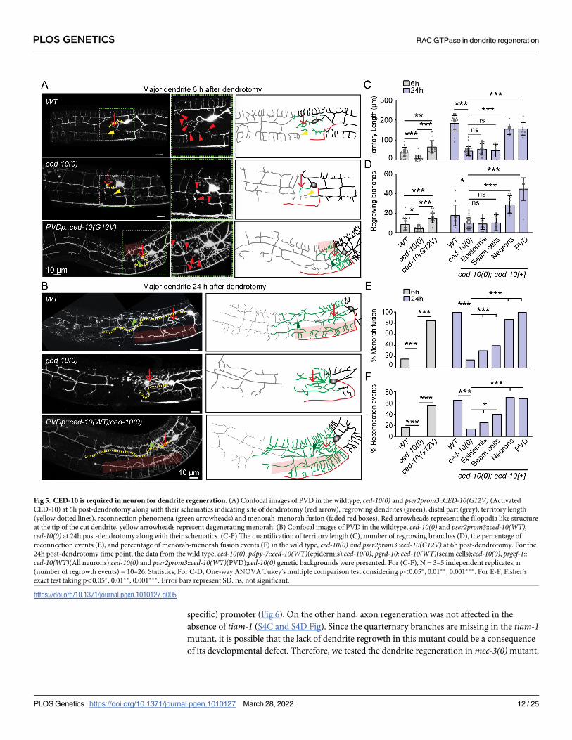
specific) promoter (Fig 6). On the other hand, axon regeneration was not affected in the
absence of tiam-1 (S4C and S4D Fig). Since the quarternary branches are missing in the tiam-1mutant, it is possible that the lack of dendrite regrowth in this mutant could be a consequence
of its developmental defect. Therefore, we tested the dendrite regeneration inmec-3(0)mutant,
Fig 5. CED-10 is required in neuron for dendrite regeneration. (A) Confocal images of PVD in the wildtype, ced-10(0) and pser2prom3::CED-10(G12V) (Activated
CED-10) at 6h post-dendrotomy along with their schematics indicating site of dendrotomy (red arrow), regrowing dendrites (green), distal part (grey), territory length
(yellow dotted lines), reconnection phenomena (green arrowheads) and menorah-menorah fusion (faded red boxes). Red arrowheads represent the filopodia like structure
at the tip of the cut dendrite, yellow arrowheads represent degenerating menorah. (B) Confocal images of PVD in the wildtype, ced-10(0) and pser2prom3::ced-10(WT);ced-10(0) at 24h post-dendrotomy along with their schematics. (C-F) The quantification of territory length (C), number of regrowing branches (D), the percentage of
reconnection events (E), and percentage of menorah-menorah fusion events (F) in the wild type, ced-10(0) and pser2prom3::ced-10(G12V) at 6h post-dendrotomy. For the
24h post-dendrotomy time point, the data from the wild type, ced-10(0), pdpy-7::ced-10(WT)(epidermis);ced-10(0), pgrd-10::ced-10(WT)(seam cells);ced-10(0), prgef-1::
ced-10(WT)(All neurons);ced-10(0) and pser2prom3::ced-10(WT)(PVD);ced-10(0) genetic backgrounds were presented. For (C-F), N = 3–5 independent replicates, n
(number of regrowth events) = 10–26. Statistics, For C-D, One-way ANOVA Tukey’s multiple comparison test considering p<0.05�, 0.01��, 0.001���. For E-F, Fisher’s
exact test taking p<0.05�, 0.01��, 0.001���. Error bars represent SD. ns, not significant.
https://doi.org/10.1371/journal.pgen.1010127.g005
PLOS GENETICS RAC GTPase in dendrite regeneration
PLOS Genetics | https://doi.org/10.1371/journal.pgen.1010127 March 28, 2022 12 / 25
Fig 6. TIAM-1 acts upstream to CED-10 for dendrite regeneration. (A) Confocal images of dendrotomized PVD neuron in the wild type, unc-73(0), ced-12(0), tiam-1(0), pser2prom3::tiam-1WT (5ng); tiam-1(0), pser2prom3::tiam-1(T548F)5ng;tiam-1(0) and pser2prom3::ced-10(G12V) 5ng;tiam-1(0) at 24h post-injury. The regrowing
dendrites, axon, and the distal part of dendrite are represented in green, red, and grey color, respectively. The green arrowheads represent reconnection events and the
faded red rectangular boxes indicate menorah-menorah fusion event. (B-C) The quantification of territory length (B), and the the number of regrowing branches (C) in
the above mentioned genotypes. N = 3–5 independent replicates, n (number of regrowth events) = 10–29. (D) The bar chart represents the percentage of reconnection
events for the above mentioned genotypes, N = 3–5 independent replicates, n (number of regrowth events) = 10–29. Statistics, For B-C, one-way ANOVA with Tukey’s
multiple comparison test, p<0.05�, 0.01��, 0.001���. For D, Fisher’s exact test, p<0.05�, 0.01��, 0.001���. Error bars represent SD. ns, not significant.
https://doi.org/10.1371/journal.pgen.1010127.g006
PLOS GENETICS RAC GTPase in dendrite regeneration
PLOS Genetics | https://doi.org/10.1371/journal.pgen.1010127 March 28, 2022 13 / 25
in which the higher-order branches were completely absent (S4A and S4B Fig) as reported
before [59]. Upon dendrotomy inmec-3(0), the primary dendrites regrew and the territory
length was comparable to that of the control (S4E, S4F and S4G Fig). The proximal dendrites
inmec-3(0) also could reconnect with their distal counterparts (S4E–S4H Fig). Therefore, a
defect in dendrite regeneration may not always be correlated to the lack of developmental
branching in PVD neuron. Moreover, we addressed whether GEF activity of TIAM-1 would
be critical for dendrite regeneration. Since the GEF activity of TIAM-1 was not required for
developmental branching of PVD neuron, the expression of a GEF dead mutant of TIAM-1
(T548F) rescued the developmental branching phenotype in tiam-1mutant (S4A and S4B Fig)
as seen before [60]. However, upon performing dendrotomy in tiam-1(0) expressing pser-2prom3-tiam-1(T548F), we found that the territory covered by the PVD dendrites and number
of branches were not rescued in this background (Fig 6). Therefore, GEF activity of TIAM-1 is
specifically required for dendrite regeneration.
To test whether CED-10 activation is limiting in tiam-1mutant background, we expressed
the constitutively activated form of ced-10 in PVD neuron in the tiam-1(0). The activated form
of CED-10 could bypass the requirement of TIAM-1 in both territory extent as well as recon-
nection phenomena in dendrite regeneration (Fig 6B and 6C). Thus, the RhoGEF TIAM-1
acts upstream of CED-10 GTPase for dendritic regeneration.
Discussion
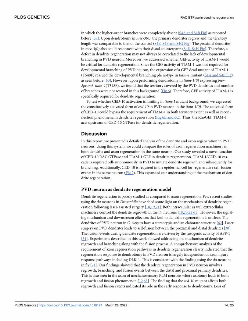
In this report, we presented a detailed analysis of the dendrite and axon regeneration in PVD
neurons. Using this system, we could compare the roles of axon regeneration machinery in
both dendrite and axon regeneration in the same neuron. Our study revealed a novel function
of CED-10 RAC GTPase and TIAM-1 GEF in dendrite regeneration. TIAM-1/CED-10 cas-
cade is required cell-autonomously in PVD to initiate dendrite regrowth and subsequently for
branching. Additionally, CED-10 is required in the epidermal cell for regenerative self-fusion
events in the same neuron (Fig 7). This expanded our understanding of the mechanism of den-
drite regeneration.
PVD neuron as dendrite regeneration model
Dendrite regeneration is poorly studied as compared to axon regeneration. Few recent studies
using the da neurons in Drosophila have shed some light on the mechanism of dendrite regen-
eration following laser-assisted surgery [18,19,21]. Both intracellular as well extracellular
machinery control the dendrite regrowth in the da neurons [18,20,22,61]. However, the signal-
ing mechanism and downstream effectors that lead to dendrite regeneration is unclear. The
dendrites of PVD neuron in C. elegans have a stereotypic and an elaborate structure [62]. Laser
surgery on PVD dendrites leads to self-fusion between the proximal and distal dendrites [30].
The fusion events during dendrite regeneration are driven by the fusogenic activity of AFF-1
[31]. Experiments described in this work allowed addressing the mechanism of dendrite
regrowth and branching along with the fusion process. A comprehensive analysis of the
requirement of axon regeneration pathways in dendrite regeneration clearly indicated that the
regeneration response to dendrotomy in PVD neuron is largely independent of axon injury
response pathways including DLK-1. This is consistent with the finding using the da neurons
in fly [21]. Our findings showed that the dendrite regeneration in PVD neuron involves
regrowth, branching, and fusion events between the distal and proximal primary dendrites.
This is also seen in the axon of mechanosensory PLM neurons where axotomy leads to both
regrowth and fusion phenomenon [12,63]. The finding that the ced-10mutant affects both
regrowth and fusion events indicated its role in the early response to dendrotomy. Loss of
PLOS GENETICS RAC GTPase in dendrite regeneration
PLOS Genetics | https://doi.org/10.1371/journal.pgen.1010127 March 28, 2022 14 / 25
tiam-1, the GTP exchange factor for CED-10 GTPase also showed similar perturbation in
regrowth and fusion events following dendrotomy. These results indicated that this conserved
RAC GTPase and its upstream activator TIAM-1 orchestrate the early events after dendrite
injury.
PVD neuron is known for its role in harsh touch response and proprioception of C. elegans
[29]. In future, the anatomical features of dendrite and axon regeneration can be correlated
with behavioral assays using the PVD neuron as an injury model.
Role of RAC GTPase in dendrite regeneration
The RAC GTPases have been known to play a broader role in developmental processes involv-
ing cell debris engulfment [64,65], migration, and axon guidance [53]. The RAC GTPases con-
trol these developmental processes through various downstream effectors of actin and
microtubule cytoskeleton [55,66,67]. The previous finding that ced-10mutant had a partial
reduction in the axon regrowth in C. elegansmotor neuron [51] prompted us to test its role in
dendrite regeneration. It was seen that ced-10 acts genetically downstream to Phosphatidylser-
ine (PS) signal to activate DLK/MLK MAP Kinase pathway in regeneration [51]. However, we
did not observe any effect on dendrite regeneration parameters in the mutants affecting either
PS or in dlk-1/mlk-1mutants. The cell-autonomous role of TIAM-1 and CED-10 in dendrite
Fig 7. A model describing the roles of CED-10 and TIAM-1 in dendrite regeneration process in PVD neuron.
https://doi.org/10.1371/journal.pgen.1010127.g007
PLOS GENETICS RAC GTPase in dendrite regeneration
PLOS Genetics | https://doi.org/10.1371/journal.pgen.1010127 March 28, 2022 15 / 25
regeneration is novel. The RAC GTPases are well-known for their role in F-actin dynamics
[68,69], and actin dynamics is a major player in dendritic remodelling during neuronal plastic-
ity [70,71]. It might be possible that CED-10 GTPase induces optimal F-actin dynamics suited
for regrowth and branching that is observed after dendrite injury.
Our finding that CED-10 plays a cell non-autonomous function in the surrounding epithe-
lial cells for the menorah-menorah fusion events during regeneration is very intriguing. It was
seen that for the fusion to take place, AFF-1 fusogen was delivered from the surrounding seam
cells, which are of epidermal origin. CED-10 may initiate the epidermal response to dendrot-
omy, which might lead to the release of vesicles containing AFF-1 from epidermal cells. Epi-
dermal cells are known for responding to dendrite injury. In case of da neurons in Drosophila,
the PS pathway in epidermal cells controls the dendrotomy induced engulfment of degener-
ated distal dendrites [50,72]. Therefore, in case of PVD neuron as well, the surrounding epi-
dermis might regulate the fate of the injured dendrites.
Materials and methods
C. elegans strains and genetics
The C. elegans strains were grown and maintained at 20˚C on OP50 bacterial lawns seeded
onto Nematode Growth Medium (NGM) plates [73]. The loss of function mutation is repre-
sented as (0) and gain of function alleles are represented as (gf), for example, the loss of func-
tion allele of dlk-1, tm4024 is represented as dlk-1(0) and gain of function allele of egl-19,
ad695 is represented as egl-19(gf). The mutants used in this study are mostly deletion or substi-
tution mutants unless otherwise mentioned (S1 Table). These mutants were taken from Cae-
norhabditis Genetics Centre (CGC) and genotyped using their respective genotyping primers.
Molecular cloning and creating transgenes
Destination vector with PVD neuron-specific promoter, pser2prom3 [4.1kb]::Gateway
[pNBRGWY99] was made by InFusion cloning (Takara). ser2prom3 Promoter region was
amplified from the fosmid WRM0623bG06 using the primers: 5’-ccatgattacgccaagtaaaagtttag-
taaattaactgc-3’ and 5’-tggccaatcccggggtatgtgttgtgatgtcac-3’ and GWY vector backbone was
amplified from pCZGY553 using 5’-ccccgggattggcca-3’ and 5’-ttggcgtaatcatgg-3’.
For pan-neuronal and epidermal rescue of ced-10, prgef::GWY [pCZGY66] and pdpy-7::
GWY [pNBRGWY44], respectively, were recombined with the entry clone of ced-10 WT[pNBRGWY88] [74] using the LR recombination (Invitrogen).
For PVD specific expression of wildtype and constitutively active ced-10, pNBRGWY99
was recombined with ced-10 WT [pNBRGWY88] and ced-10 constitutively active (G12V)[pNBRGWY89] entry clone plasmids [74], respectively, using the LR recombination
(Invitrogen).
For PVD specific expression of tiam-1, tiam-1 cDNA was ligated with the pser2prom3 con-
taining backbone DNA from pNBRGWY99 by InFusion cloning. Primers used to amplify
tiam-1 cDNA from total cDNA were 5’-tccgaattcgcccttatgggctcacgcctctca-3’, and 5’-aaggaa-
catcgaaattcaaaatagcagctttcttgtaca-3’. The primers used to amplify vector backbone are 5’-
cagctttcttgtaca-3’ and 5’-aagggcgaattcgga-3’.
The GEF dead tiam-1 (T548F) [60] substitution mutant was generated using Q5 Site-
Directed Muatgenesis kit using the primers 5’- attgttggtcTTTgagaagaaatatgtcagcgatc-3’ and 5’-
tcttgcagagccatcgcc-3’ in pserprom3::tiam-1 (wild-type cDNA, NBR46).
The clones made using various molecular techniques were then injected in the gonads of
young adult worms along with co-injection markers such as pttx-3::RFP or pmyo-2::mCherryand the F1 progeny were then isolated and checked for the formation of transgenic lines. Each
PLOS GENETICS RAC GTPase in dendrite regeneration
PLOS Genetics | https://doi.org/10.1371/journal.pgen.1010127 March 28, 2022 16 / 25
transgene was checked for two high transmission lines. List of transgenes used in this study are
listed in S2 Table.
Laser system, dendrotomy and axotomy
Dendrotomy and axotomy experiments were conducted at the L4 stage of C. elegans using the
Bruker ULTIMA multi-photon microscopy system coupled with two SpectraPhysics tunable
Infra-red femtosecond lasers (λ = 690–1040 nm) having automated dispersion compensation
(MaiTai with DeepSee)[45]. The output of lasers was controlled using Conoptics pockel cells
with superior temporal resolution (~μs) and simultaneous imaging and severing experiments
was performed by two independent set of X-Y scan galvanometer mirrors. All the multi-pho-
ton images reported here (unless otherwise mentioned) were done using 920 nm (pulse
width = 80 fps) and laser ablation was done at 720 nm laser (pulse width = 80 fps, irradiation
time: 20ms, avg. laser power: 23mW) using a water immersion Olympus objective (60X / 0.9
NA). Worms were immobilized using either Levamisole hydrochloride (10mM) (Sigma
L0380000) as a paralyzing drug agent or with polystyrene beads (Polysciences 00876–15) of
diameter 0.1 μm as friction enhancing agent on 5% agarose pads mounted with Corning cover
glass (cat No. 2855–25). Nikon Ti-2 microscope equipped with ultraviolet laser (Micropoint
laser, 337nm, 3ns Pulse, Max Pulse enery-200μJ, Max Average Power- 4mW) system was also
used to assist the laser injury using using 100X/1.40NA oil immersion objective.
PVD dendrites were severed with the first laser shot at the first branch point (~10μm away
from cell body) followed by one (two-shot experiment, Fig 1A) or more (multi-shot experi-
ment, S1A Fig) consecutive shots with a relative distance of 10–15μm from the previous shot
creating a visible gap with no fragments left at the cut site. The axons were severed distally at
the segment fasciculating with the ventral nerve cord (VNC) (~50 μm away from the cell
body). A clean transection was ensured by making two shots 5 μm apart at a distance of 10 μm
away from the distal-most lateral to ventral transition (LV) point of PVDR and PVDL. After
severing, worms were recovered from the agarose pad using an aspirator with 1X M9 solution
onto freshly seeded NGM plates for further observation.
Imaging
To observe the regenerative response, injured worms were imaged at 3, 24 and 48 hours (h)
after injury. The worms were paralyzed and mounted in 10 mM Levamisole hydrochloride
(Sigma) solution on slides containing medium on 5% agarose (Sigma) pads. The worms were
imaged with 63X/1.4NA oil objective of Nikon A1R confocal system at a voxel resolution of
0.41 μm x 0.41 μm x 1 μm and tile imaging module using imaging lasers 488 nm(GFP), 543
nm(mCherry/RFP) with 1–1.8 AU pinhole at 512x512 pixel frame size for further analysis.
Dendrite regeneration analysis and quantification
Dendrite regeneration was quantified based on the regrowth and fusion parameters. The terri-
tory covered by regenerated dendrite (Fig 1B, yellow dotted line) was estimated using Simple
Neurite Tracer plugin in Fiji-ImageJ by tracking the longest regrowing dendrite from the cell
body to the tip of the dendrite using hessian based tracking onto the Z stack of PVD neuron.
‘Total number of branches’ were then also calculated using the same plugin from the point of
emergence (cell body or dendrite) to the termination of every branch.
PVD dendrites have the capability to fuse after injury [30,31]. We also observed reconnec-
tion between the regenerating proximal primary dendrite with distal primary dendrite or distal
menorah (Fig 1B, green arrow), which were evaluated as reconnection events using ‘depth
coded’ confocal images, the population of worms showing proximal and distal dendrites on
PLOS GENETICS RAC GTPase in dendrite regeneration
PLOS Genetics | https://doi.org/10.1371/journal.pgen.1010127 March 28, 2022 17 / 25
the same depth was calculated and represented as a fraction showing reconnections. Long
menorah having more than one secondary dendrite connected to it (Fig 1B, faded red rectan-
gular boxes), was classified as menorah-menorah fusion, and the percentage of PVD neurons
showing menorah-menorah fusion was quantified. Following injury, distal dendrite shows
hallmarks of Wallerian degeneration [75,76], which were estimated as the percentage of PVD
neurons showing distal part degeneration (Fig 1C, grey dotted neurites).
Axon regeneration analysis and quantification
Analysis of the axon regeneration was carried out by ImageJ. Neurites were traced, and their
lengths were quantified using the Simple Neurite Tracer plugin of ImageJ. Volumetric visuali-
zation using the ‘3D project’ of confocal stacks facilitated a qualitative estimate and descriptors
of regenerated neurites based on the mCHERRY::RAB-3 localization. Regenerative growth
was classified into the neurite growth from the cut point of the axon, conversion of the adja-
cent tertiary dendrites to axon like identity as per the localization of mCHERRY::RAB-3 punc-
tae, and ectopic branches from the cell body, proximal axon, or converted neurites.
Quants obtained from ImageJ for both dendritic and axonal regeneration were further ana-
lyzed by Excel and Graphpad to get statistical information.
Statistical analysis
Statistical analyses were compassed using Graphpad Prism software (Prism 8 V8.2.1(441).
Two samples were analyzed using the unpaired two-tailed t test. The statistical analysis of mul-
tiple samples was performed using One way-ANOVA with Tukey’s multiple comparisons test.
The data that was used for ANOVA analysis was naturally occurring data having normal distri-
bution spread which was not further processed. To compare the population data, the fraction
values with respect to each sample were calculated and compared using the two-tailed Chi-
square Fisher’s exact contingency test. For each bar plots, the number of samples (n) and the
number of biological replicates (N) are mentioned In the respective figure legends. The signifi-
cance considered for all statistical experiments are p<0.05�, 0.01��, 0.001���.
Supporting information
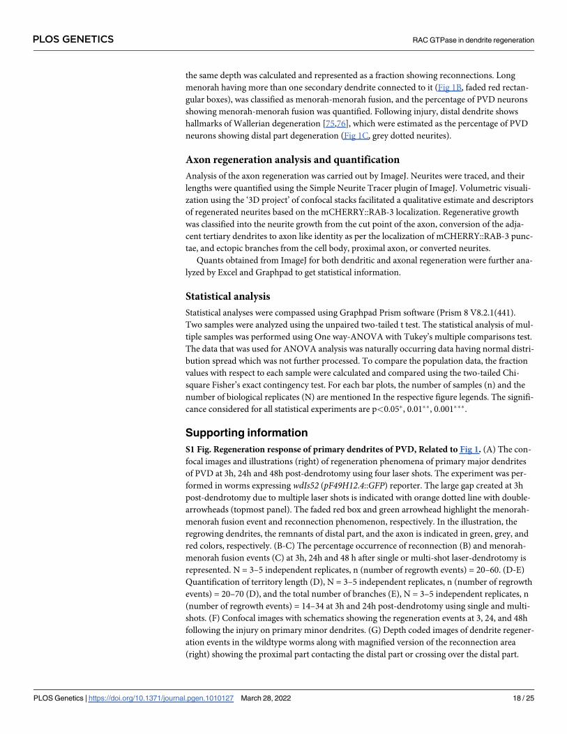
S1 Fig. Regeneration response of primary dendrites of PVD, Related to Fig 1. (A) The con-
focal images and illustrations (right) of regeneration phenomena of primary major dendrites
of PVD at 3h, 24h and 48h post-dendrotomy using four laser shots. The experiment was per-
formed in worms expressing wdIs52 (pF49H12.4::GFP) reporter. The large gap created at 3h
post-dendrotomy due to multiple laser shots is indicated with orange dotted line with double-
arrowheads (topmost panel). The faded red box and green arrowhead highlight the menorah-
menorah fusion event and reconnection phenomenon, respectively. In the illustration, the
regrowing dendrites, the remnants of distal part, and the axon is indicated in green, grey, and
red colors, respectively. (B-C) The percentage occurrence of reconnection (B) and menorah-
menorah fusion events (C) at 3h, 24h and 48 h after single or multi-shot laser-dendrotomy is
represented. N = 3–5 independent replicates, n (number of regrowth events) = 20–60. (D-E)
Quantification of territory length (D), N = 3–5 independent replicates, n (number of regrowth
events) = 20–70 (D), and the total number of branches (E), N = 3–5 independent replicates, n
(number of regrowth events) = 14–34 at 3h and 24h post-dendrotomy using single and multi-
shots. (F) Confocal images with schematics showing the regeneration events at 3, 24, and 48h
following the injury on primary minor dendrites. (G) Depth coded images of dendrite regener-
ation events in the wildtype worms along with magnified version of the reconnection area
(right) showing the proximal part contacting the distal part or crossing over the distal part.
PLOS GENETICS RAC GTPase in dendrite regeneration
PLOS Genetics | https://doi.org/10.1371/journal.pgen.1010127 March 28, 2022 18 / 25
(H) Quantification of reconnection and menorah-menorah fusion events counted from the
depth-coded vs regular z-projected images. (I) Quantification of degeneration of the distal
parts in the ’reconnection’ vs ‘no reconnection’ events. For, H-I, N = 3–4 independent repli-
cates, n (number of regrowth events) = 20–30. Statistics, for B-C, Fisher’s exact test, taking
p<0.05�, 0.001���. For D-E, one-way ANOVA with Tukey’s multiple comparisons method,
taking p<0.001���. For H-I, unpaired t test, taking p<0.05�, 0.001���. Error bars represent SD.
ns, not significant.
(TIF)
S2 Fig. The dendrite regeneration does not require DLK/MLK pathway, Related to Fig 2.
(A) Confocal images of the regeneration events of minor dendrites in wild-type and dlk-1(0);mlk-1(0) backgrounds at 24h post-dendrotomy. The schematics representing site of dendritic
injury (red arrow), regenerated dendrite (green), distal part (grey), reconnection phenomena
(green arrowheads) and menorah-menorah fusion (semi-transparent red boxes). Territory
length is represented as yellow dotted lines in the confocal image. (B-C) The territory length
(B) and the number of regrowing branches (C) at3-6h and 24h post-dendrotomy. N = 3 inde-
pendent replicates, n (number of regrowth events) = 7–19. (D) The genetic pathway involving
rpm-1, controlling axon growth termination. (E) Representative confocal images showing the
developmental phenotype of PVD in various mutants in rpm-1 pathway. In the schematics, the
axon in shown in red. Please note that in rpm-1mutants, an overshooting of axon is noticed.
(F) The quantification of axonal length of PVD neurons in the wild-type, dlk-1(0),mlk-1(0),dlk-1(0); mlk-1(0), rpm-1(ju23), rpm-1(ok364) and rpm-1(ok364);dlk-1(tm4024)mutants at L4
stage, N = 3–4 independent replicates, n (number of PVD imaged) = 8–27. Statistics, for B-C
& F one-way ANOVA with Tukey’s multiple comparison method taking p<0.05�, 0.01��,
0.001���. Error bars represent SD. ns, not significant.
(TIF)
S3 Fig. CED-10 is required for dendrite regeneration, related to Fig 5. (A) Confocal images
of PVD neuron in the wild-type, ced-10(0), andmig-2(0) is shown along with its illustrations
(right) indicating the axon in red. (B) The axonal defect at L4 stage is calculated as percentage
defect. N = 3 independent replicates, n (number of PVD imaged) = 10–12. (C) Confocal
images of axon regeneration events in the wild-type, ced-10(0) andmig-2(0) at 24h post-axot-
omy along with their schematics indicating site of axonal injury with red arrow, regrowing
axon from severed end in green and ectopic neurites in orange color. (D-E) Quantification of
axon regeneration as growth from the severed end (D) and length of ectopic neurites (E) in the
wild-type, ced-10(0) andmig-2(0) at 24h post-axotomy. N = 3–4 independent replicates, n
(number of regrowth events) = 14–25. (F) Confocal image of dendrite regeneration inmig-2(0) at 24h post-dendrotomy. The illustration indicating the regrowing dendrites in green
color, distal part in grey color, reconnection phenomenon with green arrowheads, menorah-
menorah fusion with faint red rectangular boxes. (G) The territory length in the wild-type and
mig-2(0) at 24h post-dendrotomy, N = 3–4 independent replicates, n (number of regrowth
events) = 10–11. (H) The confocal images of PVD neuron at L4 stage expressing pser2prom3::
ced-10(G12V) extrachromosomal transgenes. The pser2prom3::ced-10(G12V) plasmid was
injected at 1ng/μl, 5ng/μl, and 10ng/μl concentrations to obtain these lines. (I) Quantification
of number of ectopic neurites emerging out of cell body or adjacent dendrites at L4 stage in
the wild-type and transgenic background expressing pser2prom3::ced-10(G12V) extrachromo-
somal arrays, N = 3 independent replicates, n (number of PVD imaged) = 11–20. Statistics, for
B, Fisher’s exact test, for D-E & H, one-way ANOVA with Tukey’s multiple comparison
method, and for G, unpaired t test, p<0.05�, 0.01��, 0.001���. Error bars represent SD. ns, not
PLOS GENETICS RAC GTPase in dendrite regeneration
PLOS Genetics | https://doi.org/10.1371/journal.pgen.1010127 March 28, 2022 19 / 25
significant.
(TIF)
S4 Fig. Developmental phenotype and regeneration phenomena of PVD neuron in Rho/
RAC-GEF mutants, related to Fig 6. (A) Confocal images of PVD neuron in the wild-type,
unc-73(0), tiam-1(0), pser2prom3::tiam-1(T548F);tiam-1(0), pser2prom3::ced-10(G12V);tiam-1(0), and mec-3(0) background at L4 stage. (B) The percentage of PVDs showing quaternary
branches in the wild-type, tiam-1(0), pser2prom3::tiam-1(T548F);tiam-1(0), pser2prom3::ced-10(G12V);tiam-1(0) andmec-3(0) backgrounds, N = 3 independent replicates, n (number of
PVD imaged) = 12–15. (C) Confocal images of axon regeneration at 24h post-axotomy in the
wild-type and tiam-1(0) backgrounds. The regenerated axon from the severed end is shown in
green color in the illustration. (D) The quantification of axon regrowth from the severed end
in the wild-type and tiam-1(0), N = 3 independent replicates, n (number of regrowth events) =
10–12. (E) The confocal images of dendrite regeneration at 24h post-dendrotomy in the wild-
type andmec-3(0) is shown along with their schematics representing the site of injury (red
arrow), regenerated dendrites (green), reconnection events (green arrowhead), and the meno-
rah-menorah fusion event (faint red rectangular box). (F-G) The territory length (F) and the
number of regrowing branches (G) in the wild-type andmec-3(0) at 24h post-dendrotomy,
N = 3 independent replicates, n (number of regrowth events) = 11–14. (H) Percentage of
worms showing reconnection phenomena at 24h post-dendrotomy in the wild-type andmec-3(0), N = 3 independent replicates, n (number of regrowth events) = 11–14. Statistics, for, B &
H, Fisher’s exact test, for D & F-G, unpaired t test considering p<0.05�, 0.01��, 0.001���. Error
bars represent SD. ns, not significant.
(TIF)
S1 Table. List of C. elegans strains used in this paper.
(XLSX)
S2 Table. List of strains carrying extrachromosomal transgenes used in this paper.
(XLSX)
Acknowledgments
We thank Yuji Kohara for cDNAs. We thank National BioResource Project (NBRP), Japan,
and Caenorhabditis Genetics Center (CGC) for strains. We thank Sandhya Koushika, Yishi
Jin, Andrew Chisholm, Kavita Babu, and Cori Bargmann for the help with strains and plas-
mids. We thank Erik A. Lundquist for providing reagents to manipulate small GTPases. We
thank Bhavani Shankar Sahu for his comments on the manuscript.
Author Contributions
Conceptualization: Harjot Kaur Brar, Swagata Dey, Anindya Ghosh-Roy.
Data curation: Harjot Kaur Brar, Swagata Dey, Devashish Pande.
Formal analysis: Harjot Kaur Brar, Swagata Dey, Devashish Pande.
Funding acquisition: Swagata Dey, Anindya Ghosh-Roy.
Investigation: Harjot Kaur Brar, Swagata Dey, Smriti Bhardwaj, Devashish Pande.
Methodology: Harjot Kaur Brar, Swagata Dey, Smriti Bhardwaj, Pallavi Singh, Shirshendu
Dey.
Project administration: Anindya Ghosh-Roy.
PLOS GENETICS RAC GTPase in dendrite regeneration
PLOS Genetics | https://doi.org/10.1371/journal.pgen.1010127 March 28, 2022 20 / 25
Resources: Smriti Bhardwaj, Pallavi Singh, Anindya Ghosh-Roy.
Software: Shirshendu Dey.
Supervision: Anindya Ghosh-Roy.
Visualization: Harjot Kaur Brar, Swagata Dey, Smriti Bhardwaj, Devashish Pande.
Writing – original draft: Harjot Kaur Brar, Swagata Dey, Anindya Ghosh-Roy.
Writing – review & editing: Harjot Kaur Brar, Swagata Dey, Anindya Ghosh-Roy.
References1. Dierssen M, Ramakers GJA. Dendritic pathology in mental retardation: from molecular genetics to neu-
robiology. Genes, Brain Behav. 2006; 5: 48–60. https://doi.org/10.1111/j.1601-183X.2006.00224.x
PMID: 16681800
2. Dindot S V., Antalffy BA, Bhattacharjee MB, Beaudet AL. The Angelman syndrome ubiquitin ligase
localizes to the synapse and nucleus, and maternal deficiency results in abnormal dendritic spine mor-
phology. Hum Mol Genet. 2007; 17: 111–118. https://doi.org/10.1093/hmg/ddm288 PMID: 17940072
3. Garey LJ, Ong WY, Patel TS, Kanani M, Davis A, Mortimer AM, et al. Reduced dendritic spine density
on cerebral cortical pyramidal neurons in schizophrenia. J Neurol Neurosurg Psychiatry. 1998; 65: 446–
53. https://doi.org/10.1136/jnnp.65.4.446 PMID: 9771764
4. Kaufmann WE, Moser HW. Dendritic anomalies in disorders associated with mental retardation. Cereb
Cortex. 2000; 10: 981–91. https://doi.org/10.1093/cercor/10.10.981 PMID: 11007549
5. Mizielinska SM, Greenwood SM, Tummala H, Connolly CN. Rapid dendritic and axonal responses to
neuronal insults. Biochem Soc Trans. 2009; 37: 1389–1393. https://doi.org/10.1042/BST0371389
PMID: 19909282
6. Park JS, Bateman MC, Goldberg MP. Rapid Alterations in Dendrite Morphology during Sublethal Hyp-
oxia or Glutamate Receptor Activation. Neurobiol Dis. 1996; 3: 215–227. https://doi.org/10.1006/nbdi.
1996.0022 PMID: 8980022
7. Hori N, Carpenter DO. Functional and morphological changes induced by transient in vivo ischemia.
Exp Neurol. 1994/10/01. 1994; 129: 279–289. https://doi.org/10.1006/exnr.1994.1170 PMID: 7957741
8. Risher WC, Ard D, Yuan J, Kirov SA. Recurrent spontaneous spreading depolarizations facilitate acute
dendritic injury in the ischemic penumbra. J Neurosci. 2010/07/28. 2010; 30: 9859–9868. https://doi.
org/10.1523/JNEUROSCI.1917-10.2010 PMID: 20660268
9. Gao X, Deng P, Xu ZC, Chen J. Moderate traumatic brain injury causes acute dendritic and synaptic
degeneration in the hippocampal dentate gyrus. PLoS One. 2011; 6. https://doi.org/10.1371/journal.
pone.0024566 PMID: 21931758
10. Swann JW, Al-Noori S, Jiang M, Lee CL. Spine loss and other dendritic abnormalities in epilepsy. Hip-
pocampus. 2000; 10: 617–625. https://doi.org/10.1002/1098-1063(2000)10:5<617::AID-HIPO13>3.0.
CO;2-R PMID: 11075833
11. Gitler D, Spira ME. Real time imaging of calcium-induced localized proteolytic activity after axotomy and
its relation to growth cone formation. Neuron. 1998/07/09. 1998; 20: 1123–1135. https://doi.org/10.
1016/s0896-6273(00)80494-8 PMID: 9655501
12. Ghosh-Roy A, Wu Z, Goncharov A, Jin Y, Chisholm AD. Calcium and Cyclic AMP Promote Axonal
Regeneration in Caenorhabditis elegans and Require DLK-1 Kinase. J Neurosci. 2010; 30: 3175–3183.
https://doi.org/10.1523/JNEUROSCI.5464-09.2010 PMID: 20203177
13. Hammarlund M, Nix P, Hauth L, Jorgensen EM, Bastiani M. Axon regeneration requires a conserved
MAP kinase pathway. Science. 2009; 323: 802–806. https://doi.org/10.1126/science.1165527 PMID:
19164707
14. Yan D, Wu Z, Chisholm AD, Jin Y. The DLK-1 Kinase Promotes mRNA Stability and Local Translation
in C. elegans Synapses and Axon Regeneration. Cell. 2009; 138: 1005–1018. https://doi.org/10.1016/j.
cell.2009.06.023 PMID: 19737525
15. Hao Y, Frey E, Yoon C, Wong H, Nestorovski D, Holzman LB, et al. An evolutionarily conserved mecha-
nism for cAMP elicited axonal regeneration involves direct activation of the dual leucine zipper kinase
DLK. Elife. 2016/06/09. 2016; 5. https://doi.org/10.7554/eLife.14048 PMID: 27268300
PLOS GENETICS RAC GTPase in dendrite regeneration
PLOS Genetics | https://doi.org/10.1371/journal.pgen.1010127 March 28, 2022 21 / 25
16. Ghosh-Roy A, Goncharov A, Jin Y, Chisholm AD. Kinesin-13 and Tubulin Posttranslational Modifica-
tions Regulate Microtubule Growth in Axon Regeneration. Dev Cell. 2012; 23: 716–728. https://doi.org/
10.1016/j.devcel.2012.08.010 PMID: 23000142
17. Li C, Hisamoto N, Matsumoto K. Axon Regeneration Is Regulated by Ets–C/EBP Transcription Com-
plexes Generated by Activation of the cAMP/Ca2+ Signaling Pathways. Ashrafi K, editor. PLOS Genet.
2015; 11: e1005603. https://doi.org/10.1371/journal.pgen.1005603 PMID: 26484536
18. Song Y, Ori-McKenney KM, Zheng Y, Han C, Jan LY, Jan YN. Regeneration of Drosophila sensory neu-
ron axons and dendrites is regulated by the Akt pathway involving Pten and microRNA bantam. Genes
Dev. 2012; 26: 1612–1625. https://doi.org/10.1101/gad.193243.112 PMID: 22759636
19. Thompson-Peer KL, DeVault L, Li T, Jan LY, Jan YN. In vivo dendrite regeneration after injury is differ-
ent from dendrite development. Genes Dev. 2016; 30: 1776–1789. https://doi.org/10.1101/gad.282848.
116 PMID: 27542831
20. DeVault L, Li T, Izabel S, Thompson-Peer KL, Jan LY, Jan YN. Dendrite regeneration of adult Drosoph-
ila sensory neurons diminishes with aging and is inhibited by epidermal-derived matrix metalloprotei-
nase 2. Genes Dev. 2018/03/23. 2018; 32: 402–414. https://doi.org/10.1101/gad.308270.117 PMID:
29563183
21. Stone MC, Albertson RM, Chen L, Rolls MM. Dendrite Injury Triggers DLK-Independent Regeneration.
Cell Rep. 2014; 6: 247–253. https://doi.org/10.1016/j.celrep.2013.12.022 PMID: 24412365
22. Nye DMR, Albertson RM, Weiner AT, Hertzler JI, Shorey M, Goberdhan DCI, et al. The receptor tyro-
sine kinase Ror is required for dendrite regeneration in Drosophila neurons. Akhmanova A, editor.
PLOS Biol. 2020; 18: e3000657. https://doi.org/10.1371/journal.pbio.3000657 PMID: 32163406
23. Weiner AT, Seebold DY, Torres-Gutierrez P, Folker C, Swope RD, Kothe GO, et al. Endosomal Wnt
signaling proteins control microtubule nucleation in dendrites. PLoS Biol. 2020; 18: e3000647. https://
doi.org/10.1371/journal.pbio.3000647 PMID: 32163403
24. Rao K, Stone MC, Weiner AT, Gheres KW, Zhou C, Deitcher DL, et al. Spastin, atlastin, and ER relocali-
zation are involved in axon but not dendrite regeneration. Mol Biol Cell. 2016/11/02. 2016; 27: 3245–
3256. https://doi.org/10.1091/mbc.E16-05-0287 PMID: 27605706
25. Feng C, Thyagarajan P, Shorey M, Seebold DY, Weiner AT, Albertson RM, et al. Patronin-mediated
minus end growth is required for dendritic microtubule polarity. J Cell Biol. 2019; 218: 2309–2328.
https://doi.org/10.1083/jcb.201810155 PMID: 31076454
26. Hertzler JI, Simonovitch SI, Albertson RM, Weiner AT, Nye DMR, Rolls MM. Kinetochore proteins sup-
press neuronal microtubule dynamics and promote dendrite regeneration. Mol Biol Cell. 2020; 31:
2125–2138. https://doi.org/10.1091/mbc.E20-04-0237-T PMID: 32673176
27. Chuang M, Goncharov A, Wang S, Oegema K, Jin Y, Chisholm AD. The microtubule minus-end-binding
protein patronin/PTRN-1 is required for axon regeneration in C. elegans. Cell Rep. 2014/12/02. 2014; 9:
874–883. https://doi.org/10.1016/j.celrep.2014.09.054 PMID: 25437544
28. Chatzigeorgiou M, Yoo S, Watson JD, Lee WH, Spencer WC, Kindt KS, et al. Specific roles for DEG/
ENaC and TRP channels in touch and thermosensation in C. elegans nociceptors. Nat Neurosci. 2010/
06/01. 2010; 13: 861–868. https://doi.org/10.1038/nn.2581 PMID: 20512132
29. Tao L, Porto D, Li Z, Fechner S, Lee SA, Goodman MB, et al. Parallel Processing of Two Mechanosen-
sory Modalities by a Single Neuron in C. elegans. Dev Cell. 2019/11/19. 2019; 51: 617–631 e3. https://
doi.org/10.1016/j.devcel.2019.10.008 PMID: 31735664
30. Oren-Suissa M, Gattegno T, Kravtsov V, Podbilewicz B. Extrinsic Repair of Injured Dendrites as a Para-
digm for Regeneration by Fusion in Caenorhabditis elegans. Genetics. 2017; 206: 215–230. https://doi.
org/10.1534/genetics.116.196386 PMID: 28283540
31. Kravtsov V, Oren-Suissa M, Podbilewicz B. The fusogen AFF-1 can rejuvenate the regenerative poten-
tial of adult dendritic trees by self-fusion. Development. 2017; 144: 2364–2374. https://doi.org/10.1242/
dev.150037 PMID: 28576774
32. Oren-Suissa M, Hall DH, Treinin M, Shemer G, Podbilewicz B. The fusogen EFF-1 controls sculpting of
mechanosensory dendrites. Science. 2010; 328: 1285–8. https://doi.org/10.1126/science.1189095
PMID: 20448153
33. Stone MC, Nguyen MM, Tao J, Allender DL, Rolls MM. Global Up-Regulation of Microtubule Dynamics
and Polarity Reversal during Regeneration of an Axon from a Dendrite. Doxsey S, editor. Mol Biol Cell.
2010; 21: 767–777. https://doi.org/10.1091/mbc.e09-11-0967 PMID: 20053676
34. He Z, Jin Y. Intrinsic Control of Axon Regeneration. Neuron. 2016/05/07. 2016; 90: 437–451. https://
doi.org/10.1016/j.neuron.2016.04.022 PMID: 27151637
35. Shin JE, Cho Y, Beirowski B, Milbrandt J, Cavalli V, DiAntonio A. Dual Leucine Zipper Kinase Is
Required for Retrograde Injury Signaling and Axonal Regeneration. Neuron. 2012; 74: 1015–1022.
https://doi.org/10.1016/j.neuron.2012.04.028 PMID: 22726832
PLOS GENETICS RAC GTPase in dendrite regeneration
PLOS Genetics | https://doi.org/10.1371/journal.pgen.1010127 March 28, 2022 22 / 25
36. Nix P, Hisamoto N, Matsumoto K, Bastiani M. Axon regeneration requires coordinate activation of p38
and JNK MAPK pathways. Proc Natl Acad Sci U S A. 2011; 108: 10738–10743. https://doi.org/10.1073/
pnas.1104830108 PMID: 21670305
37. Nakata K, Abrams B, Grill B, Goncharov A, Huang X, Chisholm AD, et al. Regulation of a DLK-1 and
p38 MAP kinase pathway by the ubiquitin ligase RPM-1 is required for presynaptic development. Cell.
2005/02/15. 2005; 120: 407–420. https://doi.org/10.1016/j.cell.2004.12.017 PMID: 15707898
38. Smith CJ, Watson JD, Spencer WC, O’Brien T, Cha B, Albeg A, et al. Time-lapse imaging and cell-spe-
cific expression profiling reveal dynamic branching and molecular determinants of a multi-dendritic noci-
ceptor in C. elegans. Dev Biol. 2010/06/12. 2010; 345: 18–33. https://doi.org/10.1016/j.ydbio.2010.05.
502 PMID: 20537990
39. DiAntonio A, Haghighi AP, Portman SL, Lee JD, Amaranto AM, Goodman CS. Ubiquitination-depen-
dent mechanisms regulate synaptic growth and function. Nature. 2001/07/27. 2001; 412: 449–452.
https://doi.org/10.1038/35086595 PMID: 11473321
40. Lewcock JW, Genoud N, Lettieri K, Pfaff SL. The ubiquitin ligase Phr1 regulates axon outgrowth
through modulation of microtubule dynamics. Neuron. 2007/11/23. 2007; 56: 604–620. https://doi.org/
10.1016/j.neuron.2007.09.009 PMID: 18031680
41. Schaefer AM, Hadwiger GD, Nonet ML. rpm-1, a conserved neuronal gene that regulates targeting and
synaptogenesis in C. elegans. Neuron. 2000/06/06. 2000; 26: 345–356. https://doi.org/10.1016/s0896-
6273(00)81168-x PMID: 10839354
42. Bradke F, Fawcett JW, Spira ME. Assembly of a new growth cone after axotomy: the precursor to axon
regeneration. Nat Rev Neurosci. 2012/02/16. 2012; 13: 183–193. https://doi.org/10.1038/nrn3176
PMID: 22334213
43. Qiu J, Cai D, Dai H, McAtee M, Hoffman PN, Bregman BS, et al. Spinal axon regeneration induced by
elevation of cyclic AMP. Neuron. 2002/06/28. 2002; 34: 895–903. https://doi.org/10.1016/s0896-6273
(02)00730-4 PMID: 12086638
44. Zou Y, Chiu H, Zinovyeva A, Ambros V, Chuang CF, Chang C. Developmental decline in neuronal
regeneration by the progressive change of two intrinsic timers. Science (80-). 2013/04/20. 2013; 340:
372–376. https://doi.org/10.1126/science.1231321 PMID: 23599497
45. Basu A, Dey S, Puri D, Saha N Das, Sabharwal V, Thyagarajan P, et al. let-7 miRNA controls CED-7
homotypic adhesion and EFF-1–mediated axonal self-fusion to restore touch sensation following injury.
Proc Natl Acad Sci U S A. 2017; 114: E10206–E10215. https://doi.org/10.1073/pnas.1704372114
PMID: 29109254
46. Wang XW, Li Q, Liu CM, Hall PA, Jiang JJ, Katchis CD, et al. Lin28 Signaling Supports Mammalian
PNS and CNS Axon Regeneration. Cell Rep. 2018/09/06. 2018; 24: 2540–2552 e6. https://doi.org/10.
1016/j.celrep.2018.07.105 PMID: 30184489
47. Huang H, Miao L, Yang L, Liang F, Wang Q, Zhuang P, et al. AKT-dependent and -independent path-
ways mediate PTEN deletion-induced CNS axon regeneration. Cell Death Dis. 2019/03/01. 2019; 10:
203. https://doi.org/10.1038/s41419-018-1289-z PMID: 30814515
48. Wang Q, Fan H, Li F, Skeeters SS, Krishnamurthy V V, Song Y, et al. Optical control of ERK and AKT
signaling promotes axon regeneration and functional recovery of PNS and CNS in Drosophila. Elife.
2020/10/07. 2020; 9. https://doi.org/10.7554/eLife.57395 PMID: 33021199
49. Hisamoto N, Tsuge A, Pastuhov SI, Shimizu T, Hanafusa H, Matsumoto K. Phosphatidylserine expo-
sure mediated by ABC transporter activates the integrin signaling pathway promoting axon regenera-
tion. Nat Commun. 2018/08/08. 2018; 9: 3099. https://doi.org/10.1038/s41467-018-05478-w PMID:
30082731
50. Sapar ML, Ji H, Wang B, Poe AR, Dubey K, Ren X, et al. Phosphatidylserine Externalization Results
from and Causes Neurite Degeneration in Drosophila. Cell Rep. 2018/08/30. 2018; 24: 2273–2286.
https://doi.org/10.1016/j.celrep.2018.07.095 PMID: 30157423
51. Pastuhov SI, Fujiki K, Tsuge A, Asai K, Ishikawa S, Hirose K, et al. The Core Molecular Machinery Used
for Engulfment of Apoptotic Cells Regulates the JNK Pathway Mediating Axon Regeneration in Caenor-
habditis elegans. J Neurosci. 2016/09/16. 2016; 36: 9710–9721. https://doi.org/10.1523/JNEUROSCI.
0453-16.2016 PMID: 27629720
52. Neumann B, Coakley S, Giordano-Santini R, Linton C, Lee ES, Nakagawa A, et al. EFF-1-mediated
regenerative axonal fusion requires components of the apoptotic pathway. Nature. 2015/01/09. 2015;
517: 219–222. https://doi.org/10.1038/nature14102 PMID: 25567286
53. Lundquist EA, Reddien PW, Hartwieg E, Horvitz HR, Bargmann CI. Three C. elegans Rac proteins and
several alternative Rac regulators control axon guidance, cell migration and apoptotic cell phagocytosis.
Development. 2001/11/21. 2001; 128: 4475–4488. https://doi.org/10.1242/dev.128.22.4475 PMID:
11714673
PLOS GENETICS RAC GTPase in dendrite regeneration
PLOS Genetics | https://doi.org/10.1371/journal.pgen.1010127 March 28, 2022 23 / 25
54. Kuhn TB, Brown MD, Bamburg JR. Rac1-dependent actin filament organization in growth cones is nec-
essary for beta1-integrin-mediated advance but not for growth on poly-D-lysine. J Neurobiol. 1998/12/
19. 1998; 37: 524–540. https://doi.org/10.1002/(sici)1097-4695(199812)37:4<524::aid-neu3>3.0.co;2-
h PMID: 9858256
55. Quinn CC, Pfeil DS, Wadsworth WG. CED-10/Rac1 mediates axon guidance by regulating the asym-
metric distribution of MIG-10/lamellipodin. Curr Biol. 2008/05/24. 2008; 18: 808–813. https://doi.org/10.
1016/j.cub.2008.04.050 PMID: 18499456
56. Kishore RS, Sundaram M V. ced-10 Rac and mig-2 function redundantly and act with unc-73 trio to con-
trol the orientation of vulval cell divisions and migrations in Caenorhabditis elegans. Dev Biol. 2002/01/
11. 2002; 241: 339–348. https://doi.org/10.1006/dbio.2001.0513 PMID: 11784116
57. Reiner DJ, Lundquist EA. Small GTPases. WormBook. 2016/05/25. 2018; 2018: 1–65. https://doi.org/
10.1895/wormbook.1.67.2 PMID: 27218782
58. Zheng C, Diaz-Cuadros M, Chalfie M. GEFs and Rac GTPases control directional specificity of neurite
extension along the anterior-posterior axis. Proc Natl Acad Sci U S A. 2016; 113: 6973–6978. https://
doi.org/10.1073/pnas.1607179113 PMID: 27274054
59. Smith CJ, O’Brien T, Chatzigeorgiou M, Spencer WC, Feingold-Link E, Husson SJ, et al. Sensory Neu-
ron Fates Are Distinguished by a Transcriptional Switch that Regulates Dendrite Branch Stabilization.
Neuron. 2013; 79: 266–280. https://doi.org/10.1016/j.neuron.2013.05.009 PMID: 23889932
60. Tang LT, Diaz-Balzac CA, Rahman M, Ramirez-Suarez NJ, Salzberg Y, Lazaro-Pena MI, et al. TIAM-1/
GEF can shape somatosensory dendrites independently of its GEF activity by regulating F-actin locali-
zation. Elife. 2019/01/30. 2019; 8. https://doi.org/10.7554/eLife.38949 PMID: 30694177
61. Kitatani Y, Tezuka A, Hasegawa E, Yanagi S, Togashi K, Tsuji M, et al. Drosophila miR-87 promotes
dendrite regeneration by targeting the transcriptional repressor Tramtrack69. PLoS Genet. 2020/08/09.
2020; 16: e1008942. https://doi.org/10.1371/journal.pgen.1008942 PMID: 32764744
62. Inberg S, Meledin A, Kravtsov V, Iosilevskii Y, Oren-Suissa M, Podbilewicz B. Lessons from Worm Den-
dritic Patterning. Annu Rev Neurosci. 2019/04/03. 2019; 42: 365–383. https://doi.org/10.1146/annurev-
neuro-072116-031437 PMID: 30939099
63. Neumann B, Nguyen KC, Hall DH, Ben-Yakar A, Hilliard MA. Axonal regeneration proceeds through
specific axonal fusion in transected C. elegans neurons. Dev Dyn. 2011/03/19. 2011; 240: 1365–1372.
https://doi.org/10.1002/dvdy.22606 PMID: 21416556
64. Ellis RE, Jacobson DM, Horvitz HR. Genes required for the engulfment of cell corpses during pro-
grammed cell death in Caenorhabditis elegans. Genetics. 1991/09/01. 1991; 129: 79–94. https://doi.
org/10.1093/genetics/129.1.79 PMID: 1936965
65. Reddien PW, Horvitz HR. CED-2/CrkII and CED-10/Rac control phagocytosis and cell migration in Cae-
norhabditis elegans. Nat Cell Biol. 2000/03/09. 2000; 2: 131–136. https://doi.org/10.1038/35004000
PMID: 10707082
66. Struckhoff EC, Lundquist EA. The actin-binding protein UNC-115 is an effector of Rac signaling during
axon pathfinding in C. elegans. Development. 2002/12/31. 2003; 130: 693–704. https://doi.org/10.
1242/dev.00300 PMID: 12506000
67. Saenz-Narciso B, Gomez-Orte E, Zheleva A, Gastaca I, Cabello J. Control of developmental networks
by Rac/Rho small GTPases: How cytoskeletal changes during embryogenesis are orchestrated. Bioes-
says. 2016/10/30. 2016; 38: 1246–1254. https://doi.org/10.1002/bies.201600165 PMID: 27790724
68. Skau CT, Waterman CM. Specification of Architecture and Function of Actin Structures by Actin Nucle-
ation Factors. Annu Rev Biophys. 2015/06/23. 2015; 44: 285–310. https://doi.org/10.1146/annurev-
biophys-060414-034308 PMID: 26098516
69. Dominguez R, Holmes KC. Actin structure and function. Annu Rev Biophys. 2011/02/15. 2011; 40:
169–186. https://doi.org/10.1146/annurev-biophys-042910-155359 PMID: 21314430
70. Hotulainen P, Hoogenraad CC. Actin in dendritic spines: connecting dynamics to function. J Cell Biol.
2010/05/12. 2010; 189: 619–629. https://doi.org/10.1083/jcb.201003008 PMID: 20457765
71. Chazeau A, Mehidi A, Nair D, Gautier JJ, Leduc C, Chamma I, et al. Nanoscale segregation of actin
nucleation and elongation factors determines dendritic spine protrusion. EMBO J. 2014/10/09. 2014;
33: 2745–2764. https://doi.org/10.15252/embj.201488837 PMID: 25293574
72. Han C, Song Y, Xiao H, Wang D, Franc NC, Jan LY, et al. Epidermal cells are the primary phagocytes in
the fragmentation and clearance of degenerating dendrites in Drosophila. Neuron. 2014/01/15. 2014;
81: 544–560. https://doi.org/10.1016/j.neuron.2013.11.021 PMID: 24412417
73. Brenner S. The genetics of Caenorhabditis elegans. Genetics. 1974/05/01. 1974; 77: 71–94. https://doi.
org/10.1093/genetics/77.1.71 PMID: 4366476
PLOS GENETICS RAC GTPase in dendrite regeneration
PLOS Genetics | https://doi.org/10.1371/journal.pgen.1010127 March 28, 2022 24 / 25
74. Puri D, Ponniah K, Biswas K, Basu A, Lundquist EA, Ghosh-Roy A. WNT Signaling Establishes Microtu-
bule Polarity in Neuron Through the Regulation of Kinesin-13 Family Microtubule Depolymerizing Fac-
tor. SSRN Electron J. 2019. https://doi.org/10.2139/ssrn.3456296
75. Kerschensteiner M, Schwab ME, Lichtman JW, Misgeld T. In vivo imaging of axonal degeneration and
regeneration in the injured spinal cord. Nat Med. 2005; 11: 572–577. https://doi.org/10.1038/nm1229
PMID: 15821747
76. Waller AV. Experiments on the section of the glossopharyngeal and hypoglossal nerves of the frog, and
observations of the alterations produced thereby in the structure of their primitive fibres. Abstr Pap Com-
mun to R Soc London. 1851; 5: 924–925. https://doi.org/10.1098/rspl.1843.0224
PLOS GENETICS RAC GTPase in dendrite regeneration
PLOS Genetics | https://doi.org/10.1371/journal.pgen.1010127 March 28, 2022 25 / 25
Related Documents