DOI: 10.1002/adma.200702770 Dendrimer-Functionalized Shell-crosslinked Iron Oxide Nanoparticles for In-Vivo Magnetic Resonance Imaging of Tumors** By Xiangyang Shi, * Su He Wang, * Scott D. Swanson, Song Ge, Zhengyi Cao, Mary E. Van Antwerp, Kevin J. Landmark, and James R. Baker, Jr.* Non-invasive diagnosis and detection of early-stage tumors is regarded as one of the current challenges in the biomedical sciences. Magnetic resonance (MR) imaging is a powerful, non-invasive imaging technique because of its high spatial resolution and tomographic capabilities. However, the signal sensitivity of MR imaging for specific biological targets is largely dependent on the specificity and selectivity of the ligand used to target magnetic nanoparticles (NPs) to specific tissues. Development of tumor-targeted magnetic NPs is necessary to enhance the MR signal sensitivity for in-vivo tumor detection. Various proteins such as transferrin, [1,2] anti-carcinoembryonic antigen monoclonal antibody rch 24, [3] herceptin, [4–6] and chlorotoxin [7] have been conjugated onto iron oxide NP surfaces. Unfortunately, these protein ligands tend to display immunogenecity and the biological macromolecules used are very expensive and not available for many types of cancer, which thereby limits their applications. One of the most widely used cancer-targeting ligands is folic acid (FA), which targets FA receptors (FAR) that are overexpressed in several human carcinomas including breast, ovary, endometrium, kidney, lung, head and neck, brain, and myeloid cancers. [8–10] Several groups have investigated the conjugation of folic acid (FA) onto iron oxide NPs for targeting tumor cells. [11–17] However, many of these reports are limited to in-vitro studies. This is largely a result of difficulties related to the in-vivo stability and macrophage uptake of many FA-modified magnetic NPs. It implies that a biocompatible and robust polymer coating onto iron oxide NP surfaces may be essential for a successful in-vivo MR imaging of a tumor. The authors have recently embarked on the surface modification of iron oxide NPs with dendrimers for biomedical imaging applications. Dendrimers are a new class of highly branched, monodispersed, and synthetic macromolecules with well-defined structure, composition, and architecture. Den- drimers, especially poly(amidoamine) (PAMAM) dendrimers, have been shown to be capable of conjugating targeting ligands, imaging agents, and drug molecules for targeted cancer therapeutics. [18–21] It is expected that appropriately manip- ulating the iron oxide NP surfaces with dendrimer chemistry may offer possibilities for sensing of various biological systems. Early work has shown that carboxy-terminated PAMAM dendrimers can be successfully assembled onto Fe 3 O 4 NPs for intracellular uptake studies. [22] However, because of the large amount of carboxy groups on the dendrimer surface, the Fe 3 O 4 NPs modified with FA do not show specific binding to the FAR-expressing cells in vitro. In a previous work, it has been shown that Fe 3 O 4 NPs modified through an approach that combines a layer-by-layer (LbL) self-assembly technique [23–34] and dendrimer chemistry [18–20] can specifically target tumor cells overexpressing FAR in vitro. [35] In these studies, a bilayer composed of polystyrene sulfonate sodium salt (PSS) and FA- and FI (fluorescein isothiocyanate)-functionalized PAMAM dendrimers of generation 5 (G5.NH 2 -FI-FA) were assembled onto Fe 3 O 4 NPs through electrostatic LbL assembly, followed by acetylation of the remaining surface amine groups of the assembled G5 dendrimers. Unfortunately, in-vivo data show that most of these bilayer-modified Fe 3 O 4 NPs accumulate in the liver of mice, which suggests that the particles lack in-vivo stability (unpublished results). Development of a robust polymer shell coating onto Fe 3 O 4 is necessary to achieve a successful in-vivo MR image of a tumor. Approaches to accomplish this involve increasing the polymer layer thickness and/or chemically crosslinking the polymer shells. [36–41] Literature reports show that poly(glutamic acid) (PGA) and poly(L-lysine) (PLL) multilayers can be successfully self- assembled on planar substrates [42,43] and display very good biocompatibility for implant coatings. [44] In this present study, iron oxide NPs are assembled with multilayers of PGA and PLL, followed by assembly with G5.NH 2 -FI-FA dendrimers. The interlayers are then crosslinked through EDC COMMUNICATION [*] Dr. X. Shi, Dr. S. H. Wang, Z. Cao, M. E. Van Antwerp Prof. J. R. Baker, Jr. Michigan Nanotechnology Institute for Medicine and Biological Sciences University of Michigan, Ann Arbor, MI 48109 (USA) E-mail: [email protected]; [email protected]; [email protected] Dr. S. D. Swanson Department of Radiology University of Michigan, Ann Arbor, MI 48109 (USA) S. Ge, K. J. Landmark Department of Physics University of Michigan, Ann Arbor, MI 48109 (USA) [**] X. Shi and S. H. Wang contributed equally to this work. This project has been funded in whole or in part by the National Institutes of Health (NIH) (under the contract # NIH 1 RO1 EB002657, NOI-CO-97111, and NIH 1 RO1 CA119409) and the Michigan Economic Development Corporation-Life Sciences Corridor Fund (under award GR-472). The authors thank Sasha Meshinchi for his assistance with the TEM experiments and valuable discussions. Supporting Information is available online from Wiley InterScience or from the author. Adv. Mater. 2008, 20, 1671–1678 ß 2008 WILEY-VCH Verlag GmbH & Co. KGaA, Weinheim 1671

Welcome message from author
This document is posted to help you gain knowledge. Please leave a comment to let me know what you think about it! Share it to your friends and learn new things together.
Transcript
COM
MU
DOI: 10.1002/adma.200702770 NICATIO
N
Dendrimer-Functionalized Shell-crosslinked Iron OxideNanoparticles for In-Vivo Magnetic Resonance Imagingof Tumors**
By Xiangyang Shi,* Su He Wang,* Scott D. Swanson, Song Ge, Zhengyi Cao,
Mary E. Van Antwerp, Kevin J. Landmark, and James R. Baker, Jr.*
Non-invasive diagnosis and detection of early-stage tumors
is regarded as one of the current challenges in the biomedical
sciences. Magnetic resonance (MR) imaging is a powerful,
non-invasive imaging technique because of its high spatial
resolution and tomographic capabilities. However, the signal
sensitivity of MR imaging for specific biological targets is
largely dependent on the specificity and selectivity of the ligand
used to target magnetic nanoparticles (NPs) to specific tissues.
Development of tumor-targeted magnetic NPs is necessary to
enhance the MR signal sensitivity for in-vivo tumor detection.
Various proteins such as transferrin,[1,2] anti-carcinoembryonic
antigen monoclonal antibody rch 24,[3] herceptin,[4–6] and
chlorotoxin[7] have been conjugated onto iron oxide NP
surfaces. Unfortunately, these protein ligands tend to display
immunogenecity and the biological macromolecules used are
very expensive and not available for many types of cancer,
which thereby limits their applications. One of the most widely
used cancer-targeting ligands is folic acid (FA), which targets
FA receptors (FAR) that are overexpressed in several human
carcinomas including breast, ovary, endometrium, kidney,
lung, head and neck, brain, and myeloid cancers.[8–10] Several
groups have investigated the conjugation of folic acid (FA)
onto iron oxide NPs for targeting tumor cells.[11–17] However,
many of these reports are limited to in-vitro studies. This is
largely a result of difficulties related to the in-vivo stability and
[*] Dr. X. Shi, Dr. S. H. Wang, Z. Cao, M. E. Van AntwerpProf. J. R. Baker, Jr.Michigan Nanotechnology Institute for Medicine andBiological SciencesUniversity of Michigan, Ann Arbor, MI 48109 (USA)E-mail: [email protected]; [email protected];[email protected]
Dr. S. D. SwansonDepartment of RadiologyUniversity of Michigan, Ann Arbor, MI 48109 (USA)
S. Ge, K. J. LandmarkDepartment of PhysicsUniversity of Michigan, Ann Arbor, MI 48109 (USA)
[**] X. Shi and S. H. Wang contributed equally to this work. This projecthas been funded in whole or in part by the National Institutes ofHealth (NIH) (under the contract # NIH 1 RO1 EB002657,NOI-CO-97111, and NIH 1 RO1 CA119409) and the MichiganEconomic Development Corporation-Life Sciences Corridor Fund(under award GR-472). The authors thank Sasha Meshinchi for hisassistance with the TEM experiments and valuable discussions.Supporting Information is available online from Wiley InterScienceor from the author.
Adv. Mater. 2008, 20, 1671–1678 � 2008 WILEY-VCH Verlag G
macrophage uptake of many FA-modified magnetic NPs. It
implies that a biocompatible and robust polymer coating onto
iron oxide NP surfaces may be essential for a successful in-vivo
MR imaging of a tumor.
The authors have recently embarked on the surface
modification of iron oxide NPs with dendrimers for biomedical
imaging applications. Dendrimers are a new class of highly
branched, monodispersed, and synthetic macromolecules with
well-defined structure, composition, and architecture. Den-
drimers, especially poly(amidoamine) (PAMAM) dendrimers,
have been shown to be capable of conjugating targeting
ligands, imaging agents, and drugmolecules for targeted cancer
therapeutics.[18–21] It is expected that appropriately manip-
ulating the iron oxide NP surfaces with dendrimer chemistry
may offer possibilities for sensing of various biological systems.
Early work has shown that carboxy-terminated PAMAM
dendrimers can be successfully assembled onto Fe3O4 NPs for
intracellular uptake studies.[22] However, because of the large
amount of carboxy groups on the dendrimer surface, the Fe3O4
NPs modified with FA do not show specific binding to the
FAR-expressing cells in vitro. In a previous work, it has been
shown that Fe3O4 NPs modified through an approach that
combines a layer-by-layer (LbL) self-assembly technique[23–34]
and dendrimer chemistry[18–20] can specifically target tumor
cells overexpressing FAR in vitro.[35] In these studies, a bilayer
composed of polystyrene sulfonate sodium salt (PSS) and FA-
and FI (fluorescein isothiocyanate)-functionalized PAMAM
dendrimers of generation 5 (G5.NH2-FI-FA) were assembled
onto Fe3O4 NPs through electrostatic LbL assembly, followed
by acetylation of the remaining surface amine groups of the
assembled G5 dendrimers. Unfortunately, in-vivo data show
that most of these bilayer-modified Fe3O4 NPs accumulate in
the liver of mice, which suggests that the particles lack in-vivo
stability (unpublished results). Development of a robust
polymer shell coating onto Fe3O4 is necessary to achieve a
successful in-vivo MR image of a tumor. Approaches to
accomplish this involve increasing the polymer layer thickness
and/or chemically crosslinking the polymer shells.[36–41]
Literature reports show that poly(glutamic acid) (PGA) and
poly(L-lysine) (PLL) multilayers can be successfully self-
assembled on planar substrates[42,43] and display very good
biocompatibility for implant coatings.[44] In this present study,
iron oxide NPs are assembled with multilayers of PGA and
PLL, followed by assembly with G5.NH2-FI-FA dendrimers.
The interlayers are then crosslinked through EDC
mbH & Co. KGaA, Weinheim 1671
COM
MUNIC
ATIO
N
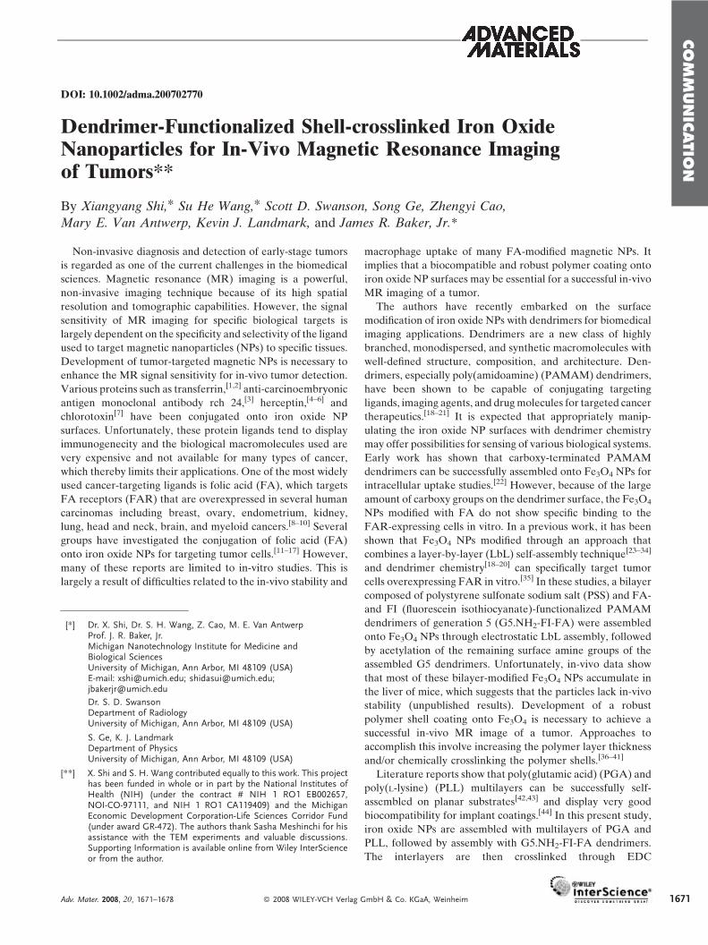
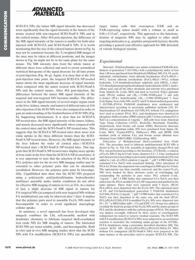
Figure 1. a) Schematic representation of the procedure for fabricating multifunctional shell-crosslinked iron oxide NPs. b) A TEM image of SCIO-FA NPs.c) A negatively phosphotungstic acid-stained TEM image of SCIO-FA NPs.
1672
(1-ethyl-3-[3-dimethylaminopropyl]carbodiimide hydrochlor-
ide) chemistry to covalently link the hydroxy groups of iron
oxide, the carboxy groups of PGA, and the amino groups of
PLL and the dendrimers. The remaining amino groups of the
dendrimers are finally acetylated to neutralize the surface
charge (Fig. 1a). The formed shell-crosslinked iron oxide
(SCIO) NPs are characterized using zeta-potential analysis,
Fourier-transform infrared (FT-IR) spectroscopy, transmis-
sion electron microscopy (TEM), and relaxivity measure-
ments. Combined flow cytometry and in-vitro and in-vivo MR
imaging studies show that the FA-modified SCIO NPs can
specifically bind to tumor cells and an early-stage tumor model
overexpressing FAR. To our knowledge, this is the first
example of the fabrication of multifunctional iron oxide NPs
using shell-crosslinked polymer multilayers combined with
dendrimer chemistry for in-vivo MR imaging studies. The
present approach using crosslinked polymer shells to functio-
nalize Fe3O4NPs with biological ligands opens a new avenue to
fabricating various NPs for a range of in-vivo biological sensing
and therapeutic applications.
LbL self-assembly, shell crosslinking, and chemical mod-
ification of the SCIO NPs were monitored by zeta-potential
www.advmat.de � 2008 WILEY-VCH Verlag GmbH
measurement (Table S1, Supporting Information). It is clear
that the positively charged Fe3O4 NPs become negatively
charged after PGA modification. After PLL assembly, the
Fe3O4 NPs again reverse to being positively charged. Further
PGA modification decreases the surface charge. The changes
in charge of the Fe3O4 NPs indicate the successful electrostatic
assembly of (PGA/PLL)2/PGA/G5.NH2-FI-FA multilayers.
The EDC crosslinking reaction does not result in a significant
change of the surface charge of the Fe3O4 NPs. However, after
the acetylation reaction, the zeta potentials of both (PGA/
PLL)2/PGA/G5.NHAc-FI and (PGA/PLL)2/PGA/G5.NHAc-
FI-FANPs are close to neutral because of the conversion of the
dendrimer surface amine groups to acetamide groups. This is in
sharp contrast to the findings in a previous work,[35] wherein
acetylation of dendrimer/PSS bilayer-modified Fe3O4 NPs did
not result in Fe3O4 NPs with close to a neutral charge. This is
presumably a result of the EDC crosslinking reaction, which
makes the dendrimer amines that interact with the PGA
polymer through electrostatic interaction covalently couple
with the PGA.
The fabricated SCIO NPs were characterized by TEM (Fig.
1b and 1c). The morphology of the FA-modified SCIO
& Co. KGaA, Weinheim Adv. Mater. 2008, 20, 1671–1678
COM
MUNIC
ATIO
N
(SCIO-FA) NPs does not show significant change after the
assembly and crosslinking of the polymers and dendrimers
when compared with the pristine Fe3O4 NPs (Fig. 1b).[22,35] A
negatively stained TEM image using phosphotungstic acid
(Fig. 1c) clearly shows that all Fe3O4 NPs are surrounded with
the bright rings of the polymer multilayers, which confirms the
successful self-assembly process. The non-targeted SCIO
(SCIO-NonFA) NPs display a morphology similar to that of
SCIO-FA NPs (images not shown). The fabricated SCIO NPs
are very stable both in aqueous solution and in a cell culture
medium for at least 6 months at Fe concentrations of up to
10mg mL�1. It is worth noting that five layers of the
polyelectrolyte (PGA and PLL) were selected to deposit onto
the Fe3O4 NPs in this work. This is because the assembly of five
layers of polyelectrolytes plus one layer of G5 dendrimers is
sufficient to maintain the shape of the intact hollow polymer
capsules once the Fe3O4 core particles are removed (see below
in Fig. 2). This is also consistent with previous reports related to
the formation of polyelectrolyte multilayer capsules.[25,45] It is
likely that fewer layers of the PGA/PLL assemblymay result in
a weak stability of the SCIONPs for in-vivo applications, while
the assembly of more PGA/PLL layers might introduce issues
related to the colloidal stability of the SCIONPs as discussed in
a previous report.[35]
The EDC chemical crosslinking reaction was confirmed by
FT-IR spectrometry (Supporting Information, Fig. S1). The
absorbance of the amide bond of (PGA/PLL)2/PGA/
G5.NH2-FI-FA-modified Fe3O4 NPs increased after EDC
crosslinking when compared with the same NPs before EDC
crosslinking. Although FT-IR spectroscopy is not a very
effective approach to characterize the intensity of the amide
bond before and after shell crosslinking (because the PGA and
PLL polymers, and the dendrimers that were used themselves,
also contain many amide bonds), the FT-IR spectra qualita-
tively verify the formation of amide bonds between the
carboxy groups of PGA and the amine groups of PLL and
dendrimers. To further confirm the improvement of the
mechanical stability after EDC shell crosslinking, the mor-
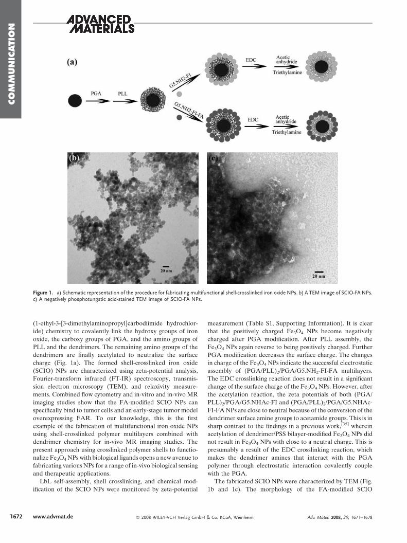
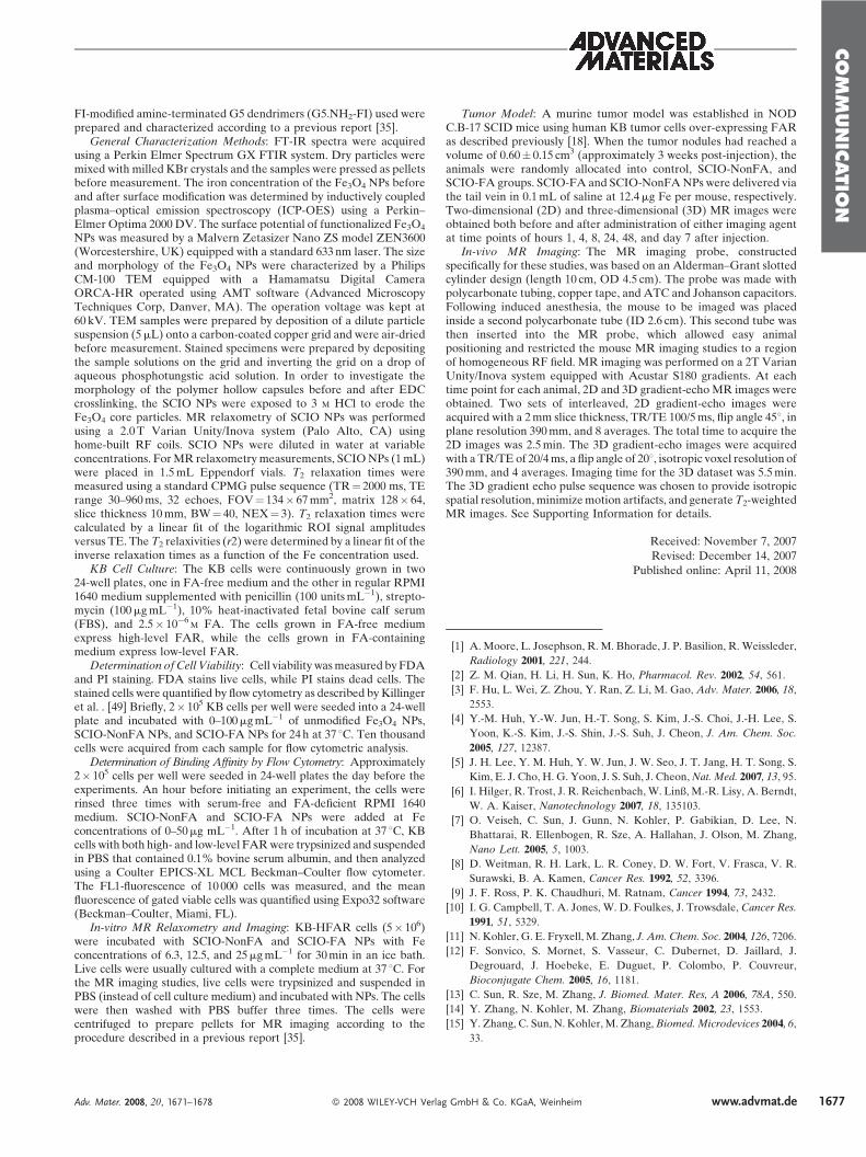
phology of the polymer shells after the removal of the iron
Figure 2. TEM images of (PGA/PLL)2/PGA/G5.NH2-FI-FA hollow polymer cintact hollow polymer capsules before EDC crosslinking. b) A magnified imagcapsule indicated by a horizontal arrow. d) (PGA/PLL)2/PGA/G5.NH2-FI-FA
Adv. Mater. 2008, 20, 1671–1678 � 2008 WILEY-VCH Verla
oxide core particles was investigated (Fig. 2). It is clear that
before EDC crosslinking, intact hollow polymer nanocapsules
can be formed (Fig. 2a–c). Some of the capsules have a
completely empty interior (Fig. 2b), while some of the capsules
still contain iron residue as a result of incomplete removal (Fig.
2c). In sharp contrast, after EDC crosslinking, most of the
capsules formed are broken (Fig. 2d). This is presumably
because of the huge rupture force induced instantaneously by
the swelling and dissolution of iron oxide cores, which further
confirms that the mechanical stability of the SCIO NPs is
significantly improved, while the permeability of the capsules
after EDC crosslinking is decreased. This provides further
evidence to show the successful EDC crosslinking reaction.
The transverse relaxation time (T2) of water protons in an
aqueous solution of fabricated SCIO NPs was measured at
2Tesla with a Carr–Purcell–Meiboom–Gill (CPMG) pulse
sequence and the measured data were used to compute the
transverse relaxivity (r2) (the transverse relaxation rate per mM
of iron). The r2 of uncoated Fe3O4 NPs, SCIO-FA NPs, and
SCIO-NonFANPs as a function of Fe concentration are shown
in the Supporting Information (Fig. S2). The uncoated Fe3O4
NPs show the highest r2 relaxivity (r2¼ 100.4 s�1mM�1),
whereas the r2 relaxivities of SCIO-FA and SCIO-NonFA
NPs are somewhat reduced at 46.3 and 78.8 s�1mM�1,
respectively. The polymer coating onto the Fe3O4 NPs shields
water molecules from their surfaces, and causes the lower r2relaxivity of SCIO NPs. Compared with SCIO-NonFA NPs,
the presence of the FA moieties of the SCIO-FA NPs may
significantly increase the hydrophobicity and hindrance of the
coating layers, thereby enhancing the shielding effect.[46] As a
consequence, SCIO-FA NPs exhibit a lower r2 relaxivity than
that of SCIO NPs without FA.
The cytotoxicity of the functionalized SCIO NPs was
evaluated by fluorescein diacetate (FDA) and propidium
iodide (PI) staining. Cell viability data (Supporting Informa-
tion, Fig. S3) show that the KB cells (a human epithelial
carcinoma cell line) treated by SCIO NPs with or without FA
conjugation display a similar percentage of FDA positive cells
to the KB cells treated by unmodified Fe3O4 NPs at an Fe
apsules after iron oxide core removal. a) (PGA/PLL)2/PGA/G5.NH2-FI-FAe of the capsule indicated by a vertical arrow. c) A magnified image of thehollow polymer capsules after EDC crosslinking.
g GmbH & Co. KGaA, Weinheim www.advmat.de 1673
COM
MUNIC
ATIO
N
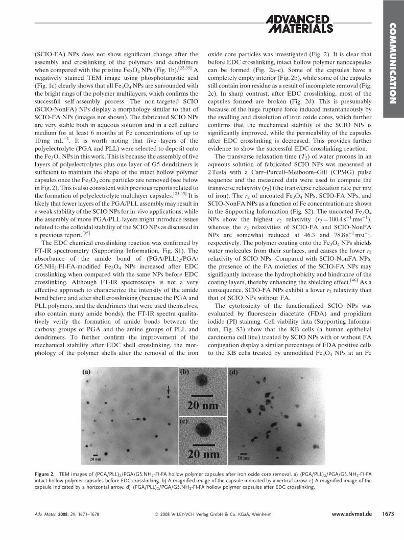
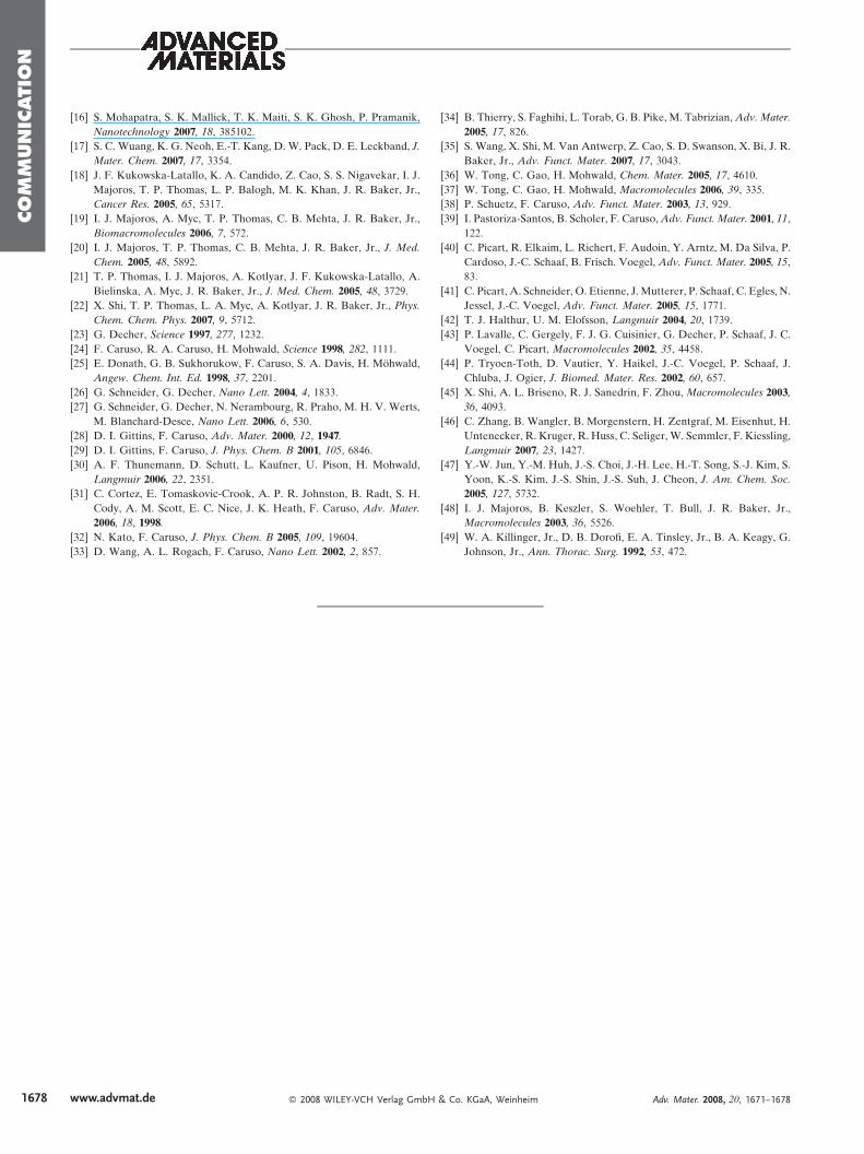
Figure 3. In-vitro flow cytometric analysis of binding of SCIO NPs with KB cells and MR images of cell pellets. Dose-dependent binding of SCIO-FA andSCIO-NonFA NPs with KB cells expressing high- (a) and low-level (b) FAR. T2 weighted spin-echo images of KB-HFAR cells incubated with functionalizedSCIO-FA and SCIO-NonFANPs with Fe concentrations of 0, 6.3, 12.5, 25.0mgmL�1 c). The color change from red to blue indicates the gradual decrease ofMR signal intensity.
Table 1. T2 of KB-HFAR cells treated with functionalized Fe3O4 NPs.
Fe concentration [mgmL�1] T2 [s]
SCIO-NonFA SCIO-FA
0 (PBS control) 1.16W 0.016 1.16W 0.016
6.3 0.387W 0.004 0.104W 0.005
12.5 0.105W 0.006 0.062W 0.0009
25.0 0.061W 0.0001 0.03W 0.0014
1674
concentration of 0–100mgmL�1. This indicates that the SCIO
NPs are biocompatible at an Fe concentration of up to
100mgmL�1.
The FA and FI dyes on the modified G5 dendrimer surfaces
were used as targeting ligands and imaging molecules,
respectively. The attached FI moieties afford a flow cytometric
study of the binding of SCIO NPs with target cells. KB cells
that express both high and low levels of FAR (denoted as
KB-HFAR and KB-LFAR, respectively) were selected for the
intracellular uptake of SCIO NPs. Figure 3a and 3b illustrate
the dose-dependent cellular uptake of the binding of SCIO-FA
and SCIO-NonFA NPs. At an Fe concentration above
2.5mgmL�1, KB-HFAR cells exposed to SCIO-FA NPs show
remarkably higher fluorescence signals than those treated with
SCIO-NonFA NPs without FA (Fig. 3a). With the increase of
Fe concentration, the mean fluorescence levels off. This
implies that the high affinity of FAR mediates specific uptake
of the NPs. Both SCIO-NonFA and SCIO-FA NPs display
much less uptake in KB-LFAR cells than in KB-HFAR cells,
even at an Fe concentration of up to 50mgmL�1 (Fig. 3b).
However, in the studied concentration range, SCIO-FA NPs
exhibit more uptake in KB-LFAR cells than SCIO-NonFA
NPs without FA modification, which is similar to what was
observed with these cells in a previous study.[35] This is a result
of the larger number of FA moieties per SCIO-FA NP
compared with a single FA-modified dendrimer, which
www.advmat.de � 2008 WILEY-VCH Verlag GmbH
facilitates polyvalent binding, thereby significantly increasing
the binding affinity of Fe3O4 NPs to each KB-LFAR cell.
Iron oxide NPs affect theMR signal by dephasing transverse
magnetization and hence reducing the value of T2. A targeted
iron oxide NP would have a major benefit in tumor imaging by
specifically detecting tumors that overexpress the FAR. To
study the effect of the SCIO-FA NPs on tumor cells, the T2 of
KB-HFAR cells exposed to differing Fe concentrations of
SCIO-FA NPs were measured. The T2 values of KB-HFAR
cell pellets treated with SCIO-FA NPs dramatically decreased
as a function of Fe concentration (Table 1). In contrast, the
decreasing trend of T2 values as a function of Fe concentration
for the same KB cells treated with SCIO-NonFA NPs is
significantly less than that of the KB cells treated with
SCIO-FA NPs. In the T2-weighted spin-echo MR images (the
color change from red to blue indicates the gradual decrease of
& Co. KGaA, Weinheim Adv. Mater. 2008, 20, 1671–1678
COM
MUNIC
ATIO
N
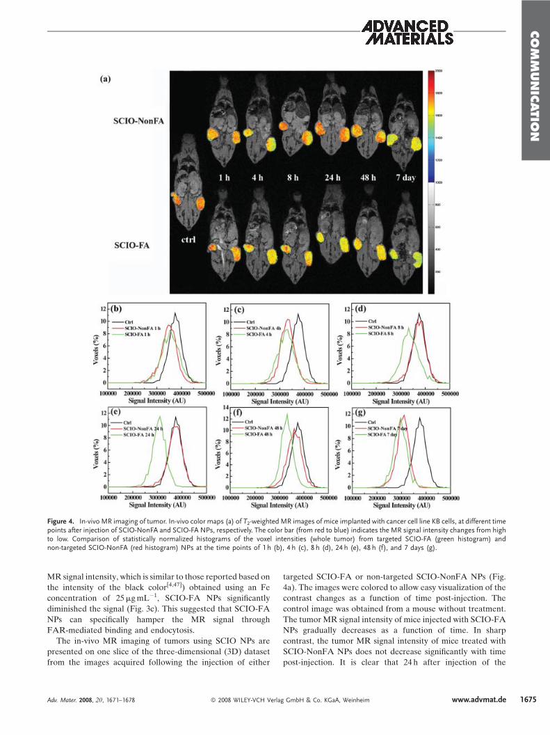
Figure 4. In-vivo MR imaging of tumor. In-vivo color maps (a) of T2-weighted MR images of mice implanted with cancer cell line KB cells, at different timepoints after injection of SCIO-NonFA and SCIO-FA NPs, respectively. The color bar (from red to blue) indicates the MR signal intensity changes from highto low. Comparison of statistically normalized histograms of the voxel intensities (whole tumor) from targeted SCIO-FA (green histogram) andnon-targeted SCIO-NonFA (red histogram) NPs at the time points of 1 h (b), 4 h (c), 8 h (d), 24 h (e), 48 h (f), and 7 days (g).
MR signal intensity, which is similar to those reported based on
the intensity of the black color[4,47]) obtained using an Fe
concentration of 25mgmL�1, SCIO-FA NPs significantly
diminished the signal (Fig. 3c). This suggested that SCIO-FA
NPs can specifically hamper the MR signal through
FAR-mediated binding and endocytosis.
The in-vivo MR imaging of tumors using SCIO NPs are
presented on one slice of the three-dimensional (3D) dataset
from the images acquired following the injection of either
Adv. Mater. 2008, 20, 1671–1678 � 2008 WILEY-VCH Verla
targeted SCIO-FA or non-targeted SCIO-NonFA NPs (Fig.
4a). The images were colored to allow easy visualization of the
contrast changes as a function of time post-injection. The
control image was obtained from a mouse without treatment.
The tumor MR signal intensity of mice injected with SCIO-FA
NPs gradually decreases as a function of time. In sharp
contrast, the tumor MR signal intensity of mice treated with
SCIO-NonFA NPs does not decrease significantly with time
post-injection. It is clear that 24 h after injection of the
g GmbH & Co. KGaA, Weinheim www.advmat.de 1675
COM
MUNIC
ATIO
N
1676
SCIO-FA NPs, the tumor MR signal intensity has decreased
more significantly than the signal intensity in the tumors of the
mouse treated with non-targeted SCIO-NonFA NPs and in
the control mouse. After 48 h post-injection, the difference of
the MR signal intensity of the tumors is smaller for both mice
injected with SCIO-FA and SCIO-NonFA NPs. It is worth
mentioning that the size of the colored tumors shown in Fig. 4a
may not be consistent because the T2-weighted MR images of
the mice may be taken at different positions, and the images
shown in Fig. 4a might not be in the same plane for the same
mouse. The MR intensity data from the whole tumor at
different slices were collected and used to create normalized
statistical histograms of the signal decrease for all time points
of post-injection (Fig. 4b–g). Again, it is clear that at the 24 h
post-injection time point, the targeted SCIO-FA NP-treated
tumor shows the most significant decrease of signal intensity
when compared with the tumor treated with SCIO-NonFA
NPs and the control mouse. After 48 h post-injection, the
differences between the tumor MR signal intensity of the
targeted and non-targeted NPs becomes smaller. The differ-
ences in the MR signal intensity of several major organs (such
as the liver, kidney, muscle, and tumor) of different mice at 24 h
after injection of the SCIONPs were also compared in order to
gain an understanding of the biodistribution of SCIO NPs (Fig.
S4, Supporting Information). It is clear that for SCIO-FA
NP-treated mice, the MR signal intensity of the tumor, kidney,
and muscle decreased more significantly when compared with
the control mice and the SCIO-NonFA NP-treated mice. This
suggests that the SCIO-FA NP-treated mice show more iron
oxide uptake in the three different tissues than the SCIO-
NonFA NP-treated mice. However, the MR signal intensity of
the liver follows the order of: control mice> SCIO-FA
NP-treated mice> SCIO-NonFA NP-treated mice. This sug-
gests that SCIO-NonFA NP-treated mice display more uptake
of iron oxide in the liver than the SCIO-FANP-treated mice. It
is very important to note that the selection of the PGA and
PLL polymer pair for the in-vivo MR imaging studies may be
extended to other polymer pairs that can be chemically
crosslinked. However, the polymer pairs must be biocompa-
tible. Unpublished data show that the SCIO NPs prepared
using a poly(acrylic acid)/poly(allylamine hydrochloride)
multilayer assembly under similar conditions do not allow
for effective MR imaging of tumors in vivo at 24 h. As a matter
of fact, a slight decrease of MR signal in tumors for
FA-targeted NPs (as compared with non-targeted NPs without
FA modification) can only be observed after 7 days. It implies
that the polymer pairs used to assemble Fe3O4 NPs must be
biocompatible in order to avoid significant macrophage
cellular uptake.
In summary, a novel approach has been developed that
uniquely combines the LbL self-assembly method with
dendrimer chemistry to fabricate targeted shell-crosslinked
iron oxide NPs for MR imaging of tumors. The fabricated
SCIO NPs are water-soluble, stable, and biocompatible. Both
in-vitro and in-vivo MR imaging studies show that the SCIO
NPs with FA modification (SCIO-FA NPs) can specifically
www.advmat.de � 2008 WILEY-VCH Verlag GmbH
target tumor cells that overexpress FAR and an
FAR-expressing tumor model with a volume as small as
0.60� 0.15 cm3, respectively. This approach to the functiona-
lization of magnetic NPs may be applied to other small
targetingmolecules (e.g., peptides and growth factors), thereby
providing a general cost-effective approach for MR detection
of various biological systems.
Experimental
Materials: Ethylenediamine core amine-terminated PAMAM den-drimers of generation 5 (G5.NH2) with a polydispersity index of lessthan 1.08 were purchased fromDendritech (Midland, MI). FA, FI, aceticanhydride, triethylamine, ferric chloride hexahydrate (FeCl3�6H2O>99%), ferrous chloride tetrahydrate (FeCl2�4H2O> 99%), sodiumhydroxide, 2-(N-morpholino)ethane sulphonic acid (MES), 1-ethyl-3-[3-dimethylaminopropyl]carbodiimide hydrochloride (EDC), hydro-chloric acid, and all the other chemicals and solvents were purchasedfrom Aldrich (St. Louis, MO) and used as received. Poly(L-glutamicacid) (PGA) sodium salt (Mw¼ 15 000–50 000 gmol�1) and poly(L-lysine) (PLL) hydrobromide (Mw¼ 15 000–30 000 gmol�1) werefrom Sigma. Iron oxide NPs, and FI- and FA-functionalized generation5 (G5.NH2-FI-FA) PAMAM dendrimers were synthesized andcharacterized according to previously published methods [35]. FI-functionalized G5 dendrimers (G5.NH2-FI) without FA conjugationwere used as control. PGA, PLL, and dendrimers were dissolved inphosphate-buffered saline (PBS) solution (pH 7.4) that contained 0.5 M
NaCl at a concentration of 1mgmL�1. KB cells were from AmericanType Tissue Collection (ATCC, Rockville, Maryland). Penicillin,streptomycin, fetal bovine calf serum (FBS), fluorescein diacetate(FDA), and propidium iodide (PI) were purchased from Sigma (St.Louis, MO). Trypsin-EDTA, Dulbecco’s PBS, and RPMI 1640medium (with or without FA), and bovine serum albumin wereobtained from GIBCO-BRL (Gaithersburg, MD).
Fabrication of Multifunctional Shell-crosslinked Iron OxideNPs: The procedure used to fabricate multifunctional SCIO NPs isshown in Fig. 1a. The LbL assembly of oppositely charged PGA andPLL was performed according to the literature. [27] Briefly, a solutionof Fe3O4 NPs (5mg in 0.5mLwater, diameter 8.4� 1.4 nm, synthesizedand characterized according to previously published methods [35]) wasadded to 1mL of a PGA solution (1mgmL�1, pH 7.4 PBS buffer thatcontained 0.5M NaCl) with occasional shaking. After adsorption ofPGA for 20min, the suspensionwas centrifuged at 8000 rpm for 10min.The supernatant was then carefully removed, and the coated Fe3O4
NPs were washed by three alternate cycles of centrifuging andresuspending the particles in pure water. PLL solution (1mL,1mgmL�1, pH 7.4 PBS buffer that contained 0.5 M NaCl) was thenadded into the PGA-modified Fe3O4 NP suspension and purified in thesame manner. These steps were repeated until 5 layers, (PGA/PLL)2PGA, were deposited onto the Fe3O4 NPs. The outermost layerof FI- and FA-functionalized generation 5 PAMAM dendrimers(G5.NH2-FI-FA) (1mgmL�1, pH 7.4 PBS buffer that contained 0.5 M
NaCl) was then deposited in the same way and the final (PGA/PLL)2PGA/G5.NH2-FI-FA-modified Fe3O4 NPs were dispersed into50� 10�3
M MES buffer (pH¼ 5.5) and EDC (12–18mg) was added tocrosslink the hydroxy groups of the Fe3O4 NPs and the amino groups ofPLL and the dendrimers with the carboxy groups of PGA. Themixturewas shaken overnight, followed by three cycles of centrifugation/redispersion (in water) to remove residual reactants. The SCIO NPswith FAmodification (SCIO-FANPs) were subjected to an acetylationreaction to neutralize the remaining amine groups of the G5.NH2-FI-FA dendrimers, using a procedure described elsewhere [48]. Thecontrol SCIO NPs (Fe3O4/(PGA/PLL)2/PGA/G5.NHAc-FI NPs)without FA conjugation (SCIO-NonFA NPs) were prepared in thesame manner as the procedure used to prepare SCIO-FA NPs. The
& Co. KGaA, Weinheim Adv. Mater. 2008, 20, 1671–1678
COM
MUNIC
ATIO
N
FI-modified amine-terminated G5 dendrimers (G5.NH2-FI) used wereprepared and characterized according to a previous report [35].
General Characterization Methods: FT-IR spectra were acquiredusing a Perkin Elmer Spectrum GX FTIR system. Dry particles weremixed with milled KBr crystals and the samples were pressed as pelletsbefore measurement. The iron concentration of the Fe3O4 NPs beforeand after surface modification was determined by inductively coupledplasma–optical emission spectroscopy (ICP-OES) using a Perkin–Elmer Optima 2000 DV. The surface potential of functionalized Fe3O4
NPs was measured by a Malvern Zetasizer Nano ZS model ZEN3600(Worcestershire, UK) equipped with a standard 633nm laser. The sizeand morphology of the Fe3O4 NPs were characterized by a PhilipsCM-100 TEM equipped with a Hamamatsu Digital CameraORCA-HR operated using AMT software (Advanced MicroscopyTechniques Corp, Danver, MA). The operation voltage was kept at60 kV. TEM samples were prepared by deposition of a dilute particlesuspension (5mL) onto a carbon-coated copper grid and were air-driedbefore measurement. Stained specimens were prepared by depositingthe sample solutions on the grid and inverting the grid on a drop ofaqueous phosphotungstic acid solution. In order to investigate themorphology of the polymer hollow capsules before and after EDCcrosslinking, the SCIO NPs were exposed to 3 M HCl to erode theFe3O4 core particles. MR relaxometry of SCIO NPs was performedusing a 2.0T Varian Unity/Inova system (Palo Alto, CA) usinghome-built RF coils. SCIO NPs were diluted in water at variableconcentrations. ForMR relaxometrymeasurements, SCIONPs (1mL)were placed in 1.5mL Eppendorf vials. T2 relaxation times weremeasured using a standard CPMG pulse sequence (TR¼ 2000 ms, TErange 30–960ms, 32 echoes, FOV¼ 134� 67mm2, matrix 128� 64,slice thickness 10mm, BW¼ 40, NEX¼ 3). T2 relaxation times werecalculated by a linear fit of the logarithmic ROI signal amplitudesversus TE. TheT2 relaxivities (r2) were determined by a linear fit of theinverse relaxation times as a function of the Fe concentration used.
KB Cell Culture: The KB cells were continuously grown in two24-well plates, one in FA-free medium and the other in regular RPMI1640 medium supplemented with penicillin (100 unitsmL�1), strepto-mycin (100mgmL�1), 10% heat-inactivated fetal bovine calf serum(FBS), and 2.5� 10�6
M FA. The cells grown in FA-free mediumexpress high-level FAR, while the cells grown in FA-containingmedium express low-level FAR.
Determination of Cell Viability: Cell viability wasmeasured byFDAand PI staining. FDA stains live cells, while PI stains dead cells. Thestained cells were quantified by flow cytometry as described byKillingeret al. . [49] Briefly, 2� 105 KB cells per well were seeded into a 24-wellplate and incubated with 0–100mgmL�1 of unmodified Fe3O4 NPs,SCIO-NonFA NPs, and SCIO-FA NPs for 24h at 37 8C. Ten thousandcells were acquired from each sample for flow cytometric analysis.
Determination of Binding Affinity by Flow Cytometry: Approximately2� 105 cells per well were seeded in 24-well plates the day before theexperiments. An hour before initiating an experiment, the cells wererinsed three times with serum-free and FA-deficient RPMI 1640medium. SCIO-NonFA and SCIO-FA NPs were added at Feconcentrations of 0–50mg mL�1. After 1 h of incubation at 37 8C, KBcells with both high- and low-level FARwere trypsinized and suspendedin PBS that contained 0.1% bovine serum albumin, and then analyzedusing a Coulter EPICS-XL MCL Beckman–Coulter flow cytometer.The FL1-fluorescence of 10 000 cells was measured, and the meanfluorescence of gated viable cells was quantified using Expo32 software(Beckman–Coulter, Miami, FL).
In-vitro MR Relaxometry and Imaging: KB-HFAR cells (5� 106)were incubated with SCIO-NonFA and SCIO-FA NPs with Feconcentrations of 6.3, 12.5, and 25mgmL�1 for 30min in an ice bath.Live cells were usually cultured with a complete medium at 37 8C. Forthe MR imaging studies, live cells were trypsinized and suspended inPBS (instead of cell culture medium) and incubated with NPs. The cellswere then washed with PBS buffer three times. The cells werecentrifuged to prepare pellets for MR imaging according to theprocedure described in a previous report [35].
Adv. Mater. 2008, 20, 1671–1678 � 2008 WILEY-VCH Verla
Tumor Model: A murine tumor model was established in NODC.B-17 SCID mice using human KB tumor cells over-expressing FARas described previously [18]. When the tumor nodules had reached avolume of 0.60� 0.15 cm3 (approximately 3 weeks post-injection), theanimals were randomly allocated into control, SCIO-NonFA, andSCIO-FA groups. SCIO-FA and SCIO-NonFANPs were delivered viathe tail vein in 0.1mL of saline at 12.4mg Fe per mouse, respectively.Two-dimensional (2D) and three-dimensional (3D) MR images wereobtained both before and after administration of either imaging agentat time points of hours 1, 4, 8, 24, 48, and day 7 after injection.
In-vivo MR Imaging: The MR imaging probe, constructedspecifically for these studies, was based on an Alderman–Grant slottedcylinder design (length 10 cm, OD 4.5 cm). The probe was made withpolycarbonate tubing, copper tape, and ATC and Johanson capacitors.Following induced anesthesia, the mouse to be imaged was placedinside a second polycarbonate tube (ID 2.6 cm). This second tube wasthen inserted into the MR probe, which allowed easy animalpositioning and restricted the mouse MR imaging studies to a regionof homogeneous RF field. MR imaging was performed on a 2T VarianUnity/Inova system equipped with Acustar S180 gradients. At eachtime point for each animal, 2D and 3D gradient-echo MR images wereobtained. Two sets of interleaved, 2D gradient-echo images wereacquired with a 2mm slice thickness, TR/TE 100/5ms, flip angle 458, inplane resolution 390mm, and 8 averages. The total time to acquire the2D images was 2.5min. The 3D gradient-echo images were acquiredwith a TR/TE of 20/4ms, a flip angle of 208, isotropic voxel resolution of390mm, and 4 averages. Imaging time for the 3D dataset was 5.5min.The 3D gradient echo pulse sequence was chosen to provide isotropicspatial resolution,minimizemotion artifacts, and generateT2-weightedMR images. See Supporting Information for details.
Received: November 7, 2007Revised: December 14, 2007
Published online: April 11, 2008
[1] A. Moore, L. Josephson, R. M. Bhorade, J. P. Basilion, R. Weissleder,
Radiology 2001, 221, 244.
[2] Z. M. Qian, H. Li, H. Sun, K. Ho, Pharmacol. Rev. 2002, 54, 561.
[3] F. Hu, L. Wei, Z. Zhou, Y. Ran, Z. Li, M. Gao, Adv. Mater. 2006, 18,
2553.
[4] Y.-M. Huh, Y.-W. Jun, H.-T. Song, S. Kim, J.-S. Choi, J.-H. Lee, S.
Yoon, K.-S. Kim, J.-S. Shin, J.-S. Suh, J. Cheon, J. Am. Chem. Soc.
2005, 127, 12387.
[5] J. H. Lee, Y. M. Huh, Y. W. Jun, J. W. Seo, J. T. Jang, H. T. Song, S.
Kim, E. J. Cho, H. G. Yoon, J. S. Suh, J. Cheon,Nat. Med. 2007, 13, 95.
[6] I. Hilger, R. Trost, J. R. Reichenbach, W. Linß, M.-R. Lisy, A. Berndt,
W. A. Kaiser, Nanotechnology 2007, 18, 135103.
[7] O. Veiseh, C. Sun, J. Gunn, N. Kohler, P. Gabikian, D. Lee, N.
Bhattarai, R. Ellenbogen, R. Sze, A. Hallahan, J. Olson, M. Zhang,
Nano Lett. 2005, 5, 1003.
[8] D. Weitman, R. H. Lark, L. R. Coney, D. W. Fort, V. Frasca, V. R.
Surawski, B. A. Kamen, Cancer Res. 1992, 52, 3396.
[9] J. F. Ross, P. K. Chaudhuri, M. Ratnam, Cancer 1994, 73, 2432.
[10] I. G. Campbell, T. A. Jones, W. D. Foulkes, J. Trowsdale, Cancer Res.
1991, 51, 5329.
[11] N. Kohler, G. E. Fryxell, M. Zhang, J. Am. Chem. Soc. 2004, 126, 7206.
[12] F. Sonvico, S. Mornet, S. Vasseur, C. Dubernet, D. Jaillard, J.
Degrouard, J. Hoebeke, E. Duguet, P. Colombo, P. Couvreur,
Bioconjugate Chem. 2005, 16, 1181.
[13] C. Sun, R. Sze, M. Zhang, J. Biomed. Mater. Res, A 2006, 78A, 550.
[14] Y. Zhang, N. Kohler, M. Zhang, Biomaterials 2002, 23, 1553.
[15] Y. Zhang, C. Sun, N. Kohler, M. Zhang,Biomed.Microdevices 2004, 6,
33.
g GmbH & Co. KGaA, Weinheim www.advmat.de 1677
COM
MUNIC
ATIO
N
1678
[16] S. Mohapatra, S. K. Mallick, T. K. Maiti, S. K. Ghosh, P. Pramanik,
Nanotechnology 2007, 18, 385102.
[17] S. C. Wuang, K. G. Neoh, E.-T. Kang, D. W. Pack, D. E. Leckband, J.
Mater. Chem. 2007, 17, 3354.
[18] J. F. Kukowska-Latallo, K. A. Candido, Z. Cao, S. S. Nigavekar, I. J.
Majoros, T. P. Thomas, L. P. Balogh, M. K. Khan, J. R. Baker, Jr.,
Cancer Res. 2005, 65, 5317.
[19] I. J. Majoros, A. Myc, T. P. Thomas, C. B. Mehta, J. R. Baker, Jr.,
Biomacromolecules 2006, 7, 572.
[20] I. J. Majoros, T. P. Thomas, C. B. Mehta, J. R. Baker, Jr., J. Med.
Chem. 2005, 48, 5892.
[21] T. P. Thomas, I. J. Majoros, A. Kotlyar, J. F. Kukowska-Latallo, A.
Bielinska, A. Myc, J. R. Baker, Jr., J. Med. Chem. 2005, 48, 3729.
[22] X. Shi, T. P. Thomas, L. A. Myc, A. Kotlyar, J. R. Baker, Jr., Phys.
Chem. Chem. Phys. 2007, 9, 5712.
[23] G. Decher, Science 1997, 277, 1232.
[24] F. Caruso, R. A. Caruso, H. Mohwald, Science 1998, 282, 1111.
[25] E. Donath, G. B. Sukhorukow, F. Caruso, S. A. Davis, H. Mohwald,
Angew. Chem. Int. Ed. 1998, 37, 2201.
[26] G. Schneider, G. Decher, Nano Lett. 2004, 4, 1833.
[27] G. Schneider, G. Decher, N. Nerambourg, R. Praho, M. H. V. Werts,
M. Blanchard-Desce, Nano Lett. 2006, 6, 530.
[28] D. I. Gittins, F. Caruso, Adv. Mater. 2000, 12, 1947.
[29] D. I. Gittins, F. Caruso, J. Phys. Chem. B 2001, 105, 6846.
[30] A. F. Thunemann, D. Schutt, L. Kaufner, U. Pison, H. Mohwald,
Langmuir 2006, 22, 2351.
[31] C. Cortez, E. Tomaskovic-Crook, A. P. R. Johnston, B. Radt, S. H.
Cody, A. M. Scott, E. C. Nice, J. K. Heath, F. Caruso, Adv. Mater.
2006, 18, 1998.
[32] N. Kato, F. Caruso, J. Phys. Chem. B 2005, 109, 19604.
[33] D. Wang, A. L. Rogach, F. Caruso, Nano Lett. 2002, 2, 857.
www.advmat.de � 2008 WILEY-VCH Verlag GmbH
[34] B. Thierry, S. Faghihi, L. Torab, G. B. Pike, M. Tabrizian,Adv. Mater.
2005, 17, 826.
[35] S. Wang, X. Shi, M. Van Antwerp, Z. Cao, S. D. Swanson, X. Bi, J. R.
Baker, Jr., Adv. Funct. Mater. 2007, 17, 3043.
[36] W. Tong, C. Gao, H. Mohwald, Chem. Mater. 2005, 17, 4610.
[37] W. Tong, C. Gao, H. Mohwald, Macromolecules 2006, 39, 335.
[38] P. Schuetz, F. Caruso, Adv. Funct. Mater. 2003, 13, 929.
[39] I. Pastoriza-Santos, B. Scholer, F. Caruso,Adv. Funct. Mater. 2001, 11,
122.
[40] C. Picart, R. Elkaim, L. Richert, F. Audoin, Y. Arntz, M. Da Silva, P.
Cardoso, J.-C. Schaaf, B. Frisch. Voegel, Adv. Funct. Mater. 2005, 15,
83.
[41] C. Picart, A. Schneider, O. Etienne, J. Mutterer, P. Schaaf, C. Egles, N.
Jessel, J.-C. Voegel, Adv. Funct. Mater. 2005, 15, 1771.
[42] T. J. Halthur, U. M. Elofsson, Langmuir 2004, 20, 1739.
[43] P. Lavalle, C. Gergely, F. J. G. Cuisinier, G. Decher, P. Schaaf, J. C.
Voegel, C. Picart, Macromolecules 2002, 35, 4458.
[44] P. Tryoen-Toth, D. Vautier, Y. Haikel, J.-C. Voegel, P. Schaaf, J.
Chluba, J. Ogier, J. Biomed. Mater. Res. 2002, 60, 657.
[45] X. Shi, A. L. Briseno, R. J. Sanedrin, F. Zhou, Macromolecules 2003,
36, 4093.
[46] C. Zhang, B. Wangler, B. Morgenstern, H. Zentgraf, M. Eisenhut, H.
Untenecker, R. Kruger, R. Huss, C. Seliger, W. Semmler, F. Kiessling,
Langmuir 2007, 23, 1427.
[47] Y.-W. Jun, Y.-M. Huh, J.-S. Choi, J.-H. Lee, H.-T. Song, S.-J. Kim, S.
Yoon, K.-S. Kim, J.-S. Shin, J.-S. Suh, J. Cheon, J. Am. Chem. Soc.
2005, 127, 5732.
[48] I. J. Majoros, B. Keszler, S. Woehler, T. Bull, J. R. Baker, Jr.,
Macromolecules 2003, 36, 5526.
[49] W. A. Killinger, Jr., D. B. Dorofi, E. A. Tinsley, Jr., B. A. Keagy, G.
Johnson, Jr., Ann. Thorac. Surg. 1992, 53, 472.
& Co. KGaA, Weinheim Adv. Mater. 2008, 20, 1671–1678
Related Documents


![Enhanced blood-brain-barrier penetrability and …potential usage for brain cancer therapy [28, 47]. All the studies demonstrated that the functionalized dendrimer-based nanoparticles](https://static.cupdf.com/doc/110x72/5fda1a5f03a67a71e2142194/enhanced-blood-brain-barrier-penetrability-and-potential-usage-for-brain-cancer.jpg)


