1999, 73(1):608. J. Virol. Aymeric de Parseval and John H. Elder Activity Unique Gene Product with Partial Rev (Tat) Protein and Characterization of a Immunodeficiency Virus Transactivating Demonstration that orf2 Encodes the Feline http://jvi.asm.org/content/73/1/608 Updated information and services can be found at: These include: REFERENCES http://jvi.asm.org/content/73/1/608#ref-list-1 at: This article cites 37 articles, 22 of which can be accessed free CONTENT ALERTS more» articles cite this article), Receive: RSS Feeds, eTOCs, free email alerts (when new http://journals.asm.org/site/misc/reprints.xhtml Information about commercial reprint orders: http://journals.asm.org/site/subscriptions/ To subscribe to to another ASM Journal go to: on January 29, 2014 by guest http://jvi.asm.org/ Downloaded from on January 29, 2014 by guest http://jvi.asm.org/ Downloaded from

Welcome message from author
This document is posted to help you gain knowledge. Please leave a comment to let me know what you think about it! Share it to your friends and learn new things together.
Transcript
1999, 73(1):608. J. Virol.
Aymeric de Parseval and John H. Elder ActivityUnique Gene Product with Partial Rev (Tat) Protein and Characterization of aImmunodeficiency Virus Transactivating Demonstration that orf2 Encodes the Feline
http://jvi.asm.org/content/73/1/608Updated information and services can be found at:
These include:
REFERENCEShttp://jvi.asm.org/content/73/1/608#ref-list-1at:
This article cites 37 articles, 22 of which can be accessed free
CONTENT ALERTS more»articles cite this article),
Receive: RSS Feeds, eTOCs, free email alerts (when new
http://journals.asm.org/site/misc/reprints.xhtmlInformation about commercial reprint orders: http://journals.asm.org/site/subscriptions/To subscribe to to another ASM Journal go to:
on January 29, 2014 by guesthttp://jvi.asm
.org/D
ownloaded from
on January 29, 2014 by guest
http://jvi.asm.org/
Dow
nloaded from
JOURNAL OF VIROLOGY,0022-538X/99/$04.0010
Jan. 1999, p. 608–617 Vol. 73, No. 1
Copyright © 1999, American Society for Microbiology. All Rights Reserved.
Demonstration that orf2 Encodes the Feline ImmunodeficiencyVirus Transactivating (Tat) Protein and Characterization of a
Unique Gene Product with Partial Rev ActivityAYMERIC DE PARSEVAL AND JOHN H. ELDER*
Department of Molecular Biology, The Scripps Research Institute, La Jolla, California 92037
Received 28 May 1998/Accepted 5 October 1998
The long PCR technique was used to amplify the three size classes of viral mRNAs produced in cells infectedby feline immunodeficiency virus (FIV). We identified in the env region a new splice acceptor site that generatedtwo transcripts, each coding for an 11-kDa protein, p11rev, whose function is unknown. The small-size class ofmRNAs included two bicistronic orf2/rev mRNAs and two rev-like mRNAs, consisting only of the second exonof rev and coding for a 15-kDa protein, p15rev. p15rev contained the minimal effector domain of Rev and wassufficient to mediate partial Rev activity. The bicistronic mRNAs encoded two distinct proteins, one of 23 kDacorresponding to Rev and a 9-kDa protein encoded by the orf2 gene. The orf2 gene product is a protein of 79amino acids with characteristics similar to those of the Tat (transactivator) proteins of the ungulate lentivi-ruses. Transient expression assays, using the FIV long terminal repeat (LTR) to drive transcription of thebacterial gene for chloramphenicol acetyltransferase demonstrated that the orf2 gene transactivates geneexpression an average of 14- to 20-fold above the basal level. Deletion mutants of the FIV LTR were generatedto locate sequences responsive to transactivation mediated by the orf2 gene. A 5* deletion mutant that removedthe AP1 site resulted in residual low-level transactivation by orf2. Further experiments using LTR mutantswith internal deletions identified three regions located between positions 2126 and 247 relative to the cap sitethat were important for orf2-directed transactivation. These regions include the AP1 site, a C/EBP tandemrepeat, and an ATF site.
Feline immunodeficiency virus (FIV) (25) is a lentivirus as-sociated with a slow progressive disease in the domestic catinvolving multiple organ systems (for a review, see reference3). FIV has a disease pattern similar to that of human immu-nodeficiency virus (HIV), but its genomic organization is lesscomplex and is more closely related to those of the ungulatelentiviruses visna virus and caprine arthritis encephalitis virus(CAEV) (6). Like with other lentiviruses, three size classes ofviral mRNAs are produced in FIV-infected cells: full-length(9.2-kb), intermediate (5.2- and 4.4-kb), and small multiplyspliced (1.7- and 1.4-kb) mRNAs which represent the gag/pol,vif/env, env, orf2/rev, and rev transcripts, respectively (27, 37).The multiply spliced mRNAs are produced shortly after infec-tion and encode Rev and the orf2 gene product, which areessential for the viral life cycle. Rev functions through a spe-cific cis-acting RNA target, the Rev-responsive element(RRE), and allows the nuclear export and cytoplasmic accu-mulation of unspliced (gag/pol) and singly spliced (vif/env andenv) viral mRNAs. FIV Rev is a 23-kDa protein whose 80N-terminal amino acids are encoded by the N terminus of envjoined in frame to 73 residues from orfH, located at the 39 endof the genome (27). Although FIV Rev does not share aleucine-rich effector domain found in HIV type 1 (HIV-1) Revand the Rev-like proteins of many other retroviruses, muta-tional analyses showed that a short region in the second exonof FIV Rev was functionally interchangeable with the HIV-1Rev effector domain (19). The effector domain of FIV Revspanned a region of 27 amino acids downstream from a stretchof basic residues in the second exon of Rev. A short open
reading frame (ORF), orf2, coincides by its size and location tothe tat genes of visna virus and CAEV (7, 10, 14). The orf2gene product has been shown to produce a three- to fivefoldtransactivation of the FIV long terminal repeat (LTR) (33, 35,38). Whether this low-level transactivation is significant re-mains to be defined. Nonetheless, the orf2 gene product is anessential component for the virus life cycle. orf2-defective FIVfailed to productively infect feline T cells and primary periph-eral blood lymphocytes (PBLs) (36). Furthermore, the FIV34TF10 infectious molecular clone, which contains a stopcodon in the orf2 gene, replicated poorly in T cells (26). Sub-stitution of the stop codon for a tryptophan codon allowed theefficient replication of the virus in T cells and primary PBLs(38), suggesting that an intact orf2 gene influences the host celltropism of FIV.
Here, we report the amplification in a single PCR of thethree different size classes of viral mRNAs produced in FIV-infected cells by using the long PCR technology. We haveidentified a new splice acceptor site located in the env regionthat generates two mRNAs producing a Rev-related protein,p11rev. In addition, we have characterized another Rev-relatedprotein, p15rev, consisting only of the second exon of Rev. Thelatter protein displayed some degree of Rev activity, althoughless than the wild-type protein. Finally, using improved trans-fection protocols, we demonstrate that the orf2 gene encodes apotent transactivator of the FIV LTR. Deletional analysis ofthe FIV LTR demonstrated that a region located betweenpositions 2126 and 247, relative to the cap site, contains AP1,a C/EBP tandem repeat, and ATF sites that are important forthe orf2 gene-mediated transactivation of the FIV LTR.
MATERIALS AND METHODS
Cells and virus. Primary PBLs were obtained by Ficoll-Paque gradient puri-fication from blood of a specific-pathogen-free cat. PBLs were maintained in
* Corresponding author. Mailing address: The Scripps ResearchInstitute, Department of Molecular Biology, 10550 N. Torrey PinesRd., La Jolla, CA 92037. Phone: (619) 784-8270. Fax: (619) 784-2750.E-mail: [email protected].
608
on January 29, 2014 by guesthttp://jvi.asm
.org/D
ownloaded from
RPMI 1640 medium supplemented with 15% heat-inactivated fetal bovine serum(Gemini Bioproducts, Calabasa, Calif.), 2 mM L-glutamine (Sigma, St. Louis,Mo.), 1 mM sodium pyruvate (Sigma), 10 mM HEPES buffer (Sigma), 13nonessential amino acids (Sigma), 13 b-mercaptoethanol (Gibco BRL, Gaith-ersburg, Md.), 7.5 mg of concanavalin A (Sigma) per ml, 100 U of humanrecombinant interleukin-2 (a gift of Hoffmann-La Roche) per ml, and 50 mg ofgentamicin (Gemini Bioproducts) per ml. Crandell feline kidney (CrFK) andHeLa cells, obtained from the American Type Culture Collection, were main-tained in Dulbecco’s modified Eagle’s medium supplemented with 10% fetalbovine serum. The San Diego isolate of FIV, PPR (26), was used for the analysisof the transcriptional pattern of FIV.
Synthetic oligonucleotide primers. LA4 (59-ACTTGAGAAGAGTGATTGAGGAAGTGAAGC-39; nucleotides 373 to 403 [sense]), LA7 (59-TAAGCAGCTGCTAGCGCTTTAACTATGAGTCATGTTCAGC-39; nucleotides 9237 to9198 [antisense]), and LA11 (59-CAAAATGGATTCATATGACATATCTTCCTC-39; nucleotides 8914 to 8885 [antisense]) were designed from the sequence ofthe PPR molecular clone (GenBank accession no. 36968 [26]).
RNA extraction and cDNA synthesis. Total RNA was extracted from PBLs,104-C1 cells, and MCH5-4 cells by using an RNeasy kit (Qiagen, Chatsworth,Calif.) as specified by the manufacturer. Briefly, total RNA (0.5 to 1 mg) washeated at 70°C for 5 min and cooled on ice. First-strand cDNA synthesis wascarried out in a total volume of 50 ml containing 50 mM Tris-HCl (pH 8.3), 75mM KCl, 3 mM MgCl2, 10 mM dithiothreitol, 50 mM deoxynucleoside triphos-phates, 10 U of RNase inhibitor (Promega, Madison, Wis.) 50 U of Moloneymurine leukemia virus reverse transcriptase (Stratagene, La Jolla, Calif.), and 0.3mg of oligo(dT)15 primer (Promega). The reaction mixture was then incubated at37°C for 2 h, and 1 to 5 ml was used for subsequent amplifications.
PCR amplification. One to 5 ml of the cDNA reaction mixture was amplifiedby long PCR (2). Amplification was carried out in a total volume of 100 mlcontaining 20 mM Tris-HCl (pH 8.55 at 25°C), 16 mM (NH4)2SO4, 150 mg ofbovine serum albumin per ml, 3.5 mM MgCl2, 250 mM each deoxynucleosidetriphosphate, 100 mM Tris base, and 700 ng of each primer. The PCR mixturewas overlaid with 75 ml of mineral oil (Promega); after 1 min of incubation at94°C, 5 U of Taq (Promega) and 1:64 U of Deep Vent (New England Biolabs,Beverly, Mass.) polymerases were added, and the mixture was incubated at 94and 68°C for 10 s and 7 min, respectively. The cycle was repeated 25 to 35 timesin a Perkin-Elmer Cetus thermocycler.
Cloning and sequencing of amplified cDNAs. The PCR-amplified cDNAs werecleaned up (Promega), gel purified, and cloned in the TA cloning vector (pCR3;Invitrogen, La Jolla, Calif.) under the control of the cytomegalovirus and T7promoters. Sequences were determined by the dideoxy-chain termination pro-cedure (30), using a Sequenase version 2.0 kit (United States Biochemical,Cleveland, Ohio).
In vitro transcription-translation and immunoprecipitations. The orf2/rev andrev-like cDNAs cloned in pCR3 were transcribed and translated in vitro by usingT7 polymerase and the coupled transcription-translation reticulocyte lysate sys-tem (Promega).
Construction of 5* deletion and internal deletion mutants. The 59 deletionmutants have been previously described (35). Internal deletion mutations of theFIV LTR were generated by PCR-ligation-PCR (1). Briefly, two fragments of theFIV LTR, one corresponding to the 59 end of U3 to the nucleotide immediatelyupstream of the site to be deleted and the second corresponding to the nucleo-tide immediately downstream of the site to be deleted to the 39 end of U5, wereamplified by using Deep-Vent DNA polymerase (New England Biolabs). ThePCR products were gel purified, phosphorylated, and then ligated. The fusiongene was next amplified by using a primer pair specific for the 59 end of U3 andthe 39 end of U5. The PCR product was then inserted in pFIVLTR-CAT deletedof the wild-type LTR insert. We generated five single internal deletion mutantscorresponding to the first AP4 site, the AP1 site, the C/EBP tandem repeat, theNF1 site, and the ATF site.
Transfections and CAT assays. DNAs were prepared by using Qiagen midi-and maxiprep kits or by the Merlin service offered by Bio101 (Vista, Calif.). TheLTR-chloramphenicol acetyltransferase (CAT) and RRE-CAT constructs usedin this study have been previously reported (27, 35). Constructs (1 mg of target,5 or 10 mg of effector plasmids) were transfected in triplicate in CrFK cells byusing a calcium phosphate precipitation procedure as previously described (38).For HeLa cells, 1 and 10 mg of LTR-CAT construct were used. Followingtransfection, cells were incubated for 40 h and lysed, and CAT activity wasassayed on 20 mg of cell extract by phase extraction according to the protocol ofSeed and Sheen (31). In our previous study (38), CAT activity was assayed on cellextracts that were normalized by a b-galactosidase assay. However, the orf2 genehas been shown to transactivate the Rous sarcoma virus promoter of our b-ga-lactosidase construct (pRSV-bGal), which causes an error in normalizing cellextracts in the presence of orf2 cotransfection (39). Therefore, in the presentstudy, CAT activity was assayed by using 20 mg of protein per cell extract.
RESULTS
Long PCR amplification of the different size classes of FIVtranscripts. Long PCR has been recently described and shownto be an efficient method to amplify long DNA targets with
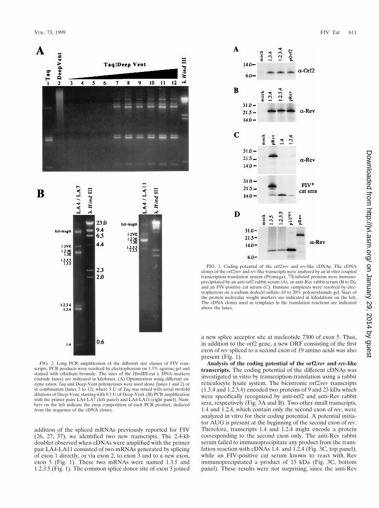
high fidelity (2). Long PCR combines modifications of stan-dard PCR buffers and thermal cycling profiles with a combi-nation of two polymerases, providing both processivity and39-59 proofreading exonuclease activity. Here, we used longPCR to investigate the gene expression of FIV. PBLs from aspecific-pathogen-free cat were infected with FIV-PPR (26).At 15 days postinfection, cells were harvested and total RNAwas extracted. One microgram of total RNA was reverse tran-scribed by using an RNase H-minus reverse transcriptase andoligo(dT)15 primer; 1 ml of the cDNA mixture was subse-quently amplified with primers LA4 and LA7, located in exons1 and 4 respectively, which are present in all viral transcripts(26, 37) (Fig. 1). We used the PCR buffer conditions describedby Barnes (2) but a different combination of polymerases: theTaq DNA polymerase from Thermus aquaticus as a processivepolymerase, and the Deep-Vent DNA polymerase from Ther-moccocus litoralis as a proofreading polymerase. Long PCRamplification was carried out with Taq polymerase alone and incombination with various amounts of Deep-Vent polymerase.The use of Taq alone resulted in the amplification of fourbands: 3 of 1.6, 1.5, and 0.5 kb, previously reported to corre-spond to exons 1.2.3.4, exons 1.3.4, and exons 1.4 (27, 37); andone band of about 3.5 kb (Fig. 2A, lane 1). No detectableamplicon was observed when Deep-Vent alone was used (Fig.2A, lane 2). Only the 1.6-, 1.5-, and 0.5-kb products wereobserved when amplification with Taq or Deep-Vent was car-ried out under classic PCR buffer conditions (data not shown).Since long PCR amplification required a very low level ofpolymerase with 39 exonuclease activity (2), we optimized thePCR conditions by combining 5 U of Taq with decreasingamounts of Deep-Vent, from 1/2 to 1/1,024 U. Long PCR using1/2 to 1/16 U of Deep-Vent failed to amplify any detectableproduct (Fig. 2A, lanes 3 to 6). However, long PCR carried outwith enzyme combinations using 1/32 to 1/1,024 U of Deep-Vent resulted in the amplication of a complex pattern of bands(Fig. 2A, lanes 7 to 12). These results are consistent with thosepreviously reported (2). Three other major products of 9.4, 4.0,and 3.5 kb were observed under these conditions (Fig. 2A,lanes 7 to 12; Fig. 2B, left panel). Long PCR amplification withcDNAs from two FIV-infected T-cell lines and FIV-infectedCrFK cells resulted in the same pattern of amplified products(data not shown). To verify that these three new amplifiedproducts represented the unspliced full-length mRNA and thesingly spliced env mRNAs, we performed a long PCR withprimers LA4 and LA11 (Fig. 1). LA11 is located 59 of the spliceacceptor at nucleotide 8944 of exon 4 (Fig. 1). This spliceacceptor is used by all of the multiply spliced mRNAs to joinexons 1, 2, and 3 to exon 4 (27, 37). Therefore, only unsplicedor partially spliced mRNAs could be amplified with this primerset. Indeed, we observed three major products correspondingto the full-length mRNA and singly spliced mRNAs; a minorproduct, a doublet at 2.4 kb, was also amplified by this primerset (Fig. 2B, right panel).
To exclude artifacts due to DNA contamination or to the useof a long annealing or extension time, the same PCRs with theprimer sets LA4-LA7 and LA4-LA11 were performed onDNase-treated total RNA and genomic DNA extracted fromFIV-infected cells. No detectable amplicon was observed afteramplification on total RNA, and a single 9-kb band corre-sponding to the full-length proviral DNA was amplified whengenomic DNA was used as a template (data not shown).
To verify that the amplified products represented FIVspliced transcripts, the PCR-amplified cDNAs were gel puri-fied, cloned in the TA vector, and sequenced as described inMaterials and Methods. A schematic representation of thedifferent FIV exons and transcripts is depicted in Fig. 1. In
VOL. 73, 1999 FIV Tat 609
on January 29, 2014 by guesthttp://jvi.asm
.org/D
ownloaded from
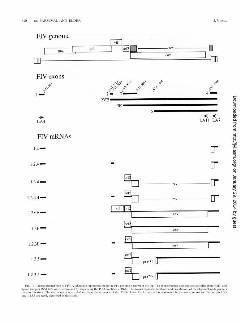
FIG. 1. Transcriptional map of FIV. A schematic representation of the FIV genome is shown at the top. The exon structure and locations of splice donor (SD) andsplice acceptor (SA) sites were determined by sequencing the PCR-amplified cDNAs. The arrows represent locations and orientations of the oligonucleotide primersused in this study. The viral transcripts are deduced from the sequence of the cDNA clones. Each transcript is designated by its exon composition. Transcripts 1.3.5and 1.2.3.5 are newly described in this study.
610 DE PARSEVAL AND ELDER J. VIROL.
on January 29, 2014 by guesthttp://jvi.asm
.org/D
ownloaded from
addition of the spliced mRNAs previously reported for FIV(26, 27, 37), we identified two new transcripts. The 2.4-kbdoublet observed when cDNAs were amplified with the primerpair LA4-LA11 consisted of two mRNAs generated by splicingof exon 1 directly, or via exon 2, to exon 3 and to a new exon,exon 5 (Fig. 1). These two mRNAs were named 1.3.5 and1.2.3.5 (Fig. 1). The common splice donor site of exon 3 joined
a new splice acceptor site at nucleotide 7300 of exon 5. Thus,in addition to the orf2 gene, a new ORF consisting of the firstexon of rev spliced to a second exon of 19 amino acids was alsopresent (Fig. 1).
Analysis of the coding potential of the orf2/rev and rev-liketranscripts. The coding potential of the different cDNAs wasinvestigated in vitro by transcription-translation using a rabbitreticulocyte lysate system. The bicistronic orf2/rev transcripts(1.3.4 and 1.2.3.4) encoded two proteins of 9 and 23 kDa whichwere specifically recognized by anti-orf2 and anti-Rev rabbitsera, respectively (Fig. 3A and B). Two other small transcripts,1.4 and 1.2.4, which contain only the second exon of rev, wereanalyzed in vitro for their coding potential. A potential initia-tor AUG is present at the beginning of the second exon of rev.Therefore, transcripts 1.4 and 1.2.4 might encode a proteincorresponding to the second exon only. The anti-Rev rabbitserum failed to immunoprecipitate any product from the trans-lation reaction with cDNAs 1.4. and 1.2.4 (Fig. 3C, top panel),while an FIV-positive cat serum known to react with Revimmunoprecipitated a product of 15 kDa (Fig. 3C, bottompanel). These results were not surprising, since the anti-Rev
FIG. 2. Long PCR amplification of the different size classes of FIV tran-scripts. PCR products were resolved by electrophoresis on 1.3% agarose gel andstained with ethidium bromide. The sizes of the HindIII-cut l DNA markers(outside lanes) are indicated in kilobases. (A) Optimization using different en-zyme ratios. Taq and Deep-Vent polymerases were used alone (lanes 1 and 2) orin combination (lanes 3 to 12), where 5 U of Taq was mixed with serial twofolddilutions of Deep-Vent, starting with 0.5 U of Deep-Vent. (B) PCR amplificationwith the primer pairs LA4-LA7 (left panel) and LA4-LA11 (right panel). Num-bers on the left indicate the exon composition of each PCR product, deducedfrom the sequence of the cDNA clones.
FIG. 3. Coding potential of the orf2/rev and rev-like cDNAs. The cDNAclones of the orf2/rev and rev-like transcripts were analyzed by an in vitro coupledtranscription-translation system (Promega). 35S-labeled proteins were immuno-precipitated by an anti-orf2 rabbit serum (A), an anti-Rev rabbit serum (B to D),and an FIV-positive cat serum (C). Immune complexes were resolved by elec-trophoresis on a sodium dodecyl sulfate–10 to 20% polyacrylamide gel. Sizes ofthe protein molecular weight markers are indicated in kilodaltons on the left.The cDNA clones used as templates in the translation reactions are indicatedabove the lanes.
VOL. 73, 1999 FIV Tat 611
on January 29, 2014 by guesthttp://jvi.asm
.org/D
ownloaded from
peptide serum was raised against an oligopeptide from the firstexon of Rev (27). The 15-kDa product, which we refer to hereas p15rev, was also immunoprecipitated in the pRev translationreaction (Fig. 3C, bottom panel), suggesting that a degree ofinternal initiation occurred from the AUG located in the sec-ond exon of rev. The coding potential of transcripts 1.3.5 and1.2.3.5 was also investigated for the ability to direct the syn-thesis of the new ORF consisting of the first exon of rev joinedin frame to 19 amino acid residues encoded by a 39 ORFlocated in the env region. In vitro translation reactions per-formed with cDNAs 1.3.5 and 1.2.3.5 were immunoprecipi-tated with the anti-Rev peptide serum, and an 11-kDa product,termed p11rev, was detected (Fig. 3D). A product of similar sizewas also immunoprecipitated when the translation reactionwas carried out with a cDNA clone encoding only the p11rev
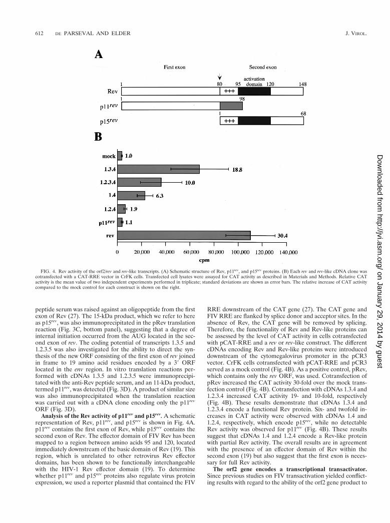
ORF (Fig. 3D).Analysis of the Rev activity of p11rev and p15rev. A schematic
representation of Rev, p11rev, and p15rev is shown in Fig. 4A.p11rev contains the first exon of Rev, while p15rev contains thesecond exon of Rev. The effector domain of FIV Rev has beenmapped to a region between amino acids 95 and 120, locatedimmediately downstream of the basic domain of Rev (19). Thisregion, which is unrelated to other retrovirus Rev effectordomains, has been shown to be functionally interchangeablewith the HIV-1 Rev effector domain (19). To determinewhether p11rev and p15rev proteins also regulate virus proteinexpression, we used a reporter plasmid that contained the FIV
RRE downstream of the CAT gene (27). The CAT gene andFIV RRE are flanked by splice donor and acceptor sites. In theabsence of Rev, the CAT gene will be removed by splicing.Therefore, the functionality of Rev and Rev-like proteins canbe assessed by the level of CAT activity in cells cotransfectedwith pCAT-RRE and a rev or rev-like construct. The differentcDNAs encoding Rev and Rev-like proteins were introduceddownstream of the cytomegalovirus promoter in the pCR3vector. CrFK cells cotransfected with pCAT-RRE and pCR3served as a mock control (Fig. 4B). As a positive control, pRev,which contains only the rev ORF, was used. Cotransfection ofpRev increased the CAT activity 30-fold over the mock trans-fection control (Fig. 4B). Cotransfection with cDNAs 1.3.4 and1.2.3.4 increased CAT activity 19- and 10-fold, respectively(Fig. 4B). These results demonstrate that cDNAs 1.3.4 and1.2.3.4 encode a functional Rev protein. Six- and twofold in-creases in CAT activity were observed with cDNAs 1.4 and1.2.4, respectively, which encode p15rev, while no detectableRev activity was observed for p11rev (Fig. 4B). These resultssuggest that cDNAs 1.4 and 1.2.4 encode a Rev-like proteinwith partial Rev activity. The overall results are in agreementwith the presence of an effector domain of Rev within thesecond exon (19) but also suggest that the first exon is neces-sary for full Rev activity.
The orf2 gene encodes a transcriptional transactivator.Since previous studies on FIV transactivation yielded conflict-ing results with regard to the ability of the orf2 gene product to
FIG. 4. Rev activity of the orf2/rev and rev-like transcripts. (A) Schematic structure of Rev, p11rev, and p15rev proteins. (B) Each rev and rev-like cDNA clone wascotransfected with a CAT-RRE vector in CrFK cells. Transfected cell lysates were assayed for CAT activity as described in Materials and Methods. Relative CATactivity is the mean value of two independent experiments performed in triplicate; standard deviations are shown as error bars. The relative increase of CAT activitycompared to the mock control for each construct is shown on the right.
612 DE PARSEVAL AND ELDER J. VIROL.
on January 29, 2014 by guesthttp://jvi.asm
.org/D
ownloaded from
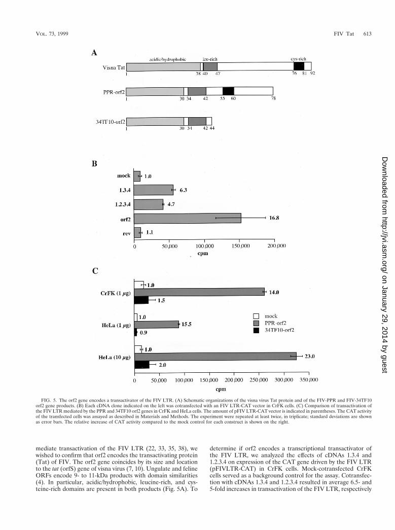
mediate transactivation of the FIV LTR (22, 33, 35, 38), wewished to confirm that orf2 encodes the transactivating protein(Tat) of FIV. The orf2 gene coincides by its size and locationto the tat (orfS) gene of visna virus (7, 10). Ungulate and felineORFs encode 9- to 11-kDa products with domain similarities(4). In particular, acidic/hydrophobic, leucine-rich, and cys-teine-rich domains are present in both products (Fig. 5A). To
determine if orf2 encodes a transcriptional transactivator ofthe FIV LTR, we analyzed the effects of cDNAs 1.3.4 and1.2.3.4 on expression of the CAT gene driven by the FIV LTR(pFIVLTR-CAT) in CrFK cells. Mock-cotransfected CrFKcells served as a background control for the assay. Cotransfec-tion with cDNAs 1.3.4 and 1.2.3.4 resulted in average 6.5- and5-fold increases in transactivation of the FIV LTR, respectively
FIG. 5. The orf2 gene encodes a transactivator of the FIV LTR. (A) Schematic organizations of the visna virus Tat protein and of the FIV-PPR and FIV-34TF10orf2 gene products. (B) Each cDNA clone indicated on the left was cotransfected with an FIV LTR-CAT vector in CrFK cells. (C) Comparison of transactivation ofthe FIV LTR mediated by the PPR and 34TF10 orf2 genes in CrFK and HeLa cells. The amount of pFIV LTR-CAT vector is indicated in parentheses. The CAT activityof the transfected cells was assayed as described in Materials and Methods. The experiment were repeated at least twice, in triplicate; standard deviations are shownas error bars. The relative increase of CAT activity compared to the mock control for each construct is shown on the right.
VOL. 73, 1999 FIV Tat 613
on January 29, 2014 by guesthttp://jvi.asm
.org/D
ownloaded from
(Fig. 5B). Furthermore, transactivation of the FIV LTR in thepresence of a vector expressing the orf2 gene of FIV-PPR was17-fold above the basal level, while in the presence of a Rev-expressing vector, no increase in CAT activity was observed(Fig. 5B). These results demonstrate that the orf2 gene en-codes a trans-acting factor that significantly increases geneexpression directed by the FIV LTR.
We next investigated the ability of the 34TF10 orf2 gene totransactivate the FIV LTR. FIV-PPR contains an intact orf2,while FIV-34TF10 contains a truncated orf2 due to a termina-tion codon at amino acid 44 (26, 34) (Fig. 5A). Cotransfectionof PPR orf2 and 34TF10 orf2 resulted in average 14- and1.5-fold increases in transactivation of the FIV LTR over themock cotransfected control, respectively (Fig. 5C). The sameexperiment was also performed with HeLa cells. Since thebasal activity of the FIV LTR is very low in HeLa cells com-pared to CrFK cells, the cotransfections were performed witheither 1 or 10 mg of pFIVLTR-CAT. As shown in Fig. 5C,relative degrees of transactivation in HeLa cells were similar tothose seen in CrFK cells (Fig. 5C). These results demonstratethat (i) the orf2 gene encodes the FIV Tat protein and (ii) anintact orf2 gene is necessary for transactivation of the FIVLTR.
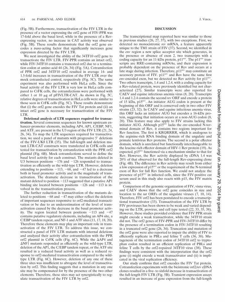
Deletional analysis of LTR sequences required for transac-tivation. Several consensus sequences for known upstream en-hancer-promoter elements, including AP4, AP1, C/EBP, NF1,and ATF, are present in the U3 region of the FIV LTR (21, 24,26, 34). To map the LTR sequences required for transactiva-tion, we used a panel of mutants with progressive 59 deletionsof the FIV-UK8 LTR (35). The full-length and deletion mu-tant LTR-CAT constructs were transfected in CrFK cells andtested for transactivation by cotransfection with the PPR orf2plasmid (Fig. 6B). Mock cotransfections served to define thebasal level activity for each construct. The mutants deleted inU3 between positions 2176 and 2126 responded to transac-tivation as efficiently as the wild-type LTR. However, deletionextending to positions 2113, 268, and 247 led to a decreaseboth in basal promoter activity and in the magnitude of trans-activation. The dramatic decrease in transactivation of themutant deleted to position 2113 suggested that a putative AP1binding site located between positions 2126 and 2113 is in-volved in the transactivation process.
The further reduction in transactivation of the mutants de-leted to positions 268 and 247 may either reflect the deletionof important sequences responsive to orf2-mediated transacti-vation or be due to an underestimation of the level of trans-activation caused by the decrease in the basal promoter activ-ity. The region located between positions 2113 and 247contains putative regulatory elements, including an AP4 site, aC/EBP tandem repeat, and NF-1 and ATF sites (11, 17, 18, 20).Therefore these elements may play an important role in trans-activation of the FIV LTR. To address this issue, we con-structed a panel of FIV LTR mutants with internal deletionsand analyzed their activity after cotransfection with the PPRorf2 plasmid in CrFK cells (Fig. 6C). While the DAP4 andDNF1 mutants responded as efficiently as the wild-type LTR,deletion of the AP1, the C/EBP tandem repeat, or the ATF siteresulted in a reduced basal activity as well as a reduced re-sponse to orf2-mediated transactivation compared to the wild-type LTR (Fig. 6C). However, deletion of any one of thesethree sites was insufficient to abrogate the level of transactiva-tion by orf2. This finding suggests that a mutation in any onesite may be compensated for by the presence of the two otherelements. Therefore, these sites may act synergistically to reg-ulate transactivation of the FIV LTR by orf2.
DISCUSSION
The transcriptional map reported here was similar to thosein previous studies (26, 27, 37), with two exceptions. First, wedetected no monocistronic rev mRNA, a feature apparentlyunique to the TM1 strain of FIV (37). Second, we identified inthe env region a new splice acceptor site which generates, inthe presence or absence of exon 2, two transcripts with acoding capacity for an 11-kDa protein, p11rev. The p11rev tran-scripts are RRE-containing mRNAs, and their expression isprobably dependent on the presence of Rev and occurs at alate stage during infection. Therefore, p11rev may constitute anaccessory protein of FIV. p11rev and Rev have the same firstenv-encoded exon, but we detected no Rev activity for p11rev.Two others transcripts, 1.4 and 1.2.4, with a coding capacity fora Rev-related protein, were previously identified but not char-acterized (27). Similar transcripts were also reported forCAEV and equine infectious anemia virus (8, 28). Transcripts1.4 and 1.2.4 contain the second rev ORF and encode a proteinof 15 kDa, p15rev. An initiator AUG codon is present at thebeginning of this ORF and is conserved only in two other FIVstrains (23, 32). In CAEV and equine infectious anemia virus,this ORF lacks an initiator AUG but encodes a 16-kDa pro-tein, suggesting that initiation occurs at a non-AUG codon (8,28). This feature may also apply to FIV strains lacking thisinitiator AUG. Although p15rev lacks the env-encoded N-ter-minal domain of Rev, it contains two regions important forRev function. The first is KRQRRRR, which is analogous tothe arginine-rich RNA binding domain of the ungulate andprimate lentivirus Rev proteins. The second is a polar effectordomain, which is unrelated but functionally interchangeable tothe leucine-rich effector domain of HIV-1 Rev protein (19). Asexpected, p15rev functioned via a mechanism similar to that forRev. However, the Rev activity displayed by cDNA 1.4 was20% of that observed for the full-length Rev-expressing clone(Fig. 4B). The difference in Rev activity may result from eithera low expression of p15rev or the requirement of the N-terminalexon of Rev for full Rev function. We could not analyze thepresence of p15rev in infected cells, since the FIV-positive catserum used in this study also reacts with p15, the FIV matrixprotein.
Comparison of the genomic organization of FIV, visna virus,and CAEV shows that the orf2 gene coincides in size andlocation to the tat ORFs of the ungulate viruses (7, 10, 14).Therefore, FIV orf2 has been postulated to encode a transcrip-tional transactivator (33). Transactivation of the FIV LTR byFIV proviruses has been shown to be weak and varied depend-ing on the LTR, provirus, and cell type tested (22, 33, 35, 38).However, these studies provided evidence that FIV PPR strainmight encode a weak transactivator, while the 34TF10 straindid not. The orf2 genes of FIV-PPR and FIV-34TF10 differ bythe presence of a termination codon in FIV-34TF10 resultingin a truncated orf2 gene (26, 34). Truncation and mutation ofthe orf2 gene were also reported to impair the ability of FIV toefficiently replicate in PBLs and feline T cells (36, 38). Mu-tagenesis of the termination codon of 34TF10 orf2 to trypto-phan codon resulted in an efficient replication of PBLs andfeline T cells by the orf2-repaired 34TF10 virus (38). Thesefindings were consistent with the interpretation that the orf2gene (i) might encode a weak transactivator and (ii) is impli-cated in the viral replication efficiency.
Our study confirms that orf2 encodes the FIV Tat protein.Cotransfection experiments with the bicistronic orf2/rev cDNAclones resulted in a five- to sixfold increase in transactivation ofthe full-length FIV LTR (Fig. 5B). Transient expression assaysresulted in an increase of gene expression from the full-length
614 DE PARSEVAL AND ELDER J. VIROL.
on January 29, 2014 by guesthttp://jvi.asm
.org/D
ownloaded from
FIG. 6. Deletion of the LTR sequences required for transactivation. (A) Deletion mutants of the FIV LTR promoter. The putative cis-acting regulatory elementscontained in the U3 region of the LTR are indicated at the top. The panel of 59 deletion mutants was previously described (35) and kindly provided by J. F. Thompsonand colleagues. wt, wild type. (B and C) Transactivation of 59 deletion mutants (B) and internal deletion mutants (C) of the FIV LTR by PPR orf2. Each deletion mutantwas cotransfected in CrFK cells with the PPR orf2 vector (gray bars). Cotransfection of each mutant with pCR3 defined baseline activity (blank bars). CAT activitywas assayed as described in Materials and Methods. The experiment was repeated twice, in triplicate; standard deviations are shown as error bars. The percentage ofwild-type LTR activity of each deletion mutant in response to transactivation by orf2, as well as the relative increase of CAT activity compared to the basal level foreach deletion mutant, is shown on the right.
VOL. 73, 1999 FIV Tat 615
on January 29, 2014 by guesthttp://jvi.asm
.org/D
ownloaded from
FIV LTR-CAT construct cotransfected with PPR orf2 in CrFKand HeLa cells. Average 14- to 17-fold increases in LTR ac-tivity were observed in CrFK cells cotransfected with the PPRorf2 gene, while no significant level of transactivation wasdetected with the truncated orf2 gene of FIV-34TF10 (Fig.5C). orf2 has been previously reported to transactivate the FIVLTR three- to fivefold above the basal level in CrFK cells. Asimilar degree of transactivation has also been shown in HeLacells, using 10 mg of pFIVLTR-CAT (38). In the present study,transactivation assays performed with HeLa cells and 1 and 10mg of pFIVLTR-CAT resulted in average 15- and 23-fold in-creases in LTR activity, respectively (Fig. 5C). We attribute thedifferences in transactivation observed between the data pre-sented here and those previously reported as reflecting betterDNA transfection efficiency rather than simply lower basalLTR activity.
The orf2 gene product was not detected in infected cells byimmunoprecipitation of labeled cells extracts. Difficulties inidentifying visna virus Tat protein in infected cells have alsobeen reported (7). However, the orf2 gene from cDNA clones1.3.4, 1.2.3.4, and pOrf2 directed the in vitro synthesis of a9-kDa polypeptide that was specifically immunoprecipitated byan anti-orf2 polypeptide serum. We are currently investigatingthe presence of orf2 gene product in infected cells by using apanel FIV-positive sera and monoclonal antibodies.
Several cis-acting regulatory elements, including AP4, AP1,C/EBP, NF1, and ATF, are present in the FIV LTR (21, 24, 26,34). Site-directed and deletion mutagenesis have shown thatthe putative AP4/AP1 and ATF sites are required for full basalpromoter activity (13, 15, 33, 35), and DNase I footprint anal-ysis has identified three major binding domains covering theAP1, the C/EBP tandem repeat, and the ATF sites (35). Tolocate sequences in the U3 region of the FIV LTR that areimportant for promoter activity in basal and transactivation, weused a panel of 59 deletion mutants of the FIV LTR. Ourresults demonstrated that a region located between positions2126 and 247 relative to the cap site was essential for LTRactivity in response to transactivation by FIV orf2. Importantly,we showed that a 59 deletion in the U3 region of the FIV LTRextending to position 2113 that removed a putative AP1 siteresulted in a dramatic reduction in promoter activity in re-sponse to transactivation by orf2. However, the present studyindicates that although internal deletion of the AP1, C/EBP, orATF motif resulted in reduced promoter activity, the level oftransactivation of these mutants by orf2 was similar to that ofthe wild-type LTR. Deletion of these sites has been shown toreduce the basal activity of the FIV LTR (13, 16, 33), anddeletion of both the AP1 and ATF sites resulted in a dramaticloss of basal LTR activity (13). These sites have also beenidentified as major protein binding domains by DNase foot-print analysis and gel mobility shift assays (13, 16, 35). Gelsupershift assays have shown that the AP1 and ATF sites wererecognized by AP1- and ATF-like proteins in CrFK cells (13).Together with the results presented here, these data stronglysuggest that the AP1, C/EBP, and ATF sites cooperate intranscriptional regulation of the FIV LTR by orf2.
There are interesting similarities among the activities of thevisna virus, CAEV, and FIV LTRs. Secondary structure anal-ysis of the R-U5 region of the visna virus, CAEV, and FIVLTRs revealed no stem-loop structure analogous to the HIVTAR region (9, 39). Also, experiments in our laboratory havefailed to show direct binding of the orf2 gene product to theFIV LTR (5). Unlike the HIV LTR, which has very low basalactivity, the visna virus, CAEV, and FIV LTRs have relativelyhigh basal activity. Furthermore, visna virus and CAEV Tatproteins have been shown to mediate transactivation through
an AP1/AP4 motif (12, 14), and here we demonstrated that anAP1 site is at least one of the targets of the FIV orf2-mediatedtransactivation. Finally, a cluster of cysteine residues containedwithin the carboxy-terminal domain of the visna virus andCAEV Tat proteins has been shown to be important for Tatfunction (29). There are also four cysteine residues in the orf2gene product of FIV, and we demonstrated that a truncatedorf2 gene (34TF10 orf2) that lacks this cluster of cysteineresidues failed to transactivate the FIV LTR. These observa-tions suggest that these proteins share a common transactiva-tion mechanism by direct interaction with cellular transcriptionfactors.
ACKNOWLEDGMENTS
We thank Udayan Chatterji, Ying-Chuan Lin, Laure Moutouh, andHuldrych Gunthard for careful reading and criticism of the manu-script, and we thank C. J. Kiser for assistance in preparation of themanuscript. We also thank James Neil for providing the LTR-CATconstructs and Tom Phillips for the RRE-CAT construct.
This work was supported in part by grants from the National Insti-tute of Allergy and Infectious Diseases (AI 25825), the National In-stitute of Mental Health (MH 47680), and the National Institutes ofHealth (GM 48870).
REFERENCES
1. Ali, S. A., and A. Steinkasserer. 1995. PCR-ligation-PCR mutagenesis: aprotocol for creating gene fusions and mutations. BioTechniques 18:746–750.
2. Barnes, W. M. 1994. PCR amplification of up to 35-kb DNA with highfidelity and high yield from l-bacteriophage templates. Proc. Natl. Acad. Sci.USA. 91:2216–2220.
3. Bendinelli, M., M. Pistello, S. Lombardi, A. Poli, C. Garzelli, D. Matteucci,L. Ceccherini-Nelli, G. Malvaldi, and F. Tozzini. 1995. Feline immunodefi-ciency virus: an interesting model for AIDS studies and an important catpathogen. Clin. Microbiol. Rev. 8:87–112.
4. Carruth, L. M., J. M. Hardwick, B. A. Morse, and J. E. Clements. 1994.Visna virus Tat protein: a potent transcription factor with both activator andsuppressor domains. J. Virol. 68:6137–6146.
5. Chatterji, U., and J. H. Elder. Unpublished observation.6. Clements, J. E., and S. L. Payne. 1994. Molecular basis of the pathobiology
of lentiviruses. Virus Res. 32:97–109.7. Davis, J. L., and J. E. Clements. 1989. Characterization of a cDNA clone
encoding the visna virus transactivating protein. Proc. Natl. Acad. Sci. USA86:414–418.
8. Gazit, A., P. Mashiah, H. Kalinski, A. Gast, R. Rosin-Abersfeld, S. R.Tronick, and A. Yaniv. 1996. Two species of Rev proteins, with distinct Ntermini, are expressed by caprine arthritis encephaltis virus. J. Virol. 70:2674–2677.
9. Gdovin, S. L., and J. E. Clements. 1992. Molecular mechanisms of visna virusTat: identification of the targets for transcriptional activation and evidencefor a post-transcriptional effect. Virology 188:438–450.
10. Gourdou, I., V. Mazarin, G. Querat, N. Sauze, and R. Vigne. 1989. The openreading frame S of visna virus genome is a trans-activating gene. Virology171:170–178.
11. Gronostajski, R. M. 1986. Analysis of nuclear factor I binding to DNA usingdegenerate oligonucleatides. Nucleic Acids Res. 14:9117–9132.
12. Hess, J. L., J. A. Small, and J. E. Clements. 1989. Sequences in the visnavirus long terminal repeat that control transcriptional activity and respond toviral trans-activation: involvement of AP-1 sites in basal activity and trans-activation. J. Virol. 63:3001–3015.
13. Ikeda, Y., Y. Inoshima, Y. Kawaguchi, K. Maeda, M. Kohmoto, C. Kai, T.Miyazawa, and T. Mikami. 1998. Protein-binding properties of the putativeAP-1 and ATF sequences in the feline immunodeficiency virus long terminalrepeat. J. Gen. Virol. 79:95–99.
14. Kalinski, H., P. Mashiah, D. Rotem, Y. Orzech, L. Sherman, T. Miki, A.Yaniv, A. Gazit, and S. R. Tronick. 1994. Characterization of cDNAs speciesencoding the Tat protein of caprine arthritis encephalitis virus. Virology204:828–834.
15. Kawaguchi, Y., J. Norimine, T. Miyazawa, C. Kai, and T. Mikami. 1992.Sequences within the feline immunodeficiency virus long terminal repeatthat regulate gene expression and respond to activation by feline herpes virustype 1. Virology 190:465–468.
16. Kawaguchi, Y., K. Tomonaga, K. Maeda, M. Ono, T. Miyazawa, M. Koh-moto, Y. Tohya, and T. Mikami. 1995. The C/EBP site in the feline immu-nodeficiency virus (FIV) long terminal repeat (LTR) is necessary for itsefficient replication and is also involved in the inhibition of FIV LTR-directed gene expression by pseudorabies virus ICP4. Virology 208:492–499.
17. Landschultz, W. H., P. F. Johnson, E. Y. Adashi, B. J. Graves, and S. L.
616 DE PARSEVAL AND ELDER J. VIROL.
on January 29, 2014 by guesthttp://jvi.asm
.org/D
ownloaded from
McKnight. 1988. Isolation of a recombinant copy of the gene encodingC/EBP. Genes Dev. 2:786–800.
18. Lin, Y.-S., and M. R. Green. 1988. Interaction of a common cellular tran-scription factor, ATF, with regulatory elements in both E1a- and cyclicAMP-inducible promotors. Proc. Natl. Acad. Sci. USA 85:414–417.
19. Mancuso, V. A., T. J. Hope, L. Zhu, D. Derse, T. Phillips, and T. G. Parslow.1994. Posttranscriptional effector domains in the Rev proteins of felineimmunodeficiency virus and equine infectious anemia virus. J. Virol. 68:1998–2001.
20. Mermod, N., T. J. Williams, and R. Tjian. 1988. Enhancer binding factorsAP-4 and AP-1 act in concert to activate SV40 late transcription in vitro.Nature (London) 332:557–561.
21. Miyazawa, T., M. Fukasawa, A. Hasegawa, N. Maki, K. Ikuta, E. Takahashi,M. Hayami, and T. Mikami. 1991. Molecular cloning of a novel isolate offeline immunodeficiency virus biologically and genetically different from theoriginal U.S. isolate. J. Virol. 65:1572–1577.
22. Miyazawa, T., M. Kohmoto, Y. Kawaguchi, K. Tomonaga, T. Toyosaki, K.Ikuta, A. Adachi, and T. Mikami. 1993. The AP-1 binding site in the felineimmunodeficiency virus long terminal repeat is not required for virus repli-cation in feline T lymphocytes. J. Gen. Virol. 74:1573–1580.
23. Morikawa, S., H. Lutz, A. Aubert, and D. H. L. Bishop. 1991. Identificationof conserved and variable regions in the envelope glycoprotein sequences oftwo feline immunodeficiency viruses isolated in Zurich, Switzerland. VirusRes. 21:53–63.
24. Olmsted, R. A., A. K. Barnes, J. K. Yamamoto, V. M. Hirsch, R. H. Purcell,and P. R. Johnson. 1989. Molecular cloning of feline immunodeficiencyvirus. Proc. Natl. Acad. Sci. USA 86:2448–2452.
25. Pedersen, N. C., E. H. Ho, M. L. Brown, and J. K. Yamamoto. 1987. Isolationof a T-lymphotropic virus from domestic cats with an immunodeficiency-likesyndrome. Science 235:790–793.
26. Phillips, T. R., R. Talbott, C. Lamont, S. Muir, K. Lovelace, and J. H. Elder.1990. Comparison of two host cell range variants of feline immunodeficiencyvirus. J. Virol. 64:4605–4613.
27. Phillips, T. R., C. Vamont, D. Konings, B. Shacklett, C. Hamson, P. Luciw,and J. H. Elder. 1992. Identification of the Rev transactivation and Rev-responsive elements of feline immunodeficiency virus. J. Virol. 66:5464–5471.
28. Rosin-Arbesfeld, R., M. Rivlin, S. Noiman, P. Mashiah, A. Yaniv, T. Miki,S. R. Tronick, and A. Gazit. 1993. Structural and functional characterizationof rev-like transcripts of equine anemia virus. J. Virol. 67:5640–5646.
29. Saltarelli, M. J., R. Schoborg, S. L. Gdovin, and J. E. Clements. 1993. TheCAEV tat gene trans-activates the viral LTR and is necessary for efficientviral replication. Virology 197:35–44.
30. Sanger, F., S. Nicklen, and A. R. Coulson. 1977. DNA sequencing withchain-terminating inhibitors. Proc. Natl. Acad. Sci. USA 74:5463–5467.
31. Seed, B., and J.-Y. Sheen. 1988. A simple phase-extraction assay for chlor-amphenicol acetyltransferase activity. Genes 67:271–277.
32. Sodora, D. L., J. Courcelle, J. Brojatsch, A. Berson, Y. C. Wang, S. W. Dow,E. A. Hoover, and J. I. Mullins. 1995. Analysis of a feline immunodeficiencyvirus provirus reveals patterns of gene sequence conservation distinct fromhuman immunodeficiency virus type 1. AIDS Res. Hum. Retroviruses 11:531–533.
33. Sparger, E., B. Shacklett, L. Renshaw-Gegg, P. Barry, N. Pedersen, J. Elder,and P. Luciw. 1992. Regulation of gene expression directed by the longterminal repeat of the feline immunodeficiency virus. Virology 187:165–177.
34. Talbott, R. L., E. E. Sparger, K. M. Lovelace, W. M. Fitch, N. C. Pedersen,P. A. Luciw, and J. H. Elder. 1989. Nucleotide sequence and genomicorganization of feline immunodeficiency virus. Proc. Natl. Acad. Sci. USA86:5743–5747.
35. Thompson, J. F., J. H. Elder, and J. C. Neil. 1994. Cis- and trans-regulationof feline immunodeficiency virus: identification of functional binding sites inthe long terminal repeat. J. Gen. Virol. 75:545–554.
36. Tomonaga, K., T. Miyazawa, J.-I. Sakuragi, T. Mori, A. Adachi, and T.Mikami. 1993. The feline immunodeficiency virus ORF-A gene facilitatesefficient viral replication in established T-cell lines and peripheral bloodlymphocytes. J. Virol. 67:5889–5895.
37. Tomonaga, K., Y.-S. Shin, M. Fukasawa, T. Miyazawa, A. Adachi, and T.Mikami. 1993. Feline immunodeficiency virus gene expression: analysis ofthe RNA splicing pattern and the monocistronic rev mRNA. J. Gen. Virol.74:2409–2417.
38. Waters, A. K., A. P. de Parseval, D. L. Lerner, J. C. Niels, F. J. Thompson,and J. H. Elder. 1996. Influence of ORF2 on host cell tropism of felineimmunodeficiency virus. Virology 215:10–16.
39. Waters, A. K., and J. H. Elder. Unpublished observation.
VOL. 73, 1999 FIV Tat 617
on January 29, 2014 by guesthttp://jvi.asm
.org/D
ownloaded from
Related Documents