DELAYED INHIBITION OF Nogo-A DOES NOT ALTER INJURY-INDUCED AXONAL SPROUTING BUT ENHANCES RECOVERY OF COGNITIVE FUNCTION FOLLOWING EXPERIMENTAL TRAUMATIC BRAIN INJURY IN RATS P. M. LENZLINGER, a,b1 S. SHIMIZU, a1,2 N. MARKLUND, a,e1 * H. J. THOMPSON, a M. E. SCHWAB, c K. E. SAATMAN, a,e R. C. HOOVER, a F. M. BAREYRE, c M. MOTTA, a A. LUGINBUHL, a R. PAPE, a A. K. CLOUSE, a C. MORGANTI-KOSSMANN d AND T. K. MCINTOSH a,e a Traumatic Brain Injury Laboratory, Department of Neurosurgery, Uni- versity of Pennsylvania, 105 Hayden Hall, 3320 Smith Walk, Philadelphia, PA 19104, USA b Division of Trauma Surgery, Department of Surgery, University Hos- pital, Raemistrasse 100, CH-8091 Zurich, Switzerland c Brain Research Institute, University and Swiss Federal Institute of Technology (ETH) Zurich, Winterthurerstrasse 190, CH-8057 Zurich, Switzerland d Department of Trauma Surgery, Monash University, Victoria, Australia e Veterans Administration Medical Center, Philadelphia, PA 19104, USA Abstract—Traumatic brain injury causes long-term neurolog- ical motor and cognitive deficits, often with limited recovery. The inability of CNS axons to regenerate following traumatic brain injury may be due, in part, to inhibitory molecules associated with myelin. One of these myelin-associated pro- teins, Nogo-A, inhibits neurite outgrowth in vitro, and inhibi- tion of Nogo-A in vivo enhances axonal outgrowth and sprouting and improves outcome following experimental CNS insults. However, the involvement of Nogo-A in the neu- robehavioral deficits observed in experimental traumatic brain injury remains unknown and was evaluated in the present study using the 11C7 monoclonal antibody against Nogo-A. Anesthetized, male Sprague–Dawley rats were sub- jected to either lateral fluid percussion brain injury of mod- erate severity (2.5–2.6 atm) or sham injury. Beginning 24 h post-injury, monoclonal antibody 11C7 (n17 injured, n6 shams included) or control Ab (IgG) (n16 injured, n5 shams included) was infused at a rate of 5 l/h over 14 days into the ipsilateral ventricle using osmotic minipumps con- nected to an implanted cannula. Rats were assessed up to 4 weeks post-injury using tests for neurological motor function (composite neuroscore, and sensorimotor test of adhesive paper removal) and, at 4 weeks, cognition was assessed using the Morris water maze. Hippocampal CA3 pyramidal neuron damage and corticospinal tract sprouting, using an anterograde tracer (biotinylated dextran amine), were also evaluated. Brain injury significantly increased sprouting from the uninjured corticospinal tract but treatment with monoclo- nal antibody 11C7 did not further increase the extent of sprouting nor did it alter the extent of CA3 cell damage. Animals treated with 11C7 showed no improvement in neu- rologic motor deficits but did show significantly improved cognitive function at 4 weeks post-injury when compared with brain-injured, IgG-treated animals. To our knowledge, the present findings are the first to suggest that (1) traumatic brain injury induces axonal sprouting in the corticospinal tract and this sprouting may be independent of myelin-asso- ciated inhibitory factors and (2) that post-traumatic inhibition of Nogo-A may promote cognitive recovery unrelated to sprouting in the corticospinal tract or neuroprotective effects on hippocampal cell loss following experimental traumatic brain injury. © 2005 Published by Elsevier Ltd on behalf of IBRO. Key words: head injury, myelin inhibitors, tract-tracing, cog- nition, hippocampus. Traumatic brain injury (TBI) accounts for 26% of all trauma-related deaths in the United States (Sosin et al., 1995) and remains one of the leading causes of injury- related deaths and disability in the Western Hemisphere (Simpson, 1997). Of the 1.5–2 million people who sustain TBI in the United States each year, approximately 70,000 – 90,000 will suffer from long-term disability with dramatic impact on the victims and their families and enormous associated socioeconomic costs (Thurman, 2001). It has been estimated that in the United States alone, 2.5– 6.5 million individuals are living with the physical, psychologi- cal and economical consequences of TBI (Consensus Conference, 1999). Despite improvements in the overall prognosis for TBI victims, a large number of surviving TBI victims will suffer from significant long-term disabilities such as impairment of information processing, perceptual function, learning ability and memory (Levin, 1990). Al- though numerous approaches to the treatment of experi- mental TBI have been evaluated using standardized, clin- ically relevant laboratory models (Povlishock et al., 1994; Laurer and McIntosh, 2001), there is currently no pharma- cological therapy with proven clinical benefit. Most exper- 1 These authors contributed equally to the manuscript. 2 Present address: Department of Pharmacology and Physiology, Drexel University College of Medicine, 245 North 15th Street, Phila- delphia, PA, USA. *Correspondence to: N. Marklund, Department of Neurosurgery, Upp- sala University Hospital, SE-756 48 Uppsala, Sweden. Tel: 46-18- 611-49-69; fax: 46-18-55-86-17. E-mail address: [email protected] (N. Marklund). Abbreviations: ANOVA, analysis of variance; BDA, biotinylated dex- tran amine; CNS, central nervous system; CST, corticospinal tract; DAB, 3,3-diaminobenzidine; FP, fluid percussion; GAP, growth-asso- ciated protein; mAb, monoclonal antibody; MAG, myelin-associated glycoprotein; MWM, Morris Water Maze; NgR, Nogo-66 receptor; OMgp, oligodendrocyte-myelin glycoprotein; PBS, phosphate-buffered saline; PFA, paraformaldehyde; PNS, peripheral nervous system; SCI, spinal cord injury; S.E.M., standard error of the mean; TB, Tris buffer; TBI, traumatic brain injury; TBST, Tris-buffered saline pH 8.0 contain- ing 0.5% Triton X-100. Neuroscience 134 (2005) 1047–1056 0306-4522/05$30.000.00 © 2005 Published by Elsevier Ltd on behalf of IBRO. doi:10.1016/j.neuroscience.2005.04.048 1047

Welcome message from author
This document is posted to help you gain knowledge. Please leave a comment to let me know what you think about it! Share it to your friends and learn new things together.
Transcript

DIRE
PNKMCa
vPb
pc
TSd
e
AiTbattsCrbpNjepssi
1
2
Dd*s6EAtDcgOssTi
Neuroscience 134 (2005) 1047–1056
0d
ELAYED INHIBITION OF Nogo-A DOES NOT ALTERNJURY-INDUCED AXONAL SPROUTING BUT ENHANCESECOVERY OF COGNITIVE FUNCTION FOLLOWING
XPERIMENTAL TRAUMATIC BRAIN INJURY IN RATSnw(punaetnsArcwtbtcosobI
Kn
Tt1r(T9iabmcCpvsftmiL
. M. LENZLINGER,a,b1 S. SHIMIZU,a1,2
. MARKLUND,a,e1* H. J. THOMPSON,a M. E. SCHWAB,c
. E. SAATMAN,a,e R. C. HOOVER,a F. M. BAREYRE,c
. MOTTA,a A. LUGINBUHL,a R. PAPE,a A. K. CLOUSE,a
. MORGANTI-KOSSMANNd AND T. K. MCINTOSHa,e
Traumatic Brain Injury Laboratory, Department of Neurosurgery, Uni-ersity of Pennsylvania, 105 Hayden Hall, 3320 Smith Walk, Philadelphia,A 19104, USA
Division of Trauma Surgery, Department of Surgery, University Hos-ital, Raemistrasse 100, CH-8091 Zurich, Switzerland
Brain Research Institute, University and Swiss Federal Institute ofechnology (ETH) Zurich, Winterthurerstrasse 190, CH-8057 Zurich,witzerland
Department of Trauma Surgery, Monash University, Victoria, Australia
Veterans Administration Medical Center, Philadelphia, PA 19104, USA
bstract—Traumatic brain injury causes long-term neurolog-cal motor and cognitive deficits, often with limited recovery.he inability of CNS axons to regenerate following traumaticrain injury may be due, in part, to inhibitory moleculesssociated with myelin. One of these myelin-associated pro-eins, Nogo-A, inhibits neurite outgrowth in vitro, and inhibi-ion of Nogo-A in vivo enhances axonal outgrowth andprouting and improves outcome following experimentalNS insults. However, the involvement of Nogo-A in the neu-
obehavioral deficits observed in experimental traumaticrain injury remains unknown and was evaluated in theresent study using the 11C7 monoclonal antibody againstogo-A. Anesthetized, male Sprague–Dawley rats were sub-
ected to either lateral fluid percussion brain injury of mod-rate severity (2.5–2.6 atm) or sham injury. Beginning 24 host-injury, monoclonal antibody 11C7 (n�17 injured, n�6hams included) or control Ab (IgG) (n�16 injured, n�5hams included) was infused at a rate of 5 �l/h over 14 daysnto the ipsilateral ventricle using osmotic minipumps con-
These authors contributed equally to the manuscript.Present address: Department of Pharmacology and Physiology,rexel University College of Medicine, 245 North 15th Street, Phila-elphia, PA, USA.Correspondence to: N. Marklund, Department of Neurosurgery, Upp-ala University Hospital, SE-756 48 Uppsala, Sweden. Tel: �46-18-11-49-69; fax: �46-18-55-86-17.-mail address: [email protected] (N. Marklund).bbreviations: ANOVA, analysis of variance; BDA, biotinylated dex-
ran amine; CNS, central nervous system; CST, corticospinal tract;AB, 3,3-diaminobenzidine; FP, fluid percussion; GAP, growth-asso-iated protein; mAb, monoclonal antibody; MAG, myelin-associatedlycoprotein; MWM, Morris Water Maze; NgR, Nogo-66 receptor;Mgp, oligodendrocyte-myelin glycoprotein; PBS, phosphate-bufferedaline; PFA, paraformaldehyde; PNS, peripheral nervous system; SCI,pinal cord injury; S.E.M., standard error of the mean; TB, Tris buffer;
cBI, traumatic brain injury; TBST, Tris-buffered saline pH 8.0 contain-
ng 0.5% Triton X-100.
306-4522/05$30.00�0.00 © 2005 Published by Elsevier Ltd on behalf of IBRO.oi:10.1016/j.neuroscience.2005.04.048
1047
ected to an implanted cannula. Rats were assessed up to 4eeks post-injury using tests for neurological motor function
composite neuroscore, and sensorimotor test of adhesiveaper removal) and, at 4 weeks, cognition was assessedsing the Morris water maze. Hippocampal CA3 pyramidaleuron damage and corticospinal tract sprouting, using annterograde tracer (biotinylated dextran amine), were alsovaluated. Brain injury significantly increased sprouting fromhe uninjured corticospinal tract but treatment with monoclo-al antibody 11C7 did not further increase the extent ofprouting nor did it alter the extent of CA3 cell damage.nimals treated with 11C7 showed no improvement in neu-
ologic motor deficits but did show significantly improvedognitive function at 4 weeks post-injury when comparedith brain-injured, IgG-treated animals. To our knowledge,
he present findings are the first to suggest that (1) traumaticrain injury induces axonal sprouting in the corticospinalract and this sprouting may be independent of myelin-asso-iated inhibitory factors and (2) that post-traumatic inhibitionf Nogo-A may promote cognitive recovery unrelated toprouting in the corticospinal tract or neuroprotective effectsn hippocampal cell loss following experimental traumaticrain injury. © 2005 Published by Elsevier Ltd on behalf of
BRO.
ey words: head injury, myelin inhibitors, tract-tracing, cog-ition, hippocampus.
raumatic brain injury (TBI) accounts for 26% of allrauma-related deaths in the United States (Sosin et al.,995) and remains one of the leading causes of injury-elated deaths and disability in the Western HemisphereSimpson, 1997). Of the 1.5–2 million people who sustainBI in the United States each year, approximately 70,000–0,000 will suffer from long-term disability with dramatic
mpact on the victims and their families and enormousssociated socioeconomic costs (Thurman, 2001). It haseen estimated that in the United States alone, 2.5–6.5illion individuals are living with the physical, psychologi-
al and economical consequences of TBI (Consensusonference, 1999). Despite improvements in the overallrognosis for TBI victims, a large number of surviving TBIictims will suffer from significant long-term disabilitiesuch as impairment of information processing, perceptualunction, learning ability and memory (Levin, 1990). Al-hough numerous approaches to the treatment of experi-ental TBI have been evaluated using standardized, clin-
cally relevant laboratory models (Povlishock et al., 1994;aurer and McIntosh, 2001), there is currently no pharma-
ological therapy with proven clinical benefit. Most exper-
ieiwcprS
trriHCooBltmtpmta
albiMKaong1ci((2NciisOrplsaua
ct
saP2aiptshit(fWhcnt
AacdapRm
S
AairlLINdidiodrcFpt
T
Oswag(epwK
P. M. Lenzlinger et al. / Neuroscience 134 (2005) 1047–10561048
mental studies designed to evaluate neuroprotective strat-gies in TBI have focused on inhibition of the secondary
njury cascade in the immediate post-injury period,hereas enhancement of the endogenous regenerativeapacity has been limited to neural or progenitor cell trans-lantation and/or administration of neurotrophic factors (foreviews see Royo et al., 2003; Conte et al., 2003;chouten et al., 2004).
In contrast to axons of the peripheral nervous sys-em (PNS), axons in the mammalian CNS exhibit aeduced or limited regenerative response to injury. Theeasons for this poor regenerative capacity observed innjured fiber tracts in the CNS are presently unclear.owever, it was shown more than two decades ago thatNS axons have the intrinsic capacity for regenerativeutgrowth when provided with a permissive environmentr a peripheral nerve graft (David and Aguayo, 1981;enfey and Aguayo, 1982). Although CNS neurons may
ack the capacity to express a gene repertoire charac-eristic of PNS neurons in response to axotomy (Neu-ann and Woolf, 1999; Bonilla et al., 2002), the lack of
rophic factors and the presence of chondroitin sulfateroteglycans (CSPGs) produced by the astroglial scaray also reduce local axonal growth following injury to
he CNS (for review see Fawcett and Asher, 1999; Silvernd Miller, 2004).
Inhibitors of axonal growth such as Nogo-A, myelin-ssociated glycoprotein (MAG) and oligodendrocyte-mye-
in glycoprotein (OMgp), first identified in CNS myelin, haveeen shown to be particularly hostile to axonal outgrowth
n response to CNS injury (Caroni and Schwab, 1988a,b;cKerracher et al., 1994; Mukhopadhyay et al., 1994;ottis et al., 2002; Wang et al., 2002a). The monoclonalntibody IN-1 (mAb IN-1), raised against a protein fractionf CNS myelin (Caroni et al., 1988a,b), can effectivelyeutralize the inhibitory effects of myelin on axonal out-rowth in vitro (Bandtlow et al., 1990; Spillmann et al.,998) and in vivo (Thallmair et al., 1998). The gene en-oding the protein substrate of the IN-1 mAb has beendentified and termed “Nogo,” of which three isoformsNogo-A, B and C) exist, derived from alternative splicingNogo A/B) and promotor usage (Nogo C) (Chen et al.,000; Grandpre et al., 2000; Prinjha et al., 2000). A centralogo-A-specific (NiG) domain and a 66 amino acid extra-ellular region (Nogo-66), common to all isoforms of Nogo,nhibit neurite outgrowth and cause growth cone collapsen vitro (Oertle et al., 2003). Nogo-66 is expressed on theurface of oligodendrocytes and, in addition to MAG andMgp, can inhibit axonal outgrowth by binding to the neu-
onal Nogo-66 receptor (NgR) (Fournier et al., 2001). Im-ortant for the inhibitory effects of Nogo-A and NgR is the
ocalization of Nogo-A and NgR at sites of axon-myelin andynaptic contact (Wang et al., 2002b). However, NiG maylso contribute to axon outgrowth inhibition through a yetndefined receptor (Fournier et al., 2001; Grados-Munrond Fournier, 2003; Oertle et al., 2003).
Since widespread axonal damage, a hallmark of bothlinical and experimental TBI, may be an important con-
ributor to the cognitive, neurological motor and behavioral iequelae of both experimental and human TBI (Adams etl., 1989; Christman et al., 1994; Meaney et al., 1995;ierce et al., 1996; Gennarelli et al., 1998; Graham et al.,000; Buki et al., 2000; Saatman et al., 2003), promotion ofxonal outgrowth may be an attractive treatment strategy
n TBI. Although attempts to enhance recovery and im-rove outcome following CNS lesions by targeting inhibi-ory molecules in myelin have been reported (for reviewee David and Lacroix, 2003; Schwab, 2004), this strategyas not been evaluated to date in clinically relevant exper-
mental models of TBI. In the present study, we adminis-ered the monoclonal mouse IgG anti-Nogo-A antibodymAb 11C7) into the ipsilateral ventricle beginning 24 hollowing lateral fluid percussion (FP) brain injury in rats.
e evaluated the effects of Nogo-A inhibition on neurobe-avioral deficits and histological damage using neurologi-al motor and cognitive function, hippocampal CA3 neuro-al damage and sprouting of the injured corticospinalracts (CST) as outcome measures.
EXPERIMENTAL PROCEDURES
ll animals were housed in pairs with food and water providedd libitum in a 12-h light/dark cycle. The animals were kept in theolony for 1 week prior to any surgical procedures. All proceduresescribed herein were approved by the Institutional Animal Carend Use Committee of the University of Pennsylvania and wereerformed in accordance with standards published by the Nationalesearch Council (1996).Throughout the study, every effort wasade to minimize animal numbers and suffering.
urgical procedures and brain injury
dult male Sprague–Dawley rats (n�65, weight 379�8 g) werenesthetized (sodium pentobarbital, 60 mg/kg, i.p.) and placed
nto a stereotaxic frame. The scalp and temporal muscle wereeflected and a 5-mm craniectomy was made in the skull over theeft hemisphere between lambda and bregma. A modified plasticuer-Lok cap was cemented (DentSply®, Dentsply Internationalnc., York, PA, USA) over the craniectomy and filled with saline.inety minutes after anesthesia, a subset of the animals, ran-omly assigned to FP brain injury or sham injury at the time of
nduction of anesthesia (n�45), was attached to the FP injuryevice via the Luer-Lok fitting and subjected to lateral FP brain
njury of moderate severity (2.57�0.05 atm) via the rapid deliveryf a pressurized pulse of saline to the intact dura, as originallyescribed (McIntosh et al., 1989). The Luer-Lok cap was thenemoved and the skin sutured. Sham-injured animals (n�20) re-eived anesthesia and all surgical procedures, but did not undergoP brain injury. Animals recovered after surgery/injury on heatingads to maintain normothermia and were subsequently returnedo their cages.
reatment groups and antibody application
n the day following brain injury, the composite neuroscore (NS,ee below) was used to stratify the animals into two groups, whichere randomly assigned to receive either the Nogo-A inhibitoryntibody mAb 11C7 (groups NogoI and NogoS; mAb11C7 a kindift from Novartis Pharma, Basel, Switzerland) or a control IgG Abgroups ConI and ConS; mouse IgG, Sigma) in phosphate-buff-red saline (PBS) at pH 7.4. For antibody delivery, osmotic mini-umps (ALZET 2ML2, Cupertino, CA, USA) delivering 5 �l/h for 2eeks were attached to a cannula for i.c.v. infusion (Brain Infusionit II, ALZET) and implanted according to the manufacturer’s
nstructions. The mini-pumps were filled with mAb solution under
saswptctm(2lwfitncTia(aiap
B
Ns2bbsTartrpw42aets
at(msic
pcwdcp4batiiT
ao
I
FaaucocwphpuSn1(t5wSt
sasawahwii
H
haaptcCmbmTwsa
aoimlficmtfa
P. M. Lenzlinger et al. / Neuroscience 134 (2005) 1047–1056 1049
terile conditions, attached to a 40 mm long polyethylene catheternd infusion cannula, and primed overnight at 37 °C in 0.9%odium chloride. Twenty-four hours following injury, the animalsere re-anesthetized (sodium pentobarbital, 60 mg/kg, i.p.) andlaced into a stereotaxic frame. To accommodate the mini-pumps,he existing scalp incision was reopened and a s.c. pocket wasreated between the scapulae by blunt dissection. The craniec-omy was enlarged anteromedially to allow for stereotaxic place-ent of the infusion cannula at 0.9 mm posterior to bregma
AP �0.9), 1.4 mm lateral to midline (ML 1.4) and a depth of.5 mm from the surface of the cortex (DV � 2.50) into the left
ateral ventricle. Using sterile technique, the pump assemblyas inserted into the s.c. pocket and the cannula, containingve 0.5 mm spacers (to adjust the depth of the cannula from 4o 2.5 mm) was inserted stereotaxically at the above coordi-ates. The cannula was then kept in place with a “cap” of dentalement, over which the scalp was sutured. Beginning 24 h afterBI or sham-injury, mAb 11C7 was infused into surviving brain-
njured (NogoI, n�20) or sham-injured (NogoS, n�10) animalst a rate of 5 �l/h for 14 days. Separate groups of brain-injuredConI, n�20) or sham-injured (ConS, n�10) animals receivedn identical volume of control Ab (IgG). Following the behav-
oral assessment on day 14 post-injury, all animals were re-nesthetized (sodium pentobarbital, 60 mg/kg, i.p.) and theump assembly was removed.
ehavioral assessment
eurological motor function was evaluated at 24 h post-injury totratify the animals into equal treatment groups (vide infra) and at, 3 and 4 weeks following injury using a previously describedattery of tests (McIntosh et al., 1989; Saatman et al., 1997). Allehavioral evaluations were performed by an experienced ob-erver, blinded to the injury and treatment status of each animal.he composite neuroscore is a battery of four tests including: leftnd right forelimb flexion during suspension by the tail, left andight hindlimb flexion when the forelimbs remain on a surface andhe hindlimbs are lifted up and back by the tail, the ability to resistight and left lateral pulsion, and the ability to stand on an inclinedlane in the left, right and vertical directions. Scoring for each testas performed on an integer scale from 0 (severely impaired) to(uninjured). Baseline evaluation of these tasks was performed
4 h prior to injury. For the inclined plane tests, scores (0–4) weressigned in each direction based on the animal’s performance atach time point relative to baseline performance. The mean ofhese scores was then added to yield a composite motor functioncore of 28.
The adhesive paper removal test has been previously used tossess sensorimotor deficits following sensorimotor cortex abla-ion (Schallert et al., 1982), middle cerebral artery occlusionModo et al., 2000) and experimental TBI (Riess et al., 2001). Theean latency (max.�120 s) over three trials to remove the adhe-
ive tapes (0.5�1 in., Avery®, Pasadena, CA, USA) from thepsilateral (left) and the contralateral (right) forepaws was re-orded at 2, 3 and 4 weeks post-injury.
Visuospatial learning ability was evaluated using the MWM asreviously described (Morris, 1984; Pierce et al., 1998). The MWMonsisted of a 1.8 m diameter circular tank painted black and filledith water maintained at 24–26 °C. A black, circular (10 cmiameter) Plexiglas platform, onto which the animals could es-ape, was submerged 1 cm below the surface of the water andlaced in a standard position (southwest quadrant of the tank at5 cm from the rim). The animal’s ability to find the platform aftereing introduced at one of four designated entry points (W, N, End S) facing the maze wall was then assessed by subjectinghem to 24 training trials over a 3-day interval at 4 weeks post-njury. Each animal was given eight trials/day (with a 20-minnterval between trials with a maximal duration of 60 s per trial).
he latencies to reach the platform and the swim distance and sverage swim speed were recorded and averaged for each groupf four trials.
njections of tract-tracer
ollowing cognitive evaluation at 4 weeks post-injury, a subset ofnimals was randomly assigned to receive injections of biotinyl-ted dextran amine (BDA, BDA-10,000 neuronal tracer kit, Molec-lar Probes, Eugene, OR, USA) into the contralateral (right) motorortex (n�12 injured, n�5 shams) to evaluate the reorganizationf unlesioned corticospinal fibers crossing the midline in order toompensate for the injury (Wiessner et al., 2003). Briefly, animalsere anesthetized (sodium pentobarbital i.p., 60 mg/kg) andlaced in a stereotaxic frame. The scalp was opened and, using aand drill, a 1-mm craniectomy was placed in the appropriateosition to allow for pressure injection of 800 nl of BDA in PBSsing an ultra micropump (UMP 2, World Precision Instruments,arasota, FL, USA) and a Hamilton syringe with a 30 gaugeeedle. The cortical injections of BDA were made at a rate of
�l/min using the coordinates 0.5 mm posterior to bregmaAP �0.5), 2.5 mm lateral to midline (2.5 ML) and 1.5 mm belowhe surface of the cortex (DV 1.5). The needle was left in place for
min before withdrawal. The scalp was sutured and the animalsere allowed to survive for 15 days following the BDA injection.even brain-injured animals treated with mAb 11C7 and five
reated with IgG control were evaluated for sprouting.In animals receiving the contralateral cortical injections, the
pinal cord and the brain were removed in toto following perfusiont 43 days post-injury. After overnight post-fixation in 4% PFA,pecimens were transferred to 0.05 M Tris buffer (TB) at pH 7.4nd kept at 4 °C. The remaining groups of rats were euthanizedith sodium pentobarbital at 33 days following FP or sham injury,nd transcardially perfused with 0.9% saline containing 1000 U ofeparin/l followed by 4% paraformaldehyde (PFA). The brainsere removed and postfixed overnight at 4 °C in PFA, transferred
nto 30% sucrose solution for 3–4 days, snap frozen in �20 °Csopentane (2-methylbutane), and stored at �80 °C.
istological assessment
Tract-tracing. The tissue was embedded in a glutaralde-yde-polymerized protein matrix, and cut into 50 �m sections onvibratome (Bareyre et al., 2002). Free-floating sections (Herzog
nd Brosamle, 1997) were washed in 50 mM Tris-buffered salineH 8.0 containing 0.5% Triton X-100 (TBST) for 3�30 min andhen incubated overnight at 4 °C in an avidin–biotin–peroxidaseomplex in TBST (ABC Elite, Vector Laboratories, Burlingame,A, USA). The following day, sections were pre-incubated for 10in in 0.4% ammonium nickel sulfate and 0.0015% 3,3-diamino-enzidine (DAB, Sigma) in TB and then reacted with 0.4% am-onium nickel sulfate, 0.0015% DAB and 0.004% H2O2 in TB.he reaction was monitored under a microscope and stopped byashing extensively in TB. Tissue sections were then dried ontolides, dehydrated in an ascending series of alcohols and xylenesnd coverslipped with Eukitt (Kindler, Freiburg, Germany).
Corticospinal projections were quantified by observers (F.M.Bnd M.E.S), blinded with respect to the treatment and injury statusf the animals. The numbers of axon collaterals crossing the ML
n the cervical cord were counted using a light microscope at 200�agnification on 40 consecutive sections starting at the standard
evel C4 (cervical level 4). Quantification of the total number ofbers labeled in the CST was also performed under light micros-opy at 400� magnification in three separate sections. This infor-ation was used to generate a ratio of sprouting fibers per the
otal number of labeled fibers (�100) for each animal to accountor differences in BDA labeling between individual rats (Bareyre etl., 2002).
Evaluation of CA3 cell damage. For semiquantitative mea-
urement of cell damage in the CA3 region of the hippocampus,
dlewtmntMlcv
S
RineitrpNpnWWpccO
N
Oiaetm1
a
cdrcmNaipP1di1ca
S
Fmrst2
C
Mtottfsiawrr(
FoIs ximum s
P. M. Lenzlinger et al. / Neuroscience 134 (2005) 1047–10561050
ensitometric analysis was performed. Coronal 25 �m sectionsocated at �4.8 mm bregma and stained with Cresyl Violet werevaluated for each animal. Data from tissue sections from animalsith 33 and 42 days survival following FP brain injury were pooled
ogether. Hippocampal CA3 cell damage was assessed by nor-alizing the total area of cells that exhibited Nissl staining oformal intensity in the ipsilateral CA3 region to that in the con-
ralateral CA3 region. Using an image analysis software (MCID/4; Imaging Research, St. Catherines, Ontario, Canada), a gray
evel threshold was set for detection of the entire CA3 pyramidalell layer in each animal bilaterally at bregma �4.8 mm, as pre-iously published (Saatman et al., 2001).
tatistical analysis
esults of the composite neuroscore, the percent of midline cross-ng fibers and CA3 cell damage ratios were analyzed using aonparametric Kruskal-Wallis analysis of variance (ANOVA) atach time-point followed by post hoc Mann-Whitney U test if
ndicated. The adhesive paper test latencies were normally dis-ributed. Right and left sided latencies were then evaluated withepeated measure two-way ANOVAs. All parametric data areresented as the mean�standard error of the mean (S.E.M.).onparametric data are presented as medians with individual dataoints for each animal. Morris water maze latencies were notormally distributed and were therefore analyzed with Kruskal-allis ANOVA on each trial block, followed by post hoc Mann-hitney U tests as indicated. For clarity, MWM latencies are
resented in Fig. 3 as the mean�S.E.M. A P-value of �0.05 wasonsidered statistically significant. All statistical analyses wereompleted using STATISTICA® (’98 Edition, Statsoft Inc., Tulsa,K, USA) software.
RESULTS
euromotor function
f the 45 animals subjected to FP brain injury, five diedmmediately following TBI, leaving an “n” of 40 for behavioralnalysis. Seven brain-injured and nine sham-injured animalsvenly distributed among the mAb groups were excluded dueo infection, death from secondary anesthesia or post-trau-atic weight loss leaving five ConS, 16 ConI, six NogoS and7 NogoI included for behavioral evaluation.
At 24 h post-injury, the composite neuroscore revealed
ig. 1. Group median and individual scores (dots) of composite neurof rats receiving either the Nogo-A inhibitory mAb 11C7 (NogoI) or cogG-treated group (ConS) performed significantly better than controignificant difference between the brain-injured groups was found. Ma
significant motor deficit in all brain-injured animals when 1
ompared with sham-injured control animals (P�0.05;ata not shown). After stratification, brain-injured animalsandomized to treatment with mAb 11C7 or vehicle hadomparable composite neuroscores at the start of treat-ent at 24 h post-injury (median�10 and 11 points in theogoI and ConI groups, respectively). Brain injury causedsignificant motor deficit at 2 weeks post-injury in brain-
njured, vehicle-treated (ConI group) animals when com-ared with sham-injured, vehicle-treated animals (ConS,�0.05, Fig. 1). The composite neuroscores of mAb1C7-treated, brain-injured animals were not significantlyifferent than the scores of control mAb-treated, brain-
njured animals at 2, 3 or 4 weeks postinjury (P�n.s., Fig.). No statistically significant difference was found in theomposite neuroscore between sham-injured groups atny time point examined (Fig. 1).
ensorimotor function
ollowing pump removal at 2 weeks post-injury, sensori-otor function was assessed using the adhesive paper
emoval test at 2 weeks (Fig. 2a and 2b). No statisticallyignificant differences were observed among any of thereatment groups for either the left (Fig. 2a) or right (Fig.b) forepaw.
ognitive function
ean latencies to find the submerged platform for the sixraining blocks (each block consisted of trials, 4 per dayver a 3 day period) in the MWM were compared amonghe four treatment groups. Swim speed and swim dis-ances, automatically calculated from the video recording,or the treated and untreated brain-injured groups were nottatistically different from the sham-injured groups, indicat-
ng that post-traumatic motor deficits did not impair thenimal’s ability to function in the water maze task. At 4eeks post-injury in trial blocks 2–6, brain-injured animals
eceiving control Ab had significantly longer latencies toeach the platform than their sham-injured controlsP�0.05, Fig. 3). Brain-injured animals receiving mAb
otor function test at 2, 3 and 4 weeks following lateral FP brain injuryse IgG (ConI) for 2 weeks following injury. The sham-injured, controlated, brain-injured animals at 2 weeks post-injury (* P�0.05). Nocore is 28.
score mntrol moul IgG-tre
1C7 (NogoI) found the hidden platform significantly faster

ttTs
C
Bmbtwssaw
wi
H
BNhfec
IC
FaC e four tre
P. M. Lenzlinger et al. / Neuroscience 134 (2005) 1047–1056 1051
han brain-injured animals receiving control Ab (ConI) atrial blocks 2, 3 and 6, with 2 blocks, (P�0.05, Fig. 3).here were no significant differences between the twoham groups.
ST sprouting
rain-injured ConI animals had a higher percentage ofidline crossing fibers, expressed as the ratio of the num-er of crossing fibers to the total number of labeled con-ralateral fibers, from the uninjured CST when comparedith ConS animals (P�0.05; Fig. 4c). Examples of BDAtaining in the upper cervical cord of a mAb 11C7-treated,ham-injured (Fig. 4a) and brain-injured (Fig. 4b) animalre shown. A slight increase in the ratio of crossing fibers
0
10
20
30
40
50
60
2 w ks
Tim
late
ncy t
o r
em
ove p
ap
er
(s)
0
10
20
30
40
50
60
2 wks
Ti
late
nc
y t
o r
em
ov
e p
ap
er
(s)
ig. 2. Mean latencies (�S.E.M.) to remove an adhesive paper from thnimals receiving mAb 11C7 (NogoI, open circles) or control IgG (CoonS, filled squares). No statistically significant differences among th
as observed in mAb 11C7-treated, brain-injured animals s
hen compared with control IgG-treated brain-injured an-mals (Fig. 4c).
ippocampal CA3 cell damage
rain-injured (ConI) animals showed a marked loss ofissl staining in the CA3 pyramidal layer in the ipsilateralippocampus compared with sham-injured (ConS) animalsollowing FP brain injury (P�0.05; Fig. 5). No significantffect of mAb 11C7 treatment on the extent of hippocampalell damage in injured brains was observed (Fig. 5).
DISCUSSION
n the present study, significantly increased sprouting ofST fibers was observed following TBI, although the
ks 4 w ks
t-injury
)
ks 4 wks
st- in ju ry
and right (B) forepaws at 2, 3 and 4 weeks post-injury in brain-injuredcircles) compared with sham-injured controls (NogoS, open squares;atment groups were found.
(A)
3 w
e pos
(B
3 w
m e p o
e left (A)nI, filled
prouting was not influenced by mAb 11C7 treatment.

Abnbsntbmp
ic(eppift2Nfblf
eaafaph(s(qcM
ceattppBcal2etsw
safggtntIamtitoIis(itt
F1sld s (NogoS
P. M. Lenzlinger et al. / Neuroscience 134 (2005) 1047–10561052
nimals treated with mAb 11C7, a monoclonal IgG anti-ody against Nogo-A, showed significantly attenuated cog-itive deficits by 4 weeks post-injury after experimental FPrain injury. To our knowledge, this is the first in vivo studyupporting a beneficial effect of Nogo-A inhibition on cog-itive function in an experimental model of TBI. The func-ional improvement was achieved with the interventioneginning at 24 h post-injury, making this beneficial treat-ent strategy potentially interesting for use in brain-injuredatients.
Improvement of neurological motor function followingnhibition of Nogo-A has been observed following spinalord injury (SCI) (Merkler et al., 2001), and ischemiaPapadopoulos et al., 2002; Wiessner et al., 2003). How-ver, in our study, treatment with mAb 11C7 did not im-rove trauma-induced neurologic motor deficits when com-ared with vehicle treatment. Although improved neurolog-
cal motor function was observed with IN-1 treatmentollowing SCI in rats, no change in the withdrawal responseo a nociceptive stimulus was observed (Merkler et al.,001), suggesting a different role (or lack of role) forogo-A in the recovery of sensory versus motor deficits
ollowing CNS lesions. It is possible that a longer-termehavioral evaluation may be required to show the estab-
ishment of significant improvement in neurologic motorunction.
The hippocampal formation is particularly vulnerable toxperimental and clinical TBI (Kotapka et al., 1994; Tatend Bigler, 2000) and this vulnerability has been associ-ted with impaired cognitive performance commonly seenollowing TBI (Lyeth et al., 1990; Hicks et al., 1993; Kline etl., 2002). Although previous studies have failed to showositive influences on cognitive performance following in-ibition of Nogo-A, regeneration of lesioned acetylcholineAch)-positive septohippocampal axons has been ob-erved with IN-1 treatment following fimbria/fornix lesionsCadelli and Schwab, 1991). In addition, seizures, fre-uently associated with TBI, have been reported to in-rease hippocampal Nogo-A mRNA (Meier et al., 2003;
0
10
20
30
40
50
60
1 2
t
mean
late
ncy (
s)
†*
ig. 3. Mean latencies (�S.E.M.) to find the hidden platform over six bmonth following experimental TBI. Brain-injured, control IgG-treated
ham-injured, control mAb-treated animals (ConS) at trial blocks 2–6earned to find the hidden platform significantly faster than brain-injureifference was observed between the two sham-injured animal group
ingorance et al., 2004), suggesting that Nogo-A may “
ontribute to hippocampal pathology following TBI. Wevaluated hippocampal cell damage as a possible mech-nism for the observed improvement in the visuospatialest, but no differences in CA3 cell damage between thereatment groups were found. Since growth-associatedrotein (GAP)-43, a marker for and contributor to neuronallasticity and sprouting (Benowitz and Routtenberg, 1997;omze et al., 2001; Fischer et al., 2004), has been impli-ated in the regenerative response following TBI (Emery etl., 2000) and GAP-43 gene expression is increased fol-
owing IN-1 treatment in uninjured animals (Bareyre et al.,002), it is possible that increased GAP-43 expression (notvaluated in the present study) may have been related tohe observed improvement in cognitive function. Furthertudies to address these mechanistic possibilities arearranted.
Traumatically induced axotomy, even adjacent to theoma, may not cause acute neuronal death (Singleton etl., 2002). Therefore, axonal outgrowth may be enhancedollowing TBI if growth-inhibitory factors are blocked oruidance molecules are upregulated. For successful re-eneration, axons in the CNS must make contacts withargets that may be thousands of cell diameters away,avigating with the growth cone in response to both at-ractant and repellant cues (Patel and Van Vactor, 2002).n the present study, we attempted to document the mech-nistic correlates associated with any observed improve-ent in functional recovery following FP brain injury and
reatment with the Nogo-A inhibitor mAb 11C7 by evaluat-ng the number of compensatory midline crossing fibers athe cervical level following contralateral cortical injectionsf BDA as previously described (Wiessner et al., 2003).
nterestingly, an increase in the number of crossing fibersn brain-injured versus sham-injured animals was ob-erved while treatment with mAb 11C7 induced a minimalnon-significant) increase in the number of midline cross-ng fibers by 4-weeks postinjury. We believe these findingso be extraordinarily novel and the first to suggest thatraumatic injury to the CNS is capable of inducing an acute
4 5 6
blocks
* * †*
ur trials per block, two blocks per day) over a 3-day period, beginning(ConI, filled circles) performed significantly worse in the MWM than5). Brain-injured animals receiving mAb 11C7 (NogoI, open circles)at trial block 2, 3 and 6 († P�0.05). No overall statistically significantversus ConS, filled and open squares, respectively).
3
raining
†*
locks (foanimals(* P�0.0
d controls
sprouting” response. We could not demonstrate a corre-

lmsa
rafic1cioec
c1re
TiSrsph2e
FBicr1
P. M. Lenzlinger et al. / Neuroscience 134 (2005) 1047–1056 1053
ation between the degree of sprouting and neurologicalotor function recovery in individual animals (data not
hown) perhaps because of the relatively small number ofnimals used in this analysis.
BDA tract tracing is a valuable method for evaluatingegenerating and sprouting axons following CNS injuries,nd following CST lesions and treatment with IN-1, CSTbers sprouted from the denervated spinal cord in order toompensate for the deafferented CST (Thallmair et al.,998; Bareyre et al., 2002). In addition, midline crossingorticorubral and corticopontine fibers originating in thentact tract were observed following unilateral pyramidot-my (Z’Graggen et al., 2000) and ischemia (Papadopoulost al., 2002). Although axonal injury is widespread and
A
C
ConS C0.0
0.1
0.2
0.3
0.4
0.5
0.6
0.7
0.8
0.9
1.0
1.1
1.2
p< 0.05
Rat
ioof
cerv
ical
mid
line
cros
sing
fiber
s(%
)
ig. 4. Representative pictures from the cervical cord at C4 from 11DA-positive axonal profiles crossing the midline (arrows, B), scale
ncreased ratio of midline crossing fibers to the total number of labeledord of brain-injured animals when compared with their respective shegard to the treatment status of the animals. ConI, brain-injured, cont1C7-treated; NogoS, sham-injured, 11C7-treated.
onsidered an important contributor to the functional defi- 1
its observed in TBI (Gennarelli, 1996; Maxwell et al.,997), the optimal anatomical site for evaluating axonalegeneration and sprouting using BDA tracing followingxperimental TBI remains to be established.
Pharmacologic targeting of myelin inhibitors followingBI has been the focus of studies in other models of CNS
njuries (for review see David et al., 2003; Lee et al., 2003;chwab, 2004). In vivo, implantation of hybridoma cells
eleasing IN-1 to inhibit Nogo-A has been reported totimulate long-distance regeneration in the spinal cord andromote recovery of function following a dorsal spinal cordemisection (Schnell and Schwab, 1990; Merkler et al.,001), CST lesion (Schnell and Schwab, 1993; Thallmairt al., 1998; Raineteau et al., 2001), SCI (Bregman et al.,
B
NogoS NogoI
p< 0.05
ed sham- (A) and brain-injured animals (B) showing an example of�m. Following contralateral cortical BDA injections, there was an
the uninjured CST (medians and individual data points) in the cervicald controls (* P�0.05; Fig. 4C), but with no significant difference witheated; ConS, sham-injured, control IgG-treated; NogoI, brain-injured,
onI
C7-treatbar�250axons inam-injurerol IgG-tr
995) and permanent focal ischemia (Papadopoulos et al.,

2fatitttaaitRpf(oaoNtN
i2pvemGwmi(cA1ti
bf(olphibms
lcapttTCtpmaappme
A4GaStSM
A
B
B
B
B
B
B
B
Fomrw*t
P. M. Lenzlinger et al. / Neuroscience 134 (2005) 1047–10561054
002) in rats. Reported caveats in these studies are theact that the mAb IN-1 has relatively low stability andntigen affinity (Fiedler et al., 2002), and that the implan-ation of hybridoma cells may cause tumor growth andmmunological reactions (Brosamle et al., 2000). Adminis-ration of the NgR antagonist NEP 1–40 and a soluble andruncated form of the NgR, NgREcto, has also been showno improve functional recovery following SCI in both ratsnd mice (Fournier et al., 2002; Grandpre et al., 2002; Lind Strittmatter, 2003; Lee et al., 2004). Furthermore,
nhibition of important mediators of NgR signaling such ashe intracellular GTPase Rho or its downstream effectorho-associated kinase (ROK), has been observed to im-rove long-distance regeneration and functional outcomeollowing SCI in mice (Dergham et al., 2002) and ratsFournier et al., 2003). The optimal target to promote ax-nal outgrowth in CNS injuries has yet to be determinednd though inhibition of NgR or Rho might promote axonalutgrowth by attenuating the effects of MAG, OMgp andogo-66, axonal outgrowth inhibition mediated by the cen-
rally Nogo-A specific region (NiG) and independent ofgR would still be present.
Modified IN-1 antibodies have been generated withmproved affinity to Nogo-A (IN-1 Fab) (Brosamle et al.,000). The IN-1 Fab possesses good tissue penetrationroperties, shows potent neutralizing effects on Nogo-A initro and promotes axonal regeneration in vivo (Brosamlet al., 2000; Fiedler et al., 2002). More recently, newonoclonal anti-Nogo-A antibodies of the immunoglobulin
1 (IgG1)-subtype (11C7 and 7B12) have been generated,hich bind to Nogo-A specific domains. Using osmoticini-pumps to facilitate delivery of these newer antibodies,
mproved sensorimotor function following CST lesionsBrosamle et al., 2000), focal ischemia or photothromboticortical injury (Wiessner et al., 2003) have been reported.lthough we did not evaluate the brain penetration of mAb1C7, the related antibody 7B12 has recently been showno penetrate the blood–brain-barrier (BBB) and, following
* *
ConS ConI NogoS NogoI0.00
0.25
0.50
0.75
1.00
1.25
1.50
Treatment
Den
sio
met
ric
Rat
io(i
psi
/co
ntr
a)
ig. 5. Densitometric assessment of CA3 cell damage following shamr FP brain injury (ratio of ipsilateral to contralateral hemisphere;edian and individual data points). Compared with sham injury, TBI
esulted in a significantly decreased area of CA3 pyramidal neuronsith normal Nissl staining (ConS vs ConI and Nogo S vs Nogo I; bothP�0.05). No significant difference between 11C7 and control mAb
reatment was found.
.c.v. infusion for 2 weeks, to be widely distributed in the
rain with a gradient from the ventricle at 2 and 4 weeksollowing ischemia or photothrombotic cortical injuryWiessner et al., 2003). In addition, although we did notbserve any effects of mAb 11C7 on hippocampal CA3 cell
oss, the improved cognitive function observed in theresent report suggests that mAb 11C7 likely reached theippocampal formation and/or other brain regions involved
n learning. We also cannot rule out the possibility thateneficial effects in hippocampal projection areas followingAb 11C7 treatment may have contributed to the ob-
erved neurobehavioral improvements.Although TBI causes a transient increase in proteins
inked to increased regeneration and/or rearrangement ofellular cytoarchitecture, such as polysialated neural cell-dhesion molecule (PSA-NCAM), microtubule-associatedrotein (MAP)1b and GAP-43 (Emery et al., 2000, 2003),
here is a distinct lack of CNS plasticity reported afterraumatic CNS injury. In the present study, we report thatBI alone resulted in an increase in the number of crossingST fibers, suggestive of a regenerative attempt or plas-
icity in the injured brain. In addition to inhibitory moleculesresent in myelin, the astrocytic response to CNS injuryay result in the formation of glial barrier that may act asphysical impediment to regenerating axons (Fawcett et
l., 1999; Silver et al., 2004). The results provided in theresent report indicate that Nogo-A may play a role inreventing cognitive recovery following TBI, and that phar-acological inhibition of Nogo-A may be a treatment strat-gy with possible clinical potential.
cknowledgments—This study was supported by NIH RO1-NS0978, NS P50-08803, and GM RO1-34690, and a Merit Reviewrant from the Veterans Administration. Vehicle IgG and 11C7ntibodies were generously provided by Novartis, Inc., Basel,witzerland. Dr. Lenzlinger was supported, in part, by a NIH
raining grant T32 NS07413-04 and a Research Fellowship by thewiss National Science Foundation (SNF). We thank Mrs. Jeannearks for preparation of the manuscript.
REFERENCES
dams JH, Doyle D, Ford I, Gennarelli TA, Graham DI, McClellan DR(1989) Diffuse axonal injury in head injury: definition, diagnosis,and grading. Histopathology 15:49–59.
andtlow C, Zachleder T, Schwab ME (1990) Oligodendrocytes arrestneurite growth by contact inhibition. J Neurosci 10:3837–3848.
areyre FM, Haudenschild B, Schwab ME (2002) Long-lasting sprout-ing and gene expression changes induced by the monoclonalantibody IN-1 in the adult spinal cord. J Neurosci 22:7097–7110.
enfey M, Aguayo AJ (1982) Extensive elongation of axons from ratbrain into peripheral nerve grafts. Nature 296:150–152.
enowitz LI, Routtenberg A (1997) GAP-43: an intrinsic determinant ofneuronal development and plasticity. Trends Pharmacol Sci20:84–91.
omze HM, Bulsara KR, Iskandar BJ, Caroni P, Skene JH (2001)Spinal axon regeneration evoked by replacing two growth coneproteins in adult neurons. Nat Neurosci 4:38–43.
onilla IE, Tanabe K, Strittmatter SM (2002) Small proline-rich repeatprotein 1A is expressed by axotomized neurons and promotesaxonal outgrowth. J Neurosci 22:1303–1315.
regman BS, Kunkel-Bagden E, Schnell L, Dai HN, Gao D, SchwabME (1995) Recovery from spinal cord injury mediated by antibod-
ies to neurite growth inhibitors. Nature 378:498–501.
B
B
C
C
C
C
C
C
C
D
D
D
E
E
F
F
F
F
F
F
G
G
G
G
G
G
H
H
K
K
K
L
L
L
L
L
L
M
M
M
M
M
P. M. Lenzlinger et al. / Neuroscience 134 (2005) 1047–1056 1055
rosamle C, Huber AB, Fiedler M, Skerra A, Schwab ME (2000)Regeneration of lesioned corticospinal tract fibers in the adult ratinduced by a recombinant, humanized IN-1 antibody fragment.J Neurosci 20:8061–8068.
uki A, Okonkwo DO, Wang KK, Povlishock JT (2000) Cytochrome crelease and caspase activation in traumatic axonal injury. J Neu-rosci 20:2825–2834.
adelli D, Schwab ME (1991) Regeneration of lesioned septohip-pocampal acetylcholinesterase-positive axons is improved by an-tibodies against the myelin-associated neurite growth inhibitorsNI-35/250. Eur J Neurosci 3:825–832.
aroni P, Schwab ME (1988a) Antibody against myelin-associatedinhibitor of neurite growth neutralizes nonpermissive substrateproperties of CNS white matter. Neuron 1:85–96.
aroni P, Schwab ME (1988b) Two membrane protein fractions fromrat central myelin with inhibitory properties for neurite growth andfibroblast spreading. J Cell Biol 106:1281–1288.
hen MS, Huber AB, van der Haar ME, Frank M, Schnell L, SpillmannAA, Christ F, Schwab ME (2000) Nogo-A is a myelin-associatedneurite outgrowth inhibitor and an antigen for monoclonal antibodyIN-1. Nature 403:434–439.
hristman CW, Grady MS, Walker SA, Holloway KL, Povlishock JT(1994) Ultrastructural studies of diffuse axonal injury in humans.J Neurotrauma 11:173–186.
onsensus Conference (1999) Rehabilitation of persons with trau-matic brain injury. NIH Consensus Development Panel on Reha-bilitation of Persons With Traumatic Brain Injury. JAMA282:974–983.
onte V, Royo NC, Shimizu S, Saatman KE, Watson DJ, Graham DI,Stocchetti N, McIntosh TK (2003) Neurotrophic factors: pathophys-iology and therapeutic applications in traumatic injury. EurJ Trauma 29:335–355.
avid S, Aguayo AJ (1981) Axonal elongation into peripheral nervoussystem “bridges” after central nervous system injury in adult rats.Science 214:931–933.
avid S, Lacroix S (2003) Molecular approaches to spinal cord repair.Annu Rev Neurosci 26:411–440.
ergham P, Ellezam B, Essagian C, Avedissian H, Lubell WD, McKerracher L (2002) Rho signaling pathway targeted to promotespinal cord repair. J Neurosci 22:6570–6577.
mery DL, Raghupathi R, Saatman KE, Fischer I, Grady MS, McIntoshTK (2000) Bilateral growth-related protein expression suggests atransient increase in regenerative potential following brain trauma.J Comp Neurol 424:521–531.
mery DL, Royo NC, Fischer I, Saatman KE, McIntosh TK (2003)Plasticity following injury to the adult central nervous system: isrecapitulation of a developmental state worth promoting? J Neu-rotrauma 20:1271–1292.
awcett JW, Asher RA (1999) The glial scar and central nervoussystem repair. Brain Res Bull 49:377–391.
iedler M, Horn C, Bandtlow C, Schwab ME, Skerra A (2002) Anengineered IN-1 F(ab) fragment with improved affinity for theNogo-A axonal growth inhibitor permits immunochemical detectionand shows enhanced neutralizing activity. Protein Eng 15:931–941.
ischer D, He Z, Benowitz LI (2004) Counteracting the Nogo receptorenhances optic nerve regeneration if retinal ganglion cells are in anactive growth state. J Neurosci 24:1646–1651.
ournier AE, Gould GC, Liu BP, Strittmatter SM (2002) Truncatedsoluble Nogo receptor binds Nogo-66 and blocks inhibition of axongrowth by myelin. J Neurosci 22:8876–8883.
ournier AE, Grandpre T, Strittmatter SM (2001) Identification of areceptor mediating Nogo-66 inhibition of axonal regeneration. Na-ture 409:341–346.
ournier AE, Takizawa BT, Strittmatter SM (2003) Rho kinase inhibi-tion enhances axonal regeneration in the injured CNS. J Neurosci
23:1416–1423.ennarelli TA (1996) The spectrum of traumatic axonal injury. Neuro-pathol Appl Neurobiol 22:509–513.
ennarelli TA, Thibault LE, Graham DI (1998) Diffuse axonal injury: animportant form of traumatic brain injury. Neuroscientist 4:202–215.
rados-Munro EM, Fournier AE (2003) Myelin-associated inhibitors ofaxon regeneration. J Neurosci Res 74:479–485.
raham DI, Raghupathi R, Saatman KE, Meaney DF, McIntosh TK(2000) Tissue tears in the white matter after lateral fluid percussionbrain injury in the rat: relevance to human brain injury. Acta Neu-ropathol 99:117–124.
randpre T, Li S, Strittmatter SM (2002) Nogo-66 receptor antagonistpeptide promotes axonal regeneration. Nature 417:547–551.
randpre T, Nakamura F, Vartanian T, Strittmatter SM (2000) Identi-fication of the Nogo inhibitor of axon regeneration as a Reticulonprotein. Nature 403:439–444.
erzog A, Brosamle C (1997) ‘Semifree-floating’ treatment: a simpleand fast method to process consecutive sections for immunohis-tochemistry and neuronal tracing. J Neurosci Methods 72:57–63.
icks RR, Smith DH, Lowenstein DH, Saint Marie RL, McIntosh TK(1993) Mild experimental brain injury in the rat induces cognitivedeficits associated with regional neuronal loss in the hippocampus.J Neurotrauma 10:405–414.
line AE, Massucci JL, Marion DW, Dixon CE (2002) Attenuation ofworking memory and spatial acquisition deficits after a delayed andchronic bromocriptine treatment regimen in rats subjected to trau-matic brain injury by controlled cortical impact. J Neurotrauma19:415–425.
otapka MJ, Graham DI, Adams JH, Gennarelli TA (1994) Hippocam-pal pathology in fatal human head injury without high intracranialpressure. J Neurotrauma 11:317–324.
ottis V, Thibault P, Mikol D, Xiao ZC, Zhang R, Dergham P, Braun PE(2002) Oligodendrocyte-myelin glycoprotein (OMgp) is an inhibitorof neurite outgrowth. J Neurochem 82:1566–1569.
aurer HL, McIntosh TK (2001) Pharmacologic therapy in traumaticbrain injury: update on experimental treatment strategies. CurrPharm Design 7:1505–1516.
ee DH, Strittmatter SM, Sah DW (2003) Targeting the Nogo receptorto treat central nervous system injuries. Nat Rev Drug Discov2:872–878.
ee J-K, Kim J-E, Sivula M, Strittmatter SM (2004) Nogo receptorantagonism promotes stroke recovery by enhancing axonal plas-ticity. J Neurosci 24:6209–6217.
evin HS (1990) Memory deficit after closed head injury. J Clin ExpNeuropsychol 12:129–153.
i S, Strittmatter SM (2003) Delayed systemic Nogo-66 receptor an-tagonist promotes recovery from spinal cord injury. J Neurosci23:4219–4227.
yeth BG, Jenkins LW, Hamm RJ, Dixon CE, Phillips LL, Clifton GL,Young HG, Hayes RL (1990) Prolonged memory impairment in theabsence of hippocampal cell death following traumatic brain injuryin the rat. Brain Res 526:249–258.
axwell WL, Povlishock JT, Graham DI (1997) A mechanistic analysisof nondisruptive axonal injury: a review. J Neurotrauma 14:419–440.
cIntosh TK, Vink R, Noble L, Yamakami I, Fernyak S, Faden AI(1989) Traumatic brain injury in the rat: Characterization of a lateralfluid percussion model. Neuroscience 28:233–244.
cKerracher L, David S, Jackson DL, Kottis V, Dunn RJ, Braun PE(1994) Identification of myelin-associated glycoprotein as a majormyelin-derived inhibitor of neurite growth. Neuron 13:805–811.
eaney DF, Smith DH, Shreiber DI, Bain AC, Miller RT, Ross DT,Gennarelli TA (1995) Biomechanical analysis of experimental dif-fuse axonal injury. J Neurotrauma 12:689–694.
eier S, Brauer AU, Heimrich B, Schwab ME, Nitsch R, Savaskan NE(2003) Molecular analysis of Nogo expression in the hippocampusduring development and following lesion and seizure. FASEB J
17:1153–1155.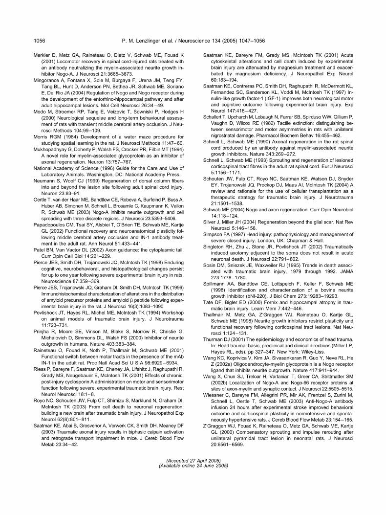
M
M
M
M
M
N
N
O
P
P
P
P
P
P
R
R
R
S
S
S
S
S
S
S
S
S
S
S
S
S
T
T
T
W
W
W
Z
P. M. Lenzlinger et al. / Neuroscience 134 (2005) 1047–10561056
erkler D, Metz GA, Raineteau O, Dietz V, Schwab ME, Fouad K(2001) Locomotor recovery in spinal cord-injured rats treated withan antibody neutralizing the myelin-associated neurite growth in-hibitor Nogo-A. J Neurosci 21:3665–3673.
ingorance A, Fontana X, Sole M, Burgaya F, Urena JM, Teng FY,Tang BL, Hunt D, Anderson PN, Bethea JR, Schwab ME, SorianoE, Del Rio JA (2004) Regulation of Nogo and Nogo receptor duringthe development of the entorhino-hippocampal pathway and afteradult hippocampal lesions. Mol Cell Neurosci 26:34–49.
odo M, Stroemer RP, Tang E, Veizovic T, Sowniski P, Hodges H(2000) Neurological sequelae and long-term behavioural assess-ment of rats with transient middle cerebral artery occlusion. J Neu-rosci Methods 104:99–109.
orris RGM (1984) Development of a water maze procedure forstudying spatial learning in the rat. J Neurosci Methods 11:47–60.
ukhopadhyay G, Doherty P, Walsh FS, Crocker PR, Filbin MT (1994)A novel role for myelin-associated glycoprotein as an inhibitor ofaxonal regeneration. Neuron 13:757–767.
ational Academy of Science (1996) Guide for the Care and Use ofLaboratory Animals. Washington, DC: National Academy Press.
eumann S, Woolf CJ (1999) Regeneration of dorsal column fibersinto and beyond the lesion site following adult spinal cord injury.Neuron 23:83–91.
ertle T, van der Haar ME, Bandtlow CE, Robeva A, Burfeind P, Buss A,Huber AB, Simonen M, Schnell L, Brosamle C, Kaupmann K, VallonR, Schwab ME (2003) Nogo-A inhibits neurite outgrowth and cellspreading with three discrete regions. J Neurosci 23:5393–5406.
apadopoulos CM, Tsai SY, Alsbiei T, O’Brien TE, Schwab ME, KartjeGL (2002) Functional recovery and neuroanatomical plasticity fol-lowing middle cerebral artery occlusion and IN-1 antibody treat-ment in the adult rat. Ann Neurol 51:433–441.
atel BN, Van Vactor DL (2002) Axon guidance: the cytoplasmic tail.Curr Opin Cell Biol 14:221–229.
ierce JES, Smith DH, Trojanowski JQ, McIntosh TK (1998) Enduringcognitive, neurobehavioral, and histopathological changes persistfor up to one year following severe experimental brain injury in rats.Neuroscience 87:359–369.
ierce JES, Trojanowski JQ, Graham DI, Smith DH, McIntosh TK (1996)Immunohistochemical characterization of alterations in the distributionof amyloid precursor proteins and amyloid � peptide following exper-imental brain injury in the rat. J Neurosci 16(3):1083–1090.
ovlishock JT, Hayes RL, Michel ME, McIntosh TK (1994) Workshopon animal models of traumatic brain injury. J Neurotrauma11:723–731.
rinjha R, Moore SE, Vinson M, Blake S, Morrow R, Christie G,Michalovich D, Simmons DL, Walsh FS (2000) Inhibitor of neuriteoutgrowth in humans. Nature 403:383–384.
aineteau O, Fouad K, Noth P, Thallmair M, Schwab ME (2001)Functional switch between motor tracts in the presence of the mAbIN-1 in the adult rat. Proc Natl Acad Sci U S A 98:6929–6934.
iess P, Bareyre F, Saatman KE, Cheney JA, Lifshitz J, Raghupathi R,Grady MS, Neugebauer E, McIntosh TK (2001) Effects of chronic,post-injury cyclosporin A administration on motor and sensorimotorfunction following severe, experimental traumatic brain injury. RestNeurol Neurosci 18:1–8.
oyo NC, Schouten JW, Fulp CT, Shimizu S, Marklund N, Graham DI,McIntosh TK (2003) From cell death to neuronal regeneration:building a new brain after traumatic brain injury. J Neuropathol ExpNeurol 62(8):801–811.
aatman KE, Abai B, Grosvenor A, Vorwerk CK, Smith DH, Meaney DF(2003) Traumatic axonal injury results in biphasic calpain activationand retrograde transport impairment in mice. J Cereb Blood Flow
Metab 23:34–42.aatman KE, Bareyre FM, Grady MS, McIntosh TK (2001) Acutecytoskeletal alterations and cell death induced by experimentalbrain injury are attenuated by magnesium treatment and exacer-bated by magnesium deficiency. J Neuropathol Exp Neurol60:183–194.
aatman KE, Contreras PC, Smith DH, Raghupathi R, McDermott KL,Fernandez SC, Sanderson KL, Voddi M, McIntosh TK (1997) In-sulin-like growth factor-1 (IGF-1) improves both neurological motorand cognitive outcome following experimental brain injury. ExpNeurol 147:418–427.
challert T, Upchurch M, Lobaugh N, Farrar SB, Spirduso WW, Gilliam P,Vaughn D, Wilcox RE (1982) Tactile extinction: distinguishing be-tween sensorimotor and motor asymmetries in rats with unilateralnigrostriatal damage. Pharmacol Biochem Behav 16:455–462.
chnell L, Schwab ME (1990) Axonal regeneration in the rat spinalcord produced by an antibody against myelin-associated neuritegrowth inhibitors. Nature 343:269–272.
chnell L, Schwab ME (1993) Sprouting and regeneration of lesionedcorticospinal tract fibres in the adult rat spinal cord. Eur J Neurosci5:1156–1171.
chouten JW, Fulp CT, Royo NC, Saatman KE, Watson DJ, SnyderEY, Trojanowski JQ, Prockop DJ, Maas AI, McIntosh TK (2004) Areview and rationale for the use of cellular transplantation as atherapeutic strategy for traumatic brain injury. J Neurotrauma21:1501–1538.
chwab ME (2004) Nogo and axon regeneration. Curr Opin Neurobiol14:118–124.
ilver J, Miller JH (2004) Regeneration beyond the glial scar. Nat RevNeurosci 5:146–156.
impson FA (1997) Head injury: pathophysiology and management ofsevere closed injury. London, UK: Chapman & Hall.
ingleton RH, Zhu J, Stone JR, Povlishock JT (2002) Traumaticallyinduced axotomy adjacent to the soma does not result in acuteneuronal death. J Neurosci 22:791–802.
osin DM, Sniezek JE, Waxweiler RJ (1995) Trends in death associ-ated with traumatic brain injury, 1979 through 1992. JAMA273:1778–1780.
pillmann AA, Bandtlow CE, Lottspeich F, Keller F, Schwab ME(1998) Identification and characterization of a bovine neuritegrowth inhibitor (bNI-220). J Biol Chem 273:19283–19293.
ate DF, Bigler ED (2000) Fornix and hippocampal atrophy in trau-matic brain injury. Learn Mem 7:442–446.
hallmair M, Metz GA, Z’Graggen WJ, Raineteau O, Kartje GL,Schwab ME (1998) Neurite growth inhibitors restrict plasticity andfunctional recovery following corticospinal tract lesions. Nat Neu-rosci 1:124–131.
hurman DJ (2001) The epidemiology and economics of head trauma.In: Head trauma: basic, preclinical and clinical directions (Miller LP,Hayes RL, eds), pp 327–347. New York: Wiley-Liss.
ang KC, Koprivica V, Kim JA, Sivasankaran R, Guo Y, Neve RL, HeZ (2002a) Oligodendrocyte-myelin glycoprotein is a Nogo receptorligand that inhibits neurite outgrowth. Nature 417:941–944.
ang X, Chun SJ, Treloar H, Vartanian T, Greer CA, Strittmatter SM(2002b) Localization of Nogo-A and Nogo-66 receptor proteins atsites of axon-myelin and synaptic contact. J Neurosci 22:5505–5515.
iessner C, Bareyre FM, Allegrini PR, Mir AK, Frentzel S, Zurini M,Schnell L, Oertle T, Schwab ME (2003) Anti-Nogo-A antibodyinfusion 24 hours after experimental stroke improved behavioraloutcome and corticospinal plasticity in normotensive and sponta-neously hypertensive rats. J Cereb Blood Flow Metab 23:154–165.
’Graggen WJ, Fouad K, Raineteau O, Metz GA, Schwab ME, KartjeGL (2000) Compensatory sprouting and impulse rerouting afterunilateral pyramidal tract lesion in neonatal rats. J Neurosci
20:6561–6569.(Accepted 27 April 2005)(Available online 24 June 2005)
Related Documents