Research Article Dehydroepiandrosterone Prevents H 2 O 2 -Induced BRL-3A Cell Oxidative Damage through Activation of PI3K/Akt Pathways rather than MAPK Pathways Longlong Li, Yao Yao, Zhihao Jiang, Jinlong Zhao, Ji Cao, and Haitian Ma Key Laboratory of Animal Physiology and Biochemistry, College of Veterinary Medicine, Nanjing Agricultural University, Nanjing 210095, China Correspondence should be addressed to Haitian Ma; [email protected] Received 26 October 2018; Accepted 3 April 2019; Published 28 April 2019 Academic Editor: Alin Ciobica Copyright © 2019 Longlong Li et al. This is an open access article distributed under the Creative Commons Attribution License, which permits unrestricted use, distribution, and reproduction in any medium, provided the original work is properly cited. Dehydroepiandrosterone (DHEA) is a popular dietary supplement that has well-known benefits in animals and humans, but there is not enough information about the mechanisms underlying its effects. The present study aimed at investigating these mechanisms through in vitro experiments on the effects of DHEA on rat liver BRL-3A cells exposed to oxidative stress through H 2 O 2 . The findings showed that DHEA increased the antioxidant enzyme activity, decreased ROS generation, and inhibited apoptosis in H 2 O 2 -treated cells. These effects of DHEA were not observed when the cells were pretreated with known antagonists of sex hormones (Trilostane, Flutamide, or Fulvestrant). Furthermore, treatment with estradiol and testosterone did not have the same protective effects as DHEA. Thus, the beneficial effects of DHEA were associated with mechanisms that were independent of steroid hormone pathways. With regard to the mechanism underlying the antiapoptotic effect of DHEA, pretreatment with DHEA was found to induce a significant decrease in the protein expression of Bax and caspase-3 and a significant increase in the protein expression of PI3K and p-Akt in H 2 O 2 -treated BRL-3A cells. These effects of DHEA were abolished when the cells were pretreated with the PI3K inhibitor LY294002. No changes were observed on the p-ERK1/2, p-p38, and p-JNK protein levels in H 2 O 2 -induced BRL-3A cells pretreated with DHEA. In conclusion, our data demonstrate that DHEA protects BRL-3A cells against H 2 O 2 -induced oxidative stress and apoptosis through mechanisms that do not involve its biotransformation into steroid hormones or the activation of sex hormone receptors. Importantly, the protective effect of DHEA on BRL-3A cells was mainly associated with PI3K/Akt signaling pathways, rather than MAPK signaling pathways. 1. Introduction Oxidative stress, which is caused by an increase in the production of reactive oxygen species (ROS), plays an impor- tant role in the development of liver diseases like fatty liver, alcohol liver, and liver injury [1]. Oxidative stress affects cell functioning by damaging lipids, proteins, and enzymes, and this subsequently leads to the apoptosis of the affected cells [2]. Hydrogen peroxide (H 2 O 2 ), which is a prominent ROS, is closely involved in the induction of liver oxidative stress [3]. High H 2 O 2 levels are responsible for lipid peroxidation and DNA damage, which eventually lead to the apoptosis of hepatic cells [4, 5]. The H 2 O 2 -induced apoptosis of hepa- tocytes involves the inhibition of antioxidative mechanisms and apoptosis-associated regulatory proteins like Bcl-2 family proteins and caspases. Thus, inhibition of proapopto- tic pathways might be a feasible way of preventing or stalling liver damage caused by excess H 2 O 2 production. Further- more, since oxidative stress has been implicated in the majority of liver injuries [6, 7], another treatment strategy for liver injury might be the use of active antioxidant molecules that ameliorate liver oxidative stress. The incidence of and susceptibility to liver diseases is known to increase with age [8]. There is some speculation that the aging-related degenerative changes observed in humans is associated with an aging-related marked decline in the levels of dehydroepiandrosterone (DHEA). In fact, the decrease in circulating DHEA levels is associated with Hindawi Oxidative Medicine and Cellular Longevity Volume 2019, Article ID 2985956, 14 pages https://doi.org/10.1155/2019/2985956

Welcome message from author
This document is posted to help you gain knowledge. Please leave a comment to let me know what you think about it! Share it to your friends and learn new things together.
Transcript

Research ArticleDehydroepiandrosterone Prevents H2O2-Induced BRL-3ACell Oxidative Damage through Activation of PI3K/AktPathways rather than MAPK Pathways
Longlong Li, Yao Yao, Zhihao Jiang, Jinlong Zhao, Ji Cao, and Haitian Ma
Key Laboratory of Animal Physiology and Biochemistry, College of Veterinary Medicine, Nanjing Agricultural University,Nanjing 210095, China
Correspondence should be addressed to Haitian Ma; [email protected]
Received 26 October 2018; Accepted 3 April 2019; Published 28 April 2019
Academic Editor: Alin Ciobica
Copyright © 2019 Longlong Li et al. This is an open access article distributed under the Creative Commons Attribution License,which permits unrestricted use, distribution, and reproduction in any medium, provided the original work is properly cited.
Dehydroepiandrosterone (DHEA) is a popular dietary supplement that has well-known benefits in animals and humans, but thereis not enough information about the mechanisms underlying its effects. The present study aimed at investigating these mechanismsthrough in vitro experiments on the effects of DHEA on rat liver BRL-3A cells exposed to oxidative stress through H2O2. Thefindings showed that DHEA increased the antioxidant enzyme activity, decreased ROS generation, and inhibited apoptosis inH2O2-treated cells. These effects of DHEA were not observed when the cells were pretreated with known antagonists of sexhormones (Trilostane, Flutamide, or Fulvestrant). Furthermore, treatment with estradiol and testosterone did not have the sameprotective effects as DHEA. Thus, the beneficial effects of DHEA were associated with mechanisms that were independent ofsteroid hormone pathways. With regard to the mechanism underlying the antiapoptotic effect of DHEA, pretreatment withDHEA was found to induce a significant decrease in the protein expression of Bax and caspase-3 and a significant increase inthe protein expression of PI3K and p-Akt in H2O2-treated BRL-3A cells. These effects of DHEA were abolished when the cellswere pretreated with the PI3K inhibitor LY294002. No changes were observed on the p-ERK1/2, p-p38, and p-JNK proteinlevels in H2O2-induced BRL-3A cells pretreated with DHEA. In conclusion, our data demonstrate that DHEA protects BRL-3Acells against H2O2-induced oxidative stress and apoptosis through mechanisms that do not involve its biotransformation intosteroid hormones or the activation of sex hormone receptors. Importantly, the protective effect of DHEA on BRL-3A cells wasmainly associated with PI3K/Akt signaling pathways, rather than MAPK signaling pathways.
1. Introduction
Oxidative stress, which is caused by an increase in theproduction of reactive oxygen species (ROS), plays an impor-tant role in the development of liver diseases like fatty liver,alcohol liver, and liver injury [1]. Oxidative stress affects cellfunctioning by damaging lipids, proteins, and enzymes, andthis subsequently leads to the apoptosis of the affected cells[2]. Hydrogen peroxide (H2O2), which is a prominent ROS,is closely involved in the induction of liver oxidative stress[3]. High H2O2 levels are responsible for lipid peroxidationand DNA damage, which eventually lead to the apoptosisof hepatic cells [4, 5]. The H2O2-induced apoptosis of hepa-tocytes involves the inhibition of antioxidative mechanisms
and apoptosis-associated regulatory proteins like Bcl-2family proteins and caspases. Thus, inhibition of proapopto-tic pathways might be a feasible way of preventing or stallingliver damage caused by excess H2O2 production. Further-more, since oxidative stress has been implicated in themajority of liver injuries [6, 7], another treatment strategyfor liver injury might be the use of active antioxidantmolecules that ameliorate liver oxidative stress.
The incidence of and susceptibility to liver diseases isknown to increase with age [8]. There is some speculationthat the aging-related degenerative changes observed inhumans is associated with an aging-related marked declinein the levels of dehydroepiandrosterone (DHEA). In fact,the decrease in circulating DHEA levels is associated with
HindawiOxidative Medicine and Cellular LongevityVolume 2019, Article ID 2985956, 14 pageshttps://doi.org/10.1155/2019/2985956

multiple metabolic consequences including autoimmunediseases, aberrations in lipid metabolism, type 2 diabetes,and oxidative stress-related diseases [9]. Recently, DHEAwas reported to exhibit antioxidative effects under conditionsof acute as well as chronic oxidative stress [10–12], and theseantioxidant effects have been confirmed through in vivo[13, 14] and in vitro [15] experiments, including our recentstudy in which DHEA treatment was found to protect vari-ous types of cells against oxidative damage [16, 17]. Althoughthese beneficial effects of DHEA are known, there is notenough information about the mechanisms through whichit exerts these effects.
DHEA is essentially a precursor protein that has thepotential to transform into estrogens or androgens in organssuch as the kidney, brain, gonads, and liver [18, 19], and itcan exert various physiological effects by binding receptors,such as the estrogen receptor, androgen receptor, and otherhighly specific receptors, in the target tissues. Some studieshave reported that DHEA executes its effects mainly throughconversion into sex steroids and activation of androgen orestrogen receptors [20–22]. For example, our recent studyfound that DHEA reduced lipid droplet accumulation inprimary hepatocytes from the chicken through its biotrans-formation into steroid hormones [23], and Mills et al.showed that DHEA promotes the healing of cutaneousinjuries by activating estrogen receptors [20]. In contrast,there is also some evidence that the positive effects of DHEAare independent of the activation of sex steroid receptors[24–26]. This means that DHEA may exert its physiologicaleffects as a neurosteroid by directly binding to neurotrans-mitter receptors. However, the mechanisms underlying itseffects and its efficacy remain unclear in the absence ofsufficient supporting data. The present study sought to fillin this gap in the literature.
Based on the findings of the literature so far, the aims ofthe present study were to determine whether DHEA protectsH2O2-exposed BRL-3A cells from oxidative stress and apo-ptosis and to identify the signaling pathways and mecha-nisms that may be involved in the effects of DHEA. Webelieve that these findings will shed light on the antioxidativemechanisms of DHEA, which may have potential for thetreatment of oxidative stress-induced conditions in humans.
2. Materials and Methods
2.1. Reagents. Testosterone, estradiol, DHEA, dimethyl sulf-oxide (DMSO), and penicillin-streptomycin were obtainedfrom Sigma-Aldrich (St. Louis, USA). The bicinchoninic acid(BCA), ROS, and cell apoptosis assay kits were from theBeyotime Institute of Biotechnology (Shanghai, China). Therabbit anti-caspase 3, anti-Bax, anti-3β-HSD (3β-hydroxy-steroid dehydrogenase), and anti-17β-HSD (17β-hydroxy-steroid dehydrogenase) antibodies were obtained fromAbcam (London, England). The rabbit ERK1/2, anti-PI3K(phosphatidylinositol-3-kinase), anti-Akt, anti-JNK, anti-phosphorylated (p)-ERK1/2, anti-p-Akt, and anti-p-JNKantibodies were purchased from Cell Signaling Technology(CST, Boston, USA). The rabbit anti-tubulin β, anti-p38,and anti-p-p38 antibodies, as well as the goat anti-rabbit
horseradish peroxidase-conjugated IgG were obtained fromBoster Biological Technology Co. (Wuhan, China). Fulves-trant (an estrogen receptor (ER) antagonist), Flutamide (anandrogen receptor (AR) antagonist), Trilostane (a 3β-HSDinhibitor), and LY294002 (PI3K inhibitor) were obtainedfrom Selleck Chemicals (Houston, USA). Testosterone,estradiol, and DHEA were dissolved in DMSO, which wasthen diluted in DMEM-F12 medium. The DMSO contentin the working solutions was less than 0.1% of the totalvolume.
2.2. Cell Culture. Rat liver BRL-3A cells (ATCC, Manassas,USA) were passaged every 3-4 days in DMEM-F12 medium(HyClone Laboratories Inc., Los Angeles, USA) containing10% fetal bovine serum, 1% of 100 U/mL penicillin, and100 μg/mL streptomycin at 37°C in a 5% CO2 atmosphere.The cells were used for the experiments when they reached80-90% confluence.
2.3. Testosterone and Estradiol Measurement byRadioimmunoassay. BRL-3A cells were grown in 6-wellplates (1 × 10⁶ cells/well) and treated with DHEA at dosesof 0, 1, 10, or 100 μMDHEA for 24 h. The BRL-3A cells werethen harvested and ultrasonically disrupted on ice. Next, thecells were centrifuged at 2500 ×g for 10 min at 4°C. Thesupernatants were extracted, and the estradiol and testoster-one contents were measured using radioimmunoassay kits(Beifang Biotechnology Research Institute, Beijing, China).
2.4. ROS Analysis. BRL-3A cells were pretreated with vehicle,10 μM Trilostane, 10 μM Flutamide, or 1 μM Fulvestrant for60 min. The cells were then treated with DHEA (0, 1, 10, or100 μM), testosterone (12.0 nM), or estradiol (6.3 nM) for24 h and subsequently treated with 150 μMH2O2 for another2 h. Intracellular ROS content was analyzed with the fluorop-robe 2′,7′-dichlorodihydrofluorescein diacetate (H2DCF-DA) and dihydroethidium (DHE) as shown in our recentreports [16, 17]. Briefly, BRL-3A cells were incubated withH2DCF-DA for 30 min at 37°C, washed three times, andresuspended in cold PBS, after which they were immediatelysubjected to flow cytometry analysis with FACSCalibur™(Becton Dickinson, San Jose, USA).
2.5. Cell Apoptosis Analysis. The cell treatments were thesame as described in Section 2.4 for ROS analysis, and cellapoptosis was examined as reported in our studies [16, 17].Briefly, BRL-3A cells were collected and washed with coldPBS, placed in 195 μL Annexin V-FITC binding solution,incubated in 5 μL Annexin V-FITC and 10 μL propidiumiodide in the dark for 30 min, and immediately subjected toflow cytometry analysis with FACSCalibur™.
2.6. Measurement of Antioxidant Parameters. The treatmentsfor the different cell groups were the same as described forthe ROS analysis. For the measurements, the cells wereharvested, disrupted ultrasonically in ice, and centrifuged at2500 ×g for 10 min at 4°C. The supernatants were collectedand stored -20°C for subsequent analysis. The activities ofcatalase (CAT), peroxidase (POD), superoxide dismutase(SOD), and glutathione peroxidase (GSH-Px) were measured
2 Oxidative Medicine and Cellular Longevity

using commercial kits following the manufacturer’s protocol(Nanjing Jiancheng Bioengineering Institute, Nanjing,China), and the data were normalized to the protein concen-tration as determined by a BCA protein assay kit.
2.7. Real-Time Quantitative RT-PCR. BRL-3A cells weregrown in 6-well plates (1 × 10⁶ cells per well) and treatedwith 0, 1, 10, or 100 μM DHEA for 24 h, then they wereexposed to 150 μM H2O2 for another 2 h. After incubation,the cells were harvested and total RNA was extracted usingthe TRIZOL reagent kit (Invitrogen, USA) according to themanufacturer’s protocols. Total RNA (2 μg) were reversetranscribed into cDNA using the SuperScript II kit (Promega,USA) according to the manufacturer’s recommendation. Analiquot of a complementary DNA sample was mixed with20 μL SYBR Green PCR Master Mix (Roche, Switzerland) inthe presence of 10 pmol of each forward and reverse primersfor β-actin (used as an internal control), Bcl-2, and Bax(Table 1). All samples were analyzed in duplicate using theiQ5 Sequence Detection System (Bio-Rad, California, USA)and programmed to conduct one cycle (95°C for 3 min)and 40 cycles (95°C for 20 s, 60°C for 30 s, and 72°C for30 s). The 2-ΔΔCT method was used to calculate the foldchange in mRNA levels. The primers used were designedby Primer Premier 5 (Premier Biosoft International, PaloAlto, USA) and synthesized by Invitrogen Biological Co.(Shanghai, China).
2.8. Western Blotting. BRL-3A cells were grown in 6-wellplates (1 × 10⁶ cells/well) and treated with 50 μM phos-phatidylinositol 3-kinase (PI3K) inhibitor (LY294002) orvehicle for 1 h before being exposed to 0, 1, 10, or 100 μMDHEA for 24 h, and then they were treated with 150 μMH2O2 for another 2 h. The western blotting method wasbased on our recent description in [23]. Briefly, after incuba-tion, the cells were scraped and the protein concentration wasmeasured using BCA protein determination kits (BeyotimeInstitute of Biotechnology, Shanghai, China). The extractedprotein was separated on 10% sodium dodecyl sulfate-polyacrylamide gel electrophoresis (SDS-PAGE) and trans-ferred onto PVDF membranes (Millipore, Bedford, MA,USA). The membranes were blocked for 3 h with 5% BSAin TBST and then incubated overnight at 4°C with thefollowing rabbit polyclonal antibodies (dilution): PI3K,phosphorylated- (p-) Akt (1 : 1000), Akt (1 : 1000), p-ERK1/2 (1 : 1000), ERK1/2 (1 : 1000), p-JNK (1 : 1000), JNK(1 : 1000), p38 (1 : 500), p-p38 (1 : 500), caspase-3 (1 : 500),and Bax (1 : 5000). After washing with TBST, goat anti-
rabbit IgG with horseradish peroxidase conjugated(1 : 10000) in washing solution was added and incubated for2 h at room temperature. Immunoreactivity proteins weredetected by SuperSignal chemiluminescence, and the pro-tein bands were digitally imaged for densitometric quanti-fication using an ECL SuperSignal™ West Pico substrate(Pierce, Rockford, USA). Tubulin β monoclonal antibody(1 : 10000) was used as the loading control, and all proteinexpression levels were normalized to tubulin β.
2.9. Data Analysis and Statistics. Data were expressed asmeans ± standard error (SE). Differences were analyzedusing one-way analysis of variance (ANOVA) followed bypost hoc tests. Differences were considered significant atP < 0 05. All statistical analyses were performed with SPSS20.0 for Windows (StatSoft Inc., Tulsa, USA).
3. Results
3.1. Biotransformation of DHEA in BRL-3A Cells. Thetestosterone and estradiol content were not detected in thevehicle-treated group, while the content of both hormoneswas significantly higher in the DHEA-treated BRL-3A cellsthan in the vehicle-treated cells, and it increased as thedose of DHEA increased from 1 to 100 μM (P < 0 05)(Figures 1(a) and 1(b)). In keeping with these findings,DHEA treatment resulted in a significant increase in theprotein expression of 3β-HSD and 17β-HSD in a dose-dependent way (P < 0 05) (Figures 1(c)–1(e)). These findingsare in keeping with the known biotransformation pathwaysthrough which DHEA is converted to sex hormones.
3.2. DHEA Increased Antioxidant Enzyme Activity in H2O2-Induced BRL-3A Cells. As shown in Figure 2, the SOD,POD, CAT, and GSH-Px activities were significantly lowerin the H2O2-treated BRL-3A cells than in the vehicle-treated cells (P < 0 01). Pretreatment with 1–100 μM DHEAbefore H2O2 treatment caused the SOD, POD, CAT, andGSH-Px activities to be significantly higher than those inthe cells that were treated only with H2O2 (P < 0 05). Theseeffects of DHEA improved as its dose was increased.
The effects of 100 μM DHEA on the activity of theantioxidant enzymes were not altered when the cells werepretreated with Trilostane (the 3β-HSD inhibitor), Fluta-mide (the AR antagonist), and Fulvestrant (the ER antago-nist) (Figure 2). Furthermore, in H2O2-induced BRL-3Acells that were pretreated with 12.0 nM testosterone and6.3 nM estradiol, the antioxidant enzyme activities were
Table 1: Primer sequences of the target genes and β-actin (internal control) used for RT-PCR.
Gene GenBank accession number Primer sequences (5′-3′) Orientation Product size (bp)
β-Actin NM 031144CCCTGTGCTGCTCACCGA
ACAGTGTGGGTGACCCCGTCForwardReverse
186
Bax NM 007527GCAGGGAGGATGGCTGGGGAGATCCAGACAAGCAGCCGCTCACG
ForwardReverse
352
Bcl-2 NM 016993CGACTTTGCAGAGATGTCCACATCCACAGAGCGATGTTGT
ForwardReverse
202
3Oxidative Medicine and Cellular Longevity

significantly lower than those in the H2O2-treated cells thatwere pretreated with 100 μM DHEA (P < 0 01), but therewas no significant difference in enzyme activity betweenH2O2-treated BRL-3A cells pretreated with 12.0 nM testos-terone and 6.2 nM estradiol and the cells treated only withH2O2 (P > 0 05) (Figure 2). The doses of testosterone andestradiol used in this experiment were approximately equalto the estradiol and testosterone concentrations detected inBRL-3A cells that were treated with 100 μM DHEA for 24 h.
3.3. DHEA Inhibited H2O2-Induced ROS Generation in BRL-3A Cells. The ROS content was significantly higher in H2O2-treated BRL-3A cells than in the vehicle-treated cells(P < 0 01). Pretreatment of the H2O2-treated cells with1-100 μM DHEA resulted in a significant decrease inthe ROS content in a dose-dependent pattern (P < 0 01). Pre-treatment with Trilostane, Flutamide, or Fulvestrant did notalter these effects of 100 μM DHEA (P > 0 05). However,the ROS content in H2O2-treated BRL-3A cells pretreatedwith 12.0 nM testosterone and 6.2 nM estradiol was signifi-cantly higher than that in the H2O2-treated cells pretreatedwith 100 μM DHEA (P < 0 01), but it was not significantlydifferent from the ROS content of the cells that were treatedonly with H2O2 (P > 0 05) (Figure 3).
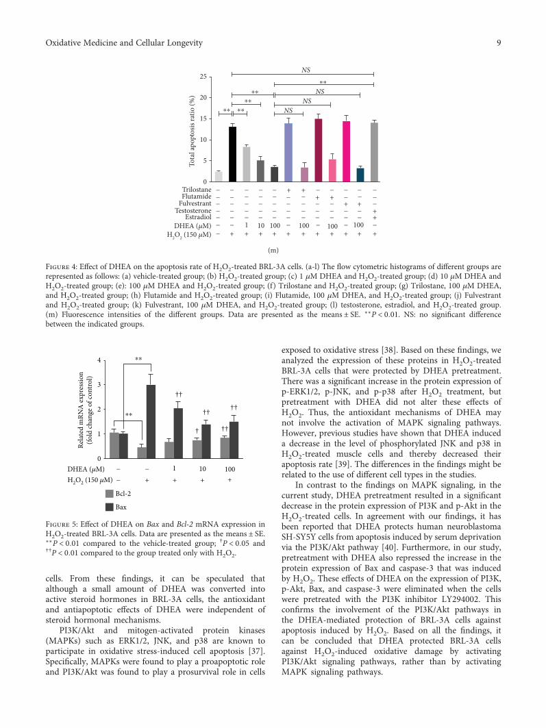
3.4. DHEA Reduced H2O2-Induced Apoptosis in BRL-3ACells. The apoptosis rate was markedly higher in theH2O2-treated BRL-3A cells than in the vehicle-treatedcells (P < 0 01), while it was significantly lower in theH2O2-treated cells that were pretreated with 1-100 μMDHEA, which showed a dose-dependent effect (P < 0 01)(Figure 4). Pretreatment with Trilostane, Flutamide, or Ful-vestrant did not alter these anti-apoptotic effects of 100 μMDHEA (P > 0 05) (Figure 4). Pretreatment with 12.0 nMtestosterone and 6.2 nM estradiol did not change the apopto-sis rate of the H2O2-treated cells (P > 0 05). In fact, the apo-ptosis rate was significantly higher in H2O2-treated cellspretreated with 12.0 nM testosterone and 6.2 nM estradiolthan in H2O2-treated cells pretreated with 100 μM DHEA(P < 0 01) (Figure 4).
3.5. DHEA Regulated Apoptosis-Related Factor mRNAExpression in H2O2-Induced BRL-3A Cells. H2O2-treatedBRL-3A cells showed significantly higher Bax mRNAexpression and significantly lower Bcl-2 mRNA expressionthan the vehicle-treated BRL-3A cells (P < 0 01). Pretreat-ment with DHEA at doses of 1-100 μM reversed theseeffects of H2O2 on the Bax mRNA level (P < 0 01), whilepretreatment with DHEA at doses of 10-100 μM reversed
0 �휇MND
0.0
0.5
1.0
1.5
2.0
Estr
adio
l con
tent
(ng/
mL)
1 �휇M 10 �휇M 100 �휇M
⁎⁎
⁎⁎
⁎
(a)
0 �휇M
ND0
1
2
3
4
Testo
stero
ne co
nten
t (ng
/mL)
1 �휇M 10 �휇M 100 �휇M
⁎⁎
⁎⁎
⁎
(b)
Tubulin �훽
33 kDa
55 kDa
42 kDa
0 1 10 100 DHEA (�휇M)
3�훽-HSD
17�훽-HSD
(c)
0 �휇M0.0
0.5
1.0
1.5
2.0
3�훽-H
SD p
rote
in ex
pres
sion
1 �휇M 10 �휇M 100 �휇M
⁎⁎⁎⁎
⁎
(d)
0 �휇M0
1
2
3
4
17�훽
-HSD
pro
tein
expr
essio
n
1 �휇M 10 �휇M 100 �휇M
⁎⁎
⁎⁎
⁎
(e)
Figure 1: Biotransformation of DHEA in BRL-3A cells. (a) Estradiol content. (b) Testosterone content. (c) Immunoblot of 3β-HSD and17β-HSD in BRL-3A cells treated with 0-100 μM DHEA. (d) 3β-HSD protein expression. (e) 17β-HSD protein expression. Data arepresented as the means ± SE. ∗P < 0 05 and ∗∗P < 0 01, compared to the vehicle-treated group.
4 Oxidative Medicine and Cellular Longevity

the inhibitory effect of H2O2 on Bcl-2 mRNA expression(P < 0 05) (Figure 5).
3.6. DHEA Regulated MAPK, PI3K, and p-AKT Protein Levelin H2O2-Induced BRL-3A Cells. PI3K and p-AKT proteinexpression was significantly lower in the H2O2-treatedBRL-3A cells than in the vehicle-treated cells (P < 0 01)(Figures 6(a)–6(c)), while p-ERK1/2, p-JNK, and p-p38protein expression was significantly higher (P < 0 01)(Figures 6(a) and 6(d)–6(f)). Pretreatment with 1-100 μMDHEA reversed the effects of H2O2 on PI3K and p-AKT pro-tein expression (P < 0 05) (Figures 6(a)–6(c)), but it did notalter the effects of H2O2 on p-ERK1/2, p-JNK, and p-p38protein expression (P > 0 05) (Figures 6(a) and 6(d)-6(f)).However, these effects of DHEA on PI3K and p-AKT expres-sion were significantly inhibited upon pretreatment of H2O2-treated cells with LY294002 (P < 0 01) (Figures 6(g)–6(i)).
3.7. DHEA Downregulated Caspase-3 and Bax Protein Levelsin H2O2-Induced BRL-3A Cells. The protein expression of
Bax and caspase-3 was significantly higher in the H2O2-treated BRL-3A cells than in the vehicle-treated cells(P < 0 01) (Figures 7(a)–7(c)). Pretreatment with 1-100 μMDHEA led to a significant decrease in Bax and caspase-3protein expression in the H2O2-treated cells (P < 0 05)(Figures 7(a)–7(c)). However, these effects of DHEA wereinhibited in H2O2-treated BRL-3A cells that were pretreatedwith LY294002 (P < 0 05) (Figures 7(d)–7(f)).
4. Discussion
In the present study, our in vitro experiments on rat liver cellsshow that DHEA exerts antioxidative and antiapoptoticeffects through pathways that are independent of steroidhormones and their receptors; instead, the PI3K/p-AKTpathways seem to be closely involved with these protectiveeffects of DHEA on the liver cells.
BRL-3A rat liver cells were used as an in vitro model forstudying the effects of H2O2, as this cell type is susceptibleto oxidative damage. Furthermore, H2O2 is well known as
0
TrilostaneFlutamide
FulvestrantTestosterone
Estradiol
DHEA (�휇M)H2O2 (150 �휇M)
20
40
SOD
activ
ity (U
/mg
prot
)
60
NSNS
NS
NS
⁎⁎
⁎⁎
⁎⁎
⁎⁎⁎⁎
− + + + + + + + + ++ +
− − − − − − −− − − − − − −
− − +− −
− − − − − + + − − −− −
− − +− −
− − − − − − − + + −− −− − − − − − − − − −+ +
1 10 100 100 100 100− − − − − −
(a)
0
5
10
POD
activ
ity (U
/mg
prot
)
15
NSNS
NS
NS
⁎⁎⁎⁎
⁎⁎⁎⁎
⁎
TrilostaneFlutamide
FulvestrantTestosterone
Estradiol
DHEA (�휇M)H2O2 (150 �휇M) − + + + + + + + + ++ +
− − − − − − −− − − − − − −
− − +−
− − − − − + + − − −− −
− − +− −
− − − − − − − + + −− −− − − − − − − − −
−−+ +
1 10 100 100 100 100− − − − − −
(b)
0
4
2
8
6
10
CAT
activ
ity (U
/mg
prot
)
12 NS
NSNS
NS
⁎⁎
⁎⁎
⁎⁎
⁎⁎ ⁎
TrilostaneFlutamide
FulvestrantTestosterone
Estradiol
DHEA (�휇M)H2O2 (150 �휇M) − + + + + + + + + ++ +
− − − − − − −− − − − − − −
− − +− −
− − − − − + + − − −− −
− − +− −
− − − − − − − + + −− −− − − − − − − − − −+ +
1 10 100 100 100 100− − − − − −
(c)
0.0
0.5
1.0G
SH-P
x ac
tivity
(U/m
g pr
ot)
1.5NS
NSNS
NS
⁎⁎⁎⁎
⁎⁎⁎⁎ ⁎
TrilostaneFlutamide
FulvestrantTestosterone
Estradiol
DHEA (�휇M)H2O2 (150 �휇M) − + + + + + + + + ++ +
− − − − − − −− − − − − − −
− − +− −
− − − − − + + − − −− −
− − +− −
− − − − − − − + + −− −− − − − − − − − − −+ +
1 10 100 100 100 100− − − − − −
(d)
Figure 2: Effects of DHEA on antioxidant enzyme activities in H2O2-treated BRL-3A cells. (a) Superoxidase dismutase (SOD) activity. (b)Peroxidase (POD) activity. (c) Catalase (CAT) activity. (d) Glutathione peroxidase (GSH-Px) activity. Data are presented as the means ±SE. ∗P < 0 05 and ∗∗P < 0 01. NS: no significant difference between the indicated groups.
5Oxidative Medicine and Cellular Longevity
1010
50
100
150
200
102 103 104 105
FL1-A
Coun
t
106 107.2
Gate: P1
(a)
0
100
200
300
400
Coun
t
101 102 103 104 105
FL1-A106 107.2
Gate: P1
(b)
101 102 103 104 105
FL1-A106 107.2
0
100
200
300
400
Coun
t
Gate: P1
(c)
101 102 103 104 105
FL1-A106 107.2
0
100
200
300
400
Coun
t
Gate: P1
(d)
101 102 103 104 105
FL1-A106 107.2
0
100
200
300
400Co
unt
Gate: P1
(e)
101 102 103 104 105
FL1-A106 107.2
0
200
400
600
800
Coun
t
Gate: P1
(f)
101 102 103 104 105
FL1-A106 107.2
0
50
100
150
200
Coun
t
Gate: P1
(g)
101 102 103 104 105
FL1-A106 107.2
0
200
400
600
800
Coun
t
Gate: P1
(h)
101 102 103 104 105
FL1-A106 107.2
0
50
100
150
200
Coun
tGate: P1
(i)
101 102 103 104 105
FL1-A106 107.2
0
200
400
600
800
Coun
t
Gate: P1
(j)
101 102 103 104 105
FL1-A106 107.2
0
50
100
150
200
Coun
t
Gate: P1
(k)
101 102 103 104 105
FL1-A106 107.2
0
200
400
600
800
Coun
t
Gate: P1
(l)
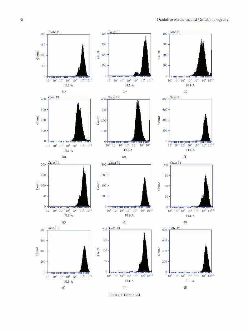
Figure 3: Continued.
6 Oxidative Medicine and Cellular Longevity

an ROS that easily penetrates cells and reacts with metal ionsto produce highly reactive hydroxyl radicals that can causesevere cellular injuries. Here, exposure to 150 μM of H2O2led to the loss of antioxidant activity and generation of ROSin the BRL-3A cells. Similarly, H2O2 has been shown toinduce oxidative damage via its inhibitory effects on antioxi-dant enzyme activity [27, 28]. Furthermore, in the presentstudy, treatment with DHEA reversed these effects of H2O2.This is also in keeping with the findings of previous studiesin which DHEA was reported to inhibit in vitro glucose-induced ROS generation [29] and in vivo ROS generation,as well as to increase the in vivo protein expression ofNADPH oxidase [30]. In particular, DHEA was found toexert antioxidant effects by inducing an increase in catalaseexpression, activating the thioredoxin system, and suppress-ing superoxide anion production [14, 31]. Also in this study,DHEA was found to promote the activity of the antioxidantenzymes such as SOD, CAT, POD, and GSH-Px in theH2O2-treated cells. Thus, all these findings demonstrate thatDHEA protects the cell against oxidative damage by pro-moting the activity of antioxidant enzymes and inhibitingROS production.
Another target of DHEA is cellular apoptotic pathways,as we found that BRL-3A cells that were pretreated withDHEA were protected against the apoptosis-promotingeffects of H2O2. Among the proteins involved in apoptosis,Bax and Bcl-2, which are members of the Bcl family ofproteins, play an important role as regulators of the initialstages of apoptosis [32]. In this study, DHEA inhibited theH2O2-induced downregulation of Bcl-2 mRNA expressionand upregulation of Bax mRNA expression. Caspases are alsoimportant players in the apoptosis pathways, and activatedcaspase-3, in particular, plays a major proapoptotic role
[33]. Our results showed that pretreatment with DHEA ledto a significant decrease in the protein expression of Baxand caspase-3 in BRL-3A cells that were treated with H2O2.Therefore, DHEA might protect liver cells from the effectsof H2O2 through its regulatory effects on downstreamapoptosis-related proteins of the Bcl-2 and caspase family.
As DHEA has been reported to exert its physiologicaleffects via bioconversion into sex hormones and activationof estrogen/androgen receptors, we also investigated whetherDHEA exerted its antioxidative effects via such hormonalmechanisms. First, direct measurement of the testosteroneand estradiol contents showed that they had increased inthe BRL-3A cells after DHEA treatment. Consistent withthese changes in the active hormone content, a significantincrease was observed in the protein expression of 3β-HSDand 17β-HSD after DHEA treatment. In agreement withour findings, DHEA has been shown to promote the testos-terone and estradiol levels in both in vivo and in vitro settings[34, 35]. These findings can be explained through the processof bioconversion wherein 3β-HSD catalyzes the rapidconversion of DHEA into androstenedione in peripheralorgans such as the brain, liver, kidney, and gonads, afterwhich androstenedione is converted into testosterone andestradiol by 17β-HSD and aromatase, respectively [36].However, although there was evidence of the bioconversionof DHEA, we found that pretreating H2O2-treated BRL-3Acells with Trilostane (a 3β-HSD inhibitor), Flutamide (anAR antagonist), and Fulvestrant (an ER antagonist) did notalter the effects of DHEA on the ROS content, antioxidantenzyme activity, or apoptosis rate. Furthermore, pretreat-ment with testosterone and estradiol did not have the sameeffects as pretreatment with DHEA on the ROS content, anti-oxidant activity, or apoptosis rate of H2O2-treated BRL-3A
0
1
2
3
4
ROS
(mea
n flu
ores
cenc
e int
ensit
y)
5NS
NSNS
NS
⁎⁎
⁎⁎⁎⁎
⁎⁎ ⁎⁎
TrilostaneFlutamide
FulvestrantTestosterone
EstradiolDHEA (�휇M)
− − − − −+
− − −+ +
− − 1 −− +++ + + +
− 100 −−
− − − − − − − − − −+ +
+ + ++ +
− − − − − − − − +− −− − − − − − − − −
−+− −
− − −−
10010010
−
100
−− −−
− + − −
H2O2 (150 �휇M)
(m)
Figure 3: Effects of DHEA on the ROS level in H2O2-treated BRL-3A cells. (a-l) The flow cytometric histograms of different groups arerepresented as follows: (a) vehicle-treated group; (b) H2O2-treated group; (c) 1 μM DHEA and H2O2-treated group; (d) 10 μM DHEAand H2O2-treated group; (e) 100 μM DHEA and H2O2-treated group; (f) Trilostane and H2O2-treated group; (g) Trilostane, 100 μMDHEA, and H2O2-treated group; (h) Flutamide and H2O2-treated group; (i) Flutamide, 100 μM DHEA, and H2O2-treated group;(j) Fulvestrant and H2O2-treated group; (k) Fulvestrant, 100 μM DHEA, and H2O2-treated group; (l) testosterone, estradiol, andH2O2-treated group. (m) Fluorescence intensities of the different groups. Data are presented as the means ± SE. ∗∗P < 0 01. NS: nosignificant difference between the indicated groups.
7Oxidative Medicine and Cellular Longevity

103102.4
103
104
105
106
106.9 Gate: P1Q1-UL0.3%
Q1-UR2.6%
Q1-LL96.6%
Q1-LR0.5%
104 105
FL1-A
FL2-
A
106 107.2
(a)
Q1-UL1.9%
Q1-UR6.9%
Q1-LL85.6%
Q1-LR5.6%
Gate: P1
102.4103
104
105
106
106.9
FL2-
A
103 104 105
FL1-A106 107.2
(b)
Q1-UL1.4%
Q1-UR7.0%
Q1-LL90.3%
Q1-LR1.4%
Gate: P1
102.2
103
104
105
106
106.9
FL2-
A
103.1 104 105
FL1-A106 107.2
(c)
Q1-UL0.9%
Q1-UR5.3%
Q1-LL93.2%
Q1-LR0.6%
Gate: P1
102.2
103
104
105
106
106.9
FL2-
A
103.1 104 105
FL1-A106 107.2
(d)
Q1-UL0.7%
Q1-UR2.9%
Q1-LL95.6%
Q1-LR0.8%
Gate: P1
102.2103
104
105
106
106.9FL
2-A
103.1 104 105
FL1-A106 107.2
(e)
Q1-UL2.2%
Q1-UR7.8%
Q1-LL81.9%
Q1-LR8.1%
Gate: P1
102.4103
104
105
106
106.9
FL2-
A
103 104 105
FL1-A106 107.2
(f)
Q1-UL0.5%
Q1-UR2.8%
Q1-LL96.0%
Q1-LR0.8%
Gate: P1
102.2
103
104
105
106
106.9
FL2-
A
103.1 104 105
FL1-A106 107.2
(g)
Q1-UL2.1%
Q1-UR9.2%
Q1-LL82.5%
Q1-LR6.3%
Gate: P1
102.4
103
104
105
106
106.9
FL2-
A
103 104 105
FL1-A106 107.2
(h)
Q1-UL1.8%
Q1-UR3.9%
Q1-LL89.7%
Q1-LR4.5%
Gate: P1
102.4103
104
105
106
106.9FL
2-A
103 104 105
FL1-A106 107.2
(i)
Q1-UL2.4%
Q1-UR7.6%
Q1-LL83.0%
Q1-LR7.1%
Gate: P1
102.4103
104
105
106
106.9
FL2-
A
103 104 105
FL1-A106 107.2
(j)
Gate: P1
102.4103
104
105
106
106.9
FL2-
A
103 104 105
FL1-A106 107.2
Q1-UL2.5%
Q1-UR3.7%
Q1-LL88.4%
Q1-LR5.3%
(k)
Gate: P1
102.4103
104
105
106
106.9
FL2-
A
103 104 105
FL1-A106 107.2
Q1-UL2.2%
Q1-UR7.5%
Q1-LL83.2%
Q1-LR7.1%
(l)
Figure 4: Continued.
8 Oxidative Medicine and Cellular Longevity
cells. From these findings, it can be speculated thatalthough a small amount of DHEA was converted intoactive steroid hormones in BRL-3A cells, the antioxidantand antiapoptotic effects of DHEA were independent ofsteroid hormonal mechanisms.
PI3K/Akt and mitogen-activated protein kinases(MAPKs) such as ERK1/2, JNK, and p38 are known toparticipate in oxidative stress-induced cell apoptosis [37].Specifically, MAPKs were found to play a proapoptotic roleand PI3K/Akt was found to play a prosurvival role in cells
exposed to oxidative stress [38]. Based on these findings, weanalyzed the expression of these proteins in H2O2-treatedBRL-3A cells that were protected by DHEA pretreatment.There was a significant increase in the protein expression ofp-ERK1/2, p-JNK, and p-p38 after H2O2 treatment, butpretreatment with DHEA did not alter these effects ofH2O2. Thus, the antioxidant mechanisms of DHEA maynot involve the activation of MAPK signaling pathways.However, previous studies have shown that DHEA induceda decrease in the level of phosphorylated JNK and p38 inH2O2-treated muscle cells and thereby decreased theirapoptosis rate [39]. The differences in the findings might berelated to the use of different cell types in the studies.
In contrast to the findings on MAPK signaling, in thecurrent study, DHEA pretreatment resulted in a significantdecrease in the protein expression of PI3K and p-Akt in theH2O2-treated cells. In agreement with our findings, it hasbeen reported that DHEA protects human neuroblastomaSH-SY5Y cells from apoptosis induced by serum deprivationvia the PI3K/Akt pathway [40]. Furthermore, in our study,pretreatment with DHEA also repressed the increase in theprotein expression of Bax and caspase-3 that was inducedby H2O2. These effects of DHEA on the expression of PI3K,p-Akt, Bax, and caspase-3 were eliminated when the cellswere pretreated with the PI3K inhibitor LY294002. Thisconfirms the involvement of the PI3K/Akt pathways inthe DHEA-mediated protection of BRL-3A cells againstapoptosis induced by H2O2. Based on all the findings, itcan be concluded that DHEA protected BRL-3A cellsagainst H2O2-induced oxidative damage by activatingPI3K/Akt signaling pathways, rather than by activatingMAPK signaling pathways.
0
5
10
15
20
Tota
l apo
ptos
is ra
tio (%
)
25NS
NSNS
NS
⁎⁎
⁎⁎
⁎⁎
⁎⁎ ⁎⁎
TrilostaneFlutamide
FulvestrantTestosterone
EstradiolDHEA (�휇M)
− −− − − − −+
− − −+ +
− − 1 −− +++ + + +
− 100 −−
− − − − − − − − − −+ +
+ + ++ +
− − − − − − − − +−− −− − − − − − − − − +− −
− − −
10010010
−
100
−− − − + − −
H2O2 (150 �휇M)
(m)
Figure 4: Effect of DHEA on the apoptosis rate of H2O2-treated BRL-3A cells. (a-l) The flow cytometric histograms of different groups arerepresented as follows: (a) vehicle-treated group; (b) H2O2-treated group; (c) 1 μM DHEA and H2O2-treated group; (d) 10 μM DHEA andH2O2-treated group; (e): 100 μM DHEA and H2O2-treated group; (f) Trilostane and H2O2-treated group; (g) Trilostane, 100 μM DHEA,and H2O2-treated group; (h) Flutamide and H2O2-treated group; (i) Flutamide, 100 μM DHEA, and H2O2-treated group; (j) Fulvestrantand H2O2-treated group; (k) Fulvestrant, 100 μM DHEA, and H2O2-treated group; (l) testosterone, estradiol, and H2O2-treated group.(m) Fluorescence intensities of the different groups. Data are presented as the means ± SE. ∗∗P < 0 01. NS: no significant differencebetween the indicated groups.
0
1
2
3
4
Relat
ed m
RNA
expr
essio
n(fo
ld ch
ange
of c
ontro
l)
DHEA (�휇M) − − 1− +++ +
10010H2O2 (150 �휇M)
Bcl-2
⁎⁎
⁎⁎
††
†† ††
†† †
Bax
Figure 5: Effect of DHEA on Bax and Bcl-2 mRNA expression inH2O2-treated BRL-3A cells. Data are presented as the means ± SE.∗∗P < 0 01 compared to the vehicle-treated group; †P < 0 05 and††P < 0 01 compared to the group treated only with H2O2.
9Oxidative Medicine and Cellular Longevity

DHEA (�휇M) −
− + + + +
− 1 10010
H2O2 (150 �휇M)
55 kDaTubulin �훽
p38p-p38
PI3K
p-ERK1/2
AKT
p-AKT
JNK
p-JNK
ERK1/2
38 kDa38 kDa
46 kDa54 kDa46 kDa54 kDa
42 kDa44 kDa42 kDa44 kDa60 kDa
60 kDa
110 kDa
(a)
0.0
0.5
1.0
1.5
PI3K
pro
tein
expr
essio
n
DHEA (�휇M) −
− + + + +
− 1 10010H2O2 (150 �휇M)
⁎⁎
†††††
(b)
p-A
KT/A
KT
0.0
0.5
1.0
1.5
DHEA (�휇M) −
− + + + +
− 1 10010H2O2 (150 �휇M)
⁎⁎
††
††
††
(c)
p-p3
8/p3
8
0
1
2
3
4
DHEA (�휇M) −
− + + + +
− 1 10010H2O2 (150 �휇M)
⁎⁎
(d)
p-JN
K/JN
K
0.0
0.5
1.0
2.0
1.5
2.5
DHEA (�휇M) −
− + + + +
− 1 10010H2O2 (150 �휇M)
⁎⁎
(e)
p-ER
H1/
2ERK
1/2
0
1
2
3
DHEA (�휇M) −
− + + + +
− 1 10010H2O2 (150 �휇M)
⁎⁎
(f)
Tubulin �훽
PI3K
AKT
p-AKT
−
−
− −
+
+ +
+ + +
+ +
− − 1+
10010H2O2 (150 �휇M)
DHEA (�휇M)
LY294002 (50 �휇M)
(g)
0.0
0.5
1.0
1.5
PI3K
pro
tein
expr
essio
n
DHEA (�휇M) −−
− +
+
+ +
+ + +
+ +− − 1
+10010
H2O2 (150 �휇M)
LY294002 (50 �휇M)
⁎⁎
(h)
DHEA (�휇M) −−
− −
+
+ +
+ + +
+ +− − 1
+10010
H2O2 (150 �휇M)
LY294002 (50 �휇M)0.0
0.5
1.0
1.5
p-A
KT/A
KT
⁎⁎
(i)
Figure 6: Effects of DHEA on PI3K, p-Akt, and MAPK protein expression in H2O2-treated BRL-3A cells. (a) Immunoblot of PI3K, p-Akt,and MAPKs in BRL-3A cells pretreated with 1-100 μM DHEA (24 h) and exposed to 150 μM H2O2 (2 h). (b) PI3K proteinexpression. (c) p-Akt protein expression. (d) p-p38 protein expression. (e) p-JNK protein expression. (f) p-ERK1/2 proteinexpression. (g) Immunoblot of PI3K and p-Akt in BRL-3A cells pretreated with 50 μM of the PI3K inhibitor LY294002 or vehiclefor 1 h before exposure to 0, 1, 10, or 100 μM DHEA (24 h) and then 150 μM H2O2 (2 h). (h) PI3K protein expression. (i) p-Aktprotein expression. Data are presented as the means ± SE. ∗∗P < 0 01 compared to the vehicle-treated group; †P < 0 05 and ††P < 0 01compared to the group treated only with H2O2.
10 Oxidative Medicine and Cellular Longevity

In conclusion, our data demonstrate that DHEAexerted a protective effect on BRL-3A cells that wereexposed to H2O2 by inhibiting the production of ROS,promoting the activity of antioxidant enzymes, andregulating the expression of apoptosis-related proteins.Furthermore, the antiapoptosis mechanisms of DHEAinvolved the activation of the PI3K/Akt signaling pathways,rather than the MAPK signaling pathways. Importantly,these effects of DHEA are independent of androgen and
estrogen receptor pathways (Figure 8). This could meanthat DHEA directly interacts with specific receptors toexert these effects, but this needs to be explored throughfuture investigations into its mechanisms. This informa-tion not only increases our understanding of the molec-ular mechanisms of DHEA, but it also highlights thepotential applications of DHEA in the treatment ofdiseases, especially liver diseases, caused by oxidativestress.
55 kDa
32 kDa
21 kDa
Tubulin �훽
Caspase-3
Bax
DHEA (�휇M) −
− + + + +
− 1 10010H2O2 (150 �휇M)
(a)
Bax
prot
ein
expr
essio
n
0.0
1.0
0.5
1.5
2.0
DHEA (�휇M) −
− + + + +
− 1 10010H2O2 (150 �휇M)
⁎⁎
† †† ††
(b)
Casp
ase-
3 pr
otei
n ex
pres
sion
0
1
2
3
DHEA (�휇M) −
− + + + +
− 1 10010H2O2 (150 �휇M)
⁎⁎
† ††††
(c)
55 kDa
32 kDa21 kDa
Tubulin �훽
Caspase-3
Bax
DHEA (�휇M) −
−−
−
+ + + +
+− + + + +
− 1 10010
H2O2 (150 �휇M)
LY294002 (150 �휇M)
(d)
Bax
prot
ein
expr
essio
n
0
1
2
3
DHEA (�휇M) −
−
− −
+
+ +
+ + +
+ +
− − 1+
10010H2O2 (150 �휇M)
LY294002 (50 �휇M)
⁎⁎
(e)
Casp
ase-
3 pr
otei
n ex
pres
sion
0
1
2
3
DHEA (�휇M) −
−
− −
+
+ +
+ + +
+ +− − 1
+10010
H2O2 (150 �휇M)
LY294002 (50 �휇M)
⁎⁎
(f)
Figure 7: Effects of DHEA on Bax and caspase-3 protein expression in H2O2-treated BRL-3A cells. (a) Immunoblot of Bax and caspase-3 inBRL-3A cells pretreated with 1-100 μM DHEA (24 h) and then exposed to 150 μM H2O2 (2 h). (b) Bax protein expression. (c) Caspase-3protein expression. (d) Immunoblot of Bax and caspase-3 in BRL-3A cells pretreated with 50 μM of the phosphatidylinositol 3-kinase(PI3K) inhibitor LY294002 or vehicle for 1 h before exposure to 0, 1, 10, or 100 μM DHEA (24 h) and then 150 μM H2O2 (2 h). (e) Baxprotein expression. (f) Caspase-3 protein expression. Data are presented as the means ± SE. ∗∗P < 0 01 compared to the vehicle-treatedgroup; †P < 0 05 and ††P < 0 01 compared to the group treated only with H2O2.
11Oxidative Medicine and Cellular Longevity

Data Availability
The data used to support the findings of this study areavailable from the corresponding author upon request.
Conflicts of Interest
The authors declare that they have no competing interests.
Authors’ Contributions
Haitian Ma conceived and designed the experiments.Longlong Li and Yao Yao performed the experiments.Longlong Li, Yao Yao, Zhihao Jiang, and Ji Cao analyzedthe data. Longlong Li and Jinlong Zhao organized thefigures in the article. Longlong Li and Haitian Ma wrotethis manuscript. Longlong Li and Yao Yao contributedequally to this work.
Acknowledgments
This work was supported by the National Natural Sci-ence Foundation of China (No. 31572483), the Funda-mental Research Funds for the Central Universities (No.KYDZ201901), and the Priority Academic Program Devel-opment (PAPD) of the Natural Science Foundation of theJiangsu Higher Education Institutions of China.
References
[1] T. M. Leung and N. Nieto, “CYP2E1 and oxidant stress inalcoholic and non-alcoholic fatty liver disease,” Journal ofHepatology, vol. 58, no. 2, pp. 395–398, 2013.
[2] B. Halliwell, “Antioxidants and human disease: a generalintroduction,” Nutrition Reviews, vol. 55, no. 1, Part 2,pp. S44–S49, 1997.
[3] J. Jiang, S. Yu, Z. Jiang et al., “N-Acetyl-serotonin protectsHepG2 cells from oxidative stress injury induced by hydrogenperoxide,” Oxidative Medicine and Cellular Longevity,vol. 2014, Article ID 310504, 15 pages, 2014.
Bcl-2
Bax
p -Akt
PI3K MAPK Signaling pathway
DHEA receptor?
ROS
Androgen receptor
Estrogen receptor
Red: activation effectiveBlue: inhibition effective
Stimulatory modification
Inhibitory modification
No impact
Estrogen
Testosterone
3�훽-HSD PI3K/Aktsignaling pathwayMAPK signaling pathwayAntioxidant enzymes
DHEA
(p-ERK1/2, p-JNK, and p-p38)
H2O2
(SOD, POD, CAT, and GSH-Px)Antioxidant enzymes
Caspase-3 Apoptosis
Figure 8: Schematic diagram of the potential mechanisms through which DHEA protects H2O2-treated BRL-3A cells from oxidative damageand apoptosis. There are at least three parallel regulation mechanisms that contribute to the protective effects of DHEA on H2O2-treatedBRL-3A cells: (1) increasing cellular antioxidative enzyme activities, thus decreasing the levels of intracellular reactive oxygen species andreducing oxidative damage; (2) reducing caspase-3 protein levels through the activation of PI3K/Akt signaling pathways, rather thanMAPK signaling pathways; and (3) activating specific receptors rather than androgen and estrogen receptors.
12 Oxidative Medicine and Cellular Longevity
[4] L. Jian, L. Xin, M. Yufang, and H. Yifan, “Protective effect ofcalycosin-7-O-β-D-glucopyranoside against oxidative stressof BRL-3A cells induced by thioacetamide,” PharmacognosyMagazine, vol. 11, no. 43, pp. 524–532, 2015.
[5] R. G. Li, T. T. Li, L. Hao, X. Xu, and J. Na, “Hydrogen peroxidereduces lead-induced oxidative stress to mouse brain andliver,” Bulletin of Environmental Contamination & Toxicology,vol. 82, no. 4, pp. 419–422, 2009.
[6] N. Dubey, A. M. Khan, and R. Raina, “Sub-acute deltamethrinand fluoride toxicity induced hepatic oxidative stress and bio-chemical alterations in rats,” Bulletin of Environmental Con-tamination and Toxicology, vol. 91, no. 3, pp. 334–338, 2013.
[7] J. H. Kim, A. Qu, J. K. Reddy, B. Gao, and F. J. Gonzalez,“Hepatic oxidative stress activates the Gadd45b gene by wayof degradation of the transcriptional repressor STAT3,” Hepa-tology, vol. 59, no. 2, pp. 695–704, 2014.
[8] M. Hoare, T. Das, and G. Alexander, “Ageing, telomeres,senescence, and liver injury,” Journal of Hepatology, vol. 53,no. 5, pp. 950–961, 2010.
[9] K. Bácsi, J. Kósa, A. Lazáry et al., “Significance of dehydroepi-androsterone and dehydroepiandrosterone sulfate in differentdiseases,” Orvosi Hetilap, vol. 148, no. 14, pp. 651–657, 2007.
[10] E. D. de La Roque, N. Bellance, R. Rossignol et al., “Dehydro-epiandrosterone reverses chronic hypoxia/reoxygenation-induced right ventricular dysfunction in rats,” EuropeanRespiratory Journal, vol. 40, no. 6, pp. 1420–1429, 2012.
[11] D. K. de Souza, M. F. M. Ribeiro, and L. C. R. Kucharski,“Effects of dehydroepiandrosterone (DHEA) and lactate onglucose uptake in the central nervous system,” NeuroscienceLetters, vol. 507, no. 1, pp. 62–66, 2012.
[12] G. De Pergola, “The adipose tissue metabolism: role of testos-terone and dehydroepiandrosterone,” International Journal ofObesity, vol. 24, Supplement 2, pp. S59–S63, 2000.
[13] M. Aragno, E. Brignardello, E. Tamagno, V. Gatto, O. Danni,and G. Boccuzzi, “Dehydroepiandrosterone administrationprevents the oxidative damage induced by acute hyperglyce-mia in rats,” Journal of Endocrinology, vol. 155, no. 2,pp. 233–240, 1997.
[14] A. Yildirim, M. Gumus, S. Dalga, Y. N. Sahin, and F. Akcay,“Dehydroepiandrosterone improves hepatic antioxidantsystems after renal ischemia-reperfusion injury in rabbits,”Annals of Clinical and Laboratory Science, vol. 33, no. 4,pp. 459–464, 2003.
[15] M. Aragno, E. Tamagno, V. Gatto et al., “Dehydroepiandros-terone protects tissues of streptozotocin-treated rats againstoxidative stress,” Free Radical Biology & Medicine, vol. 26,no. 11-12, pp. 1467–1474, 1999.
[16] L. Li, J. Zhao, C. Ge, L. Yu, and H. Ma, “Dehydroepiandroster-one rehabilitate BRL-3A cells oxidative stress damage inducedby hydrogen peroxide,” Journal of Cellular Physiology, vol. 233,no. 8, pp. 6262–6272, 2018.
[17] X. Ding, D. Wang, L. Li, and H. Ma, “Dehydroepiandrosteroneameliorates H2O2-induced Leydig cells oxidation damage andapoptosis through inhibition of ROS production and activa-tion of PI3K/Akt pathways,” International Journal of Biochem-istry & Cell Biology, vol. 70, pp. 126–139, 2016.
[18] F. Labrie, V. Luu-The, C. Labrie et al., “Endocrine andintracrine sources of androgens in women: inhibition of breastcancer and other roles of androgens and their precursordehydroepiandrosterone,” Endocrine Reviews, vol. 24, no. 2,pp. 152–182, 2003.
[19] W. Arlt, H. G. Justl, F. Callies et al., “Oral dehydroepiandros-terone for adrenal androgen replacement: pharmacokineticsand peripheral conversion to androgens and estrogens inyoung healthy females after dexamethasone suppression,”The Journal of Clinical Endocrinology and Metabolism,vol. 83, no. 6, pp. 1928–1934, 1998.
[20] S. J. Mills, J. J. Ashworth, S. C. Gilliver, M. J. Hardman, andG. S. Ashcroft, “The sex steroid precursor DHEA acceleratescutaneous wound healing via the estrogen receptors,” Journalof Investigative Dermatology, vol. 125, no. 5, pp. 1053–1062, 2005.
[21] M. Iruthayanathan, Y. H. Zhou, and G. V. Childs, “Dehydro-epiandrosterone restoration of growth hormone gene expres-sion in aging female rats, in vivo and in vitro: evidence foractions via estrogen receptors,” Endocrinology, vol. 146,no. 12, pp. 5176–5187, 2005.
[22] A. Qin, J. Qin, Y. Jin et al., “DHEA improves the antioxidantcapacity of endometrial stromal cells and improves endome-trium receptivity via androgen receptor,” European Journalof Obstetrics & Gynecology and Reproductive Biology,vol. 198, pp. 120–126, 2016.
[23] L. Li, C. Ge, D. Wang, L. Yu, J. Zhao, and H. Ma, “Dehydroepi-androsterone reduces accumulation of lipid droplets inprimary chicken hepatocytes by biotransformation mediatedvia the cAMP/PKA-ERK1/2 signaling pathway,” Biochimicaet Biophysica Acta-Molecular and Cell Biology of Lipids,vol. 1863, no. 6, pp. 625–638, 2018.
[24] R. L. Jesse, K. Loesser, D. M. Eich, Y. Z. Qian, M. L. Hess, andJ. E. Nestler, “Dehydroepiandrosterone inhibits human plate-let aggregation in vitro and in vivoa,” Annals of the New YorkAcademy of Sciences, vol. 774, pp. 281–290, 1995.
[25] M. R. I. Williams, T. Dawood, S. Ling et al., “Dehydroepian-drosterone increases endothelial cell proliferation in vitroand improves endothelial function in vivo by mechanismsindependent of androgen and estrogen receptors,” Journalof Clinical Endocrinology and Metabolism, vol. 89, no. 9,pp. 4708–4715, 2004.
[26] T. Simoncini, P. Mannella, L. Fornari, G. Varone, A. Caruso,and A. R. Genazzani, “Dehydroepiandrosterone modulatesendothelial nitric oxide synthesis via direct genomic andnongenomic mechanisms,” Endocrinology, vol. 144, no. 8,pp. 3449–3455, 2003.
[27] R. Vergara, F. Parada, S. Rubio, and F. J. Pérez, “Hypoxiainduces H2O2 production and activates antioxidant defencesystem in grapevine buds through mediation of H2O2 and eth-ylene,” Journal of Experimental Botany, vol. 63, no. 11,pp. 4123–4131, 2012.
[28] P. Mahakunakorn, M. Tohda, Y. Murakami, K. Matsumoto,H. Watanabe, and O. Vajaragupta, “Cytoprotective andcytotoxic effects of curcumin: dual action on H2O2-inducedoxidative cell damage in NG108-15 cells,” Biological and Phar-maceutical Bulletin, vol. 26, no. 5, pp. 725–728, 2003.
[29] E. Huerta-García, J. L. Ventura-Gallegos, M. E. C. Victoriano,A. Montiél-Dávalos, G. Tinoco-Jaramillo, and R. López-Marure, “Dehydroepiandrosterone inhibits the activationand dysfunction of endothelial cells induced by high glucoseconcentration,” Steroids, vol. 77, no. 3, pp. 233–240, 2012.
[30] J. P. G. Camporez, E. H. Akamine, A. P. Davel, C. R. Franci,L. V. Rossoni, and C. R. de Oliveira Carvalho, “Dehydroepi-androsterone protects against oxidative stress-induced endo-thelial dysfunction in ovariectomized rats,” Journal ofPhysiology, vol. 589, no. 10, Part 10, pp. 2585–2596, 2011.
13Oxidative Medicine and Cellular Longevity

[31] P. F. Mohan and M. S. Jacobson, “Inhibition of macrophagesuperoxide generation by dehydroepiandrosterone,” AmericanJournal of the Medical Sciences, vol. 306, no. 1, pp. 10–15, 1993.
[32] J. S. Lee, W.-K. Jung, M. H. Jeong, T. R. Yoon, and H. K. Kim,“Sanguinarine induces apoptosis of HT-29 human coloncancer cells via the regulation of Bax/Bcl-2 ratio and caspase-9-dependent pathway,” International Journal of Toxicology,vol. 31, no. 1, pp. 70–77, 2012.
[33] E. Swanton, P. Savory, S. Cosulich, P. Clarke, andP. Woodman, “Bcl-2 regulates a caspase-3/caspase-2 apoptoticcascade in cytosolic extracts,” Oncogene, vol. 18, no. 10,pp. 1781–1787, 1999.
[34] L. Liu, D. Wang, L. Li, X. Ding, and H. Ma, “Dehydroepian-drosterone inhibits cell proliferation and improves viabilityby regulating S phase and mitochondrial permeability in pri-mary rat Leydig cells,” Molecular Medicine Reports, vol. 14,no. 1, pp. 705–714, 2016.
[35] M. Leblanc, C. Labrie, A. Bélanger, B. Candas, and F. Labrie,“Pharmacokinetics of oral dehydroepiandrosterone (DHEA)in the ovariectomised cynomolgus monkey,” Journal of SteroidBiochemistry and Molecular Biology, vol. 81, no. 2, pp. 159–164, 2002.
[36] W. Arlt, J. Haas, F. Callies et al., “Biotransformation of oraldehydroepiandrosterone in elderly men: significant increasein circulating estrogens,” Journal of Clinical Endocrinology &Metabolism, vol. 84, no. 6, pp. 2170–2176, 1999.
[37] X. Zeng, S. P. Yu, T. Taylor, M. Ogle, and L. Wei, “Protectiveeffect of apelin on cultured rat bone marrow mesenchymalstem cells against apoptosis,” Stem Cell Research, vol. 8,no. 3, pp. 357–367, 2012.
[38] F. Labrie, V. Luu-The, C. Labrie, and J. Simard, “DHEA and itstransformation into androgens and estrogens in peripheraltarget tissues: intracrinology,” Frontiers in Neuroendocrinol-ogy, vol. 22, no. 3, pp. 185–212, 2001.
[39] S. Jeon, J. Hur, and J. Kim, “DHEA alleviates oxidative stress ofmuscle cells via activation of Nrf2 Pathway,” Applied Biochem-istry and Biotechnology, vol. 176, no. 1, pp. 22–32, 2015.
[40] M. Leskiewicz, M. Regulska, B. Budziszewska et al., “Effects ofneurosteroids on hydrogen peroxide- and staurosporine-induced damage of human neuroblastoma SH-SY5Y cells,”Journal of Neuroscience Research, vol. 86, no. 6, pp. 1361–1370, 2008.
14 Oxidative Medicine and Cellular Longevity

Stem Cells International
Hindawiwww.hindawi.com Volume 2018
Hindawiwww.hindawi.com Volume 2018
MEDIATORSINFLAMMATION
of
EndocrinologyInternational Journal of
Hindawiwww.hindawi.com Volume 2018
Hindawiwww.hindawi.com Volume 2018
Disease Markers
Hindawiwww.hindawi.com Volume 2018
BioMed Research International
OncologyJournal of
Hindawiwww.hindawi.com Volume 2013
Hindawiwww.hindawi.com Volume 2018
Oxidative Medicine and Cellular Longevity
Hindawiwww.hindawi.com Volume 2018
PPAR Research
Hindawi Publishing Corporation http://www.hindawi.com Volume 2013Hindawiwww.hindawi.com
The Scientific World Journal
Volume 2018
Immunology ResearchHindawiwww.hindawi.com Volume 2018
Journal of
ObesityJournal of
Hindawiwww.hindawi.com Volume 2018
Hindawiwww.hindawi.com Volume 2018
Computational and Mathematical Methods in Medicine
Hindawiwww.hindawi.com Volume 2018
Behavioural Neurology
OphthalmologyJournal of
Hindawiwww.hindawi.com Volume 2018
Diabetes ResearchJournal of
Hindawiwww.hindawi.com Volume 2018
Hindawiwww.hindawi.com Volume 2018
Research and TreatmentAIDS
Hindawiwww.hindawi.com Volume 2018
Gastroenterology Research and Practice
Hindawiwww.hindawi.com Volume 2018
Parkinson’s Disease
Evidence-Based Complementary andAlternative Medicine
Volume 2018Hindawiwww.hindawi.com
Submit your manuscripts atwww.hindawi.com
Related Documents