Copyright University of F lorida 1998 Degenerative Myelopathy Of German Shepherd Dogs • A chronic, progressive neurodegenerative disease • Initial signs are due to TL spinal cord disease • Represents an auto- immune disorder

Degenerative Myelopathy Copyright University of Florida 1998 Of German Shepherd Dogs A chronic, progressive neurodegenerative disease Initial signs are.
Dec 19, 2015
Welcome message from author
This document is posted to help you gain knowledge. Please leave a comment to let me know what you think about it! Share it to your friends and learn new things together.
Transcript

Copyright University of Florida 1998
Degenerative Myelopathy
Of German Shepherd Dogs
• A chronic, progressive neurodegenerative disease
• Initial signs are due to TL spinal cord disease
• Represents an auto-immune disorder
Copyright University of Florida 1998
Degenerative Myelopathy
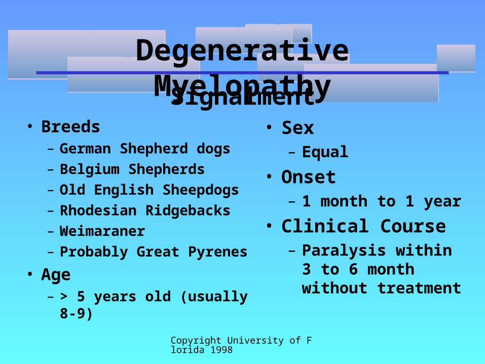
Signalment• Breeds
– German Shepherd dogs
– Belgium Shepherds
– Old English Sheepdogs
– Rhodesian Ridgebacks
– Weimaraner
– Probably Great Pyrenes
• Age– > 5 years old (usually 8-9)
• Sex– Equal
• Onset– 1 month to 1 year
• Clinical Course– Paralysis within 3 to 6
month without treatment

Copyright University of Florida 1998
Degenerative Myelopathy
Similar Conditions in Human Beings
• Multiple Sclerosis– Immune-related demyelinating disorder
• Amyotrophic Lateral Sclerosis– Axonal losing disease
• Genetic
• Free-radical association

Copyright University of Florida 1998
Degenerative Myelopathy
Progression
Time Clinical Disease

Copyright University of Florida 1998
Degenerative Myelopathy
Early Clinical Signs
• Mild Spinal Ataxia– Diminished Proprioception
– Slight Hyper-reflexia in Rear Legs
• Rear Leg Weakness– Slight Muscle Atrophy
• Occasionally, Atypical LMN Dysfunction

Copyright University of Florida 1998
Degenerative Myelopathy
Late Clinical Signs
• Severe Spinal Ataxia– Conscious Proprioceptive
Deficits
– Unconscious Proprioceptive Deficits
– Crossed-extensor Reflex
– Babinski’s Sign

Copyright University of Florida 1998
Degenerative Myelopathy
Late Clinical Signs
• Severe Motor Weakness– Loss of Weight Bearing
– Moderate Rear Leg Muscle Atrophy

Copyright University of Florida 1998
Degenerative Myelopathy
Histopathology
• Axon and myelin loss– Swollen axons
– Patchy demyelination
• Astrocyte proliferation• Increase in vasculature

Copyright University of Florida 1998
Degenerative Myelopathy
Diagnosis
• Physical and Neurologic Examination– History of chronic progressive posterior paresis
in susceptible breed– TL (non-localized) dysfunction

Copyright University of Florida 1998
Degenerative Myelopathy
Diagnosis
• EMG– Needle EMG- -normal– NCV- -normal– Repetitive Nerve Stimulation- -non-
decremental– Spinal Evoked Potential- -abnormal
Copyright University of Florida 1998
Degenerative Myelopathy
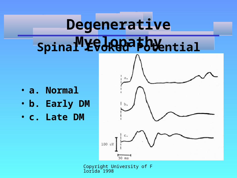
Spinal Evoked Potential
• a. Normal• b. Early DM• c. Late DM

Copyright University of Florida 1998
Degenerative Myelopathy
Diagnosis
• CSF tap (lumbar)– Increased protein with normal cells– Elevated inflammatory proteins– Increased acetylcholinesterase levels (2 X
normal)

Copyright University of Florida 1998
Degenerative Myelopathy
Diagnosis
• Spinal Radiographs– Plain radiographs- -spondylosis & spinal
arthritis– Myelography- -no significant lesions
• Immune Studies

Copyright University of Florida 1998
Degenerative Myelopathy
CSF Analysis
• Lumbar CSF tap and analysis– Normal cellularity– Slight to moderate increase in protein
content 80-120 mg/dl
• Normal BB Barrier– 258.4 + 92.7

Copyright University of Florida 1998
Degenerative Myelopathy
2-D Electrophoresis of CSF
• Normal • DM

Copyright University of Florida 1998
Degenerative Myelopathy
CSF Cholinesterase

Copyright University of Florida 1998
Degenerative Myelopathy
CSF Inflammatory Markers
• Increased Inflammatory Markers– IL6– ICAM– Leukotreine C, D, E
• No Markers for:– viral and bacterial infection– prion infection– TNF
Copyright University of Florida 1998
Degenerative Myelopathy
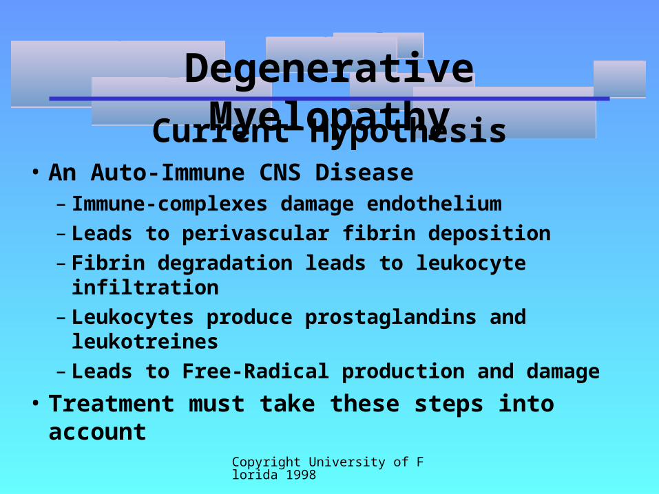
Current Hypothesis• An Auto-Immune CNS Disease
– Immune-complexes damage endothelium– Leads to perivascular fibrin deposition– Fibrin degradation leads to leukocyte infiltration– Leukocytes produce prostaglandins and
leukotreines– Leads to Free-Radical production and damage
• Treatment must take these steps into account
Related Documents