A GLÂNDULA PITUITÁRIA FICA ABAIXO DO CÉREBRO EM UM BOLSO DA LINHA MÉDIA OU FOSSA DO OSSO ESFENÓIDE CONHECIDA COMO A SELA TURCA, IMAGINATIVAMENTE CHAMADA POR ANATOMISTAS POR CAUSA DE SUA SEMELHANÇA COM UMA SELA DE CAVALO TURCO. EMBRIOLOGICAMENTE, ANATOMICAMENTE E FUNCIONALMENTE A GLÂNDULA HUMANA É DIVIDIDA EM DOIS LÓBULOS. O lobo anterior constitui 2/3 do volume da glândula e do lobo posterior de 1/3. Tal como acontece com todas as outras glândulas endócrinas, os sintomas surgem como o resultado de uma hipo ou hipersecreção de hormônios. CONEXÕES ANATÔMICAS E FUNCIONAIS DO EIXO HIPOTÁLAMO- HIPOFISÁRIO. A parte posterior da glândula pituitária (lobo neural ou neurohypophysis) é embriológica e anatomicamente contínua com o hipotálamo, é uma área de matéria cinzenta na parte basal da parte anterior do cérebro em torno do terceiro ventrículo. Os neurônios no hipotálamo se projetam diretamente para a glândula pituitária posterior e cerca de 100.000 axônios formam o trajeto do nervo hipófise. A glândula pituitária posterior é, portanto, formada a partir de axônios e terminais nervosos de neurônios hipotalâmicos; hormônios armazenados nos terminais são liberados na circulação geral em

Deficiência do crescer infantil juvenil por comprometimento de hormônios hipofisários associados.
Jan 27, 2017
Welcome message from author
This document is posted to help you gain knowledge. Please leave a comment to let me know what you think about it! Share it to your friends and learn new things together.
Transcript

A GLÂNDULA PITUITÁRIA FICA ABAIXO DO CÉREBRO EM UM BOLSO DA LINHA MÉDIA OU FOSSA DO OSSO ESFENÓIDE CONHECIDA COMO A SELA
TURCA, IMAGINATIVAMENTE CHAMADA POR ANATOMISTAS POR CAUSA DE SUA SEMELHANÇA COM UMA SELA DE CAVALO TURCO.
EMBRIOLOGICAMENTE, ANATOMICAMENTE E FUNCIONALMENTE A GLÂNDULA HUMANA É DIVIDIDA EM DOIS LÓBULOS.
O lobo anterior constitui 2/3 do volume da glândula e do lobo posterior
de 1/3. Tal como acontece com todas as outras glândulas endócrinas, os
sintomas surgem como o resultado de uma hipo ou hipersecreção de
hormônios.
CONEXÕES ANATÔMICAS E FUNCIONAIS DO EIXO HIPOTÁLAMO-
HIPOFISÁRIO.
A parte posterior da glândula pituitária (lobo neural ou
neurohypophysis) é embriológica e anatomicamente contínua com o
hipotálamo, é uma área de matéria cinzenta na parte basal da parte
anterior do cérebro em torno do terceiro ventrículo. Os neurônios no
hipotálamo se projetam diretamente para a glândula pituitária posterior
e cerca de 100.000 axônios formam o trajeto do nervo hipófise. A
glândula pituitária posterior é, portanto, formada a partir de axônios e
terminais nervosos de neurônios hipotalâmicos; hormônios
armazenados nos terminais são liberados na circulação geral em

resposta à excitação elétrica. Ao redor dos nervos terminais sofrem
modificações conhecidas como astrócitos pituicitos e essas células
parecem ter um papel importante no controle local da liberação de
hormônio. O lobo anterior (ou adenohipófise) é anatomicamente
distinto do hipotálamo e consiste de uma coleção de células endócrinas.
Foram identificados inicialmente três tipos de células diferentes de
acordo com a sua capacidade para assumir colorações histológicas
gerais; estas foram denominadas cromófobas, acidófilas e basófilas.
Técnicas de imunohistoquímica mais recentes permitiu a classificação
das células por seus produtos específicos de secreção.
Cerca de 50% das células secretoras da adeno-hipófise são somatotrófos
(que sintetizam somatotrofina ou GH), 10 a 25% (lactotrófos sintetizam
prolactina), 15 a 20% (corticotrófos sintetizam ACTH), 10 a 15%
(gonadotrófos sintetizam gonadotrofinas LH e FSH), e 3 a 5% (tireotrófos
sintetizam TSH). Algumas células, normalmente os cromófobos, não
colorem com qualquer um dos anticorpos para os vários dos hormônios
da pituitária anterior, embora à microscopia eletrônica revele que estas
células contêm grânulos secretórios. Enquanto a glândula pituitária
anterior, não seja anatomicamente relacionada com o hipotálamo, ele
está funcionalmente ligado com esta parte do cérebro. As células
nervosas do hipotálamo secretam neurohormônios que, através de um

sistema de vasos portais hipofisários na eminência mediana, agem sobre
as células endócrinas do lobo anterior para estimular ou inibir a sua
síntese e secreção. Dentro do hipotálamo, existem grupos distintos de
células nervosas, denominados núcleos, dispostas bilateralmente em
torno do terceiro ventrículo. Aqueles que se preocupam com as
secreções hormonais da glândula pituitária tendem a ser distribuídos
mais medialmente enquanto aqueles preocupados com funções
autônomas, como a regulação da temperatura, ingestão de alimentos,
saciedade e estimulação simpática de secreções adrenomedulares,
tendem a se localizar mais lateralmente. A glândula pituitária mantém
suas conexões anatômicas e funcionais, com o cérebro e ainda fica fora
da barreira hemato-encefálica. A parte anterior da sela túrcica é o
tubérculo da sela que é ladeado por projeções semelhantes a asas do
esfenóide conhecidos como os processos crinóides anterior. A parte
posterior, conhecida como o dorso da sela, é flanqueada pelos processos
crinóides posterior.

Estes processos crinóides são os pontos de fixação do diafragma da sela,
um reflexo da dura-máter que envolve o cérebro. Desta forma, toda a
glândula pituitária está rodeada por dura-máter de modo que a
membrana aracnoideia e, assim, o fluido cerebrospinal, não pode entrar
na sela turca. O suprimento de sangue para o eixo hipotálamo-
hipofisário é complexo, mas define a relação funcional entre o
hipotálamo e adenohipófise. Qualquer interrupção do fluxo de sangue
prejudica o controle hipotalâmico da secreção adeno- hipofisária. O
hipotálamo recebe seu suprimento de sangue a partir do polígono de
Willis, enquanto a neurohipófise e a adenohipófise recebem o sangue
das artérias hipofisárias inferior e superior, respectivamente. O plexo
capilar dos drenos da artéria hipofisária inferior no seio dural embora
alguns desses capilares na forma haste neurais veia porta "curto" que
drenam para a glândula pituitária anterior.
SÍNDROME DE SHEEHAN
Durante a gravidez, há um aumento de aproximadamente 50% no
volume da glândula pituitária. Isto é principalmente devido à hiperplasia
dos lactótrofos que secretam prolactina para preparar os seios para a
lactação. Assim, enquanto o volume da pituitária aumenta a fossa em
que a glândula pituitária se encontra não consegue suportar o seu
crescimento. A queda súbita da pressão arterial após um evento, como
uma hemorragia pós-parto causa isquemia da glândula, dano celular e
edema. Por sua vez, os resultados do edema da glândula pituitária (que
já é ampliada na hiperplasia dos lactótrofos) restringindo ainda mais o
fluxo normal de sangue para a glândula. O resultado é um enfarte na
glândula que provoca a perda das suas secreções.
DEFICIÊNCIA DE CRESCIMENTO E SOMATOTROFINA

Um exemplo para facilitar a compreensão: a idade é plotada no (X) eixo
horizontal. Dois conjuntos de dados normais são plotados. Altura (o
conjunto superior das curvas) está representada no lado esquerdo
vertical (Y) e de peso (o conjunto inferior das curvas) no eixo Y à direita.
Um menino, “neste caso, foi inicialmente designado por falta de
prosperar”, um termo geralmente utilizado para as crianças com menos
de 2 anos de idade que não estão focados em peso (ou seja, inclinar-se
para a sua altura). Para interpretar este caso, é necessário entender o
uso de gráficos de crescimento; a apresentação inicial estava mais perto
do 3º percentil para peso e do que 3º percentil para a altura. Em outras
palavras é uma criança de baixa estatura e com peso superior. Assim, ele
não estava conseguindo evoluir, ele não estava conseguindo crescer.
Com os pais no percentil 25, ele teria sido o esperado, todas as outras
coisas sendo iguais, também o crescer ao longo dessa linha. O
crescimento pós-natal nunca corresponde a esta com apenas um
aumento de 20 vezes em massa e um aumento de 3 a 4 vezes em
comprimento. Na infância, há um período de rápido crescimento seguido
de um período de pouco crescimento, com um retorno da aceleração do
crescimento, ou melhor, um surto de crescimento puberal e uma fase de
desaceleração até a altura final. O crescimento intra-uterino é regulado
pelo sistema endócrino, fatores maternos e genéticos, embora os
determinantes do crescimento pré-natal são pouco compreendidos. As
concentrações de GH no plasma fetal são muito elevadas e ainda
crianças com deficiência de GH, e mesmo aqueles com anencefalia,

podem ter o comprimento normal do corpo no momento do nascimento.
As influências maternas (intrauterinas) têm sido difíceis de definir, mas a
má nutrição materna é o fator mais importante que leva ao baixo peso
ao nascer e à duração a nível mundial.
A ingestão de álcool e o tabagismo são outros fatores adversos sobre o
crescimento fetal e infecções maternas, como rubéola, toxoplasmose e
citomegalovírus levar a muitas alterações, bem como à baixa estatura.
O GH E OS IGF:
A família IGF é composta por 3 membros (insulina, IGF-1 e IGF-2) com
semelhanças estruturais comuns de compartilhamento. Existem formas
variantes do IGF. O IGF-1 e o IGF-2, também têm funções metabólicas,
mas também desempenha um papel importante na maturação e
crescimento do feto, criança, infantil e juvenil.
DISABLED CHILD AND YOUTH GROWTH BY OTHER COMMITMENTS
PITUITARY HORMONE ASSOCIATES.
LOW HEIGHT: THE PITUITARY GLAND IS BELOW THE BRAIN IN A POCKET
OR AVERAGE LINE OF BONE FOSSA SPHENOID KNOWN AS THE TURKISH
SADDLE, IMAGINATIVELY CALL FOR ANATOMISTS BECAUSE OF HIS
LIKENESS WITH A SADDLE HORSE TURKISH. EMBRYOLOGICALLY,
ANATOMICALLY AND FUNCTIONALLY THE GLAND IS DIVIDED INTO TWO
LOBES. PHYSIOLOGY-ENDOCRINOLOGY-NEUROENDOCRINOLOGY-
GENETICS-ENDOCRINE-PEDIATRICS (SUBDIVISION OF ENDOCRINOLOGY):
DR. JOÃO SANTOS CAIO JR. ET DRA. HENRIQUETA VERLANGIERI CAIO.
The anterior lobe is two-thirds of the volume of the gland and the
posterior lobe is a third of the volume of the gland. As all other
endocrine glands, symptoms arise as the result of a hypo-or hyper
hormones.
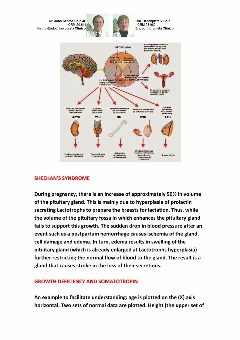
THE ANATOMICAL AND FUNCTIONAL CONNECTIONS OF THE
HYPOTHALAMIC-PITUITARY AXIS.
The posterior part of the pituitary (neurohypophysis or neural lobe) is
anatomically and embryologically continuous with the hypothalamus, an
area of gray matter in the basal part of the forebrain around the third
ventricle. Neurons in the hypothalamus project directly to the posterior
pituitary gland about 100.000 axons form the path of pituitary nerve.

The posterior pituitary is thus formed from axons and nerve terminals to
hypothalamic neurons; hormones stored in the terminal are released
into the general circulation in response to electrical excitation. Around
the nerve terminals are modified astrocytes known as pituicytes and
these cells appear to play an important role in local control of hormone
release. The previous (or adenohypophysis) lobe is anatomically distinct
from the hypothalamus and consists of a collection of endocrine cells.
Three different cells were initially identified according to their ability to
assume general histological staining; these were chromophobes,
acidophils and basophils. Most recent immunohistochemical techniques
enable sorting the cells by their specific secretory products.
Approximately 50% of the secretory cells are anterior pituitary
somatotrophs (which synthesize somatotropin or growth hormone, GH),
10 to 25% (Lactotrophs making prolactin), 15 to 20% corticotrophic
hormone (ACTH), 10 to 15% gonadotrophs (LH and FSH), and 3% to 5%
thyrotrophs (TSH). Some cells normally chromophobes not stain with
any of the various antibodies to the hormones of the anterior pituitary,
although electron microscopy reveals that these cells contain secretory
granules. While the anterior pituitary gland, is not anatomically
associated with the hypothalamus, it is functionally connected with this
part of the brain.

The nerve cells of the hypothalamus secrete neurohormones which, via a
pituitary portal vasculature in the median eminence, act on the
endocrine cells of the anterior lobe to stimulate or inhibit the synthesis
and secretion. Within the hypothalamus, different groups of nerve cells
called nuclei, bilaterally disposed around the third ventricle. Those
concerned with the hormonal secretions of the pituitary gland tend to be
distributed more medial while those concerned with autonomous
functions such as temperature regulation, food intake and satiety and
sympathetic stimulation of adrenomedullary secretions, tend to be more
laterally located. The pituitary gland maintains its anatomical and
functional connections with the brain still gets outside of the blood brain
barrier. The anterior part of the sella is the tuber of the saddle which is
flanked by wing-like sphenoid processes known as crinoid previous
projections. The back, known as the back of the saddle, is flanked by
later processes crinoids. These crinoids processes are the attachment
points of the diaphragm seals, a reflection of the dura mater surrounding
the brain. Therefore, all of the pituitary gland is surrounded by dura so
that the arachnoid membrane and thus the cerebrospinal fluid can not
enter the sella turcica. The blood supply to the hypothalamic-pituitary
axis is complex, but it defines the functional relationship between the
hypothalamus and anterior pituitary. Any interruption of blood flow
affect the hypothalamic control of pituitary adeno secretion. The
hypothalamus receives its blood supply from the circle of Willis, while
the neurohypophysis and adenohypophysis receives blood from the
lower and upper, respectively hypophyseal arteries. The capillary plexus
drains the inferior hypophyseal artery dural sinus although some of
these capillaries as neural stem vein "short" door that drain into the
anterior pituitary gland.
SHEEHAN'S SYNDROME
During pregnancy, there is an increase of approximately 50% in volume
of the pituitary gland. This is mainly due to hyperplasia of prolactin
secreting Lactotrophs to prepare the breasts for lactation. Thus, while
the volume of the pituitary fossa in which enhances the pituitary gland
fails to support this growth. The sudden drop in blood pressure after an
event such as a postpartum hemorrhage causes ischemia of the gland,
cell damage and edema. In turn, edema results in swelling of the
pituitary gland (which is already enlarged at Lactotrophs hyperplasia)
further restricting the normal flow of blood to the gland. The result is a
gland that causes stroke in the loss of their secretions.
GROWTH DEFICIENCY AND SOMATOTROPIN
An example to facilitate understanding: age is plotted on the (X) axis
horizontal. Two sets of normal data are plotted. Height (the upper set of

curves) is shown on the left vertical (Y) and weight (lower set of curves)
on the Y axis to the right. A boy, "this case was initially referred to as'
failure to thrive", a term generally used for children under 2 years of age
who are not focused on weight (e.g., leaning his height). To interpret this
case, it is necessary to understand the use of growth charts; the initial
presentation was closer to the third percentile for weight, than third
percentile for height. In other words it is a child of short stature and
weighing. Thus, he was unable to move, he was not getting growth.
Wtith parents on the 25th percentile, it would have been expected, all
other things being equal, also growing along that line. Postnatal growth
never matches this with just a 20-fold increase in mass and an increase
of 3-4 times in length. In childhood, there is a period of rapid growth
followed by a period of steady growth, with a mid-childhood
acceleration, the pubertal growth spurt and a deceleration phase until
final height. In involutionary years, there is a period of shrinkage, which
reflects the changes of bone marrow fat. Intrauterine growth is
regulated by the endocrine system, maternal and genetic factors,
although the determinants of pre-natal growth are poorly understood.
GH concentrations in fetal plasma is very high and even children with GH
deficiency hormone, and even those with anencephaly, may have
normal body length at birth. Maternal (uterine) influences have been
difficult to define, but poor maternal nutrition is the most important
factor that leads to low birth weight and duration worldwide. Alcohol
intake and smoking are other adverse factors on fetal growth and
maternal infections such as rubella, toxoplasmosis and cytomegalovirus
lead to many changes, and short stature.
THE GH AND IGF:
The IGF family consists of 3 members (insulin, IGF-1 and IGF-2) common
share structural similarities. There are variant forms of the IGF. The IGF-1
and IGF-2 also has metabolic functions, but also play a major role in fetal
growth, maturation, infants, children and juveniles.
Dr. João Santos Caio Jr.
Endocrinologia – Neuroendocrinologista
CRM 20611
Dra. Henriqueta V. Caio
Endocrinologista – Medicina Interna
CRM 28930
AUTORIZADO O USO DOS DIREITOS AUTORAIS COM CITAÇÃO DOS
AUTORES PROSPECTIVOS ET REFERÊNCIA BIBLIOGRÁFICA.
Como saber mais:
1. Os genes para essas proteínas provavelmente evoluíram de um
ancestral comum, mesmo que os genes sejam localizados em
cromossomos diferentes (cromossomo 6 para prolactina e cromossomo
17 para GH)...
http://hormoniocrescimentoadultos.blogspot.com
2. Os genes para GH, prolactina e lactogênio placentário têm uma
organização estrutural eliminada na excisão e é chamada de íntron (o
processo de reconhecimento de introns e exons em genes para definir
quais trechos serão transcritos em uma cadeia de RNA guarda uma
admirável complexidade) separando cinco éxons...
http://longevidadefutura.blogspot.com
3. É um segmento de DNA de um gene eucarioto cuja transcrição
sobrevive ao processo de excisão (ou splicing)...
http://imcobesidade.blogspot.com

Referências Bibliográficas:
Caio Jr, João Santos, Dr.; Endocrinologista, Neuroendocrinologista, Caio,H. V., Dra. Endocrinologista, Medicina Interna – Van Der
Häägen Brazil, São Paulo, Brasil; Lee SL, Sadovsky Y, Swirnoff AH, Polish JA, Goda P, et al. 1996. Luteinizing hormone deficiency
and female infertility in mice lacking; the transcription factor NGFI-A (Egr-1); Science 273:1219–21; Li S, Crenshaw EB III, Rawson
EJ, Simmons; DM, Swanson LW, Rosenfeld MG. 1990; Dwarf locus mutants lacking three pituitary cell-types result from
mutations in the POUdomain gene pit-1. Nature 347:528–33 Lin CR, Koussi C, O’Connell S, Briata P, Szeto D, et al. 1999; Pitx2
regulates lung asymmetry, cardiac positioning and pituitary and tooth morphogenesis. Nature 401:279–82 Lin SC, Li S, Drolet
DW, Rosenfeld MG. 1994; Pituitary ontogeny of the Snell dwarf mouse reveals Pit-1-independent and Pit-1- dependent origins
of the thyrotrope. Development 120:515–22; Lin SC, Lin CR, Gukovsky I, Lusis AJ, Sawchenko PE, Rosenfeld MG. 1993. Molecular
basis of the little mouse phenotype and implications for cell type-specific growth. Nature 364:208–13 Lipkin SM, Naar AM, Kalla
KA, Sack RA, Rosenfeld MG. 1993. Identification of a novel zinc finger protein binding a conserved element critical for Pit-1-
dependent growth hormone gene expression. Genes Dev. 7:1674–87; Logan M, Tabin CJ. 1999. Role of Pitx1 upstream of Tbx4
in specification of hindlimb identity. Science 283:1736–39 Lu MF, Pressman C, Dyer R, Johnson RL, Martin JF. 1999. Function of
Rieger syndrome gene in left-right asymmetry and craniofacial development. Nature 401: 276–78; Martin GR. 1998. The roles of
FGFs in the early development of vertebrate limbs. Genes Dev. 12:1571–86 Mead PE.
Site Van Der Häägen Brazil
www.vanderhaagenbrazil.com.br
www.clinicavanderhaagen.com.br
www.crescimentoinfoco.com
www.obesidadeinfoco.com.br
http://drcaiojr.site.med.br
http://dracaio.site.med.br
Joao Santos Caio Jr http://google.com/+JoaoSantosCaioJr google.com/+JoãoSantosCaioJrvdh google.com/+VANDERHAAGENBRAZILvdh Video http://youtu.be/woonaiFJQwY VAN DER HAAGEN BRAZI
Instagram https://instagram.com/clinicascaio/ Google Maps: http://maps.google.com.br/maps/place?cid=5099901339000351730&q=Van+Der+Haagen+Brasil&hl=pt&sll=-23.578256,46.645653&sspn=0.005074,0.009645&ie =UTF8&ll=-23.575591,-46.650481&spn=0,0&t = h&z=17
Related Documents