Defective Secretion of Islet Hormones in Chromogranin- B Deficient Mice Stefanie Obermu ¨ ller 1. , Federico Calegari 2,3.¤ , Angus King 4 , Anders Lindqvist 1 , Ingmar Lundquist 1 , Albert Salehi 1 , Maura Francolini 3 , Patrizia Rosa 3 , Patrik Rorsman 5 , Wieland B. Huttner 2,4 *, Sebastian Barg 1,6 * 1 Department of Clinical Sciences-Malmo ¨ , Lund University, Malmo ¨ , Sweden, 2 Max Planck Institute of Molecular Cell Biology and Genetics, Dresden, Germany, 3 CNR Institute of Neuroscience, Department of Medical Pharmacology, University of Milan, Milan, Italy, 4 Department of Neurobiology, University of Heidelberg, Heidelberg, Germany, 5 Oxford Centre for Diabetes, Endocrinology and Metabolism (OCDEM), University of Oxford, Churchill Hospital, Oxford, United Kingdom, 6 Department of Medical Cell Biology, Uppsala University, Uppsala, Sweden Abstract Granins are major constituents of dense-core secretory granules in neuroendocrine cells, but their function is still a matter of debate. Work in cell lines has suggested that the most abundant and ubiquitously expressed granins, chromogranin A and B (CgA and CgB), are involved in granulogenesis and protein sorting. Here we report the generation and characterization of mice lacking chromogranin B (CgB-ko), which were viable and fertile. Unlike neuroendocrine tissues, pancreatic islets of these animals lacked compensatory changes in other granins and were therefore analyzed in detail. Stimulated secretion of insulin, glucagon and somatostatin was reduced in CgB-ko islets, in parallel with somewhat impaired glucose clearance and reduced insulin release, but normal insulin sensitivity in vivo. CgB-ko islets lacked specifically the rapid initial phase of stimulated secretion, had elevated basal insulin release, and stored and released twice as much proinsulin as wildtype (wt) islets. Stimulated release of glucagon and somatostatin was reduced as well. Surprisingly, biogenesis, morphology and function of insulin granules were normal, and no differences were found with regard to b-cell stimulus-secretion coupling. We conclude that CgB is not required for normal insulin granule biogenesis or maintenance in vivo, but is essential for adequate secretion of islet hormones. Consequentially CgB-ko animals display some, but not all, hallmarks of human type-2 diabetes. However, the molecular mechanisms underlying this defect remain to be determined. Citation: Obermu ¨ ller S, Calegari F, King A, Lindqvist A, Lundquist I, et al. (2010) Defective Secretion of Islet Hormones in Chromogranin-B Deficient Mice. PLoS ONE 5(1): e8936. doi:10.1371/journal.pone.0008936 Editor: Kathrin Maedler, University of Bremen, Germany Received July 13, 2009; Accepted January 11, 2010; Published January 28, 2010 Copyright: ß 2010 Obermu ¨ ller et al. This is an open-access article distributed under the terms of the Creative Commons Attribution License, which permits unrestricted use, distribution, and reproduction in any medium, provided the original author and source are credited. Funding: S.O. was a fellow of the Diabetes Programme at Lund University (DPLU), S.B. was supported by the Swedish Science Council (Vetenskapsra ˚det), the European Foundation for the Study of Diabetes, and long-term fellowships from European Molecular Biology Organization (EMBO), the Knut and Alice Wallenberg Foundation, Zetterlings stiftelse, and Go ¨ ran Gustafsson stiftelse. W.B.H. was supported by grants from the Deutsche Forschungsgemeinschaft (DFG) and the Fonds der Chemischen Industrie. The funders had no role in study design, data collection and analysis, decision to publish, or preparation of the manuscript. Competing Interests: The authors have declared that no competing interests exist. * E-mail: [email protected] (WBH); [email protected] (SB) . These authors contributed equally to this work. ¤ Current address: DFG Research Center and Cluster of Excellence for Regenerative Therapies Dresden, Technische Universita ¨t Dresden, c/o Max Planck Institute of Molecular Cell Biology and Genetics, Dresden, Germany Introduction Granins form a family of highly acidic proteins that are primarily found in the lumen of dense-core secretory granules of endocrine cells and neurons. The most abundant members of this family are chromogranin A (CgA), chromogranin B (CgB), and secretogranin II (SgII) which are characterized by i) highly hydrophilic, acidic primary amino acid sequences, ii) the presence of multiple dibasic sites as potential targets for proteolytic processing, iii) a multitude of post-translational modifications, and iv) the tendency to self-aggregate at low pH/high Ca 2+ conditions typical of the lumen of the trans-Golgi network (TGN) and secretory granules[1,2,3,4]. The precise function of chromo- granins is still debated, but evidence points in four main directions. First, both CgA and CgB have been implicated in granulogen- esis [1,5]. Down-regulation of CgA [6] and CgB [7] resulted in the loss of secretory granules in PC12 cells and overexpression induced granule biogenesis in non-secretory cells [6,7,8], suggesting that granin expression is essential and sufficient to the neuroendocrine phenotype. This view has been challenged by the findings that granules can form independently of CgA in PC12 [9] and mouse chromaffin cells [10,11] and that in non- secretory cells exogenous granins accumulate in dense compart- ments that may resemble, but are not identical to, secretory granules [9,12,13]. Second, granins play a role in the sorting and packaging of neuropeptides in granules within the TGN [14,15,16]. For example, in the TGN SgIII binds both to the proposed sorting receptor carboxypeptidase E (CPE) [17] and CgA and mediates targeting of proopiomelanocortin-derived peptides to granules [18]. Similarly, targeting of CgB to granules depends both on its self-aggregation and binding to an unknown sorting receptor PLoS ONE | www.plosone.org 1 January 2010 | Volume 5 | Issue 1 | e8936

Welcome message from author
This document is posted to help you gain knowledge. Please leave a comment to let me know what you think about it! Share it to your friends and learn new things together.
Transcript
Defective Secretion of Islet Hormones in Chromogranin-B Deficient MiceStefanie Obermuller1., Federico Calegari2,3.¤, Angus King4, Anders Lindqvist1, Ingmar Lundquist1,
Albert Salehi1, Maura Francolini3, Patrizia Rosa3, Patrik Rorsman5, Wieland B. Huttner2,4*, Sebastian
Barg1,6*
1 Department of Clinical Sciences-Malmo, Lund University, Malmo, Sweden, 2 Max Planck Institute of Molecular Cell Biology and Genetics, Dresden, Germany, 3 CNR
Institute of Neuroscience, Department of Medical Pharmacology, University of Milan, Milan, Italy, 4 Department of Neurobiology, University of Heidelberg, Heidelberg,
Germany, 5 Oxford Centre for Diabetes, Endocrinology and Metabolism (OCDEM), University of Oxford, Churchill Hospital, Oxford, United Kingdom, 6 Department of
Medical Cell Biology, Uppsala University, Uppsala, Sweden
Abstract
Granins are major constituents of dense-core secretory granules in neuroendocrine cells, but their function is still a matterof debate. Work in cell lines has suggested that the most abundant and ubiquitously expressed granins, chromogranin Aand B (CgA and CgB), are involved in granulogenesis and protein sorting. Here we report the generation andcharacterization of mice lacking chromogranin B (CgB-ko), which were viable and fertile. Unlike neuroendocrine tissues,pancreatic islets of these animals lacked compensatory changes in other granins and were therefore analyzed in detail.Stimulated secretion of insulin, glucagon and somatostatin was reduced in CgB-ko islets, in parallel with somewhatimpaired glucose clearance and reduced insulin release, but normal insulin sensitivity in vivo. CgB-ko islets lackedspecifically the rapid initial phase of stimulated secretion, had elevated basal insulin release, and stored and releasedtwice as much proinsulin as wildtype (wt) islets. Stimulated release of glucagon and somatostatin was reduced as well.Surprisingly, biogenesis, morphology and function of insulin granules were normal, and no differences were found withregard to b-cell stimulus-secretion coupling. We conclude that CgB is not required for normal insulin granule biogenesisor maintenance in vivo, but is essential for adequate secretion of islet hormones. Consequentially CgB-ko animals displaysome, but not all, hallmarks of human type-2 diabetes. However, the molecular mechanisms underlying this defect remainto be determined.
Citation: Obermuller S, Calegari F, King A, Lindqvist A, Lundquist I, et al. (2010) Defective Secretion of Islet Hormones in Chromogranin-B Deficient Mice. PLoSONE 5(1): e8936. doi:10.1371/journal.pone.0008936
Editor: Kathrin Maedler, University of Bremen, Germany
Received July 13, 2009; Accepted January 11, 2010; Published January 28, 2010
Copyright: � 2010 Obermuller et al. This is an open-access article distributed under the terms of the Creative Commons Attribution License, which permitsunrestricted use, distribution, and reproduction in any medium, provided the original author and source are credited.
Funding: S.O. was a fellow of the Diabetes Programme at Lund University (DPLU), S.B. was supported by the Swedish Science Council (Vetenskapsradet), theEuropean Foundation for the Study of Diabetes, and long-term fellowships from European Molecular Biology Organization (EMBO), the Knut and Alice WallenbergFoundation, Zetterlings stiftelse, and Goran Gustafsson stiftelse. W.B.H. was supported by grants from the Deutsche Forschungsgemeinschaft (DFG) and theFonds der Chemischen Industrie. The funders had no role in study design, data collection and analysis, decision to publish, or preparation of the manuscript.
Competing Interests: The authors have declared that no competing interests exist.
* E-mail: [email protected] (WBH); [email protected] (SB)
. These authors contributed equally to this work.
¤ Current address: DFG Research Center and Cluster of Excellence for Regenerative Therapies Dresden, Technische Universitat Dresden, c/o Max Planck Institute ofMolecular Cell Biology and Genetics, Dresden, Germany
Introduction
Granins form a family of highly acidic proteins that are
primarily found in the lumen of dense-core secretory granules of
endocrine cells and neurons. The most abundant members of this
family are chromogranin A (CgA), chromogranin B (CgB), and
secretogranin II (SgII) which are characterized by i) highly
hydrophilic, acidic primary amino acid sequences, ii) the presence
of multiple dibasic sites as potential targets for proteolytic
processing, iii) a multitude of post-translational modifications,
and iv) the tendency to self-aggregate at low pH/high Ca2+
conditions typical of the lumen of the trans-Golgi network (TGN)
and secretory granules[1,2,3,4]. The precise function of chromo-
granins is still debated, but evidence points in four main directions.
First, both CgA and CgB have been implicated in granulogen-
esis [1,5]. Down-regulation of CgA [6] and CgB [7] resulted in
the loss of secretory granules in PC12 cells and overexpression
induced granule biogenesis in non-secretory cells [6,7,8],
suggesting that granin expression is essential and sufficient to
the neuroendocrine phenotype. This view has been challenged by
the findings that granules can form independently of CgA in
PC12 [9] and mouse chromaffin cells [10,11] and that in non-
secretory cells exogenous granins accumulate in dense compart-
ments that may resemble, but are not identical to, secretory
granules [9,12,13].
Second, granins play a role in the sorting and packaging of
neuropeptides in granules within the TGN [14,15,16]. For
example, in the TGN SgIII binds both to the proposed sorting
receptor carboxypeptidase E (CPE) [17] and CgA and mediates
targeting of proopiomelanocortin-derived peptides to granules
[18]. Similarly, targeting of CgB to granules depends both on its
self-aggregation and binding to an unknown sorting receptor
PLoS ONE | www.plosone.org 1 January 2010 | Volume 5 | Issue 1 | e8936
[19], which suggests that granins act as an assembly factor in the
TGN that may recruit other proteins into the budding granule
[16].
Third, granins may participate in the structural matrix of the
granule lumen [20] and facilitate storage of transmitter molecules
such as ATP and catecholamines [21,22]. Granins have pH-
buffering capability and bind and release large amounts of ATP,
Ca2+ and catecholamines [3,22]. Knockout of CgA leads to a
reduction in the amount of catecholamine stored in chromaffin
granules [23].
Fourth, granin-derived peptides are secreted during regulated
exocytosis and might thus exert hormonal, autocrine, and
paracrine activities [4,24,25,26]. The perhaps best known example
is pancreastatin, a CgA derived peptide with strong inhibitory
action on insulin secretion [25]. Antisera against CgB have been
reported to stimulate insulin release [27].
Here we report the generation of a mouse line that lacks CgB
(CgB-ko). These mice were viable and fertile but exhibited defects
in hormone secretion from pancreatic islets, which were paralleled
by slight glucose intolerance. Surprisingly, synthesis and function
of insulin granules were normal, which argues against a role of
CgB in granulogenesis.
Results
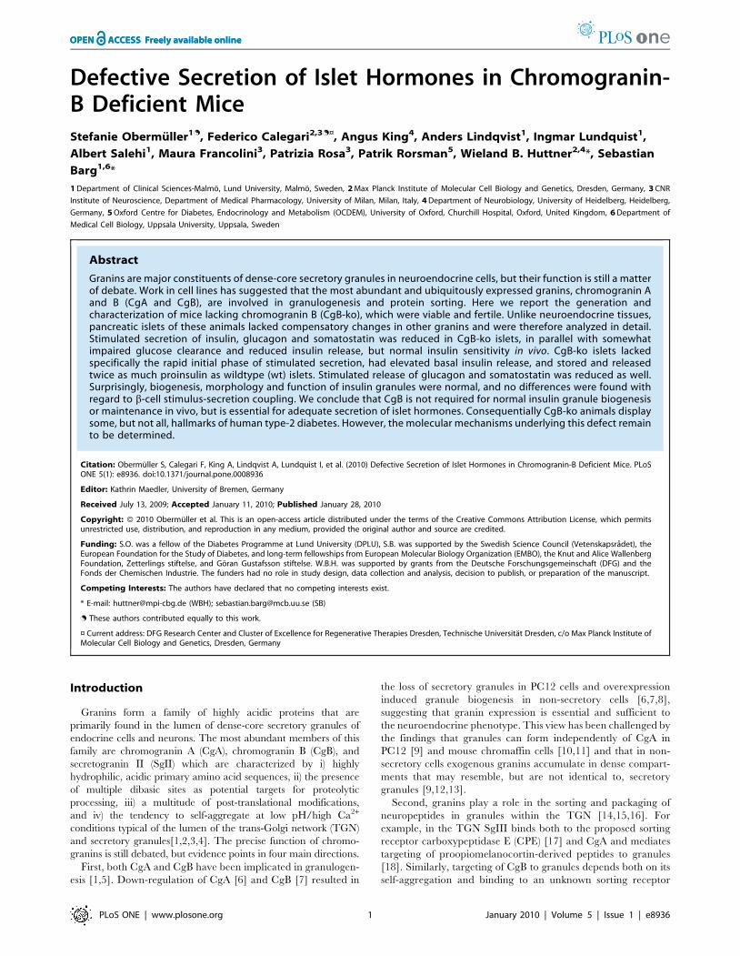
Generation of CgB-ko MiceThe deletion of the mCgB gene was obtained by homologous
recombination of a targeting vector which replaced 117 bases of
the proximal promoter region and the first 29 bases of the coding
sequence of CgB with the neomycin resistance gene (Fig 1A). ES
clones were screened for homologous recombination by Southern
blot analysis and positive clones were injected into C57BL/6J
blastocysts to generate chimeras. CgB-ko mice were identified in
the F2 by Southern blot analyses (Fig 1B) and lack of CgB protein
confirmed by Western blot analysis of adrenal tissue (Fig 1C). Like
CgA-ko mice [11], CgB-ko animals are viable and fertile, and
showed no obvious phenotype with respect to weight, mating
performance and overall behavior.
Expression of Other GraninsThe lack of an obvious phenotype in CgB-ko mice could be
explained by compensatory overexpression of other granins such
as CgA and SgII [11]. We therefore compared the abundance of
CgA and SgII in adrenal, pituitary and isolated pancreatic islets
obtained from 3–6 months old wt or CgB-ko mice, of both sexes,
Figure 1. Generation of the CgB-ko mouse line. (A) Top, map of the mCgb locus (exons indicated as black bars) and region encoding the probe(probe A) used for Southern blot analysis. Bottom, construct used for homologous recombination containing the neomycin (Neo) and HSV-TKcassettes used for double drug selection. Relevant restriction sites and scale in kb are indicated. (B) Southern blot of HindIII-digested genomic DNA ofCgB wt (+/+), heterozygous (+/-), and CgB-ko (2/2) mice. (C) Western blot for CgB in adrenals glands of wt (+/+), heterozygous (+/2) and CgB-ko(2/2) mice, and PC12 cells as control. Note the higher molecular weight of rat CgB (rCgB) from PC12 cells compared to that of mouse CgB (mCgB).(D) Western blot analysis of CgA and SgII expression, in adrenal gland, pituitary gland and pancreatic islets from wt (+/+) and CgB-ko (2/2) mice.Tubulin (tub) was used as loading control. (E) Quantification of experiments as in (E), for CgA (black bars) and SgII (white bars). Values representexpression relative to the corresponding wt sample (100%) after internal normalization with the tubulin signal.doi:10.1371/journal.pone.0008936.g001
Hormone Secretion in CgB-ko
PLoS ONE | www.plosone.org 2 January 2010 | Volume 5 | Issue 1 | e8936
by Western blot analysis (Fig 1D). CgB-ko mice had i) increased
levels of both CgA and SgII in adrenal glands (167625% of wt,
n = 8 and 163620%, n = 12, respectively), ii) similar levels of CgA
expression but a robust increase of SgII in pituitary glands
(97614%, n = 9 and 207639%, n = 10, respectively), and iii)
similar levels of both CgA and SgII expression in islets (73622%,
n = 4 and 131617%, n = 4, respectively) (Fig 1D and E). Thus, the
lack of CgB induces increased expression of other granins in some,
but not all, endocrine tissues. Since the changes were minor in
pancreatic islets, we decided to focus our study on the endocrine
pancreas, where effects of CgB ablation can be studied in the
absence of compensatory effects on other granins. Importantly, the
expression of the granule membrane proteins carboxypeptidase E
(CPE) and phogrin (IA-2b) was not altered in islets obtained from
CgB-ko as compared to those obtained from wt mice (9367%,
n = 4 and 10069%, n = 4; not shown), indicating that the overall
constituents of secretory granules are similar in the absence of
CgB.
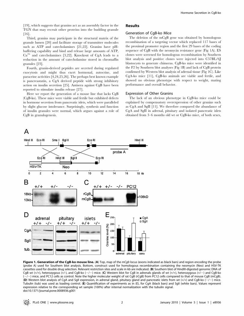
Decreased Hormone Secretion from Islets of LangerhansIn Vitro
Islets were isolated and hormone release measured at glucose
concentrations ranging from 0 to 25 mM (Fig 2). Insulin secretion
from wt islets was observed at glucose .5 mM, and increased
sigmoidally with the glucose concentration to a maximum rate of
3.660.5 ng/(islet*h) at 25 mM glucose. Half-maximal stimulation
(EC50) was reached at 13 mM glucose (Fig 2A). Strikingly, islets
from CgB-ko mice had a nearly linear response to glucose with a
maximum release rate of only 2.260.4 ng/(islet*h) at 25 mM
glucose. Insulin release at low glucose (2–8 mM) was significantly
higher in CgB-ko than in wt islets, but still lower than in wt islets in
stimulatory glucose (16–25 mM; p,0.01 at 2–25 mM glucose; see
Fig 2A). Insulin content was similar in both groups, with 2468 ng
insulin/islet found in wt (2962% of total protein, n = 9) and 3469
ng/islet in the CgB-ko islets (3464% of total protein, n = 8; data
not shown).
Glucagon was measured in the same samples (Fig 2B). As
expected, glucose had a bimodal effect on glucagon release with a
maximal inhibition at 8 mM in both wt and CgB-ko islets. At 0–2
mM and again at 25 mM glucose, release in the CgB-ko was
significantly reduced compared to wt. In the absence of glucose,
CgB-ko islets released 2666 pg/(islet21*h), i.e. 30% less than wt
islets which released 3865 pg/(islet21*h). Thus, in CgB-ko islets
stimulated glucagon secretion at both low and high glucose
concentrations was reduced compared to wt, while secretion at
intermediate blood glucose (8 mM point) was unchanged.
Somatostatin was released from wt islets at a rate of 1.160.1
pmol/(islet*h) at 0 mM glucose, and increased sigmoidally with the
glucose concentration (EC50 = 4.4 mM) to a maximum of 5.760.4
pmol/(islet*h) in 25 mM glucose (Fig 2C). Islets from CgB-ko mice
showed a similar glucose dependent increase of somatostatin
release (EC50 = 6.6 mM), but secretion was reduced to about half
of that measured from wt islets, at all glucose concentrations
(p,0.01) (Fig 2C).
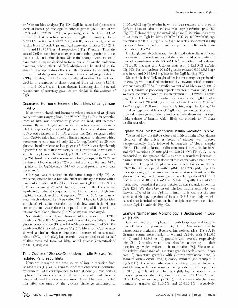
Time Course of Glucose-Dependent Insulin Release fromIsolated Pancreatic Islets
Next, we measured the time course of insulin secretion from
isolated islets (Fig 3A-B). Similar to what is observed in perifusion
experiments, wt islets responded to high glucose (20 mM) with a
biphasic timecourse characterized by a transient rapid phase of
release followed by a slower sustained phase. The peak rate 4–6
min after the onset of the glucose challenge amounted to
0.10560.002 ng/(islet*min) in wt, but was reduced to a third in
CgB-ko islets (maximum 0.03460.004 ng/(islet*min); p,0.002)
(Fig 3B). Release during the sustained phase (8–20 min) was slower
in wt than in CgB-ko islets (0.00760.002 vs. 0.02260.002 ng/
(islet*min), p,0.001) (Fig 3A–B). CgB-ko islets also showed slightly
increased basal secretion, confirming the results with static
incubations (Fig 2A).
Unlike glucose, depolarization by elevated extracellular K+ does
not sustain insulin release beyond the initial rapid phase. After 15
min of stimulation with 50 mM K+, wt islets had released
0.7160.05 ng/islet and CgB-ko islets only 0.4560.03 ng/islet
(Fig 3C). For comparison, 20 mM glucose released 0.8360.11 ng/
islet in wt and 0.4960.1 ng/islet in the CgB-ko (Fig 3C).
Since the lack of CgB might affect insulin storage or proinsulin
processing, we quantified proinsulin by enzyme-linked immuno-
sorbent assay (ELISA). Proinsulin content of wt islets was 5.461.2
ng/islet, similar to previously reported values in mouse [28]. CgB-
ko islets contained twice as much proinsulin, 11.262.0 ng/islet
(Fig 3D). Likewise, proinsulin secretion from CgB-ko islets
stimulated with 20 mM glucose was elevated, with 85614 and
154625 pg/islet*30 min in wt and CgB-ko, respectively (Fig 3E).
Taken together, ablation of CgB leads to modest increase in
proinsulin storage and release and selectively decreases the rapid
initial release of insulin, which likely corresponds to 1st phase
insulin secretion.
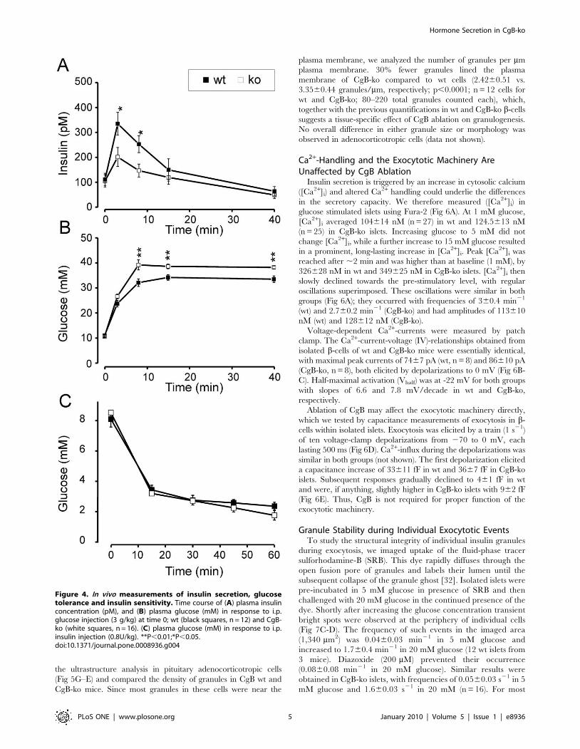
CgB-ko Mice Exhibit Abnormal Insulin Secretion In VivoWe tested how the defects observed in islets might affect glucose
tolerance of the mice. A bolus of glucose was injected
intraperitoneally (i.p.), followed by analysis of blood samples
(Fig 4). The initial plasma insulin concentration was similar in wt
and CgB-ko mice (100622 pM vs. 93621 pM; Fig 4). All mice
responded to the glucose challenge with a transient increase in
plasma insulin, which then declined to baseline with a half-time of
,10 min. The peak in plasma insulin was higher in the wt
(332646 pM), compared with CgB-ko (200638 pM) (Fig 4A).
Correspondingly, the wt mice were somewhat more resistant to the
glucose challenge and plasma glucose reached peaks of 33.961.1
mM in wt and 38.360.9 mM in CgB-ko (Fig 4B). Lack of CgB
might affect peripheral glucose uptake, as was recently shown for
CgA [29]. We therefore tested whether insulin sensitivity was
likewise affected in the CgB-ko animals. This was not the case
since a single i.p. injection of insulin (0.8 U/kg body weight)
caused near identical reductions in blood glucose over time in both
wt and CgB-ko animals (Fig 4C).
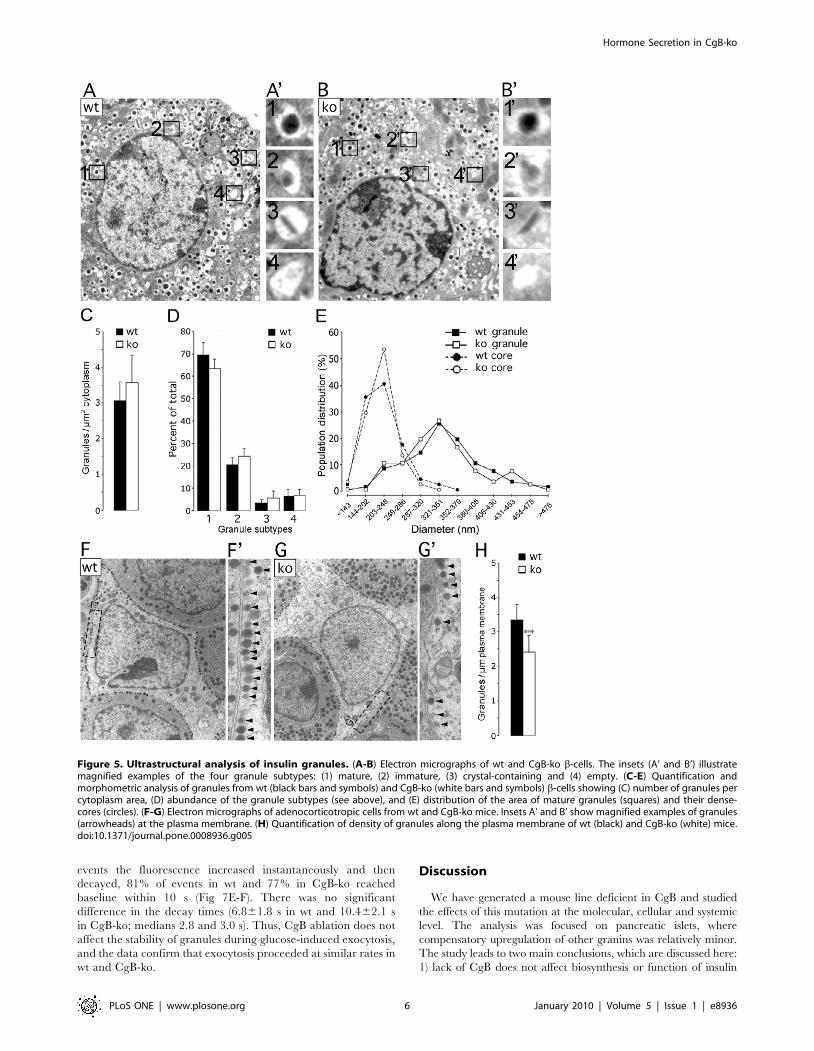
Granule Number and Morphology Is Unchanged in CgB-ko b-Cells
Granins have been implicated in both biogenesis and matura-
tion of secretory granules [1,5,6,7,8,16]. We tested this by
ultrastructure analysis of b-cells within isolated islets (Fig 5 A,B).
Granule counts were similar in wt and CgB-ko with 3.160.6
(n = 9) and 3.560.8 (n = 9) granules/mm2 cytosol, respectively
(Fig 5C). Granules were then classified according to their
morphology, which reflects their maturation [30]. We assessed
the relative abundance of 1) mature granules with electron-dense
core, 2) immature granules with electron-translucent core, 3)
granules with a crystal and, 4) empty granules (see examples in
Fig 5A’-B’). The relative abundance of all types was similar in wt
and CgB-ko, with most granules having a dense or opaque core
(,70%, Fig 5D). Wt cells had a slightly higher proportion of
mature granules than CgB-ko (mean6sd: 74.364.5% and
68.064.4%, respectively; p,0.01), and correspondingly fewer
immature granules (21.963.5% and 26.063.1%, respectively;
Hormone Secretion in CgB-ko
PLoS ONE | www.plosone.org 3 January 2010 | Volume 5 | Issue 1 | e8936
p,0.02) (n = 9 cells wt and CgB-ko; 100–500 total granules
counted each). Finally, the size of mature granules and their cores
was analyzed. Histograms of the cross-section granule area in wt
and CgB-ko essentially overlap (peaks around 0.09 mm2) (Fig 5E;
squares), as do the histograms for the cross-section area of the
electron-dense core (peaks around 0.03 mm2). The corresponding
average value for the granule diameter (0.34 mm, assuming
spherical geometry) is in good agreement with earlier measure-
ments (357 nm) [31]. Therefore, lack of CgB has no major affect
on insulin granule biogenesis or maturation, but causes a modest
increase in immature granules at the expense of those that are
mature. These changes are unlikely to be a primary cause of
defected insulin secretion.
Some of the granules near the plasma membrane may be
docked and therefore more likely to undergo exocytosis. However,
the proportion of docked granules, defined as visually connected to
the plasma membrane, was similar in wt and CgB-ko (9.363.5%
and 9.863.3%, respectively). Moreover, no difference was
observed in the proportion of granules found within 1.5 granule
diameters of the plasma membrane of wt and CgB-ko b-cells
(2067.7% and 19.867.2%, respectively) (Fig 5F) (n = 9 cells for wt
and CgB-ko; 100–500 total granules counted each).
Chromogranin dependent effects on granulogenesis have been
observed in other tissues [5,6,7,8,9]. Since we failed to detect any
morphological changes in CgB-ko insulin granules, we reasoned
that such effects may be tissue specific. To test this, we repeated
Figure 2. Hormone secretion from isolated pancreatic islets.Quantification of insulin (A), glucagon (B) and somatostatin (C) releasefrom isolated wt (black squares) and CgB-ko (white squares) islets
during 1 h incubation at various glucose concentrations (0–25 mM);n = 8. **P,0.01; ***P,0.001.doi:10.1371/journal.pone.0008936.g002
Figure 3. Timecourse of glucose- and K+-induced insulinrelease from isolated islets. (A) Isolated wt (black) and CgB-ko(white) islets were incubated in the presence of 1 mM (circles) or20 mM (squares) glucose. Insulin in the medium was measured at 2 minintervals during 20 min after adjusting the glucose concentration; n = 4.(B) Same data as in A, but plotted as release per 2 min. (C) Insulinsecretion from wt islets (black) and CgB-ko (white) islets was measuredafter 15 min incubation in the following conditions: 1 mM gluco-se+5 mM K+ (1G), 20 mM glucose+5 mM K+ (20 G) and 1 mMglucose+50 mM K+ (50 K+); n = 10. (D) Proinsulin content of wt (black)and CgB-ko islets (white); (n = 8 each). (E) Proinsulin release during30 min batch incubations in buffer containing 20 mM glucose.*P,0.05; **P,0.01; ***P,0.001.doi:10.1371/journal.pone.0008936.g003
Hormone Secretion in CgB-ko
PLoS ONE | www.plosone.org 4 January 2010 | Volume 5 | Issue 1 | e8936
the ultrastructure analysis in pituitary adenocorticotropic cells
(Fig 5G–E) and compared the density of granules in CgB wt and
CgB-ko mice. Since most granules in these cells were near the
plasma membrane, we analyzed the number of granules per mm
plasma membrane. 30% fewer granules lined the plasma
membrane of CgB-ko compared to wt cells (2.4260.51 vs.
3.3560.44 granules/mm, respectively; p,0.0001; n = 12 cells for
wt and CgB-ko; 80–220 total granules counted each), which,
together with the previous quantifications in wt and CgB-ko b-cells
suggests a tissue-specific effect of CgB ablation on granulogenesis.
No overall difference in either granule size or morphology was
observed in adenocorticotropic cells (data not shown).
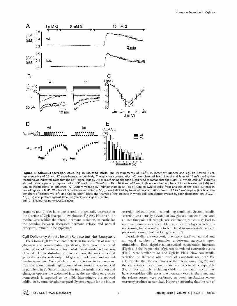
Ca2+-Handling and the Exocytotic Machinery AreUnaffected by CgB Ablation
Insulin secretion is triggered by an increase in cytosolic calcium
([Ca2+]i) and altered Ca2+ handling could underlie the differences
in the secretory capacity. We therefore measured ([Ca2+]i) in
glucose stimulated islets using Fura-2 (Fig 6A). At 1 mM glucose,
[Ca2+]i averaged 104614 nM (n = 27) in wt and 124.5613 nM
(n = 25) in CgB-ko islets. Increasing glucose to 5 mM did not
change [Ca2+]i, while a further increase to 15 mM glucose resulted
in a prominent, long-lasting increase in [Ca2+]i. Peak [Ca2+]i was
reached after ,2 min and was higher than at baseline (1 mM), by
326628 nM in wt and 349625 nM in CgB-ko islets. [Ca2+]i then
slowly declined towards the pre-stimulatory level, with regular
oscillations superimposed. These oscillations were similar in both
groups (Fig 6A); they occurred with frequencies of 360.4 min21
(wt) and 2.760.2 min21 (CgB-ko) and had amplitudes of 113610
nM (wt) and 128612 nM (CgB-ko).
Voltage-dependent Ca2+-currents were measured by patch
clamp. The Ca2+-current-voltage (IV)-relationships obtained from
isolated b-cells of wt and CgB-ko mice were essentially identical,
with maximal peak currents of 7467 pA (wt, n = 8) and 86610 pA
(CgB-ko, n = 8), both elicited by depolarizations to 0 mV (Fig 6B-
C). Half-maximal activation (Vhalf) was at -22 mV for both groups
with slopes of 6.6 and 7.8 mV/decade in wt and CgB-ko,
respectively.
Ablation of CgB may affect the exocytotic machinery directly,
which we tested by capacitance measurements of exocytosis in b-
cells within isolated islets. Exocytosis was elicited by a train (1 s21)
of ten voltage-clamp depolarizations from 270 to 0 mV, each
lasting 500 ms (Fig 6D). Ca2+-influx during the depolarizations was
similar in both groups (not shown). The first depolarization elicited
a capacitance increase of 33611 fF in wt and 3667 fF in CgB-ko
islets. Subsequent responses gradually declined to 461 fF in wt
and were, if anything, slightly higher in CgB-ko islets with 962 fF
(Fig 6E). Thus, CgB is not required for proper function of the
exocytotic machinery.
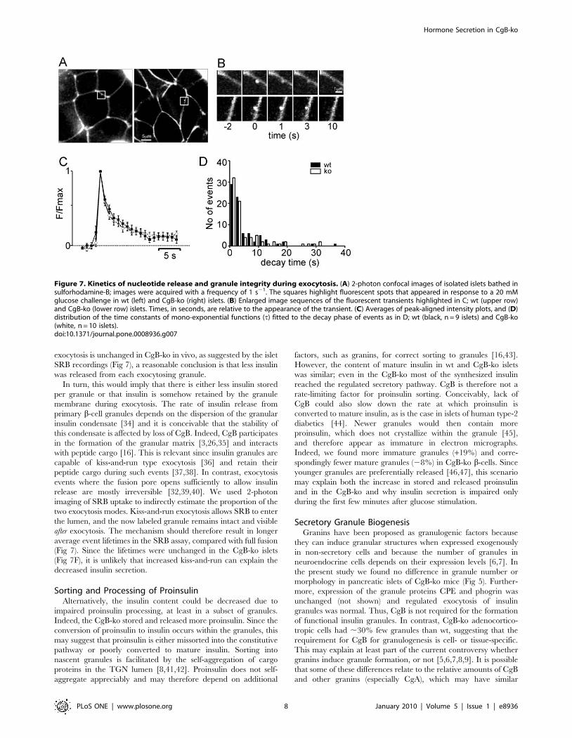
Granule Stability during Individual Exocytotic EventsTo study the structural integrity of individual insulin granules
during exocytosis, we imaged uptake of the fluid-phase tracer
sulforhodamine-B (SRB). This dye rapidly diffuses through the
open fusion pore of granules and labels their lumen until the
subsequent collapse of the granule ghost [32]. Isolated islets were
pre-incubated in 5 mM glucose in presence of SRB and then
challenged with 20 mM glucose in the continued presence of the
dye. Shortly after increasing the glucose concentration transient
bright spots were observed at the periphery of individual cells
(Fig 7C-D). The frequency of such events in the imaged area
(1,340 mm2) was 0.0460.03 min21 in 5 mM glucose and
increased to 1.760.4 min21 in 20 mM glucose (12 wt islets from
3 mice). Diazoxide (200 mM) prevented their occurrence
(0.0860.08 min21 in 20 mM glucose). Similar results were
obtained in CgB-ko islets, with frequencies of 0.0560.03 s21 in 5
mM glucose and 1.660.03 s21 in 20 mM (n = 16). For most
Figure 4. In vivo measurements of insulin secretion, glucosetolerance and insulin sensitivity. Time course of (A) plasma insulinconcentration (pM), and (B) plasma glucose (mM) in response to i.p.glucose injection (3 g/kg) at time 0; wt (black squares, n = 12) and CgB-ko (white squares, n = 16). (C) plasma glucose (mM) in response to i.p.insulin injection (0.8U/kg). **P,0.01;*P,0.05.doi:10.1371/journal.pone.0008936.g004
Hormone Secretion in CgB-ko
PLoS ONE | www.plosone.org 5 January 2010 | Volume 5 | Issue 1 | e8936
events the fluorescence increased instantaneously and then
decayed, 81% of events in wt and 77% in CgB-ko reached
baseline within 10 s (Fig 7E-F). There was no significant
difference in the decay times (6.861.8 s in wt and 10.462.1 s
in CgB-ko; medians 2.8 and 3.0 s). Thus, CgB ablation does not
affect the stability of granules during glucose-induced exocytosis,
and the data confirm that exocytosis proceeded at similar rates in
wt and CgB-ko.
Discussion
We have generated a mouse line deficient in CgB and studied
the effects of this mutation at the molecular, cellular and systemic
level. The analysis was focused on pancreatic islets, where
compensatory upregulation of other granins was relatively minor.
The study leads to two main conclusions, which are discussed here:
1) lack of CgB does not affect biosynthesis or function of insulin
Figure 5. Ultrastructural analysis of insulin granules. (A-B) Electron micrographs of wt and CgB-ko b-cells. The insets (A’ and B’) illustratemagnified examples of the four granule subtypes: (1) mature, (2) immature, (3) crystal-containing and (4) empty. (C-E) Quantification andmorphometric analysis of granules from wt (black bars and symbols) and CgB-ko (white bars and symbols) b-cells showing (C) number of granules percytoplasm area, (D) abundance of the granule subtypes (see above), and (E) distribution of the area of mature granules (squares) and their dense-cores (circles). (F-G) Electron micrographs of adenocorticotropic cells from wt and CgB-ko mice. Insets A’ and B’ show magnified examples of granules(arrowheads) at the plasma membrane. (H) Quantification of density of granules along the plasma membrane of wt (black) and CgB-ko (white) mice.doi:10.1371/journal.pone.0008936.g005
Hormone Secretion in CgB-ko
PLoS ONE | www.plosone.org 6 January 2010 | Volume 5 | Issue 1 | e8936
granules, and 2) islet hormone secretion is generally decreased in
the absence of CgB (except at low glucose; Fig 2A). However, the
mechanisms behind the altered hormone secretion, in particular
the paradox between decreased hormone release and normal
exocytosis, remain to be explained.
CgB-Deficiency Affects Insulin Release but Not ExocytosisIslets from CgB-ko mice had defects in the secretion of insulin,
glucagon and somatostatin. Specifically, they lacked the rapid
initial phase of insulin secretion, while basal insulin release was
elevated. Despite disturbed insulin secretion, the mice appeared
generally healthy with only mild glucose intolerance and normal
insulin sensitivity. We speculate that this is due to two reasons:
First, secretion of insulin, glucagon and somatostatin were reduced
in parallel (Fig 2). Since somatostatin inhibits insulin secretion and
glucagon opposes the actions of insulin, the net effect on glucose
homeostasis is expected to be mild. Interestingly, the reduced
inhibition by somatostatin may partially compensate for the insulin
secretion defect, at least in stimulating conditions. Second, insulin
secretion was actually elevated at low glucose concentrations and
at later timepoints during glucose stimulation, which may lead to
improved glucose clearance. The cause for this hypersecretion is
not known, but it is unlikely to be related to somatostatin since it
plays only a minor role at low glucose [33].
Paradoxically, the exocytotic machinery itself was normal and
an equal number of granules underwent exocytosis upon
stimulation. Both depolarization-evoked capacitance increases
(Fig 6) and the frequencies of glucose-stimulated exocytosis events
(Fig 7) were similar in wt and CgB-ko islets. How can insulin
secretion be different when rates of exocytosis are not? We
acknowledge that the conditions of the release assay (Fig 2a) and
the capacitance measurements are not necessarily comparable
(Fig 6). For example, including cAMP in the patch pipette may
have overridden differences that normally exist in the islets, and
the release assays were performed as batch incubations where
secretory products accumulate. However, assuming that the rate of
Figure 6. Stimulus-secretion coupling in isolated islets. (A) Measurements of [Ca2+]i in intact wt (upper) and CgB-ko (lower) islets,representative of 25 and 27 experiments, respectively. The glucose concentration (G) was changed from 1 to 5 and later to 15 mM during therecording, as indicated. Note that the Ca2+-signal lags by .2 min, reflecting the time b-cell need to metabolize the sugar. (B) Whole-cell Ca2+-currentselicited by voltage-clamp depolarizations (50 ms from 270 mV to 240, 220, 0 and +20 mV) in b-cells on the periphery of intact isolated wt (left) andCgB-ko (right) islets, as indicated. (C) Current-voltage (IV) relationships in wt (black) CgB-ko (white) cells, from analysis of the peak currents inrecordings as in B. (D) Whole-cell capacitance recordings (DCm, lower) elicited by trains of depolarizations from 270 to 0 mV (top) in b-cells on theperiphery of isolated wt (left) and CgB-ko (right) islets. (E) Analysis of the increase in whole-cell capacitance evoked by each depolarization (DCm,n–DCm,n21) and plotted against time; wt (black) and CgB-ko (white).doi:10.1371/journal.pone.0008936.g006
Hormone Secretion in CgB-ko
PLoS ONE | www.plosone.org 7 January 2010 | Volume 5 | Issue 1 | e8936
exocytosis is unchanged in CgB-ko in vivo, as suggested by the islet
SRB recordings (Fig 7), a reasonable conclusion is that less insulin
was released from each exocytosing granule.
In turn, this would imply that there is either less insulin stored
per granule or that insulin is somehow retained by the granule
membrane during exocytosis. The rate of insulin release from
primary b-cell granules depends on the dispersion of the granular
insulin condensate [34] and it is conceivable that the stability of
this condensate is affected by loss of CgB. Indeed, CgB participates
in the formation of the granular matrix [3,26,35] and interacts
with peptide cargo [16]. This is relevant since insulin granules are
capable of kiss-and-run type exocytosis [36] and retain their
peptide cargo during such events [37,38]. In contrast, exocytosis
events where the fusion pore opens sufficiently to allow insulin
release are mostly irreversible [32,39,40]. We used 2-photon
imaging of SRB uptake to indirectly estimate the proportion of the
two exocytosis modes. Kiss-and-run exocytosis allows SRB to enter
the lumen, and the now labeled granule remains intact and visible
after exocytosis. The mechanism should therefore result in longer
average event lifetimes in the SRB assay, compared with full fusion
(Fig 7). Since the lifetimes were unchanged in the CgB-ko islets
(Fig 7F), it is unlikely that increased kiss-and-run can explain the
decreased insulin secretion.
Sorting and Processing of ProinsulinAlternatively, the insulin content could be decreased due to
impaired proinsulin processing, at least in a subset of granules.
Indeed, the CgB-ko stored and released more proinsulin. Since the
conversion of proinsulin to insulin occurs within the granules, this
may suggest that proinsulin is either missorted into the constitutive
pathway or poorly converted to mature insulin. Sorting into
nascent granules is facilitated by the self-aggregation of cargo
proteins in the TGN lumen [8,41,42]. Proinsulin does not self-
aggregate appreciably and may therefore depend on additional
factors, such as granins, for correct sorting to granules [16,43].
However, the content of mature insulin in wt and CgB-ko islets
was similar; even in the CgB-ko most of the synthesized insulin
reached the regulated secretory pathway. CgB is therefore not a
rate-limiting factor for proinsulin sorting. Conceivably, lack of
CgB could also slow down the rate at which proinsulin is
converted to mature insulin, as is the case in islets of human type-2
diabetics [44]. Newer granules would then contain more
proinsulin, which does not crystallize within the granule [45],
and therefore appear as immature in electron micrographs.
Indeed, we found more immature granules (+19%) and corre-
spondingly fewer mature granules (28%) in CgB-ko b-cells. Since
younger granules are preferentially released [46,47], this scenario
may explain both the increase in stored and released proinsulin
and in the CgB-ko and why insulin secretion is impaired only
during the first few minutes after glucose stimulation.
Secretory Granule BiogenesisGranins have been proposed as granulogenic factors because
they can induce granular structures when expressed exogenously
in non-secretory cells and because the number of granules in
neuroendocrine cells depends on their expression levels [6,7]. In
the present study we found no difference in granule number or
morphology in pancreatic islets of CgB-ko mice (Fig 5). Further-
more, expression of the granule proteins CPE and phogrin was
unchanged (not shown) and regulated exocytosis of insulin
granules was normal. Thus, CgB is not required for the formation
of functional insulin granules. In contrast, CgB-ko adenocortico-
tropic cells had ,30% few granules than wt, suggesting that the
requirement for CgB for granulogenesis is cell- or tissue-specific.
This may explain at least part of the current controversy whether
granins induce granule formation, or not [5,6,7,8,9]. It is possible
that some of these differences relate to the relative amounts of CgB
and other granins (especially CgA), which may have similar
Figure 7. Kinetics of nucleotide release and granule integrity during exocytosis. (A) 2-photon confocal images of isolated islets bathed insulforhodamine-B; images were acquired with a frequency of 1 s21. The squares highlight fluorescent spots that appeared in response to a 20 mMglucose challenge in wt (left) and CgB-ko (right) islets. (B) Enlarged image sequences of the fluorescent transients highlighted in C; wt (upper row)and CgB-ko (lower row) islets. Times, in seconds, are relative to the appearance of the transient. (C) Averages of peak-aligned intensity plots, and (D)distribution of the time constants of mono-exponential functions (t) fitted to the decay phase of events as in D; wt (black, n = 9 islets) and CgB-ko(white, n = 10 islets).doi:10.1371/journal.pone.0008936.g007
Hormone Secretion in CgB-ko
PLoS ONE | www.plosone.org 8 January 2010 | Volume 5 | Issue 1 | e8936
granulogenic function and thus compensate for the lack of CgB. In
this scenario, granulogenesis may not be affected in cells that
normally contain relatively small amounts of CgB since it is
compensated for by the presence of other, more abundant granins.
In contrast to granulogenesis, we observed changes in hormone
secretion in the CgB-ko that were actually more pronounced than
in a CgA-ko [48]. Evidently, these changes are not compensated
for by other granins.
CgB and Human DiseaseCgB-deficiency in mice leads to a phenotype with some
hallmarks of human type-2 diabetes including loss of the initial
rapid phase of insulin secretion and hypersecretion of proinsulin.
Polymorphisms in either the CgB gene or factors affecting its
expression could thus predispose to human diabetes. Such
polymorphisms have already been linked to hypertension [49]
and schizophrenia [50], a disease that correlates with increased
risk for type-2 diabetes. Moreover, ablation of the forkhead
transcription factor Foxa2 in mice induced hyperinsulinemic
hypoglycemia in parallel with a 3-fold increase in CgB mRNA
[51]. Clearly, further studies are required to understand the
mechanism by which CgB affects secretion and whether the
protein plays any role in diabetes. Such studies may be facilitated
by the CgB-ko mouse model described here.
Materials and Methods
Ethics StatementAnimal experiments in Lund were approved by the local ethics
committee (Malmo/Lunds djurforsoksetiska namnd at Lunds
tingsratt) and conformed to Swedish animal protection laws and
applicable guidelines (djurskyddslagen 1988:534; djurskyddsfor-
ordningen 1988:539; djurskyddsmyndigheten DFS 2004:4). Those
in Germany were approved by the Regierungsprasidium Dresden,
Germany (license number: 24D-9168.24-9-2005-3) and con-
formed to German law.
Generation of CgB-ko MiceFor details on the generation of the CgB-ko line see [52].
Briefly, the targeting construct used for homologous recombina-
tion in E14.1-129/Ola ES cells included the H. simplex virus
thymidine kinase gene, 2.8 kb of mCgB proximal promoter region,
the neomycin resistance gene, and the full genomic sequence,
BglII-EcoRI excised, of intron 1 and exon 2 of mCghb
corresponding to 3.6 kb (Fig 1A). ES cells were cultured in
presence of 2 mM gancyclovir (Syntex Pharmaceuticals) and 300
mg/ml neomycin (Gibco). After double selection, clones were
screened by Southern blot analysis of HindIII digested genomic
DNA using 1.6 kb fragment from intron 1 of mCgb as probe. Five
positive clones were injected into C57/Bl-6 blastocysts and four
generated chimeras showed germline transmission. Subsequent
generations were obtained by intercrossing and outcrossing with
C57BL/6J mice and genotyped by Southern blot analysis. Finally,
lines of wt and CgB-ko mice were established from 12 CgB+/2
founders, with .95% C57BL/6J genetic background. Generation
of the two lines involved 5 generations of littermate inbreeding to
homogenate their genetic background. Age and sex-matched pairs
were used.
Islet and b-cell PreparationAnimal experiments were approved by local authorities of the
University of Lund, Sweden; University of Heidelberg, Germany;
University of Milan, Italy and by the Regierungsprasidium
Dresden, Germany. Mice were killed by cervical dislocation, the
pancreas quickly excised and islets isolated by standard collagenase
P (Sigma-Aldrich, Stockholm, Sweden) digestion. The islets were
maintained in short-term tissue culture (,24 h) in RPMI-1640
supplemented with 10% calf serum, 100 U/ml penicillin and
10 mg/ml streptomycin (all from Life Technologies, Taby,
Sweden).
Electron MicroscopyPancreatic islets were fixed in 2.5% glutaraldehyde for 1 h,
treated with 1% osmium tetroxide, dehydrated and embedded in
Durcupan (Sigma-Aldrich). Freshly dissected pituitaries were fixed
in 4% paraformaldehyde, 0.2% glutaraldehyde in 0.12 M
cacodylate buffer (pH 7.4, 2 h on ice), postfixed in 1%
osmiumtetroxide (1 h) and embedded in EMBed-812 (Science
Services). Samples were then sectioned (60–80 nm), mounted on
Cu-grids and contrasted with uranyl acetate and lead citrate and
examined by electron microscopy (Morgagni, FEI, Eindhoven,
Netherlands or JEM 1230, Jeol-USA, Peabody, MA).
Hormone Release MeasurementsRelease from isolated islets was carried out at 37uC in Krebs-
Ringer bicarbonate buffer (KRB; pH 7.4) containing additional
0.1% BSA, 1 mM glucose, 10 mM HEPES and gassed with 95%
O2 and 5% CO2. After preincubation for 30 min, the medium was
replaced with KRB with the indicated concentrations of glucose or
KCl (50 mM, equimolarly replacing NaCl). All batches contained
12 islets in 1 ml solution, except in Fig 3A,B (50 islets in 1.5 ml).
For in vivo measurements, glucose was injected intraperitoneally (3
g/kg body weight, in 0.9% NaCl) and blood was sampled every 2
min [53]. Insulin was quantified by radioimmunoassay [54], using
insulin(I-125) as tracer and an antibody against mouse-insulin (LR-
9011 and LR1013K, Linco, Tyreso, Sweden). Glucagon, somato-
statin and proinsulin were analyzed with a commercially available
radio-immuno assay kits (RB 310 and RB306, Euro-Diagnostica,
Malmo, Sweden) or ELISA (Rat Proinsulin 10-1185-01, Merco-
dia, Sweden). Islet insulin content was determined following acidic
alcohol extraction. Plasma glucose concentrations were deter-
mined enzymatically. Insulin for in vivo testing was from
NovoNordisk, Bagsvaerd, Denmark (NovoRapid).
Measurements of Intracellular Calcium[Ca2+]i was recorded by ratiometric microfluorimetry [31].
Islets were pre-loaded with 3 mM fura-2-acetomethoxyester
(Molecular Probes) and imaged at .510 nm using a 63x/1.25
objective (Carl Zeiss) and alternating excitation light (350/380 nm,
10 Hz) in a buffer containing (in mM) 140 NaCl, 3.6 KCl, 2
NaHCO3, 0.5 NaH2PO4, 0.5 MgSO4, 5 HEPES (pH 7.4), 2.5
CaCl2 and glucose as indicated, at 32uC. We focused several cell
diameters into the islets to decrease the influence of peripheral
non-b-cells. [Ca2+]i was calculated offline using the equation
[Ca2+]i = KdRmax-Rð ÞR-Rminð Þ .
F350
F380assuming a Kd of 224 nM. Rmax
was estimated after addition of 60 mM ionomycin and background
after quenching the fluorescence with 1 mM MnCl2.
ElectrophysiologyMeasurements of exocytosis and calcium currents were
performed in the whole-cell configuration using an EPC-10
amplifier (HEKA Elektronik). The intracellular solution consisted
of (in mM) 125 CsCl, 10 NaCl, 1 MgCl2, 3 ATP-Mg, 0.1 cAMP,
0.05 EGTA and 5 HEPES (pH 7.15). The extracellular solution
(EC) was (in mM) 138 NaCl, 5.6 KCl, 1.2 MgCl2, 2.6 CaCl2, 5 D-
glucose, and 5 mM HEPES (pH 7.40) and held at 32uC.
Hormone Secretion in CgB-ko
PLoS ONE | www.plosone.org 9 January 2010 | Volume 5 | Issue 1 | e8936
Capacitance was measured using 500 Hz, 20 mV sine waves. b-
cells were identified by their electrophysiological properties[55].
Rhodamine Uptake and Image AnalysisIslets were held with a suction pipette and incubated in EC with
1 mM sulforhodamine B (SRB) for 5 min. Exocytosis was then
stimulated by adjusting glucose to 20 mM. SRB fluorescence was
imaged at 1 Hz with 2-photon confocal microscopy (LSM510-
Meta, Carl Zeiss) with a 406 objective (1.2 C-Apochromat, Carl
Zeiss). SRB was excited at 800 nm and fluorescence detected at
,560 nm. Exocytosis was detected manually in records obtained
,2-15 min after solution change and quantified in circular regions
(0.5 mm diameter) centered on the event location. The pre-
stimulatory value was subtracted as local background.
StatisticsResults are expressed as mean6S.E.M, except in Figs 1+5
where mean6S.D. is shown. Error bars smaller than the symbols
are omitted. Significance of differences was assessed using
Student’s t-test.
Acknowledgments
We wish to thank Britt-Marie Nilsson and Kristina Borglid (Lund) for
excellent technical assistance, and Michaela Wilsch-Brauninger (Dresden)
for the processing of pituitary tissues for electron microcopy, Marcus
Michel and Eckhard Lammert (Dresden) for performing immunostaining
and critically reading the manuscript.
Author Contributions
Conceived and designed the experiments: SO FC AK PR WH SB.
Performed the experiments: SO FC AK ABL IL AS MF SB. Analyzed the
data: SO FC AK ABL SB. Contributed reagents/materials/analysis tools:
FC PR. Wrote the paper: AK SB.
References
1. Huttner WB, Gerdes HH, Rosa P (1991) The granin (chromogranin/
secretogranin) family. Trends Biochem Sci 16: 27–30.
2. Rosa P, Gerdes HH (1994) The granin protein family: markers for
neuroendocrine cells and tools for the diagnosis of neuroendocrine tumors.
J Endocrinol Invest 17: 207–225.
3. Winkler H, Fischer-Colbrie R (1992) The chromogranins A and B: the first 25
years and future perspectives. Neuroscience 49: 497–528.
4. Taupenot L, Harper KL, O’Connor DT (2003) The chromogranin-secreto-
granin family. N Engl J Med 348: 1134–1149.
5. Kim T, Zhang CF, Sun Z, Wu H, Loh YP (2005) Chromogranin A deficiency in
transgenic mice leads to aberrant chromaffin granule biogenesis. J Neurosci 25:
6958–6961.
6. Kim T, Tao-Cheng JH, Eiden LE, Loh YP (2001) Chromogranin A, an ‘‘on/
off’’ switch controlling dense-core secretory granule biogenesis. Cell 106:
499–509.
7. Huh YH, Jeon SH, Yoo SH (2003) Chromogranin B-induced secretory granule
biogenesis: comparison with the similar role of chromogranin A. J Biol Chem
278: 40581–40589.
8. Beuret N, Stettler H, Renold A, Rutishauser J, Spiess M (2004) Expression of
regulated secretory proteins is sufficient to generate granule-like structures in
constitutively secreting cells. J Biol Chem 279: 20242–20249.
9. Day R, Gorr SU (2003) Secretory granule biogenesis and chromogranin A:
master gene, on/off switch or assembly factor? Trends Endocrinol Metab 14:
10–13.
10. Mahapatra NR, O’Connor DT, Vaingankar SM, Hikim AP, Mahata M, et al.
(2005) Hypertension from targeted ablation of chromogranin A can be rescued
by the human ortholog. J Clin Invest 115: 1942–1952.
11. Hendy GN, Li T, Girard M, Feldstein RC, Mulay S, et al. (2006) Targeted
ablation of the chromogranin a (Chga) gene: normal neuroendocrine dense-core
secretory granules and increased expression of other granins. Mol Endocrinol
20: 1935–1947.
12. Borgonovo B, Ouwendijk J, Solimena M (2006) Biogenesis of secretory granules.
Curr Opin Cell Biol 18: 365–370.
13. Malosio ML, Giordano T, Laslop A, Meldolesi J (2004) Dense-core granules: a
specific hallmark of the neuronal/neurosecretory cell phenotype. J Cell Sci 117:
743–749.
14. Iacangelo AL, Eiden LE (1995) Chromogranin A: current status as a precursor
for bioactive peptides and a granulogenic/sorting factor in the regulated
secretory pathway. Regul Pept 58: 65–88.
15. Rosa P, Hille A, Lee RW, Zanini A, De Camilli P, et al. (1985) Secretogranins I
and II: two tyrosine-sulfated secretory proteins common to a variety of cells
secreting peptides by the regulated pathway. J Cell Biol 101: 1999–2011.
16. Natori S, Huttner WB (1996) Chromogranin B (secretogranin I) promotes
sorting to the regulated secretory pathway of processing intermediates derived
from a peptide hormone precursor. Proc Natl Acad Sci U S A 93: 4431–4436.
17. Cool DR, Normant E, Shen F, Chen HC, Pannell L, et al. (1997)
Carboxypeptidase E is a regulated secretory pathway sorting receptor: genetic
obliteration leads to endocrine disorders in Cpe(fat) mice. Cell 88: 73–83.
18. Hosaka M, Watanabe T, Sakai Y, Uchiyama Y, Takeuchi T (2002)
Identification of a chromogranin A domain that mediates binding to
secretogranin III and targeting to secretory granules in pituitary cells and
pancreatic beta-cells. Mol Biol Cell 13: 3388–3399.
19. Kromer A, Glombik MM, Huttner WB, Gerdes HH (1998) Essential role of the
disulfide-bonded loop of chromogranin B for sorting to secretory granules is
revealed by expression of a deletion mutant in the absence of endogenous granin
synthesis. J Cell Biol 140: 1331–1346.
20. Westphal CH, Muller L, Zhou A, Zhu X, Bonner-Weir S, et al. (1999) The
neuroendocrine protein 7B2 is required for peptide hormone processing in vivo
and provides a novel mechanism for pituitary Cushing’s disease. Cell 96:
689–700.
21. Nanavati C, Fernandez JM (1993) The secretory granule matrix: a fast-acting
smart polymer. Science 259: 963–965.
22. Helle KB, Reed RK, Ehrhart M, Aunis D, Hogue Angeletti R (1990)
Chromogranin A: osmotically active fragments and their susceptibility to
proteolysis during lysis of the bovine chromaffin granules. Acta Physiol Scand
138: 565–574.
23. Montesinos MS, Machado JD, Camacho M, Diaz J, Morales YG, et al. (2008)
The crucial role of chromogranins in storage and exocytosis revealed using
chromaffin cells from chromogranin A null mouse. J Neurosci 28: 3350–3358.
24. Aardal S, Helle KB, Elsayed S, Reed RK, Serck-Hanssen G (1993) Vasostatins,
comprising the N-terminal domain of chromogranin A, suppress tension in
isolated human blood vessel segments. J Neuroendocrinol 5: 405–412.
25. Tatemoto K, Efendic S, Mutt V, Makk G, Feistner GJ, et al. (1986)
Pancreastatin, a novel pancreatic peptide that inhibits insulin secretion. Nature
324: 476–478.
26. Helle KB (2004) The granin family of uniquely acidic proteins of the diffuse
neuroendocrine system: comparative and functional aspects. Biol Rev Camb
Philos Soc 79: 769–794.
27. Karlsson E, Stridsberg M, Sandler S (2000) Chromogranin-B regulation of IAPP
and insulin secretion. Regul Pept 87: 33–39.
28. Borjesson A, Carlsson C (2007) Altered proinsulin conversion in rat pancreatic
islets exposed long-term to various glucose concentrations or interleukin-1beta.
J Endocrinol 192: 381–387.
29. Gayen JR, Saberi M, Schenk S, Biswas N, Vaingankar SM, et al. (2009) A novel
pathway of insulin sensitivity in chromogranin a null mice: a crucial role for
pancreastatin in glucose homeostasis. J Biol Chem 284: 28498–28509.
30. Orci L, Lambert AE, Kanazawa Y, Amherdt M, Rouiller C, et al. (1971)
Morphological and biochemical studies of B cells of fetal rat endocrine pancreas
in organ culture. Evidence for (pro) insulin biosynthesis. J Cell Biol 50: 565–582.
31. Olofsson CS, Gopel SO, Barg S, Galvanovskis J, Ma X, et al. (2002) Fast insulin
secretion reflects exocytosis of docked granules in mouse pancreatic B-cells.
Pflugers Arch 444: 43–51.
32. Takahashi N, Kishimoto T, Nemoto T, Kadowaki T, Kasai H (2002) Fusion
pore dynamics and insulin granule exocytosis in the pancreatic islet. Science 297:
1349–1352.
33. Hauge-Evans AC, King AJ, Carmignac D, Richardson CC, Robinson IC, et al.
(2009) Somatostatin secreted by islet delta-cells fulfills multiple roles as a
paracrine regulator of islet function. Diabetes 58: 403–411.
34. Michael DJ, Ritzel RA, Haataja L, Chow RH (2006) Pancreatic beta-cells
secrete insulin in fast- and slow-release forms. Diabetes 55: 600–607.
35. Lukinius A, Stridsberg M, Wilander E (2003) Cellular expression and specific
intragranular localization of chromogranin A, chromogranin B, and synapto-
physin during ontogeny of pancreatic islet cells: an ultrastructural study.
Pancreas 27: 38–46.
36. Tsuboi T, Rutter GA (2003) Multiple Forms of ‘‘Kiss-and-Run’’ Exocytosis
Revealed by Evanescent Wave Microscopy. Curr Biol 13: 563–567.
37. Obermuller S, Lindqvist A, Karanauskaite J, Galvanovskis J, Rorsman P, et al.
(2005) Selective nucleotide-release from dense-core granules in insulin-secreting
cells. J Cell Sci 118: 4271–4282.
38. Tsuboi T, McMahon HT, Rutter GA (2004) Mechanisms of dense core vesicle
recapture following ‘‘kiss and run’’ (‘‘cavicapture’’) exocytosis in insulin-secreting
cells. J Biol Chem 279: 47115–47124.
Hormone Secretion in CgB-ko
PLoS ONE | www.plosone.org 10 January 2010 | Volume 5 | Issue 1 | e8936

39. MacDonald PE, Braun M, Galvanovskis J, Rorsman P (2006) Release of small
transmitters through kiss-and-run fusion pores in rat pancreatic beta cells. Cell
Metab 4: 283–290.
40. Ma L, Bindokas VP, Kuznetsov A, Rhodes C, Hays L, et al. (2004) Direct
imaging shows that insulin granule exocytosis occurs by complete vesicle fusion.
Proc Natl Acad Sci U S A 101: 9266–9271.
41. Gerdes HH, Rosa P, Phillips E, Baeuerle PA, Frank R, et al. (1989) The primary
structure of human secretogranin II, a widespread tyrosine-sulfated secretory
granule protein that exhibits low pH- and calcium-induced aggregation. J Biol
Chem 264: 12009–12015.
42. Chanat E, Huttner WB (1991) Milieu-induced, selective aggregation of regulated
secretory proteins in the trans-Golgi network. J Cell Biol 115: 1505–1519.
43. Molinete M, Irminger JC, Tooze SA, Halban PA (2000) Trafficking/sorting and
granule biogenesis in the beta-cell. Semin Cell Dev Biol 11: 243–251.
44. Kahn SE, Halban PA (1997) Release of incompletely processed proinsulin is the
cause of the disproportionate proinsulinemia of NIDDM. Diabetes 46:
1725–1732.
45. Zhu X, Orci L, Carroll R, Norrbom C, Ravazzola M, et al. (2002) Severe block
in processing of proinsulin to insulin accompanied by elevation of des-64,65
proinsulin intermediates in islets of mice lacking prohormone convertase 1/3.
Proc Natl Acad Sci U S A 99: 10299–10304.
46. Duncan RR, Greaves J, Wiegand UK, Matskevich I, Bodammer G, et al. (2003)
Functional and spatial segregation of secretory vesicle pools according to vesicle
age. Nature 422: 176–180.
47. Rhodes CJ, Halban PA (1987) Newly synthesized proinsulin/insulin and stored
insulin are released from pancreatic B cells predominantly via a regulated, ratherthan a constitutive, pathway. J Cell Biol 105: 145–153.
48. Portela-Gomes GM, Gayen JR, Grimelius L, Stridsberg M, Mahata SK (2008)
The importance of chromogranin A in the development and function ofendocrine pancreas. Regul Pept 151: 19–25.
49. Zhang K, Rao F, Rana BK, Gayen JR, Calegari F, et al. (2009) AutonomicFunction in Hypertension: Role of Genetic Variation at the Catecholamine
Storage Vesicle Protein Chromogranin B. Circ Cardiovasc Genet 2: 46–56.
50. Iijima Y, Inada T, Ohtsuki T, Senoo H, Nakatani M, et al. (2004) Associationbetween chromogranin b gene polymorphisms and schizophrenia in the
Japanese population. Biol Psychiatry 56: 10–17.51. Gao N, White P, Doliba N, Golson ML, Matschinsky FM, et al. (2007) Foxa2
controls vesicle docking and insulin secretion in mature Beta cells. Cell Metab 6:267–279.
52. King A (1996) Dissertation: Generation of Chromogranin B deficient mice.
Heidelberg, Germany: University of Heidelberg.53. Rerup C, Lundquist I (1966) Blood glucose level in mice. 1. Evaluation of a new
technique of multiple serial sampling. Acta Endocrinol (Copenh) 52: 357–367.54. Heding L (1966) A simplified insulin radioimmunoassay method. In: Donato
L, Milhaud G., Sirchis J., eds. Labelled proteins in tracer studies. Brussels:
Euratom. pp 345–350.55. Barg S, Galvanovskis J, Gopel SO, Rorsman P, Eliasson L (2000) Tight coupling
between electrical activity and exocytosis in mouse glucagon-secreting a-cells.Diabetes 49: 1500–1510.
Hormone Secretion in CgB-ko
PLoS ONE | www.plosone.org 11 January 2010 | Volume 5 | Issue 1 | e8936
Related Documents


