Defective peroxisomal catabolism of branched fatty acyl coenzyme A in mice lacking the sterol carrier protein-2/sterol carrier protein-x gene function Udo Seedorf, 1,2,6 Martin Raabe, 1 Peter Ellinghaus, 1,7 Frank Kannenberg, 1 Manfred Fobker, 1,2 Thomas Engel, 1 Simone Denis, 3 Fred Wouters, 4 Karel W.A. Wirtz, 4 Ronald J.A. Wanders, 3 Nobuyo Maeda, 5 and Gerd Assmann 1,2 1 Institute for Arteriosclerosis Research and 2 Institute for Clinical Chemistry and Laboratory Medicine (Zentrallaboratorium), Westfalian Wilhelms-University, D-48129 Mu ¨ nster, Germany; 3 Department of Pediatrics, Academic Medical Center, University of Amsterdam, 1105 AZ Amsterdam, The Netherlands; 4 Center for Biomembranes and Lipid Enzymology, University of Utrecht, De Uithof, Utrecht, The Netherlands; 5 Department of Pathology, University of North Carolina at Chapel Hill, Chapel Hill, North Carolina 27599-7525 USA Gene targeting in mice was used to investigate the unknown function of Scp2, encoding sterol carrier protein-2 (SCP2; a peroxisomal lipid carrier) and sterol carrier protein-x (SCPx; a fusion protein between SCP2 and a peroxisomal thiolase). Complete deficiency of SCP2 and SCPx was associated with marked alterations in gene expression, peroxisome proliferation, hypolipidemia, impaired body weight control, and neuropathy. Along with these abnormalities, catabolism of methyl-branched fatty acyl CoAs was impaired. The defect became evident from up to 10-fold accumulation of the tetramethyl-branched fatty acid phytanic acid in Scp2(-/-) mice. Further characterization supported that the gene disruption led to inefficient import of phytanoyl-CoA into peroxisomes and to defective thiolytic cleavage of 3-ketopristanoyl-CoA. These results corresponded to high-affinity binding of phytanoyl-CoA to the recombinant rat SCP2 protein, as well as high 3-ketopristanoyl-CoA thiolase activity of the recombinant rat SCPx protein. [Key Words: Gene targeting; peroxisomes; b-oxidation; Refsum disease; cholesterol; steroid hormones] Received December 10, 1997; revised version accepted February 9, 1998. Sterol carrier protein-2 (SCP2) was isolated originally as a ‘‘cytosolic’’ factor required for efficient in vitro conver- sion of 7-dehydrocholesterol to cholesterol, catalyzed by microsomal sterol-D 7 -reductase (Noland et al. 1980). Subsequently, it was shown that the protein was identi- cal to the nonspecific lipid transfer protein (ns-LTP), which had been purified based on its ability to catalyze the exchange of a variety of phospholipids between membranes in vitro (Bloj and Zilversmit 1977). More re- cently, it could be demonstrated that purified SCP2 binds fatty acids and fatty acyl Coenzyme A (CoA) with similar or even higher affinity than sterols (Stolowich et al. 1997). Cloning and sequencing of SCP2 cDNAs showed that the protein comprises a carboxy-terminal SKL peroxisomal targeting signal (Seedorf and Assmann 1991), and immunocytochemical studies confirmed the predominant localization of SCP2 within peroxisomes (Keller et al. 1989; Ossendorp and Wirtz 1993). Several lines of indirect evidence exist that appear to support a role of SCP2 in adrenal and ovarian steroidogenesis (for review, see Pfeifer et al. 1993a). In addition, cell culture studies suggested a potential participation of SCP2 in cytosolic sterol transport to the plasma membrane (Puglielli et al. 1995; Baum et al. 1997). However, the localization of SCP2 in peroxisomes makes it difficult to understand how the protein might carry out these func- tions in the intact cell. Thus, the biological function of SCP2 is not clear. The SCP2-encoding gene (Scp2) comprises 16 exons, spanning ∼100 kb on human chromosome 1p32. Tran- scription initiation is controlled by two distant promot- ers that were mapped immediately upstream of the first exon (P1) and exon 12 (P2) (Ohba et al. 1994, 1995). P2 is used to generate SCP2-encoding transcripts, which com- 6 Corresponding author. E-MAIL [email protected]; FAX 49-251-8356208. 7 This study contains part of a thesis work performed in partial fulfill- ment of the requirements of the Westfalian Wilhelms-University, Mu ¨ n- ster, Germany. GENES & DEVELOPMENT 12:1189–1201 © 1998 by Cold Spring Harbor Laboratory Press ISSN 0890-9369/98 $5.00; www.genesdev.org 1189 Cold Spring Harbor Laboratory Press on August 17, 2019 - Published by genesdev.cshlp.org Downloaded from

Welcome message from author
This document is posted to help you gain knowledge. Please leave a comment to let me know what you think about it! Share it to your friends and learn new things together.
Transcript

Defective peroxisomal catabolismof branched fatty acyl coenzyme Ain mice lacking the sterol carrierprotein-2/sterol carrier protein-xgene functionUdo Seedorf,1,2,6 Martin Raabe,1 Peter Ellinghaus,1,7 Frank Kannenberg,1 Manfred Fobker,1,2
Thomas Engel,1 Simone Denis,3 Fred Wouters,4 Karel W.A. Wirtz,4 Ronald J.A. Wanders,3
Nobuyo Maeda,5 and Gerd Assmann1,2
1Institute for Arteriosclerosis Research and 2Institute for Clinical Chemistry and Laboratory Medicine (Zentrallaboratorium),Westfalian Wilhelms-University, D-48129 Munster, Germany; 3Department of Pediatrics, Academic Medical Center,University of Amsterdam, 1105 AZ Amsterdam, The Netherlands; 4Center for Biomembranes and Lipid Enzymology,University of Utrecht, De Uithof, Utrecht, The Netherlands; 5Department of Pathology, University of North Carolinaat Chapel Hill, Chapel Hill, North Carolina 27599-7525 USA
Gene targeting in mice was used to investigate the unknown function of Scp2, encoding sterol carrierprotein-2 (SCP2; a peroxisomal lipid carrier) and sterol carrier protein-x (SCPx; a fusion protein between SCP2and a peroxisomal thiolase). Complete deficiency of SCP2 and SCPx was associated with marked alterations ingene expression, peroxisome proliferation, hypolipidemia, impaired body weight control, and neuropathy.Along with these abnormalities, catabolism of methyl-branched fatty acyl CoAs was impaired. The defectbecame evident from up to 10-fold accumulation of the tetramethyl-branched fatty acid phytanic acid inScp2(−/−) mice. Further characterization supported that the gene disruption led to inefficient import ofphytanoyl-CoA into peroxisomes and to defective thiolytic cleavage of 3-ketopristanoyl-CoA. These resultscorresponded to high-affinity binding of phytanoyl-CoA to the recombinant rat SCP2 protein, as well as high3-ketopristanoyl-CoA thiolase activity of the recombinant rat SCPx protein.
[Key Words: Gene targeting; peroxisomes; b-oxidation; Refsum disease; cholesterol; steroid hormones]
Received December 10, 1997; revised version accepted February 9, 1998.
Sterol carrier protein-2 (SCP2) was isolated originally asa ‘‘cytosolic’’ factor required for efficient in vitro conver-sion of 7-dehydrocholesterol to cholesterol, catalyzed bymicrosomal sterol-D7-reductase (Noland et al. 1980).Subsequently, it was shown that the protein was identi-cal to the nonspecific lipid transfer protein (ns-LTP),which had been purified based on its ability to catalyzethe exchange of a variety of phospholipids betweenmembranes in vitro (Bloj and Zilversmit 1977). More re-cently, it could be demonstrated that purified SCP2binds fatty acids and fatty acyl Coenzyme A (CoA) withsimilar or even higher affinity than sterols (Stolowich etal. 1997). Cloning and sequencing of SCP2 cDNAsshowed that the protein comprises a carboxy-terminal
SKL peroxisomal targeting signal (Seedorf and Assmann1991), and immunocytochemical studies confirmed thepredominant localization of SCP2 within peroxisomes(Keller et al. 1989; Ossendorp and Wirtz 1993). Severallines of indirect evidence exist that appear to support arole of SCP2 in adrenal and ovarian steroidogenesis (forreview, see Pfeifer et al. 1993a). In addition, cell culturestudies suggested a potential participation of SCP2 incytosolic sterol transport to the plasma membrane(Puglielli et al. 1995; Baum et al. 1997). However, thelocalization of SCP2 in peroxisomes makes it difficult tounderstand how the protein might carry out these func-tions in the intact cell. Thus, the biological function ofSCP2 is not clear.
The SCP2-encoding gene (Scp2) comprises 16 exons,spanning ∼100 kb on human chromosome 1p32. Tran-scription initiation is controlled by two distant promot-ers that were mapped immediately upstream of the firstexon (P1) and exon 12 (P2) (Ohba et al. 1994, 1995). P2 isused to generate SCP2-encoding transcripts, which com-
6Corresponding author.E-MAIL [email protected]; FAX 49-251-8356208.7This study contains part of a thesis work performed in partial fulfill-ment of the requirements of the Westfalian Wilhelms-University, Mun-ster, Germany.
GENES & DEVELOPMENT 12:1189–1201 © 1998 by Cold Spring Harbor Laboratory Press ISSN 0890-9369/98 $5.00; www.genesdev.org 1189
Cold Spring Harbor Laboratory Press on August 17, 2019 - Published by genesdev.cshlp.orgDownloaded from

bine the coding information provided by exons 12–16. Inaddition, alternate transcription initiation at P1 leads toproduction of a second transcript that includes the cod-ing information provided by exons 1–16. The respectivegene product consists of 547 amino acids and was namedsterol carrier protein-x (SCPx) (Seedorf and Assmann1991). SCPx represents a fusion protein between a thio-lase domain, extending from amino acids 1–404, andSCP2, which is located at the carboxyl terminus (Ossen-dorp et al. 1991). It is known from previous in vitro stud-ies that SCPx has similar lipid transfer activity as SCP2and that the substrate specificity of the SCPx thiolaseshows a preference for straight medium chain acyl-CoAsubstrates and tetramethyl-branched 3-ketopristanoyl-CoA (Seedorf et al. 1994a; Wanders et al. 1997). Thus, theSCPx-associated thiolase differs from the initially iden-tified peroxisomal thiolase that is assumed traditionallyto play a major role in peroxisomal b-oxidation of mostnaturally occurring substrates, including bile acids andvery long chain fatty-acids (VLCFA) (Hijikata et al. 1987;Schram et al. 1987).
In the present study, we investigated the biologicalfunction of Scp2 by using gene targeting in mice. Thephenotypic abnormalities of the Scp2(−/−) knockout(KO) mice revealed a profound impact of the gene dis-ruption on the in vivo degradation of branched-chainfatty acyl-CoAs coming from the metabolism of tetra-methyl-branched fatty acid phytanic acid. On the otherhand, the serum concentrations of cholesterol, steroids,VLCFA, and long-chain fatty acids were not affected inScp2(−/−) mice. This leads to our conclusion that oneprincipal function of Scp2 resides in the major peroxi-somal pathway, which mediates the degradation ofmethyl-branched fatty acyl-CoA substrates in mice.
Results
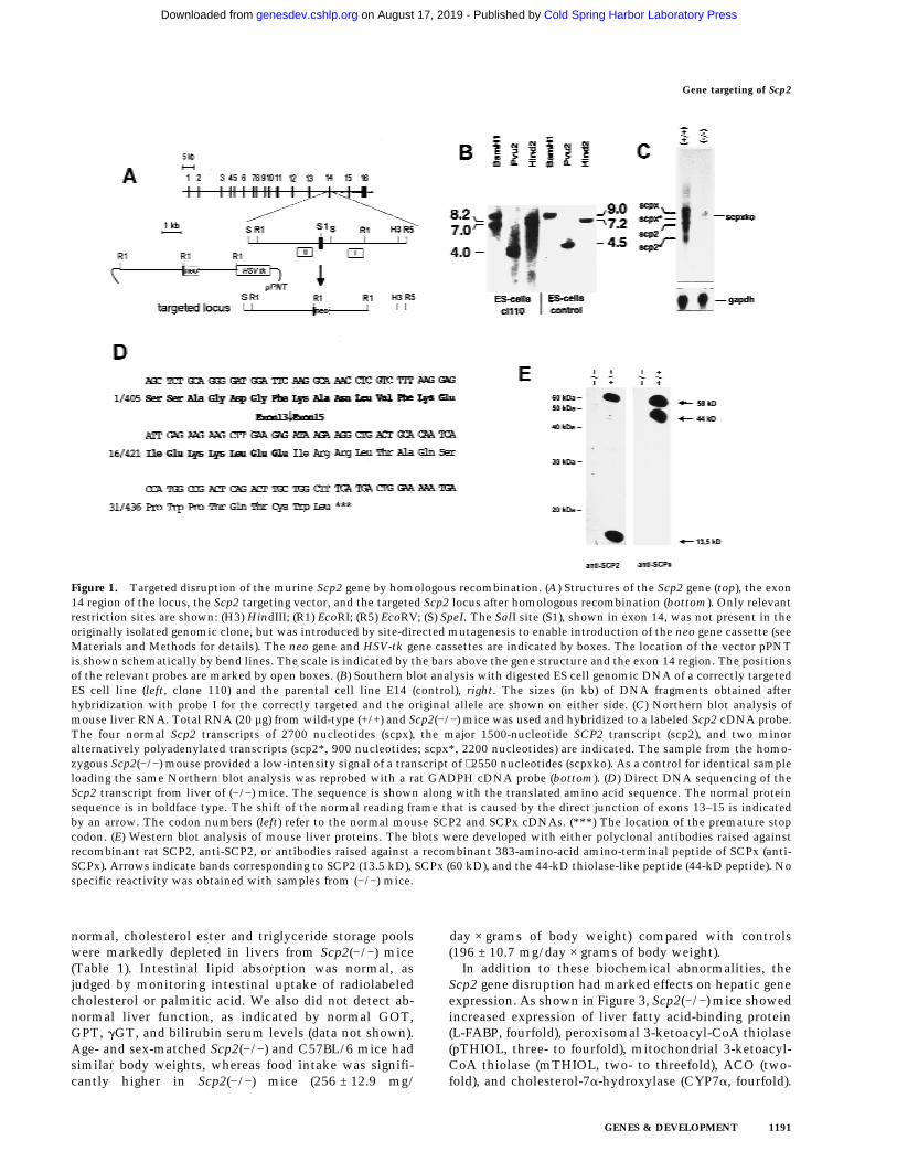
To produce SCP2 and SCPx deficiency, we introduced agene disruption at the exon 14 region of the gene (Fig.1A). Transfection of the targeting vector into mouse E14ES cells and subsequent positive–negative selection pro-vided 182 clones. Southern blot analysis led to the iden-tification of two correctly targeted clones (cl20, cl110)with only a single neo gene copy in the targeted locus(Fig. 1B). Injection of recombinant cl110 embryonic stemcells in blastocysts obtained from C57BL/6 donors, fol-lowed by embryo transfer into CD1 foster mothers gen-erated five chimeras. Among these, we identified threetransmitters that were crossed further with C57BL/6mice.
Complete elimination of the Scp2 gene function wasconfirmed by Northern blot experiments, showing a low-intensity signal derived from a truncated Scp2 transcriptin (−/−) mice that was ∼150 nucleotides smaller than thecorresponding transcript in normal C57BL/6 mice (Fig.1C). When primers, flanking the region of the Scp2cDNA encoded by exon 14, were used for PCR amplifi-cation with reverse transcribed poly(A) RNA, isolatedfrom (−/−) mice, a DNA fragment was obtained thatagain was ∼150 bp smaller than normal. DNA sequenc-
ing of this DNA fragment revealed that the Scp2 targetedallele led to abnormal splicing resulting in exon 14 skip-ping (Fig. 1D). The cDNA sequence predicted proteinsequences of SCP2 and SCPx that were normal until po-sition 22 and 427, respectively. Thereafter, the directjunction to the coding information of exon 15 resulted ina frame-shift, thereby creating a premature stop 18codons downstream. Results from earlier site-directedmutagenesis studies lead to the conclusion that the pre-dicted SCP2 variant should clearly lack any residual lipidtransfer activity (Seedorf et al. 1994b). In contrast, thethiolase-like domain (residues 1–404) was preserved inthe predicted SCPx variant, which may suggest the pos-sibility of residual thiolase activity. However, becausethe carboxy-terminal peroxisomal targeting signal is ab-sent, the variant should no longer be imported into per-oxisomes and therefore, should be at least functionallyinactive. Moreover, as shown in Figure 1E, liver extractssubjected to Western blot analyses developed with anti-SCP2 or anti-SCPx antibodies revealed complete absenceof the two proteins in (−/−) mice. Homozygous trans-genes also lacked the previously identified peroxisomal44-kD thiolase-like peptide that was considered to resultfrom proteolytic processing of SCPx (Seedorf et al.1994a).
Genotyping showed that heterozygous (+/−) and ho-mozygous (−/−) transgenes were viable. When kept understandard laboratory conditions, (−/−) mice developednormally and had no developmental abnormalities. Wedid not observe differences in the incidence of (+/+),(+/−), and (−/−) mice from the Mendelian distribution[27% (+/+), 52% (+/−), 21% (−/−), n = 141] indicating thatthe (−/−) allele did not affect the viability at 3–4 weeks ofage. In addition, (−/−) males and females reached fertilityat the normal age of ∼6 weeks. Interbreeding between(−/−) males and (−/−) females gave rise to viable progeny.The litters were of comparable sizes as that found in(+/+) or (+/−) interbreeding. In 6- to 8-week-old males,testosterone and glucocorticoid concentrations werewithin the normal range. No differences between thetwo strains were also found for progesterone in nonpreg-nant females under baseline conditions. Whereas plasmainsulin and cholesterol concentrations were normal, tri-glycerides were slightly higher and free fatty acid andglucose concentrations were moderately lower in (−/−)mice (Table 1).
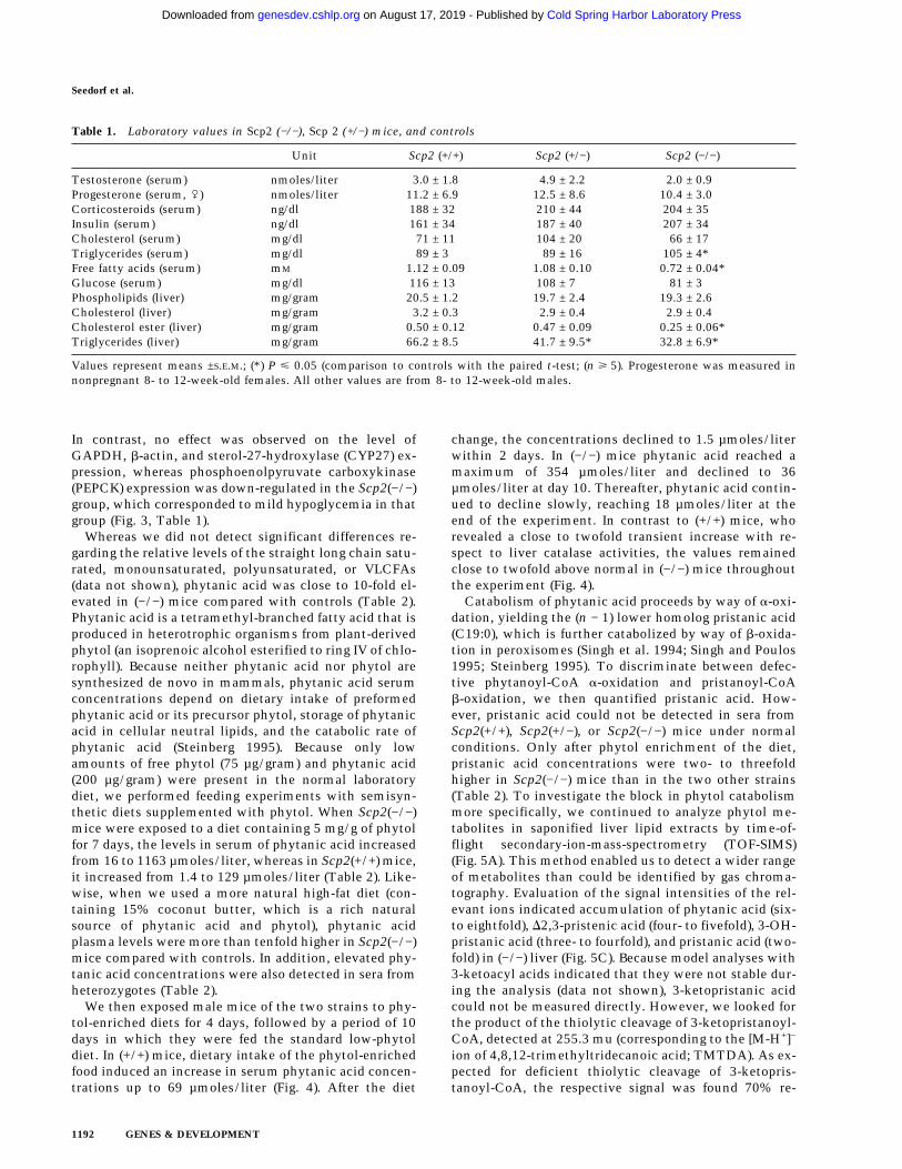
All major tissues of the Scp2(−/−) mice were examinedby light microscopy at various times after birth and com-pared with those of heterozygous and wild-type mice.Although the organ systems appeared morphologicallynormal, we observed more intense diaminobenzidinestaining (DAB, stains specifically peroxisomes) in frozenliver sections from Scp2(−/−) mice than from controls(Fig. 2A). Enzyme activity levels of the peroxisomalmarker catalase were 1.8-fold elevated in Scp2(−/−) liver.Likewise, peroxisomal palmitoyl-CoA oxidase (ACO),mitochondrial butyryl-CoA dehydrogenase, and total3-ketooctanoyl-CoA thiolase activities were all two- tothreefold higher in Scp2(−/−) mice than in controls (Fig.2B). Whereas the hepatic levels of phospholipids were
Seedorf et al.
1190 GENES & DEVELOPMENT
Cold Spring Harbor Laboratory Press on August 17, 2019 - Published by genesdev.cshlp.orgDownloaded from
normal, cholesterol ester and triglyceride storage poolswere markedly depleted in livers from Scp2(−/−) mice(Table 1). Intestinal lipid absorption was normal, asjudged by monitoring intestinal uptake of radiolabeledcholesterol or palmitic acid. We also did not detect ab-normal liver function, as indicated by normal GOT,GPT, gGT, and bilirubin serum levels (data not shown).Age- and sex-matched Scp2(−/−) and C57BL/6 mice hadsimilar body weights, whereas food intake was signifi-cantly higher in Scp2(−/−) mice (256 ± 12.9 mg/
day × grams of body weight) compared with controls(196 ± 10.7 mg/day × grams of body weight).
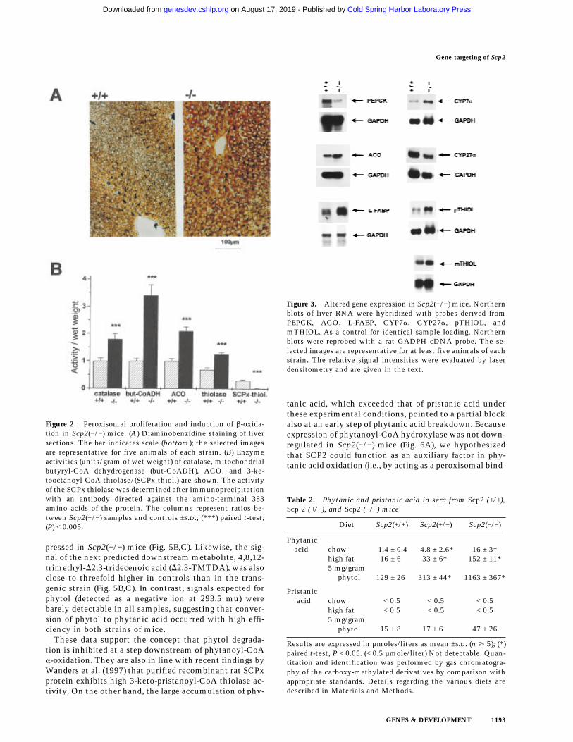
In addition to these biochemical abnormalities, theScp2 gene disruption had marked effects on hepatic geneexpression. As shown in Figure 3, Scp2(−/−) mice showedincreased expression of liver fatty acid-binding protein(L-FABP, fourfold), peroxisomal 3-ketoacyl-CoA thiolase(pTHIOL, three- to fourfold), mitochondrial 3-ketoacyl-CoA thiolase (mTHIOL, two- to threefold), ACO (two-fold), and cholesterol-7a-hydroxylase (CYP7a, fourfold).
Figure 1. Targeted disruption of the murine Scp2 gene by homologous recombination. (A) Structures of the Scp2 gene (top), the exon14 region of the locus, the Scp2 targeting vector, and the targeted Scp2 locus after homologous recombination (bottom). Only relevantrestriction sites are shown: (H3) HindIII; (R1) EcoRI; (R5) EcoRV; (S) SpeI. The SalI site (S1), shown in exon 14, was not present in theoriginally isolated genomic clone, but was introduced by site-directed mutagenesis to enable introduction of the neo gene cassette (seeMaterials and Methods for details). The neo gene and HSV-tk gene cassettes are indicated by boxes. The location of the vector pPNTis shown schematically by bend lines. The scale is indicated by the bars above the gene structure and the exon 14 region. The positionsof the relevant probes are marked by open boxes. (B) Southern blot analysis with digested ES cell genomic DNA of a correctly targetedES cell line (left, clone 110) and the parental cell line E14 (control), right. The sizes (in kb) of DNA fragments obtained afterhybridization with probe I for the correctly targeted and the original allele are shown on either side. (C) Northern blot analysis ofmouse liver RNA. Total RNA (20 µg) from wild-type (+/+) and Scp2(−/−) mice was used and hybridized to a labeled Scp2 cDNA probe.The four normal Scp2 transcripts of 2700 nucleotides (scpx), the major 1500-nucleotide SCP2 transcript (scp2), and two minoralternatively polyadenylated transcripts (scp2*, 900 nucleotides; scpx*, 2200 nucleotides) are indicated. The sample from the homo-zygous Scp2(−/−) mouse provided a low-intensity signal of a transcript of ∼2550 nucleotides (scpxko). As a control for identical sampleloading the same Northern blot analysis was reprobed with a rat GADPH cDNA probe (bottom). (D) Direct DNA sequencing of theScp2 transcript from liver of (−/−) mice. The sequence is shown along with the translated amino acid sequence. The normal proteinsequence is in boldface type. The shift of the normal reading frame that is caused by the direct junction of exons 13–15 is indicatedby an arrow. The codon numbers (left) refer to the normal mouse SCP2 and SCPx cDNAs. (***) The location of the premature stopcodon. (E) Western blot analysis of mouse liver proteins. The blots were developed with either polyclonal antibodies raised againstrecombinant rat SCP2, anti-SCP2, or antibodies raised against a recombinant 383-amino-acid amino-terminal peptide of SCPx (anti-SCPx). Arrows indicate bands corresponding to SCP2 (13.5 kD), SCPx (60 kD), and the 44-kD thiolase-like peptide (44-kD peptide). Nospecific reactivity was obtained with samples from (−/−) mice.
Gene targeting of Scp2
GENES & DEVELOPMENT 1191
Cold Spring Harbor Laboratory Press on August 17, 2019 - Published by genesdev.cshlp.orgDownloaded from
In contrast, no effect was observed on the level ofGAPDH, b-actin, and sterol-27-hydroxylase (CYP27) ex-pression, whereas phosphoenolpyruvate carboxykinase(PEPCK) expression was down-regulated in the Scp2(−/−)group, which corresponded to mild hypoglycemia in thatgroup (Fig. 3, Table 1).
Whereas we did not detect significant differences re-garding the relative levels of the straight long chain satu-rated, monounsaturated, polyunsaturated, or VLCFAs(data not shown), phytanic acid was close to 10-fold el-evated in (−/−) mice compared with controls (Table 2).Phytanic acid is a tetramethyl-branched fatty acid that isproduced in heterotrophic organisms from plant-derivedphytol (an isoprenoic alcohol esterified to ring IV of chlo-rophyll). Because neither phytanic acid nor phytol aresynthesized de novo in mammals, phytanic acid serumconcentrations depend on dietary intake of preformedphytanic acid or its precursor phytol, storage of phytanicacid in cellular neutral lipids, and the catabolic rate ofphytanic acid (Steinberg 1995). Because only lowamounts of free phytol (75 µg/gram) and phytanic acid(200 µg/gram) were present in the normal laboratorydiet, we performed feeding experiments with semisyn-thetic diets supplemented with phytol. When Scp2(−/−)mice were exposed to a diet containing 5 mg/g of phytolfor 7 days, the levels in serum of phytanic acid increasedfrom 16 to 1163 µmoles/liter, whereas in Scp2(+/+) mice,it increased from 1.4 to 129 µmoles/liter (Table 2). Like-wise, when we used a more natural high-fat diet (con-taining 15% coconut butter, which is a rich naturalsource of phytanic acid and phytol), phytanic acidplasma levels were more than tenfold higher in Scp2(−/−)mice compared with controls. In addition, elevated phy-tanic acid concentrations were also detected in sera fromheterozygotes (Table 2).
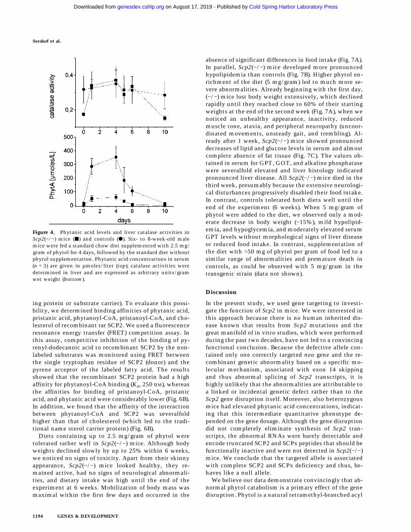
We then exposed male mice of the two strains to phy-tol-enriched diets for 4 days, followed by a period of 10days in which they were fed the standard low-phytoldiet. In (+/+) mice, dietary intake of the phytol-enrichedfood induced an increase in serum phytanic acid concen-trations up to 69 µmoles/liter (Fig. 4). After the diet
change, the concentrations declined to 1.5 µmoles/literwithin 2 days. In (−/−) mice phytanic acid reached amaximum of 354 µmoles/liter and declined to 36µmoles/liter at day 10. Thereafter, phytanic acid contin-ued to decline slowly, reaching 18 µmoles/liter at theend of the experiment. In contrast to (+/+) mice, whorevealed a close to twofold transient increase with re-spect to liver catalase activities, the values remainedclose to twofold above normal in (−/−) mice throughoutthe experiment (Fig. 4).
Catabolism of phytanic acid proceeds by way of a-oxi-dation, yielding the (n − 1) lower homolog pristanic acid(C19:0), which is further catabolized by way of b-oxida-tion in peroxisomes (Singh et al. 1994; Singh and Poulos1995; Steinberg 1995). To discriminate between defec-tive phytanoyl-CoA a-oxidation and pristanoyl-CoAb-oxidation, we then quantified pristanic acid. How-ever, pristanic acid could not be detected in sera fromScp2(+/+), Scp2(+/−), or Scp2(−/−) mice under normalconditions. Only after phytol enrichment of the diet,pristanic acid concentrations were two- to threefoldhigher in Scp2(−/−) mice than in the two other strains(Table 2). To investigate the block in phytol catabolismmore specifically, we continued to analyze phytol me-tabolites in saponified liver lipid extracts by time-of-flight secondary-ion-mass-spectrometry (TOF-SIMS)(Fig. 5A). This method enabled us to detect a wider rangeof metabolites than could be identified by gas chroma-tography. Evaluation of the signal intensities of the rel-evant ions indicated accumulation of phytanic acid (six-to eightfold), D2,3-pristenic acid (four- to fivefold), 3-OH-pristanic acid (three- to fourfold), and pristanic acid (two-fold) in (−/−) liver (Fig. 5C). Because model analyses with3-ketoacyl acids indicated that they were not stable dur-ing the analysis (data not shown), 3-ketopristanic acidcould not be measured directly. However, we looked forthe product of the thiolytic cleavage of 3-ketopristanoyl-CoA, detected at 255.3 mu (corresponding to the [M-H+]−
ion of 4,8,12-trimethyltridecanoic acid; TMTDA). As ex-pected for deficient thiolytic cleavage of 3-ketopris-tanoyl-CoA, the respective signal was found 70% re-
Table 1. Laboratory values in Scp2 (−/−), Scp 2 (+/−) mice, and controls
Unit Scp2 (+/+) Scp2 (+/−) Scp2 (−/−)
Testosterone (serum) nmoles/liter 3.0 ± 1.8 4.9 ± 2.2 2.0 ± 0.9Progesterone (serum, /) nmoles/liter 11.2 ± 6.9 12.5 ± 8.6 10.4 ± 3.0Corticosteroids (serum) ng/dl 188 ± 32 210 ± 44 204 ± 35Insulin (serum) ng/dl 161 ± 34 187 ± 40 207 ± 34Cholesterol (serum) mg/dl 71 ± 11 104 ± 20 66 ± 17Triglycerides (serum) mg/dl 89 ± 3 89 ± 16 105 ± 4*Free fatty acids (serum) mM 1.12 ± 0.09 1.08 ± 0.10 0.72 ± 0.04*Glucose (serum) mg/dl 116 ± 13 108 ± 7 81 ± 3Phospholipids (liver) mg/gram 20.5 ± 1.2 19.7 ± 2.4 19.3 ± 2.6Cholesterol (liver) mg/gram 3.2 ± 0.3 2.9 ± 0.4 2.9 ± 0.4Cholesterol ester (liver) mg/gram 0.50 ± 0.12 0.47 ± 0.09 0.25 ± 0.06*Triglycerides (liver) mg/gram 66.2 ± 8.5 41.7 ± 9.5* 32.8 ± 6.9*
Values represent means ±S.E.M.; (*) P ø 0.05 (comparison to controls with the paired t-test; (n ù 5). Progesterone was measured innonpregnant 8- to 12-week-old females. All other values are from 8- to 12-week-old males.
Seedorf et al.
1192 GENES & DEVELOPMENT
Cold Spring Harbor Laboratory Press on August 17, 2019 - Published by genesdev.cshlp.orgDownloaded from
pressed in Scp2(−/−) mice (Fig. 5B,C). Likewise, the sig-nal of the next predicted downstream metabolite, 4,8,12-trimethyl-D2,3-tridecenoic acid (D2,3-TMTDA), was alsoclose to threefold higher in controls than in the trans-genic strain (Fig. 5B,C). In contrast, signals expected forphytol (detected as a negative ion at 293.5 mu) werebarely detectable in all samples, suggesting that conver-sion of phytol to phytanic acid occurred with high effi-ciency in both strains of mice.
These data support the concept that phytol degrada-tion is inhibited at a step downstream of phytanoyl-CoAa-oxidation. They are also in line with recent findings byWanders et al. (1997) that purified recombinant rat SCPxprotein exhibits high 3-keto-pristanoyl-CoA thiolase ac-tivity. On the other hand, the large accumulation of phy-
tanic acid, which exceeded that of pristanic acid underthese experimental conditions, pointed to a partial blockalso at an early step of phytanic acid breakdown. Becauseexpression of phytanoyl-CoA hydroxylase was not down-regulated in Scp2(−/−) mice (Fig. 6A), we hypothesizedthat SCP2 could function as an auxiliary factor in phy-tanic acid oxidation (i.e., by acting as a peroxisomal bind-
Figure 2. Peroxisomal proliferation and induction of b-oxida-tion in Scp2(−/−) mice. (A) Diaminobenzidine staining of liversections. The bar indicates scale (bottom); the selected imagesare representative for five animals of each strain. (B) Enzymeactivities (units/gram of wet weight) of catalase, mitochondrialbutyryl-CoA dehydrogenase (but-CoADH), ACO, and 3-ke-tooctanoyl-CoA thiolase/(SCPx-thiol.) are shown. The activityof the SCPx thiolase was determined after immunoprecipitationwith an antibody directed against the amino-terminal 383amino acids of the protein. The columns represent ratios be-tween Scp2(−/−) samples and controls ±S.D.; (***) paired t-test;(P) < 0.005.
Figure 3. Altered gene expression in Scp2(−/−) mice. Northernblots of liver RNA were hybridized with probes derived fromPEPCK, ACO, L-FABP, CYP7a, CYP27a, pTHIOL, andmTHIOL. As a control for identical sample loading, Northernblots were reprobed with a rat GADPH cDNA probe. The se-lected images are representative for at least five animals of eachstrain. The relative signal intensities were evaluated by laserdensitometry and are given in the text.
Table 2. Phytanic and pristanic acid in sera from Scp2 (+/+),Scp 2 (+/−), and Scp2 (−/−) mice
Diet Scp2(+/+) Scp2(+/−) Scp2(−/−)
Phytanicacid chow 1.4 ± 0.4 4.8 ± 2.6* 16 ± 3*
high fat 16 ± 6 33 ± 6* 152 ± 11*5 mg/gram
phytol 129 ± 26 313 ± 44* 1163 ± 367*
Pristanicacid chow < 0.5 < 0.5 < 0.5
high fat < 0.5 < 0.5 < 0.55 mg/gram
phytol 15 ± 8 17 ± 6 47 ± 26
Results are expressed in µmoles/liters as mean ±S.D. (n ù 5); (*)paired t-test, P < 0.05. (< 0.5 µmole/liter) Not detectable. Quan-titation and identification was performed by gas chromatogra-phy of the carboxy-methylated derivatives by comparison withappropriate standards. Details regarding the various diets aredescribed in Materials and Methods.
Gene targeting of Scp2
GENES & DEVELOPMENT 1193
Cold Spring Harbor Laboratory Press on August 17, 2019 - Published by genesdev.cshlp.orgDownloaded from
ing protein or substrate carrier). To evaluate this possi-bility, we determined binding affinities of phytanic acid,pristanic acid, phytanoyl-CoA, pristanoyl-CoA, and cho-lesterol of recombinant rat SCP2. We used a fluorescenceresonance energy transfer (FRET) competition assay. Inthis assay, competitive inhibition of the binding of py-renyl-dodecanoic acid to recombinant SCP2 by the non-labeled substrates was monitored using FRET betweenthe single tryptophan residue of SCP2 (donor) and thepyrene acceptor of the labeled fatty acid. The resultsshowed that the recombinant SCP2 protein had a highaffinity for phytanoyl-CoA binding (Kd, 250 nM), whereasthe affinities for binding of pristanoyl-CoA, pristanicacid, and phytanic acid were considerably lower (Fig. 6B).In addition, we found that the affinity of the interactionbetween phytanoyl-CoA and SCP2 was severalfoldhigher than that of cholesterol (which led to the tradi-tional name sterol carrier protein) (Fig. 6B).
Diets containing up to 2.5 mg/gram of phytol weretolerated rather well in Scp2(−/−) mice. Although bodyweights declined slowly by up to 25% within 6 weeks,we noticed no signs of toxicity. Apart from their skinnyappearance, Scp2(−/−) mice looked healthy, they re-mained active, had no signs of neurological abnormali-ties, and dietary intake was high until the end of theexperiment at 6 weeks. Mobilization of body mass wasmaximal within the first few days and occurred in the
absence of significant differences in food intake (Fig. 7A).In parallel, Scp2(−/−) mice developed more pronouncedhypolipidemia than controls (Fig. 7B). Higher phytol en-richment of the diet (5 mg/gram) led to much more se-vere abnormalities. Already beginning with the first day,(−/−) mice lost body weight extensively, which declinedrapidly until they reached close to 60% of their startingweights at the end of the second week (Fig. 7A), when wenoticed an unhealthy appearance, inactivity, reducedmuscle tone, ataxia, and peripheral neuropathy (uncoor-dinated movements, unsteady gait, and trembling). Al-ready after 1 week, Scp2(−/−) mice showed pronounceddecreases of lipid and glucose levels in serum and almostcomplete absence of fat tissue (Fig. 7C). The values ob-tained in serum for GPT, GOT, and alkaline phosphatasewere severalfold elevated and liver histology indicatedpronounced liver disease. All Scp2(−/−) mice died in thethird week, presumably because the extensive neurologi-cal disturbances progressively disabled their food intake.In contrast, controls tolerated both diets well until theend of the experiment (6 weeks). When 5 mg/gram ofphytol were added to the diet, we observed only a mod-erate decrease in body weight (−15%), mild hypolipid-emia, and hypoglycemia, and moderately elevated serumGPT levels without morphological signs of liver diseaseor reduced food intake. In contrast, supplementation ofthe diet with >50 mg of phytol per gram of food led to asimilar range of abnormalities and premature death incontrols, as could be observed with 5 mg/gram in thetransgenic strain (data not shown).
Discussion
In the present study, we used gene targeting to investi-gate the function of Scp2 in mice. We were interested inthis approach because there is no human inherited dis-ease known that results from Scp2 mutations and thegreat manifold of in vitro studies, which were performedduring the past two decades, have not led to a convincingfunctional conclusion. Because the defective allele con-tained only one correctly targeted neo gene and the re-combinant genetic abnormality based on a specific mo-lecular mechanism, associated with exon 14 skippingand thus abnormal splicing of Scp2 transcripts, it ishighly unlikely that the abnormalities are attributable toa linked or incidental genetic defect rather than to theScp2 gene disruption itself. Moreover, also heterozygousmice had elevated phytanic acid concentrations, indicat-ing that this intermediate quantitative phenotype de-pended on the gene dosage. Although the gene disruptiondid not completely eliminate synthesis of Scp2 tran-scripts, the abnormal RNAs were barely detectable andencode truncated SCP2 and SCPx peptides that should befunctionally inactive and were not detected in Scp2(−/−)mice. We conclude that the targeted allele is associatedwith complete SCP2 and SCPx deficiency and thus, be-haves like a null allele.
We believe our data demonstrate convincingly that ab-normal phytol catabolism is a primary effect of the genedisruption. Phytol is a natural tetramethyl-branched acyl
Figure 4. Phytanic acid levels and liver catalase activities inScp2(−/−) mice (j) and controls (d). Six- to 8-week-old malemice were fed a standard chow diet supplemented with 2.5 mg/gram of phytol for 4 days, followed by the standard diet withoutphytol supplementation. Phytanic acid concentrations in serum(n = 3) are given in µmoles/liter (top); catalase activities weredetermined in liver and are expressed as arbitrary units/gramwet weight (bottom).
Seedorf et al.
1194 GENES & DEVELOPMENT
Cold Spring Harbor Laboratory Press on August 17, 2019 - Published by genesdev.cshlp.orgDownloaded from

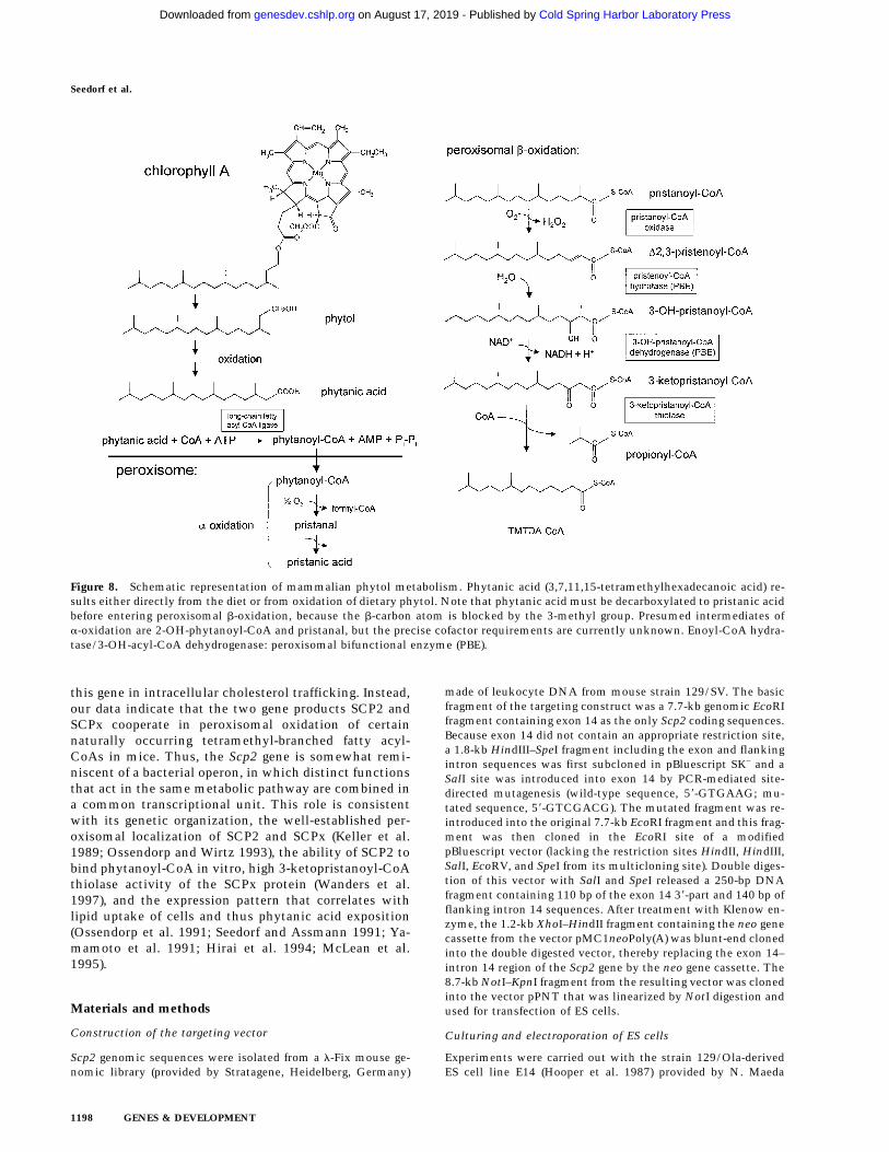
alcohol, originating from the isoprenoic side chain es-terified to ring IV of chlorophyll. In humans, the dailydietary intake of phytol and its product phytanic acid isin the order of 100 mg/day, but intake varies greatlydepending on dietary habits (Steinberg 1995). As illus-trated in Figure 8, normal catabolism starts with the con-version of phytol to phytanic acid, followed by activationto phytanoyl-CoA in the cytoplasm. Phytanoyl-CoA isthen imported into peroxisomes followed by a-oxida-tion, which involves hydroxylation at the a-carbon po-sition by phytanoyl-CoA hydroxylase (PHYH). Subse-quently, 2-OH-phytanoyl-CoA is converted to pristanicacid and formyl-CoA (Wanders et al. 1994; Croes et al.1997). Whereas formyl-CoA is further catabolized inmitochondria, pristanic acid is activated to pristanoyl-CoA, which is then subject to six cycles of peroxisomalb-oxidation. The intermediates of the first cycle areD2,3-pristenoyl-CoA (produced by pristanoyl-CoA oxi-dase), 3-OH-pristanoyl-CoA, and 3-ketopristanoyl-CoA(produced by a peroxisomal bifunctional enzyme). Finally,
3-ketopristanoyl-CoA is substrate for thiolytic cleavage,catalyzed by a 3-ketopristanoyl-CoA thiolase, which yieldsthe (n-3) lower homolog of pristanoyl-CoA (4,8,12-trimeth-yltridecanoyl-CoA) and propionyl-CoA (for review, seeSteinberg 1995). Although studies on mice are not avail-able, it now appears that this pathway operates in a similarway in rats and humans (Watkins et al. 1994; Singh andPoulos 1995).
We detected pronounced accumulation of phytanicacid in Scp2(−/−) mice, which exceeded that of pristanicacid and the downstream catabolic intermediates by sev-eralfold. This may be explained by inhibition of initialphytanic acid activation, phytanoyl-CoA import intoperoxisomes, or phytanoyl-CoA a-hydroxylation. Abnor-mal activation was unlikely because recent studies dem-onstrated convincingly that phytanoyl-CoA ligation ismediated by a common long chain fatty acyl-CoA ligase(Watkins et al. 1996), whereas metabolism of longstraight chain fatty acids was apparently not affected bythe gene disruption. Because PHYH was expressed nor-
Figure 5. Defective phytol catabolism in Scp2(−/−) mice. (A) Detection of phytanic acid and its catabolic metabolites in the liver byTOF-SIMS analysis. (Top) Mass spectrum of the phytanic acid standard (1 pg). The compound was detected as a negative ion at 311.5mu, representing the [M-H+]− ion. (Middle) Lipid extracts were prepared from liver of Scp2(−/−) mice and subjected to TOF-SIMSanalysis using identical conditions as for the standard. Arrows point to phytanic acid (311.5 mu), and the expected signals forD2,3-pristenic acid (295.5 mu), pristanic acid (297.5 mu), and 3-OH-pristanic acid (313.5 mu). The signals obtained for the 18 carbonatoms straight chain fatty acids C18:0, C18:1, and C18:2 are also marked. (Bottom) The same analysis with a liver sample from anScp2(+/+) mouse. The images are representative for five mice of each genotype and both sexes. (B) Monitoring of ions correspondingto the predicted products of thiolase-mediated 3-ketopristanoyl-CoA cleavage (at 255.5 mu, TMTDA; at 253.5 mu, D2,3-TMTDA). (C)Quantitative evaluation of the signal intensities based on five mice of each strain and sex. The columns indicate mean values of folddifference between either (−/−) mice or controls; ±S.E.M.. Black bars (−/−); hatched bars (+/+).
Gene targeting of Scp2
GENES & DEVELOPMENT 1195
Cold Spring Harbor Laboratory Press on August 17, 2019 - Published by genesdev.cshlp.orgDownloaded from

mally in Scp2(−/−) mice, we also excluded secondarydown-regulation of PHYH expression. On the basis ofthese considerations, we hypothesized that the lipid car-rier function of SCP2 may be involved in peroxisomalphytanoyl-CoA uptake (i.e., by acting as phytanoyl-CoAbinding protein). The analysis, which was performedwith a very specific FRET competition assay on the pu-rified recombinant rat protein, revealed a much higheraffinity for binding of phytanoyl-CoA than of pristanoyl-CoA, phytanic acid, pristanic acid, or cholesterol. In ad-dition, the Kd value was within a physiologically mean-ingful range (250 nM), thus supporting the postulated in-direct role of SCP2 in peroxisomal phytanoyl-CoAuptake.
Impaired phytanoyl-CoA import into peroxisomeswould lead to the expectation that the production ofdownstream intermediates, which are generated in per-oxisomes from phytanoyl-CoA, should be repressed inScp2(−/−) mice. In contrast, evaluation of TOF-SIMS sig-nals, which corresponded to these intermediates, indi-cated even higher concentrations than in controls after
challenging mice with high dosages of dietary phytol.Although the twofold increase in pristanic acid did notreach statistical significance, accumulation of D2,3-pris-tenic acid (four- to fivefold) and 3-OH-pristanic acid(three- to fourfold) was highly significant. In contrast,3-ketopristanic acid was not stable enough to withstandalkaline extraction and subsequent TOF-SIMS or GC-MSanalyses. We could, however, detect a very significant70% repression of the signals produced by the down-stream products of the 3-ketopristanoyl-CoA thiolase re-action in Scp2(−/−) mice (4,8,12-trimethyltridecanoicacid and 4,8,12-trimethyl-D2,3-tridecenoic acid). To-gether with enrichment of upstream intermediates inpristanic acid b-oxidation, the latter result supportedvery clearly inhibition at the level of 3-ketopristanoyl-CoA cleavage. These in vivo data corresponded to recentstudies, which demonstrated high specific activity of re-combinant rat SCPx to catalyze the thiolytic cleavage of3-ketopristanoyl-CoA in vitro (Wanders et al. 1997).Thus, our data appear to indicate a dual effect of the genedisruption, consisting of reduced peroxisomal phytanoyl-CoA import combined with defective thiolytic cleavageof 3-ketopristanoyl-CoA. Whereas the first effect seemsto relate to the phytanoyl-CoA carrier function of SCP2,the second may reflect the enzymatic activity associatedwith SCPx. This hypothesis appears very compelling, be-cause it may clarify why evolution has established a mo-lecular basis for coexpression of the two Scp2-encodedfunctions by fusing two originally separated SCP2 andthiolase genes into one common transcriptional unit.The fused gene is present in all vertebrates and could betraced back to Drosophila melanogaster (GenBank ac-cession no. X97685). In contrast, two separated geneswere identified in Caenorhabditis elegans and severalyeast species (Pfeifer et al. 1993b; Bunya et al. 1997).Interestingly, an ancient precursor of SCP2 could beidentified even in the primitive methanogenic archaeonMethanococcus jannaschii (Bult et al. 1996), in whommethyl-branched fatty acids play a prominent role.
As expected, (−/−) mice revealed a higher increase ofphytanic acid concentrations than controls, when wechallenged the two strains of mice transiently with phy-tol-enriched diets. However, after switching to the lowphytol diet, Scp2(−/−) mice eliminated phytanic acidfrom the bloodstream at a surprisingly high initial rate,followed by a much slower decline after 6 days. Becauseit is known from studies on patients with Refsum dis-ease that excess phytanic acid can be taken up by cellsand stored in triglycerides (Steinberg 1995), the high ini-tial rate most likely reflected cellular uptake and storagerather than high residual activity for phytanic acidbreakdown. On the basis of the slow rate of the latedecline, we calculated this activity to ∼10%. This was inline with our other findings—10-fold higher steady-stateconcentrations of phytanic acid and ∼10-fold increasedphytol toxicity. However, whether the residual activityis attributable to compensatory up-regulation of peroxi-somal straight chain b-oxidation, the presence in ourmodel of the SCP2-like activity associated with the 80-kD precursor of 17b-hydroxysteroid dehydrogenase type
Figure 6. Expression of phytanoyl-CoA hydroxylase in Scp2(−/−)mice. Northern blots of liver RNA were hybridized with a la-beled PYHH cDNA probe (A). Specific phytanoyl-CoA-bindingactivity of recombinant rat SCP2 (B). The columns representmeans of 1/Kd ± S.D. of five independent determinations.
Seedorf et al.
1196 GENES & DEVELOPMENT
Cold Spring Harbor Laboratory Press on August 17, 2019 - Published by genesdev.cshlp.orgDownloaded from

IV (Leenders et al. 1996), or an alternative pathway forphytanic acid oxidation, cannot be decided from ourdata.
Baum et al. (1997) reported that overexpression ofSCP2 in rat hepatoma cells inhibited cholesterol esteri-fication and HDL secretion, whereas plasma membranecholesterol was significantly increased. In addition,Puglielli et al. (1995) showed that treatment of humanskin fibroblasts with SCP2 anti-sense oligonucleotidesled to inhibition of cholesterol net transfer to the plasmamembrane. In view of these results, we found it surpris-ing that Scp2(−/−) mice had significantly lower hepaticcholesterol ester storage than controls. Because the genedisruption also lowered free fatty acid and triglycerideconcentrations very effectively, our results seem to in-dicate decreased availability of fatty acids for intracellu-lar lipid esterification rather than a specific abnormalityin cytosolic free cholesterol trafficking. It appeared in-teresting to us that hepatic hypolipidemia was associ-ated with peroxisome proliferation and induction of per-oxisomal and mitochondrial fatty acid b-oxidation inScp2(−/−) mice. As is known from treatment of rodentswith fibrates, induction of b-oxidation and peroxisomeproliferation can lead to fatty acid hypermetabolism andhypolipidemia (for review, see Lemberger et al. 1996).The signals mediating peroxisome proliferation andmodulation of gene expression in Scp2(−/−) mice are cur-rently unknown. One possibility consists of a direct orindirect effect of accumulating phytol metabolites onnuclear signal transduction pathways (i.e., the peroxi-some proliferator activated receptor PPARa being themost likely candidate in this respect).
Earlier studies provided several lines of indirect evi-dence that appear to support a role for SCP2 in adrenaland ovarian steroidogenesis (Pfeifer et al. 1993a). SCP2 isabundant in steroidogenic glands and trophic hormonesstimulate steroidogenesis along with SCP2 gene expres-sion (Trzeciak et al. 1987; Rennert et al. 1991). In addi-tion, SCP2 enhanced the movement of cholesterol be-tween vesicles and isolated mitochondria in vitro, whichcorresponded to increased pregnenolone synthesis in thein vitro system (Chanderbhan et al. 1982; Xu et al. 1991).Moreover, overexpression of SCP2 in COS cells engi-neered to produce progestins increased steroid formation(Yamamoto et al. 1991). On the other hand, no correla-tion existed between SCP2 expression and side chaincleavage activity in a variety of human tissue specimen(Yanase et al. 1996) and, a priori, it is not very clear howa peroxisomal protein would stimulate the net transferof free cholesterol to mitochondria directly. So far, thephenotypic characterization of the Scp2(−/−) mouse hasnot provided any convincing evidence for a role of SCP2in steroidogenesis in vivo. The absence of developmentalabnormalities or salt wasting and the fact that (−/−) micehad no abnormalities affecting fertility, seemed to ex-clude an obligatory role of SCP2 in general steroidogen-esis. This was in line with normal adrenal morphologyand normal plasma concentrations of testosterone, pro-gesterone, and glucocorticoids. On the other hand, amore subtle defect may be masked by compensatorymechanisms or depend on appropriate stress conditions.
In summary, the current phenotypic characterizationof the Scp2(−/−) KO mouse model did not provide im-mediate convincing evidence for an obligatory role of
Figure 7. Phenotypic abnormalities inScp2(−/−) mice after dietary phytol en-richment. (A) Six- to 8-week-old malemice (n = 5 in each group) were fed thestandard chow diet (control) or the samediet supplemented with 2.5 or 5 mg/gram of phytol for up to 6 weeks. Bodyweights (top) and food intake (bottom,given as grams of food consumed in the4-day period) were monitored daily. Re-sults were obtained for the first 4 days,when decline of body weight was maxi-mal. When Scp2(−/−) mice were fed thediet containing 5 mg/gram of phytol,they continued to mobilize body massuntil they reached close to 60% of theirstarting weights. In the third week, theydeveloped neuropathy, reduced theirfood intake, and died. (B) Serum triglyc-eride concentrations in the six experi-mental groups. The columns representmeans ± S.D.. (C) Abdominal tissues in(+/+) (left) and (−/−) mice (right) after 10days on the 5 mg/gram phytol diet. Notethe normal appearance of the kidneysbut hyperplastic and dark structures ofthe adrenal glands. In addition, lack of virtually any fat tissue, which is prominent in the (+/+) mouse, is evident in the (−/−) mouse.The abnormalities developed in the absence of significant reductions in food intake.
Gene targeting of Scp2
GENES & DEVELOPMENT 1197
Cold Spring Harbor Laboratory Press on August 17, 2019 - Published by genesdev.cshlp.orgDownloaded from
this gene in intracellular cholesterol trafficking. Instead,our data indicate that the two gene products SCP2 andSCPx cooperate in peroxisomal oxidation of certainnaturally occurring tetramethyl-branched fatty acyl-CoAs in mice. Thus, the Scp2 gene is somewhat remi-niscent of a bacterial operon, in which distinct functionsthat act in the same metabolic pathway are combined ina common transcriptional unit. This role is consistentwith its genetic organization, the well-established per-oxisomal localization of SCP2 and SCPx (Keller et al.1989; Ossendorp and Wirtz 1993), the ability of SCP2 tobind phytanoyl-CoA in vitro, high 3-ketopristanoyl-CoAthiolase activity of the SCPx protein (Wanders et al.1997), and the expression pattern that correlates withlipid uptake of cells and thus phytanic acid exposition(Ossendorp et al. 1991; Seedorf and Assmann 1991; Ya-mamoto et al. 1991; Hirai et al. 1994; McLean et al.1995).
Materials and methods
Construction of the targeting vector
Scp2 genomic sequences were isolated from a l-Fix mouse ge-nomic library (provided by Stratagene, Heidelberg, Germany)
made of leukocyte DNA from mouse strain 129/SV. The basicfragment of the targeting construct was a 7.7-kb genomic EcoRIfragment containing exon 14 as the only Scp2 coding sequences.Because exon 14 did not contain an appropriate restriction site,a 1.8-kb HindIII–SpeI fragment including the exon and flankingintron sequences was first subcloned in pBluescript SK− and aSalI site was introduced into exon 14 by PCR-mediated site-directed mutagenesis (wild-type sequence, 58-GTGAAG; mu-tated sequence, 58-GTCGACG). The mutated fragment was re-introduced into the original 7.7-kb EcoRI fragment and this frag-ment was then cloned in the EcoRI site of a modifiedpBluescript vector (lacking the restriction sites HindII, HindIII,SalI, EcoRV, and SpeI from its multicloning site). Double diges-tion of this vector with SalI and SpeI released a 250-bp DNAfragment containing 110 bp of the exon 14 38-part and 140 bp offlanking intron 14 sequences. After treatment with Klenow en-zyme, the 1.2-kb XhoI–HindII fragment containing the neo genecassette from the vector pMC1neoPoly(A) was blunt-end clonedinto the double digested vector, thereby replacing the exon 14–intron 14 region of the Scp2 gene by the neo gene cassette. The8.7-kb NotI–KpnI fragment from the resulting vector was clonedinto the vector pPNT that was linearized by NotI digestion andused for transfection of ES cells.
Culturing and electroporation of ES cells
Experiments were carried out with the strain 129/Ola-derivedES cell line E14 (Hooper et al. 1987) provided by N. Maeda
Figure 8. Schematic representation of mammalian phytol metabolism. Phytanic acid (3,7,11,15-tetramethylhexadecanoic acid) re-sults either directly from the diet or from oxidation of dietary phytol. Note that phytanic acid must be decarboxylated to pristanic acidbefore entering peroxisomal b-oxidation, because the b-carbon atom is blocked by the 3-methyl group. Presumed intermediates ofa-oxidation are 2-OH-phytanoyl-CoA and pristanal, but the precise cofactor requirements are currently unknown. Enoyl-CoA hydra-tase/3-OH-acyl-CoA dehydrogenase: peroxisomal bifunctional enzyme (PBE).
Seedorf et al.
1198 GENES & DEVELOPMENT
Cold Spring Harbor Laboratory Press on August 17, 2019 - Published by genesdev.cshlp.orgDownloaded from
(University of North Carolina). The cells were cultured onG418-resistant mouse embryonic fibroblast feeder layers as de-scribed earlier (Zhang et al. 1992). ES cells (3 × 10⁷ cells) wereresuspended in 0.5 ml of PBS containing 25 µg of linearizedtargeting vector and electroporated for 1 sec with a Bio-RadGenePulser at 200 µF and 300 V per 0.4 cm. Cells were thenseeded on eight Petri dishes (diameter, 10 cm) coated with fi-broblast feeder layers. Selection with G418 (200 µg/ml, GIBCO)was started after 1 day and selection with gancyclovir (2 µM)after 2 days. After 12 days of growth, individual colonies werepicked. Each colony was scraped from the plate with a sterileglass capillary and transferred to a 24-multiwell plate coatedwith fibroblast feeder layers and containing 1 ml of growth me-dium supplemented with 200 µg/ml G418, 2 µM Gancyclovir,and 100 U/ml of penicillin and streptomycin. After 2 days, eachcolony was disrupted with trypsin [0.025% wt/vol]. Four to 8days later, the cells were trypsinized again and ∼90 % of thecells were removed for DNA isolation. The remaining cellswere transferred into new coated multiwell plates and after thecells were grown to a final density of 1.5 × 10⁶ to 2.5 × 10⁶ cellsper well, they were frozen in growth medium containing 10%dimethylsulfoxide.
DNA analysis of ES cells and mice
Cells were lysed in 200 µl of 1% SDS, 25 µg of proteinase K permilliliter for 12–16 hr at 55°C. Thereafter, 100 µl of saturatedNaCl was added, mixed, and centrifuged in an Eppendorf bench-top centrifuge at maximal speed for 15 min. The DNA wasethanol precipitated from the supernatant and dissolved in 25 µlof Tris-EDTA buffer (TE). Eight microliters of this solution wasdigested with the appropriate restriction enzyme, fractionatedin 0.8% agarose gels and transferred to nitrocellulose (0.1-µmpore size; Schleicher & Schuell). Hybridization was performedas described (Raabe et al. 1996) with final washes in 0.1× SSC,0.1% (wt/vol) SDS at 65°C for 30 min. DNA for genotype analy-sis was isolated from mouse tail tips as described by Laird et al.(1991).
RNA and Western blot analyses, PCR, and DNA sequencing
Total RNA was isolated from mouse tissues with the guandini-um–thiocyanate–phenol–chloroform extraction procedure (Chom-czynski and Sacchi 1987) followed by selection of poly(A) RNAon oligo(dT) cellulose. Northern blots were hybridized with di-goxigenin-labeled probes prepared by random priming using acommercially available kit (Boehringer, Mannheim, Germany).All probes were obtained from a mouse liver cDNA library byPCR amplification with appropriate primers. Quantificationwas carried out relative to expression of GAPDH, detected witha probe derived from a 1.3-kb PstI fragment from pGAPDH (Fortet al. 1985) containing rat glyceraldehyde-3-phosphate dehydro-genase cDNA, the probe for detection of Scp2 expression was a0.45-kb PstI fragment from pBS-mSCPx containing mouse sterolcarrier protein X cDNA (Seedorf et al. 1993). The membraneswere rinsed twice in 0.1% SDS, 2× SSC at room temperature andthen twice in 1% SDS, 0.5× SSC at 68°C for 15 min. Bands werevisualized using the chemiluminescence substrate CDP-Star(Tropix-Serva, Heidelberg, Germany). DNA sequencing was per-formed on an automated laser fluorescence DNA sequencer(Pharmacia, Upsala, Sweden) according to the instructionmanual of the supplier. Detection of SCP2-related peptides byWestern blot analysis was described earlier (Seedorf et al.1994a).
Dietary intervention studies, and histological andanatomical analyses
In most experiments, 6- to 24-week-old male mice were used.However, the defect in phytol catabolism was confirmed to bepresent also in a group of 25 female mice. Mice were fed astandard chow diet [(Altrumin, Hanover, Germany) containing0.8 mg/gram (wt/wt) of various sterols, mainly cholesterol andb-sitosterol, 0.075 mg/gram (wt/wt) of nonesterified phytol and0.2 mg/gram (wt/wt) of phytanic acid] and water of pH 3.4–3.6ad libitum. The high fat diet consisted of standard chow supple-mented with 1% cholesterol, 15% coconut butter, and 0.5%cholate. Phytol-enriched diets were prepared from these diets byadding 1–50 mg/gram (wt/wt) of phytol (Aldrich, St. Louis,MO). Animals were kept individually, and food intake and bodyweights were monitored daily. Tissues were dissected routinelybetween 9 and 10 a.m. (to exclude variations that might beattributable to circadian regulation) after lethal anesthesia withavertin (Sigma). Tissues were fixed in phosphate-buffered form-aldehyde (pH 7.2) embedded in paraffin, sectioned at 5 µm, andstained with appropriate stains.
Binding of fatty acids to SCP2
Binding constants for the interaction between recombinant ratSCP2 and phytanic acid, phytanoyl-CoA, pristanic acid, or pris-tanoyl-CoA were determined by competing bound pyrene-la-beled dodecanoic acid with the nonlabeled substrates. Bindingof pyrenyl-dodecanoic acid was monitored using FRET betweenthe single tryptophan residue of SCP2 (donor) and the pyreneacceptor of the labeled fatty acid. The signals were corrected fordirect excitation of pyrene at 280 nm. The competitor-induceddecrease in sensitized emission was fitted to a binding equationderived from the rate equations of the relevant bimolar bindingreactions.
Analytical techniques, serum chemistry, and statisticalanalyses
Serum samples were taken by orbital bleeding or heart punc-ture. Serum chemistry was performed by routine clinical testson a Hitachi 747 analyzer with sample volumes of 0.15 ml.Hormones were measured using commercially available radio-immunoassay kits (Diagnostic Products Corp., Los Angeles,CA). Fatty acids and phytanic and pristanic acid in serum weremeasured by gas chromatography. Identification was achievedwith appropriate standards and verified by mass spectrometry asdescribed earlier. Analyses in the liver of phytol metaboliteswere performed by TOF-SIMS as described earlier (Seedorf et al.1995). All measurements were performed at least in triplicates.Statistical analyses were performed with the paired t-test. Val-ues of P ø 0.05 were considered statistically significant.
Acknowledgments
We thank B. Glass, K. Kluckman, and D. Lee for expert techni-cal assistance. Dr. R. Voss assisted in standardizing TOF-SIMS-based metabolite quantitation. This work was supported bygrants from the Deutsche Forschungsgemeinschaft (Se 459/2-2),the Interdisziplinares Klinisches Forschungszentrum (ProjectA4) of the Medical Faculty, University of Munster, the Boeh-ringer Ingelheim Stiftung, Bristol Myers Squibb, and theBayer AG.
The publication costs of this article were defrayed in part bypayment of page charges. This article must therefore be herebymarked ‘‘advertisement’’ in accordance with 18 USC section1734 solely to indicate this fact.
Gene targeting of Scp2
GENES & DEVELOPMENT 1199
Cold Spring Harbor Laboratory Press on August 17, 2019 - Published by genesdev.cshlp.orgDownloaded from

References
Baum, C.L., E.J. Reschly, A.K. Gayen, M.E. Groh, and K. Schad-ick. 1997. Sterol carrier protein-2 overexpression enhancessterol cycling and inhibits cholesterol ester synthesis andhigh density lipoprotein cholesterol secretion. J. Biol. Chem.272: 6490–6498.
Bloj, B. and D.B. Zilversmit. 1977. Rat liver proteins capable oftransferring phosphatidylethanolamine. Purification andtransfer activity for other phospholipids and cholesterol. J.Biol. Chem. 252: 1613–1619.
Bult, C.J., O. White, G.J. Olsen, L. Zhou, R.D. Fleischmann,G.G. Sutton, J.A. Blake, L.M. Fitzgerald, R.A. Clayton, J.D.Gocayne, A.R. Kerlavage, B.A. Dougherty, J.F. Tomb, M.D.Adams, C.I. Reich, R. Overbeek, E.F. Kirkness, K.G. Wein-stock, J.M. Merrick, A. Glodek, J.L. Scott, N.S.M. Geogha-gen, J.F. Weidman, J.L. Fuhrmann, J.C. Venter, et al. 1996.Complete genome sequence of the methanogenic archaeon,Methanococcus jannaschii. Science 273: 1058–1073.
Bunya, M., M. Maebuchi, T. Hashimoto, S. Yokota, and T. Ka-miryo. 1997. A second isoform of 3-ketoacyl-CoA thiolasefound in Caenorhabditis elegans, which is similar to sterolcarrier protein-x but lacks the sequence of sterol carrier pro-tein-2. Eur. J. Biochem. 245: 252–259.
Chanderbhan, R., B.J. Noland, T.J. Scallen, and G.V. Vahouny.1982. Sterol carrier protein-2—Delivery of cholesterol fromadrenal lipid droplets to mitochondria for pregnenolone syn-thesis. J. Biol. Chem. 257: 8928–8934.
Chomczynski, P. and N. Sacchi. 1987. Single-step method ofRNA isolation by acid guanidinium thiocyanate phenolchloroform extraction. Anal. Biochem. 162: 156–159.
Croes, K., P.P. van Veldhoven, G.P. Mannaerts, and M. Casteels.1997. Production of formyl-CoA during peroxisomal alpha-oxidation of 3-methyl-branched fatty acids. FEBS Lett.407: 197–200.
Fort, P., L. Marty, M. Piechaczyk, S. el Sabrouty, C. Dani, P.Jeanteur, and J.M. Blanchard. 1985. Various rat adult tis-sues express only one major mRNA species from the glycer-aldehyde-3-phosphate-dehydrogenase multigenic family.Nucleic Acids Res. 13: 1431–1442.
Hijikata, M., N. Ishii, H. Kagamiyama, T. Osumi, and T. Hashi-moto. 1987. Structural analysis of cDNA for rat peroxisomal3-ketoacyl-CoA thiolase. J. Biol. Chem. 262: 8151–8158.
Hirai, A., T. Kino, K. Tokinaga, K. Tahara, Y. Tamura, and S.Yoshida. 1994. Regulation of sterol carrier protein-2 (SCP2)gene-expression in rat peritoneal-macrophages during foamcell-formation—A key role for free-cholesterol content. J.Clin. Invest. 94: 2215–2223.
Hooper, M., K. Hardy, A. Handyside, S. Hunter, and M. Monk.1987. HPRT-deficient (Lesch-Nyhan) mouse embryos de-rived from germline colonization by cultured cells. Nature326: 292–295.
Keller, G.A., T.J. Scallen, D. Clarke, P.A. Maher, S.K. Krisans,and S.J. Singer. 1989. Subcellular-localization of sterol car-rier protein-2 in rat hepatocytes—Its primary localization toperoxisomes. J. Cell Biol. 108: 1353–1361.
Laird, P.W., A. Zijderveld, K. Linders, M.A. Rudnicki, R. Jae-nisch, and A. Berns. 1991. Simplified mammalian DNA iso-lation procedure. Nucleic Acids Res. 19: 4293.
Leenders, F., J.G. Tesdorpf, M. Markus, T. Engel, U. Seedorf, andJ. Adamski. 1996. Porcine 80-kda protein reveals intrinsic17-beta-hydroxysteroid dehydrogenase, fatty acyl-CoA-hy-dratase/dehydrogenase, and sterol transfer activities. J. Biol.Chem. 271: 5438–5442.
Lemberger, T., B. Desvergne, and W. Wahli. 1996. Peroxisomeproliferator-activated receptors: a nuclear receptor signaling
pathway in lipid physiology. Ann. Rev. Cell Dev. Biol.12: 335–363.
McLean, M.P., J.T. Billheimer, K.J. Warden, and R.B. Irby. 1995.Differential expression of hepatic sterol carrier proteins inthe streptozotocin-treated diabetic rat. Endocrinology136: 3360–3368.
Noland, B.J., R.E. Arebalo, E. Hansbury, and T.J. Scallen. 1980.Purification and properties of sterol carrier protein-2. J. Biol.Chem. 255: 4282–4289.
Ohba, T., H. Rennert, S.M. Pfeifer, Z.G. He, R. Yamamoto, J.A.Holt, J.T. Billheimer, and J.F. Strauss. 1994. The structure ofthe human sterol carrier protein-x/sterol carrier protein-2gene (scp2). Genomics 24: 370–374.
Ohba, T., J.A. Holt, J.T. Billheimer, and J.F. Strauss. 1995. Hu-man sterol carrier protein-x/sterol carrier protein-2 gene hastwo promoters. Biochemistry 34: 10660–10668.
Ossendorp, B.C. and K.W.A. Wirtz. 1993. The nonspecific lipid-transfer protein (sterol carrier protein-2) and its relationshipto peroxisomes. Biochimie 75: 191–200.
Ossendorp, B.C., G.P.H. van Heusden, A.L.J. de Beer, K. Bos,G.L. Schouten, and K.W.A. Wirtz. 1991. Identification of thecDNA clone which encodes the 58-kda protein containingthe amino-acid-sequence of rat-liver nonspecific lipid- trans-fer protein (sterol carrier protein-2)—Homology with rat per-oxisomal and mitochondrial 3-oxoacyl-CoA thiolases. Eur. J.Biochem. 201: 233–239.
Pfeifer, S.M., E.E. Furth, T. Ohba, Y.J. Chang, H. Rennert, N.Sakuragi, J.T. Billheimer, and J.F. Strauss. 1993a. Sterol car-rier protein-2—A role in steroid-hormone synthesis. J. Ste-roid Biochem. Mol. Biol. 47: 167–172.
Pfeifer, S.M., N. Sakuragi, A. Ryan, A.L. Johnson, R.G. Deeley,J.T. Billheimer, M.E. Baker, and J.F. Strauss. 1993b. Chickensterol carrier protein-2/sterol carrier protein-x: cDNA clon-ing reveals evolutionary conservation of structure and regu-lated expression. Arch. Biochem. Biophys. 304: 287–293.
Puglielli, L., A. Rigotti, A.V. Greco, M.J. Santos, and F. Nervi.1995. Sterol carrier protein-2 is involved in cholesteroltransfer from the endoplasmic reticulum to the plasmamembrane in human fibroblasts. J. Biol. Chem. 270: 18723–18726.
Raabe, M., U. Seedorf, H. Hameister, P. Ellinghaus, and G. Ass-mann. 1996. Structure and chromosomal assignment of themurine sterol carrier protein-2 gene (Scp2) and two relatedpseudogenes by in situ hybridization. Cytogenet. CellGenet. 73: 279–281.
Rennert, H., A. Amsterdam, J.T. Billheimer, and J.F. Strauss.1991. Regulated expression of sterol carrier protein-2 in theovary—A key role for cyclic-AMP. Biochemistry 30: 11280–11285.
Schram, A.W., S. Goldfischer, C.W. van Roermund, K.E. Brou-wer, J. Collins, T. Hashimoto, H.S. Heymans, H. van denBosch, R.B. Schutgens, J.M. Tager, et al. 1987. Human per-oxisomal 3-oxoacyl-coenzyme A thiolase deficiency. Proc.Natl. Acad. Sci. 84: 2494–2496.
Seedorf, U. and G. Assmann. 1991. Cloning, expression, andnucleotide-sequence of rat-liver sterol carrier protein-2cDNAs. J. Biol. Chem. 266: 630–636.
Seedorf, U., M. Raabe, and G. Assmann. 1993. Cloning, expres-sion and sequences of mouse sterol-carrier protein-x-encod-ing cDNAs and a related pseudogene. Gene 123: 165–172.
Seedorf, U., P. Brysch, T. Engel, K. Schrage, and G. Assmann.1994a. Sterol carrier protein-x is peroxisomal 3-oxoacyl co-enzyme-A thiolase with intrinsic sterol carrier and lipidtransfer activity. J. Biol. Chem. 269: 21277–21283.
Seedorf, U., S. Scheek, T. Engel, C. Steif, H.J. Hinz, and G.Assmann. 1994b. Structure-activity studies of human sterol
Seedorf et al.
1200 GENES & DEVELOPMENT
Cold Spring Harbor Laboratory Press on August 17, 2019 - Published by genesdev.cshlp.orgDownloaded from
carrier protein-2. J. Biol. Chem. 269: 2613–2618.Seedorf, U., M. Fobker, R. Voss, K. Meyer, F. Kannenberg, D.
Meschede, K. Ullrich, J. Horst, A. Benninghoven, and G. Ass-mann. 1995. Smith-Lemli-Opitz syndrome diagnosed by us-ing time-of-flight secondary-ion mass-spectrometry. Clin.Chem. 41: 548–552.
Singh, H. and A. Poulos. 1995. Substrate-specificity of rat-livermitochondrial carnitine palmitoyl transferase-I—Evidenceagainst alpha-oxidation of phytanic acid in rat-liver mito-chondria. FEBS Lett. 359: 179–183.
Singh, H., K. Beckman, and A. Poulos. 1994. Peroxisomal beta-oxidation of branched chain fatty acids in rat liver. Evidencethat carnitine palmitoyltransferase I prevents transport ofbranched chain fatty acids into mitochondria. J. Biol. Chem.269: 9514–9520.
Steinberg, D. 1995. Refsum disease. In The metabolic and mo-lecular basis of inherited disease (ed. C.R. Scriver, A.L.Beaudet, W.S. Sly, and D. Valle), pp. 2351–2370. McGraw-Hill, New York, NY.
Stolowich, N.J., A. Frolov, B. Atshaves, E.J. Murphy, C.A. Jolly,J.T. Billheimer, A.I. Scott, and F. Schroeder. 1997. The sterolcarrier protein-2 fatty acid binding site: an NMR, circulardichroic, and fluorescence spectroscopic determination. Bio-chemistry 36: 1719–1729.
Trzeciak, W.H., E.R. Simpson, T.J. Scallen, G.V. Vahouny, andM.R. Waterman. 1987. Studies on the synthesis of sterolcarrier protein-2 in rat adrenocortical-cells in monolayer-culture-regulation by ACTH and dibutyryl cyclic 38,58-AMP.J. Biol. Chem. 262: 3713–3717.
Wanders, R.A., C.W.T. van Roermund, D.S.M. Schor, H.J. Ten-brink, and C. Jakobs. 1994. 2-hydroxyphytanic acid oxidaseactivity in rat and human liver and its deficiency in theZellweger-syndrome. Biochim. Biophys. Acta Mol. BasisDis. 1227: 177–182.
Wanders, R.A., S. Denis, F. Wouters, K.W.A. Wirtz, and U. Seed-orf. 1997. Sterol carrier protein-x (SCPx) is a peroxisomalbranched-chain beta-ketothiolase specifically reacting with3-oxo-pristanoyl-CoA: A new, unique role for SCPx inbranched-chain fatty acid metabolism in peroxisomes. Bio-chem. Biophys. Res. Commun. 236: 565–569.
Watkins, P.A., A.E. Howard, and S.J. Mihalik. 1994. Phytanicacid must be activated to phytanoyl-CoA prior to its alpha-oxidation in rat liver peroxisomes. Biochim. Biophys. Acta1214: 288–294.
Watkins, P.A., A.E. Howard, S.J. Gould, J. Avigan, and S.J. Mi-halik. 1996. Phytanic acid activation in rat liver peroxisomesis catalyzed by long-chain acyl-CoA synthetase. J. Lipid Res.37: 2288–2295.
Xu, T.S., E.P. Bowman, D.B. Glass, and J.D. Lambeth. 1991.Stimulation of adrenal mitochondrial cholesterol side-chaincleavage by GTP, steroidogenesis activator polypeptide(SAP), and sterol carrier protein-2. J. Biol. Chem. 266: 6801–6807.
Yamamoto, R., C.B. Kallen, G.O. Babalola, H. Rennert, J.T. Bill-heimer, and J.F. Strauss. 1991. Cloning and expression of acDNA encoding human sterol carrier protein-2. Proc. Natl.Acad. Sci. 88: 463–467.
Yanase, T., T. Hara, Y. Sakai, R. Takayanagi, and H. Nawata.1996. Expression of sterol carrier protein-2 (SCP2) in humanadrenocortical tissue. Eur. J. Endocrinol. 134: 501–507.
Zhang, S.H., R.L. Reddick, J.A. Piedrahita, and N. Maeda. 1992.Spontaneous hypercholesterolemia and arterial lesions inmice lacking apolipoprotein-E. Science 258: 468–471.
Gene targeting of Scp2
GENES & DEVELOPMENT 1201
Cold Spring Harbor Laboratory Press on August 17, 2019 - Published by genesdev.cshlp.orgDownloaded from
12:1998, Genes Dev. Udo Seedorf, Martin Raabe, Peter Ellinghaus, et al. function
genein mice lacking the sterol carrier protein-2/sterol carrier protein-x Defective peroxisomal catabolism of branched fatty acyl coenzyme A
References
http://genesdev.cshlp.org/content/12/8/1189.full.html#ref-list-1
This article cites 43 articles, 21 of which can be accessed free at:
License
ServiceEmail Alerting
click here.right corner of the article or
Receive free email alerts when new articles cite this article - sign up in the box at the top
Cold Spring Harbor Laboratory Press
Cold Spring Harbor Laboratory Press on August 17, 2019 - Published by genesdev.cshlp.orgDownloaded from
Related Documents