GENOME INTEGRITY Yasaei and Slijepcevic Genome Integrity 2010, 1:3 http://www.genomeintegrity.com/content/1/1/3 Open Access RESEARCH BioMed Central © 2010 Yasaei and Slijepcevic; licensee BioMed Central Ltd. This is an Open Access article distributed under the terms of the Creative Commons Attribution License (http://creativecommons.org/licenses/by/2.0), which permits unrestricted use, distribution, and repro- duction in any medium, provided the original work is properly cited. Research Defective Artemis causes mild telomere dysfunction Hemad Yasaei and Predrag Slijepcevic* Abstract Background: Repair of DNA double strand breaks by non-homologous end joining (NHEJ) requires several proteins including Ku, DNA-PKcs, Artemis, XRCC4, Ligase IV and XLF. Two of these proteins, namely Ku and DNA-PKcs, are also involved in maintenance of telomeres, chromosome end-structures. In contrast, cells defective in Ligase IV and XRCC4 do not show changes in telomere length or function suggesting that these proteins are not involved in telomere maintenance. Since a mouse study indicated that defective Artemis may cause telomere dysfunction we investigated the effects of defective Artemis on telomere maintenance in human cells. Results: We observed significantly elevated frequencies of telomeric fusions in two primary fibroblast cell lines established from Artemis defective patients relative to the control cell line. The frequencies of telomeric fusions increased after exposure of Artemis defective cells to ionizing radiation. Furthermore, we observed increased incidence of DNA damage at telomeres in Artemis defective cells that underwent more than 32 population doublings using the TIF (Telomere dysfunction Induced Foci) assay. We have also inhibited the expression levels of DNA-PKcs in Artemis defective cell lines by either using synthetic inhibitor (IC86621) or RNAi and observed their greater sensitivity to telomere dysfunction relative to control cells. Conclusion: These results suggest that defective Artemis causes a mild telomere dysfunction phenotype in human cell lines. Background There is increasing evidence that the maintenance of telomeres, physical ends of chromosomes, and DNA damage response mechanisms are interlinked. The first observation of a telomere dysfunction phenotype in a DNA damage response defective environment was reported in the case of Ataxia telangiectasia (AT) cells. The telomere dysfunction phenotype in cells from AT patients or ATM (AT mutated) defective mice ranges from accelerated telomere shortening to end-to-end chromosome fusions and extra-chromosomal telomeric fragments [1,2]. Following the observation of telomere dysfunction associated with the ATM defect, a number of DNA damage response factors have been shown to affect telomere maintenance. Most notably, proteins involved in the repair of DNA double strand breaks (DSBs) either by Non-Homologous End Joining (NHEJ) or homologous recombination (HR) including Ku, DNA-PKcs, RAD54, RAD51D and BRCA1 if dysfunctional, will cause a severe telomere dysfunction phenotype [3-6]. So far, at least 17 DNA damage response proteins have been shown to affect telomere maintenance [7]. It is not yet clear as to why the interplay between telomere maintenance and DNA damage response is required. However, it is certain that both pathways are essential for chromosome integ- rity maintenance and perhaps their interaction is impor- tant for the stable chromosome segregation. One of the key pathways required for the stable segre- gation of chromosomes is NHEJ. The key players in this pathway are Ku 70/86 and DNA-PKcs, both shown to be involved in telomere maintenance [3]. Other proteins involved in NHEJ include: Artemis, Ligase IV, XRCC4 and XLF [8]. Previous studies have shown that Ligase IV and XRCC4 do not have effect on telomere length or function [9]. However, it is not clear yet whether the remaining two NHEJ proteins, namely Artemis and XLF, affect telomere maintenance. * Correspondence: [email protected] 1 Brunel Institute of Cancer Genetics and Pharmacogenomics, Division of Biosciences, School of Health Sciences and Social Care, Brunel University, Uxbridge, Middlesex, UB8 3PH, UK Full list of author information is available at the end of the article

Welcome message from author
This document is posted to help you gain knowledge. Please leave a comment to let me know what you think about it! Share it to your friends and learn new things together.
Transcript
GENOME INTEGRITYYasaei and Slijepcevic Genome Integrity 2010, 1:3http://www.genomeintegrity.com/content/1/1/3
Open AccessR E S E A R C H
ResearchDefective Artemis causes mild telomere dysfunctionHemad Yasaei and Predrag Slijepcevic*
AbstractBackground: Repair of DNA double strand breaks by non-homologous end joining (NHEJ) requires several proteins including Ku, DNA-PKcs, Artemis, XRCC4, Ligase IV and XLF. Two of these proteins, namely Ku and DNA-PKcs, are also involved in maintenance of telomeres, chromosome end-structures. In contrast, cells defective in Ligase IV and XRCC4 do not show changes in telomere length or function suggesting that these proteins are not involved in telomere maintenance. Since a mouse study indicated that defective Artemis may cause telomere dysfunction we investigated the effects of defective Artemis on telomere maintenance in human cells.
Results: We observed significantly elevated frequencies of telomeric fusions in two primary fibroblast cell lines established from Artemis defective patients relative to the control cell line. The frequencies of telomeric fusions increased after exposure of Artemis defective cells to ionizing radiation. Furthermore, we observed increased incidence of DNA damage at telomeres in Artemis defective cells that underwent more than 32 population doublings using the TIF (Telomere dysfunction Induced Foci) assay. We have also inhibited the expression levels of DNA-PKcs in Artemis defective cell lines by either using synthetic inhibitor (IC86621) or RNAi and observed their greater sensitivity to telomere dysfunction relative to control cells.
Conclusion: These results suggest that defective Artemis causes a mild telomere dysfunction phenotype in human cell lines.
BackgroundThere is increasing evidence that the maintenance oftelomeres, physical ends of chromosomes, and DNAdamage response mechanisms are interlinked. The firstobservation of a telomere dysfunction phenotype in aDNA damage response defective environment wasreported in the case of Ataxia telangiectasia (AT) cells.The telomere dysfunction phenotype in cells from ATpatients or ATM (AT mutated) defective mice rangesfrom accelerated telomere shortening to end-to-endchromosome fusions and extra-chromosomal telomericfragments [1,2]. Following the observation of telomeredysfunction associated with the ATM defect, a number ofDNA damage response factors have been shown to affecttelomere maintenance. Most notably, proteins involved inthe repair of DNA double strand breaks (DSBs) either by
Non-Homologous End Joining (NHEJ) or homologousrecombination (HR) including Ku, DNA-PKcs, RAD54,RAD51D and BRCA1 if dysfunctional, will cause a severetelomere dysfunction phenotype [3-6]. So far, at least 17DNA damage response proteins have been shown toaffect telomere maintenance [7]. It is not yet clear as towhy the interplay between telomere maintenance andDNA damage response is required. However, it is certainthat both pathways are essential for chromosome integ-rity maintenance and perhaps their interaction is impor-tant for the stable chromosome segregation.
One of the key pathways required for the stable segre-gation of chromosomes is NHEJ. The key players in thispathway are Ku 70/86 and DNA-PKcs, both shown to beinvolved in telomere maintenance [3]. Other proteinsinvolved in NHEJ include: Artemis, Ligase IV, XRCC4and XLF [8]. Previous studies have shown that Ligase IVand XRCC4 do not have effect on telomere length orfunction [9]. However, it is not clear yet whether theremaining two NHEJ proteins, namely Artemis and XLF,affect telomere maintenance.
* Correspondence: [email protected] Brunel Institute of Cancer Genetics and Pharmacogenomics, Division of Biosciences, School of Health Sciences and Social Care, Brunel University, Uxbridge, Middlesex, UB8 3PH, UKFull list of author information is available at the end of the article
BioMed Central© 2010 Yasaei and Slijepcevic; licensee BioMed Central Ltd. This is an Open Access article distributed under the terms of the CreativeCommons Attribution License (http://creativecommons.org/licenses/by/2.0), which permits unrestricted use, distribution, and repro-duction in any medium, provided the original work is properly cited.

Yasaei and Slijepcevic Genome Integrity 2010, 1:3http://www.genomeintegrity.com/content/1/1/3
Page 2 of 12
Artemis has exonuclease and endonuclease activities inthe presence of DNA-PKcs and ATP [10]. It is requiredfor V(D)J recombination and people with mutations inthe gene coding for Artemis show immunodeficiency andradiosensitivity [11]. Thus, the human disease due todefective Artemis is named RS-SCID (radio-sensitivesevere combined immunodeficiency disease).
A study of cells from Artemis defective mice [12]revealed slightly elevated frequencies of end-to-end chro-mosome fusions, a cytological sign of telomere dysfunc-tion. Furthermore, analysis of a primary fibroblast cellline from an RS-SCID patient showed accelerated short-ening of telomeres relative to the normal control cell line[13]. These studies point to the possibility that Artemis,similarly to the other two NHEJ proteins, Ku and DNA-PKcs, may have a role in telomere maintenance. This pos-sibility is further supported by observations that a closehomologue of Artemis, a protein named Apollo, isdirectly involved in telomere maintenance, most likelytrue interactions with the telomeric protein TRF2 [14,15].
In this study we analyzed spontaneous and radiationinduced chromosomal abnormalities and monitoredrepair kinetics of ionizing radiation (IR) induced DSBsoccurring within telomeric sequences in Artemis defec-tive human cells. Furthermore, we either inhibited orknocked-down DNA-PKcs and monitored the effect ofthis procedure on telomeres. Our results suggest thatdefective Artemis causes a mild telomere dysfunctionphenotype.
ResultsSpontaneous telomeric fusions in Artemis defective cellsWe started by analyzing spontaneous chromosomeabnormalities in two Artemis defective cell lines and acontrol cell line using FISH with the telomeric PNA (pep-tide nucleic acid) probe. The total number of end-to-endchromosome fusions (telomeric fusions) and chromo-some breaks were recorded. No significant differencesbetween frequencies of spontaneous chromosome breakswere observed between Artemis defective and controlcells (Fig. 1A). However, we observed low but signifi-cantly higher frequencies of telomeric fusions in the twoArtemis defective cell lines compared to the normal con-trol cell line (p < 0.001) (Fig. 1B). The majority of thesefusions were sister chromatid unions (~62%) (Fig. 1C).Given the significantly elevated level of telomeric fusionsin Artemis defective cells relative to control cells, ourresults indicate that the Artemis defect may cause a mildtelomere dysfunction phenotype in human cells.
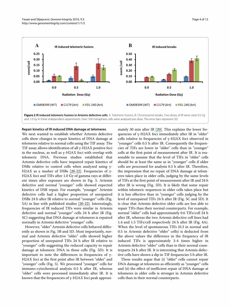
Radiation induced telomere dysfunction in Artemis defective cellsWe next examined whether telomere dysfunction is ele-vated following exposure of Artemis defective cells to
ionizing radiation (IR). Interestingly, frequencies of telo-meric fusions were significantly higher in Artemis defec-tive cell lines than in the control cell line (p < 0.001) afterirradiating cells with 1.0 Gy of gamma rays (Fig. 2A). Themajority of telomeric fusions were of chromatid type(56%) but we also observed some chromosome type telo-meric fusions i.e. dicentric chromosomes (30%). Only theCJ179 cell line showed statistically significant differencein telomeric fusions relative to the control line at bothdoses used (0.5 and 1.0 Gy) (Fig. 2A). Relative insensitiv-ity of the F01-240 cell line to the formation of telomericfusions at the dose of 0.5 Gy can be attributed to a type ofmutation present in this cell line that may lead to quanti-tatively different effects at different doses. Therefore, ourresults suggest that Artemis defective cell lines showslightly but significantly elevated IR induced telomericfusions in comparison to the control cell line. This furtherindicates the presence of a mild telomere dysfunctionphenotype as a result of Artemis deficiency in humancells.
Our results have also confirmed that the two Artemisdefective cell lines exhibit significantly higher frequenciesof IR induced chromosome breaks compared to the nor-mal control cell line (p < 0.001) (Fig. 2B). This is similar topublished result [16].
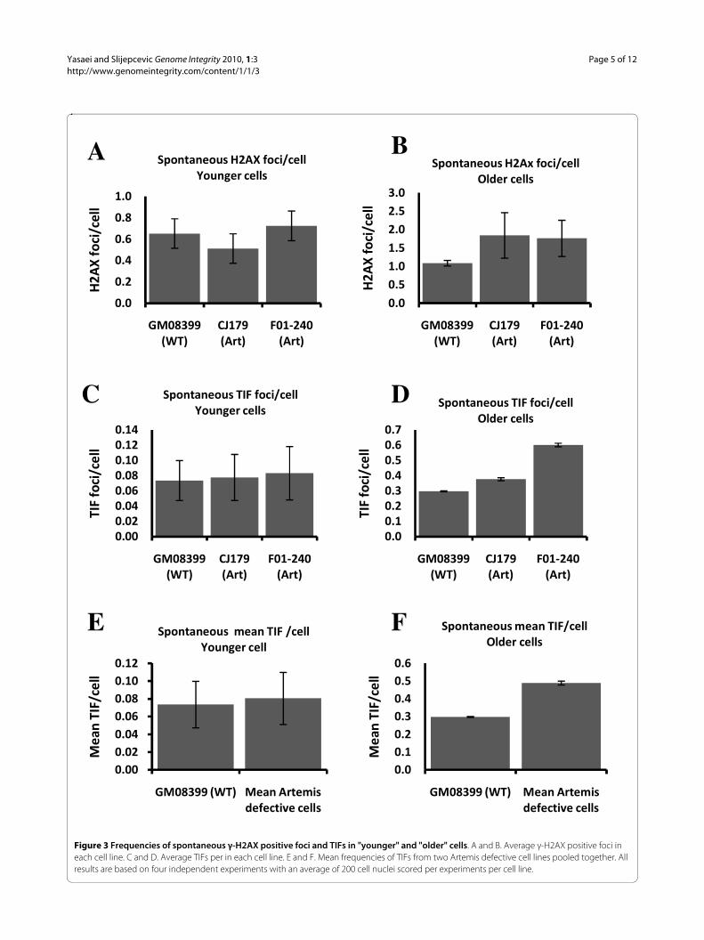
Analysis of Artemis defective human cell lines using immuno FISHTo confirm the presence of a mild telomere dysfunctionphenotype in Artemis defective cell lines we used themodified version of the TIF (Telomere dysfunctionInduced Foci) assay. This assay usually relies on simulta-neous detection of telomeres (antibodies against TRF1 orTRF2) and DNA damage (antibody against a DNA dam-age marker) [17]. However, instead of antibodies againsttelomeric proteins we used telomeric PNA in combina-tion with an antibody against a DNA damage marker, γ-H2AX (immuno-FISH). Given that telomeres shorten inproliferating primary human fibroblasts [18] and that thisshortening can affect telomere function [19] we decidedto analyze cells in relatively early passages ("younger"cells) as well as cells in late passages ("older" cells). Cellswith PD (population doubling) value below 16 repre-sented "younger" cells and cells with PD value of 32-33represented "older" cells.
Artemis defective and normal "younger" cells had simi-lar frequencies of spontaneous γ-H2AX positive foci (Fig.3A) and this is in line with published results [20]. How-ever, γ-H2AX positive spontaneous foci increased inArtemis defective and normal "older" cells relative to"younger" cells (Fig. 3B). Interestingly, the difference inspontaneous frequencies of γ-H2AX positive focibetween Artemis defective and normal "older" cells wasstatistically significant (p < 0.001). This could mean that

Yasaei and Slijepcevic Genome Integrity 2010, 1:3http://www.genomeintegrity.com/content/1/1/3
Page 3 of 12
"older" Artemis defective cells are less efficient in repair-ing endogenous DNA DSBs than the normal control cellsof the same age.
The immuno-FISH analysis showed no difference inspontaneous TIF frequencies between Artemis defectiveand normal "younger" cells (Fig. 3C and 3E). Therefore,the observed mild spontaneous telomere dysfunction inArtemis defective cells, which manifests as elevated levelof telomeric fusions (Fig. 1B) cannot be linked to theresults of immuno-FISH analysis. However, it is impor-tant to note that the average PD of cells used in experi-ments described in Fig. 1 was PD 24 (ranging from 19 to32). Therefore, it is possible that the observed mild spon-
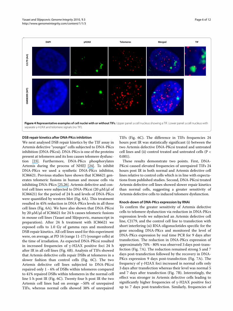
taneous telomere dysfunction occurs only in "older" cells.In line with this possibility "older" Artemis defective cellshad a significantly higher frequency of spontaneous TIFsthan normal "older" cells (Fig. 3D and 3F). Representativeexamples of TIFs are shown in Fig 4. Based on theseresults it seems reasonable to argue that telomere dys-function in Artemis defective cells increases with PDnumber. When Artemis defective cells are relatively"young" (low PDs) they show functional telomeres. How-ever, when Artemis defective cells become "older" (PD32+) but not senescent yet, they show a small but signifi-cant increase in spontaneous TIF frequency relative tonormal cells of similar age (Fig. 3D and 3F).
Figure 1 Spontaneous chromosome abnormalities in Artemis defective cells. A. Chromosome breaks. B. Telomeric fusions. C. Example of a telo-meric fusion in a CJ179 cell: sister chromatid union. Error bars represent standard deviation (SD). Frequencies of abnormalities per cell were calculated from three separate experiments. The total number of metaphase cells analysed were: GM08399 - 111, CJ179 - 193 and F01-240 - 174.
����
����
����
����
����
����
���
���� �����
���� ������
�������������
����
����
!"#��$��#%&��'�#(#&#(��)��$*&
����
����
����
���
����
����
���� �����
���� ������
�������������
����
����
!"#��$��#%&��� #(��+���%&+#�&B A
C
Yasaei and Slijepcevic Genome Integrity 2010, 1:3http://www.genomeintegrity.com/content/1/1/3
Page 4 of 12
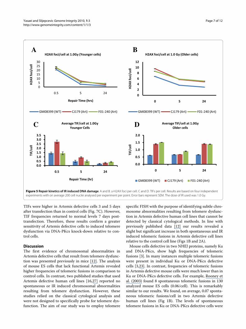
Repair kinetics of IR induced DNA damage at telomeresWe next wanted to establish whether Artemis defectivecells show changes in repair kinetics of DNA damage attelomeres relative to normal cells using the TIF assay. TheTIF assay allows identification of all γ-H2AX positive fociin the nucleus, as well as γ-H2AX foci with overlap withtelomeric DNA. Previous studies established thatArtemis defective cells have impaired repair kinetics ofDSBs relative to control cells when analyzed using γ-H2AX as a marker of DSBs [20-22]. Frequencies of γ-H2AX foci and TIFs after 1.0 Gy of gamma rays at differ-ent times after exposure are shown in Fig. 5. Artemisdefective and normal "younger" cells showed expectedkinetics of DSB repair. For example, "younger" Artemisdefective cells had a higher proportion of unrepairedDSBs 24 h after IR relative to normal "younger" cells (Fig.5A) in line with published studies [20-22]. Interestingly,frequencies of IR induced TIFs were similar in Artemisdefective and normal "younger" cells 24 h after IR (Fig.5C) suggesting that DNA damage at telomeres is repairednormally in Artemis defective "younger" cells.
However, "older" Artemis defective cells behaved differ-ently as shown in Fig. 5B and 5D. Most importantly, nor-mal and Artemis defective "older" cells showed higherproportion of unrepaired TIFs 24 h after IR relative to"younger" cells suggesting the reduced capacity to repairdamage at telomeric DNA in these cells (Fig. 5D). It isimportant to note the differences in frequencies of γ-H2AX foci at the first point after IR between "older" and"younger" cells (Fig. 5). We processed "younger" cells forimmuno-cytochemical analysis 0.5 h after IR, whereas"older" cells were processed immediately after IR. It isknown that the frequencies of γ-H2AX foci peak approxi-
mately 30 min after IR [20]. This explains the lower fre-quencies of γ-H2AX foci immediately after IR in "older"cells relative to frequencies of γ-H2AX foci observed in"younger" cells 0.5 h after IR. Consequently the frequen-cies of TIFs are lower in "older" cells than in "younger"cells at the first point of measurement after IR. It is rea-sonable to assume that the level of TIFs in "older" cellsshould be at least the same as in "younger" cells if oldercells are processed for analysis 0.5 h after IR. Therefore,the impression that no repair of DNA damage at telom-eres takes place in older cells, judging by the same levelsof TIFs at the first point of measurement after IR and 24 hafter IR is wrong (Fig. 5D). It is likely that some repairwithin telomeric sequences in older cells takes place butit is less effective than in "younger" cells judging by thelevel of unrepaired TIFs 24 h after IR (Fig. 5C and 5D). Itis clear that Artemis defective older cells are less able torepair TIFs than their normal counterparts. For example,normal "older" cells had approximately 0.6 TIFs/cell 24 hafter IR, whereas the two Artemis defective cell lines had1.4 and 1.5 TIFs/cell respectively 24 h after IR (Fig. 4A).When the level of spontaneous TIFs (0.3 in normal and0.5 in Artemis defective "older" cells) is deducted fromthe above values the difference in the frequency of IRinduced TIFs is approximately 3-4 times higher inArtemis defective "older" cells than in their normal coun-terparts 24 h after IR. It is interesting that Artemis defec-tive cells have shown a dip in TIF frequencies 5 h after IR.
These results argue that (i) "older" cells cannot repairDNA damage at telomeres as efficiently as "younger" cellsand (ii) the effect of inefficient repair of DNA damage attelomeres in older cells is stronger in Artemis defectivecells than in their normal counterparts.
Figure 2 IR induced telomeric fusions in Artemis defective cells. A. Telomeric fusions. B. Chromosome breaks. Two doses of IR were used: 0.5 Gy and 1.0 Gy in three independent experiments. Over 100 metaphase cells were analysed per dose. The error bars represent SD.
����
����
����
����
����
����
��� ��� ���
����
����
,$-+$�+#���.#&���/�
0,�+�-%��-��� #(��+��1%&+#�&
���������� ��������� ������������
����
����
����
����
����
����
��� ��� ���
����
�����
,$-+$�+#��.#&���/�
0,�+�-%��-�)��$*&
���������� ��������� ������������
A B
Yasaei and Slijepcevic Genome Integrity 2010, 1:3http://www.genomeintegrity.com/content/1/1/3
Page 5 of 12
Figure 3 Frequencies of spontaneous γ-H2AX positive foci and TIFs in "younger" and "older" cells. A and B. Average γ-H2AX positive foci in each cell line. C and D. Average TIFs per in each cell line. E and F. Mean frequencies of TIFs from two Artemis defective cell lines pooled together. All results are based on four independent experiments with an average of 200 cell nuclei scored per experiments per cell line.
���
���
���
��
���
���
���� �����
���� ������
�������������
2��
3�1#
�+��
�
!"#��$��#%&�2��3�1#�+��� 4#%�5����� &�
���������������������
���� �����
���� ������
�������������
2��
3�1#
�+��
�
!"#��$��#%&�2��6�1#�+��� 7 -����� &
�������������������������������
���� �����
���� ������
�������������
�0��
1#�+
���
!"#��$��#%&��0��1#�+��� 4#%�5����� &
�����������������������
���� �����
���� ������
�������������
�0��
1#�+
���
!"#��$��#%&��0��1#�+��� 7 -����� &
���������������������������
���� ����� ��$������(+&�-�1���+����� &
��$
���0
����
!"#��$��#%&��(�$���0����� 4#%�5�����
��������������������
���� ����� ��$������(+&�-�1���+����� &
��$
���0
����
!"#��$��#%&�(�$���0���� 7 -����� &
B A
E
D C
F
Yasaei and Slijepcevic Genome Integrity 2010, 1:3http://www.genomeintegrity.com/content/1/1/3
Page 6 of 12
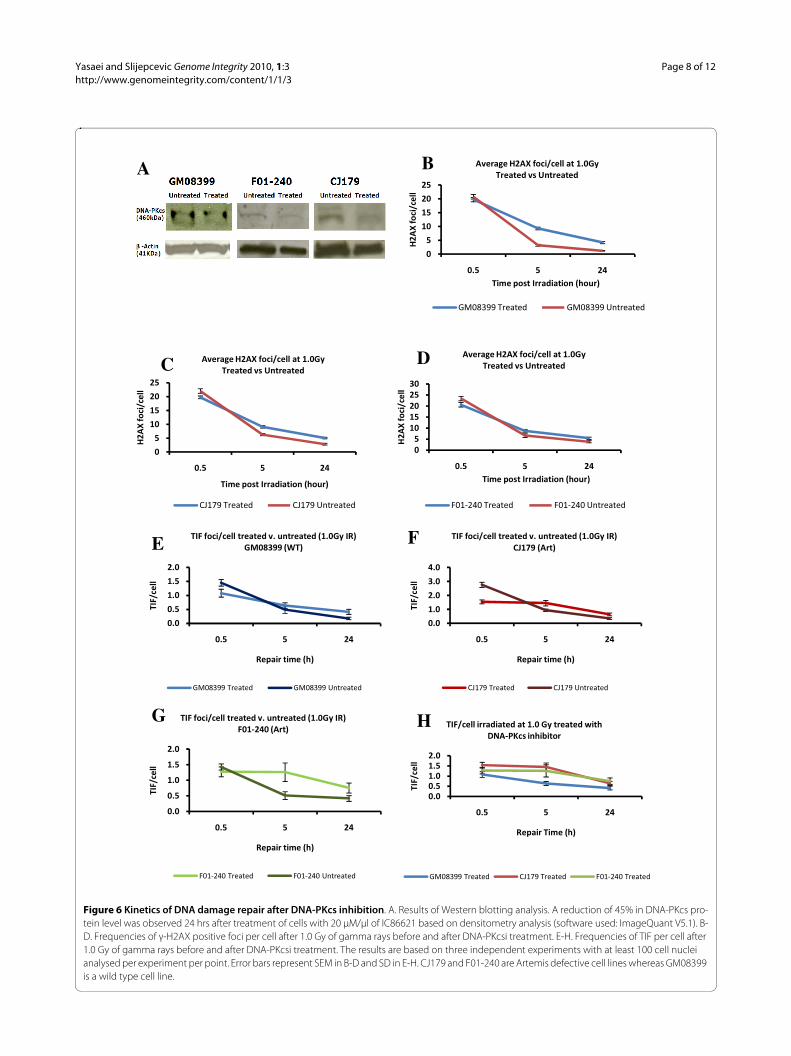
DSB repair kinetics after DNA-PKcs inhibitionWe next analysed DSB repair kinetics by the TIF assay inArtemis defective "younger" cells subjected to DNA-PKcsinhibition (DNA-PKcsi). DNA-PKcs is one of the proteinspresent at telomeres and its loss causes telomere dysfunc-tion [23]. Furthermore, DNA-PKcs phosphorylatesArtemis during the process of NHEJ [24]. To inhibitDNA-PKcs we used a synthetic DNA-PKcs inhibitor,IC86621. Previous studies have shown that IC86621 gen-erates telomeric fusions in human and mouse cells viainhibiting DNA-PKcs [25,26]. Artemis defective and con-trol cell lines were subjected to DNA-PKcsi (20 μM/μl ofIC86621) for the period of 24 h and levels of DNA-PKcswere quantified by western blot (Fig. 6A). This treatmentresulted in 45% reduction in DNA-PKcs levels in all threecell lines (Fig. 6A). We have also shown that DNA-PKcsiby 20 μM/μl of IC86621 for 24 h causes telomeric fusionsin mouse cell lines (Yasaei and Slijepcevic, manuscript inpreparation). After 24 h treatment with IC86621 weexposed cells to 1.0 Gy of gamma rays and monitoredDSB repair kinetics. All cell lines used for this experimentwere, on average, at PD 16 (range 11-17) (younger cells) atthe time of irradiation. As expected DNA-PKcsi resultedin increased frequencies of γ-H2AX positive foci 24 hafter IR in all cell lines (Fig. 6B). Analysis of TIFs showedthat Artemis defective cells repair DSBs at telomeres in aslower fashion than control cells (Fig. 6C). The twoArtemis defective cell lines subjected to DNA-PKcsirepaired only 1 - 6% of DSBs within telomeres comparedto 41% repaired DSBs within telomeres in the normal cellline 5 h post IR (Fig. 6C). Twenty four h post IR the twoArtemis cell lines had on average ~50% of unrepairedTIFs, whereas normal cells showed 38% of unrepaired
TIFs (Fig. 6C). The difference in TIFs frequencies 24hours post IR was statistically significant (i) between thetwo Artemis defective DNA-PKcsi treated and untreatedcell lines and (ii) control treated and untreated cells (P <0.001).
These results demonstrate two points. First, DNA-PKcsi caused elevated frequencies of unrepaired TIFs 24hours post IR in both normal and Artemis defective celllines relative to control cells which is in line with expecta-tions from published studies. Second, DNA-PKcsi treatedArtemis defective cell lines showed slower repair kineticsthan normal cells, suggesting a greater sensitivity ofArtemis defective cells to induced telomere dysfunction.
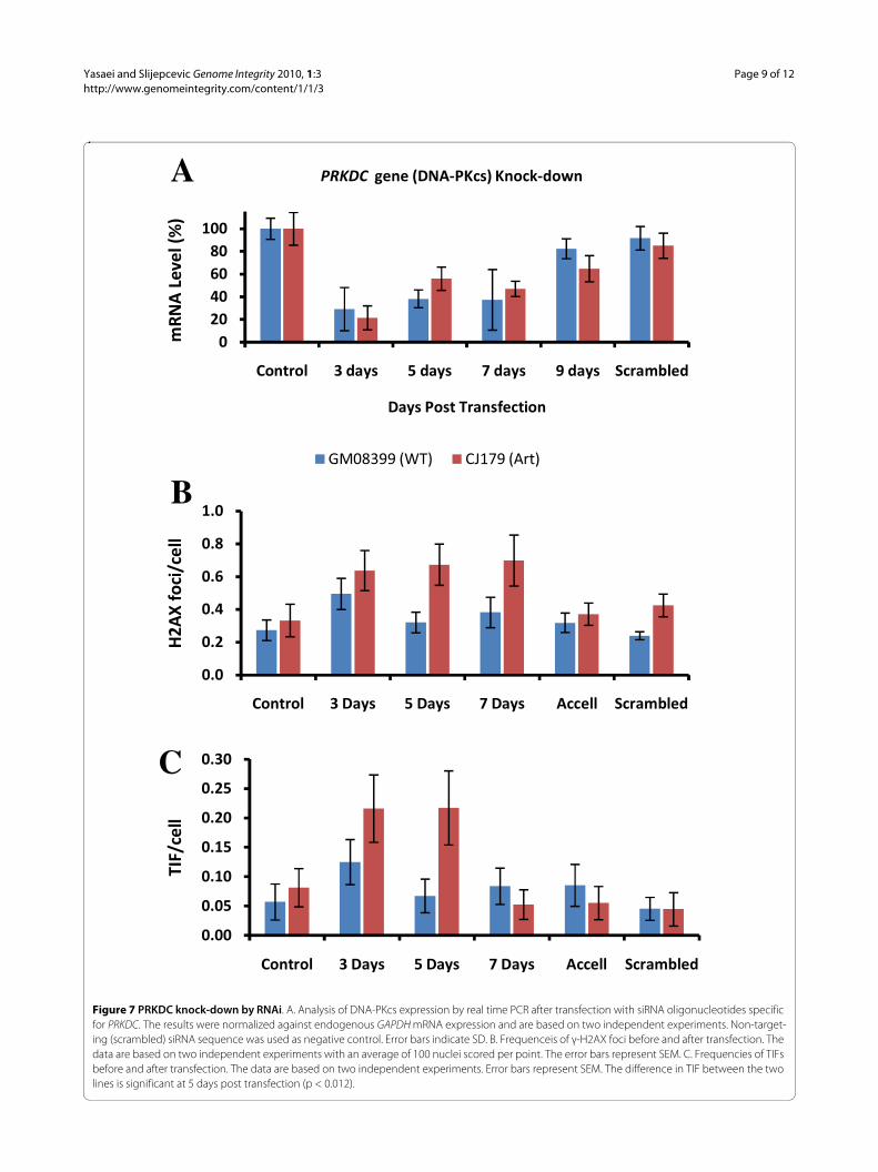
Knock-down of DNA-PKcs expression by RNAiTo confirm the greater sensitivity of Artemis defectivecells to telomere dysfunction via reduction in DNA-PKcsexpression levels we subjected an Artemis defective cellline, CJ179, and the control cell line to transfection withshort interfering (si) RNA oligonuclotides specific for thegene encoding DNA-PKcs and monitored the level ofDNA-PKcs expression by real time PCR for 9 days aftertransfection. The reduction in DNA-PKcs expression ofapproximately 70% - 80% was observed 3 days post-trans-fection (Fig. 7A). The reduction remained strong 5 and 7days post-transfection followed by the recovery in DNA-PKcs expression 9 days post-transfection (Fig. 7A). Thefrequency of γ-H2AX foci increased in normal cells only3 days after transfection whereas their level was normal 5and 7 days after transfection (Fig. 7B). Interestingly, theeffect was stronger in Artemis defective cells leading tosignificantly higher frequencies of γ-H2AX positive fociup to 7 days post-transfection. Similarly, frequencies of
Figure 4 Representative examples of cell nuclei with or without TIFs. Upper panel: a cell nucleus showing a TIF. Lower panel: a cell nucleus with separate γ-H2AX and telomere signals (no TIF).
�
.�80� �� #(���&�92��3� ���5�-� �0��
����
���
����
�
���
���
���
Yasaei and Slijepcevic Genome Integrity 2010, 1:3http://www.genomeintegrity.com/content/1/1/3
Page 7 of 12
TIFs were higher in Artemis defective cells 3 and 5 daysafter transfection than in control cells (Fig. 7C). However,TIF frequencies returned to normal levels 7 days post-transfection. Therefore, these results confirm a greatersensitivity of Artemis defective cells to induced telomeredysfunction via DNA-PKcs knock-down relative to con-trol cells.
DiscussionThe first evidence of chromosomal abnormalities inArtemis defective cells that result from telomere dysfunc-tion was presented previously in mice [12]. The analysisof mouse ES cells that lack functional Artemis revealedhigher frequencies of telomeric fusions in comparison tocontrol cells. In contrast, two published studies that usedArtemis defective human cell lines [16,27] reported nospontaneous or IR induced chromosomal abnormalitiesresulting from telomere dysfunction. However, thesestudies relied on the classical cytological analysis andwere not designed to specifically probe for telomere dys-function. The aim of our study was to employ telomere
specific FISH with the purpose of identifying subtle chro-mosome abnormalities resulting from telomere dysfunc-tion in Artemis defective human cell lines that cannot bedetected by classical cytological methods. In line withpreviously published data [12] our results revealed aslight but significant increase in both spontaneous and IRinduced telomeric fusions in Artemis defective cell linesrelative to the control cell line (Figs 1B and 2A).
Mouse cells defective in two NHEJ proteins, namely Kuand DNA-PKcs, show high frequencies of telomericfusions [3]. In many instances multiple telomeric fusionswere present in individual Ku or DNA-PKcs defectivecells [3,23]. In contrast, frequencies of telomeric fusionsin Artemis defective mouse cells were much lower than inKu or DNA-PKcs defective cells. For example, Rooney etal. (2003) found 8 spontaneous telomeric fusions in 130analyzed mouse ES cells (0.06/cell). This is remarkablysimilar to our results. We found, on average, 0.07 sponta-neous telomeric fusions/cell in two Artemis defectivehuman cell lines (Fig 1B). The levels of spontaneoustelomere fusions in Ku or DNA-PKcs defective cells were
Figure 5 Repair kinetics of IR induced DNA damage. A and B. γ-H2AX foci per cell. C and D. TIFs per cell. Results are based on four independent experiments with on average 200 cell nuclei analysed per experiment per point. Error bars represent SEM. The dose of IR used was 1.0 Gy.
������������
��� � ��
2��
3�1#
�+��
�
,�"$+���+(���'�&�
2��3�1#�+��� �$�����/��4#%�5����� &�
���������� ��������� ������������
�
�
�
�
��
��
� � ��
2��
3�1#
�+��
�
2��3�1#�+��� �$������/��7 -����� &�
���������� ��������� ������������
������������������������
��� � ��
�0��
��
,�"$+���+(���'��
����$5���0���� �$�����/4#%�5����� &
���
���
���
���
���
� � ��
�0��
��
����$5���0���� �$�����/7 -����� &
���������� ��������� ������������
D C
B A
Yasaei and Slijepcevic Genome Integrity 2010, 1:3http://www.genomeintegrity.com/content/1/1/3
Page 8 of 12
Figure 6 Kinetics of DNA damage repair after DNA-PKcs inhibition. A. Results of Western blotting analysis. A reduction of 45% in DNA-PKcs pro-tein level was observed 24 hrs after treatment of cells with 20 μM/μl of IC86621 based on densitometry analysis (software used: ImageQuant V5.1). B-D. Frequencies of γ-H2AX positive foci per cell after 1.0 Gy of gamma rays before and after DNA-PKcsi treatment. E-H. Frequencies of TIF per cell after 1.0 Gy of gamma rays before and after DNA-PKcsi treatment. The results are based on three independent experiments with at least 100 cell nuclei analysed per experiment per point. Error bars represent SEM in B-D and SD in E-H. CJ179 and F01-240 are Artemis defective cell lines whereas GM08399 is a wild type cell line.
�
�
��
��
��
��
��� � ��
2��
3�1#
�+��
�
�+(��"#&��0��$-+$�+#���'#%��
����$5��2��3�1#�+��� �$�����/���$��-��&�:����$��-
��������������� �����������������
�
�
��
��
��
��
��� � ��
2��
3�1#
�+��
�
�+(��"#&��0��$-+$�+#���'#%��
����$5��2��3�1#�+��� �$�����/���$��-��&�:����$��-
������������ ��������������
��
����������
��� � ��
2��
3�1#
�+��
�
�+(��"#&��0��$-+$�+#���'#%��
����$5��2��3�1#�+��� �$�����/���$��-��&�:����$��-
��������������� �����������������
���
���
���
���
���
��� � ��
�0��
��
,�"$+���+(���'�
�0��1#�+��� ����$��-����%����$��-�����/�0,����� �����
��������������� �����������������
���
���
���
���
���
��� � ��
�0��
��
,�"$+���+(���'�
�0��1#�+��� ����$��-����%����$��-�����/�0,����� ������
������������ ��������������
���
���
���
���
���
��� � ��
�0��
��
,�"$+���+(���'�
�0��1#�+��� ����$��-����%����$��-�����/�0,��������������
��������������� �����������������
���������������
��� � ��
�0��
��
,�"$+���+(���'�
�0���� �+��$-+$��-�$������/����$��-�;+�'�.<��8=�&�+�'+)+�#�
��������������� ������������ ���������������
B A
G H
F E
C D
Yasaei and Slijepcevic Genome Integrity 2010, 1:3http://www.genomeintegrity.com/content/1/1/3
Page 9 of 12
Figure 7 PRKDC knock-down by RNAi. A. Analysis of DNA-PKcs expression by real time PCR after transfection with siRNA oligonucleotides specific for PRKDC. The results were normalized against endogenous GAPDH mRNA expression and are based on two independent experiments. Non-target-ing (scrambled) siRNA sequence was used as negative control. Error bars indicate SD. B. Frequenceis of γ-H2AX foci before and after transfection. The data are based on two independent experiments with an average of 100 nuclei scored per point. The error bars represent SEM. C. Frequencies of TIFs before and after transfection. The data are based on two independent experiments. Error bars represent SEM. The difference in TIF between the two lines is significant at 5 days post transfection (p < 0.012).
��������
���
�#���# ��-$/& ��-$/& ��-$/& �-$/& !��$() �-
(,<
��>
���
��?�
.$/&�8#&����$�&1���+#�
����� 5�����.<��8=�&��=�#�*�-#;�
���������� ���������
���
���
���
��
���
���
�#���# ��.$/& ��.$/& ��.$/& ���� !��$() �-
2��
3�1#
�+��
�
����
����
����
����
����
����
����
�#���# ��.$/& ��.$/& ��.$/& ���� !��$() �-
�0��
��
B
A
C
Yasaei and Slijepcevic Genome Integrity 2010, 1:3http://www.genomeintegrity.com/content/1/1/3
Page 10 of 12
> 10 times higher [3]. This suggests that the effect of dys-functional Artemis on telomeres in mammalian cells ismuch milder in comparison with Ku or DNA-PKcsdefects Furthermore, we have demonstrated that (i) DNAdamage occurring within the telomeric DNA is repairedless efficiently in "older" cells (PD 32+) irrespectivewhether they are Artemis defective or normal and (ii)Artemis defective "older" cells show higher proportion ofDNA damage at telomeres than their normal counter-parts (Fig. 5D). This means that the presence of a DSBwithin telomeric sequences (TIFs) causes a problem forrepair mechanisms in "older" cells leading to differencesin TIF frequencies 24 h after IR between "older" and"younger" cells. It has recently been shown that telomericsequences pose a significant challenge to DNA replica-tion machinery causing replication-dependent abnormal-ities very similar to aphidicolin-induced fragile sites [28].The notion that telomeric sequences may behave as frag-ile sites originated from observations that interstitial telo-meric sequences in Chinese hamster cells are prone to IRinduced chromosome breakage [29]. Interestingly, Kruket al. [30] found that UV induced damage at telomereswas repaired less efficiently than in transcribing DNAsequences but more efficiently that in other non-codingDNA sequences. More importantly Kruk et al. [30] foundthat DNA repair efficiency in cells from older donors waslower than in cells from younger donors. This is similar toour observation that TIFs persist at telomeres in "older"cells whereas "younger" cells repair IR induced damage attelomeres efficiently.
Finally, we have shown that DNA-PKcsi causes thegreater effect on telomeres in Artemis defective cells thanin control cells after approximately 45% reduction inDNA-PKcs expression (Fig 6). The greater effect ofreduced DNA-PKcs expression on Artemis defective cellsrelative to control cells was confirmed by RNAi resultingin elevated frequencies of TIFs in Artemis defective cells5 days after transfection. However, mechanisms behindthe greater sensitivity of Artemis defective cells to telom-ere dysfunction relative to control cells remain unclear. Itis worth pointing out that Artemis is involved in therepair of only a proportion of DSBs. For example, ~90% ofIR induced DSBs are repaired relatively quickly andrequire Ku, DNA-PKcs and LIG-4/XRCC4 [20]. Theremaining fraction of 10% DSBs are repaired by the com-bined action of ATM and Artemis [20] and they occurwithin heterochromatic regions of the genome [31].Telomeres are typical heterochromatic parts of thegenome [32,33]. It is therefore possible that Artemis andATM are required specifically to repair of DSBs occurringwithin telomeric sequences. If this is true than we canargue that when Artemis is defective, as in the case of twocell lines used in this study, DSBs at telomeres cannot be
repaired efficiently leading to cytological manifestation oftelomere dysfunction (Fig. 1 and 2).
ConclusionWe have shown that Artemis defect in human cell linescauses a mild telomere dysfunction phenotype detectableat the cytological level. However, mechanisms by whichdysfunctional Artemis affects telomere maintenanceremain unclear.
MethodsCell culture and irradiationThe human primary fibroblast cell lines defective inArtemis, CJ179 and F01-240 were kindly provided byProf. P.A. Jeggo, University of Sussex, Brighton UK.Details of mutations in each cell line have been describedpreviously [20]. The normal human primary fibroblast,GM08399 was purchased from the Coriell Institute forMedical Research. All cell lines were cultured in D-MEMmedium (Gibco/Invitrogen) supplemented with 10% fetalcalf serum (Gibco/Invitrogen). The cultures were incu-bated at 37°C in the atmosphere of 10% CO2. Cells wereirradiated, using a Cobalt60 source (dose rate), at 80-90%confluency either in tissue culture flasks for chromosomeanalysis or on polyprep slides (Sigma) for TIF assay.
Telo-FISH and Cytogenetic AnalysisMetaphase preparation was performed as described pre-viously [34]. Briefly, cells were subjected to irradiationwith gamma rays and incubated for 24 hours. The last 7 hof incubation 10 μl/ml of colcemid (10 μg/ml) (Sigma)was added to cell cultures followed by trypsinization,treatment with the hypotonic solution (5.6 g/l of KCl),fixation with methanol/acetic acid and slide preparation.Telo-FISH was performed as described previously [34].Briefly, after appropriate washing slides were hybridizedwith the Cy-3 labelled telomeric PNA probe (CCCTAA)3and left in a dark humidified chamber for 2 hours. Afterthis slides were washed in 70% formamide and stainedwith the DAPI mounting medium (Vector Laboratories).Chromosomal analysis was performed using a Ziess-Axioplan2 microscope equipped with a CCD camera andMetaSystem image acquisition software (Imaging Associ-ate).
Telomere dysfunction induced foci (TIF) assayThe TIF assay described here is based on the detection of(i) DNA damage by an antibody against DNA damagemarker γ-H2AX and (ii) telomeres by the synthetic PNAprobe. Briefly, cells were incubated in tissue culture flasktwo days prior to experiment and after reaching a 80-90%confluency they were trypsinized and 1.0 ml of cell sus-pension (containing roughly 50,000 cells) was placed ontopolyprep slides (Sigma) which were tranferred into petri
Yasaei and Slijepcevic Genome Integrity 2010, 1:3http://www.genomeintegrity.com/content/1/1/3
Page 11 of 12
dishes and left to grow for 24 h. Slides were then rinsed inPBS and fixed in 4% formaldehyde in PBS for 15 minutes.Cells were permeabilized in 0.2% (v/v) of Triton-X in dis-tilled water at 4.0°C for 10 minutes and blocked with 0.5%(w/v) Bovine Serum Albumin (BSA) in PBS for 30 min-utes. Anti-phospho-Histone H2A.X (Ser139) (Upstate)was diluted 1:500 with 0.5% BSA and 100 μl of resultingsolution was added onto each slide and left to incubatefor 1 hour in a damp container. After three washes insterile tris-buffered saline tween-20 (TBS-T, pH7.4; 0.15M NaCl, 0.268 mM KCl, 0.025 M tris-base, 500 μl/1ltween-20) for 3 minutes slides were incubated with 100 μlof secondary anti-goat antibody conjugated with Fluores-cein isothiocyanate (FITC) and washed as above. Afterthis slides were placed in 4% formaldehyde for 20 minutesfor cross-fixing to preserve antibodies. The next stepinvolved hybridization with telomeric PNA and this wasperformed as described above with the exception thatTBS-T was used far washing instead of PBS.
Western blottingWestern blotting has been carried out as described previ-ously [35]. Briefly, cells grown in P100 petri dish werelyzed with 900 μl of lysis buffer (5× sample buffer; 10% (v/v) sodium dodecyl sulphate, 250 mM tris pH 8.0, 50% (v/v) glycerol, 0.01% (w/v) bromophenol blue). In addition,50 μl of protease inhibitor (Roche) plus 50 μl of beta-mer-captoethanol was added and left for at least one minute.Cells were scraped off and collected in an eppendorf tube.Cells were then mechanically sheared ten times using a 1ml syringe and a 23 g needle. Samples were spun at13,000 RPM for 5 minutes at 4°C. Proteins were quanti-fied using RC-DC protein assay (Bio-rad). Equal concen-trations of 50 μg/50 μl were loaded onto a 4% precast gel(Bio-rad) until the high molecular weight protein marker(Invitrogen) was well separated. Following a wet blottingtransfer to a polyvinylidine fluoride (PVDF) membraneand blocking with 5% (w/v) semi-skimmed milk (Marvel),the membrane was incubated with the mouse monoclo-nal primary antibody (DNA-PKcs, AB-4, Neomarker,Thermo Fisher Scientific) and β-acting (Abcam, Cam-bridge, UK) in a 1:500 and 1:1000 dilution respectively at4°C overnight. This was followed by a secondary incuba-tion period of 45 minutes with an HRP conjugated anti-mouse monoclonal rabbit antibody (Abcam, Cambridge,UK) diluted of 1:5000. The antibody detection wasachived using chemiluminescence technique (ECL plus,GE healthcare) and protein levels were quantified usingImageQuant V5.1 (Molecular Dynamics, Piscataway, NJ).
siRNA transfectionThe two primary fibroblast cell lines (GM08399 andCJ179) were plated at a seeding density of 0.045 × 10⁶
cells/1/2 ml roughly equating to 40% confluency in a 24-
well plate. All siRNA transfections were done in duplicateusing Dharmacon Accell SMARTpool siRNA reagent(Thermo Fisher Scientific, Lafayette. CO) targetingPRKDC gene (NM_006904) and a non-targeting (scram-bled) siRNA as a negative control at a final concentrationof 1 μM/μl and followed Dharmacon recommended pro-tocol. Transfection was performed in a passive mannerusing a serum-free Accell transfection medium (Dharma-con). Cells were plated 24 h prior to transfection andincubated at 37°C in the incubator with the CO2 concen-tration adjusted to 10%. Total RNA were extracted at var-ious time points as shown in Fig 6. The sequences of thehuman PRKDC gene siRNA were: UCUUGU-GUUUAUUGGAUC, GGAAGAAGCUCAUUUGAUU,CGAUCAACACGGAAUUAUU and CUUUUACAUAGCAUGGUUA as recommended by Dharmacon(Thermo Fisher Scientific, Lafayette. CO).
Real-time quantitative PCRA two step reverse-transcription PCR was performedusing SYBR green 1 dye (Applied Biosystems) and expres-sion of mRNA quantified in real-time with an ABI prism7900HT sequence detection system (Applied Biosys-tems). The relative gene expression of DNA-PKcs weremeasured against the endogenous GAPDH gene and cal-culated using ΔCt. The sequence of DNA-PKcs andGAPDH primer were: forward 5'-CCGGACGGAC-CTACTACGACT-3' and reverse 5'-AGAACGACCTG-GGCA TCCT-3', forward 5'-GAAGGTGAAGGTCGGAGT-3' and reverse 5'-GAAGATGG TGATGG-GATTTC-3' respectively.
Competing interestsThe authors declare that they have no competing interests.
Authors' contributionsHY carried out experiments. PS designed experiments with the help of HY. PShas written the manuscript. HY and PS have read and approved the final manu-script.
AcknowledgementsThis study was supported by a grant from European Commission RISC-RAD contract FI6R-CT2003-508842.
Author DetailsBrunel Institute of Cancer Genetics and Pharmacogenomics, Division of Biosciences, School of Health Sciences and Social Care, Brunel University, Uxbridge, Middlesex, UB8 3PH, UK
References1. Metcalfe JA, Parkhill J, Campbell L, Stacey M, Biggs P, Byrd PJ, Taylor AMR:
Accelerated telomere shortening in ataxia telangiectasia. Nat Genet 1996, 13:350-353.
2. Hande MP, Balajee AS, Tchirkov A, Wynshaw-Boris A, Lansdorp PM: Extra-chromosomal telomeric DNA in cells from Atm-/- mice and patients with ataxia-telangiectasia. Hum Mol Genet 2001, 10:519-528.
3. Bailey SM, Meyne J, Chen DJ, Kurimasa A, Li GC, Lehnert BE, Goodwin EH: DNA double-strand break repair proteins are required to cap the ends
Received: 15 January 2010 Accepted: 26 May 2010 Published: 26 May 2010This article is available from: http://www.genomeintegrity.com/content/1/1/3© 2010 Yasaei and Slijepcevic; licensee BioMed Central Ltd. This is an Open Access article distributed under the terms of the Creative Commons Attribution License (http://creativecommons.org/licenses/by/2.0), which permits unrestricted use, distribution, and reproduction in any medium, provided the original work is properly cited.Genome Integrity 2010, 1:3
Yasaei and Slijepcevic Genome Integrity 2010, 1:3http://www.genomeintegrity.com/content/1/1/3
Page 12 of 12
of mammalian chromosomes. Proceedings of the National Academy of Sciences of the United States of America 1999, 96:14899-14904.
4. Jaco I, Munoz P, Goytisolo F, Wesoly J, Bailey S, Taccioli G, Blasco MA: Role of Mammalian Rad54 in Telomere Length Maintenance. Mol Cell Biol 2003, 23:5572-5580.
5. Tarsounas M, Muñoz P, Claas A, Smiraldo PG, Pittman DL, Blasco MA, West SC: Telomere Maintenance Requires the RAD51D Recombination/Repair Protein. Cell 2004, 117:337-347.
6. McPherson JP, Hande MP, Poonepalli A, Lemmers B, Zablocki E, Migon E, Shehabeldin A, Porras A, Karaskova J, Vukovic B, Squire J, Hakem R: A role for Brca1 in chromosome end maintenance. Hum Mol Genet 2006, 15:831-838.
7. Slijepcevic P: The role of DNA damage response proteins at telomeres--an "integrative" model. DNA Repair 2006, 5:1299-1306.
8. Sekiguchi JM, Ferguson DO: DNA Double-Strand Break Repair: A Relentless Hunt Uncovers New Prey. Cell 2006, 124:260-262.
9. d'Adda di Fagagna F, Hande MP, Tong W-M, Roth D, Lansdorp PM, Wang Z-Q, Jackson SP: Effects of DNA nonhomologous end-joining factors on telomere length and chromosomal stability in mammalian cells. Current Biology 2001, 11:1192-1196.
10. Goodarzi AA, Yu Y, Riballo E, Douglas P, Walker SA, Ye R, Harer C, Marchetti C, Morrice N, Jeggo PA, Lees-Miller SP: DNA-PK autophosphorylation facilitates Artemis endonuclease activity. EMBO J 2006, 25:3880-3889.
11. Poinsignon C, de Chasseval Regina, Soubeyrand Sebastien, Moshous Despina, Fischer Alain, Robert J, Haché G, Villartay J-Pd: Phosphorylation of Artemis following irradiation-induced DNA damage. European Journal of Immunology 2004, 34:3146-3155.
12. Rooney S, Alt FW, Lombard D, Whitlow S, Eckersdorff M, Fleming J, Fugmann S, Ferguson DO, Schatz DG, Sekiguchi J: Defective DNA Repair and Increased Genomic Instability in Artemis-deficient Murine Cells. J Exp Med 2003, 197:553-565.
13. Cabuy E, Newton C, Joksic G, Woodbine L, Koller B, Jeggo P, Slijepcevic P: Accelerated telomere shortening and telomere abnormalities in radiosensitive cell lines. Radiation Research 2005, 164:53-62.
14. Lenain C, Bauwens S, Amiard S, Brunori M, Giraud-Panis M-Jp, Gilson E: The Apollo 52 Exonuclease Functions Together with TRF2 to Protect Telomeres from DNA Repair. Cell 2006, 16:1303-1310.
15. van Overbeek M, de Lange T: Apollo, an Artemis-Related Nuclease, Interacts with TRF2 and Protects Human Telomeres in S Phase. Cell 2006, 16:1295-1302.
16. Darroudi F, Wiegant W, Meijers M, Friedl AA, Burg M van der, Fomina J, van Dongen JJM, van Gent DC, Zdzienicka MZ: Role of Artemis in DSB repair and guarding chromosomal stability following exposure to ionizing radiation at different stages of cell cycle. Mutation Research/Fundamental and Molecular Mechanisms of Mutagenesis 2007, 615:111-124.
17. Takai H, Smogorzewska A, de Lange T: DNA Damage Foci at Dysfunctional Telomeres. Cell 2003, 13:1549-1556.
18. Harley CB, Futcher AB, CW G: Telomeres shorten during ageing of human fibroblasts. Nature 1990, 345:458-460.
19. Shay JW, Wright WE: Senescence and immortalization: role of telomeres and telomerase. Carcinogenesis 2005, 26:867-874.
20. Riballo E, Kuhne M, Rief N, Doherty A, Smith GCM, Recio Ma-J, Reis C, Dahm K, Fricke A, Krempler A, Parker AR, Jackson SP, Gennery A, Jeggo PA, Löbrich M: A Pathway of Double-Strand Break Rejoining Dependent upon ATM, Artemis, and Proteins Locating to gamma-H2AX Foci. Molecular Cell 2004, 16:715-724.
21. Wang J, Pluth JM, Cooper PK, Cowan MJ, Chen DJ, Yannone SM: Artemis deficiency confers a DNA double-strand break repair defect and Artemis phosphorylation status is altered by DNA damage and cell cycle progression. DNA Repair 2005, 4:556-570.
22. Krempler A, Deckbar D, Jeggo PA, Lobrich M: An imperfect G2 M checkpoint contributes to chromosome instability following irradiation of S and G2 phase cells. Cell Cycle 2007, 6:1682-1687.
23. Bailey SM, Brenneman MA, Halbrook J, Nickoloff JA, Ullrich RL, Goodwin EH: The kinase activity of DNA-PK is required to protect mammalian telomeres. DNA Repair 2004, 3:225-233.
24. Ma Y, Pannicke U, Schwarz K, Lieber MR: Hairpin Opening and Overhang Processing by an Artemis/DNA-Dependent Protein Kinase Complex in Nonhomologous End Joining and V(D)J Recombination. Cell 2002, 108:781-794.
25. Zhang Y, Zhou J, Cao X, Zhang Q, Lim CUK, Ullrich RL, Bailey SM, Liber HL: Partial deficiency of DNA-PKcs increases ionizing radiation-induced mutagenesis and telomere instability in human cells. Cancer letters 2007, 250:63-73.
26. Williams ES, Klingler R, Ponnaiya B, Hardt T, Schrock E, Lees-Miller SP, Meek K, Ullrich RL, Bailey SM: Telomere Dysfunction and DNA-PKcs Deficiency: Characterization and Consequence. Cancer Res 2009, 69:2100-2107.
27. Musio A, Veronica M, Cristina S, Francesca R, Laura F, Silvia G, Gaetana L, Luigi DN, Domenico D, Roberto C, Vezzoni P, Villa A: Damaging-agent sensitivity of Artemis-deficient cell lines. European Journal of Immunology 2005, 35:1250-1256.
28. Sfeir A, Kosiyatrakul ST, Hockemeyer D, MacRae SL, Karlseder J, Schildkraut CL, de Lange T: Mammalian Telomeres Resemble Fragile Sites and Require TRF1 for Efficient Replication. Cell 2009, 138:90-103.
29. Alvarez L, Evans J, Wilks R, Lucas J, Brown J, Giaccia A: Chromosomal radiosensitivity at intrachromosomal telomeric sites. Genes Chromosomes Cancer 1993, 8:8-14.
30. Kruk P, Rampino N, Bohr V: DNA damage and repair in telomeres: relation to aging. Proceedings of the National Academy of Sciences 1995, 92:258-262.
31. Goodarzi AA, Noon AT, Deckbar D, Ziv Y, Shiloh Y, Löbrich M, Jeggo PA: ATM Signaling Facilitates Repair of DNA Double-Strand Breaks Associated with Heterochromatin. Molecular Cell 2008, 31:167-177.
32. Garcia-Cao M, O'Sullivan R, Peters AHFM, Jenuwein T, Blasco MA: Epigenetic regulation of telomere length in mammalian cells by the Suv39 h1 and Suv39 h2 histone methyltransferases. Nat Genet 2004, 36:94-99.
33. Gonzalo S, Blasco MA: Role of Rb family in the epigenetic definition of chromatin. Cell Cycle 2005, 4:752-756.
34. Al-Wahiby S, Slijepcevic P: Chromosomal aberrations involving telomeres in BRCA1 deficient human and mouse cell lines. Cytogenetics and Genome Research 2005, 109:491-496.
35. Cabuy E, Newton C, Slijepcevic P: BRCA1 knock-down causes telomere dysfunction in mammary epithelial cells. Cytogenetics and Genome Research 2008, 122:336-342.
doi: 10.1186/2041-9414-1-3Cite this article as: Yasaei and Slijepcevic, Defective Artemis causes mild telomere dysfunction Genome Integrity 2010, 1:3
http://www.ncbi.nlm.nih.gov/entrez/query.fcgi?cmd=Retrieve&db=PubMed&dopt=Abstract&list_uids=2342578
Related Documents