Deep-Red Emitting Mn 4+ Doped Mg 2 TiO 4 Nanoparticles Mina M. Medic ́ , † Mikhail G. Brik, ‡,§,∥ Goran Draz ̌ ic ́ , ⊥ Z ̌ eljka M. Antic ́ , † Vesna M. Lojpur, † and Miroslav D. Dramic ́ anin* ,† † University of Belgrade, Vinč a Institute of Nuclear Sciences, P.O. Box 522, Belgrade 11001, Serbia ‡ College of Sciences, Chongqing University of Posts and Telecommunications, Chongqing 400065, People’s Republic of China § Institute of Physics, University of Tartu, Tartu 50411, Estonia ∥ Institute of Physics, Jan Dlugosz University, PL-42200 Czestochowa, Poland ⊥ Laboratory for Materials Chemistry, National Institute of Chemistry, Hajdrihova 19, 1000 Ljubljana, Slovenia ABSTRACT: Synthesis, structure, morphology, and detailed spectroscopic and crystal-field analysis of Mn 4+ doped Mg 2 TiO 4 nanoparticles (NPs) are presented. These Mg 2 TiO 4 :Mn 4+ NPs are obtained through a Pechini-type polymerized complex route and calcination at 600 °C, and are approximately 10 nm in diameter and loosely agglomerated into 1-μm particles, as evidenced from transmission electron microscopy. These NPs exhibit strong, sharp red emission at 658 nm (with a 1.2 ms emission decay) as a result of the spin-forbidden 2 E g → 4 A 2g electron transition of the tetravalent manganese ions. No signatures of the presence of manganese ions in divalent or trivalent valence states are observed in the NPs with either photoluminescence or diffuse reflection spectroscopy. The energy levels of the Mn 4+ ions in a trigonal crystal field of Mg 2 TiO 4 are calculated using the exchange-charge model and are well matched with the experimental photoluminescence excitation and emission spectra. The absolute values of the calculated crystal-field parameters (CFPs) are similar to those reported for trigonal point symmetry at Mn 4+ dopant sites (Y 2 Ti 2 O 7 ,Y 2 Sn 2 O 7 , and Na 2 SiF 6 ). It is also observed that the contributions of covalent and exchange effects to the CFPs are nearly eight times greater than the point-charge contribution. 1. INTRODUCTION Red emission of the Mn 4+ ions incorporated in different materials is used in various applications, such as lighting, 1−6 holographic recording, 7,8 and thermoluminescence dosimetry. 9 Tetravalent manganese is a promising candidate for replacing the well-established rare-earth Eu 3+ activator in red phosphors, since the availability of rare earths is expected to be significantly limited in the near future. Mn 4+ emission can improve the color-rendering index of phosphor-converted white-light- emitting diodes. 10 For example, the quality of the YAG:Ce white light can be improved with the addition of a red-emitting phosphor that can be efficiently excited by blue light (∼470 nm). 10 Additionally, deep-red emission complements the use of phosphors in biolabeling because biological species absorb less radiation in the higher-wavelength region of the visible spectrum. Here, the electronic configuration of the Mn 4+ ions is 3d 3 ; in a crystal field, the 8 LS terms of such an ion in a free state are split into a number of energy levels, which can be analyzed by the Tanabe−Sugano diagram. 11 Mn 4+ as an impurity can be incorporated into a number of materials, which can be conditionally referred to as “oxides” (if the surrounding environment is made of oxygen ions) and “fluorides” (if surrounded by fluorine ions). As a rule, the Mn 4+ ions always encounter a so-called strong crystal field, which is characterized by a sharp red emission resulting from the spin-forbidden 2 E g → 4 A 2g transition of the manganese ions. The energy of the 2 E g excited state does not depend on the crystal-field strength. 11 However, depending on the host, the spectral position of the 2 E g → 4 A 2g transition maxima can be detected over a wide range: from 620 nm in K 2 SiF 6 to 723 nm in SrTiO 3 . 12 As previously reported, 12 the covalent interaction between Mn ions and their surrounding environment is responsible for such varied emission wavelengths. Among the various materials that can be doped with Mn 4+ ions, crystals with the spinel structure are a particularly noteworthy example. To begin with, they are chemically stable. Second, they have a cubic structure, and the Mn 4+ site is characterized by octahedral or trigonal symmetry, which makes crystal-field analysis and comparison with the Tanabe−Sugano diagram more reliable without needing to consider low- symmetry crystal-field effects. Examples of earlier studies of Mn-doped spinel materials can be given as follows: Mn-related luminescence in ZnGa 2 O 4 is reported in refs 13 and 14. The optical absorption spectra of a series of spinel samples Li[Ni 0.5 Mn 1.5‑x Ti x ]O 4 is detailed in ref 15 for various concentrations (x). The luminescence characteristics of the ZnAl 2 O 4 :Mn nanophosphor are analyzed in ref 16, and the Received: September 22, 2014 Revised: November 15, 2014 Article pubs.acs.org/JPCC © XXXX American Chemical Society A DOI: 10.1021/jp5095646 J. Phys. Chem. C XXXX, XXX, XXX−XXX

Welcome message from author
This document is posted to help you gain knowledge. Please leave a comment to let me know what you think about it! Share it to your friends and learn new things together.
Transcript
Deep-Red Emitting Mn4+ Doped Mg2TiO4 NanoparticlesMina M. Medic,† Mikhail G. Brik,‡,§,∥ Goran Drazic,⊥ Zeljka M. Antic,† Vesna M. Lojpur,†
and Miroslav D. Dramicanin*,†
†University of Belgrade, Vinca Institute of Nuclear Sciences, P.O. Box 522, Belgrade 11001, Serbia‡College of Sciences, Chongqing University of Posts and Telecommunications, Chongqing 400065, People’s Republic of China§Institute of Physics, University of Tartu, Tartu 50411, Estonia∥Institute of Physics, Jan Dlugosz University, PL-42200 Czestochowa, Poland⊥Laboratory for Materials Chemistry, National Institute of Chemistry, Hajdrihova 19, 1000 Ljubljana, Slovenia
ABSTRACT: Synthesis, structure, morphology, and detailed spectroscopic andcrystal-field analysis of Mn4+ doped Mg2TiO4 nanoparticles (NPs) are presented.These Mg2TiO4:Mn4+ NPs are obtained through a Pechini-type polymerizedcomplex route and calcination at 600 °C, and are approximately 10 nm in diameterand loosely agglomerated into 1-μm particles, as evidenced from transmissionelectron microscopy. These NPs exhibit strong, sharp red emission at 658 nm(with a 1.2 ms emission decay) as a result of the spin-forbidden 2Eg → 4A2gelectron transition of the tetravalent manganese ions. No signatures of thepresence of manganese ions in divalent or trivalent valence states are observed inthe NPs with either photoluminescence or diffuse reflection spectroscopy. Theenergy levels of the Mn4+ ions in a trigonal crystal field of Mg2TiO4 are calculatedusing the exchange-charge model and are well matched with the experimentalphotoluminescence excitation and emission spectra. The absolute values of thecalculated crystal-field parameters (CFPs) are similar to those reported for trigonal point symmetry at Mn4+ dopant sites(Y2Ti2O7, Y2Sn2O7, and Na2SiF6). It is also observed that the contributions of covalent and exchange effects to the CFPs arenearly eight times greater than the point-charge contribution.
1. INTRODUCTION
Red emission of the Mn4+ ions incorporated in differentmaterials is used in various applications, such as lighting,1−6
holographic recording,7,8 and thermoluminescence dosimetry.9
Tetravalent manganese is a promising candidate for replacingthe well-established rare-earth Eu3+ activator in red phosphors,since the availability of rare earths is expected to be significantlylimited in the near future. Mn4+ emission can improve thecolor-rendering index of phosphor-converted white-light-emitting diodes.10 For example, the quality of the YAG:Cewhite light can be improved with the addition of a red-emittingphosphor that can be efficiently excited by blue light (∼470nm).10 Additionally, deep-red emission complements the use ofphosphors in biolabeling because biological species absorb lessradiation in the higher-wavelength region of the visiblespectrum.Here, the electronic configuration of the Mn4+ ions is 3d3; in
a crystal field, the 8 LS terms of such an ion in a free state aresplit into a number of energy levels, which can be analyzed bythe Tanabe−Sugano diagram.11 Mn4+ as an impurity can beincorporated into a number of materials, which can beconditionally referred to as “oxides” (if the surroundingenvironment is made of oxygen ions) and “fluorides” (ifsurrounded by fluorine ions). As a rule, the Mn4+ ions alwaysencounter a so-called strong crystal field, which is characterizedby a sharp red emission resulting from the spin-forbidden 2Eg
→ 4A2g transition of the manganese ions. The energy of the 2Egexcited state does not depend on the crystal-field strength.11
However, depending on the host, the spectral position of the2Eg → 4A2g transition maxima can be detected over a widerange: from 620 nm in K2SiF6 to 723 nm in SrTiO3.
12 Aspreviously reported,12 the covalent interaction between Mnions and their surrounding environment is responsible for suchvaried emission wavelengths.Among the various materials that can be doped with Mn4+
ions, crystals with the spinel structure are a particularlynoteworthy example. To begin with, they are chemically stable.Second, they have a cubic structure, and the Mn4+ site ischaracterized by octahedral or trigonal symmetry, which makescrystal-field analysis and comparison with the Tanabe−Suganodiagram more reliable without needing to consider low-symmetry crystal-field effects. Examples of earlier studies ofMn-doped spinel materials can be given as follows: Mn-relatedluminescence in ZnGa2O4 is reported in refs 13 and 14. Theoptical absorption spectra of a series of spinel samplesLi[Ni0.5Mn1.5‑xTix]O4 is detailed in ref 15 for variousconcentrations (x). The luminescence characteristics of theZnAl2O4:Mn nanophosphor are analyzed in ref 16, and the
Received: September 22, 2014Revised: November 15, 2014
Article
pubs.acs.org/JPCC
© XXXX American Chemical Society A DOI: 10.1021/jp5095646J. Phys. Chem. C XXXX, XXX, XXX−XXX
sol−gel preparation of the Mg2TiO4:Mn4+ phosphor is detailedin ref 17.With the emergence of nanoscience and nanotechnology,
nanophosphorsnanoscale phosphorescent materialshavebecome the subject of extensive research18 and haveconsequently exhibited novel properties,19 leading to manynanoscale applications, especially in various high-performancedisplays and devices. Although quantum size effects are muchless pronounced with nanophosphors than with semiconduct-ing quantum dots or noble-metal nanoparticles (NPs), thereduction of phosphor dimensionality provides many interest-ing features and functionalities. Specifically, nanophosphorshave reduced (or negligible) light scattering compared to bulkpowders (which is important for overall phosphor efficiency),their emission kinetics are altered (in most cases emissiondecays are longer compared to those in bulk), and they allowfor the tuning of optical properties with nanocomposites (likein core−shell nanophosphors) or with crystal-field engineering(in solid solutions).Magnesium titanates are usually synthesized by a high-
temperature solid-state reaction at ∼1400 °C, producingpowders with relatively large, nonuniform particles.20−23
Reaction temperatures needed for Mg2TiO4 formation can bereduced with the use of wet chemical methods. To date,Mg2TiO4 particles have been successfully synthesized via sol−gel, coprecipitation, and polymeric precursor methods, as wellas peroxide routes.10,24−27 So far, the smallest Mg2TiO4particles (∼40 nm in diameter) were obtained using a peroxideroute with a temperature treatment at 550 °C for 8 h.27
In the present work, we report on the synthesis of Mn4+
doped Mg2TiO4 NPs of about 10 nm in diameter. We thenpresent a detailed analysis on their morphology and structure.We also provide results of spectroscopic and crystal-fieldstudies of Mn4+ emission for these inverse-spinel Mg2TiO4nanophosphors.
2. MATERIALS AND METHODS
2.1. Synthesis of Mn4+ Doped Mg2TiO4 Nanoparticles.For the synthesis of 2 wt % Mn4+ doped Mg2TiO4nanoparticles, titanium(IV)-isopropoxide, magnesium(II)-ni-trate, citric acid, and ethylene glycol were mixed in a 1:2:5:20molar ratio. In the first step, titanium(IV)-isopropoxide (AlfaAesar, 97%) was dissolved in ethylene glycol (Lach-Ner, 99%)under constant magnetic stirring. Then, citric acid (Kemika,
99.5%) was added to this solution and stirred until completedissolution was observed. Next, a prescribed amount of MgO(Alfa Aesar, 96%) was dissolved in hot, concentrated nitric acid,dried by evaporation and added to a mixture of titanium(IV)-isopropoxide/EG/CA and manganese nitrate (Aldrich, 99.9%).This mixture was stirred for 1 h at 60 °C until it becametransparent and was stirred further at 130 °C for a few hours topromote polymerization and remove excess solvents, resultingin a resin-like product. In order to obtain black, amorphousprecursor, the resin was fired at 350 °C for 30 min and groundto a powder. The precursor powder was further calcined at 600°C for 1 h to produce pure-phase Mn4+ doped Mg2TiO4nanoparticles.
2.2. Instruments and Measurements. The size,morphology, and crystallinity of nanoparticles were studiedby high-resolution transmission electron microscopy(HRTEM), a 200 kV Cs-probe-corrected cold-field-emissiontransmission electron microscope (Jeol ARM 200 CF), coupledwith a Gatan Quantum ER electron energy-loss spectroscopy(EELS) system and an energy-dispersive X-ray spectroscope(Jeol Centurio 100 mm2). Samples were dispersed in ethanoland placed on a copper lacy-carbon grid. EELS and EDXSspectra and mapping were collected in scanning-transmissionmode (STEM).X-ray diffraction measurements were performed using a
Rigaku SmartLab diffractometer. Diffraction data were recordedin the 2θ range from 10° to 90°, at a counting speed of 5°/minin 0.02° steps. Parameters for the samples’ crystal structurewere derived from a Rietveld refinement of the XRD data usingTopas Academic software. Photoluminescence measurementswere performed at room temperature with a Fluorolog-3 ModelFL3−221 spectrofluorometer system (Horiba JobinYvon),utilizing a 450 W xenon lamp as the excitation source foremission measurements and a xenon−mercury pulsed lamp forlifetime measurements. The emission spectra were scannedover the wavelength range of 425 to 740 nm under 385 nmexcitation. The excitation spectrum was recorded over thewavelength range of 325 to 600 nm by monitoring the emissionat 660 nm. The TBX-04-D PMT (Horiba JobinYvon) detectorwas used for both lifetime and steady-state acquisitions. Theline intensities and positions of the measured spectra werecalibrated with a standard mercury−argon lamp. Photo-luminescence measurements were performed on pelletsprepared from the NP powders under a load of 5 tons and
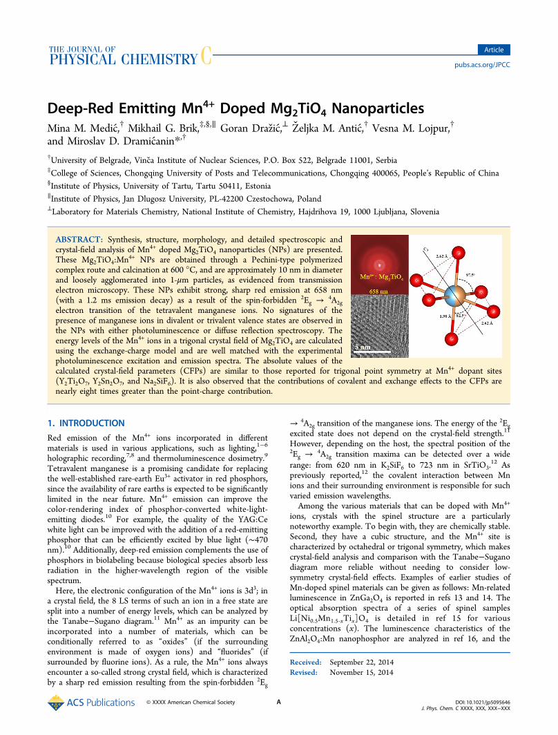
Figure 1. (a) HRTEM image and SAED pattern (inset) of Mg2TiO4 nanoparticles. (b) A subsection of the same region at higher magnification.
The Journal of Physical Chemistry C Article
DOI: 10.1021/jp5095646J. Phys. Chem. C XXXX, XXX, XXX−XXX
B
without any additives. Diffuse spectral reflectance measure-ments were performed on the Thermo Evolution 600spectrometer equipped with an integrating sphere, usingBaSO4 as a blank. Crystal-field calculations were performedusing the exchange-charge model (ECM),28 which permits theanalysis of crystal-field effects without requiring any a prioriassumptions regarding the point symmetry of the impurity ion’ssite.
3. RESULTS AND DISCUSSION
3.1. Size and Morphology of Mn4+ Doped Mg2TiO4NPs. The structure and morphology of Mn4+ doped Mg2TiO4powder were analyzed with TEM/STEM, EELS, and EDXS. Itwas found that the powder consisted of NPs of about 10 nm indiameter, agglomerated into micron-size particles. A lower-magnification figure (Figure 1a) reveals a mesoporosity of a fewnanometers. Uniform, relatively sharp rings with visibleindividual spots in the selected-area electron diffraction(SAED) pattern (inset) corresponded to a Mg2TiO4 (Fd3-mS) spinel structure and was characteristic of randomlyoriented, roughly 10 nm particles. No other crystalline phasewas detected with SAED. Examining the terminal planes of thenanoparticles, we found that no substantial amorphous phasewas present near the crystals.EDXS analysis showed a homogeneous distribution of
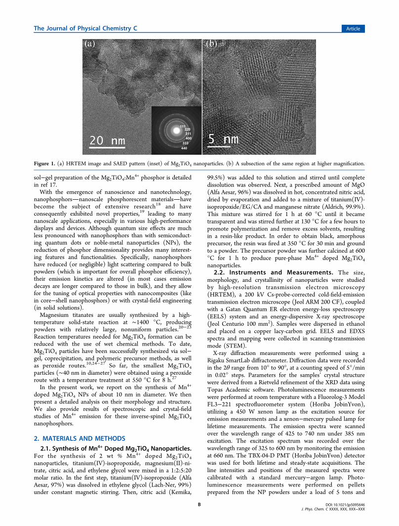
cations in NPs with about 2 wt % of Mn. EELS spectra revealsmall Mn L3 and L2 edges at 642 and 652 eV with anapproximate L3/L2 ratio of 1.2. On the basis of studies29
reporting differences in EELS spectra as a function of thevalence state of manganese, such as when L3 and L2 lines areabout 10 eV apart and the ratio of the lines is less than 2, wecan assume that the Mn is in the 4+ valence state. In Figure 2a,an EELS spectrum with Ti, O, and Mn edges is presented. InFigure 2b the background-subtracted L3 and L2 lines of Mn areshown. Using the Gatan EELS Advisor code for simulatingEELS spectra,30 we found that 2 wt % of Mn is just above thedetection limit, meaning that the valence state of manganesecould not be measured with the high precision.3.2. Crystal Structure of NPs. An X-ray diffraction pattern
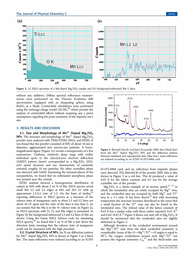
for Mn4+ doped Mg2TiO4 NPs is shown in Figure 3 as a blackline. The main reflections were indexed according to an ICDD
01-072-6968 card, and no reflections from impurity phaseswere detected. The Rietveld fit of the powder XRD data is alsoshown in Figure 3 as a red line. This fit produced a value of8.43 Å for the lattice constant and 6.3 nm for the averagecrystallite size of the powder.Mg2TiO4 is a classic example of an inverse spinel,31−33 in
which the tetrahedral sites are solely occupied by Mg2+ ions,and the octahedral sites are occupied by both Mg2+ and Ti4+
ions in a 1:1 ratio. It has been shown33 that with increasingtemperature the structure becomes disordered in the sense thata small fraction of the Ti4+ ions can also be found in thetetrahedral sites. The refined value of the lattice constant of8.43 Å has a similar value with those earlier reported: 8.41 Å31
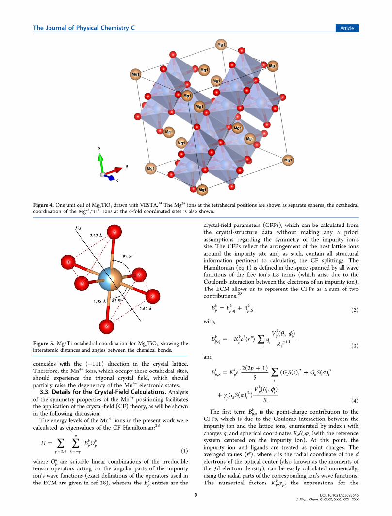
and 8.44−8.45 Å.32 Figure 4 shows one unit cell of Mg2TiO4. Itshould be mentioned that the octahedral sites are slightlydeformed in Figure 5.Although deviation of the octahedral environment around
the Mg2+/Ti4+ ions from the ideal octahedral symmetry isconsiderable (none of the OMg2+/Ti4+O angles is equal to90°), the oxygen octahedrons around each Mg2+/Ti4+ ionpossess the trigonal symmetry C3i,
35 and the third-order axis
Figure 2. (a) EELS spectrum of a Mn-doped Mg2TiO4 sample and (b) background-subtracted Mn L lines.
Figure 3. Rietveld data fit (red line) for powder XRD data (black line)from the Mn4+ doped Mg2TiO4 NPs and the difference patternbetween simulated and experimental data (blue line); main reflectionsare indexed according to an ICDD 01-072-6968 card.
The Journal of Physical Chemistry C Article
DOI: 10.1021/jp5095646J. Phys. Chem. C XXXX, XXX, XXX−XXX
C
coincides with the (−111) direction in the crystal lattice.Therefore, the Mn4+ ions, which occupy these octahedral sites,should experience the trigonal crystal field, which shouldpartially raise the degeneracy of the Mn4+ electronic states.3.3. Details for the Crystal-Field Calculations. Analysis
of the symmetry properties of the Mn4+ positioning facilitatesthe application of the crystal-field (CF) theory, as will be shownin the following discussion.The energy levels of the Mn4+ ions in the present work were
calculated as eigenvalues of the CF Hamiltonian:28
∑ ∑== =−
H B Op k p
p
pk
pk
2,4 (1)
where Opk are suitable linear combinations of the irreducible
tensor operators acting on the angular parts of the impurityion’s wave functions (exact definitions of the operators used inthe ECM are given in ref 28), whereas the Bp
k entries are the
crystal-field parameters (CFPs), which can be calculated fromthe crystal-structure data without making any a prioriassumptions regarding the symmetry of the impurity ion’ssite. The CFPs reflect the arrangement of the host lattice ionsaround the impurity site and, as such, contain all structuralinformation pertinent to calculating the CF splittings. TheHamiltonian (eq 1) is defined in the space spanned by all wavefunctions of the free ion’s LS terms (which arise due to theCoulomb interaction between the electrons of an impurity ion).The ECM allows us to represent the CFPs as a sum of twocontributions:28
= +B B Bpk
p qk
p Sk
, , (2)
with,
∑θ ϕ
= − ⟨ ⟩ +B K e r qV
R
( , )p qk
pk p
ii
pk
i i
ip,
21
(3)
and
∑ σ
γ πθ ϕ
=+
+
+
σ
π
B K ep
G S s G S
G SV
R
2(2 1)5
( ( ) ( )
( ) )( , )
p Sk
pk
is i i
p ipk
i i
i
,2 2 2
2
(4)
The first term Bp,qk is the point-charge contribution to the
CFPs, which is due to the Coulomb interaction between theimpurity ion and the lattice ions, enumerated by index i withcharges qi and spherical coordinates Ri,θi,φi (with the referencesystem centered on the impurity ion). At this point, theimpurity ion and ligands are treated as point charges. Theaveraged values ⟨rp⟩, where r is the radial coordinate of the delectrons of the optical center (also known as the moments ofthe 3d electron density), can be easily calculated numerically,using the radial parts of the corresponding ion’s wave functions.The numerical factors Kp
k,γp, the expressions for the
Figure 4. One unit cell of Mg2TiO4 drawn with VESTA.34 The Mg2+ ions at the tetrahedral positions are shown as separate spheres; the octahedralcoordination of the Mg2+/Ti4+ ions at the 6-fold coordinated sites is also shown.
Figure 5. Mg/Ti octahedral coordination for Mg2TiO4 showing theinteratomic distances and angles between the chemical bonds.
The Journal of Physical Chemistry C Article
DOI: 10.1021/jp5095646J. Phys. Chem. C XXXX, XXX, XXX−XXX
D
polynomials Vpk, and the definitions for the operators Op
k are allgiven in refs 28 and 36 and thus are not shown here for the sakeof brevity. The second term of eq 2, Bp,S
k , is proportional to theoverlap between the wave functions of the impurity ion andligands to account for the covalent and exchange effects. At thispoint, the impurity ion and ligands are treated quantummechanically, clearly distinguishing between different orbitals ofthe ions involved in chemical bond formation. The S(s), S(σ),S(π) terms denote the overlap integrals between the d-functions of the impurity ion and the p- and s-functions of theligands: S(s) = ⟨d0|s0⟩, S(σ) = ⟨d0|p0⟩, S(π) = ⟨d1|p1⟩. The Gs,Gσ, and Gπ coefficients represent the dimensionless adjustableparameters of the ECM, whose values are determined from thepositions of the three lowest energy-absorption bands in theexperimental spectrum. These can be taken as equal, (i.e., Gs =Gσ = Gπ = G), but then must be estimated by the lowest-energyabsorption band. The summation in eq 4 includes only thenearest neighbors of an impurity ion (i.e., six ligands in the caseof an octahedral impurity center), since the overlap with theions from the second, third, etc. coordination spheres can besafely neglected.The ECM uses a small number of fitting parameters, which is
one of the strongest points of the model. It also allows forcalculating the CFPs and energy levels of impurities in crystalswithout invoking any assumptions about the impurity centersymmetry. This is also very important for a consistent analysisof the low-symmetry crystal-field effects and comparativestudies of isostructural/isoelectronic systems. The ECM hasbeen successfully used for the calculations of energy levels ofrare-earth ions28,37,38 and transition metal ions.37,39−41
3.4. Results of Crystal-Field Calculations and EmissionProperties of NPs. Using the structural data from ref 31 andthe refined lattice constant of 8.43 Å, we built up a clusterconsisting of 56,630 ions, which allowed us to account forcrystal-lattice ions located up to 75 Å from the impurity ion’ssite. The Mn4+O2− overlap integrals were calculatednumerically using the radial wave functions from refs 42 and43. Applications of eqs 1−4 resulted in the following values forthe CFPs (Table 1).
As seen in Table 1, the second contribution Bp,sk to the CFPs
values is of paramount importance, being nearly eight timesgreater than Bp,q
k in the case of B43. For a comparison, we also list
in Table 1 the values of CFPs calculated for Mn4+ ions in othercrystals with trigonal point symmetry. All the data in Table 1are consistent: the absolute values of the corresponding CFPsare similar, and the difference in signs reflects opposite trigonaldistortions (compression/elongation) along the third-order axisof rotation in different crystals.For the next step, the CF Hamiltonian (eq 1) with the CFPs
from Table 1 was diagonalized in the space spanned by all wavefunctions of 8 LS terms for the d3 electron configuration of theMn4+ ions. The Racah parameters B and C were taken as 790and 3172 cm−1, respectively, which lie in the typical range fortetravalent manganese ions.36
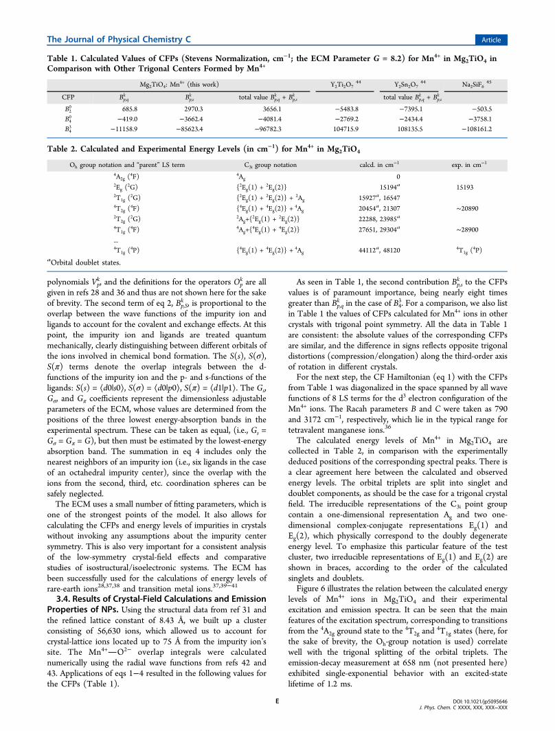
The calculated energy levels of Mn4+ in Mg2TiO4 arecollected in Table 2, in comparison with the experimentallydeduced positions of the corresponding spectral peaks. There isa clear agreement here between the calculated and observedenergy levels. The orbital triplets are split into singlet anddoublet components, as should be the case for a trigonal crystalfield. The irreducible representations of the C3i point groupcontain a one-dimensional representation Ag and two one-dimensional complex-conjugate representations Eg(1) andEg(2), which physically correspond to the doubly degenerateenergy level. To emphasize this particular feature of the testcluster, two irreducible representations of Eg(1) and Eg(2) areshown in braces, according to the order of the calculatedsinglets and doublets.Figure 6 illustrates the relation between the calculated energy
levels of Mn4+ ions in Mg2TiO4 and their experimentalexcitation and emission spectra. It can be seen that the mainfeatures of the excitation spectrum, corresponding to transitionsfrom the 4A2g ground state to the 4T2g and
4T1g states (here, forthe sake of brevity, the Oh-group notation is used) correlatewell with the trigonal splitting of the orbital triplets. Theemission-decay measurement at 658 nm (not presented here)exhibited single-exponential behavior with an excited-statelifetime of 1.2 ms.
Table 1. Calculated Values of CFPs (Stevens Normalization, cm−1; the ECM Parameter G = 8.2) for Mn4+ in Mg2TiO4 inComparison with Other Trigonal Centers Formed by Mn4+
Mg2TiO4: Mn4+ (this work) Y2Ti2O744 Y2Sn2O7
44 Na2SiF645
CFP Bp,qk Bp,s
k total value Bp,qk + Bp,s
k total value Bp,qk + Bp,s
k
B20 685.8 2970.3 3656.1 −5483.8 −7395.1 −503.5
B40 −419.0 −3662.4 −4081.4 −2769.2 −2434.4 −3758.1
B43 −11158.9 −85623.4 −96782.3 104715.9 108135.5 −108161.2
Table 2. Calculated and Experimental Energy Levels (in cm−1) for Mn4+ in Mg2TiO4
Oh group notation and “parent” LS term C3i group notation calcd. in cm−1 exp. in cm−1
4A2g (4F) 4Ag 0
2Eg (2G) {2Eg(1) +
2Eg(2)} 15194a 151932T1g (
2G) {2Eg(1) +2Eg(2)} + 2Ag 15927a, 16547
4T2g (4F) {4Eg(1) +
4Eg(2)} + 4Ag 20454a, 21307 ∼208902T2g (
2G) 2Ag+{2Eg(1) +
2Eg(2)} 22288, 23985a
4T1g (4F) 4Ag+{
4Eg(1) +4Eg(2)} 27651, 29304a ∼28900
...4T1g (
4P) {4Eg(1) +4Eg(2)} + 4Ag 44112a, 48120 4T1g (
4P)aOrbital doublet states.
The Journal of Physical Chemistry C Article
DOI: 10.1021/jp5095646J. Phys. Chem. C XXXX, XXX, XXX−XXX
E
As one can see from comparison of the refined latticeconstant of NPs with the bulk value, the lattice constant for theNPs is slightly increased. As a result, a slightly lower value ofthe 10Dq parameter (crystal field strength) is observed in NPsin comparison to the bulk particles,35,46 but the main excitationand emission peaks for these nanoparticles are very similar tothe bulk material.Figure 7 shows Kubelka−Munk function of the measured
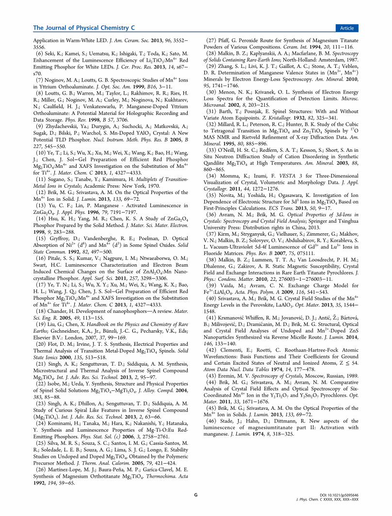
diffuse reflectance spectrum where characteristic absorption of
Mn4+ ions can be observed, and no Mn3+ spin-allowedabsorption bands are present.
4. CONCLUSIONSDetailed spectroscopic and crystal-field studies of theMg2TiO4:Mn4+ NPs are described in the present paper.Deep-red emitting (658 nm with a lifetime of 1.2 ms)Mg2TiO4:Mn4+ NPs of approximately 10 nm in diameter areobtained through the Pechini-type polymerized complex route,based on polyesterification between citric acid (CA) andethylene glycol (EG) and the use of a mixed-metal CA complexwith a stoichiometric Mg/Ti ratio of 2:1. Through thissynthesis approach, an inverse-spinel crystal structure that
incorporates Mn4+ luminescence centers is obtained at atemperature of 600 °C, following 1 h of calcination. This is amuch lower temperature and shorter time than those oftraditional solid-state reactions, which require long firing cyclesat temperatures of approximately 1400 °C. No signatures ofmanganese ions in divalent or trivalent valence states areobserved in photoluminescence and diffuse reflection spectra.The energy levels of the Mn4+ ions in Mg2TiO4 were
calculated using the crystal-field ECM. The point symmetry ofthe Mn4+ position is C3i, which is in agreement with thestructure of the crystal-field Hamiltonian and calculated patternof the energy-level splitting. The calculated energy levels ofMn4+ in a trigonal crystal field are in good agreement with theexperimental excitation and emission spectra and allow for thedesignation of all prominent spectral features. The mainexcitation and emission peaks for these nanoparticles are verysimilar to the bulk material. The values of the CFPs for Mn4+
ions in Mg2TiO4 are similar to those calculated for Mn4+ inother crystals with trigonal point symmetry at Mn4+ sites. Thecrystal-field analysis also showed that contributions of covalentand exchange effects to CFPs are nearly eight times greater thanthe point-charge contribution.
■ AUTHOR INFORMATION
Corresponding Author*Tel. +381 11 3408607; e-mail: [email protected].
Author ContributionsThe manuscript was written through contributions of allauthors. All authors have given approval to the final version ofthe manuscript.
NotesThe authors declare no competing financial interest.
■ ACKNOWLEDGMENTS
M.M., Z.A., V.L., and M.D.D. acknowledge the financialsupport of the Ministry of Education and Science of theRepublic of Serbia (Project No. 45020) and the support fromthe APV Provincial Secretariat for Science and TechnologicalDevelopment of the Republic of Serbia through Project No.114-451-1850/2014-03. M.B. acknowledges the financialsupport of the Marie Curie Initial Training NetworkLUMINET through Grant Agreement 316906 and theProgramme for the Foreign Experts offered by ChongqingUniversity of Posts and Telecommunications. Goran Drazic acknowledges the financial support of the Slovenian ResearchAgency (ARRS) through Program No. P2-0148 and Project J2-6754.
■ REFERENCES(1) Butler, K. H. Fluorescent Lamp Phosphors; The Pennsylvania StateUniversity Press: University Park, P.A., 1980.(2) Srivastava, A. M.; Soules, T. F. Luminescent Materials. In(Phosphors)Kirk-Othmer Encyclopedia of Chemical Technology; JohnWiley & Sons: New York, 1995.(3) Radkov, E. V.; Grigorov, L. S.; Setlur, A. A.; Srivastava, A. M.United States Patent Application US2006/0169998 A1.(4) Cao, R. P.; Peng, M. Y.; Song, E. H.; Qiu, J. R. ECS HighEfficiency Mn4+ Doped Sr2MgAl22O36 Red Emitting Phosphor forWhite LED. J. Solid State Sci. Technol. 2012, 1, R123−R126.(5) Liao, C. X.; Cao, R. P.; Ma, Z. J.; Li, Y.; Dong, G. P.; Sharafudeen,K. N.; Qiu, J. R. Synthesis of K2SiF6:Mn4+ Phosphor from SiO2Powders via Redox Reaction in HF/KMnO4 Solution and Their
Figure 6. Comparison of the calculated energy levels of Mn4+ ininverse-spinel Mg2TiO4 with experimental excitation (black line) andemission (red line) spectra.
Figure 7. Kubelka−Munk function, F(R), calculated from the diffusereflectance spectrum of Mn4+:Mg2TiO4 NPs.
The Journal of Physical Chemistry C Article
DOI: 10.1021/jp5095646J. Phys. Chem. C XXXX, XXX, XXX−XXX
F
Application in Warm-White LED. J. Am. Ceram. Soc. 2013, 96, 3552−3556.(6) Seki, K.; Kamei, S.; Uematsu, K.; Ishigaki, T.; Toda, K.; Sato, M.Enhancement of the Luminescence Efficiency of Li2TiO3:Mn4+ RedEmitting Phosphor for White LEDs. J. Cer. Proc. Res. 2013, 14, s67−s70.(7) Noginov, M. A.; Loutts, G. B. Spectroscopic Studies of Mn4+ Ionsin Yttrium Orthoaluminate. J. Opt. Soc. Am. 1999, B16, 3−11.(8) Loutts, G. B.; Warren, M.; Taylor, L.; Rakhimov, R. R.; Ries, H.R.; Miller, G.; Noginov, M. A.; Curley, M.; Noginova, N.; Kukhtarev,N.; Caulfield, H. J.; Venkateswarlu, P. Manganese-Doped YttriumOrthoaluminate: A Potential Material for Holographic Recording andData Storage. Phys. Rev. 1998, B 57, 3706.(9) Zhydachevskii, Ya.; Durygin, A.; Suchocki, A.; Matkovskii, A.;Sugak, D.; Bilski, P.; Warchol, S. Mn-Doped YAlO3 Crystal: A NewPotential TLD Phosphor. Nucl. Instrum. Meth. Phys. Res. B 2005, B227, 545−550.(10) Ye, T.; Li, S.; Wu, X.; Xu, M.; Wei, X.; Wang, K.; Bao, H.; Wang,J.; Chen, J. Sol−Gel Preparation of Efficient Red PhosphorMg2TiO4:Mn4+ and XAFS Investigation on the Substitution of Mn4+
for Ti4+. J. Mater. Chem. C 2013, 1, 4327−4333.(11) Sugano, S.; Tanabe, Y.; Kamimura, H. Multiplets of Transition-Metal Ions in Crystals; Academic Press: New York, 1970.(12) Brik, M. G.; Srivastava, A. M. On the Optical Properties of theMn4+ Ion in Solid. J. Lumin. 2013, 133, 69−72.(13) Yu, C. F.; Lin, P. Manganese - Activated Luminescence inZnGa2O4. J. Appl. Phys. 1996, 79, 7191−7197.(14) Hsu, K. H.; Yang, M. R.; Chen, K. S. A Study of ZnGa2O4
Phosphor Prepared by the Solid Method. J. Mater. Sci. Mater. Electron.1998, 9, 283−288.(15) Gryffroy, D.; Vandenberghe, R. E.; Poelman, D. OpticalAbsorption of Ni2+ (d8) and Mn4+ (d3) in Some Spinel Oxides. SolidState Commun. 1992, 82, 497−500.(16) Pitale, S. S.; Kumar, V.; Nagpure, I. M.; Ntwaeaborwa, O. M.;Swart, H.C. Luminescence Characterization and Electron BeamInduced Chemical Changes on the Surface of ZnAl2O4:Mn Nano-crystalline Phosphor. Appl. Surf. Sci. 2011, 257, 3298−3306.(17) Ye, T. N.; Li, S.; Wu, X. Y.; Xu, M.; Wei, X.; Wang, K. X.; Bao,H. L.; Wang, J. Q.; Chen, J. S. Sol−Gel Preparation of Efficient RedPhosphor Mg2TiO4:Mn4+ and XAFS Investigation on the Substitutionof Mn4+ for Ti4+. J. Mater. Chem. C 2013, 1, 4327−4333.(18) Chander, H. Development of nanophosphorsA review. Mater.Sci. Eng. R. 2005, 49, 113−155.(19) Liu, G.; Chen, X. Handbook on the Physics and Chemistry of RareEarths; Gschneidner, K.A., Jr., Bunzli, J.-C. G., Pecharsky, V.K., Eds;Elsevier B.V.: London, 2007, 37, 99−169.(20) Flot, D. M.; Irvine, J. T. S. Synthesis, Electrical Properties andThermal Analysis of Transition Metal-Doped Mg2TiO4 Spinels. SolidState Ionics 2000, 135, 513−518.(21) Singh, A. K.; Senguttuvan, T. D.; Siddiquia, A. M. Synthesis,Microstructural and Thermal Analysis of Inverse Spinel CompoundMg2TiO4. Int. J. Adv. Res. Sci. Technol. 2013, 2, 95−97.(22) Isobe, M.; Ueda, Y. Synthesis, Structure and Physical Propertiesof Spinel Solid Solutions Mg2TiO4−MgTi2O4. J. Alloy. Compd. 2004,383, 85−88.(23) Singh, A. K.; Dhillon, A.; Senguttuvan, T. D.; Siddiquia, A. M.Study of Curious Spiral Like Features in Inverse Spinel Compound(Mg2TiO4). Int. J. Adv. Res. Sci. Technol. 2013, 2, 63−66.(24) Kominami, H.; Tanaka, M.; Hara, K.; Nakanishi, Y.; Hatanaka,Y. Synthesis and Luminescence Properties of Mg-Ti-O:Eu Red-Emitting Phosphors. Phys. Stat. Sol. (c) 2006, 3, 2758−2761.(25) Silva, M. R. S.; Souza, S. C.; Santos, I. M. G.; Cassia-Santos, M.R.; Soledade, L. E. B.; Souza, A. G.; Lima, S. J. G.; Longo, E. StabilityStudies on Undoped and Doped Mg2TiO4, Obtained by the PolymericPrecursor Method. J. Therm. Anal. Calorim. 2005, 79, 421−424.(26) Martínez-Lope, M. J.; Baura-Pena, M. P.; Garíca-Clavel, M. E.Synthesis of Magnesium Orthotitanate Mg2TiO4. Thermochima. Acta1992, 194, 59−65.
(27) Pfaff, G. Peroxide Route for Synthesis of Magnesium TitanatePowders of Various Compositions. Ceram. Int. 1994, 20, 111−116.(28) Malkin, B. Z.; Kaplyanskii, A. A.; Macfarlane, B. M. Spectroscopyof Solids Containing Rare-Earth Ions; North-Holland: Amsterdam, 1987.(29) Zhang, S. L.; Livi, K. J. T.; Gaillot, A. C.; Stone, A. T.; Veblen,D. R. Determination of Manganese Valence States in (Mn3+, Mn4+)Minerals by Electron Energy-Loss Spectroscopy. Am. Mineral. 2010,95, 1741−1746.(30) Menon, N. K.; Krivanek, O. L. Synthesis of Electron EnergyLoss Spectra for the Quantification of Detection Limits. Microsc.Microanal. 2002, 8, 203−215.(31) Barth, T.; Posnjak, E. Spinel Structures: With and WithoutVariate Atom Equipoints. Z. Kristallogr. 1932, 82, 325−341.(32) Millard, R. L.; Peterson, R. C.; Hunter, B. K. Study of the Cubicto Tetragonal Transition in Mg2TiO4 and Zn2TiO4 Spinels by 17OMAS NMR and Rietveld Refinement of X-ray Diffraction Data. Am.Mineral. 1995, 80, 885−896.(33) O’Neill, H. St. C.; Redfern, S. A. T.; Kesson, S.; Short, S. An inSitu Neutron Diffraction Study of Cation Disordering in SyntheticQandilite Mg2TiO4 at High Temperatures. Am. Mineral. 2003, 88,860−865.(34) Momma, K.; Izumi, F. VESTA 3 for Three-DimensionalVisualization of Crystal, Volumetric and Morphology Data. J. Appl.Crystallogr. 2011, 44, 1272−1276.(35) Novita, M.; Yoshida, H.; Ogasawara, K. Investigation of IonDependence of Electronic Structure for 3d3 Ions in Mg2TiO4 Based onFirst-Principles Calculations. ECS Trans. 2013, 50, 9−17.(36) Avram, N. M.; Brik, M. G. Optical Properties of 3d-Ions inCrystals: Spectroscopy and Crystal Field Analysis; Springer and TsinghuaUniversity Press: Distribution rights in China, 2013.(37) Kirm, M.; Stryganyuk, G.; Vielhauer, S.; Zimmerer, G.; Makhov,V. N.; Malkin, B. Z.; Solovyev, O. V.; Abdulsabirov, R. Y.; Korableva, S.L. Vacuum-Ultraviolet 5d-4f Luminescence of Gd3+ and Lu3+ Ions inFluoride Matrices. Phys. Rev. B 2007, 75, 075111.(38) Malkin, B. Z.; Lummen, T. T. A.; Van Loosdrecht, P. H. M.;Dhalenne, G.; Zakirov, A. R. Static Magnetic Susceptibility, CrystalField and Exchange Interactions in Rare Earth Titanate Pyrochlores. J.Phys.: Condens. Matter. 2010, 22, 276003−1−276003−11.(39) Vaida, M.; Avram, C. N. Exchange Charge Model forFe3+:LiAl5O8. Acta. Phys. Polym. A 2009, 116, 541−543.(40) Srivastava, A. M.; Brik, M. G. Crystal Field Studies of the Mn4+
Energy Levels in the Perovskite, LaAlO3. Opt. Mater. 2013, 35, 1544−1548.(41) Krsmanovic Whiffen, R. M.; Jovanovic, D. J.; Antic, Z.; Bartova,B.; Milivojevic, D.; Dramicanin, M. D.; Brik, M. G. Structural, Opticaland Crystal Field Analyses of Undoped and Mn2+-Doped ZnSNanoparticles Synthesized via Reverse Micelle Route. J. Lumin. 2014,146, 133−140.(42) Clementi, E.; Roetti, C. Roothaan-Hartree-Fock AtomicWavefunctions: Basis Functions and Their Coefficients for Groundand Certain Excited States of Neutral and Ionized Atoms, Z ≤ 54.Atom Data Nucl. Data Tables 1974, 14, 177−478.(43) Eremin, M. V. Spectroscopy of Crystals, Moscow, Russian, 1989.(44) Brik, M. G.; Srivastava, A. M.; Avram, N. M. ComparativeAnalysis of Crystal Field Effects and Optical Spectroscopy of Six-Coordinated Mn4+ Ion in the Y2Ti2O7 and Y2Sn2O7 Pyrochlores. Opt.Mater. 2011, 33, 1671−1676.(45) Brik, M. G.; Srivastava, A. M. On the Optical Properties of theMn4+ Ion in Solids. J. Lumin. 2013, 133, 69−72.(46) Stade, J.; Hahn, D.; Dittmann, R. New aspects of theluminescence of magnesiumtitanate part II: Activation withmanganese. J. Lumin. 1974, 8, 318−325.
The Journal of Physical Chemistry C Article
DOI: 10.1021/jp5095646J. Phys. Chem. C XXXX, XXX, XXX−XXX
G
Related Documents


