Deep Neural Networks for Improving Computer-Aided Diagnosis, Segmentation and Text/Image Parsing in Radiology Le Lu, Ph.D. Joint work with Holger R. Roth, Hoo-chang Shin, Ari Seff, Xiaosong Wang, Mingchen Gao, Isabella Nogues, Ronald M. Summers Radiology and Imaging Sciences, National Institutes of Health Clinical Center [email protected]

Welcome message from author
This document is posted to help you gain knowledge. Please leave a comment to let me know what you think about it! Share it to your friends and learn new things together.
Transcript

Deep Neural Networks for Improving Computer-Aided Diagnosis, Segmentation and Text/Image Parsing in Radiology
Le Lu, Ph.D.
Joint work with Holger R. Roth, Hoo-chang Shin, Ari Seff, Xiaosong Wang, Mingchen Gao, Isabella Nogues, Ronald M. Summers
Radiology and Imaging Sciences, National Institutes of Health Clinical Center

Application Focus: Cancer Imaging
American Cancer Society: Cancer Facts and Figures 2016. Atlanta, Ga: American Cancer
Society, 2016. Last accessed February 1, 2016.
http://www.cancer.gov/types/common-cancers
Cancer Type
Lung (Bronchus)
Colorectal
Pancreatic Breast (F-M)
Prostate
Estimated New Cases
224,390 134,490 53,070 246,660 – 2,600
180,890
Estimated Deaths
158,080 49,190 41,780 40,450 – 440
26,120

Overview: Three Key Problems (I)
• Computer-aided Detection (CADe) and Diagnosis (CADx) • Lung, Colon pre-cancer detection; bone and vessel imaging (13 conference papers
in CVPR/ECCV/ICCV/MICCAI/WACV/CIKM, 12 patents, 6 years of industrial R&D)
• Lymph node, colon polyp, bone lesion detection using Deep CNN + Random View Aggregation (http://arxiv.org/abs/1505.03046, TMI 2016a; MICCAI 2014a)
• Empirical analysis on Lymph node detection and interstitial lung disease (ILD) classification using CNN (http://arxiv.org/abs/1602.03409, TMI 2016b)
• Non-deep models for CADe using compositional representation (MICCAI 2014b) and +mid-level cues (MICCAI 2015b); deep regression based multi-label ILD prediction (MICCAI 2016 in submission); missing label issue in ILD (ISBI 2016)
• Clinical Impact: producing various high performance “second or first reader” CAD use cases and applications effective imaging based prescreening tools on a cloud based platform for large population

Overview: Three Key Problems (II)
• Semantic Segmentation in Medical Image Analysis • “DeepOrgan” for pancreas segmentation (MICCAI 2015a) via scanning superpixels
using multi-scale deep features (“Zoom-out”) and probability map embedding http://arxiv.org/abs/1506.06448
• Deep segmentation on pancreas and lymph node clusters with HED (Holistically-nested neural networks, Xie & Tu, 2015) as building blocks to learn unary (segmentation mask) and pairwise (labeling segmentation boundary) CRF terms + spatial aggregation or + structured optimization (The focus of MICCAI 2016 submissions since this is a much needed task Small datasets; (de-)compositional representation is still the key.)
• CRF: conditional random fields
• Clinical Impact: semantic segmentation can help compute clinically more accurate and desirable imaging bio-markers!

Overview: Three Key Problems (III)
• Interleaved or Joint Text/Image Deep Mining on a Large-Scale Radiology Image Database “large” datasets; no labels (~216K 2D key images/slices extracted from >60K unique patients)
• Interleaved Text/Image Deep Mining on a Large-Scale Radiology Image Database (CVPR 2015, a proof of concept study)
• Interleaved Text/Image Deep Mining on a Large-Scale Radiology Image Database for Automated Image Interpretation (its extension, JMLR 2016, to appear) http://arxiv.org/abs/1505.00670
• Learning to Read Chest X-Rays: Recurrent Neural Cascade Model for Automated Image Annotation, (CVPR 2016) http://arxiv.org/abs/1603.08486
• Unsupervised Category Discovery via Looped Deep Pseudo-Task Optimization Using a Large Scale Radiology Image Database, (ECCV 2016 in submission) http://arxiv.org/abs/1603.07965
• Clinical Impact: eventually to build an automated programmable
mechanism to parse and learn from hospital scale PACS-RIS databases to derive semantics and knowledge … • has to be deep learning based since effective image features are very hard to be hand-
crafted cross different diseases, imaging protocols and modalities.

(I) Automated Lymph Node Detection
• Difficult due to large variations in appearance, location and pose.
• Plus low contrast against surrounding tissues.
Abdominal lymph node in CT Mediastinal lymph node in CT

Previous Work
• Previous work mostly use direct 3D image feature information from CT volume.
• The state-of-the-art approaches [4,5] employ a large set of boosted 3D Haar features to build a holistic detector, in a scanning window manner.
• Curse of dimensionality leads to relatively poor performance [Lu, Barbu, et al., 2008].
*Can we represent the challenging object detection task(s) as 2D or 2.5D problems, to achieve better FROC performance?
(+ parts of Abd.)

Heterogeneous Cascade CADe
*Ingredients* (MICCAI 2014~2015, TMI 2016):
CG: Avoid exhaustive scanning window search, but use systems or
modules which can generate object hypotheses with extremely high
recall, at the expense of high false positive rates (e.g., heuristic
importance sampling) as candidate proposals.
Hundreds of Thousands potential object windows reduced to ~[40-
50] windows or 3D VOIs. Heterogeneous Cascade for Object
Detection via classification! unbalanced (hard) negative sampling
issue)
Propose, implement and evaluate 2.5D approaches using local
composites of 2D views of classification, versus one-shot 3D “yes-no”
classification. (Compositional or De-compositional Model)

Lymph Node Candidate Generation
•Mediastinum [J. Liu et al. 2014] – 388 lymph nodes in 90 patients – 3208 false-positives
• 36 FPs per patient
• Abdomen [K. Cherry et al. 2014] – 595 lymph nodes in 86 patients – 3484 false-positives
• 41 FPs per patient
•Deep Detection Proposal Generation as future work

Shallow Models: 2D View Aggregation Using a Two-Level Hierarchy of Linear Classifiers [Seff et al. MICCAI 2014]
2D slice gallery for a LN candidate VOI (45 x 45 × 45 voxels).
Axial
Coronal
Sagittal
• VOI candidates generated via a random forest classifier using voxel-level features (not the primary focus of this work), for high sensitivity but also high false positive rates.
• 2.5D: 3 sequences of orthogonal 2D slices then extracted from each candidate VOI (9 x 3 = 27 views).

HOG: Histogram of Oriented Gradients + LibLinear on processing 2D Views
HOG feature extraction
Resulting feature weights after training.
Abdominal LN axial slice.
SVM training
Note that a unified, compact HOG model is trained, regardless of axial, coronal, or sagittal views, or unifying view orientations.

Lymph Node Detection FROC Performance

Lymph Node Detection FROC Performance
Enriching HOG descriptor with other image feature channels, e.g., mid-level semantic
contours/gradients, can further lift the sensitivity for 8~10%!
About 1/3 FPs are found to be smaller lymph nodes (short axis < 10 mm).
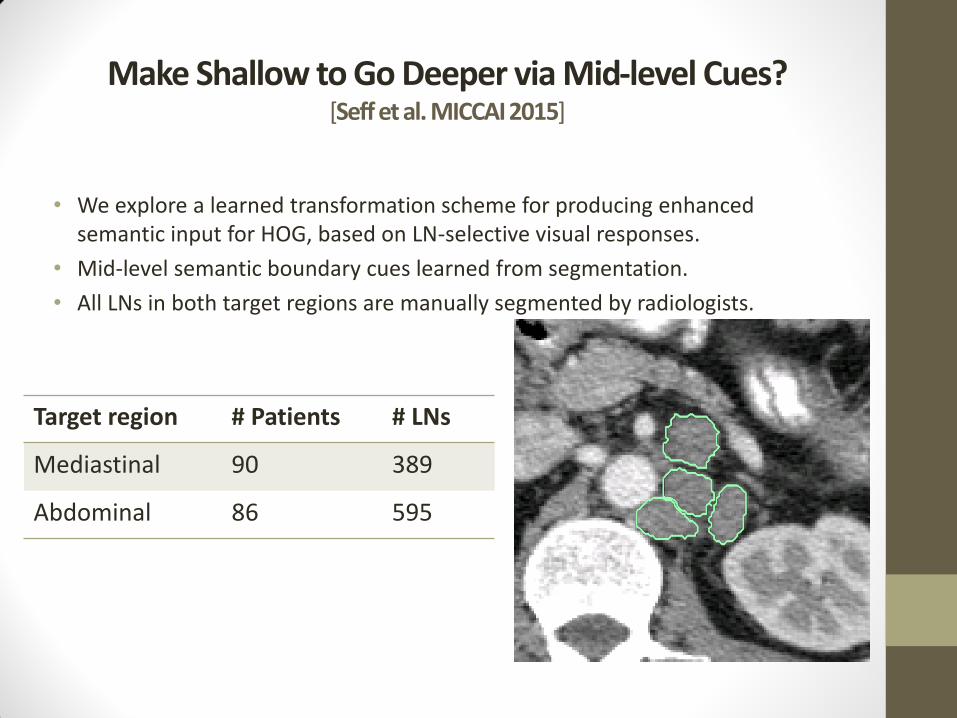
Make Shallow to Go Deeper via Mid-level Cues? [Seff et al. MICCAI 2015]
• We explore a learned transformation scheme for producing enhanced semantic input for HOG, based on LN-selective visual responses.
• Mid-level semantic boundary cues learned from segmentation.
• All LNs in both target regions are manually segmented by radiologists.
Target region # Patients # LNs
Mediastinal 90 389
Abdominal 86 595

Sketch Tokens (CVPR’13)
• Extract all patches (radius = 7 voxels) centered on a boundary pixel
• Cluster into “sketch token” classes using k-means with k = 150
• A random forest is trained for sketch token classification for input CT patches
Abdominal LN Mediastinal LN Colon Polyps

Feature Map Construction
• An enhanced, 3-channel feature map:
SumMap
MaxMap
HOG
Computation
3-Channel
Feature Map
Candidate
slice sampling
Semantic boundary cue features are extracted from a true positive mediastinal LN candidate. SumMap and MaxMap are constructed by taking the sums and maximums respectively of the pixel-level contour-class probabilities output by the sketch tokens random forest.
CT Slice

Single Template Results
• Top performing feature sets (Sum_Max_I and Sum_Max) exhibit 15%-23% greater recall than the baseline HOG at low FP rates (e.g. 3/FP scan).
• Our system outperforms the state-of-the-art deep CNN system (Roth et al., 2014) in the mediastinum, e.g. 78% vs. 70% at 3 FP/scan.
Six-fold cross-valdiation FROC curves are shown for the two target regions

Classification
• A linear SVM is trained using the new feature set; A HOG cell size of 9x9 pixels gives optimal performance.
• Separate models are trained for specific LN size ranges to form a mixture-of-templates-approach (see later slide)
Visualization of linear SVM weights for the abdominal LN detection models

• Wide distribution of LN sizes invites the application of size-specific models trained separately.
• LNs > 20 mm are especially clinically relevant
Mixture Model Results
Single template and mixture model performance for abdominal models

Deep models: Random Sets of Convolutional Neural Network Predictions [Roth et al. MICCAI 2014, TMI 2016]
Not-so-deep Convolutional Neural Network:
CIFAR-10
Trained Filters
CUDA-ConvNet: Open-source GPU accelerated code by [A. Krizhevsky et al. 2012] plus DropConnect modification by [L. Wan et al. 2013]
[H. Roth et al. MICCAI 2014]

Deep models: Random Sets of Convolutional Neural Network Predictions [Roth et al., MICCAI 2014]
Application to appearance modeling and detecting lymph node
Random translations, rotations and
scale

Convolutional Neural Network Architecture
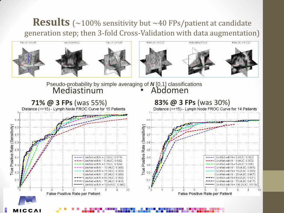
Results (~100% sensitivity but ~40 FPs/patient at candidate
generation step; then 3-fold Cross-Validation with data augmentation)
Mediastinum 71% @ 3 FPs (was 55%)
• Abdomen 83% @ 3 FPs (was 30%)
Pseudo-probability by simple averaging of N [0,1] classifications

Results (~100% sensitivity but ~40 FPs/patient at candidate
generation step)
Mediastinum 82% @ 3 FPs (was 55%)
• Abdomen 80% @ 3 FPs (was 30%)
Training mediastinum and abdomen Jointly!

Previous Work (CAD 1.0 or 2.0)
• The previous state-of-the-art work is (Feulner et al., MedIA, 2013) which shows 52.9% sensitivity at 3.1 FP/Vol on 54 Chest CT scans or 60.9% recall at 6.1 FP/Vol.
• In (Feulner et al., MedIA, 2013), “In order to compare the automatic detection results with the performance of a human, we did an experiment on the intra-human observer variability. Ten of the CT volumes were annotated a second time by the same person a few months later. The first segmentations served as ground truth, and the second ones were considered as detections.
• TPR and FP were measured in the same way as for the automatic detection. The TPR was 54.8% with 0.8 false positives per volume on average. While 0.8 FP is very low, a TPR of 54.8% shows that finding lymph nodes in CT is quite challenging also for humans.“
Table reproduced from Table 3, Feulner et al., “Lymph node detection and segmentation in chest CT data
using discriminative learning and a spatial prior”, Medical image analysis, 17(2): 254-270 (2013). Note that
Barbu et al. (2010) is not directly comparable to other papers since Axillary lymph nodes are easier to detect.
Method Body Region Number CT Vol.
Size (mm)
TP Criterion TPR (%) FP/Vol.
Kitasaka et al. (2007)
Abdomen 5 >5.0 Overlap 57.0% 58
Feuerstein et al. (2009)
Mediastinum 5 >1.5 Overlap 82.1% 113
Dornheim (2008) Neck 1 >8.0 Unknown 100% 9
Barbu et al. (2010) Axillary 101 >10.0 In box 82.3% 1.0
Feulner et al. (2013) Mediastinum 54 >10.0 In box 52.9% 3.1
Intra-obs. Var. Mediastinum 10 >10.0 In box 54.8% 0.8

Generalizable? Colon CADe Results using a deeper CNN on 1186 patients (or 2372 CTC volumes) [Roth et al., TMI 2016]
[SVM baseline] Summers, et a., Computed tomographic virtual colonoscopy computer-aided
polyp detection in a screening population, Gastroenterology, vol. 129, no. 6, pp.1832–1844, 2005.
1,186 patients with prone and supine CTC images (394/792 patients; 79/173 polyps tr/ts split)

Deep Convolutional Neural Networks for Computer-Aided Detection: CNN Architectures, Dataset Characteristics and Transfer Learning
[Shin et al., TMI 2016, in press; http://arxiv.org/abs/1602.03409]
• For a more comprehensive evaluation, we exploit three important, but previously under-studied factors of employing deep convolutional neural networks to CADe problems. provide some insights and implementation tips for MICCAI community.
• Particularly, we present
• Evaluation of different CNN architectures ranging from 5 thousand to 160 million parameters with various of depths of CNN layers;
• Impacts on performance given datasets of different scales and spatial image contexts;
• When transfer learning from pre-trained ImageNet CNN models via fine-tuning can be helpful and why?

Problem 1: Lymph node detection in CT using three-orthogonal views + random sampling + multi-scale

Problem 2.b: Slice based ILD Classification in CT, thick sliceness, no Lung segmentation
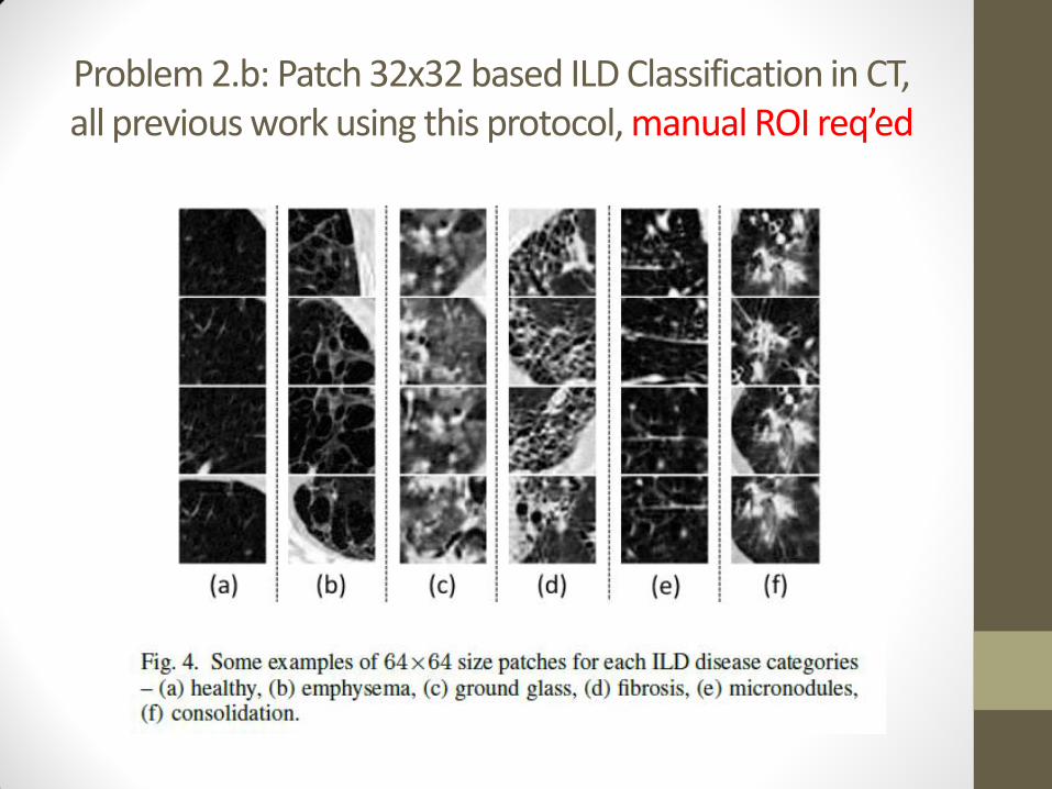
Problem 2.b: Patch 32x32 based ILD Classification in CT, all previous work using this protocol, manual ROI req’ed

Observations & Directions
• We summarize our findings as follows.
1. Deep CNN architectures in 8, even 22 layers [3], [18] can be useful even for CADe problems where the available training datasets are limited. Previously, CNN models used in medical image analysis applications are often 2~ 5 orders of magnitude smaller.
2. The tradeoff of better learning models versus more training datasets [29] should be thought carefully for finding an optimal solution of any CADe problem (e.g., mediastinal and abdominal LN detection).
3. The Datasets can be the bottleneck to further advance the field of CADe. Building progressively growing (in scales) well annotated datasets is at least with the same importance of developing new algorithms.
• As an analogy in computer vision, Scene Recognition problem has made tremendous progress, thanks to the steady and continuous development of Scene-15, MIT Indoor-67, SUN-397 and Place datasets [36], ….

4. Transfer learning from the large scale annotated natural image datasets (ImageNet) to CADe problems is validated to be consistently beneficial in our experiments. This sheds some light on cross-datasets CNN learning in medical image domain, e.g., the union of ILD [20] and LTRC datasets [38] as suggested in this paper.
5. Last, applying out-of-shelf deep CNN image features on CADe problems can be improved by either exploring/coupling the performance-complementary properties of hand-crafted features [9], [8], [11]; or CNNs trained from scratch (Roth et al., MICCAI 2014, TMI 2016) and more desirably CNNs fine-tuned on the target medical image dataset (evaluated in this paper).

Visualization on Transfer Learning (Learned from Thoracoabdominal LNs)

Better Localization after Fine-tuning?

Failure Cases


[Farag et al., arXiv-1407.8497, 2014; Roth et al., arXiv-1504.03967; Roth et al., MICCAI 2015]
(II) Semantic (Free-form) Organ Segmentation

[A. Farag et al., 2014]
• 97% avg. sensitivity/recall
• 27% avg. Dice score
(over-segmentation)
e.g., threshold at
p > 0.5
Refinement: Multi-Level Regional and Patch ConvNets Fusion
(II) Candidate Region Generation (Hand-crafted Image Features + RF) [Farag et al., arXiv-1407.8497]

Convolutional Neural Networks (AlexNet)
CUDA-ConvNet: Open-source GPU accelerated code by [Krizhevsky et al., NIPS 2012]
Trained first level filter kernels
2

Multi-Scale “Zoom-out” R-ConvNet
Zoom-out Zoom-out

P-ConvNet: Deep Patch Classification
ho
lge
r.ro
th@
nih
.gov
Ground truth Random Forest 2.5D Patch ConvNet prob.

R2-ConvNet: Regional ConvNet
~27%
Dice
score
~57%
Dice
score
~68%
Dice
score

3/2
4/2
015
h
olg
er.
roth
@nih
.gov
43
Training & Testing Performance (4-fold Cross-Validation)
Probability maps thresholded at p0=0.2, p1=0.5, and p2=0.6, calibrated in training and applied
on testing.
Dice coefficients: 84.2% (+/- 3.6%) in Training and 75.8% (+/-5.4%) in Testing (more stable by
std values)

4-fold CV Performance
Minimum surface distances: 0.94+/-0.6mm (p<0.01) with R2-ConvNet from 1.46+/-1.5mm
if just P-ConvNet is applied.
Previous state-of-the-art: [46.6% to 69.1%] DSC, all under LOO (Leave-one-patient-out).

An Above-Average Example
a) The manual ground truth annotation (in red outline)
b) The G(P2(x)) probability map
c) The final segmentation (in green outline) at p2=0.6
DSC=82.7%.

Mean 0.936 mm
Std 0.586 mm
Min 0.297 mm
Max 2.204 mm
mm mm mm mm
mm mm mm mm
mm mm mm mm
mm mm mm mm
mm mm mm mm

(III) Interleaved Text/Image Deep Mining on a Large-Scale Radiology Database (780K/60K patients) for Automated Image Interpretation
• Hoo-Chang Shin, Le Lu, Lauren Kim, Ari Seff, Jianhua Yao, Ronald M. Summers, IEEE Conf. CVPR 2015, to appear; JMLR on large scale health informatics issue (in submission)

Interleaved Text/Image Deep Mining on a Large-Scale Radiology Database
Example words embedded in the vector space using Open Source RNN based Google Word-
to-Vector modeling (visualized on 2D), trained from 1B words in 780K radiology reports and
0.2B from OpenI:an open access biomedical image search engine; http://openi.nlm.nih.gov .

Interleaved Text/Image Deep Mining on a Large-Scale Radiology Database

Interleaved Text/Image Deep Mining on a Large-Scale Radiology Database D
ise
ase
On
tolo
gy (O
D) is
an
alo
gic
al to
Wo
rdN
et to
Ima
ge
Ne
t
Shin et al., IEEE CVPR 2015, JMLR 2016 (http://arxiv.org/abs/1505.00670)
http://arxiv.org/abs/1603.08486

Unsupervised Category Discovery via Looped Deep Pseudo-Task Optimization Using a Large Scale Radiology Image Database [Wang et al. 2016] http://arxiv.org/abs/1603.07965
• Obtaining semantic labels on a large scale radiology image database (215,786 key images from 61,845 unique patients) is a prerequisite yet bottleneck to train highly effective deep convolutional neural network (CNN) models for image recognition.
• Nevertheless, conventional methods for collecting image labels (e.g., Google search followed by crowd-sourcing) are not applicable due to the formidable difficulties of medical annotation tasks for those who are not clinically trained.
• This type of image labeling task remains non-trivial even for radiologists due to uncertainty and possible drastic inter-observer variation or inconsistency.
In this paper, we present a looped deep pseudo-task optimization (LDPO) procedure for automatic category discovery of visually coherent and clinically semantic (concept) clusters.

Unsupervised Category Discovery via Looped Deep Pseudo-Task Optimization Using a Large Scale Radiology Image Database [Wang et al. 2016] http://arxiv.org/abs/1603.07965
• Our system can be initialized by domain-specific (CNN trained on radiology images and text report derived labels) or generic (ImageNet based) CNN models.
• Afterwards, a sequence of pseudo-tasks are exploited by the looped deep image feature clustering (to refine image labels) and deep CNN training/classification using new labels (to obtain more task representative deep features).
• Our method is conceptually simple and based on the hypothesized "convergence" of better labels leading to better trained CNN models which in turn feed more effective deep image features to facilitate more meaningful clustering/labels.
• We have empirically validated the convergence and demonstrated promising quantitative and qualitative results.
• Category labels of significantly higher quality than those in previous work are discovered. This allows for further investigation of the hierarchical semantic nature of the given large-scale radiology image database.

Framework of LDPO
Fine-tuned CNN model (with topic labels) or generic
Imagenet CNN model
Randomly Shuffled Images
for Each Iteration
Train 70% Val 10% Test 20%
Deep CNN features extraction and
encoding
Clustering CNN feature
(k-means or RIM)
Fine-tuning the CNN (Using renewed
cluster labels)
NLP on text reports for each
Cluster
Image Clusters with semantic text
labels
Yes
No
If converged
by evaluating
the clusters

CNN Models and Feature Encoding
• LDPO is applicable to a variety of CNN models, by analyzing the CNN activations from layers of different depths in AlexNet and GoogLeNet
• Caffe CNN implementation to perform fine-tuning on pre-trained CNN

Cluster Labeling – Samples

Five-level Hierarchical Categorization
• Form a hierarchical category tree (ontology semantics?) of (270, 64, 15, 4, 1) different class labels from bottom (leaf) to top (root). The random color coded category tree is shown.

A Sample Branch of Category Hierarchy
14
50
6 15
26
55
4 7
1
1
22 25 60 64 141 174 40 129 195 26 72 200 205 230 253 23 75 233 41 104 166 246 81 84 179 224 259
The high majority of images in the clusters of this branch are verified as CT Chest scans by radiologists.

With “Radiologist-in-the-loop” Protocol to build an annotated Large-scale Radiology Image Database Flickr 30K, MS COCO …?

Take Home Messages
1. High performance CAD systems can be build using “Stratified, Heterogeneous Cascade or Stacking; progressively pruning from large dimensional model state spaces” approaches to handle the unbalanced negative learning challenge (negatives need to be approximately sampled).
2. Full 3D approaches may capture more holistic patterns but can be very challenging to be effectively/compactly trained, even by modern learning systems not always optimal by default The issue of Complexity & Composability “curse-of-dimensionality” of trainability and generality proper balance of representation granularity/scale & size.
3. Proper image representations (e.g., random 2D/2.5D view sampling and aggregation, mid-level cues, “20-questions” hypothesis testing, …) can be critical alternatives.
4. Multi-staged algorithmic flow is not end-to-end trainable; but offer great flexibility of leveraging heterogeneous components: shallow or deep, as long as the performance goal of each step/stage is clearly defined and can compensate each other.
5. Generally speaking, it seems that “Deeper is better” if carefully handled!

Thank you!
Imaging Biomarkers and Computer-Aided Diagnosis Laboratory Clinical Image Processing & Services
Radiology and Imaging Sciences National Institutes of Health Clinical Center
[email protected]; [email protected]
Thanks NIH Intramural Research Program for support and NVIDIA for
donating Tesla K40 GPUs! All code and data (except full radiology
reports) discussed are in the process to make publicly available, or
already shared at NCI cancer image archive or Github (upon approval).
CVPR 2015, 2016 Workshop on Medical Computer Vision: How Big Data is Possible for Medical
Image Analysis, invited talks only, Boston, MA, June 11th, 2015; Las Vegas, NV, July 1st, 2016
Related Documents