Deep endometriosis: definition, diagnosis, and treatment Philippe R. Koninckx, Ph.D., a,b,c,d Anastasia Ussia, M.D., d Leila Adamyan, Ph.D., e Arnaud Wattiez, Ph.D., f and Jacques Donnez, Ph.D. g a KU Leuven, Leuven, Belgium; b Oxford University, Oxford, United Kingdom; c Universit a del Sacro Cuore, Rome, Italy; d Gruppo Italo Belga, Leuven-Rome, Villa del Rosario, Rome, Italy; e Moscow State University, Moscow, Russia; f University of Strasbourg, Strasbourg, France; and g Universit e Catholique de Louvain, Brussels, Belgium Deep endometriosis, defined as adenomyosis externa, mostly presents as a single nodule, larger than 1 cm in diameter, in the vesicou- terine fold or close to the lower 20 cm of the bowel. When diagnosed, most nodules are no longer progressive. In >95% of cases, deep endometriosis is associated with very severe pain (in >95%) and is probably a cofactor in infertility. Its prevalence is estimated to be 1% –2%. Deep endometriosis is suspected clinically and can be confirmed by ultrasonography or magnetic resonance imaging. Contrast enema is useful to evaluate the degree of sigmoid occlusion. Surgery requires expertise to identify smaller nodules in the bowel wall, and difficulty increases with the size of the nodules. Excision is feasible in over 90% of cases often requiring suture of the bowel muscularis or full-thickness defects. Segmental bowel resections are rarely needed except for sigmoid nodules. Deep endometriosis often involves the ureter causing hydronephrosis in some 5% of cases. The latter is associated with 18% ureteral lesions. Deep endometriosis surgery is associated with late complications such as late bowel and ureteral perforations, and recto-vaginal and uretero-vaginal fistulas. Al- though rare, these complications require expertise in follow-up and laparoscopic management. Pain relief after surgery is excellent and some 50% of women will conceive spontaneously, despite often severe adhesions after sur- gery. Recurrence of deep endometriosis is rare. In conclusion, defined as adenomyosis externa, deep endometriosis is a rarely a progressive and recurrent disease. The treatment of choice is surgical excision, while bowel resection should be avoided, except for the sigmoid. (Fertil Steril Ò 2012;98:564–71. Ó2012 by American Society for Reproductive Medicine.) Discuss: You can discuss this article with its authors and with other ASRM members at http:// fertstertforum.com/koninckxpr-deep-endometriosis-diagnosis-treatment/ Use your smartphone to scan this QR code and connect to the discussion forum for this article now.* * Download a free QR code scanner by searching for “QR scanner” in your smartphone’s app store or app marketplace. T he number of articles on endo- metriosis has increased expo- nentially over recent decades, creating problems (1) of quality. Fur- thermore, evidence-based medicine (EBM), which aims to apply the best available evidence gained from scien- tific methods, has the tendency to con- sider everything that is not proven as untrue, thus disregarding valuable ob- servational information. This contrib- utes to controversy, especially in surgery. Indeed, randomized controlled trials (RCTs) for rare and complex pa- thologies (e.g., hemorrhagic ascites or m€ ullerianosis) are practically impossi- ble to conduct because of the numbers required (2, 3). Surgical series always evaluate both the technique and the skill of the surgeon, whereas blinding is difficult to implement. For deep endometriosis, surgical series often deal with the entire range of this highly variable pathology, which breaches the strict inclusion and exclusion criteria of most RCTs. Moreover, we lack an animal model for this disease (4). Most published series are small and nonrandomized. They all reflect a referral bias and choices made concerning diagnosis and surgery by a specific group. This, together with the fact that deep endo- metriosis is clinically highly variable, prevents a meta-analysis, or even a sys- tematic review, of simple but obvious questions such as the importance of size, localization, and depth of infiltra- tion. In addition, many problems are too rare for meaningful conclusions. Indeed, to collect a series of only 80 cases of hydronephrosis, 20 years and 2400 interventions were needed (5). Therefore, in the light of available data we chose not to write a systematic review because this would only high- light controversy while everything considered ‘‘unproven’’ would be left out. Instead, an authority-based review is presented, reflecting the joint surgi- cal experience of >8,000 cases of deep endometriotic nodules and nu- merous discussions during congresses and live surgery. DEFINITION OF DEEP ENDOMETRIOSIS Deep endometriosis was defined arbi- trarily as endometriosis infiltrating the peritoneum by >5 mm (6). Microscopi- cally, this definition included both Received June 4, 2012; revised July 5, 2012; accepted July 6, 2012. P.R.K. has nothing to disclose. A.U. has nothing to disclose. L.A. has nothing to disclose. A.W. has noth- ing to disclose. J.D. has nothing to disclose. Reprint requests: Philippe R. Koninckx, Ph.D., vuilenbos 2, B 3360 Bierbeek, Belgium (E-mail: [email protected]). Fertility and Sterility® Vol. 98, No. 3, September 2012 0015-0282/$36.00 Copyright ©2012 American Society for Reproductive Medicine, Published by Elsevier Inc. http://dx.doi.org/10.1016/j.fertnstert.2012.07.1061 564 VOL. 98 NO. 3 / SEPTEMBER 2012

Welcome message from author
This document is posted to help you gain knowledge. Please leave a comment to let me know what you think about it! Share it to your friends and learn new things together.
Transcript
Deep endometriosis: definition,diagnosis, and treatment
Philippe R. Koninckx, Ph.D.,a,b,c,d Anastasia Ussia, M.D.,d Leila Adamyan, Ph.D.,e Arnaud Wattiez, Ph.D.,fand Jacques Donnez, Ph.D.g
a KU Leuven, Leuven, Belgium; b Oxford University, Oxford, United Kingdom; c Universit�a del Sacro Cuore, Rome, Italy;d Gruppo Italo Belga, Leuven-Rome, Villa del Rosario, Rome, Italy; e Moscow State University, Moscow, Russia;f University of Strasbourg, Strasbourg, France; and g Universit�e Catholique de Louvain, Brussels, Belgium
Deep endometriosis, defined as adenomyosis externa, mostly presents as a single nodule, larger than 1 cm in diameter, in the vesicou-terine fold or close to the lower 20 cm of the bowel. When diagnosed, most nodules are no longer progressive. In >95% of cases, deependometriosis is associated with very severe pain (in >95%) and is probably a cofactor in infertility. Its prevalence is estimated to be1% –2%. Deep endometriosis is suspected clinically and can be confirmed by ultrasonography or magnetic resonance imaging. Contrastenema is useful to evaluate the degree of sigmoid occlusion. Surgery requires expertise to identify smaller nodules in the bowel wall, anddifficulty increases with the size of the nodules. Excision is feasible in over 90% of cases often requiring suture of the bowel muscularisor full-thickness defects. Segmental bowel resections are rarely needed except for sigmoid nodules. Deep endometriosis often involvesthe ureter causing hydronephrosis in some 5% of cases. The latter is associated with 18% ureteral lesions. Deep endometriosis surgery isassociated with late complications such as late bowel and ureteral perforations, and recto-vaginal and uretero-vaginal fistulas. Al-though rare, these complications require expertise in follow-up and laparoscopic management. Pain relief after surgery is excellent
Use your smartphone
and some 50% of women will conceive spontaneously, despite often severe adhesions after sur-gery. Recurrence of deep endometriosis is rare. In conclusion, defined as adenomyosis externa,deep endometriosis is a rarely a progressive and recurrent disease. The treatment of choice issurgical excision, while bowel resection should be avoided, except for the sigmoid. (Fertil Steril�2012;98:564–71. �2012 by American Society for Reproductive Medicine.)
Discuss: You can discuss this article with its authors and with other ASRM members at http://fertstertforum.com/koninckxpr-deep-endometriosis-diagnosis-treatment/
to scan this QR codeand connect to thediscussion forum forthis article now.*
* Download a free QR code scanner by searching for “QRscanner” in your smartphone’s app store or app marketplace.
he number of articles on endo- evaluate both the technique and the questions such as the importance of
T metriosis has increased expo-nentially over recent decades,creating problems (1) of quality. Fur-thermore, evidence-based medicine(EBM), which aims to apply the bestavailable evidence gained from scien-tific methods, has the tendency to con-sider everything that is not proven asuntrue, thus disregarding valuable ob-servational information. This contrib-utes to controversy, especially insurgery. Indeed, randomized controlledtrials (RCTs) for rare and complex pa-thologies (e.g., hemorrhagic ascites orm€ullerianosis) are practically impossi-ble to conduct because of the numbersrequired (2, 3). Surgical series always
Received June 4, 2012; revised July 5, 2012; acceptedP.R.K. has nothing to disclose. A.U. has nothing to dis
ing to disclose. J.D. has nothing to disclose.Reprint requests: Philippe R. Koninckx, Ph.D., vu
Fertility and Sterility® Vol. 98, No. 3, September 201Copyright ©2012 American Society for Reproductivehttp://dx.doi.org/10.1016/j.fertnstert.2012.07.1061
564
skill of the surgeon, whereas blindingis difficult to implement. For deependometriosis, surgical series oftendeal with the entire range of thishighly variable pathology, whichbreaches the strict inclusion andexclusion criteria of most RCTs.
Moreover, we lack an animal modelfor this disease (4). Most publishedseries are small and nonrandomized.They all reflect a referral bias andchoices made concerning diagnosisand surgery by a specific group. This,together with the fact that deep endo-metriosis is clinically highly variable,prevents ameta-analysis, or even a sys-tematic review, of simple but obvious
July 6, 2012.close. L.A. has nothing to disclose. A.W. has noth-
ilenbos 2, B 3360 Bierbeek, Belgium (E-mail:
2 0015-0282/$36.00Medicine, Published by Elsevier Inc.
size, localization, and depth of infiltra-tion. In addition, many problems aretoo rare for meaningful conclusions.Indeed, to collect a series of only 80cases of hydronephrosis, 20 years and2400 interventions were needed (5).
Therefore, in the light of availabledata we chose not to write a systematicreview because this would only high-light controversy while everythingconsidered ‘‘unproven’’ would be leftout. Instead, an authority-based reviewis presented, reflecting the joint surgi-cal experience of >8,000 cases ofdeep endometriotic nodules and nu-merous discussions during congressesand live surgery.
DEFINITION OF DEEPENDOMETRIOSISDeep endometriosis was defined arbi-trarily as endometriosis infiltrating theperitoneum by >5 mm (6). Microscopi-cally, this definition included both
VOL. 98 NO. 3 / SEPTEMBER 2012
Fertility and Sterility®
adenomyosis externa and deeper typical lesions (described astype I). Typical lesions are multifocal and surgically less de-manding. Nodules of adenomyosis externa (type II and typeIII lesions) are generally unique at the level of the rectum, rec-tosigmoid, sigmoid, or vesicouterine fold. Occasionally, twonodules are present, whereas three nodules are extremelyrare (in the experience of the authors, <1 in 1,000). The diffi-culty of accurate depth estimation, has lead to the inclusion ofvariable numbers of typical lesions in most published series,the percentages of which differs between groups. This has cre-ated confusion in the literature because the pathology of thelesions is rarely mentioned. In addition, that deep endometri-osis surgery has become a skill label has contributed to theproblem of including typical lesions.
We therefore suggest that deep endometriosis should bepathologically defined as adenomyosis externa—suggestedas early as 1990 (7)—making it a homogeneous entity. Inthis article, this definition of adenomyosis externa will beused.
PREVALENCE, PAIN, AND INFERTILITYThe prevalence of deep endometriosis varies according to rec-ognition and diagnosis, but it is estimated to be just a few per-cent of the population (8). Most women have severe pain, butan estimated 5% are pain free (no hard data available). Aftersurgery, half will conceive spontaneously (9), suggestinga cause-and-effect relationship between deep endometriosisand infertility.
During the past two decades, the prevalence has appar-ently increased. Based on our experience in developing cen-ters for deep endometriosis surgery in Oxford, Rome, andStrasbourg, we consider that this reflects growing awarenessand referral. In addition, the inclusion of larger typical lesionscontributes to this apparent increase.
PATHOPHYSIOLOGY AND NATURALHISTORY: NOT PROGRESSIVE OR RECURRENTThe pathophysiology of endometriosis remains a subject ofdebate. It is still unclear whether endometrial and endometri-otic cells are genetically different (10), or whether observeddifferences are the consequence of different environments(11, 12) or of preexisting immunologic defects (13). The factthat deep and cystic endometriosis are both clonal in origin,strongly (14–16) suggests some underlying geneticmutation. Associated differences in the endometrium appearto reflect a genetic predisposition.
Deep endometriosis should not be considered a progres-sive disease. Although an endometriotic lesion must clearlyhave been growing at some point, we challenge the conceptof unavoidable progression after implantation because toour knowledge, a transition from typical to cystic or deep le-sions was never observed. Deep endometriotic lesions thatwere not removed after diagnosis were not found to be pro-gressive, some with a follow-up of 10 years. Although exactdata are not available and mainly reflect nodules that arenot painful, the joint experience of the authors suggests pro-gression in only 1 nodule out of 20. This concept of no or lowprogression is moreover consistent with clinical observations
VOL. 98 NO. 3 / SEPTEMBER 2012
that most women experience severe pain for many years, of-ten decades. In addition, the diameter of the nodules does notincrease with age or duration of symptoms (unpublished ob-servations). Indirect evidence in the Leuven area confirms thisconcept. Indeed, Ivo Brosens (personal communication) in-vestigated patients for endometriosis in the early 1980s. Thesewomen, diagnosed at the time with typical and cystic endo-metriosis, were not the same as those in whom deep endome-triosis was diagnosed in the 1990s by PK or JD working in thesame area.
Deep endometriosis should not be considered a recurrentdisease. Although surgery is highly variable and goodprospective data are lacking, recurrence rates of confirmeddeep endometriosis are convincingly low, that is,<5% (17, 18).
DIAGNOSIS AND DECISION TO PERFORMSURGERY FOR DEEP ENDOMETRIOSISBy clinical examination, only 50% of deep endometrioticnodules >3 cm in diameter were diagnosed in the mid-1990s (19). With experience and awareness, the clinical diag-nosis has probably improved. The most important conclusion,however, that the vast majority of deep endometriotic lesionswill not be diagnosed by clinical examination, remains valid.
Deep endometriosis should be suspected in all womenwith invalidating hypogastric pain, especially dysmenorrhea,deep dyspareunia, severe chronic pain, mictalgia, anddyschezia. Most pathognomonic signs are severe dyschezia,menstrual blood on stools, menstrual diarrhea, severe men-strual mictalgia, and radiation of pain to the perineum(unpublished data). Although solid data linking these symp-toms to size and localization of deep endometriosis are lack-ing, clinical symptoms remain key to suspecting deependometriosis and deciding to perform surgery.
Clinical suspicion of rectal or rectosigmoid deep endome-triosis can be confirmed by vaginal ultrasonography. The sen-sitivity and specificity of ultrasonography in the diagnosis ofdeep endometriosis remains unclear, and although reported tobe >85% and even close to 100% (20), operators were neverblinded to the clinical symptoms and rarely to the clinical ex-amination. In addition, data linking sensitivity and specificityto the diameter and localization of the nodules are lacking.Most important, however, is that the accuracy of ultrasonog-raphy varies according to the expertise of theultrasonographer. Answers to important questions such assensitivity and specificity in the absence of clinical suspicion,or after a negative clinical exam, are missing. We suggestthat ultrasonography is a useful tool when performed by anexperienced ultrasonographer in dialogue with the clinicianor surgeon. Conversely, ultrasonographic diagnosis of deependometriosis in the absence of clinical symptoms shouldnot be an indication for surgery. Although rare cases withsilent hydronephrosis without clinical symptoms at routineultrasonographic examination could contradict this state-ment, none of the authors have found this pathology,emphasizing that it must be extremely rare. Moreover, ultra-sonographic examinations are not useful for the diagnosis ofsigmoid endometriosis (20).
565
VIEWS AND REVIEWS
Clinical suspicion can be confirmed by magnetic reso-nance imaging (MRI). Magnetic resonance imaging mightbe less operator dependent and can also provide informationabout lesions at the level of the sigmoid, but the conclusionsreached are similar to those of ultrasonographic examination(21–24). Typical images of a contrast enema can occasionallyconfirm a sigmoid or high rectosigmoid lesion. Colonoscopyis almost invariably negative. Only in rare cases of verylarge nodules (Fig. 1) with a high degree of bowel occlusionwill colonoscopy be positive. The prevalence is estimated tobe <5 in 1,000 cases. CA125 measurement is not veryuseful for the management of deep endometriosis, althoughsensitivity and specificity are >80% (25).
In conclusion, deep endometriosis is suspected ordiagnosed clinically and the clinical suspicion can be con-firmed by ultrasonography or MRI. Most important is closecooperation and dialogue between the surgeon and imagingspecialist. The decision to perform surgery for deep endome-triosis is mainly clinical. Ultrasonography and MRI can beuseful tools to have a preoperative estimation of the sizeand lateral extension of lesions, larger lesions being more atrisk of causing urinary retention after surgery (26). It remainsdebated (27), however, to what extent preoperative ultraso-nography or MRI should influence the decision to performsurgery, or indeed the type of intervention to undertake fordeep endometriosis.
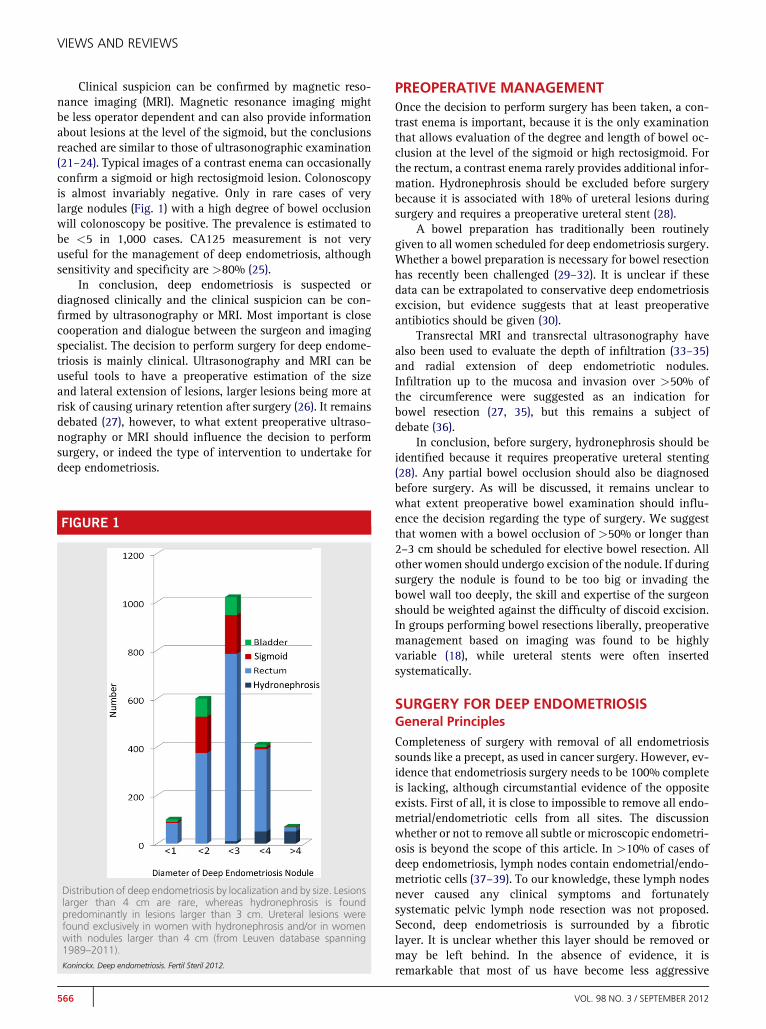
FIGURE 1
Distribution of deep endometriosis by localization and by size. Lesionslarger than 4 cm are rare, whereas hydronephrosis is foundpredominantly in lesions larger than 3 cm. Ureteral lesions werefound exclusively in women with hydronephrosis and/or in womenwith nodules larger than 4 cm (from Leuven database spanning1989–2011).Koninckx. Deep endometriosis. Fertil Steril 2012.
566
PREOPERATIVE MANAGEMENTOnce the decision to perform surgery has been taken, a con-trast enema is important, because it is the only examinationthat allows evaluation of the degree and length of bowel oc-clusion at the level of the sigmoid or high rectosigmoid. Forthe rectum, a contrast enema rarely provides additional infor-mation. Hydronephrosis should be excluded before surgerybecause it is associated with 18% of ureteral lesions duringsurgery and requires a preoperative ureteral stent (28).
A bowel preparation has traditionally been routinelygiven to all women scheduled for deep endometriosis surgery.Whether a bowel preparation is necessary for bowel resectionhas recently been challenged (29–32). It is unclear if thesedata can be extrapolated to conservative deep endometriosisexcision, but evidence suggests that at least preoperativeantibiotics should be given (30).
Transrectal MRI and transrectal ultrasonography havealso been used to evaluate the depth of infiltration (33–35)and radial extension of deep endometriotic nodules.Infiltration up to the mucosa and invasion over >50% ofthe circumference were suggested as an indication forbowel resection (27, 35), but this remains a subject ofdebate (36).
In conclusion, before surgery, hydronephrosis should beidentified because it requires preoperative ureteral stenting(28). Any partial bowel occlusion should also be diagnosedbefore surgery. As will be discussed, it remains unclear towhat extent preoperative bowel examination should influ-ence the decision regarding the type of surgery. We suggestthat women with a bowel occlusion of >50% or longer than2–3 cm should be scheduled for elective bowel resection. Allother women should undergo excision of the nodule. If duringsurgery the nodule is found to be too big or invading thebowel wall too deeply, the skill and expertise of the surgeonshould be weighted against the difficulty of discoid excision.In groups performing bowel resections liberally, preoperativemanagement based on imaging was found to be highlyvariable (18), while ureteral stents were often insertedsystematically.
SURGERY FOR DEEP ENDOMETRIOSISGeneral Principles
Completeness of surgery with removal of all endometriosissounds like a precept, as used in cancer surgery. However, ev-idence that endometriosis surgery needs to be 100% completeis lacking, although circumstantial evidence of the oppositeexists. First of all, it is close to impossible to remove all endo-metrial/endometriotic cells from all sites. The discussionwhether or not to remove all subtle or microscopic endometri-osis is beyond the scope of this article. In >10% of cases ofdeep endometriosis, lymph nodes contain endometrial/endo-metriotic cells (37–39). To our knowledge, these lymph nodesnever caused any clinical symptoms and fortunatelysystematic pelvic lymph node resection was not proposed.Second, deep endometriosis is surrounded by a fibroticlayer. It is unclear whether this layer should be removed ormay be left behind. In the absence of evidence, it isremarkable that most of us have become less aggressive
VOL. 98 NO. 3 / SEPTEMBER 2012

Fertility and Sterility®
surgically than 10 years ago, as discussed at a meeting inAtlanta between Camran Nezhat and the present authors.Third, recurrence rates of deep endometriosis requiringsurgery are so low that it would be virtually impossible todemonstrate the need for complete versus near completeexcision. Indeed, an increased recurrence rate from 1% toa few percent would be hard to prove. Finally, it remainsunclear whether deep endometriotic nodules not causingpain should be excised when there is no evidence ofprogression. Moreover, many of these nodules probably gounnoticed, without doing any apparent harm.
Absence of evidence that all remaining endometrioticcells should be removed to reduce recurrence rates or improvepain or infertility outcome is very different from carrying outincomplete surgery by leaving a large part of the nodule be-hind. Clinical impressions confirmed by reviewing our data-base suggest that the most difficult surgeries of longduration were indeed those performed in women who hadbeen operated on before, leaving deep endometriosis behindbecause of massive adhesions. Similar observations weremade in women who had undergone several IVF cycles withoocyte pickup, and puncture through a deep endometrioticnodule. We therefore suspect that trauma to endometrioticnodules could reactivate the disease, causing growth andmassive adhesions. Surgery of ‘‘fresh’’ nodules, never previ-ously operated, is anticipated as being technically mucheasier.
In conclusion, in the absence of solid evidence, we sug-gest that deep endometriosis surgery should be visually com-plete, but at the level of the bowel, a rim of fibrosis can be leftbehind. Because we found most recurrences at the posteriorfornix of the vagina, and because the vaginal cuff healswell, we specifically emphasize completeness at this level.
Discoid Excision
Excision of deep endometriosis can be technically demandingand the surgeon should balance his skills against the antici-pated difficulty and duration of surgery. A technically diffi-cult and long surgery should be expected when the noduleis >3 cm in diameter, when it is firmly attached to the ischialspine, when localized in the sigmoid, or when adhesions areexpected because of previous incomplete surgery or IVF(unpublished data from the Leuven database, confirmed clin-ically by all authors). Especially when surgery exceeds 4–5hours, the surgeon should be aware that fatigue can impairjudgment (40). This highlights the need for a team with anassistant experienced in deep endometriosis surgery.
Instruments should be adequate. First, a high-flow insuf-flator allowing a flow >20 L/min is necessary to prevent lossof pneumoperitoneum when a bleeding requires continuousaspiration. Second, a uterine manipulator should be used topush the uterus into anteversion and to prevent loss of pneu-moperitoneum when the vagina is opened. A CO2 laser is notrequired but may facilitate and speed up surgery. There is noevidence today that robotic surgery is an advantage (41, 42).
Over the years, the technique has become streamlined andclose to identical in all patients. Obviously not all steps are re-quired in all patients, but the sequence rarely differs. The steps
VOL. 98 NO. 3 / SEPTEMBER 2012
we suggest for deep endometriosis excision from the rectumor rectosigmoid are as follows, and it is important to stickto these steps, especially when surgery is difficult.
Step 1. The plane of cleavage between the bowel and theside wall has to be identified, beginning from the pelvic brim.This will free the ovaries from the side wall permitting to fixthem to the anterior wall. Together with lysis of physiologicadhesions of the sigmoid, this allows good visualization ofthe pelvis, without excessive Trendelenburg.
Step 2. Both ureters need to be identified if close to or in-volved in the endometriotic nodule being dissected. If hydro-nephrosis was missed before surgery, a ureteral stent shouldbe placed. Endometriosis close to or even surrounding theureter without hydronephrosis should not be considereda major problem and does not require a stent, because ureterallesions occur in<0.5% of cases (28). When in doubt, however,it is preferable to insert a stent, a major risk being the surgeonwho wants to prove he can do without one. When hydroneph-rosis is present, the stricture can be removed (over the stent)without a lesion in >80% of patients (28). If a lesion of theureter is identified, or if part of the ureter is very thin, espe-cially when>2 cm, repair has to be done either by prophylac-tic suturing or by resection and end-to-end anastomosis.Following this step, the lateral margins of the nodule shouldbe completely dissected.
Step 3. The posterior/dorsal margin of the nodule is dis-sected. When strongly attached to the ischial spine and/or is-chiosacral ligament, dissection should be very careful becauseof the risk of massive bleeding that can be difficult to control.At the end of this step, the pararectal spaces are identified andshould be free of disease. Care is taken to complete this stepbefore dissection of the nodule from the bowel is started. In-deed, possible lateral attachments of the nodule to the utero-sacral ligaments or cervix and attachment medially to thebowel should be carefully dissected. The end result shouldsee the endometriotic nodule remaining attached to the cen-tral part between the bowel and the uterus. Pushing the uterusinto anteversion will elevate the nodule with the bowel hang-ing from it.
Step 4. Dissection of the nodule from the bowel is thenperformed. First, the anatomy should be identified. Largernodules of the rectosigmoid are often associated with a bowelloop attaching the rectosigmoid to the uterus above the nod-ule. This increases the technical difficulty, because a rectalprobe cannot reach this loop. Technically, during this dissec-tion, the margin of the nodule is followed when dissectingfrom the bowel. When the muscularis of the bowel is reached,dissection with cold scissors is suggested with minimal coag-ulation in order to prevent late bowel lesions. This dissectionwill often include resection of part of the bowel muscularis upto the mucosa. During this dissection, small perforations ofthe mucosa may occur. Although 10 years ago such injurywas considered inevitable and necessary to achieve completeexcision, today we prefer to avoid this by leaving a fibrosislayer. Full-thickness resection with resection of a larger partof the mucosa is rare and necessary only for lesions infiltrat-ing the bowel lumen, that is, those visible by rectoscopy.Aggressive coagulation of the bowel wall, especially whenthin, is avoided. We suture and close any mucosal lesions
567

VIEWS AND REVIEWS
immediately in order to keep the duration of the bowel open-ing as short as possible.
Step 5. Dissection from the vagina is finally carried out asa last step. Care is taken not to enter the cervix, which hasa similar consistency as a nodule. Dissection of the vaginais facilitated by use of a fornix presenter; often, small endo-metriotic cysts indicate that the vaginal mucosa is close andthat part of the vaginal fornix should be removed.
Following careful hemostasis, the vaginal cuff, if opened,is closed. We close a muscularis defect of the rectum witha single-layer running transversal suture. A full-thickness de-fect is sutured in two layers. Finally, the pelvis and the upperabdomen are washed until the liquid runs clear. This may bea painstakingly long procedure, because often up to 8 L ofsolution is required.
Several technical variations of discoid excision exist. Acircular stapler can be used to remove/suture part of the bowelwall, especially in case of smaller nodules, instead of riskinga small bowel hole and double suture. It is unclear whethera running suture is superior to single sutures for muscularisor full-thickness defects. It is also unclear which size of boweldefect can be sutured and when a bowel resection becomesnecessary. We consider that bowel resection should be con-templated whenever a defect of >50% of its circumferenceis present or when the muscularis defect is>7–8 cm long. Al-though technically feasible, this suturing is challenging forthe surgeon, though too rare for anybody to have any mean-ingful experience.
For the sigmoid, the technique is essentially identical. It isadvisable to start by fixing the bowel to the round ligamentsin order to pull the sigmoid downwards and decrease mobility.Excision of sigmoid nodules is always difficult, because itfrequently requires full-thickness resection as the nodule isoften localized mesenterically, thus impairing blood flowafter excision, because of the mobility of the sigmoid, andbecause suturing high in the pelvis is technically difficult.In addition, excision of a sigmoid nodule should be balancedagainst sigmoid resection, knowing that sigmoid resectioncan be done with minimal short- and long-term risks. Thisstrongly suggests that difficult discoid resection of the sig-moid should preferably be replaced by bowel resection.
Bowel Resection
Steps 1 to 3 should be identical for discoid and bowel resec-tion. The most important thing to realize is that if dissectionis started between the nodule and vagina, bowel resectionbecomes almost inevitable. As described for the sigmoid, dis-section between the nodule and vagina will end with a mobilerectum, with the nodule attached to it. Dissection of thenodule then becomes difficult. The technique and pitfalls ofbowel resection with a circular stapler are beyond the scopeof this article.
Bladder Endometriosis
Bladder endometriosis, even when requiring full-thickness re-section, can be managed by laparoscopic surgery providedcare is taken not to damage the intramural part of the ureter.
568
For this reason, a stent should be inserted when the lesion isclose to the ureter, and the bladder should be opened ashigh as possible, permitting identification of the exact local-ization of the uretr. The bladder wall is sutured in two layers(although some consider one layer to be sufficient) and a blad-der catheter is left in place for 7–10 days. It is unclear whetherbladder healing should be checked afterwards.
Discussion of Surgery
When discoid excision or bowel resection should be done re-mains debatable. In two systematic reviews, we demon-strated that the leakage rate and long-term consequencesof bowel resection increase when the resection involves thelower part of the bowel. For sigmoid resection, leaks occurin <1% of cases, almost without long-term problems. Forlow rectal resections, leaks increase to 15% or more andcarry a lifelong risk of bowel, bladder, and sexual problemsof 30%, 30%, and 40%, respectively (43). Of almost 2,000bowel resections for endometriosis published to date, thelarge majority were lower resections. It remains unclear,however, what the exact indications were for bowel resec-tion, because it is highly variable between authors. The sizeof nodules is rarely indicated, the length of resection rangesfrom 5 to 25 cm, and the duration of surgery can be as longas 8 hours, whereas data on sexuality after surgery are lack-ing (18). Besides the observation that most articles merely de-scribe feasibility in small series, it is obvious that manygroups almost exclusively perform bowel resections and forvarious reasons. Apart from the indications mentioned, weshould be aware of many other aspects influencing surgerythat are rarely mentioned in articles. Bowel resection is notonly technically easier and faster than discoid excision, butit by definition makes the bowel surgeon coresponsible,which is medicolegally important, especially in countrieswhere gynecologists should not perform bowel surgery. Inaddition, the fact that reimbursement of bowel resection is5–10 times higher than discoid excision might be an argu-ment for some hospitals.
We therefore suggest that discoid excision should at leastbe attempted for all rectosigmoid and rectal nodules. If toodifficult for the level of expertise of the surgeon, bowel resec-tion can be performed. In what percentage of cases a bowelresection should be performed is unclear. For the rectumand rectosigmoid, we (PK) performed <1% bowel resections.This very low rate of resections is partially an historical con-sequence of PK developing the technique together with thebowel surgeons he had trained in Leuven. When discussingwhich technique should be used, it remains important thatdeep endometriosis surgery is not a skill contest. What is im-portant is that the incidence of bowel resection should pro-gressively decrease with expertise from 15%–20% to muchless. An incidence of>50% of bowel resections signals a sur-gical choice, not a technical necessity. For sigmoid lesions, weshould be much more liberal with bowel resections. To iden-tify the limits of conservative surgery, we (PK) today perform5% elective bowel resections for deep sigmoid endometrioticnodules, and another 5% is decided during surgery. It canbe argued that sigmoid endometriosis should be treated
VOL. 98 NO. 3 / SEPTEMBER 2012
Fertility and Sterility®
conservatively only in case of small lesions and by experi-enced surgeons.
We strongly oppose bowel resection that is decided uponbefore surgery, except in case of the sigmoid showing signs ofextensive occlusion. Indeed, some lesions erroneously can bejudged as endometriosis or as invasive endometriosis. Al-though data are scarce, up to 14% of bowel resections werereported in which no endometriosis was subsequently foundby pathology. Moreover, if we consider that in 12% of casesthe endometriotic nodule was located outside the muscularis,this adds up to 26% of unnecessary bowel resections (44). Thisis confirmed by the fact that we have all seen lesions stronglysuggestive of endometriosis, which during dissection provedto be no more than artifacts.
Considering the severe lifelong implications of bowel,bladder, and sexual problems following rectal and especiallylow rectal resection, we suggest that this procedure should notbe performed when avoidable, which is the case in mostwomen. This statement remains valid at least until it is dem-onstrated that complications for low rectal resection in case ofendometriosis are different from those for other indications.
POSTOPERATIVE CAREAntibiotics are given as one shot when the vagina has beenopened, whereas full-thickness resection requires 7 days ofantibiotic treatment. Following a muscularis defect andsingle-layer suture, or full-thickness resection and double-layer suture, the patient remains nil by mouth for 4 and 7days, respectively.
Deep endometriosis surgery is associated with urinary re-tention and/or bladder dysfunction (45–47). The incidence ishigher when nodules are bigger, extending laterally deep intothe pararectal space. This is not surprising given the anatomyof the parasympathetic nerve. Nerve sparing is an illusion,because the parasympathetic nerve cannot be dissectedfrom the endometriotic nodule: either endometriosis is leftin place or part of the nerve is destroyed. Urinary retentiongenerally resolves within a few weeks, occasionally after aslong as 9 months. To prevent permanent bladder retention,we prefer to leave some endometriosis unilaterally, whencomplete excision of the nodule would risk damaging theparasympathetic nerve bilaterally.
Deep endometriosis may require ureteral and bowel sur-gery and is prone to complications. Postoperative care shouldtherefore be meticulous and requires expertise. In the absenceof strict and validated guidelines for the postoperative period,we use daily C-reactive protein levels, which should alwaysgo down progressively after day 3, and we advocate liberaluse of repeat laparoscopy whenever in doubt.
The most severe complications during the first week arelate bowel perforations and late ureteral perforations. A latebowel perforation is a potentially life-threatening event,recognized as early as 1996 (48). In >90% of cases, it occursduring the first few days after surgery, but can occasionallyoccur up to postoperative day 7. It requires immediate recog-nition and therapy, which demands expertise, because apartfrom a short episode of acute pain, clinical symptoms are min-imal, the clinical examination is negative and C-reactive
VOL. 98 NO. 3 / SEPTEMBER 2012
protein levels are not yet increased. Although a contrast en-ema or computed tomographic scan can be helpful, an earlyrepeat laparoscopy is recommendedwhenever in doubt. If per-formed within 24 hours after perforation, treatment can beconservative with a bowel suture and lavage, as demonstratedin>50 cases (PK, unpublished data). If a perforation is missedand diagnosis made after 24 hours, four-quadrant peritonitiswill be found, necessitating a colostomy. Conservative treat-ment and repeated lavage have been performed occasionally,but this should be considered as experimental surgery. Lateureteral leakage should be treated with a stent inserted underlaparoscopic vision and a stitch; following blind stent inser-tion, ureteral leaks will continue in 50% of cases (28).
Other complications are rectovaginal, ureterovaginal, andvesicovaginal fistulae. Although experience is limited, vesi-covaginal and ureterovaginal fistulae can and should betreated immediately by laparoscopy, thus avoiding develop-ment of extensive fibrosis. Treatment of rectovaginal fistulaeis less clear. We are in doubt whether to perform early repeatlaparoscopy to close the defect, similar to our experience withvesicovaginal fistulae. Traditionally, however, it is advocatedto wait at least 6 weeks because some of the smaller fistulaeclose spontaneously. Thereafter, bowel resection is generallyperformed, or occasionally conservative vaginal closure. Lap-aroscopic dissection with closure of the rectovaginal fistulaecan be done after 6 weeks (only performed twice), but dissec-tion is long and difficult.
In conclusion, postoperative care after surgery for deependometriosis requires strict follow-up with early repeat lap-aroscopy to immediately treat any complications, includingbleeding, infection, late ureteral or bowel perforation, or fis-tulae. When a complication occurs >2 weeks after surgery,the risks and advantages of immediate intervention shouldbe balanced against the rule that between day 15 and 45, re-peat interventions should be avoided if possible.
RESULTSThe results of rectovaginal surgery have been described ina number of excellent articles and reviews. Pain relief is un-equivocally excellent, with >85% of women pain free (34).Recurrence rates, defined as recurrence of endometriosis,are low. The spontaneous fertility rate is 50%–60%. However,available data do not allow us to balance outcome against thesize or localization of nodules, type of surgery performed, pre-operative imaging, or pathology.
Postoperative adhesions after deep endometriosis surgeryare poorly documented. In a recent RCT (in preparation), ad-hesions were surprisingly much more severe than anticipatedin most women.
DISCUSSIONDeep endometriosis should be considered a specific entity ofendometriosis, to be defined as adenomyosis externa by pa-thology. This definition will avoid inclusion of slightly largertypical lesions in reports on deep endometriosis, which con-found the data. Furthermore, it should not be considereda progressive and recurrent disease, until proven otherwise.This is extremely important when counseling patients.
569
VIEWS AND REVIEWS
Recurrence is suggested to be almost invariably a consequenceof incomplete surgery.
Indications for deep endometriosis surgery are pain and/or infertility, and it is unclear whether a nodule without bowelocclusion that does not cause pain should be operated. Today,the indications should never be occasional findings by imag-ing techniques, in the absence of clinical symptoms.
The need for a bowel preparation is a matter of debate(29–32) for bowel resection. However, given the risk of latebowel perforation, we prefer to administer a full bowelpreparation for discoid excision, until proven otherwise.
Preoperative management and the predictive value of im-aging remain a source of controversy. Although transvaginalultrasonography and MRI are useful to confirm the clinicaldiagnosis or suspicion, these examinations unfortunatelycannot sufficiently predict the difficulty of surgery or thetype of intervention required. It would indeed be useful tohave examinations that could predict the difficulty of surgery,especially for inexperienced surgeons. Until then, we main-tain the rule initiated in Oxford: do a laparoscopy and judgethe difficulty. If too difficult, refer without further surgery.Other procedures such as transrectal ultrasonography orMRI cannot predict the need for bowel resection either. Dataare lacking to demonstrate even a predictive value.We believethat only a contrast enema can predict the degree of sigmoidobstruction, and this examination is therefore useful to decideabout elective sigmoid resection.
The completeness of resection of deep endometriosis re-mains under discussion.We suggest that for the bowel, a smalllayer of fibrosis can be left behind, thus further reducing theneed for bowel resection. Bowel resections are often avoid-able, although in 8% of cases, margins are not free. Consider-ing the number of unnecessary and avoidable bowelresections, we strongly suggest always trying discoid excisionand deciding only during surgery what type of interventionshould be made. The decision to perform a bowel resectionshould not be taken before surgery, except in case of anocclusive nodule in the sigmoid. Because shaving suggestslayer-by-layer excision, which contrast with the idea of enbloc excision of deep endometriosis as described, we preferto use the terms discoid excision or en bloc excision.
Deep endometriosis surgery is challenging and complica-tion prone. As afirst line of prevention, we recommend avoid-ing interventions that take more than 5 hours because offatigue of the surgeon. This implies that in the rare event oftwo nodules being present, for example, at the level of the rec-tum and the sigmoid, interventions should be performed intwo steps, the alternative being a bowel resection of 25 cm.To demonstrate medicolegally that surgery was performedcarefully, some of us advocate video recording of entire pro-cedures (49).Without video recording, which can demonstrateunequivocally the need for intervention and type of surgery,reimbursement will remain at a much lower level than forbowel resection. Those interested in the evolution of ideasand techniques over time are invited to read a similar articlewritten in 1996 (50).
Acknowledgments: For reviewing the manuscript and fortheir comments,we thank our current and former collaborators,
570
that is, Jean Squifflet, UCL, Belgium; Carlo De Cicco, CampusBiomedico Rome, Italy; Roberta Corona, Vrije UniversiteitBrussel, Brussels, Belgium; Karina Mailova, Moscow StateUniversity, Moscow, Russia; Ron Shonman, Tel Aviv, Israel;John Koninckx, Leuven, Belgium; Mario Malzoni, Avelino,Italy; Alfonso Rosetti, Rome, Italy; Enda McVeigh, Oxford,United Kingdom; and Errico Zpi, Rome, Italy. We also thankauthorities mentioned in the manuscript, such as Ivo Brosens,Leuven, Belgium, and Camran Nezhat, Atlanta, GA, for theirhelp with its review.
REFERENCES1. Koninckx PR, Batt RE, Hummelshoj L, McVeigh E, Ussia A, Yeh J. The ele-
phant in the room: quality control of endometriosis data. J Minim InvasiveGynecol 2010;17:637–40.
2. Ussia A, Betsas G, Corona R, De CC, Koninckx PR. Pathophysiology of cyclichemorrhagic ascites and endometriosis. J Minim Invasive Gynecol 2008;15:677–81.
3. Batt RE, Smith RA, Buck Louis GM, Martin DC, Chapron C, Koninckx PR,et al. Mullerianosis. Histol Histopathol 2007;22:1161–6.
4. Dehoux JP, Defrere S, Squifflet J, Donnez O, Polet R, Mestdagt M, et al. Is thebaboon model appropriate for endometriosis studies? Fertil Steril 2011;96:728–33.
5. De CC, Ussia A, Koninckx PR. Laparoscopic ureteral repair in gynaecologicalsurgery. Curr Opin Obstet Gynecol 2011;23:296–300.
6. Koninckx PR, Martin DC. Deep endometriosis: a consequence of infiltrationor retraction or possibly adenomyosis externa? Fertil Steril 1992;58:924–8.
7. Cornillie FJ, Oosterlynck D, Lauweryns JM, Koninckx PR. Deeply infiltratingpelvic endometriosis: histology and clinical significance. Fertil Steril 1990;53:978–83.
8. Koninckx PR. Biases in the endometriosis literature. Illustrated by 20 years ofendometriosis research in Leuven. Eur J Obstet Gynecol Reprod Biol 1998;81:259–71.
9. Donnez J, Squifflet J. Complications, pregnancy and recurrence in a pro-spective series of 500 patients operated on by the shaving technique fordeep rectovaginal endometriotic nodules. Hum Reprod 2010;25:1949–58.
10. Koninckx PR, Barlow D, Kennedy S. Implantation versus infiltration: theSampson versus the endometriotic disease theory. Gynecol Obstet Invest1999;47(Suppl 1):3–9.
11. Koninckx PR, Kennedy SH, Barlow DH. Endometriotic disease: the role ofperitoneal fluid. Hum Reprod Update 1998;4:741–51.
12. Koninckx PR. Biases in the endometriosis literature—illustrated by 20 yearsof endometriosis research in Leuven. Eur J Obstet Gynecol Reprod Biol1998;81:259–71.
13. Oosterlynck DJ, Meuleman C, Waer M, Koninckx PR. CO2-laser excision ofendometriosis does not improve the decreased natural killer activity. ActaObstet Gynecol Scand 1994;73:333–7.
14. Nabeshima H, Murakami T, Yoshinaga K, Sato K, Terada Y, Okamura K.Analysis of the clonality of ectopic glands in peritoneal endometriosis usinglaser microdissection. Fertil Steril 2003;80:1144–50.
15. Wu Y, Basir Z, Kajdacsy-Balla A, Strawn E, Macias V, Montgomery K, et al.Resolution of clonal origins for endometriotic lesions using laser capture mi-crodissection and the human androgen receptor (HUMARA) assay. FertilSteril 2003;79(Suppl 1):710–7.
16. Jimbo H, Hitomi Y, Yoshikawa H, Yano T, Momoeda M, Sakamoto A, et al.Evidence for monoclonal expansion of epithelial cells in ovarian endometrialcysts. Am J Pathol 1997;150:1173–8.
17. Roman H, Vassilieff M, Gourcerol G, Savoye G, Leroi AM, Marpeau L,et al. Surgical management of deep infiltrating endometriosis of the rec-tum: pleading for a symptom-guided approach. Hum Reprod 2011;26:274–81.
18. De Cicco C, Corona R, Schonman R, Mailova K, Ussia A, Koninckx P. Bowelresection for deep endometriosis: a systematic review. Br J Obstet Gynaecol2011;118:285–91.
VOL. 98 NO. 3 / SEPTEMBER 2012
Fertility and Sterility®
19. Koninckx PR,Meuleman C, Oosterlynck D, Cornillie FJ. Diagnosis of deep en-dometriosis by clinical examination during menstruation and plasma CA-125 concentration. Fertil Steril 1996;65:280–7.
20. Hudelist G, English J, Thomas AE, Tinelli A, Singer CF, Keckstein J. Diagnosticaccuracy of transvaginal ultrasound for non-invasive diagnosis of bowel en-dometriosis: systematic review and meta-analysis. Ultrasound Obstet Gyne-col 2011;37:257–63.
21. Saba L, Guerriero S, Sulcis R, Pilloni M, Ajossa S, Melis G, et al. MRIand "tenderness guided" transvaginal ultrasonography in the diagnosisof recto-sigmoid endometriosis. J Magn Reson Imaging 2012;35:352–60.
22. Bazot M, Gasner A, Lafont C, Ballester M, Darai E. Deep pelvic endometri-osis: limited additional diagnostic value of postcontrast in comparisonwith conventional MR images. Eur J Radiol 2011;80:e331–9.
23. BazotM, Darai E. Value of transvaginal sonography in assessing severe pelvicendometriosis. Ultrasound Obstet Gynecol 2010;36:134–5.
24. Bazot M, Lafont C, Rouzier R, Roseau G, Thomassin-Naggara I, Darai E. Di-agnostic accuracy of physical examination, transvaginal sonography, rectalendoscopic sonography, and magnetic resonance imaging to diagnosedeep infiltrating endometriosis. Fertil Steril 2009;92:1825–33.
25. Muyldermans M, Cornillie FJ, Koninckx PR. CA125 and endometriosis. HumReprod Update 1995;1:173–87.
26. Ballester M, Santulli P, Bazot M, Coutant C, Rouzier R, Darai E. Preoperativeevaluation of posterior deep-infiltrating endometriosis demonstrates a rela-tionship with urinary dysfunction and parametrial involvement. J Minim In-vasive Gynecol 2011;18:36–42.
27. Abrao MS, Podgaec S, Dias JA Jr, Averbach M, Silva LF, Marino de CF. Endo-metriosis lesions that compromise the rectum deeper than the inner muscu-laris layer have more than 40% of the circumference of the rectum affectedby the disease. J Minim Invasive Gynecol 2008;15:280–5.
28. De Cicco C, Schonman R, Craessaerts M, Van CB, Ussia A, Koninckx PR. Lap-aroscopic management of ureteral lesions in gynecology. Fertil Steril 2009;92:1424–7.
29. Cao F, Li J, Li F. Mechanical bowel preparation for elective colorectal surgery:updated systematic review and meta-analysis. Int J Colorectal Dis 2012;27:803–10.
30. Cohen SL, Einarsson JI. The role of mechanical bowel preparation in gyneco-logic laparoscopy. Rev Obstet Gynecol 2011;4:28–31.
31. Fanning J, Valea FA. Perioperative bowel management for gynecologic sur-gery. Am J Obstet Gynecol 2011;205:309–14.
32. Galandiuk S, Fry DE, Polk HC Jr. Is there a role for bowel preparation and oralor parenteral antibiotics in infection control in contemporary colon surgery?Adv Surg 2011;45:131–40.
33. Hudelist G, Tuttlies F, Rauter G, Pucher S, Keckstein J. Can transvaginal so-nography predict infiltration depth in patients with deep infiltrating endo-metriosis of the rectum? Hum Reprod 2009;24:1012–7.
34. Bassi MA, Podgaec S, Dias JA Jr, D'Amico FN, Petta CA, AbraoMS. Quality oflife after segmental resection of the rectosigmoid by laparoscopy in patientswith deep infiltrating endometriosis with bowel involvement. J Minim Inva-sive Gynecol 2011;18:730–3.
VOL. 98 NO. 3 / SEPTEMBER 2012
35. Goncalves MO, Podgaec S, Dias JA Jr, Gonzalez M, Abrao MS. Transvaginalultrasonography with bowel preparation is able to predict the number oflesions and rectosigmoid layers affected in cases of deep endometriosis,defining surgical strategy. Hum Reprod 2010;25:665–71.
36. Koninckx PR, De CC, Schonman R, Corona R, Betsas G, Ussia A. The recentarticle "Endometriosis lesions that compromise the rectum deeper thanthe inner muscularis layer have more than 40% of the circumference ofthe rectum affected by the disease."J Minim Invasive Gynecol 2008;15:774–5.
37. Gong Y, Tempfer CB. Regional lymphatic spread in women with pelvicendometriosis. Med Hypotheses 2011;76:560–3.
38. Namkung J, Kim SJ, Kim JH, Kim J, Hur SY. Rectal endometriosis withinvasion into lymph nodes. J Obstet Gynaecol Res 2011;37:1117–21.
39. Tempfer CB,Wenzl R, Horvat R, GrimmC, Polterauer S, Buerkle B, et al. Lym-phatic spread of endometriosis to pelvic sentinel lymph nodes: a prospectiveclinical study. Fertil Steril 2011;96:692–6.
40. Schonman R, De CC, Corona R, Soriano D, Koninckx PR. Accident analysis:factors contributing to a ureteric injury during deep endometriosis surgery.Br J Obstet Gynaecol 2008;115:1611–5.
41. Nezhat C, Hajhosseini B, King LP. Robotic-assisted laparoscopic treatment ofbowel, bladder, and ureteral endometriosis. J Soc Laparoendosc Surg 2011;15:387–92.
42. Nezhat C, Lewis M, Kotikela S, Veeraswamy A, Saadat L, Hajhosseini B, et al.Robotic versus standard laparoscopy for the treatment of endometriosis.Fertil Steril 2010;94:2758–60.
43. Ret Davalos ML, De Cicco C, D'Hoore A, De DB, Koninckx PR. Outcome afterrectum or sigmoid resection: a review for gynecologists. J Minim InvasiveGynecol 2007;14:33–8.
44. Meuleman C, D'Hoore A, Van CB, Beks N, D'Hooghe T. Outcome after mul-tidisciplinary CO2 laser laparoscopic excision of deep infiltrating colorectalendometriosis. Reprod Biomed Online 2009;18:282–9.
45. Ballester M, Chereau E, Dubernard G, Coutant C, Bazot M, Darai E. Urinarydysfunction after colorectal resection for endometriosis: results of a prospec-tive randomized trial comparing laparoscopy to open surgery. Am J ObstetGynecol 2011;204:303.e1–6.
46. Gabriel B, Nassif J, Trompoukis P, Lima AM, Barata S, Lang-Averous G, et al.Prevalence and outcome of urinary retention after laparoscopic surgery forsevere endometriosis—does histology provide answers? Int Urogynecol J2012;23:111–6.
47. Kovoor E, Nassif J, Miranda-Mendoza I, Lang-Averous G, Wattiez A. Long-term urinary retention after laparoscopic surgery for deep endometriosis.Fertil Steril 2011;95:803–12.
48. Koninckx PR, Timmermans B, Meuleman C, Penninckx F. Complications ofCO2-laser endoscopic excision of deep endometriosis. Hum Reprod 1996;11:2263–8.
49. Koninckx PR. Videoregistration of surgery should be used as a qualitycontrol. J Minim Invasive Gynecol 2008;15:248–53.
50. Koninckx PR, Martin DC. Surgical treatment of deeply infiltrating endometri-osis. In: Sutton C, editor. Gynecological endoscopic surgery. London: Chap-man and Hall; 1997:19–35.
571
Related Documents


![FNAC Diagnosis of Scar Endometriosis: A Report of 3 Cases with … · 2020. 7. 29. · is curative for scar endometriosis.[8] cOncLUs IOn Scar endometriosis is relatively rare entity](https://static.cupdf.com/doc/110x72/5fd095cb2c296c4af70ea6b5/fnac-diagnosis-of-scar-endometriosis-a-report-of-3-cases-with-2020-7-29-is.jpg)


![[Product Monograph Template - Standard] · Endometriosis Symptoms and physical findings associated with a previous diagnosis of endometriosis may ... Vaginal infections should be](https://static.cupdf.com/doc/110x72/5f321a38e7991861762775c6/product-monograph-template-standard-endometriosis-symptoms-and-physical-findings.jpg)