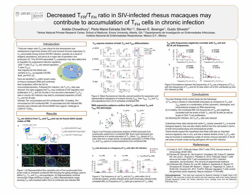
Decreased T FR /T FH ratio in SIV-infected rhesus macaques may contribute to accumulation of T FH cells in chronic infection Ankita Chowdhury 1 , Perla Maria Estrada Del Rio 1,2 , Steven E. Bosinger 1 , Guido Silvestri 1* 1 Yerkes National Primate Research Center, School of Medicine, Emory University, Atlanta, GA; 2 Departamento de Investigación en Enfermedades Infecciosas, Instituto Nacional de Enfermedades Respiratorias. México D.F., México Acknowledgments: This work was supported by National Institutes of Health Grant U19 AI096187. We would like to thank the animal care and veterinary staff at the Yerkes National Primate Research Center, the Yerkes Genomics Core, Virology Core and the Molecular Pathology Core. T FR express markers of both T FH and T REG differentiation. T FH = CD4 + CXCR5 + PD1 hi Foxp3 - T FR =CD4 + CXCR5 + PD1 hi Foxp3 + CD25 + T REG = CD4 + PD1 lo/int CD25 + Foxp3 + TFR TFH TREG Introduction Results T FR are distinct from T FH and T REG and can be found within lymph nodes of RM. 0 10 20 30 0 2 4 6 0 10 20 30 0 5 10 15 p= 0.0358 r= -0.4397 T FH (% of CD4) Frequency of T FR (% of T FH ) p= 0.0140 r= -0.5049 Frequency of T FR (% of T FH ) GC B cells (% of B cells) 0 5 10 15 20 0 10 20 30 0 5 10 15 20 0 10 20 30 p=0.0199 r= -0.5039 T FH (% of CD4) Frequency of T FR (% of T FH ) p=0.031 r= -0.4714 GC B cells (% of B cells) Frequency of T FR (% of T FH ) 0 5 10 15 0.0 0.1 0.2 0.3 0.4 0.5 0 5 10 15 20 25 p=0.0004 p=0.0078 p=0.0122 p=0.0071 T FR (% of T FH cells) T FH (% of CD4 cells) T FR (% of CD4 cells) p=0.0058 T follicular helper cells (T FH ) are critical for the development and maintenance of germinal centers (GC) and humoral immune responses (1). T FH accumulate during chronic HIV/SIV infection, possibly as a result of antigenic persistence, and serve as a major site of persistent viral production (2). This SIV/HIV-associated T FH expansion may also reflect lack of regulation by suppressive follicular regulatory CD4 + T cells (T FR ). T FR are natural regulatory T-cells (T REG ) that migrate into the follicle and, similarly to T FH , up-regulate CXCR5, Bcl6, and PD1 (3). a b RNA expression patterns confirm that T FR cells share T FH and T REG like phenotype. T FR cells decrease as a frequency of T FH cells a3er SIV infec8on. T FR cells frequencies negatively correlate with T FH cell and GC B cell frequencies. Figure 3 (a) Principal components analysis of RNA transcripts from lymphocytes sorted from 5 uninfected RM. Each circle represents the transcriptome of a sorted population of T FH (blue), T REG (green) of T FR (red) cells from a single animal. (b) Absolute expression in FPKM of key T FH and T REG genes in sorted populations from uninfected RM. Figure 4. The frequency of (a) T FH and (b) T FR cells within LN of uninfected (black), acutely infected (pink) and chronically infected (red) untreated RM. (c) Frequency of T FR cells (as a percent of T FH cells). Figure 1 (a) Representative flow cytometry plot of live lymphocytes from lymph node of untreated uninfected RM showing the gating strategy used to define T FR , T FH and T REG cell populations. (b) Representative confocal microscope image showing a single T FR cell(c) Representative image showing T FR cells localized within GCs of uninfected and infected RM. Figure 5.Correlations between the frequencies of T FR (as a frequency of T FH ) with the frequencies of T FH and GC B cells within LN of SIV uninfected (a) and SIV infected (b) RM. a b 0 103 104 105 <PE-Cy5-A>: CD127 0 20 40 60 80 100 % of Max 0 102 103 104 105 <Pacific Blue-A>: CTLA4 0 20 40 60 80 100 % of Max 0 102 103 104 105 <PE-Texas Red-A>: BCL6 0 20 40 60 80 100 % of Max 0 102 103 104 105 <FITC-A>: HELIOS 0 20 40 60 80 100 % of Max Helios Bcl6 CTLA4 CD127 -500 0 500 1000 1500 CD127 MFI p = 0.0039 p = 0.0039 0 20 40 60 80 100 % CD127 + p = 0.0313 0 500 1000 1500 2000 CTLA4 MFI p = 0.0039 p = 0.0039 p = 0.0039 0 20 40 60 80 100 % CTLA4 + p = 0.0039 p = 0.0078 G G R H 0 500 1000 1500 Bcl6 MFI p = 0.0078 p = 0.0078 0 20 40 60 80 100 % Bcl6 + p = 0.0313 p = 0.0313 0 20 40 60 80 100 % Helios + p = 0.0039 p = 0.0039 0 1000 2000 3000 Helios MFI p = 0.0078 p = 0.0078 p = 0.0078 d c b a T REG cells Non-T REG cells T FR cells T FH cells Figure 2. Mean fluorescence intensity, percent positive for expression and representative histograms of markers among T REG , Non-T REG , T FR and T FH cell populations from LN of untreated uninfected RM. T REG genes T FH genes a b Live CD3+ CD4+ T cells c a b c The main findings of the current study are the followings: (i) T FR show a shared or intermediate phenotype as compared to T FH and T REG based on a combination of flow cytometric, histological, and transcriptional analysis by RNA sequencing; (ii) In healthy, SIV-uninfected RM, the frequencies of T FR are negatively correlated with the levels of both T FH and GC B-cells as well as levels of CD4 + T-cell proliferation; (iii) following SIV infection, the T FR /T FH ratio was reduced Collectively these data indicate that while T FR closely resemble T FH in several biological aspects, they are also clearly distinct from this cell subset in terms of both immunophenotype and transcriptional profile. These results support the hypothesis that these cells play an important immune regulatory role in vivo, and that a relative decline of the T FR /T FH ratio may be involved in establishing a state of chronic immune activation in the B cell areas of lymph nodes during pathogenic HIV and SIV infection. Conclusions PCA 1 PCA 2 CD127 Helios CTLA4 Bcl6 TREG Non-TREG TFH TFR TREG Non-TREG TFH TFR Uninfected Acute Chronic T FH cells T FR cells T FR cells (of T FH cells) Here we identified T FR within lymph nodes of rhesus macaques (RM) and confirmed their localization within the GC by immunohistochemistry. Following SIV infection, the T FR /T FH ratio was reduced. Our data suggests that T FR may contribute to the regulation and proliferation of T FH and GC B-cells in vivo and that a decreased T FR /T FH ratio in chronic SIV infection may lead to unchecked expansion of both T FH and GC B-cells. Animals: Ten unvaccinated and SIV-uninfected RM, 13 healthy, SIV- immunized but SIV-uninfected RM, 13 vaccinated and SIV-infected RM. Animals were infected with SIVsmmE660 intra-vaginal challenge at 2.06X10 4 TCID 50 . TREG GC B cell TFH TFR ? ? BCL6 SAP T CELL ZONE GERMINAL CENTER CXCR5 CXCR5 FOXP3 FOXP3 (1) Crotty S. 2011. Follicular helper CD4 T cells (TFH). Annual review of immunology 29:621-663. (2) Perreau M, Savoye AL, De Crignis E, Corpataux JM, Cubas R, Haddad EK, De Leval L, Graziosi C, Pantaleo G. 2013. Follicular helper T cells serve as the major CD4 T cell compartment for HIV-1 infection, replication, and production. The Journal of experimental medicine (3)Linterman MA, Pierson W, Lee SK, Kallies A, Kawamoto S, Rayner TF, Srivastava M, Divekar DP, Beaton L, Hogan JJ, Fagarasan S, Liston A, Smith KG, Vinuesa CG. 2011. FoxP3+ follicular regulatory T cells control the germinal center response. Nature medicine 17:975-982. References

Welcome message from author
This document is posted to help you gain knowledge. Please leave a comment to let me know what you think about it! Share it to your friends and learn new things together.
Transcript
Decreased TFR/TFH ratio in SIV-infected rhesus macaques may contribute to accumulation of TFH cells in chronic infection
Ankita Chowdhury1, Perla Maria Estrada Del Rio1,2, Steven E. Bosinger1, Guido Silvestri1* 1Yerkes National Primate Research Center, School of Medicine, Emory University, Atlanta, GA; 2 Departamento de Investigación en Enfermedades Infecciosas,
Instituto Nacional de Enfermedades Respiratorias. México D.F., México
Acknowledgments: This work was supported by National Institutes of Health Grant U19 AI096187. We would like to thank the animal care and veterinary staff at the Yerkes National Primate Research Center, the Yerkes Genomics Core, Virology Core and the Molecular Pathology Core.
TFR express markers of both TFH and TREG differentiation.
TFH= CD4+CXCR5+PD1hiFoxp3-
TFR=CD4+CXCR5+PD1hiFoxp3+CD25+
TREG= CD4+PD1lo/intCD25+Foxp3+
TFR
TFH
TREG
Introduction
Results TFR are distinct from TFH and TREG and can be found within lymph nodes of RM.
0 10 20 300
2
4
6
0 10 20 300
5
10
15p= 0.0358 r= -0.4397
T FH (%
of C
D4)
Frequency of TFR (% of TFH )
p= 0.0140 r= -0.5049
Frequency of TFR (% of TFH ) GC
B c
ells
(% o
f B c
ells
)
0 5 10 15 200
10
20
30
0 5 10 15 200
10
20
30p=0.0199 r= -0.5039
T FH
(% o
f CD
4)
Frequency of TFR (% of TFH )
p=0.031 r= -0.4714
GC
B c
ells
(% o
f B c
ells
)
Frequency of TFR (% of TFH )
0
5
10
15
0.0
0.1
0.2
0.3
0.4
0.5
0
5
10
15
20
25
p=0.0004
p=0.0078
p=0.0122
p=0.0071
TFR
(% o
f TFH
cel
ls)
T FH (%
of C
D4
cells
)
T FR (%
of C
D4
cells
)
p=0.0058
T follicular helper cells (TFH) are critical for the development and maintenance of germinal centers (GC) and humoral immune responses (1). TFH accumulate during chronic HIV/SIV infection, possibly as a result of antigenic persistence, and serve as a major site of persistent viral production (2). This SIV/HIV-associated TFH expansion may also reflect lack of regulation by suppressive follicular regulatory CD4+ T cells (TFR). TFR are natural regulatory T-cells (TREG) that migrate into the follicle and, similarly to TFH, up-regulate CXCR5, Bcl6, and PD1 (3).
a
b
RNA expression patterns confirm that TFR cells share TFH and TREG like phenotype.
TFR cells decrease as a frequency of TFH cells a3er SIV infec8on.
TFR cells frequencies negatively correlate with TFH cell and GC B cell frequencies.
Figure 3 (a) Principal components analysis of RNA transcripts from lymphocytes sorted from 5 uninfected RM. Each circle represents the transcriptome of a sorted population of TFH (blue), TREG (green) of TFR (red) cells from a single animal. (b) Absolute expression in FPKM of key TFH and TREG genes in sorted populations from uninfected RM.
Figure 4. The frequency of (a) TFH and (b) TFR cells within LN of uninfected (black), acutely infected (pink) and chronically infected (red) untreated RM. (c) Frequency of TFR cells (as a percent of TFH cells).
Figure 1 (a) Representative flow cytometry plot of live lymphocytes from lymph node of untreated uninfected RM showing the gating strategy used to define TFR, TFH and TREG cell populations. (b) Representative confocal microscope image showing a single TFR cell(c) Representative image showing TFR cells localized within GCs of uninfected and infected RM.
Figure 5.Correlations between the frequencies of TFR (as a frequency of TFH) with the frequencies of TFH and GC B cells within LN of SIV uninfected (a) and SIV infected (b) RM.
a
b
0 103 104 105
<PE-Cy5-A>: CD127
0
20
40
60
80
100
% o
f Max
0 102 103 104 105
<Pacific Blue-A>: CTLA4
0
20
40
60
80
100
% o
f Max
0 102 103 104 105
<PE-Texas Red-A>: BCL6
0
20
40
60
80
100
% o
f Max
0 102 103 104 105
<FITC-A>: HELIOS
0
20
40
60
80
100
% o
f Max
Helios
Bcl6
CTLA4
CD127 TREG
NON TREG TFR
TFH-500
0
500
1000
1500
CD
127
MFI
p = 0.0039
p = 0.0039
0
20
40
60
80
100
% C
D12
7 +
p = 0.0313
TREG
NON TREG TFR
TFH0
500
1000
1500
2000
CTL
A4
MFI
p = 0.0039
p = 0.0039 p = 0.0039
0
20
40
60
80
100
% C
TLA
4 +
p = 0.0039 p = 0.0078
TREG
NON TREG TFR
TFH0
500
1000
1500
Bcl
6 M
FI
p = 0.0078 p = 0.0078
0
20
40
60
80
100
% B
cl6
+
p = 0.0313 p = 0.0313
0
20
40
60
80
100
% H
elio
s +
p = 0.0039 p = 0.0039
TREG
NON TREG TFR
TFH0
1000
2000
3000
Hel
ios
MFI
p = 0.0078
p = 0.0078
p = 0.0078
d
c
b
a TREG cells Non-TREG cells TFR cells TFH cells
Figure 2. Mean fluorescence intensity, percent positive for expression and representative histograms of markers among TREG, Non-TREG, TFR and TFH cell populations from LN of untreated uninfected RM.
TREG genes TFH genes
a b
Live CD3+ CD4+ T cells
c a b c
The main findings of the current study are the followings: (i) TFR show a shared or intermediate phenotype as compared to TFH and
TREG based on a combination of flow cytometric, histological, and transcriptional analysis by RNA sequencing;
(ii) In healthy, SIV-uninfected RM, the frequencies of TFR are negatively correlated with the levels of both TFH and GC B-cells as well as levels of CD4+ T-cell proliferation;
(iii) following SIV infection, the TFR/TFH ratio was reduced
Collectively these data indicate that while TFR closely resemble TFH in several biological aspects, they are also clearly distinct from this cell subset in terms of both immunophenotype and transcriptional profile. These results support the hypothesis that these cells play an important immune regulatory role in vivo, and that a relative decline of the TFR/TFH ratio may be involved in establishing a state of chronic immune activation in the B cell areas of lymph nodes during pathogenic HIV and SIV infection.
Conclusions
PCA 1
PC
A 2
CD127
Helios
CTLA4
Bcl6
TREG
Non-TREG TFH
TFR TREG
Non-TREG TFH
TFR
Uninfected Acute Chronic
TFH cells TFR cells TFR cells (of TFH cells)
Here we identified TFR within lymph nodes of rhesus macaques (RM) and confirmed their localization within the GC by immunohistochemistry. Following SIV infection, the TFR/TFH ratio was reduced. Our data suggests that TFR may contribute to the regulation and proliferation of TFH and GC B-cells in vivo and that a decreased TFR/TFH ratio in chronic SIV infection may lead to unchecked expansion of both TFH and GC B-cells. Animals: Ten unvaccinated and SIV-uninfected RM, 13 healthy, SIV-immunized but SIV-uninfected RM, 13 vaccinated and SIV-infected RM. Animals were infected with SIVsmmE660 intra-vaginal challenge at 2.06X10⁴ TCID50.
TREG
GC B cell
TFH
TFR ?
?
BCL6 SAP
T CELL ZONE
GERMINAL CENTER
CXCR5
CXCR5
FOXP3
FOXP3
(1) Crotty S. 2011. Follicular helper CD4 T cells (TFH). Annual review of immunology 29:621-663.
(2) Perreau M, Savoye AL, De Crignis E, Corpataux JM, Cubas R, Haddad EK, De Leval L, Graziosi C, Pantaleo G. 2013. Follicular helper T cells serve as the major CD4 T cell compartment for HIV-1 infection, replication, and production. The Journal of experimental medicine
(3) Linterman MA, Pierson W, Lee SK, Kallies A, Kawamoto S, Rayner TF, Srivastava M, Divekar DP, Beaton L, Hogan JJ, Fagarasan S, Liston A, Smith KG, Vinuesa CG. 2011. FoxP3+ follicular regulatory T cells control the germinal center response. Nature medicine 17:975-982.
References
Related Documents