DECREASED INFARCT SIZE AFTER FOCAL CEREBRAL ISCHEMIA IN MICE CHRONICALLY INFECTED WITH TOXOPLASMA GONDII D. ARSENIJEVIC, a1 * F. DE BILBAO, b1 P. VALLET, b A. HEMPHILL, c B. GOTTSTEIN, c D. RICHARD, d P. GIANNAKOPOULOS b AND W. LANGHANS e a Department of Medicine, Division of Physiology, University of Fribourg, Chemin Du Musée 5, 1700 Fribourg, Switzerland b Division of Geriatric Psychiatry, University Hospitals of Geneva, Belle-Idée, 1225 Geneva and Division of Old Age Psychiatry, Univer- sity of Lausanne, 1008 Prilly, Switzerland c Institute of Parasitology, University of Bern, 3012, Bern, Switzerland d Institut Universitaire de Cardiologie et de Pneumologie, Hopital Laval, Quebec, Canada e Institute of Animal Sciences, ETH Zurich, Schorenstrasse 16, Zurich 8603 Schwerzenbach, Switzerland Abstract—To determine whether Toxoplasma gondii infec- tion could modify biological phenomena associated with brain ischemia, we investigated the effect of permanent mid- dle cerebral artery occlusion (MCAO) on neuronal survival, inflammation and redox state in chronically infected mice. Infected animals showed a 40% to 50% decrease of infarct size compared with non-infected littermates 1, 4 and 14 days after MCAO. The resistance of infected mice may be associ- ated with increased basal levels of anti-inflammatory cyto- kines and/or a marked reduction of the MCAO-related brain induction of two pro-inflammatory cytokines, tumor necrosis factor-alpha and interferon-gamma (IFN). In addition, poten- tial anti-inflammatory/neuroprotective factors such as nerve growth factor, suppressor of cytokine signaling-3, su- peroxide dismutase activity, uncoupling protein-2 and gluta- thione (GSH) were upregulated in the brain of infected mice. Consistent with a role of GSH in central cytokine regulation, GSH depletion by diethyl maleate inhibited Toxoplasma gon- dii lesion resistance by increasing the proinflammatory cyto- kine IFN brain levels. Overall, these findings indicate that chronic toxoplasmosis decisively influences both the inflam- matory molecular events and outcome of cerebral ischemia. © 2007 IBRO. Published by Elsevier Ltd. All rights reserved. Key words: toxoplasmosis, cerebral ischemia, cytokines, redox factors, rodent. One of the possible biological consequences of infection is a change of the redox status in both CNS and peripheral tissues (Arsenijevic et al., 2001). Although both animal models and clinical studies showed that infections promote neuronal death following cerebral ischemic injury (Sacco, 2001; Emsley and Tyrrell, 2002), activation of the immune system can also result in neuroprotection (Bordet et al., 2000). In fact, the effect of infection on ischemic damage may largely depend on the regulation of reactive oxygen species by pro-inflammatory and anti-inflammatory cyto- kines as well as by the main antioxidant state regulators, namely superoxide dismutase (SOD) (Guegan et al., 1998; Murakami et al., 1998), glutathione (GSH) (Nicholls and Budd, 2000; Schulz et al., 2000; Droge, 2002), uncoupling protein-2 (UCP2) (Arsenijevic et al., 2000b; Mattiasson et al., 2003) and nerve growth factor (NGF) (Brodie, 1996; Guegan et al., 1999; Villoslada et al., 2000). In addition, the newly described suppressor of cytokine signaling (SOCS) proteins are induced in peripheral and central models of inflammation (Lebel et al., 2000; Bates et al., 2001; Larsen and Ropke, 2002; Wang and Campbell, 2002; Huang et al., 2003; Park et al., 2003; Jo et al., 2005). SOCS possibly interact with cellular redox determinants (Park et al., 2003) and transgenic SOCS expression has been shown to inhibit inflammation and apoptosis following lipopolysaccharide (LPS) injection (Jo et al., 2005). Chronic murine toxoplasmosis may be of particular interest in the study of infection-related effects on brain ischemia since it is associated with a tissue-specific regu- lation of the oxidative state (Arsenijevic et al., 2001) as well as activation of pro-inflammatory cytokines such as tumor necrosis factor-alpha (TNF), interferon-gamma (IFN) and interleukin-2 (IL-2), but also anti-inflammatory cyto- kines such as interleukin-10 (IL-10) (Arsenijevic et al., 1997, 1998). Some of these cytokines are known to en- hance neurodegeneration following ischemia (Arsenijevic et al., 2006). In order to determine how chronic infection influences the main biological phenomena associated with acute cerebral ischemia, the present study explores the impact of chronic Toxoplasma gondii infection on brain ischemic injury and inflammatory/antioxidant processes in- duced by permanent middle cerebral artery occlusion (MCAO). EXPERIMENTAL PROCEDURES All procedures were approved by the Veterinary Office of the Canton of Zurich Health Directorate and the Veterinary Office of Geneva in accordance with the Swiss Animal Care Guidelines. All efforts were made to minimize the number of animals used in this study and every effort was taken to reduce any suffering. 1 Equal first authors. *Corresponding author. Tel: 41-79-501-52-83; fax: 41-26-300-97-34. E-mail address: [email protected] (D. Arsenijevic). Abbreviations: cDNA, complementary DNA; DEM, diethyl maleate; GAPDH, glyceraldehyde 3-phosphate dehydrogenase; GSH, glutathi- one; IFN, interferon-gamma; IL-2, interleukin-2; IL-10, interleukin-10; LPS, lipopolysaccharide; MCAO, middle cerebral artery occlusion; NGF, nerve growth factor; SOCS, suppressor of cytokine signaling; SOCS-3, suppressor of cytokine signaling-3; SOD, superoxide dismutase; TNF, tumor necrosis factor-alpha; TUNEL, terminal deoxynucleotidyl trans- ferase biotin-dUTP nick end labeling; UCP2, uncoupling protein-2. Neuroscience 150 (2007) 537–546 0306-4522/07$30.000.00 © 2007 IBRO. Published by Elsevier Ltd. All rights reserved. doi:10.1016/j.neuroscience.2007.09.080 537

Welcome message from author
This document is posted to help you gain knowledge. Please leave a comment to let me know what you think about it! Share it to your friends and learn new things together.
Transcript
DM
DAPa
Fb
Bsc
d
Qe
8
AtbdiIsaakiftnptCGdkcm©
Kr
Oa1
*EAGoLnstf
Neuroscience 150 (2007) 537–546
0d
ECREASED INFARCT SIZE AFTER FOCAL CEREBRAL ISCHEMIA IN
ICE CHRONICALLY INFECTED WITH TOXOPLASMA GONDIItmn2s2msknMBpaGt(m22S(bl
iilanak1heiaiid(
ACGe
. ARSENIJEVIC,a1* F. DE BILBAO,b1 P. VALLET,b
. HEMPHILL,c B. GOTTSTEIN,c D. RICHARD,d
. GIANNAKOPOULOSb AND W. LANGHANSe
Department of Medicine, Division of Physiology, University ofribourg, Chemin Du Musée 5, 1700 Fribourg, Switzerland
Division of Geriatric Psychiatry, University Hospitals of Geneva,elle-Idée, 1225 Geneva and Division of Old Age Psychiatry, Univer-ity of Lausanne, 1008 Prilly, Switzerland
Institute of Parasitology, University of Bern, 3012, Bern, Switzerland
Institut Universitaire de Cardiologie et de Pneumologie, Hopital Laval,uebec, Canada
Institute of Animal Sciences, ETH Zurich, Schorenstrasse 16, Zurich603 Schwerzenbach, Switzerland
bstract—To determine whether Toxoplasma gondii infec-ion could modify biological phenomena associated withrain ischemia, we investigated the effect of permanent mid-le cerebral artery occlusion (MCAO) on neuronal survival,
nflammation and redox state in chronically infected mice.nfected animals showed a 40% to 50% decrease of infarctize compared with non-infected littermates 1, 4 and 14 daysfter MCAO. The resistance of infected mice may be associ-ted with increased basal levels of anti-inflammatory cyto-ines and/or a marked reduction of the MCAO-related brain
nduction of two pro-inflammatory cytokines, tumor necrosisactor-alpha and interferon-gamma (IFN�). In addition, poten-ial anti-inflammatory/neuroprotective factors such aserve growth factor, suppressor of cytokine signaling-3, su-eroxide dismutase activity, uncoupling protein-2 and gluta-hione (GSH) were upregulated in the brain of infected mice.onsistent with a role of GSH in central cytokine regulation,SH depletion by diethyl maleate inhibited Toxoplasma gon-ii lesion resistance by increasing the proinflammatory cyto-ine IFN� brain levels. Overall, these findings indicate thathronic toxoplasmosis decisively influences both the inflam-atory molecular events and outcome of cerebral ischemia.2007 IBRO. Published by Elsevier Ltd. All rights reserved.
ey words: toxoplasmosis, cerebral ischemia, cytokines,edox factors, rodent.
ne of the possible biological consequences of infection ischange of the redox status in both CNS and peripheral
Equal first authors.Corresponding author. Tel: �41-79-501-52-83; fax: �41-26-300-97-34.-mail address: [email protected] (D. Arsenijevic).bbreviations: cDNA, complementary DNA; DEM, diethyl maleate;APDH, glyceraldehyde 3-phosphate dehydrogenase; GSH, glutathi-ne; IFN�, interferon-gamma; IL-2, interleukin-2; IL-10, interleukin-10;PS, lipopolysaccharide; MCAO, middle cerebral artery occlusion; NGF,erve growth factor; SOCS, suppressor of cytokine signaling; SOCS-3,uppressor of cytokine signaling-3; SOD, superoxide dismutase; TNF�,
sumor necrosis factor-alpha; TUNEL, terminal deoxynucleotidyl trans-erase biotin-dUTP nick end labeling; UCP2, uncoupling protein-2.
306-4522/07$30.00�0.00 © 2007 IBRO. Published by Elsevier Ltd. All rights reseroi:10.1016/j.neuroscience.2007.09.080
537
issues (Arsenijevic et al., 2001). Although both animalodels and clinical studies showed that infections promoteeuronal death following cerebral ischemic injury (Sacco,001; Emsley and Tyrrell, 2002), activation of the immuneystem can also result in neuroprotection (Bordet et al.,000). In fact, the effect of infection on ischemic damageay largely depend on the regulation of reactive oxygen
pecies by pro-inflammatory and anti-inflammatory cyto-ines as well as by the main antioxidant state regulators,amely superoxide dismutase (SOD) (Guegan et al., 1998;urakami et al., 1998), glutathione (GSH) (Nicholls andudd, 2000; Schulz et al., 2000; Droge, 2002), uncouplingrotein-2 (UCP2) (Arsenijevic et al., 2000b; Mattiasson etl., 2003) and nerve growth factor (NGF) (Brodie, 1996;uegan et al., 1999; Villoslada et al., 2000). In addition,
he newly described suppressor of cytokine signalingSOCS) proteins are induced in peripheral and centralodels of inflammation (Lebel et al., 2000; Bates et al.,001; Larsen and Ropke, 2002; Wang and Campbell,002; Huang et al., 2003; Park et al., 2003; Jo et al., 2005).OCS possibly interact with cellular redox determinants
Park et al., 2003) and transgenic SOCS expression haseen shown to inhibit inflammation and apoptosis following
ipopolysaccharide (LPS) injection (Jo et al., 2005).Chronic murine toxoplasmosis may be of particular
nterest in the study of infection-related effects on brainschemia since it is associated with a tissue-specific regu-ation of the oxidative state (Arsenijevic et al., 2001) as wells activation of pro-inflammatory cytokines such as tumorecrosis factor-alpha (TNF�), interferon-gamma (IFN�)nd interleukin-2 (IL-2), but also anti-inflammatory cyto-ines such as interleukin-10 (IL-10) (Arsenijevic et al.,997, 1998). Some of these cytokines are known to en-ance neurodegeneration following ischemia (Arsenijevict al., 2006). In order to determine how chronic infection
nfluences the main biological phenomena associated withcute cerebral ischemia, the present study explores the
mpact of chronic Toxoplasma gondii infection on brainschemic injury and inflammatory/antioxidant processes in-uced by permanent middle cerebral artery occlusionMCAO).
EXPERIMENTAL PROCEDURES
ll procedures were approved by the Veterinary Office of theanton of Zurich Health Directorate and the Veterinary Office ofeneva in accordance with the Swiss Animal Care Guidelines. Allfforts were made to minimize the number of animals used in this
tudy and every effort was taken to reduce any suffering.ved.

M
MLi(SdirWb(s(lcimldifoiMMp(spg
Hn
HiMeijctpcbRwTiHMat(r
Ia
Mwpmdsts
(esUtmarlBoats(suge
ammwoani2zimflAfiogp
P
PmCcs1wo
N
Ipx1wbpbaw(
C
Wi
D. Arsenijevic et al. / Neuroscience 150 (2007) 537–546538
ice and diets
ale Swiss Webster mice of 4 months of age from Charles Riveraboratories (Wilmington, MA, USA) were used. Mice were chron-
cally infected by i.p. injection of 10 cysts of Toxoplasma gondiiMe49 strain obtained from Dr A. Hemphill, University of Bern,witzerland) (Arsenijevic et al., 1997). Less than 5% of mice diedue to infection during weeks 2 and 3. After this time point all
nfected mice survived. Acute infection with Toxoplasma gondiiesults in anorexia and body weight loss (Arsenijevic et al., 1997).
e followed infected mice body weight and food intake 7 daysefore infection and up to 28 days (chronic phase) after infectionn�10). In the chronic phase of infection, some of these mice mayhow a partial weight regain (50%) or no weight regain (50%)Arsenijevic et al., 1997). For all experiments, we used only theatter type of mice since these animals had higher basal brainytokine levels and were expected to maximally respond to a newnflammation (Arsenijevic et al., 1998). A group of non-infected
ice (n�18) was chronically underfed to mimic the food intakeevel of infected mice from days 1–28. This group was used toetermine: 1) if the reduced food intake of the infected mice may
nfluence basal brain GSH levels (n�6); 2) the effect of under-eeding on brain GSH levels after MCAO (n�6) and 3) the effectf underfeeding on ischemic lesion size (n�6). All mice were
ndividually weighed and food intake was measured daily. For theCAO study, infected and non-infected mice with and withoutCAO (n�18 mice per group) were monitored daily from 7 daysrior to and up until 3 days after operation. Daily food intakeg/mouse/day) and body weight changes after MCAO were mea-ured, and food intake changes after MCAO were calculated as aercentage of pre-ischemia average food intake for each mouseroup.
istology of infected brains compared withon-infected controls
istological analysis was performed in infected (28 days followingnfection) (n�4) and non-infected control brains (n�4) prior to
CAO. Brain slices (20 �m) were stained with hematoxylin andosin for histological identification of Toxoplasma gondii cysts and
nfiltrating immune cells (Frenkel and Escajadillo, 1987; Arseni-evic et al., 2007a). Detection of apoptosis in the brain of chroni-ally infected mice was made using terminal deoxynucleotidylransferase biotin-dUTP nick end labeling (TUNEL) labeling asreviously described (de Bilbao et al., 2000). The suppressor ofytokine signaling-3 (SOCS-3) mRNA expression was determinedy in situ hybridization in the brains of these mice (n�6 per group).iboprobe preparation and in situ hybridization histochemistryere kindly performed by Dr S. Rivest (Laval University, Canada).he rat SOCS-3 complementary DNA (cDNA) fragment that was
nitially inserted in a pEF-FLAG-1 vector (provided by Dr. Dougilton, The Walter and Eliza Hall Institute of Medical Research,elbourne, Australia) was extracted with XbaI and reinserted intopCRII (Invitrogen, Carlsbad, CA, USA). The new construct was
hen linearized with XhoI. 35S-UTP was used to label the probefor the complete in situ hybridization protocol conditions seeeference by Lebel et al., 2000).
nduction of permanent focal cerebral ischemiand volume of the infarct
ice were operated 28 days after infection, when their bodyeight and food intake had stabilized (Arsenijevic et al., 1998). Weerformed permanent MCAO in infected and non-infected controlice (n�6 for each group and post-MCAO time) as described inetails elsewhere (de Bilbao et al., 2000). All mice survived andhowed infarction after MCAO. One day, 4 days and 14 days later,he animals were perfused through the ascending aorta with a
olution of paraformaldehyde 4% in phosphate-buffered saline pPBS, pH 7.35). Brains were removed and processed for paraffinmbedding. Sections (7 �m) of the whole infarct area were cut onlides pretreated with 3-aminopropyltriethoxy-silane (Sigma, MO,SA), counterstained with Cresyl Violet for the histological iden-
ification of the nuclear boundaries and peri-infarct areas andounted in Eukitt. For each animal, quantification of the infarctedrea was performed on the Cresyl Violet–stained sections at fiveepresentative levels throughout the rostro-caudal extent of theesion (A 0.26, �0.22, �0.40, �0.70 and �1.2–4 mm relative toregma) (Franklin and Paxinos, 1997). The rostro-caudal extentf the infarct was the same in both groups of mice. The infarctedrea of each section was calculated by the subtraction of healthyissue areas of the contralateral to the ipsilateral side of theection in order to compensate for the effect of brain edemaGuegan et al., 1998) using a computer-assisted image analyzingystem (Software Morphometry, Samba 2005 TITN, Alcatel). Vol-mes of infarct (mm3) were calculated for each animal after inte-ration of areas with the distance between each level (de Bilbaot al., 2000).
To evaluate whether local alterations in cerebral vascularnatomy contribute to different susceptibility to injury in infectedice, an additional series of five non-infected and five infectedice were killed on day 1 after ischemia. Cerebral vasculatureas studied in non-infected and infected mice (non-operated andn day 1 after ischemia) after intracardial perfusion of a mixture ofn equal proportion of gelatinous water (5%) and China ink (Sen-elier, France) warmed at 40 °C (1 ml). Brains were removed and
mmersed for 24 h in 4% paraformaldehyde at 4 °C (Chen et al.,005). Cerebral vasculature was observed with a Zeiss stereooom microscope. The absence of cerebral blood flow in thenfarct area was assessed visually and by transcranial measure-
ents of cerebral blood flow that were made using laser Dopplerowmetry (Oxford Optronix Ltd., UK) just before and after MCAO.nimals were placed under a stereotactic head frame and then ane needle probe (MNP110XP, 0.48 mm diameter) was lowerednto the temporal bone surface 0.5–1 mm dorsal to the openingiving access to the MCA and wetted with a small amount ofhysiological saline.
hysiological parameters
hysiological parameters including arterial blood pressure (Kentouse tail blood pressure system RTBP2000, Kent Scientificorporation, Torrington, USA), plasma glucose (using Roche Glu-otrend Active, Rotkreuz, Switzerland) and hematocrit were mea-ured daily (n�5 for each type of mice) before MCAO and on dayand day 4 after injury. During surgery, mice were placed on aarm mat and rectal temperature was measured. During theperation, all mice had a body temperature of 38 °C.
orthern blot for UCP2 mRNA
nfected and non-infected mice subjected or not to ischemia (1 dayost-MCAO) (n�6 mice per group) were anesthetized i.p. withylazine (20 mg/kg)/ketamine (100 mg/kg) in 0.9% NaCl (100 �l/0 g body weight). They were intracardially perfused without delayith ice-cold isotonic saline. At the end of the perfusion the wholerains were quickly dissected out and frozen. Total RNA wasrepared as described before (Arsenijevic et al., 1997). Northernlot analyses were performed using the mouse UCP2 or glycer-ldehyde 3-phosphate dehydrogenase (GAPDH) cDNA labeledith 32P under standard conditions. A similar amount of total RNA
20 �g) was used in every lane.
ytokines and NGF levels in brain
e measured TNF�, IFN�, IL-10, IL-2 and NGF in the brain ofnfected and non-infected mice subjected or not to ischemia (1 day
ost-MCAO; n�6 mice per group). Brains were analyzed 1 day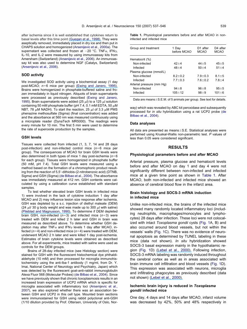
abaCsIAs(
S
WpBzw1cNpaaet
G
Tpgaf(miSwcs
1MG((btmpfuEac
satIwAwim2tw(
wpB
D
Apl
P
AbsmMa
Bi
UsiciavnmSgStbTan
Ig
O
Ti
G
H
P
A
D. Arsenijevic et al. / Neuroscience 150 (2007) 537–546 539
fter ischemia since it is well established that cytokines return toasal levels after this time point (Guegan et al., 1998). They wereseptically removed, immediately placed in dry ice and then put inHAPS solution and homogenized (Arsenijevic et al., 2000a). Theupernatant was collected and frozen at �20 °C. TNF�, IFN�,L-10, and IL-2 were measured by using immunoassay kits frommersham (Switzerland) (Arsenijevic et al., 2006). An immunoas-ay kit was also used to determine NGF (Catalys, Switzerland)Arsenijevic et al., 2006).
OD activity
e investigated SOD activity using a biochemical assay (1 dayost-MCAO; n�6 mice per group) (Ewing and Janero, 1995).rains were homogenized in phosphate-buffered saline and fro-en immediately in liquid nitrogen. Aliquots of brain supernatantsere processed as previously described (Ewing and Janero,995). Brain supernatants were added (25 �l) to a 125 �l solutionontaining 50 mM phosphate buffer (pH 7.4, 0.1 mM EDTA, 50 �MBT, 78 �M NADH). To start the reaction, 25 �l of 3.3 �M PMShenazine methosulfate (Sigma) (final concentration) was addednd the absorbance at 560 nm was measured continuously usingmicroplate reader (DynaTech MR5000). The readings were
very minute for 10 min. The first 5 min were used to determinehe rate of superoxide production by the samples.
SH levels
issues were collected from infected (1, 3, 7, 14 and 28 daysost-infection) and non-infected control mice (n�6 mice perroup). The consequences of MCAO for brain GSH levels werelso determined in both types of mice 1 day post-ischemia (n�6or each group). Tissues were homogenized in phosphate buffer50 mM, pH 7.4). Total GSH levels were measured using aethod based on the formation of a chromophoric product result-
ng from the reaction of 5,5=-dithiobis-(2-nitrobenzoic acid) (DTNB,igma) and GSH (Sigma) (de Bilbao et al., 2004). The absorbanceas immediately measured at 412 nm. GSH contents were cal-ulated by using a calibration curve established with standardamples.
To test whether elevated brain GSH levels in infected mice) were involved in the lack of cytokine induction 1 day afterCAO and 2) may influence lesion size response after ischemia,SH was depleted by a s.c. injection of diethyl maleate (DEM)
24 �l/ 30 g body weight and was made up to 300 �l with saline)Pileblad and Magnusson, 1990). To determine if DEM depletedrain GSH, non-infected (n�3) and infected mice (n�3) werereated with DEM and killed 2 h later and GSH in brain waseasured as described above. To determine whether GSH de-letion may alter TNF� and IFN� levels 1 day after MCAO, in-ected (n�4) and non-infected mice (n�4) were treated with DEM,nderwent MCAO 2 h later and were killed 1 day post-ischemia.stimates of brain cytokine levels were obtained as describedbove. For all experiments, mice treated with saline were used asontrols for the DEM groups.
Brains of 28-day infected mice (see Histology section) weretained for GSH with the fluorescent histochemical dye phthaldi-ldehyde (10 mM) and then processed for microglia immunohis-ochemistry using the anti-Iba-1 antibody (1 mg/ml, from Dr. Y.mai, National Center of Neurology and Psychiatry, Japan) whichas detected by the fluorescent goat-anti-rabbit immunoglobulinlexa Fluor 568 (Molecular Probes) (de Bilbao et al., 2004). Sincee have previously shown that chronic toxoplasmosis results in an
ncreased brain expression of UCP2 mRNA which is specific foricroglia associated with inflammatory loci (Arsenijevic et al.,007), we also explored whether there was an association be-ween GSH and UCP2 in this cell type. Mounted brain sectionsere immunostained for GSH using rabbit polyclonal anti-GSH
1/10 dilution provided by Prof. Ottersen, University of Oslo, Nor- w
ay) which was revealed by ABC kit peroxidase and subsequentlyrocessed for in situ hybridization using a rat UCP2 probe (deilbao et al., 2004).
ata analyses
ll data are presented as means�S.E. Statistical analyses wereerformed using Kruskal-Wallis non-parametric test. P values of
ess than 0.05 were considered significant.
RESULTS
hysiological parameters before and after MCAO
rterial pressure, plasma glucose and hematocrit levelsefore and after MCAO on day 1 and day 4 were notignificantly different between non-infected and infectedice at a given time point as shown in Table 1. AfterCAO, both non-infected and infected mice showed anbsence of cerebral blood flow in the infarct area.
rain histology and SOCS-3 mRNA inductionn infected mice
nlike non-infected mice, the brains of the infected micehowed many randomly located inflammatory loci (includ-
ng neutrophils, macrophages/monocytes and lympho-ytes) 28 days after infection. These loci were not colocal-
zed with intact Toxoplasma gondii cysts (Fig. 1A, B) andlso occurred around blood vessels, but not within theessels’ walls (Fig. 1C). There was no evidence of neuro-al apoptosis as determined by TUNEL labeling in theseice (data not shown). In situ hybridization showedOCS-3 basal expression mainly in the hypothalamic re-ion (Fig. 1D) (Lebel et al., 2000). Following infection,OCS-3 mRNA labeling was randomly induced throughout
he cerebral cortex as well as in areas associated withrain immune cell infiltration and blood vessels (Fig. 1D).his expression was associated with neurons, microgliand infiltrating phagocytes as previously described (dataot shown) (Lebel et al., 2000).
schemic brain injury is reduced in Toxoplasmaondii infected mice
ne day, 4 days and 14 days after MCAO, infarct volume
able 1. Physiological parameters before and after MCAO in non-nfected and infected mice
roup and treatment 1 Daybefore MCAO
D1 afterMCAO
D4 afterMCAO
ematocrit (%)Non-infected 42�4 44�5 45�5Infected 48�4 50�4 51�4
lasma glucose (mmol/L)Non-infected 8.2�0.2 7.9�0.3 8.1�5Infected 7.7�0.3 7.6�0.2 7.8�4
rterial pressure (mm Hg)Non-infected 94�8 96�8 95�5Infected 105�12 98�9 101�6
Data are means�S.E.M. of 5 animals per group. See text for details.
as decreased by 42%, 50% and 48% respectively in

Tnip
Tg
Odcfnap
triealtga
Bd
T
Fcrtmiifvgc esyl Violeb (Tox) at
D. Arsenijevic et al. / Neuroscience 150 (2007) 537–546540
oxoplasma gondii infected mice when compared withon-infected control mice (P�0.01) (Fig. 1E, F), suggest-
ng that chronic infection with Toxoplasma gondii mayrotect neocortical areas from ischemic damage.
ransient hyperphagia occurs early in Toxoplasmaondii–infected mice in response to MCAO
n the day of lesion, MCAO resulted in a significant re-uction of food intake in both infected and non-infectedontrol mice when compared with their pre-MCAO basalood intakes (�26�0.5% and �39�0.7% respectively,�18, P�0.01). The day after ischemia, both non-infectednd infected mice started to regain appetite. From this time
ig. 1. Brain histology, in situ hybridization for SOCS-3 mRNA expresross-sections of a Toxoplasma gondii–infected (for 28 days) mouse besulted in inflammatory loci found in brain tissue (A) (thin black arrow)he presence of Toxoplasma cysts was not associated with inflammaRNA in non-infected control (Cont) and infected mice (Tox) brains (
n hypothalamic region. Twenty-eight days following infection, SOCS-3nfected mice in particular in the cortical area (Tox) (n�4 for each groor SOCS-3 mRNA expression in infected brains. SOCS-3 mRNA wessels (thin arrow). Cells are visualized with Thionin staining and SOondii infection on infarct size. (E) Representative coronal sections shronically infected mice (Tox MCAO). Sections were stained with Crars�120 �m). (F) Infarct brain volumes were reduced in infected mice
oint, infected mice increased food intake more rapidly p
han the non-infected group (�3�0.2% and �28�2%,espectively, n�18, P�0.01). Infected mice showed signif-cantly enhanced hyperphagia on days 2 and 3 postisch-mia compared with their non-operated controls (28�1.2%nd 1�0.2% respectively, n�18, P�0.01). Body weight
oss induced by ischemia followed a similar temporal pat-ern to that of food intake indicating that Toxoplasmaondii–infected mice had attenuated negative energy bal-nce in response to MCAO.
rain specific alterations in GSH levelsue to infection
here was a 45% decrease in brain GSH levels on day 7
brain infarct size in Toxoplasma gondii infected mice. (A–C) Coronaled with hematoxylin and eosin (scale bar�15 �m). Note that infectionssociation with blood vessels (B, C) (thin black arrows). Note also that(B, thick arrow). (D) Representative in situ hybridization for SOCS-3�120 �m). SOCS-3 mRNA basal expression was found in particularxpression was randomly located and induced throughout the brain ofright column shows a higher magnification of selected areas specificiated with microglia/infiltrated immune cells (thick arrow) and blooder grains are black (scale bars�15 �m). (E, F) Effect of Toxoplasmachemic lesion size 4 days after MCAO in control (Cont MCAO) andt. The surrounded areas denote the size of the ischemic area (scale
1, 4 and 14 days after MCAO compared with non-infected mice (Cont).
sion andrain stainand in atory lociscale barmRNA e
up). Theas assocCS-3 silvhowing is
ostinfection when compared with non-infected control

malibce2a0(i0G
MfuG0ilGin(iw
FrwE(av
D. Arsenijevic et al. / Neuroscience 150 (2007) 537–546 541
ice (P�0.05). On day 14, as mice started to regainppetite, there was a progressive increase in brain GSH
evels in infected mice (P�0.001) up to 28 days afternfection (P�0.001) (Fig. 2A). The increase in GSH in therains of 28-day infected mice was associated with a spe-ific increase in microglia (Fig. 2B, C); only microglia withlevated UCP2 mRNA levels showed GSH labeling (Fig.D, E). In non-infected control mice, MCAO resulted in
80% decrease of brain GSH levels (from 0.52�.02 nmol/mg tissue to 2.60�0.18 nmol/mg tissue)P�0.001) (Fig. 3A). Interestingly, this decrease due toschemia was only 11% in infected animals (from 4.60�.80–4.11�0.31 nmol/mg tissue) (P�0.01). In addition,
Brai
0
2
4
6
Cont
Day 1
Day
nm
ol/
mg
tis
su
e
2B
2D
2A
ig. 2. Changes in brain GSH levels from day 1 to day 28 followingeduction in brain GSH levels on day 7. From day 14 to day 28 after infith non-infected control mice (Cont). Values (nmol/mg tissue) are preach group represents six mice. (B) GSH (fluorescent staining, white
C). Only microglia cells that have increased UCP2 mRNA (E, black arrrow) (D). Scale bars�15 �m (B, C, D); 7.5 �m (E). For interpretationersion of this article.
SH brain content in infected animals having undergone 1
CAO was significantly higher compared with non-in-ected MCAO animals (P�0.001) (Fig. 3A). Chronicallynderfed non-infected mice did not show altered brainSH levels compared with non-infected controls (2.47�.19 versus 2.60�0.07 nmol/mg tissue) (Fig. 3A) suggest-
ng that anorexia does not explain the increased brain GSHevels in infected mice. Although 1 day after ischemia,
SH levels were reduced in the chronically underfed non-nfected mice, this decrease was significantly less pro-ounced compared with the non-infected control groupP�0.01) (Fig. 3A). This was associated with a reducednfarct size in the chronically underfed group comparedith the non-infected control group (15.0�0.9 mm3 and
H
Day14 7
Day28
*
***
***
C
E
Toxoplasma gondii infection. (A) Toxoplasmosis resulted in a slightSH was significantly increased in the brain of infected mice compareds mean�S.E. (* P�0.05, *** P�0.001, comparison to saline control).specifically increased in microglia in infected mice (red, white arrow)
w an increase in GSH (immunohistochemical detection, orange, blackerences to color in this figure legend, the reader is referred to the Web
n GS
4
Day
2
2
murineection, Gsented aarrow) isrow) shoof the ref
9.3�1.1 mm3, respectively, n�6 mice, P�0.05). Al-

tuto(
I
Sfn(Mcu
Um
TG(Mrm
Il
BIotmwtosm1II(c(mblnIi
Fslom(lltawwcfMipscUaTM*
TI
TIIIN
*(a
M
D. Arsenijevic et al. / Neuroscience 150 (2007) 537–546542
hough a contributing effect cannot be excluded, thenderfeeding effect on reduced lesion size could nototally explain the much greater reduction of infarct sizebserved in the infected mice (10.2�0.4 mm3) (Fig. 1F)
ig. 3. Brain GSH, brain SOD activity and brain UCP2 mRNA expres-ion. (A) One day after MCAO, we observed a reduction in brain GSHevels in non-infected mice (Cont MCAO) compared with their non-perated controls (Cont). After MCAO, brain levels of GSH in infectedice were markedly elevated compared with non-infected animals
Cont MCAO). Underfeeding (Ufc) results in no change in brain GSHevels compared with control mice (Cont). One day after MCAO, GSHevel in underfed mice (Ufc MCAO) was reduced, however this reduc-ion was less than that found in Cont MCAO mice. (B) Brain SODctivity was increased during chronic infection (Tox) when comparedith non-infected animals (Cont). One day post-MCAO, SOD activitiesere similarly increased in both groups compared with non-infectedontrols (Cont). (C) Brain UCP2 mRNA expression was increasedollowing infection (Tox) compared with non-infected controls (Cont).CAO increased brain UCP2 mRNA levels in both infected and non-
nfected mice, but levels were higher in the former group. Values areresented as mean�S.E. for the various groups. Each group repre-ents six mice. * Indicates the statistical comparison with non-infectedontrols (Cont), a indicates the statistical comparison between Tox andfc mice, b indicates the statistical comparison between Tox MCAOnd Cont MCAO mice, c indicates the statistical comparison betweenox and Tox MCAO mice, & indicates comparison between ContCAO and Ufc MCAO (* P�0.05, *** P�0.001, ** P�0.01,
** P�0.001, a P�0.001, b P�0.001, c P�0.05, & P�0.01).
P�0.05). d
nfection increases brain SOD activity
OD activity (Fig. 3B) was higher in the brains of in-ected mice (14.2�1.2 units/mg protein) compared withon-infected mice (10.2�0.8 units/mg protein)P�0.01). This difference did not persist 1 day afterCAO (Fig. 3B) as SOD activities were similarly in-
reased in both groups (17.2�1.5 and 15.1�1.1nits/mg protein respectively).
pregulation of brain UCP2 mRNA levels in infectedice after ischemia
oxoplasmosis resulted in a 76% increase in brain UCP2/APDH mRNA ratio compared with non-infected mice
2.3�0.2 versus 1.3�0.1) (P�0.01) (Fig. 3C). One day post-CAO, there was a 62% increase in UCP2/GADPH mRNA
atio in infected mice compared with MCAO non-infectedice (3.4�0.2 versus 2.1�0.2) (P�0.001) (Fig. 3C).
nfection does not result in enhanced cytokineevels after MCAO
asal levels of proinflammatory cytokines (TNF�, IL-2,FN�) were increased in Toxoplasma gondii infected micen day 28 postinfection compared with non-infected con-rols (P�0.001) (Table 2). One day post-MCAO, infectedice had higher TNF�, IL-2 and IFN� levels comparedith MCAO non-infected animals (P�0.001). However,
hese higher levels did not significantly differ from thosebserved in infected mice not subjected to ischemia. Thisharply contrasts with the results obtained in non-infectedice which showed a marked induction of cytokine levelsday post-MCAO compared with non-operated animals.
nfected mice also had significantly higher brain levels ofL-10 and NGF compared with non-infected control miceP�0.001) (Table 2). NGF and IL-10 significantly in-reased in non-infected mice that underwent MCAOP�0.001); this induction was not observed in infectedice suggesting an attenuated immune response in therains of infected mice (Table 2). Following MCAO, NGF
evels in infected animals were doubled compared withon-infected controls (P�0.001). This was not the case forL-10 suggesting that NGF may be a more important anti-nflammatory agent in infected mice.
able 2. Basal and one day post-MCAO levels of brain TNF�, IFN�,L-2, IL-10 and NGF in non-infected and infected mice
Cont Cont MCAO Tox Tox MCAO
NF� pg/ml 100�4 800�12*** 993�51*** 1093�50a
FN� pg/ml 25�2 175�4*** 330�8*** 358�9a
L-2 pg/ml 44�2 750�23*** 1622�64*** 1804�61a
L-10 pg/ml 24�2 492�38*** 504�40*** 560�19GF pg/ml 40�3 300�12*** 600�22*** 624�20a
Values (pg/ml tissue) are presented as mean�S.E.Indicates statistical comparison with non-infected animals (Cont)*** P�0.001). Each group included six mice. See text for details.Indicates statistical comparison between Tox MCAO and ContCAO mice (a P � 0.001). Each group included six mice. See text for
etails.
tM
mDbcwmDgc1psbteti
Tgiabarimsmn
tqsoi
Fss((t ntrols;
D. Arsenijevic et al. / Neuroscience 150 (2007) 537–546 543
Depletion of brain GSH by DEM reversed neuroprotec-ion by increasing lesion size and central IFN� levels afterCAO in infected mice.
Brain GSH levels in both non-infected and infectedice were markedly decreased 2 h after an s.c. injection ofEM as seen in Fig. 4A. One day after ischemia (Fig. 4B),rain GSH levels were further reduced by DEM whenompared with non-treated MCAO groups. This reductionas associated with an increased lesion size in infectedice when compared with infected mice not treated withEM (22.2�1.7 mm3 versus 12.8�0.5 mm3, n�6 perroup, P�0.01). Non-infected mice also showed an in-rease in lesion size when treated with DEM (15.8�.5 mm3 for saline versus 20.3�1.0 mm3 for DEM, n�6er group, P�0.05). After ischemia, DEM treatment re-ulted in induction of central IFN� levels but not TNF� inoth types of mice (Fig. 4C, D). Note that, consistent with
he known transitory effect of DEM on GSH levels (Guptat al., 2000), GSH returned to basal levels 24 h after DEMreatment in the absence of ischemic damage in both
ig. 4. GSH, TNF� and IFN� were measured in non-infected and infealine). (A) DEM significantly reduced brain GSH in non-infected andignificantly reduced brain GSH levels in infected and non-infected micGupta et al., 2000), GSH returned to basal levels 24 h after DEM treatmC, D) DEM did not significantly alter brain TNF� (C) but significantlyhree to five animals per group. * P�0.001, comparison with saline co
nfected and non-infected mice (Fig. 4C, D). t
DISCUSSION
his study revealed a marked resistance of Toxoplasmaondii–infected mice to acute cerebral ischemia character-
zed by a marked decrease of infarct size one, and 4 butlso 14 days post-MCAO. The reverse effect of DEM onrain infarct size implies that GSH up-regulation may playpivotal role in the observed ischemic resistance. Our
esults make it also possible to propose additional biolog-cal mechanisms surrounding this phenomenon, such as
arked differences in pre- and/or post-ischemic cytokinetatus as well as pre- and/or post-ischemic GSH, SOCS-3RNA and UCP2 mRNA levels in infected compared withon-infected mice.
Following the acute phase of toxoplasmosis, the ini-ially produced pro-inflammatory cytokines will subse-uently induce a counter-regulatory anti-inflammatory re-ponse (Arsenijevic et al., 1997). During the chronic phasef infection, apart from the role that the adaptive specific
mmune systems (CD4, CD8 T cell activation) could play,
e after ischemia; 2 h prior to MCAO, mice were treated with DEM (ormice 2 h after treatment. (B) Twenty-four hours after ischemia, DEMhat, consistent with the known transitory effect of DEM on GSH levelse absence of ischemic damage in both infected and non-infected mice.
brain IFN� levels (D) 24 h after ischemia. Data are means�S.E. ofP�0.001, comparison with MCAO controls.
cted micinfectede. Note tent in th
increaseda
he consequence of a second inflammation caused by

ctuUNatsttiOGbgApriflgcpcfBw1UasalUia
ctTvmctfpatmstptt
gmri
MipSta2hT2erioDcIho(naivcc
mmomwttam
ibmsacos2brsbp2atidd(
D. Arsenijevic et al. / Neuroscience 150 (2007) 537–546544
erebral ischemia may be determined by the balance be-ween these two antagonistic systems. The basal up-reg-lation of antioxidant molecules such as GSH, SOD,CP2, SOCS-3 mRNA and anti-inflammatory cytokinesGF and IL-10 may represent the two first lines of defensegainst ischemic damage in infected mice. One could pos-ulate that the observed changes in redox status may be aimple epiphenomenon of the underfeeding observed inhe chronically infected mice. In fact, it has been shownhat underfeeding may result in protection from cerebralnjury by modifying redox status (Yu and Mattson, 1999).ur findings show that both the steady increase of brainSH levels by day 7 to day 28 post-infection as well as therain resistance to ischemia are specific to Toxoplasmaondii infection and did not depend on energy intake.ctually, although we did find that post-ischemic GSH wasartially dependent on energy intake, this component rep-esented only 13% of the post-ischemic GSH levels ofnfected mice. We also show that energy intake does in-uence lesion size, but alone cannot account for the de-ree of resistance observed in infected animals. A possibleausal relationship between redox status and cytokineroduction has been previously suggested in that in-reased basal GSH levels and SOD activity may resultrom high NGF levels (Guegan et al., 1998, 1999; deilbao et al., 2004; Arsenijevic et al., 2006). Consistentith their possible role in neuroprotection (Guegan et al.,999; Mattiasson et al., 2003), the differences in GSH,CP2 mRNA and NGF levels persisted between infectednd non-infected mice after MCAO. In the absence of apecific antibody for brain UCP2, it was not possible tossess the impact of UCP2 mRNA changes on protein
evels. However, previous data using UCP2 transgenic andCP2 KO mice have shown that UCP2 plays a central role
n brain neuroprotection following ischemia (Mattiasson etl., 2003; de Bilbao et al., 2004).
An additional molecular mechanism involved in thisontext may involve the absence of pro-inflammatory cy-okine induction observed in infected mice after ischemia.his phenomenon could be partly due to the already ele-ated basal levels of these cytokines in the brains of theseice (i.e. counter-regulatory mechanisms). Although one
ould argue that this finding might reflect the presence of ahreshold in cytokine levels reached in Toxoplasma-in-ected mice, this is an unlikely scenario since we havereviously demonstrated that an i.p. injection of LPS led tomarked upregulation of pro-inflammatory cytokines in
hese mice (Arsenijevic et al., 1998). Importantly, infectedice showed feeding behavior concordant with the relative
tunting of brain cytokine response after ischemia. In fact,ransient hyperphagia started earlier in infected mice com-ared with non-infected mice after MCAO. This is consis-ent with the attenuated post-ischemic induction of anorec-ic cytokines (TNF�, IFN�) in the brain.
In agreement with previous contributions, our data sug-est that both SOCS-3 and GSH are plausible candidateolecules for the regulation of secondary inflammatory
esponse and cytokine production in Toxoplasma gondii–
nfected mice (Bjorkbaek et al., 1999; Auerhammer and selmed, 2001; Park et al., 2003). The absence of pro-nflammatory cytokine induction could be partly due to thearallel elevation of the SOCS-3 mRNA after infection. TheOCS-3 is a potent inhibitor of cytokine signaling and its
ransgenic over-expression may inhibit inflammation andssociated apoptosis in vivo (Auerhammer and Melmed,001; Jo et al., 2005). Accordingly, although infected micead elevated basal pro-inflammatory cytokine levels (i.e.NF�, IFN� and IL-2) (Eizenberg et al., 1995; Bate et al.,006; Lee et al., 2006; Yu et al., 2006), there was novidence of neuronal apoptosis in their brain tissue. A keyole for GSH is suggested by the fact that the decreasednfarct size in MCAO mice chronically infected is no longerbserved when brain GSH levels have been depleted byEM treatment. This phenomenon was specifically asso-iated with the central up-regulation of pro-inflammatoryFN� but not TNF� in response to MCAO. Previous studiesave also shown that DEM may not have a marked effectn TNF induction in response to an inflammatory responseKang et al., 1999; Wang et al., 1999). Further work iseeded to get a better understanding of the complex inter-ctions of molecules regulating the inflammatory response
n this context. As recently proposed, the observed ele-ated basal SOCS-3 and GSH levels could act synergisti-ally as GSH can interact in SOCS-3 pathways to inhibitytokines (Cisowski et al., 2002).
Besides its direct effect against oxidative stress, GSHay also act via the suppression of glia-mediated inflam-ation (Wang et al., 2006). In the same line with this idea,ur contributions have shown that both UCP2 and SOCS-3RNA levels in brains infected with Toxoplasma gondiiere elevated in microglia (Arsenijevic et al., 2007a). Al-
hough we cannot exclude a possible participation of infil-rated leukocytes, our evidence suggests that GSH, UCP2nd SOCS-3 may attenuate ischemic injury in infectedice by modifying microglia activity.
Despite a widely disseminated idea, only rare stud-es have shown that pre-existing infections can enhancerain pathology in response to a subsequent new inflam-atory process (Arsenijevic et al., 1998). In contrast,
everal studies have shown that chronic infection canttenuate some peripheral and central pathological pro-esses: complete Freund’s adjuvant prevents the onsetf experimental allergic encephalomyelitis (Bach, 2001),chistosomiasis can protect against asthma (Yang et al.,007), coxsackievirus B3 can inhibit cardiomyopathy (Hu-er et al., 2006) and various infections can attenuate mu-ine models of multiple sclerosis (Sewell et al., 2002). Inome conditions, a pre-existing inflammation can inducerain neurodegeneration. This is the case for the acuteeripheral and/or central injection of LPS (Nguyen et al.,004; Cunningham et al., 2005; McColl et al., 2007; Qin etl., 2007; Spencer et al., 2007). However, it appears thathe LPS dose used in these models may be a determinantn the observed degeneration. In fact, although high LPSoses may increase cytokines and oxidative stress, lowoses of peripheral LPS have a neuroprotective effectBordet et al., 2000). This is consistent with our recent
tudy in which we observed an increase in brain NGF and
Gsceseattdm
Tgc1petplnpo
A
A
A
A
A
A
A
A
A
B
B
B
B
B
B
C
C
C
d
d
D
E
E
E
F
F
G
D. Arsenijevic et al. / Neuroscience 150 (2007) 537–546 545
SH levels 3 days after peripheral LPS treatment (Ar-enijevic et al., 2007b; Hernadfalvi et al., 2007). In thisontext, the present study provides a new model toxplore the effect of a pre-existing chronic inflammatorytatus on the outcome of brain ischemic insult. Sinceach bacterium or parasite produces different cytokinend leukocyte activation profiles and, ultimately, pat-erns of neurodegeneration, it is crucial to investigatehe molecular mechanisms surrounding the beneficial oreleterious effect of various models of chronic inflam-ation on cerebral ischemia.
CONCLUSION
his study revealed a marked resistance of Toxoplasmaondii–infected mice to acute cerebral ischemia that isharacterized by a decreased lesion size 1 day, 4 days and4 days post-MCAO. Marked differences in pre- and/orost-ischemic cytokines and antioxidant molecules couldxplain this phenomenon. In addition, our findings implyhat GSH may play a pivotal role in these processes sinceharmacological depletion of GSH resulted in increased
esion size and increased brain IFN� levels. Functionaleurological data could be useful to confirm further therotective effect of Toxoplasma gondii infection on theutcome of cerebral ischemia.
REFERENCES
rsenijevic D, Clavel S, Sanchis D, Plamondon J, Huang Q, RicquierD, Rouger L, Richard D (2007a) Induction of Ucp2 expression inbrain phagocytes and neurons following murine toxoplasmosis: Anessential role of IFN-� and an association with negative energybalance. J Neuroimmunol 186:121–132.
rsenijevic D, Hernadfalvi N, von Meyenburg C, Onteniente B, RichardD, Langhans W (2007b) Role of nerve growth factor in the in vivoregulation of glutathione in response to LPS in mice. Eur CytokineNetw 18:93–101.
rsenijevic D, de Bilbao F, Giannakopoulos P, Girardier L, Samec S,Richard D (2001) A role for interferon-� in the hypermetabolicresponse to murine toxoplasmosis. Eur Cytokine Netw 12:518 –527.
rsenijevic D, de Bilbao F, Plamondon J, Paradis E, Vallet P, RichardD, Langhans W, Giannakopoulos P (2006) Increased infarct sizeand lack of hyperphagic response after focal cerebral ischemia inperoxisome proliferator-activated receptor �-deficient mice.J Cereb Blood Flow Metab 26:433–445.
rsenijevic D, Garcia I, Vesin C, Vesin D, Arsenijevic Y, Seydoux J,Girardier L, Ryffel B, Dullo AG, Richard D (2000a) Differential rolesof tumor necrosis factor-� and interferon-� in mouse hypermeta-bolic and anorectic responses induced by LPS. Eur Cytokine Netw11:662–668.
rsenijevic D, Onuma H, Pecqueur C, Raimbault C, Manning BS,Miroux B, Couplan E, Alves-Guerra MC, Goubern M, Surwitt R,Bouillaud F, Richard D, Collins S, Ricquier D (2000b) Disruption ofthe uncoupling protein-2 gene in mice reveals a role in immunityand reactive oxygen species production. Nat Genet 26:435–439.
rsenijevic D, Girardier L, Seydoux J, Chang HR, Dulloo AG (1997)Altered energy balance and cytokine gene expression in a murinemodel of chronic infection with Toxoplasma gondii. Am J Physiol272:E908–E917.
rsenijevic D, Girardier L, Seydoux J, Pechere JC, Garcia I, Lucas R,Chang HR, Dulloo AG (1998) Metabolic cytokine responses to asecondary immunological challenge with LPS in mice with T. gondii
infection. Am J Physiol 274:E439–E445.uerhammer CJ, Melmed S (2001) The central role of SOCS-3 inintegrating the neuro-immunoendocrine interface. J Clin Invest108:1735–1740.
ach JF (2001) Protective role of infections and vaccinations onautoimmune diseases. J Autoimmun 16:347–353.
ate C, Kempster S, Last V, Williams A (2006) Interferon-gammaincreases neuronal death in response to amyloid-beta1–42. J Neu-roinflammation 3:1–7.
ates S, Read SJ, Harrison DC, Topp S, Morrow R, Gale D, MurdockP, Barone FC, Parsons AA, Gloger IS (2001) Characterisation ofgene expression changes following permanent MCAO in the ratusing subtractive hybridisation. Brain Res Mol Brain Res 93:70–80.
jorkbaek C, Elmquist JK, El-Haschimi K, Kelly J, Ahima RS, HilemanS, Flier JS (1999) Activation of SOCS-3 messenger ribonucleicacid in the hypothalamus by ciliary neurotrophic factor. Endocri-nology 140:2035–2043.
ordet R, Deplanque D, Maboudou P, Puisieux F, Pu Q, Martin A,Bastide D, Leys D, Lhermitte M, Dupuis B (2000) Increase inendogenous brain superoxide dismutase as a potential mecha-nism of lipopolysaccharide induced brain ischemic tolerance.J Cereb Blood Flow Metab 20:1190–1196.
rodie C (1996) Differential effects of Th1 and Th2 derived cytokineson NGF synthesis by mouse astrocytes. FEBS Lett 394:117–120.
hen H, Luo J, Kintner DB, Shull GE, Sun D (2005) Na�-dependentchloride transporter (NKCC1)-null mice exhibit less gray and whitematter damage after focal cerebral ischemia. J Cereb Blood FlowMetab 25:54–66.
isowski J, Zarebski A, Koj A (2002) IL-1 mediated inhibition of IL-6-induced STAT3 activation is modulated by IL-4, MAP kinase inhib-itors and redox state of HepG2 cells. Folia Histochem Cytobiol40:341–345.
unningham C, Wilcockson DC, Campion S, Lunnon K, Perry VH(2005) Central and systemic endotoxin challenges exacerbate thelocal inflammatory response and increase neuronal death duringchronic neurodegeneration. J Neurosci 25:9275–9284.
e Bilbao F, Arsenijevic D, Vallet P, Hjelle OP, Ottersen OP, Bouras C,Raffin Y, Abou K, Langhans W, Collins S, Plamondon J, Alves-Guerra MC, Haguenauer A, Garcia I, Richard D, Ricquier D, Gi-annakopoulos P (2004) Resistance to cerebral ischemic injury inUCP2 knockout mice: evidence for a role of UCP2 as a regulator ofmitochondrial glutathione levels. J Neurochem 89:1283–1292.
e Bilbao F, Guarin E, Nef S, Vallet P, Giannakopoulos P, Dubois-Dauphin M (2000) Cell death is prevented in thalamic fields by butnot in injured neocortical areas after permanent focal ischemia inmice overexpressing the anti-apoptotic protein Bcl-2. Eur J Neu-rosci 12:921–934.
roge W (2002) Free radicals in physiological control of cell function.Physiol Rev 82:47–95.
izenberg O, Faber-Elman A, Gottlieb E, Oren M, Rotter V, SchwarzM (1995) Direct involvement of p53 in programmed cell death ofoligodentrocytes. EMBO J 14:1136–1144.
msley HC, Tyrrell PJ (2002) Inflammation and infection in clinicalstroke. J Cereb Blood Flow Metab 22:1399–1419.
wing JF, Janero DR (1995) Microplate superoxide dismutase assayemploying a nonenzymatic superoxide generator. Anal Biochem232:243–248.
ranklin KBJ, Paxinos G (1997) The mouse brain in stereotaxic coor-dinates, pp 29–41. San Diego: Academic Press Inc.
renkel JK, Escajadillo A (1987) Cyst rupture as a pathogenic mech-anism of toxoplasmic encephalitis. Am J Trop Med Hyg 36:517–522.
uegan C, Ceballos-Picot I, Chevallier E, Nicole A, Onteniente B, SolaB (1999) Reduction of ischemic damage in NGF-transgenic mice:Correlation with enhancement of antioxidant enzyme activities.
Neurobiol Dis 6:180–189.
G
G
H
H
H
J
K
L
L
L
M
M
M
N
N
P
P
Q
S
S
S
S
V
W
W
W
Y
Y
Y
D. Arsenijevic et al. / Neuroscience 150 (2007) 537–546546
uegan C, Ceballos-Picot I, Nicole A, Kato H, Onteniente B, Sola B(1998) Recruitment of several neuroprotective pathways after per-manent focal ischemia in mice. Exp Neurol 154:371–380.
upta A, Gupta M, Datta M, Shkla GS (2000) Cerebral antioxidantstatus and free radical generation following glutathione depletionand subsequent recovery. Mol Cell Biochem 209:55–61.
ernadfalvi N, Langhans W, von Meyenburg C, Onteniente B, RichardD, Arsenijevic D (2007) Role of glutathione in the hyposensitivity ofLPS-pretreated mice to LPS anorexia. Eur Cytokine Netw18:86–92.
uang KC, Chen CW, Chen JC, Lin WW (2003) Statins induce sup-pressor of cytokine signaling-3 in macrophages. FEBS Lett555:385–389.
uber SA, Feidman AM, Sartini D (2006) Coxsackievirus B3 inducesT regulatory cells, which inhibit cardiomyopathy in tumor necrosisfactor-alpha transgenic mice. Cir Res 99:1109–1116.
o D, Liu D, Yao S, Collins RD, Hawiger J (2005) Intracellular proteintherapy with SOCS3 inhibits inflammation and apoptosis. Nat Med11:892–898.
ang KE, Pak YMK, Kim ND (1999) Diethylmaleate and buthioninesulfoximine, glutathione-depleting agents, differentially inhibit ex-pression of inducible nitric oxide synthase in endotoxemic mice.Nitric Oxide Biol Chem 3:265–271.
arsen L, Ropke C (2002) Suppressors of cytokine signalling. APMIS110:833–844.
ebel E, Vallieres L, Rivest S (2000) Selective involvement of inter-leukin-6 in the transcriptional activation of the suppressor of cyto-kine signaling-3 in the brain during systemic immune challenges.Endocrinology 141:3749–3763.
ee J, Shin JS, Choi IH (2006) Human brain astrocytes mediateTRAIL-mediated apoptosis after treatment with IFN-g. Yonsei MedJ 47:354–358.
attiasson G, Shamloo M, Gido G, Gido G, Mathi K, Tomasevic G, YiS, Warden CH, Castilho RF, Melcher T, Gonzalez-Zulueta K, Ni-kolich K, Wieloch T (2003) Uncoupling protein-2 prevents neuronaldeath and diminishes brain dysfunction after stroke and braintrauma. Nat Med 9:1062–1068.
cColl BW, Rothwell NJ, Allan SM (2007) Systemic inflammatorystimulus potentiates the acute phase and CXC chemokine re-sponses to experimental stroke and exacerbates brain damage viainterleukin-1 and neutrophil-dependent mechanisms. J Neurosci27:4403–4412.
urakami K, Kondo T, Kawase M, Li Y, Sato S, Chen SF, Chan PH(1998) Mitochondrial susceptibility to oxidative stress exacerbatescerebral infarction that follows permanent focal cerebral ischemiain mutant mice with manganese superoxide dismutase deficiency.J Neurosci 18:205–213.
guyen MD, D’Aigle T, Gowing G, Julien JP, Rivest S (2004) Exac-
erbation of motor neuron disease by chronic stimulation of innateimmunity in a mouse model of amyotrophic lateral sclerosis.J Neurosci 24:1340–1349.
icholls DG, Budd SL (2000) Mitochondria and neural survival. PhysiolRev 80:315–360.
ark SH, Kim KE, Hwang HY, Kim TY (2003) Regulatory effects ofSOCS on NF-KB activity in murine monocytes/macrophages. DNACell Biol 22:131–139.
ileblad E, Magnusson T (1990) Effective depletion of glutathione inrat striatum and substantia nigra by L-buthionine sulfoximine incombination with 2-cyclohexene-1-one. Life Sci 47:2333–2342.
in L, Wu X, Block ML, Liu Y, Breese GR, Hong JS, Knapp DJ, CrewsFT (2007) Systemic LPS causes chronic neuroinflammation andprogressive neurodegeneration. Glia 55:453–462.
acco RL (2001) New risk factors for stroke. Neurology 57(5 Suppl 2):S31–S34.
chulz JB, Lindenau J, Seyfried J, Dichgans J (2000) Glutathione,oxidative stress and neurodegeneration. Eur J Biochem 267:4904–4911.
ewell DL, Reinke EK, Hogan LH, Sandor M, Farby Z (2002) Immu-noregulation of CNS autoimmunity by helminth and mycobacterialinfections. Immunol Lett 82:101–110.
pencer SJ, Mouihate A, Pittman QJ (2007) Peripheral inflammationexacerbates damage after global ischemia independently of tem-perature and acute brain inflammation. Stroke 38:1570–1577.
illoslada P, Hauser SL, Bartke I, Unger J, Heald N, Rosenberg D,Cheung SW, Mobley WC, Fisher S, Genain CP (2000) Humannerve growth factor protects common marmosets against autoim-mune encephalomyelitis by switching the balance of T helper celltype 1 and 2 cytokines within the central nervous system. J ExpMed 191:1799–1806.
ang JY, Wen LL, Huang YN, Chen YT, Ku MC (2006) Dual effects ofantioxidants in neurodegeneration: direct neuroprotection againstoxidative stress and indirect protection via suppression of glia-mediated inflammation. Curr Pharm Des 12:3521–3533.
ang J, Campbell IL (2002) Cytokine signalling in the brain: Putting aSOCS in it? J Neurosci Res 67:423–427.
ang F, Wang LY, Wright D, Parmely MJ (1999) Redox imbalancedifferentially inhibits lipopolysaccharide-induced macrophage acti-vation in the liver. Infect Immun 67:5409–5416.
ang J, Zhao J, Zhang L, Yang X, Zhu X, Ji M, Sun N, Su C (2007)Schistosoma japonicum egg antigens stimulate CD4 CD25 T cellsand modulate airway inflammation in a murine model of asthma.Immunology 120:8–18.
u L, Miao H, Hou Y, Zhang B, Guo L (2006) Neuroprotective effect ofA20 on TNF-induced postischemic apoptosis. Neurochem Res31:21–32.
u ZF, Mattson MP (1999) Dietary restriction and 2-deoxyglucoseadministration reduce focal ischemic brain damage and improvebehavioural outcome: evidence for a preconditioning mechanism.
J Neurosci Res 57:830–839.(Accepted 3 October 2007)(Available online 11 October 2007)
Related Documents