1 Decreased chondrocyte proliferation and dysregulated apoptosis in the cartilage growth plate are key features of a murine model of epiphyseal dysplasia caused by a matn3 mutation Matthew P Leighton 1 , Seema Nundlall 1 , Tobias Starborg 1 , Roger S Meadows 1 , Farhana Suleman 1 , Lynette Knowles 1 , Raimund Wagener 2 , David J Thornton 1 , Karl E Kadler 1 , Raymond P Boot-Handford 1 and Michael D Briggs 1* . 1 Wellcome Trust Centre for Cell-Matrix Research, Faculty of Life Sciences, University of Manchester, Michael Smith Building, Oxford Road, Manchester, M13 9PT, UK. 2 Center for Biochemistry, University of Cologne, Germany. © 2007 The Author(s) This is an Open Access article distributed under the terms of the Creative Commons Attribution Non-Commercial License (http://creativecommons.org/licenses/by-nc/2.0/uk/) which permits unrestricted non-commercial use, distribution, and reproduction in any medium, provided the original work is properly cited. HMG Advance Access published May 21, 2007 by guest on November 17, 2015 http://hmg.oxfordjournals.org/ Downloaded from

Welcome message from author
This document is posted to help you gain knowledge. Please leave a comment to let me know what you think about it! Share it to your friends and learn new things together.
Transcript

1
Decreased chondrocyte proliferation and dysregulated apoptosis in the cartilage growth plate
are key features of a murine model of epiphyseal dysplasia caused by a matn3 mutation
Matthew P Leighton1, Seema Nundlall1, Tobias Starborg1, Roger S Meadows1, Farhana
Suleman1, Lynette Knowles1, Raimund Wagener2, David J Thornton1, Karl E Kadler1,
Raymond P Boot-Handford1 and Michael D Briggs1*.
1Wellcome Trust Centre for Cell-Matrix Research, Faculty of Life Sciences, University of
Manchester, Michael Smith Building, Oxford Road, Manchester, M13 9PT, UK.
2Center for Biochemistry, University of Cologne, Germany.
© 2007 The Author(s) This is an Open Access article distributed under the terms of the Creative Commons Attribution Non-Commercial License (http://creativecommons.org/licenses/by-nc/2.0/uk/) which permits unrestricted non-commercial use, distribution, and reproduction in any medium, provided the original work is properly cited.
HMG Advance Access published May 21, 2007 by guest on N
ovember 17, 2015
http://hmg.oxfordjournals.org/
Dow
nloaded from
2
*Correspondence should be addressed to:-
Dr Mike Briggs
Wellcome Trust Centre for Cell Matrix Research
Faculty of Life Sciences
University of Manchester
Michael Smith Building
Oxford Road
Manchester
M13 9PT, UK
+44 161 275 5082 (Tel)
+44 161 275 5642 (Fax)
by guest on Novem
ber 17, 2015http://hm
g.oxfordjournals.org/D
ownloaded from

3
Abstract
Disruption to endochondral ossification leads to delayed and irregular bone formation
and can result in a heterogeneous group of genetic disorders known as the chondrodysplasias.
One such disorder, multiple epiphyseal dysplasia (MED), is characterized by mild dwarfism
and early-onset osteoarthritis and can result from mutations in the gene encoding matrilin-3
(MATN3).
To determine the disease mechanisms that underpin the pathophysiology of MED we
generated a murine model of epiphyseal dysplasia by knocking-in a matn3 mutation. Mice
that are homozygous for the mutation develop a progressive dysplasia and have short-limbed
dwarfism that is consistent in severity with the relevant human phenotype. Mutant matrilin-3
is retained within the rough endoplasmic reticulum of chondrocytes and is associated with an
unfolded protein response. Eventually there is reduced proliferation and spatially dysregulated
apoptosis of chondrocytes in the cartilage growth plate, which is likely to be the cause of
disrupted linear bone growth and the resulting short-limbed dwarfism in the mutant mice.
by guest on Novem
ber 17, 2015http://hm
g.oxfordjournals.org/D
ownloaded from

4
Introduction
Chondrocyte proliferation and differentiation within the cartilage growth plate of
endochondral bones is the driving force behind longitudinal bone growth. For example,
chondrocyte proliferation and matrix deposition in the growth plate is the basis of long bone
growth, whilst apoptosis of hypertrophic chondrocytes plays a pivotal role in the transition
from chondrogenesis to osteogenesis (1). The regulation and control of chondrocyte
proliferation, hypertrophy and apoptosis in the relevant zones of the growth plate is therefore
critical for normal bone growth (2, 3) and any disruption to the balance between proliferation
and hypertrophy can lead to skeletal defects and in particular the chondrodysplasias (4).
The chondrodysplasias are a clinically and genetically heterogeneous group of
diseases that affect the development of the skeleton (5). There are over 200 different
phenotypes, which range in severity from relatively mild to severe and lethal forms. Although
individually rare, as a group of diseases the chondrodysplasias have an overall incidence of at
least 1 per 4,000 and result in a significant healthcare responsibility.
Many of the individual phenotypes have been grouped into ‘bone dysplasia families’
on the basis of a similar clinical and radiographic presentation and members of the same
family are postulated to share common disease mechanisms (6). Pseudoachondroplasia
(PSACH) and multiple epiphyseal dysplasia (MED) are a family of autosomal dominant
skeletal dysplasias, which share common phenotypic characteristics but encompass a wide
spectrum of severity, ranging from severe to mild phenotypes respectively (7, 8). PSACH
results exclusively from mutations in the gene encoding cartilage oligomeric matrix protein
(COMP) (9), the fifth member of the thrombospondin protein family. Some forms of MED
are allelic with PSACH and also result from COMP mutations, however, MED is genetically
heterogeneous and can also result from mutations in the genes encoding matrilin-3 (MATN3)
and collagen type IX (COL9A1, COL9A2 and COL9A3)(7). A recessive form of MED is
by guest on Novem
ber 17, 2015http://hm
g.oxfordjournals.org/D
ownloaded from

5
caused by mutations in the sulfate transporter 26A2 protein (SLC26A2/DTDST)(10). Matrilin-
3, COMP and type IX collagen are all expressed extensively in a range of skeletal tissues, but
in particular throughout the cartilage growth plate during endochondral ossification (11-14).
The matrilins are a family of extracellular matrix (ECM) proteins; matrilin-1 and
matrilin-3 are specifically expressed in cartilaginous tissues whilst matrilin-2 and matrilin-4
have a wider pattern of expression in a variety of extracellular matrices including non-skeletal
tissues (15). Matrilin-3 comprises a single von Willebrand Factor A-like domain (A-domain),
four EGF-like motifs and a coiled-coil oligomerization domain. Matrilin-3 can form hetero-
oligomers with matrilin-1 (16, 17) and has been shown to bind to COMP and collagen types II
and IX in vitro (18, 19).
All of the MED mutations identified in MATN3 are missense mutations which
primarily affect conserved residues that comprise the β-sheet of the single A-Domain of
matrilin-3 (20-22); although a single missense mutation has also been identified in the α-1
helix of the A-domain (23). Interestingly, a missense mutation (p.Thr303Met) in the first
EGF-domain of matrilin-3 has been implicated in susceptibility to hand osteoarthritis (24, 25)
and spinal disc degeneration (26) suggesting a role for MATN3 mutations in the
pathophysiology of more common cartilage-related diseases (27).
Previous studies of MATN3 mutations using transfected cells as an in vitro assay have
suggested that mutant matrilin-3 is retained within the rough endoplasmic reticulum (rER) of
chondrocytes (22, 28), where it exists as an unfolded intermediate and is associated with
ERp72 (22), a chaperone protein known to be involved in mediating disulfide bond formation
(29). Although in vivo data is very limited, due primarily to the scarcity of relevant samples,
the microscopic analysis of an iliac crest biopsy from a 10-year old boy with MED, caused by
a p.Arg121Trp mutation in matrilin-3, shows evidence of enlarged cisternae of rER due to the
retention of matrilin-3 (22). In this context, the consequences of MATN3 mutations on the
by guest on Novem
ber 17, 2015http://hm
g.oxfordjournals.org/D
ownloaded from

6
trafficking of matrilin-3 appear similar to those caused by COMP mutations in MED and
PSACH (8). For example, mutations in the type III repeats of COMP have been shown to
cause the retention of mutant COMP in enlarged cisternae of rER, along with the co-retention
of other ECM molecules. This accumulation of protein has been proposed to elicit a cell stress
response and results in an apparent increase in apoptosis (30-35).
However, the over reliance on in vitro expression systems to study the effects of
MATN3 and COMP mutations has meant that fundamental questions concerning the effect of
mutant protein expression on chondrocyte proliferation and apoptosis in the growth plate have
remained unresolved. Although a series of knock-out mice have been generated for COMP
(36) and the matrilin family of ECM proteins, such as matrilin-3 (37), matrilin-1 (38, 39) and
matrilin-2 (40), all of these mice have apparently normal skeletal development. More recently,
a second matrilin-3 deficient mouse strain was shown to have premature chondrocyte
maturation, increased bone mineral density and osteoarthritis, but no chondrodysplasia (41).
Overall these data indicate a functional redundancy within the matrilin and thrombospondin
families of proteins and strongly suggest that PSACH and MED are caused by dominant-
negative (antimorphic) mechanisms. In order to determine in vivo the disease mechanisms that
underlie the pathophysiology of MATN3 mutations we have generated a murine model of
MED by introducing a specific human disease-causing mutation (p.Val194Asp) into the A-
domain of mouse matrilin-3.
by guest on Novem
ber 17, 2015http://hm
g.oxfordjournals.org/D
ownloaded from

7
Results
Generation of a matn3 (p.V194D) knock-in mouse model of chondrodysplasia.
Mice harbouring the equivalent of the human p.Val194Asp mutation in the A-domain
of matrilin-3 were generated by homologous recombination in R1 ES cells (Fig. 1A). The
GTG → GAT mutation was introduced into exon 2 of matn3 by site-directed mutagenesis. Of
360 ES clones, 33 tested positive for homologous recombination by Southern blotting using
the external probe on SpeI digested genomic DNA (Fig. 1B). Twenty-two of which also
contained the desired mutation shown by Cla I digestion and direct DNA sequencing (Fig. 1C
& D). Six correctly-targeted clones were then transiently transfected with the pIC-Cre vector
and the deletion of the neo-tk selection cassette in FIAU-resistant ES clones was confirmed by
PCR and Southern blot analysis. ES cells from one clone were then used to generate chimeric
mice. High contribution chimeric males were mated with C57BL/6 females to generate F1
heterozygote offspring. Further crosses generated mice that were either heterozygous or
homozygous for the p.V194D mutation. Normal mendelian ratios were observed in the
offspring of all matings.
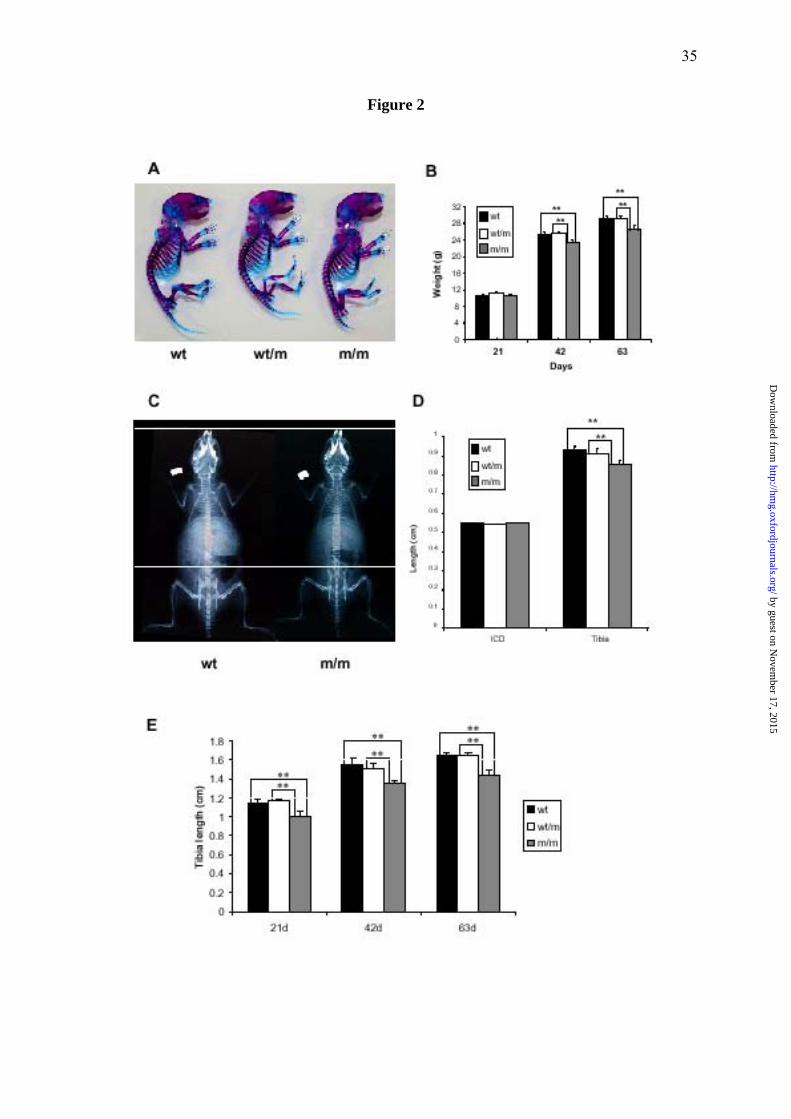
Knock-in mice are normal at birth but develop short-limbed dwarfism.
At birth the skeletons of mice heterozygous or homozygous for the p.Val194Asp
mutation appeared normal when compared with wild type littermates (Fig. 2A). At day 21 all
mice had similar body weights, however, by day 42 mice that were homozygous for the
mutation were ~7.5% lighter than either their wild type or heterozygous littermates and by
day 63 these same mice were ~9.5% lighter (Fig. 2B; n>23 mice per genotype, **p<0.01 by
one-way ANOVA). The body weights of mice heterozygous for the mutation were
indistinguishable from those of their wild type littermates.
by guest on Novem
ber 17, 2015http://hm
g.oxfordjournals.org/D
ownloaded from
8
Bone length measurements were performed on mice of all three genotypes at days 14,
21, 42 and 63. Bone measurements were made of the inner canthal distance (ICD), a measure
of intramembranous bone growth; and of the humerus, pelvis, femur and tibia, as measures of
endochondral bone growth (Fig. 2C & D; only ICD and tibia measurement are shown but the
results are representative of all bones measured). At day 14 mice that were homozygous for
the mutation had tibia lengths that were ~8.5% shorter than their both their wild type and
heterozygous littermates (Fig. 2D) and by day 21 the reduction in tibia lengths had progressed
to ~12.5% (Fig. 2E; n>6 mice per genotype, **p<0.01 by one-way ANOVA). No differences
were observed in the ICD of age-matched mice of all three genotypes at all ages confirming
that intramembranous bone growth was not affected by the matn3 mutation (Fig. 2D and data
not shown). These morphometric measurements demonstrated that mice, which are
homozygous for p.Val194Asp, are normal at birth but from day 14 develop a measurable
short-limbed dwarfism as a result of disturbed endochondral ossification. The age of onset
and progressive nature of the dwarfism in mutant mice is comparable to the ago of onset in
patients with MED, which can be as early as 2 years of age.
The cartilage growth plate is disorganised in mice harbouring the matn3 mutation.
Haematoxylin and eosin (H&E) staining of the tibia growth plates from wild type
animals at all ages showed a well organized growth plate in which the resting, proliferative
and hypertrophic zones were clearly distinguishable (Fig. 3A). Furthermore, the cells in the
proliferative zone were closely aligned in well ordered columns that were evenly spaced along
the horizontal axis of the growth plate. The tibia growth plates from mice homozygous for the
matn3 mutation also appeared normal at birth. However, from day 7 mice that were
homozygous for the mutation developed a progressively dysplastic growth plate in which the
proliferative zone had a disordered cellular organization and morphology (Fig. 3A: insert).
by guest on Novem
ber 17, 2015http://hm
g.oxfordjournals.org/D
ownloaded from
9
This dysplasia was also seen in the hypertrophic zone but no growth plate dysplasia was
evident in mice that were heterozygous for the mutation.
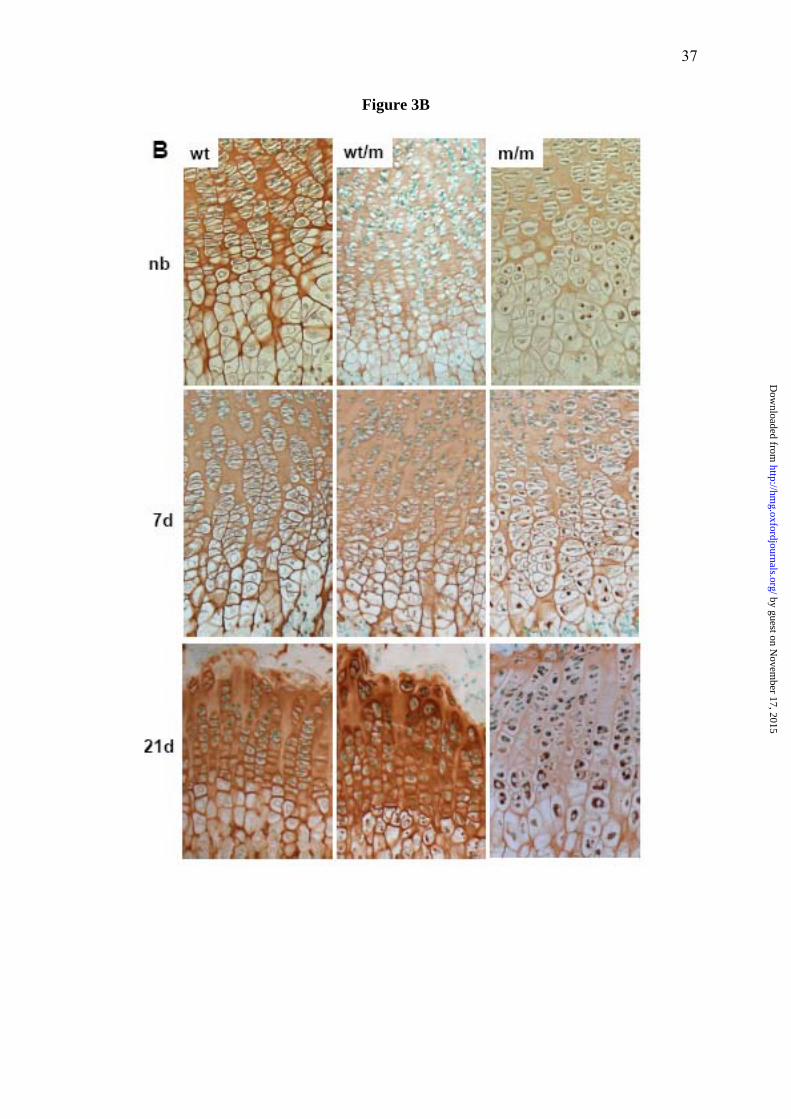
The retention of mutant matrilin-3 within the rER of chondrocytes is observed from birth.
Immuno-histochemical (IHC) staining of tibia cartilage from newborn wild type mice
showed that matrilin-3 was present throughout the ECM of the growth plate and by day 21
had a predominantly territorial localisation. In contrast, analysis of cartilage from newborn
mice that were homozygous for the matn3 mutation demonstrated that whilst matrilin-3 was
present in the ECM, there were also detectable levels retained within the pre-hypertrophic and
hypertrophic chondrocytes (Fig. 3B). By day 7 the retention of matrilin-3 had become more
widespread and was present in chondrocytes from all zones of the growth plate. Finally, by
day 21 the retention of mutant matrilin-3 was extensive and the levels of secreted protein were
markedly reduced. In contrast, chondrocytes from mice that were heterozygous for the
mutation showed no apparent retention of matrilin-3 until day 21 and at this age it was only
present in hypertrophic chondrocytes (Fig. 3B). IHC analysis using anti -matrilin-1, -COMP, -
collagen II, - collagen IX, - collagen X, -aggrecan, revealed no apparent disruption to the
localisation of these proteins in the ECM of mice homozygous for the mutation
(supplementary data).
Ultrastructural analysis of the growth plate cartilage revealed enlarged individual cisternae
of rER within the chondrocytes and disrupted chondron organization.
Transmission electron microscopy (TEM) was performed along the entire vertical axis
of the growth plate to generate a complete montage of the cartilage growth plate of a 7 day
old mouse tibia (i.e. from resting to terminal hypertrophic and mineralization zones). By
aligning images it was possible to compare directly the ultrastructure of a wild type mouse
by guest on Novem
ber 17, 2015http://hm
g.oxfordjournals.org/D
ownloaded from

10
with littermates that were either heterozygous or homozygous for the mutation (Fig. 4A). In
the wild type growth plate the resting chondrocytes were evenly spaced, whilst in the early to
late proliferative zones groups of flattened cells (i.e. 2, 4 and 8 cells per chondron) were
apparent. The morphology of chondrocytes in the growth plate of mice that were homozygous
for the mutation was strikingly different. For example, chondrocytes in the resting zone
contained numerous dilated cisternae of rER in addition to the normal rER. These dilated
cisternae gradually become larger in size as the chondrocytes underwent proliferation and in
some cells they occupied large sections of the cytoplasm, indeed there was a proportion of the
proliferating chondrocytes that were ‘wedge-shaped’ in appearance and were unable to align
closely within the 4 and 8 cell chondrons. All chondrocytes in the growth plate of mutant
mice (n>200) showed enlarged dilated cisternae of rER, which was not seen in chondrocytes
from wild type mice. Most chondrocytes from mice heterozygous for the mutation had some
distended rER, but this was not as prominent as that seen in homozygous mice.
Hypertrophic chondrocytes from wild type mice were somewhat rectangular in shape
and the condensation of chromatin was clearly apparent (42). In contrast, hypertrophic
chondrocytes from mice homozygous for p.Val194Asp mutation were more oval in shape and
showed the persistence of protein within the remnants of the rER (Fig. 4A).
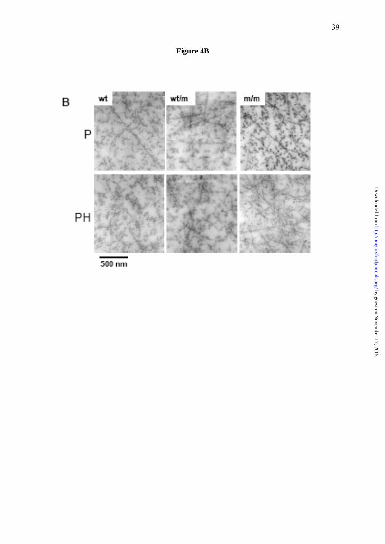
There is a disturbance to collagen networks in the mutant growth plate.
TEM analysis of the inter-territorial matrix from the proliferating and pre-hypertrophic
zones of day 7 tibia growth plates was used to study the ECM architecture of the growth plate
cartilage (Fig. 4B). The growth plate cartilage in wild type mice contained multiple randomly
arranged collagen fibrils, but individual fibrils were not always apparent because of a coating
of stained proteins (e.g. proteoglycans)(43). In contrast, the collagen fibrils in the ECM of
mice homozygous for the mutation were readily identified by their uniform diameter and
by guest on Novem
ber 17, 2015http://hm
g.oxfordjournals.org/D
ownloaded from

11
banding pattern. The differences between collagen fibrils in wild type and mutant ECM was
most apparent when the fibrils were in transverse section (i.e. cut end-on), when the fibrils
appeared as dense circles. For example, in the proliferative zone, the mutant samples showed
high contrast between the fibril surface and the inter-fibrillar space whereas in wild type the
abundance of fibril-associated material blurred the fibril boundaries. This effect was most
pronounced in the pre-hypertrophic zone; in the mutant samples the fibrils are readily seen
whereas in the wild type, the presence of surface-associated material obscured virtually all of
the fibril cross sections.
3-D reconstruction of 4-cell chondrons shows a 9-fold increase in the volume of distended
rER in chondrocytes from mutant mice.
3-D reconstructions of 140 serial TEM sections from the tibia of wild type and mutant
mice at day 7 were used to generate virtual representations of 4-cell chondrons from the
proliferative zone of the growth plate (Fig. 5 and supplemental video). In the wild type mice
the volume of the rER, expressed as a proportion of the total cell volume, was 1.3 % per
individual chondrocytes (average 7.28 μm3/549.25 μm3 cell volume; n=4). However, in
mutant mice the average volume of the distended rER per individual chondrocyte was 11.6 %
(average 55.3 μm3/476.5 μm3 cell volume; n=4), thus representing a ~9-fold increase in the
volume of the rER (Fig. 5 and supplemental video).
Chaperone proteins associated with the unfolded protein response are up-regulated in
mutant chondrocytes.
The increased expression of the rER chaperones BiP and Grp94 are classical markers
of an unfolded protein response activation in both yeast and mammalian cells (44-46).
Therefore, to determine if the retention of mutant matrilin-3 was eliciting an unfolded protein
by guest on Novem
ber 17, 2015http://hm
g.oxfordjournals.org/D
ownloaded from

12
response we performed quantitative real-time RT-PCR (qRT-PCR), Western blot analysis and
IHC to evaluate the relative levels of these chaperone proteins in the chondrocytes of wild
type mice and mice homozygous for the mutation. qRT-PCR analysis confirmed that by 3
days of age the relative levels of BiP and Grp94 were up-regulated in mutant chondrocytes by
>2- and >4-fold respectively (Fig. 6A; n=3 samples per genotype, independent t-test **p<0.05
and *p<0.01 respectively), which was confirmed by Western blot analysis (Fig. 6B; n=6
samples per genotype) and IHC (not shown).
When misfolded/unfolded mutant proteins accumulate in the rER they can induce an
unfolded protein response, which may cause rER/cell stress and the increased expression of
C/EBP homologous protein (CHOP/GADD153)(47). CHOP is a member of the C/EBP family
of bZIP transcription factors and because it’s over expression induces apoptosis we
investigated the relative levels of CHOP expression in mutant chondrocytes by qRT-PCR.
However, at 3, 5 and 21 days of age there was no detectable increase in the relative levels of
CHOP expression compared with wild type chondrocytes (Fig. 6C).
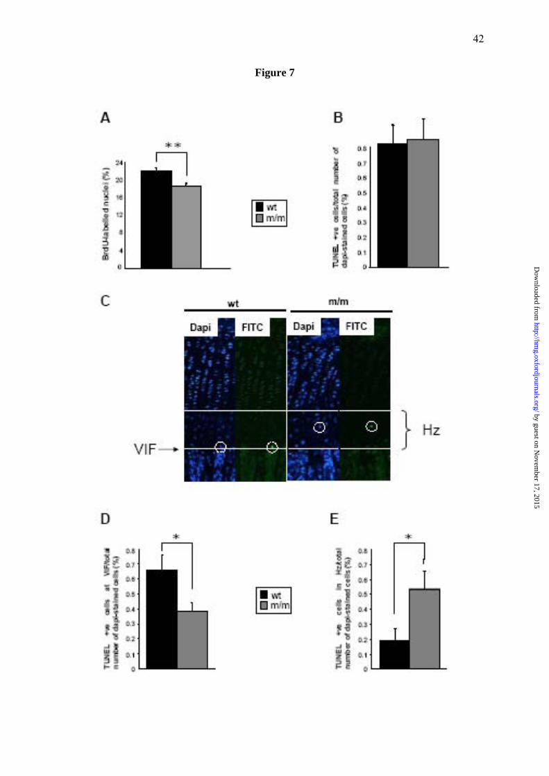
Mice homozygous for the mutation have reduced chondrocyte proliferation in the growth
plate.
In order to understand the physiological effect of up-regulated chaperone protein
expression and the unfolded protein response on chondrocyte differentiation and eventually
longitudinal bone growth we determined the relative levels of chondrocyte proliferation in the
growth plate. To quantify the levels of chondrocyte proliferation in the growth plate BrdU
labelling experiments were performed at day 21. 22% of cell nuclei within the proliferative
zone of wild type mice were labelled with BrdU, whilst only 18% of nuclei were labelled
within the proliferative zone of mice homozygous for the mutation, thus signifying an overall
decrease of 16% in the rate of chondrocyte proliferation (Fig. 7A; n>36 sections from >3 mice
by guest on Novem
ber 17, 2015http://hm
g.oxfordjournals.org/D
ownloaded from
13
per genotype, independent t-test **p<0.01). Proliferation rates in mice heterozygous for the
mutation were comparable to those of wild type mice (data not shown).
Chondrocyte apoptosis is spatially dysregulated in the growth plates of mutant mice
To determine if the unfolded protein response was having a detrimental affect on cell
viability we determined the relative levels of apoptosis in the growth plate at 21 day of age by
counting the number of TUNEL-positive cells compared to DAPI-stained cells in the
hypertrophic zone. In both the wild type and mutant growth plates approximately 0.8% of
cells were TUNEL-positive which was within normal limits (48) (Fig. 7B; typically 2-3
TUNEL-positive cells from 285-310 DAPI-stained cells per section, n>20 sections from >3
mice per genotype). However, we noticed that in the mutant growth plate, unlike wild type
mice, that apoptosis was occurring throughout the hypertrophic zone and was not just limited
to terminal hypertrophic chondrocytes at the vascular invasion front (Fig. 7C). For example,
in the wild type growth plate apoptosis was mostly restricted to terminal hypertrophic
chondrocytes at the vascular invasion front and typically ~0.7% of TUNEL-positive cells
were specifically located at the vascular invasion front. In contrast, only 0.38% of TUNEL-
positive cells were located at the vascular invasion front in mice homozygous for the mutation
(Fig. 7D; n>20 sections from >3 mice per genotype, independent t-test *p<0.05).
Furthermore, the relative number of TUNEL-positive cells throughout the entire hypertrophic
zone (i.e. upper → lower hypertophic zones) was significantly increased in the mutant growth
plate, suggesting that apoptosis was spatially dysregulated (Fig. 7E; n>20 sections from >3
mice per genotype, independent t-test *p<0.05).
by guest on Novem
ber 17, 2015http://hm
g.oxfordjournals.org/D
ownloaded from

14
Discussion
In this study we have generated a knock-in mouse model to determine the disease
mechanisms that underpin the pathophysiology of MED caused by a matrilin-3 mutation. We
introduced a matn3 mutation (p.Val189Asp) that resulted in a murine chondrodysplasia
characterised by mild short-limbed dwarfism. The equivalent human mutation in MATN3
(p.Val194Asp) has previously been shown to cause an autosomal dominant form of MED in
which patients are normal at birth, but develop mild short-limbed dwarfism during childhood
(20, 49). The final heights of MED patients with MATN3 mutations generally range from the
3rd to 75th percentile and are therefore often within normal limits (21, 49, 50). It was therefore
not surprising that this mouse model of MED did not have a severe short-limb dwarfism;
however, by considering the median bone length measurements in wild type mice as the 50th
percentile, a 12% reduction in tibia bone length (seen at day 63 in mice homozygous for the
mutation) is below the 3rd percentile (i.e. 4 S.D. below the mean). This observation is
therefore consistent with the equivalent human phenotype caused by the p.Val194Asp
mutation in which the heights of affected adult males were on the 75th percentile (49), whilst
an affected child in this family is currently growing at just below the 3rd percentile (Dr Geert
Mortier, personal communication).
Although the clinical phenotype in the mouse is only evident from day 14, the
histological phenotype is in fact observable from birth. This is characterised by the
accumulation of mutant matrilin-3 in the rER, which by day 7 leads to a disruption to
chondrocyte morphology and columnar organisation within the growth plate. Concurrently,
there was an up-regulation of BiP and Grp94, which are classical markers for UPR activation
(44-46) and by day 21 the UPR and growth plate dysplasia is associated with a reduction in
chondrocyte proliferation and spatially dysregulated apoptosis. The differences in the timing
of the clinical and histological/molecular manifestations of the murine chondrodysplasia is
by guest on Novem
ber 17, 2015http://hm
g.oxfordjournals.org/D
ownloaded from

15
therefore consistent with the natural history of the human MED phenotype and provides a
rationale for the progressive short-limbed dwarfism seen in patients (49). Interestingly, in
contrast to the human MED phenotype, which is a dominant disease, both copies of the
mutant allele were required for the mice to develop a detectable chondrodysplasia. This might
be due in part to the different physiology of mice or the inbred nature of the strains that were
used to generate this mouse model. Similar discrepancies have been observed in other mice
models of human skeletal disease such as achondroplasia (51) and mice that harbour a comp
p.Thr585Met mutation (Pirog-Garcia et al manuscript in preparation) and are suggestive of
dominance modification (52, 53).
Previous studies to elucidate the structural and functional significance of MATN3
mutations in MED have focused on the use of two different in vitro cell culture systems,
namely, primary bovine chondrocytes (28) and a mammalian immortalised cell line (22). In
both cases matrilin-3 harbouring MED mutations was retained within the rER of cells and in
one system remained associated with ERp72 as an unfolded intermediate (22). Furthermore,
electron microscopy of cartilage from an MED patient with a MATN3 mutation showed the
presence of dilated cisternae of rER; whilst IHC analysis confirmed that the retained protein
was matrilin-3 (22). However, despite these studies several fundamental questions have
remained unresolved. For example, although these in vitro studies have suggested that there is
a protein trafficking defect of mutant matrilin-3, neither study was able to demonstrate
categorically whether mutant matrilin-3 is present within the cartilage ECM, and if a
potentially abnormal ECM is able to provide a suitable environment for chondrocyte
proliferation and differentiation. IHC analysis of growth plate cartilage from mice
homozygous for the mutation demonstrated that whilst the majority of the mutant protein was
retained intracellularly, a smaller proportion was evenly distributed throughout the ECM.
Therefore, these data demonstrate for the first time that mutant matrilin-3 can be secreted to
by guest on Novem
ber 17, 2015http://hm
g.oxfordjournals.org/D
ownloaded from

16
some extent from chondrocytes, however, we were unable to determine if this mutant
matrilin-3 was present in the ECM as a homo-oligomer (i.e. [matrilin-3]4) or as a hetero-
oligomer with matrilin-1 (i.e. [matrilin-1]2 [matrilin-3]2) and it therefore remains a possibility
that mutant matrilin-3 monomers can only be secreted when they form hetero-oligomers with
normal matrilin-1 monomers.
The morphology of individual chondrocytes, chondrons and indeed the entire growth
plate was clearly disrupted in mice that were homozygous for the mutation. The accumulation
of protein within dilated cisternae of rER had the effect of altering the shape of some
chondrocytes, which were unable to form compact 4- and 8- cell chondrons during cell
proliferation. These in turn were unable to align correctly into a columnar arrangement and
gave the overall appearance of a disrupted growth plate. It is likely that this misalignment of
proliferating cells would have a detrimental affect on linear bone growth. Furthermore, the
observed changes in the structure and properties of the ECM are also likely to affect the
integrity of the tissue and have a profound affect on cell motility during the proliferation and
realignment of chondrocytes at the 2-, 4- and 8-cell stage (54, 55). The 3-D reconstruction of
a 4-cell chondron from both wild type and mutant mice elegantly illustrates the extent of the
accumulation of mutant protein within the distended rER. It is clear from this model that the
~9-fold increase in the volume of distended rER is likely to have an adverse physical affect on
the chondrocytes, in addition to the UPR. IHC indicated that the accumulation of mutant
matrilin-3 was greatest at day 21 (Fig. 3B), but due to technical constraints the 3-D
reconstructions were only performed on day 7 mice (Fig. 5), therefore the proportion of
distended rER would be even greater by day 21.
Prior to this study it was not known whether the retention of mutant matrilin-3 in the
rER of chondrocytes would initiate an UPR in vivo. By using a combination of qRT-PCR,
Western blot analysis and IHC we have been able to establish for the first time that specific
by guest on Novem
ber 17, 2015http://hm
g.oxfordjournals.org/D
ownloaded from
17
chaperone proteins, which are classical markers for UPR activation (44-46), were up-
regulated in chondrocytes from mutant mice. It is most likely that the activation of the UPR
was in direct response to the retention of mutant matrilin-3 and this is normally associated
with several downstream consequences, most notable of which is the up-regulation of CHOP,
a key mediator or rER/cell stress. Interestingly, our observation that mutant matrilin-3 steadily
accrues within the rER of chondrocytes suggests that the UPR (including ER-associated
degradation) is insufficient to prevent the accumulation of mutant protein. However, we were
unable to detect an increase in the relative levels of CHOP expression.
One key goal for generating a murine model of MED was to answer fundamental
questions regarding disease mechanisms within the context of the growth plate. Primarily,
what is the mechanistic link between the expression of a mutant protein and the decreased
linear bone growth and short-limbed dwarfism? The effect of an UPR (and potential rER/cell
stress) on chondrocyte proliferation and apoptosis was therefore determined and these data
provided novel insight into potential disease mechanisms. We were able to demonstrate for
the first time that the relative levels of chondrocyte proliferation were significantly reduced in
the growth plate of mice homozygous for the mutation. Interestingly, we did not detect an
overall increase in apoptosis; however, apoptosis was spatially dysregulated in the growth
plate and significant numbers of TUNEL-positive cells were identified away from the
vascular invasion front. These observations suggested that apoptosis was spatially
dysregulated rather than increased and are supported by the qRT-PCR data which confirmed
that there was no apparent increase in the relative expression of CHOP in mutant
chondrocytes. Bearing in mind that chondrocyte proliferation and hypertrophy in the growth
plate is vital for long bone growth, any disruption to these processes are likely to severely
delay endochondral ossification and eventually lead to short-limb dwarfism. Our data
therefore support the hypothesis that reduced chondrocyte proliferation and spatially
by guest on Novem
ber 17, 2015http://hm
g.oxfordjournals.org/D
ownloaded from

18
dysregulated apoptosis is sufficient to cause the progressive short-limb dwarfism that is
characteristic of this form of MED.
In summary therefore we have generated the first relevant murine model of multiple
epiphyseal dysplasia and identified mechanistic links between the expression of a mutant gene
product and the resulting growth plate dysplasia Ultimately this will pave the way for the
development of suitable therapeutic approaches for the treatment of diseases within PSACH-
MED bone dysplasia family.
by guest on Novem
ber 17, 2015http://hm
g.oxfordjournals.org/D
ownloaded from

19
Materials and Methods
Construction of the targeting vector and generation of chimeric mice. A targeting vector
was constructed from overlapping lambda clones isolated from a 129/sv library (56). A neo-tk
cassette, flanked by loxP sites, was cloned into Bam HI/Cla I site of pBluescript II KS
(pBSNeotk). The short arm was generated by PCR (SAF 5’-ttctgctcataccgactgg-3’ and SAR
5’-gcaccagctgtaatcatagta-3’) amplification of a 2 kb fragment of mouse genomic DNA, which
was digested with Psp OMI/Bcl I and cloned into the Not I/Bam HI sites of the pBSNeotk
vector (pBSNeotkSA). The long arm was made from two composite genomic fragments. The
5’ of the long arm, containing the mutation, was generated by PCR amplification of a 1.5 kb
fragment from mouse genomic DNA (SalIF 5’-acgcgtcgacttgtttttctgagaggacttcattc-3’ and
Mut+XhoIR 5’-cacgggctcgagcagccacctcattcacctggtcctgcggcctcccatctgtatcgataatagctaccttgg-
3’). The forward primer had a Sal I restriction site (bold), while the reverse contained the Xho
I restriction site (bold) found in exon 2, and the mutation which created a Cla I restriction site
(underlined). This was digested with Sal I and Xho I and cloned into the Xho I site of
pBSNeotkSA, disrupting the original Xho I site from the vector and leaving the Xho I site
from exon 2 intact. The correct orientation and presence of the mutation was confirmed by
digests and sequencing (pBSNeotkSA-SX). Finally the rest of the long arm, a 7 kb fragment
was removed from the genomic clone via Xho I digestion and cloned into the Xho I site of
pBSNeotkSA-SX. The orientation was confirmed leaving a continuous 8.5 kb long arm
(pBSNeotk-V194D).
The targeting vector DNA (70 μg) was linearized with Not I and used to electroporate
4 x 10⁷ R1 embryonic stem (ES) cells, grown on feeders and supplemented with LIF
(Leukaemia inhibitory factor), using a Bio-Rad gene pulser set at 0.8 kV and 3 μF for 0.1
msec. Selection with 500 μg/ml G418 began after 24 hrs for 5-6 days. DNA from clones were
digested with Spe I and analysed by Southern blot hybridisation using an external probe (Fig.
by guest on Novem
ber 17, 2015http://hm
g.oxfordjournals.org/D
ownloaded from

20
1b). In order to remove the floxed neo-tk cassette from homologously recombined clones in
vitro, ES cells were electroporated as before but with 50 μg of pIC-Cre, a mammalian
expression vector containing the Cre transcript. Cultures were selected with FIAU (0.2 μM
final concentration) for 6 days. Clones were picked, DNA isolated and detection of only the
12 kb fragment showed removal of the neo-tk cassette. PCR amplification across the neo-tk
site confirmed removal of the cassette and presence of the single remaining LoxP site.
Targeted ES cells were microinjected into DBA blastocysts to generate chimeric mice.
Chimeric males were bred with C57Bl/6 females. Offspring heterozygous for the mutation
were used to generate the matn3 p.Val194Asp mouse strain.
Analysis of the skeleton. Skeletal preparations of newborn mice were prepared as described
previously (57). Growth curves were produced by measuring body weights of littermates at
day 21, 42 and 63. Bone length measurements were taken from X-ray radiographs.
Histology and Immuno-histochemistry (IHC). Tissue samples were fixed overnight in ice-
cold 10% formalin (Histological staining and TUNEL) or 95% ethanol/5% acetic acid (IHC
and BrdU). Bone samples were then decalcified in 20% EDTA and embedded in paraffin
wax. Antibodies used were anti-matrilin-3 and anti-BrdU (Abcam). IHC was carried out on
ethanol/acetic acid-fixed samples. Briefly, endogenous peroxidase activity was quenched by a
H2O2/methanol wash, followed by hyaluronidase treatment. Samples were blocked with goat
serum and BSA in PBS for 1hr, incubated for 1hr with the primary antibody (in PBS/BSA)
and then 1hr with the secondary antibody (biotinylated goat anti-rabbit IgG, Dako
Cytomation) in PBS/BSA with goat serum. Slides were then incubated with the
ABCcomplex/HRP reagent for 30 mins, and developed using DAB. Samples were
counterstained with methyl green and mounted with VectaMount. BrdU IHC was performed
by guest on Novem
ber 17, 2015http://hm
g.oxfordjournals.org/D
ownloaded from
21
as above, except the hyaluronidase step was replaced with an antigen retrieval step (incubated
in 4M HCl for 15 mins, then neutralised with 0.1M borate buffer).
Measurements of in vivo apoptosis was carried out using the DeadEndTM fluorometric
TUNEL system (Promega) and visualised with a Zeiss Axiovision microscope. Nuclei where
stained with DAPI while apoptotic cells stained with FITC.
Ultrastructural analysis. Tibia from day 7 mice were fixed overnight in 4%
formaldehyde/2.5% gluteraldehyde in 0.1 M sodium cacodylate buffer, followed by 3 washes
in 0.1 M sodium cacodylate buffer. Sample were then incubated (2hrs, 4°C) in a secondary fix
of 1% osmium tetroxide, followed by 3 washes in water. En bloc staining of the sample was
carried out by incubation (1hr, 4°C) in 0.5% uranyl acetate, followed by water washes.
Samples were then dehydrated through an ascending graded acetone series. The acetone was
replaced with 2 changes of propylene oxide, which in turn was replaced with Spurr’s resin.
After several changes the resin was polymerised by incubating at 60°C for 48 hrs. 70 nm
sections were cut and stained with 0.3% (w/v) lead citrate and images were taken on a FEI
Tecnai 12 Biotwin electron microscope and were recorded on 4489 film (Kodak) and scanned
using an Imacon Flextight 848 scanner (Precision Camera & Video). Images from EM serial
sections were aligned, reconstructed and visualized in IMOD for Linux (58).
qRT-PCR and Western blot analysis of mutant chondrocytes. For qRT-PCR rib cages from 3
and 5 day old mice (wt and m/m) were treated with collagenase for 2 hrs (Type 1A, 2 mg ml-
1). The costal cartilage was then dissected from the rib cage and the perichondrium layer
removed. The cartilage was then further treated with collagenase for 3 hrs to digest away the
collagen matrix and release the chondrocytes. The chondrocytes were passed through a cell
strainer (70 μm) and washed twice with PBS. The cell pellet was resuspended in 500 μl
by guest on Novem
ber 17, 2015http://hm
g.oxfordjournals.org/D
ownloaded from
22
Trizol. Total RNA was then isolated according to manufacturer’s instructions (Invitrogen).
cDNA was then generated using random hexamer primers (Superscript III, Invitrogen) and
qRT-PCR perfomed using the SYBR® green PCR method for each chaperone. The following
primer sequences were used for BiP (For: 5’-ggcaccttcgatgtgtctcttc-3’and Rev: 5’-
tccatgacccgctgatcaa-3’), Grp94 (For: 5’-taagctgtatgtacgccgcgt-3’ and Rev: 5’-
ggagatcatcggaatccacaac-3’) and 18s RNA (For: 5’-gtaaaccgttgaaccccatt-3’ and Rev: 5’-
ccatccaatcggtagcg-3’). For Western blot analysis, chondrocytes were isolated as above, but
aliquots of 2 x 10⁵ chondrocytes were prepared and resuspended in 5 x SDS loading buffer.
These aliquots were run on SDS-PAGE then transferred to nitrocellulase membrane for
western blot analysis. Ponceau staining was used to confirm equal loading of total protein
isolates. Antibodies to key chaperones associated with the unfolded protein response were
used at 1:500 dilutions, namely BiP (Santa Cruz) and Grp94 (Santa Cruz).
Statistical analysis. One-way analysis of variance (ANOVA) was used to determine
differences within and between groups (used for body weight and bone length comparisons),
whilst differences in chondrocyte proliferation, apoptosis and qPCR were analysed using
independent t-test. A P-value <0.05 was considered statistically significant.
Online supplemental material. The video depicts a 3-D reconstruction comparing 4-cell
chondrons from wild type and mutant mice. Cells are color rendered; Nuclei are shown in
purple, distended rER in blue and primary cilia in red. The video shows the extent of the
accumulation of mutant matrilin-3 within the distended rER.
by guest on Novem
ber 17, 2015http://hm
g.oxfordjournals.org/D
ownloaded from

23
Acknowledgments
This work was supported by grants from the Wellcome Trust (071161/Z/03/Z to MDB) the
National Institute of Health (RO1 AR49547-01 to MDB, RBH, KEK and DJT) and Deutsche
Forschungsgemeinschaft (WA1338/2-4 to RW). This work was undertaken in the Wellcome
Trust Centre for Cell-Matrix Research and the Histology and Transgenic Mouse core facilities
of the Faculty of Life Sciences at the University of Manchester. We would like to thank Dick
Heinegard (Lund) for the COMP antibody and Tim Hardingham (Manchester) for the
aggrecan antibody.
Conflicts of Interest
There are no conflicts of interest to declare
by guest on Novem
ber 17, 2015http://hm
g.oxfordjournals.org/D
ownloaded from
24
References
1. Shapiro, I.M., Adams, C.S., Freeman, T. and Srinivas, V. (2005) Fate of the hypertrophic chondrocyte: microenvironmental perspectives on apoptosis and survival in the epiphyseal growth plate. Birth Defects Res. C. Embryo Today, 75, 330-9.
2. Kornak, U. and Mundlos, S. (2003) Genetic disorders of the skeleton: a developmental approach. Am. J. Hum. Genet., 73, 447-74.
3. Goldring, M.B., Tsuchimochi, K. and Ijiri, K. (2006) The control of chondrogenesis. J. Cell. Biochem., 97, 33-44.
4. Zelzer, E. and Olsen, B.R. (2003) The genetic basis for skeletal diseases. Nature, 423, 343-8.
5. (1998) International nomenclature and classification of the osteochondrodysplasias (1997). International Working Group on Constitutional Diseases of Bone. Am. J. Med. Genet., 79, 376-82.
6. Spranger, J. (1988) Bone dysplasia 'families'. Pathol. Immunopathol. Res., 7, 76-80. 7. Briggs, M.D. and Chapman, K.L. (2002) Pseudoachondroplasia and multiple
epiphyseal dysplasia: Mutation review, molecular interactions, and genotype to phenotype correlations. Hum. Mutat., 19, 465-78.
8. Unger, S. and Hecht, J.T. (2001) Pseudoachondroplasia and multiple epiphyseal dysplasia: New etiologic developments. Am. J. Med. Genet., 106, 244-50.
9. Kennedy, J., Jackson, G., Ramsden, S., Taylor, J., Newman, W., Wright, M.J., Donnai, D., Elles, R. and Briggs, M.D. (2005) COMP mutation screening as an aid for the clinical diagnosis and counselling of patients with a suspected diagnosis of pseudoachondroplasia or multiple epiphyseal dysplasia. Eur. J. Hum. Genet., 13, 547-55.
10. Rossi, A. and Superti-Furga, A. (2001) Mutations in the diastrophic dysplasia sulfate transporter (DTDST) gene (SLC26A2): 22 novel mutations, mutation review, associated skeletal phenotypes, and diagnostic relevance. Hum. Mutat., 17, 159-71.
11. Segat, D., Frie, C., Nitsche, P.D., Klatt, A.R., Piecha, D., Korpos, E., Deak, F., Wagener, R., Paulsson, M. and Smyth, N. (2000) Expression of matrilin-1, -2 and -3 in developing mouse limbs and heart. Matrix Biol., 19, 649-55.
12. Hedbom, E., Antonsson, P., Hjerpe, A., Aeschlimann, D., Paulsson, M., Rosa-Pimentel, E., Sommarin, Y., Wendel, M., Oldberg, A. and Heinegard, D. (1992) Cartilage matrix proteins. An acidic oligomeric protein (COMP) detected only in cartilage. J. Biol. Chem., 267, 6132-6.
13. Eyre, D. (2002) Collagen of articular cartilage. Arthritis Res., 4, 30-5. 14. Klatt, A.R., Paulsson, M. and Wagener, R. (2002) Expression of matrilins during
maturation of mouse skeletal tissues. Matrix Biol., 21, 289-96. 15. Wagener, R., Ehlen, H.W., Ko, Y.P., Kobbe, B., Mann, H.H., Sengle, G. and
Paulsson, M. (2005) The matrilins--adaptor proteins in the extracellular matrix. FEBS Lett., 579, 3323-9.
16. Wu, J.J. and Eyre, D.R. (1998) Matrilin-3 forms disulfide-linked oligomers with matrilin-1 in bovine epiphyseal cartilage. J. Biol. Chem., 273, 17433-8.
17. Klatt, A.R., Nitsche, D.P., Kobbe, B., Morgelin, M., Paulsson, M. and Wagener, R. (2000) Molecular structure and tissue distribution of matrilin-3, a filament- forming extracellular matrix protein expressed during skeletal development. J. Biol. Chem., 275, 3999-4006.
18. Mann, H.H., Ozbek, S., Engel, J., Paulsson, M. and Wagener, R. (2004) Interactions between the cartilage oligomeric matrix protein and matrilins. Implications for matrix assembly and the pathogenesis of chondrodysplasias. J. Biol. Chem., 279, 25294-8.
by guest on Novem
ber 17, 2015http://hm
g.oxfordjournals.org/D
ownloaded from

25
19. Budde, B., Blumbach, K., Ylostalo, J., Zaucke, F., Ehlen, H.W., Wagener, R., Ala-Kokko, L., Paulsson, M., Bruckner, P. and Grassel, S. (2005) Altered integration of matrilin-3 into cartilage extracellular matrix in the absence of collagen IX. Mol. Cell Biol., 25, 10465-78.
20. Chapman, K.L., Mortier, G.R., Chapman, K., Loughlin, J., Grant, M.E. and Briggs, M.D. (2001) Mutations in the region encoding the von Willebrand factor A domain of matrilin-3 are associated with multiple epiphyseal dysplasia. Nat. Genet., 28, 393-6.
21. Jackson, G.C., Barker, F.S., Jakkula, E., Czarny-Ratajczak, M., Makitie, O., Cole, W.G., Wright, M.J., Smithson, S.F., Suri, M., Rogala, P. et al. (2004) Missense mutations in the beta strands of the single A-domain of matrilin-3 result in multiple epiphyseal dysplasia. J. Med. Genet., 41, 52-9.
22. Cotterill, S.L., Jackson, G.C., Leighton, M.P., Wagener, R., Makitie, O., Cole, W.G. and Briggs, M.D. (2005) Multiple epiphyseal dysplasia mutations in MATN3 cause misfolding of the A-domain and prevent secretion of mutant matrilin-3. Hum. Mutat., 26, 557-65.
23. Maeda, K., Nakashima, E., Horikoshi, T., Mabuchi, A. and Ikegawa, S. (2005) Mutation in the von Willebrand factor-A domain is not a prerequisite for the MATN3 mutation in multiple epiphyseal dysplasia. Am. J. Med. Genet. A, 136, 285-6.
24. Stefansson, S.E., Jonsson, H., Ingvarsson, T., Manolescu, I., Jonsson, H.H., Olafsdottir, G., Palsdottir, E., Stefansdottir, G., Sveinbjornsdottir, G., Frigge, M.L. et al. (2003) Genomewide scan for hand osteoarthritis: a novel mutation in matrilin-3. Am. J. Hum. Genet., 72, 1448-59.
25. Eliasson, G.J., Verbruggen, G., Stefansson, S.E., Ingvarsson, T. and Jonsson, H. (2006) Hand radiology characteristics of patients carrying the T(303)M mutation in the gene for matrilin-3. Scand. J. Rheumatol., 35, 138-42.
26. Min, J.L., Meulenbelt, I., Riyazi, N., Kloppenburg, M., Houwing-Duistermaat, J.J., Seymour, A.B., van Duijn, C.M. and Slagboom, P.E. (2006) Association of matrilin-3 polymorphisms with spinal disc degeneration and with osteoarthritis of the CMC1 joint of the hand. Ann. Rheum. Dis.
27. Loughlin, J. (2005) The genetic epidemiology of human primary osteoarthritis: current status. Expert. Rev. Mol. Med., 7, 1-12.
28. Otten, C., Wagener, R., Paulsson, M. and Zaucke, F. (2005) Matrilin-3 mutations that cause chondrodysplasias interfere with protein trafficking while a mutation associated with hand osteoarthritis does not. J. Med. Genet., 42, 774-9.
29. Mazzarella, R.A., Srinivasan, M., Haugejorden, S.M. and Green, M. (1990) ERp72, an abundant luminal endoplasmic reticulum protein, contains three copies of the active site sequences of protein disulfide isomerase. J. Biol. Chem., 265, 1094-101.
30. Maynard, J.A., Cooper, R.R. and Ponseti, I.V. (1972) A unique rough surfaced endoplasmic reticulum inclusion in pseudoachondroplasia. Lab Invest., 26, 40-4.
31. Vranka, J., Mokashi, A., Keene, D.R., Tufa, S., Corson, G., Sussman, M., Horton, W.A., Maddox, K., Sakai, L. and Bachinger, H.P. (2001) Selective intracellular retention of extracellular matrix proteins and chaperones associated with pseudoachondroplasia. Matrix Biol., 20, 439-50.
32. Hecht, J.T., Hayes, E., Haynes, R. and Cole, W.G. (2005) COMP mutations, chondrocyte function and cartilage matrix. Matrix Biol., 23, 525-33.
33. Hecht, J.T., Hayes, E., Snuggs, M., Decker, G., Montufar-Solis, D., Doege, K., Mwalle, F., Poole, R., Stevens, J. and Duke, P.J. (2001) Calreticulin, PDI, Grp94 and BiP chaperone proteins are associated with retained COMP in pseudoachondroplasia chondrocytes. Matrix Biol., 20, 251-62.
by guest on Novem
ber 17, 2015http://hm
g.oxfordjournals.org/D
ownloaded from
26
34. Hecht, J.T., Makitie, O., Hayes, E., Haynes, R., Susic, M., Montufar-Solis, D., Duke, P.J. and Cole, W.G. (2004) Chondrocyte cell death and intracellular distribution of COMP and type IX collagen in the pseudoachondroplasia growth plate. J. Orthop. Res., 22, 759-67.
35. Hecht, J.T., Montufar-Solis, D., Decker, G., Lawler, J., Daniels, K. and Duke, P.J. (1998) Retention of cartilage oligomeric matrix protein (COMP) and cell death in redifferentiated pseudoachondroplasia chondrocytes. Matrix Biol., 17, 625-33.
36. Svensson, L., Aszodi, A., Heinegard, D., Hunziker, E.B., Reinholt, F.P., Fassler, R. and Oldberg, A. (2002) Cartilage oligomeric matrix protein-deficient mice have normal skeletal development. Mol. Cell Biol., 22, 4366-71.
37. Ko, Y., Kobbe, B., Nicolae, C., Miosge, N., Paulsson, M., Wagener, R. and Aszodi, A. (2004) Matrilin-3 is dispensable for mouse skeletal growth and development. Mol. Cell Biol., 24, 1691-9.
38. Aszodi, A., Bateman, J.F., Hirsch, E., Baranyi, M., Hunziker, E.B., Hauser, N., Bosze, Z. and Fassler, R. (1999) Normal skeletal development of mice lacking matrilin 1: redundant function of matrilins in cartilage? Mol. Cell Biol., 19, 7841-5.
39. Huang, X., Birk, D.E. and Goetinck, P.F. (1999) Mice lacking matrilin-1 (cartilage matrix protein) have alterations in type II collagen fibrillogenesis and fibril organization. Dev. Dyn., 216, 434-41.
40. Mates, L., Nicolae, C., Morgelin, M., Deak, F., Kiss, I. and Aszodi, A. (2004) Mice lacking the extracellular matrix adaptor protein matrilin-2 develop without obvious abnormalities. Matrix Biol., 23, 195-204.
41. van der Weyden, L., Wei, L., Luo, J., Yang, X., Birk, D.E., Adams, D.J., Bradley, A. and Chen, Q. (2006) Functional knockout of the matrilin-3 gene causes premature chondrocyte maturation to hypertrophy and increases bone mineral density and osteoarthritis. Am. J. Pathol., 169, 515-27.
42. Zenmyo, M., Komiya, S., Kawabata, R., Sasaguri, Y., Inoue, A. and Morimatsu, M. (1996) Morphological and biochemical evidence for apoptosis in the terminal hypertrophic chondrocytes of the growth plate. J. Pathol., 180, 430-3.
43. Eggli, P.S., Herrmann, W., Hunziker, E.B. and Schenk, R.K. (1985) Matrix compartments in the growth plate of the proximal tibia of rats. Anat. Rec., 211, 246-57.
44. Kaufman, R.J. (2002) Orchestrating the unfolded protein response in health and disease. J. Clin. Invest., 110, 1389-98.
45. Kaufman, R.J. (1999) Stress signaling from the lumen of the endoplasmic reticulum: coordination of gene transcriptional and translational controls. Genes Dev., 13, 1211-33.
46. Kozutsumi, Y., Segal, M., Normington, K., Gething, M.J. and Sambrook, J. (1988) The presence of malfolded proteins in the endoplasmic reticulum signals the induction of glucose-regulated proteins. Nature, 332, 462-4.
47. Zinszner, H., Kuroda, M., Wang, X., Batchvarova, N., Lightfoot, R.T., Remotti, H., Stevens, J.L. and Ron, D. (1998) CHOP is implicated in programmed cell death in response to impaired function of the endoplasmic reticulum. Genes Dev., 12, 982-95.
48. Chrysis, D., Nilsson, O., Ritzen, E.M. and Savendahl, L. (2002) Apoptosis is developmentally regulated in rat growth plate. Endocrine, 18, 271-8.
49. Mortier, G.R., Chapman, K., Leroy, J.L. and Briggs, M.D. (2001) Clinical and radiographic features of multiple epiphyseal dysplasia not linked to the COMP or type IX collagen genes. Eur. J. Hum. Genet., 9, 606-12.
50. Makitie, O., Mortier, G.R., Czarny-Ratajczak, M., Wright, M.J., Suri, M., Rogala, P., Freund, M., Jackson, G.C., Jakkula, E., Ala-Kokko, L. et al. (2004) Clinical and
by guest on Novem
ber 17, 2015http://hm
g.oxfordjournals.org/D
ownloaded from

27
radiographic findings in multiple epiphyseal dysplasia caused by MATN3 mutations: description of 12 patients. Am. J. Med. Genet., 125A, 278-84.
51. Li, C., Chen, L., Iwata, T., Kitagawa, M., Fu, X.Y. and Deng, C.X. (1999) A Lys644Glu substitution in fibroblast growth factor receptor 3 (FGFR3) causes dwarfism in mice by activation of STATs and ink4 cell cycle inhibitors. Hum. Mol. Genet., 8, 35-44.
52. Dixon, J. and Dixon, M.J. (2004) Genetic background has a major effect on the penetrance and severity of craniofacial defects in mice heterozygous for the gene encoding the nucleolar protein Treacle. Dev. Dyn., 229, 907-14.
53. Nadeau, J.H. (2001) Modifier genes in mice and humans. Nat. Rev. Genet., 2, 165-74. 54. Aszodi, A., Hunziker, E.B., Brakebusch, C. and Fassler, R. (2003) Beta1 integrins
regulate chondrocyte rotation, G1 progression, and cytokinesis. Genes Dev., 17, 2465-79.
55. Grashoff, C., Aszodi, A., Sakai, T., Hunziker, E.B. and Fassler, R. (2003) Integrin-linked kinase regulates chondrocyte shape and proliferation. EMBO Rep., 4, 432-8.
56. Wagener, R., Kobbe, B., Aszodi, A., Liu, Z., Beier, D.R. and Paulsson, M. (2000) Structure and mapping of the mouse matrilin-3 gene (Matn3), a member of a gene family containing a U12-type AT-AC intron. Mamm. Genome, 11, 85-90.
57. Braun, T., Rudnicki, M.A., Arnold, H.H. and Jaenisch, R. (1992) Targeted inactivation of the muscle regulatory gene Myf-5 results in abnormal rib development and perinatal death. Cell, 71, 369-82.
58. Kremer, J.R., Mastronarde, D.N. and McIntosh, J.R. (1996) Computer visualization of three-dimensional image data using IMOD. J. Struct. Biol., 116, 71-6.
by guest on Novem
ber 17, 2015http://hm
g.oxfordjournals.org/D
ownloaded from
28
Figure Legends
Figure 1. Generation of the matn3 p.Val194Asp knock-in mouse model. (A) 1: genomic
organisation of the mouse matn3 gene; individual exons are indicated by black boxes and
numbered sequentially. 2: the targeting construct with LoxP sequences indicated by
arrowheads and the site of the mutation by an asterisk. Neo, neomycin resistance cassette; TK,
thymidine kinase gene. 3: the homologous recombinant allele and 4: the recombinant allele
after cre transfection. Relevant restriction sites are: S, Spe I; P, Psp OMI; B, Bam HI; C, Cla
I; N, Not I. Probe denotes the external probe used in Southern blot detection of wild type and
homologously recombined mutant matn3 alleles. (B) Southern Blot analysis of transfected ES
cells. The probe indicated in panel (A) detects a 12 kb fragment and 6 kb fragment after
digestion with Spe I in the wild type and knock-in alleles respectively; 1 = homologous
recombinant, 2 = wild type alleles. (C) PCR amplification of the region containing the
mutation was followed by digestion with Cla I. The p.Val194Asp mutation introduces a Cla I
restriction site and mutation-positive homologously recombinant clones show the presence of
two extra digestion products (1 = wild type, 2= homologous recombinant). (D) Sequencing of
mutation-positive homologous recombinants confirmed the presence of the mutation
(GTG>GAT). Small arrows show the location of primers used for routine genotyping.
Figure 2. Phenotypic characterization of wild type mice and mice either heterozygous or
homozygous for the matn3 p.Val194Asp mutation. (A) Alcian blue (cartilage) and alizarin
red (bone) staining of whole newborn skeletons. There is no overt phenotype observed in
mice heterozygous (wt/m) or homozygous (m/m) for the mutation. (B) Mouse growth curves.
All mice have normal body weights at day 21, but from day 21 onwards the growth of m/m
mice is reduced compared to both wt and wt/m mice eventually leading to a 9.4% reduction in
by guest on Novem
ber 17, 2015http://hm
g.oxfordjournals.org/D
ownloaded from

29
body weight by day 63 (n>23 mice per genotype, **p<0.01 by one-way ANOVA). (C)
Radiographs of day 42 wild type mice and mice homozygous for p.Val194Asp, confirm that
mice homozygous for the mutation are shorter than the wild type mice. White dotted lines are
aligned at the tip of the nose and the top of the pelvis of the m/m mouse. (D) Bone length
measurements of the inner canthal distance (ICD) and the tibia from 14 day old male mice
demonstrate reduced long bone growth compared to wt littermates (n>6 mice, **p<0.01 by
one-way ANOVA). (E) Bone length measurements of the tibia at 21, 42 and 36 days of age.
A maximum reduction of ~12.5% was seen by 21 days of age (n>6 mice, **p<0.01 by one-
way ANOVA). Similar reductions in bone length were also recorded for the humerus, pelvis
and femur (data not shown). At all time points there was no statistical difference between the
respective ICD for all 3 genotypes.
Figure 3. Histological and immuno-histochemical (IHC) analysis of the tibia growth
plate shows disrupted growth plate morphology and retention of mutant matrilin-3
protein. (A) H&E staining of newborn (NB), 7 and 21 day old tibia growth plates of wild
type (wt) mice and of mice either heterozygous (wt/m) or homozygous (m/m) for the mutation
(PFA-fixed 6 μm sections). The growth plates of new born mice show no overt dysplasia.
However from day 7 and, in particular, at day 21 m/m mice have a disrupted proliferative
zone with disorganised columns (insert) and some areas of hypocellularity. (B) IHC using an
anti-matrilin-3 antibody on NB, day 7 and 21 tibia growth plates from wt, wt/m and m/m mice
(ethanol/acetic acid-fixed 6 μm sections). Chondrocytes in the hypertrophic zone of NB mice
homozygous for the mutation show the retention of mutant matrilin-3. By day 7 this retention
is more widespread and affects all zones of the growth plate. By day 21 the retention of
matrilin-3 has become more extensive and there are significantly reduced levels of staining in
the matrix. By day 21 mice heterozygous for the mutation are also exhibiting low-levels of
by guest on Novem
ber 17, 2015http://hm
g.oxfordjournals.org/D
ownloaded from

30
matrilin-3 retention but the amount of matrilin-3 in the ECM appears to be within normal
limits.
Figure 4. Ultrastructural analysis of 7 day old tibia growth plates from wild type mice,
and from mice either heterozygous or homozygous for the mutation, shows enlarged
individual cisternae of rER and altered chondrocyte morphology in mutant mice. (A)
This representative selection of images was taken from a complete TEM montage of a day 7
tibia growth plate (from resting to mineralisation zones). Resting (R), proliferative (P) and
hypertrophic (H) zones are shown. Chondrocytes in the R zone of m/m mice show enlarged
individual cisternae of rER (asterisk). In the P zone the enlarged cisternae are more
widespread within individual chondrocytes. Chondrocytes from wt/m mice show evidence of
dilated cisternae of rER (asterisk), but the overall morphology of the chondrocytes appears
within normal limits. Scale bar = 5 μm (R and P) or 10 μm (H). (B) TEM of the inter-
territorial matrix of the proliferating (P) zone and the prehypertrophic (PH) zone of day 7 tibia
growth plates. The extracellular matrix in the proliferating zone from wt mice shows collagen
fibrils with high levels of surface-associated proteins. The fibrils in m/m mice appear to have
less surface coating. wt/m samples are similar in appearance to wt. Scale bar = 500 nm.
Figure 5. Chondrocytes from mice homozygous for the mutation have increased
distended rER volume. Serial section reconstructions of cells (day 7) from wild type mice
and mice homozygous for the mutation were performed to obtain a 3-D virtual representation
of a 4-cell chondron. From this model morphological differences were assessed, in particular
calculations were made regarding chondrocyte (549.25 μm3/chondrocyte for wild type and
476.5 μm3/chondrocyte for mutant), nuclei (76.5 μm3/chondrocyte for wild type and 94
μm3/chondrocyte for mutant) and distended rER volumes (7.3 μm3/chondrocyte for wild type
by guest on Novem
ber 17, 2015http://hm
g.oxfordjournals.org/D
ownloaded from
31
and 55.3 μm3/chondrocyte for mutant). The top panel shows a representative TEM image
from the serial sections of wild type and mutant growth plate. The colours show the tracing of
different cell structures and relate to the reconstruction. The middle panel shows the 3-D
reconstruction of the 4-cell chondrons from wild type and mutant mice (chondrocyte 1
[Articular surface side]: blue, chondrocyte 2: yellow, chondrocyte 3: green, chondrocyte 4:
red [Trabecular bone side]). The bottom panel shows the internal structure of the
chondrocytes. There is a visible increase in the amount of distended rER in the chondrocytes
from mutant mice (nuclei: purple; distended rER: pale blue; primary cilia tube: red). Scale bar
= 5 μm.
Figure 6. The accumulation of mutant matrilin-3 elicits an unfolded protein response in
chondrocytes. (A) mRNA levels of the PR-associated chaperones (BiP and GRP94; see
methods for primer sequences) were determined using qRT-PCR analysis of mRNA isolated
from mutant and wild type chondrocytes at day 4. The graph indicates the relative increase in
levels of mRNA in mutant chondrocytes compared to wild type chondrocytes and in both
cases the level of mRNA was normalised against 18s RNA. BiP and Grp94 mRNA levels
were >2- and >4-fold increased respectively in mutant mice (n=3 mice; 3 separate
experiments in duplicate, independent t-test, p<0.05 [*] or p<0.01 [**]). (B) Total cellular
protein isolated from equal numbers of wild type or mutant chondrocytes (2 x 10⁵ cells) was
analysed by SDS-PAGE and Western blot using antibodies raised against BiP (~ 76 KDa) and
Grp94 (~94 KDa), the anti-Grp94 antibody consistently detected an non-specific band.
Protein levels of both chaperones were elevated in mice homozygous for the mutation at day
4. (C) The relative levels of CHOP mRNA in mutant chondrocytes was determined by qRT-
PCR. At day 3 and 5 there was no obvious upregulation of CHOP expression in mutant
by guest on Novem
ber 17, 2015http://hm
g.oxfordjournals.org/D
ownloaded from

32
chondrocytes compared to wild type (n=3 mice per genotype; 3 separate experiments,
independent t-test)
Figure 7. Cell proliferation and apoptosis are significantly affected in the mutant growth
plate. (A) 21 day old mice were administered with 0.01 ml/g of the nucleotide analogue BrdU
2 hrs prior to sacrifice. Tibia samples from mice were processed as normal and IHC was
performed using anti-BrdU antibody on ethanol-fixed 6 μM sections. The proportion of BrdU
labelled nuclei was calculated by comparing the number of BrdU-labelled nuclei with the total
number of chondrocytes in the proliferating zone (i.e. methyl green-labelled nuclei + BrdU-
labelled nuclei). Mice homozygous for the matn3 p.V194D mutation had significantly lower
proliferation rates compared to wild type and heterozygote mice (n>36 section per genotype,
independent t-test, **p<0.01).End-stage apoptosis (DNA fragmentation) was measured in
tibia of 21 day old mice (PFA-fixed 6 μM sections) using the DeadEnd™ fluorometric
TUNEL system. (B) The relative levels of apoptosis were calculated by comparing the
number of apoptotic chondrocytes (FITC-labelled nuclei) with the total number of
chondrocytes in the hypertrophic zone (DAPI-labelled nuclei + FITC-labelled nuclei). (C)
Representative images of tibia growth plates showing DAPI and FITC stained sections. White
circles highlight TUNEL-positive chondrocytes whilst the solid line indicates the start of the
hypertrophic zone and the dotted line marks the VIF. Apoptosis was occurring away from the
VIF in mice homozygous for the mutation. (D) The rate of apoptosis at the VIF for mice
homozygous for the mutation was 0.38 % compared to 0.66% for wild type mice; an overall
reduction of >40% (E) Apoptosis of chondrocytes in the hypertrophic zone was significantly
increased in mutant mice (n>20 sections per genotype, independent t-test, *p<0.05).
by guest on Novem
ber 17, 2015http://hm
g.oxfordjournals.org/D
ownloaded from

33
Online supplemental Video. Chondrocytes from mutant mice have increased levels of
distended rER. The video depicts a 3-D reconstruction comparing 4-cell chondrons from wild
type (left) and mutant (right) mice. Mutant mice have a ~9-fold increase in distended rER
volume, as a proportion of the chondrocyte volume, compared to wild type. Cells are color
rendered with nuclei shown in purple, distended rER in blue and primary cilia in red.
by guest on Novem
ber 17, 2015http://hm
g.oxfordjournals.org/D
ownloaded from

34
Figure 1
by guest on Novem
ber 17, 2015http://hm
g.oxfordjournals.org/D
ownloaded from
35
Figure 2
by guest on Novem
ber 17, 2015http://hm
g.oxfordjournals.org/D
ownloaded from

36
Figure 3A
by guest on Novem
ber 17, 2015http://hm
g.oxfordjournals.org/D
ownloaded from
37
Figure 3B
by guest on Novem
ber 17, 2015http://hm
g.oxfordjournals.org/D
ownloaded from

38 38
Figure 4A
by guest on Novem
ber 17, 2015http://hm
g.oxfordjournals.org/D
ownloaded from
39
Figure 4B
by guest on Novem
ber 17, 2015http://hm
g.oxfordjournals.org/D
ownloaded from

40
Figure 5
by guest on Novem
ber 17, 2015http://hm
g.oxfordjournals.org/D
ownloaded from

41
Figure 6
by guest on Novem
ber 17, 2015http://hm
g.oxfordjournals.org/D
ownloaded from
42
Figure 7
by guest on Novem
ber 17, 2015http://hm
g.oxfordjournals.org/D
ownloaded from

43
Abbreviations.
ANOVA One-way analysis of variance
COMP Cartilage oligomeric matrix protein
EO Endochondral Ossification
H&E Haematoxylin and Eosin
ICD Inner Canthal Distance
IHC Immuno-histochemistry
MATN3 Matrilin-3
MED Multiple epiphyseal dysplasia
PSACH Psuedoachondroplasia
qRT-PCR Real-Time RT-PCR
rER Rough Endoplasmic reticulum
TEM Transmission electron microscopy
UPR Unfolded protein response
VIF Vascular invasion front
by guest on Novem
ber 17, 2015http://hm
g.oxfordjournals.org/D
ownloaded from
Related Documents