Edited by Decline of Mediterranean Fruit Crops and Forests Associated with Fungal Trunk Pathogens Carlos Agustí-Brisach Printed Edition of the Special Issue published in Plants www.mdpi.com/journal/plants

Welcome message from author
This document is posted to help you gain knowledge. Please leave a comment to let me know what you think about it! Share it to your friends and learn new things together.
Transcript
Edited by
Decline of Mediterranean Fruit Crops and Forests Associated with Fungal Trunk Pathogens
Carlos Agustí-Brisach
Printed Edition of the Special Issue published in Plants
www.mdpi.com/journal/plants

Decline of Mediterranean Fruit Cropsand Forests Associated with FungalTrunk Pathogens

Decline of Mediterranean Fruit Cropsand Forests Associated with FungalTrunk Pathogens
Editor
Carlos Agustı-Brisach
MDPI • Basel • Beijing • Wuhan • Barcelona • Belgrade • Manchester • Tokyo • Cluj • Tianjin
Editor
Carlos Agustı-Brisach
University of Cordoba
Spain
Editorial Office
MDPI
St. Alban-Anlage 66
4052 Basel, Switzerland
This is a reprint of articles from the Special Issue published online in the open access journal Plants
(ISSN 2223-7747) (available at: https://www.mdpi.com/journal/plants/special issues/Fungal
Trunk Dis).
For citation purposes, cite each article independently as indicated on the article page online and as
indicated below:
LastName, A.A.; LastName, B.B.; LastName, C.C. Article Title. Journal Name Year, Volume Number,
Page Range.
ISBN 978-3-0365-4411-3 (Hbk)
ISBN 978-3-0365-4412-0 (PDF)
Cover image courtesy of Carlos Agustı-Brisach
© 2022 by the authors. Articles in this book are Open Access and distributed under the Creative
Commons Attribution (CC BY) license, which allows users to download, copy and build upon
published articles, as long as the author and publisher are properly credited, which ensures maximum
dissemination and a wider impact of our publications.
The book as a whole is distributed by MDPI under the terms and conditions of the Creative Commons
license CC BY-NC-ND.
Contents
About the Editor . . . . . . . . . . . . . . . . . . . . . . . . . . . . . . . . . . . . . . . . . . . . . . vii
Preface to ”Decline of Mediterranean Fruit Crops and Forests Associated with Fungal Trunk
Pathogens” . . . . . . . . . . . . . . . . . . . . . . . . . . . . . . . . . . . . . . . . . . . . . . . . . . ix
Piebiep Goufo, Ana C. Marques and Isabel Cortez
Exhibition of Local but Not Systemic Induced Phenolic Defenses in Vitis vinifera L. Affected byBrown Wood Streaking, Grapevine Leaf Stripe, and Apoplexy (Esca Complex)Reprinted from: Plants 2019, 8, 412, doi:10.3390/plants8100412 . . . . . . . . . . . . . . . . . . . . 1
Mohamed T. Nouri, Daniel P. Lawrence, Craig E. Kallsen and Florent P. Trouillas
Macrophomina Crown and Root Rot of Pistachio in CaliforniaReprinted from: Plants 2020, 9, 134, doi:10.3390/plants9020134 . . . . . . . . . . . . . . . . . . . . 21
Nahid Esparham, Hamid Mohammadi and David Gramaje
A Survey of Trunk Disease Pathogens within Citrus Trees in IranReprinted from: Plants 2020, 9, 754, doi:10.3390/plants9060754 . . . . . . . . . . . . . . . . . . . . 39
Carlos Agustı-Brisach, David Moldero, Marıa del Carmen Raya, Ignacio J. Lorite, Francisco
Orgaz and Antonio Trapero
Water Stress Enhances the Progression of Branch Dieback and Almond Decline underField ConditionsReprinted from: Plants 2020, 9, 1213, doi:10.3390/plants9091213 . . . . . . . . . . . . . . . . . . . 59
Pedro Reis, Ana Gaspar, Artur Alves, Florence Fontaine, Ines Lourenco, Jose Saramago,
Mariana Mota and Cecılia Rego
Early Season Symptoms on Stem, Inflorescences and Flowers of Grapevine Associated withBotryosphaeriaceae SpeciesReprinted from: Plants 2020, 9, 1427, doi:10.3390/plants9111427 . . . . . . . . . . . . . . . . . . . 85
Pierluigi Reveglia, Regina Billones-Baaijens, Jennifer Millera Niem, Marco Masi, Alessio
Cimmino, Antonio Evidente and Sandra Savocchia
Production of Phytotoxic Metabolites by Botryosphaeriaceae in Naturally Infected andArtificially Inoculated GrapevinesReprinted from: Plants 2021, 10, 802, doi:10.3390/plants10040802 . . . . . . . . . . . . . . . . . . 99
Francesco Calzarano, Giancarlo Pagnani, Michele Pisante, Mirella Bellocci, Giuseppe Cillo,
Elisa Giorgia Metruccio and Stefano Di Marco
Factors Involved on Tiger-Stripe Foliar Symptom Expression of Esca of GrapevineReprinted from: Plants 2021, 10, 1041, doi:10.3390/plants10061041... . . . . . . . . . . . . . . . . . 117
Juan L. Chacon-Vozmediano, David Gramaje, Maela Leon, Josep Armengol, Juan Moral,
Pedro M. Izquierdo-Canas and Jesus Martınez-Gascuena
Cultivar Susceptibility to Natural Infections Caused by Fungal Grapevine Trunk Pathogens inLa Mancha Designation of Origin (Spain)Reprinted from: Plants 2021, 10, 1171, doi:10.3390/plants10061171 . . . . . . . . . . . . . . . . . . 139
Daina Grinbergs, Javier Chilian, Carla Hahn, Marisol Reyes, Mariana Isla, Andres France
and Jorunn Børve
Silverleaf (Chondrostereum purpureum) Effects on Japanese Plum (Prunus salicina)Reprinted from: Plants 2021, 10, 2777, doi:10.3390/plants10122777 . . . . . . . . . . . . . . . . . . 153
v

Stefania Mirela Mang, Carmine Marcone, Aurel Maxim and Ippolito Camele
Investigations on Fungi Isolated from Apple Trees with Die-Back Symptoms from BasilicataRegion (Southern Italy)Reprinted from: Plants 2022, 11, 1374, doi:10.3390/plants11101374 . . . . . . . . . . . . . . . . . . 167
vi
About the Editor
Carlos Agustı Brisach
Carlos Agustı-Brisach is Assistant Professor in Plant Pathology in the Department of Agronomy
(Unit of Excellence Marıa de Maeztu 2020–2023) at the University of Cordoba, Spain. He was
graduated as BSc on Agricultural Engineering (2008), MSc on Plant Protection (2010), and PhD on
Plant Pathology (2013) at the Polytechnic University of Valencia (Valencia, Spain). He served as
postdoctoral researcher at the University of Angers, France (2013–2014), in the Kearny Agricultural
Research and Extension Centre, UC Davis, Fresno, USA (4 months; 2017), and at the University
of Cordoba, Spain (2016–2021). In addition, in 2015, he was recruited by IDAI Nature S.L, a
private company working on biostimulation for plant protection, where he coordinated the I+D+i
Department until he joined the University of Cordoba in 2016. Currently, his main research lines are
‘Etiology, epidemiology and control of wood diseases in Mediterranean woody crops and ‘Biocontrol
of diseases in woody plants by means of antagonistic microorganisms, bio-stimulants and resistance
host inducers’. The research activity of Dr. Agustı-Brisach is focused on applied plant pathology. He
is the author of 57 papers in JCR scientific journals (Hi = 14), 32 outreach publications in national
journals, and 75 contributions to national or international congresses.
vii

Preface to ”Decline of Mediterranean Fruit Crops and
Forests Associated with Fungal Trunk Pathogens”
This book was established after closing the special issue “Decline of Mediterranean Fruit Crops
and Forests Associated with Fungal Trunk Pathogens” edited by Dr. Carlos Agustı-Brisach as Guest
Editor and Mr. Everett Zhu as Manager Editor.
Tree decline has been a growing syndrome in agriculture and forest ecosystems during the
last decades causing major economic losses worldwide. The syndrome has been categorized as a
complex disease due to the wide diversity of symptoms expression, as well as the multiple fungal
species associated with the disease. Tree decline can express itself in a broad diversity of symptoms,
including chlorotic and necrotic leaves, shoot blight, branch dieback, cankers in the tree trunk, crown
rot, gummosis, internal wood discolouration, and/or reduction in root biomass and root necrosis.
When the disease progresses, the tree shows general debilitation and eventually dies. Symptoms vary
depending on the biology of the causal agent, the affected host, the environmental and agronomical
conditions, and all their interactions. A broad diversity of Mediterranean fruit crops (grapevine, olive,
and tree nuts) and forest trees (Pinus spp., Quercus spp., etc.), have been described as susceptible hosts.
Botryosphaeriaceae, Diaporthaceae and Diatrypaceae fungi have been identified as the main causal
agents of this disease syndrome. However, there are hundreds of fungal species associated with tree
decline. For all these aspects, the diagnosis of the disease is difficult, and elucidating its aetiology is
essential towards the establishment of effective management strategies.
In addition, elucidating the role of biotic and abiotic factors on the infection of fungal trunk
pathogens as well as the interactions among fungal trunk pathogens to determine synergistic or
antagonistic effects among them will provide important challenges for research.
Therefore, this book represents a collection of papers related to the etiology, epidemiology,
and control of fungal trunk diseases in several Mediterranean woody crops such as almond, citrus,
grapevine, or pistachio, among others. They have generated relevant knowledge on the etiology and
epidemiology of the tree decline syndromes, which will be useful to build a strong foundation for
developing effective management approaches to reduce the yield losses caused by these complex
diseases.
This Special Issue consists of 10 research papers. In the first paper, https://www.mdpi.com/
2223-7747/8/10/412, Goufo et al. demonstrated the hypothesis that invasion of grapevine wood
by esca-associated fungi induces the production of defensive compounds as part of locally and
systemically induced responses. Through this study, the authors concluded that the long latency
period between trunk invasion by fungi and visible foliar damage and the year-to-year fluctuation
in symptomatic expressions observed with “Esca complex” might be partially attributed to a better
utilization of constitutive defenses.
The second paper, https://www.mdpi.com/2223-7747/9/2/134, was designed two determine
the etiology of Macrophomina crown and root rot of Pistachio in California. In this study, Nouri et al.
confirmed the association of Macrophomina phaseolina with the decline of pistachio trees, representing
the first description of this fungus as a crown rot-causing agent of pistachio in California. In addition,
the authors demonstrated that the widely used clonal University of California Berkeley I (UCBI)
rootstock appeared highly susceptible to M. phaseolina, suggesting that this pathogen is an emerging
threat to the production of pistachio in California.
In the third paper, https://www.mdpi.com/2223-7747/9/6/754, Espargham et al. conducted
a survey of trunk disease pathogens within Citrus trees in Iran. In this study, a broad diversity of
ix
fungal species was associated with cankers and dieback symptoms in Citrus trees in Iran. Among the
pathogens described in this study, Lasiodiplodia theobromae and Neoscytalidium hyalinum were reported
for the first time in citrus in Iran, and several Phaeoacremonium species, Stilbocrea walteri, Peyronellaea
pinodella and Cadophora luteo-olivacea were reported in citrus trees for the first time in the world.
The fourth paper, https://www.mdpi.com/2223-7747/9/9/1213, was a contribution of the
Guest Editor of the Special Issue, Agustı-Brisach and co-workers. This study was designed to
demonstrate that abiotic factors such as water stress may play an important role enhancing the
progression of symptoms associated with almond decline under field conditions. Symptoms of
branch dieback and general decline were observed over the two experimental years, mainly in
the experimental plots subjected to high water deficiency, with Botryosphaeriaceae being the most
consistently isolated fungi, and Botryosphaeria dothidea the most frequent species. In addition, this
work revealed the need to elucidate the role of biotic and biotic factors that increase the rate of
infection of fungal trunk pathogens.
In the fifth paper, https://www.mdpi.com/2223-7747/9/11/1427, Reis et al. suggested that
early-season symptoms of Botryosphaeria dieback in grapevines may sometimes be disregarded by
growers, being mistaken with symptoms from other diseases such as downy mildew or botrytis
rot. To demonstrate it, grapevine samples showing necrosis on green shoots, dried inflorescences,
and flowers were collected in vineyards during flowering period to conduct isolation, fundal
identification and pathogenicity tests. The results of this study concluded that Diplodia seriata and
Neofusicoccum parvum were the two main species apparently responsible for these symptoms.
The sixth paper, https://www.mdpi.com/2223-7747/10/4/802, aimed to investigate the role
of phytotoxic metabolites (PMs) in the expression of Botryosphaeria dieback symptoms in naturally
infected and artificially inoculated wood using molecular and analytical chemistry techniques. In this
study, Reveglia et al., showed that (R)-mellein may be produced by fungal trunk pathogens during
infection to break down the wood. They suggested that the foliar symptoms in vineyards may be
due to a combination of PMs produced and climatic and physiological factors that require further
investigation.
In the seventh paper, https://www.mdpi.com/2223-7747/10/6/1041, Calzarano et al.
evaluated the factors involved on tiger-stripe foliar symptom expression of Esca of grapevine by
means of macro and microelement analyses and leaf reflectance measurements on leaves of both never
leaf-symptomatic vines and different categories of diseased vine shoots. Their results confirmed the
strong response of the plant to symptom expression development and the possibility of limiting this
response with calcium and magnesium applications carried out before the symptom onset.
The eighth paper, https://www.mdpi.com/2223-7747/10/6/1171, was conducted by
Chacon-Vozmediano et al., who monitored a grapevine germplasm collection including 22 white and
25 red cultivars along three growing seasons to evaluate their susceptibility to natural infections of
fungal trunk pathogens associated with grapevine trunk diseases (GTDs). The results revealed that
‘Monastrell’, ‘Graciano’, ‘Cabernet Franc’, ‘Cabernet Sauvignon’, ‘Syrah’, ‘Moscatel de Alejandrıa’,
‘Sauvignon Blanc’, and ‘Airen’ were highly susceptible to GTDs, whereas ‘Petit Verdot’, ‘Pinot Noir’,
‘Chardonnay’, and ‘Riesling’ were considered tolerant cultivars.
The ninth paper of this collection, https://www.mdpi.com/2223-7747/10/12/2777, aimed to
elucidate the etiology of silverleaf, an important fungal trunk disease Japanese plum (Prunus salicina).
In this study, Grinbergs et al. characterized the causal agent of the disease, Chondrostereum purpureum,
based on morphological and molecular characters, and demonstrated its pathogenicity on healthy
plum plants. In addition, the effects of the disease were also evaluated by determining the xylem
water potential and fruit yield and quality in healthy and Silverleaf-diseased plum trees, showing that
x

the water potential was altered in diseased trees, and fruit yield was reduced significantly compared
to fruit from healthy trees.
Finally, the last paper of this Special Issue, https://www.mdpi.com/2223-7747/11/10/1374,
was conducted by Mang and Camele, who described the etiology of apple tree dieback in Val d’Agri
(Basilicata Region, Southern Italy). These authors identified Neofusicoccum parvum, Diaporthe eres, and
Trametes versicolor as the most frequent fungi associated with the disease, among other secondary
fungi such as Pestalotiopsis funerea, Phomopsis spp. and Diaporthe foeniculina. Pathogenicity tests were
conducted on apple trees cv. Golden Delicious, with N. parvum being the most aggressive fungus and
Phomopsis sp. the least.
Carlos Agustı-Brisach
Editor
xi

plants
Article
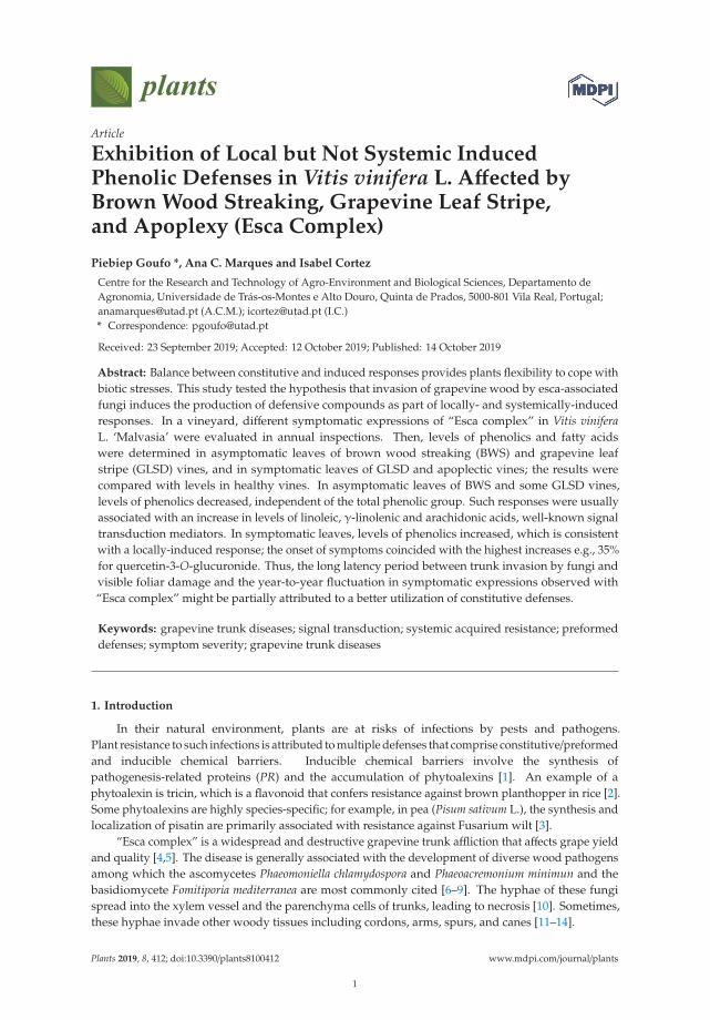
Exhibition of Local but Not Systemic InducedPhenolic Defenses in Vitis vinifera L. Affected byBrown Wood Streaking, Grapevine Leaf Stripe,and Apoplexy (Esca Complex)
Piebiep Goufo *, Ana C. Marques and Isabel Cortez
Centre for the Research and Technology of Agro-Environment and Biological Sciences, Departamento deAgronomia, Universidade de Trás-os-Montes e Alto Douro, Quinta de Prados, 5000-801 Vila Real, Portugal;[email protected] (A.C.M.); [email protected] (I.C.)* Correspondence: [email protected]
Received: 23 September 2019; Accepted: 12 October 2019; Published: 14 October 2019
Abstract: Balance between constitutive and induced responses provides plants flexibility to cope withbiotic stresses. This study tested the hypothesis that invasion of grapevine wood by esca-associatedfungi induces the production of defensive compounds as part of locally- and systemically-inducedresponses. In a vineyard, different symptomatic expressions of “Esca complex” in Vitis viniferaL. ‘Malvasia’ were evaluated in annual inspections. Then, levels of phenolics and fatty acidswere determined in asymptomatic leaves of brown wood streaking (BWS) and grapevine leafstripe (GLSD) vines, and in symptomatic leaves of GLSD and apoplectic vines; the results werecompared with levels in healthy vines. In asymptomatic leaves of BWS and some GLSD vines,levels of phenolics decreased, independent of the total phenolic group. Such responses were usuallyassociated with an increase in levels of linoleic, γ-linolenic and arachidonic acids, well-known signaltransduction mediators. In symptomatic leaves, levels of phenolics increased, which is consistentwith a locally-induced response; the onset of symptoms coincided with the highest increases e.g., 35%for quercetin-3-O-glucuronide. Thus, the long latency period between trunk invasion by fungi andvisible foliar damage and the year-to-year fluctuation in symptomatic expressions observed with“Esca complex” might be partially attributed to a better utilization of constitutive defenses.
Keywords: grapevine trunk diseases; signal transduction; systemic acquired resistance; preformeddefenses; symptom severity; grapevine trunk diseases
1. Introduction
In their natural environment, plants are at risks of infections by pests and pathogens.Plant resistance to such infections is attributed to multiple defenses that comprise constitutive/preformedand inducible chemical barriers. Inducible chemical barriers involve the synthesis ofpathogenesis-related proteins (PR) and the accumulation of phytoalexins [1]. An example of aphytoalexin is tricin, which is a flavonoid that confers resistance against brown planthopper in rice [2].Some phytoalexins are highly species-specific; for example, in pea (Pisum sativum L.), the synthesis andlocalization of pisatin are primarily associated with resistance against Fusarium wilt [3].
“Esca complex” is a widespread and destructive grapevine trunk affliction that affects grape yieldand quality [4,5]. The disease is generally associated with the development of diverse wood pathogensamong which the ascomycetes Phaeomoniella chlamydospora and Phaeoacremonium minimun and thebasidiomycete Fomitiporia mediterranea are most commonly cited [6–9]. The hyphae of these fungispread into the xylem vessel and the parenchyma cells of trunks, leading to necrosis [10]. Sometimes,these hyphae invade other woody tissues including cordons, arms, spurs, and canes [11–14].
Plants 2019, 8, 412; doi:10.3390/plants8100412 www.mdpi.com/journal/plants
1
Plants 2019, 8, 412
“Esca complex” exhibits a long latency time (several years) between wood colonization and visiblefoliar symptoms [5] and has become increasingly frequent worldwide. A ten-year survey conducted indifferent vine-growing regions of France revealed that the simplification of the woody vine structuremay have resulted in an increase in the incidence of the affliction in the country [15]. Some basicand practical cultural measures for preventing “Esca complex” have been proposed. For example,it was determined that increasing the length of cordons [15] and opting for a minimal pruning systeminstead of the standard spur-pruning [7] may help minimize the consequences of wood necroses.The foliar application of fertilizer mixtures containing calcium, magnesium, and Fucales seaweedwas found effective in reducing foliar symptoms and increasing the yield and quality of berries [4,16].The correlation between the symptomatic expression of “Esca complex” and the host physiology washighlighted by some authors [13,14,17]. This implies that characterizing the impact of the affliction ongrapevine physiology could help in finding candidate biomarkers associated with disease resistance.
Several studies have indicated that phytoalexins and in particular phenolic compounds(phenolic acids, flavonoids, anthocyanins, proanthocyanidins, and stilbenes) play a role in limiting thedevelopment of “Esca complex”. A typical reaction to wood colonization by esca-associated fungiis the accumulation of a mixture of polysaccharides (tyloses and gummosis) and the formation ofpolyphenol-rich reaction zones that obstruct the xylem to compartmentalize the fungi [18]. However,decreased levels of most phenolic compounds were observed in the xylem sap of vines with severewood symptoms [19], as well as a decreasing trend for the levels of amino acids involved in thebiosynthesis of phenolic compounds [20].
Rusjan et al. [10] found that esca-associated fungi caused the accumulation of flavonoids andstilbenes in both asymptomatic and necrotic trunks of vines. In particular, there was a high degree offlavonoid polymerization and a high level of procyanidins in the necrotic wood. However, reduction inthe levels of phenolic compounds in asymptomatic wood and no effect on the levels in symptomaticwood were reported for Pa. chlamydospora-infected young vines [12]. Further, no difference wasobserved in total analyzed phenolics in asymptomatic stems of healthy and infected vines in the studyby Magnin-Robert et al. [13]; however, a considerable accumulation of stilbenes—trans-resveratrol andtrans-vitisin B—was observed in the affected vines.
It was also demonstrated that vines respond to “Esca complex” by accumulating stilbenes innaturally infected leaves [21] and leaves infected ex vivo with Pa. chlamydospora [22]. These increaseswere accompanied with the up-regulation of phenylalanine ammonia-lyase (PAL) and stilbene synthase(StSy)—two genes involved in the biosynthesis of polyphenolic compounds—in green [22] and dryleaves [23]. The accumulation of phenolic acids and flavonoids in symptomatic and asymptomaticleaves of field-grown vines was also reported [24]. However, in the study by Martín et al. [25], it wasdemonstrated that the appearance of foliar symptoms led to a decrease in the levels of flavonoids,proanthocyanidins, and hydroxycinnamic acids in the leaves of Vitis vinifera L. ‘Tempranillo’ grownunder a dry and warm temperature. For the same cultivar grown under a hot and humid temperature,hydroxycinnamic acids levels increased in symptomatic leaves whereas flavonoids levels decreased.Interestingly, levels of trans-resveratrol in asymptomatic leaves of affected vines were slightly higherthan those in healthy leaves of non-affected vines in some vineyards in Italy [26].
It is evident from the abovementioned findings that there are different responses of phenoliccompounds to “Esca complex”. These different results can be attributed to the types and complexitiesof symptomatic and asymptomatic materials studied by different authors.
Internal wood symptoms in adult vines are characterized by two diverse shapes of necrotic areasand discolorations. One shape/discoloration is caused by F. mediterranea and is called “white rot” orsimply “esca” it is characterized by a clear/yellowish soft and spongy mass of wood usually in thecenter of the trunk or cordons, which can be observed alone or with dark-brown to black spots in thexylem vessels [10,11,14,19]. The second shape/discoloration refers to different types of brown woodnecrosis of which “dark/brown wood streaking” (BWS) is most commonly reported; BWS consistsof extended columnar strips of xylem necrosis with pink-brown to dark-brown areas or black spots
2
Plants 2019, 8, 412
around the annual growth section [13,19]. A third type of shape/discoloration (wood stripe), which ispresent in external vine wood, is also reported and the symptoms appear as a longitudinal andsuperficial yellowish-orange stripe and orange-brown discolorations of the young wood vessels locatedimmediately below the bark [27].
Two typical severity levels of leaf symptoms are observed in esca-affected grapevines. A chronicform, characterized by tiger-striped symptoms (GLS) (also named by some authors GLSD for “GrapevineLeaf Stripe Disease”), is initially characterized by chlorosis and then light-green irregular spots and/orscorching between the main veins and/or along the leaf margins. The chlorotic and drying areasgradually expand from the basal to the distal part of the leaves, and then they coalesce to becomepartial necrotic stripes. As the chlorotic tissues turn yellow-brown or red-brown, the leaves exhibita tiger stripe pattern [17,19,25–28]. GLSD symptoms are also reported in the berries and consist oftiny dark-brown or purple speckling distributed irregularly over the entire surface or scattered at thefar end (termed “black measles” by some authors) and sometimes of shriveling/withering of grapebunches [18]. “Apoplexy” consists of partial or complete sudden wilting of the crown and is consideredan acute form of the leaf symptomatic expression of “Esca complex” [23,27] or an acute form of GLSDby some authors [14]. BWS and GLSD vines are associated with a large procession of inhabitingfungi, although Pa. chlamydospora and Pm. minimun are most commonly found [18]. Although thepercentage of necrotic areas within the wood from which pathogens can be isolated is often a key factorto determine the severity of ”Esca complex,” wood necrosis is not always related to the incidenceof foliar symptoms [15]. Given this observation, the term “esca proper” is used by some authors toindicate the coexistence of “white rot” and GLSD in the same vine [13,14]. “Esca complex” is mostcommonly noted in established vineyards. In newly planted vines, scattered brown-black spots ofnecrotic xylem (without decay), often with a dark viscous ooze and a moderate/diffuse chlorosis of theleaves are observed, and the disease is termed “Petri disease” [6,8,12].
Studies have shown that many abiotic factors and cultural practices (alone or combined) mayinfluence the development of “Esca complex” and the variability of its damage [15,20,26]. For example,it has been observed that heavy rainfall followed by hot winds in mid-summer favors the onset ofapoplexy [18]. These observations were confirmed by other authors [25], who then reported that thebiosynthesis of phenolic compounds in esca-affected leaves depended on the climate under which thevines were grown.
The above literature review shows that potential defense mechanisms developed by grapevineto resist esca-associated fungi need to be explored further. Therefore, the experiment in this studywas designed to produce complementary data that would help improve the understanding of defenseevents occurring during an “Esca” invasion. It was hypothesized that esca-associated fungi inducethe production of defensive compounds in leaves as part of both a locally- and systemically-induceddefense response; local induction is defined as the enhancement of defensive traits in the organ thatis attacked, while systemic induction is the enhancement of defenses in distant and undamagedorgans, conferring broad-spectrum resistance throughout the plant [29,30]. To test this hypothesis,the accumulation of polyphenols in the leaves of vineyard-grown plants was monitored and levels offatty acids were determined. Recent studies demonstrated that fatty acids play an important role in themodulation of signal transduction pathways in systemically acquired pathogen resistance. In severalplants, the degree of resistance to pathogens was found to be directly correlated with the levels ofC16:1 (palmitoleic acid), C18:1 (oleic and elaidic acids), C18:2 (linoleic and linolelaidic acids), C18:3(α-linolenic and γ-linolenic acids), and C20:4 (achidonic acid) [1]. For example, rhizobacteria-inducedenhanced resistance to Botrytis cinerea is associated with the accumulation of C18:2 and C18:3 inPhaseolus vulgaris L. [31], while reduction in C18:1 level induces defense responses against severalpathogens by upregulating expressions of a variety of structurally diverse R genes in Arabidopsis [32].Therefore, the levels of phenolic compounds and fatty acids in asymptomatic and symptomatic leavesof grapevine affected by BWS, GLSD and apoplexy were investigated to identify infection stages atwhich plant resistance mechanisms were more efficiently activated.
3
Plants 2019, 8, 412
2. Results
2.1. Effect of Brown Wood Streaking, Grapevine Leaf Stripe and Apoplexy on the Levels of Phenolic Compoundsin Grapevine Leaves
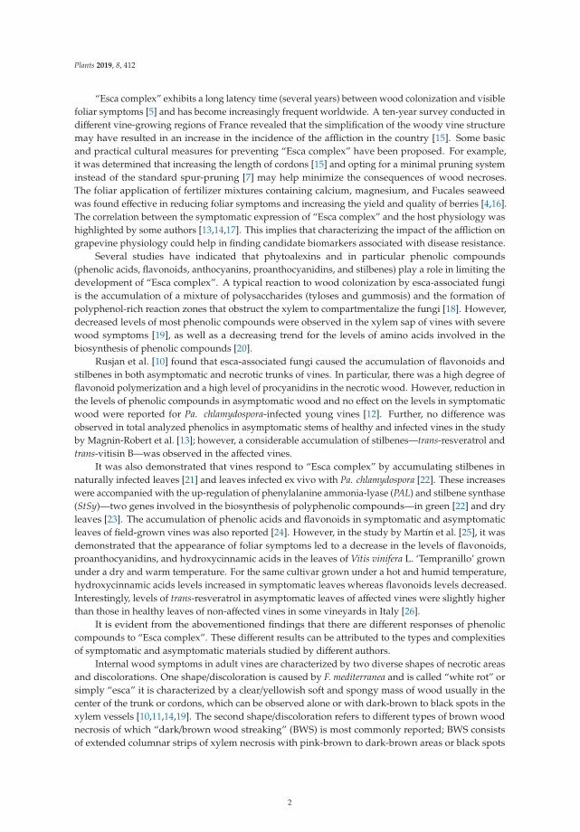
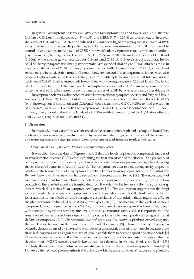
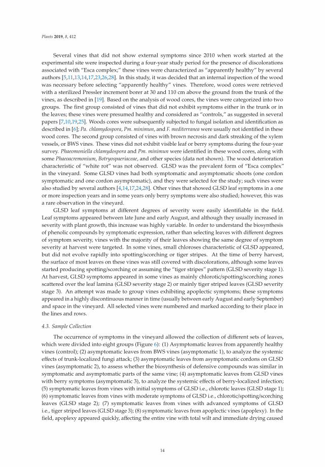
In this study, the amount of total phenolic compounds in the leaves of vines affected by “Escacomplex” was first analyzed using colorimetric methods (Figure 1). An interesting trend emerged inthat asymptomatic leaves of BWS and GLSD vines had a lower amount of TPC than that in controlleaves, with a decrease of 14% in leaves of BWS vines (asymptomatic 1). The amount of TPC wasparticularly high in leaves exhibiting the initial foliar symptoms (GLSD stage 1) as compared to that incontrol leaves, and then, it decreased in proportion to the severity of chlorosis and necrosis on the leaves.Changes in the amount of TAC, TPAC, and TFC due to BWS and GLSD were similar to changes in theamount of TPC, with some exceptions: the highest amount of TAC was measured in asymptomaticleaves of GLSD vines that had both symptomatic and asymptomatic cordons (asymptomatic 2), and a64% increase in the amount of TAC was recorded passing from chlorotic/spotting/scorching leaves(GLSD stage 2) to tiger striped (GLSD stage 3) and apoplectic leaves (Figure 1).
Figure 1. Total phenolic (TPC), anthocyanin (TAC), proanthocyanidin (TPAC), and flavonoid (TFC)content (dry weight basis) in asymptomatic and symptomatic leaves of vines affected by brown woodstreaking, grapevine leaf stripe and apoplexy. The legend is as in Figure 6. Error bars = standarddeviations (n = 4); different letters above the columns denote statistical differences (Tukey’s test;P ≤ 0.05).
The HPLC method used in this study led to the separation of 104 peaks with 95 peaksshowing phenolic characteristics. Using the information provided by the detector and reportsin the literature, the peaks were assigned to metabolites of the structure classes hydroxybenzoicacid (9), hydroxydiphenic acid (1), proanthocyanidin (9), stilbene (1), hydroxycinnamic acid (16),flavonoid (37), and anthocyanin (10). Eight metabolites were labeled “unknown,” whereas fourexhibited the characteristics of both proanthocyanidins and hydroxybenzoic acids and were labeledas “benzoic acid derivatives.” On average, quercetin-3-O-glucuronide was the major phenolic
4
Plants 2019, 8, 412
compound in the leaves (2834.43 mg· kg−1) followed by myricetin-3-O-galactoside (127.68 mg· kg−1),quercetin-3-O-glucoside (127.32 mg· kg−1), quercetin-3-O-galactoside (83.35 mg· kg−1), caftaric acid(82.78 mg· kg−1), myricetin-3-O-glucoside (60.82 mg· kg−1), kaempferol-3-O-glucoside (54.01 mg· kg−1),coutaric acid (49.00 mg· kg−1), epicatechin (37.93 mg· kg−1), quercetin-3-O-rutinoside (23.10 mg· kg−1),kaempferol-3-O-rutinoside (13.72 mg· kg−1), and epigallocatechin gallate (12.38 mg· kg−1).The levels of the remaining compounds were below 10 mg· kg−1 (Table S1). In some samples,quercetin-3-O-glucuronide and quercetin-3-O-glucoside co-eluted in the chromatograms; therefore,the levels of these two compounds were summed and used in the statistical analyses; the same wastrue for myricetin-3-O-galactoside and myricetin-3-O-glucuronide. In V. vinifera, several stilbeneshave been reported as stress response metabolites [22]. In this study, only one stilbene was detected,which was identified as trans-resveratrol, with an average content of 0.98 mg· kg−1 (Table S1). This valuewas substantially lower than 1.38–50.49 mg· kg−1, which was observed in the leaves of some Italiancultivars [26]. The non-detection of stilbenes was not surprising because stilbenic compounds areusually detected by HPLC from a filtrate obtained after several solid–liquid and liquid–liquid extractionand purification steps [33]. The clean-up step used in this study was aimed at discarding chlorophyllsand chromatography was optimized for the separation of flavonoids and proanthocyanidins.
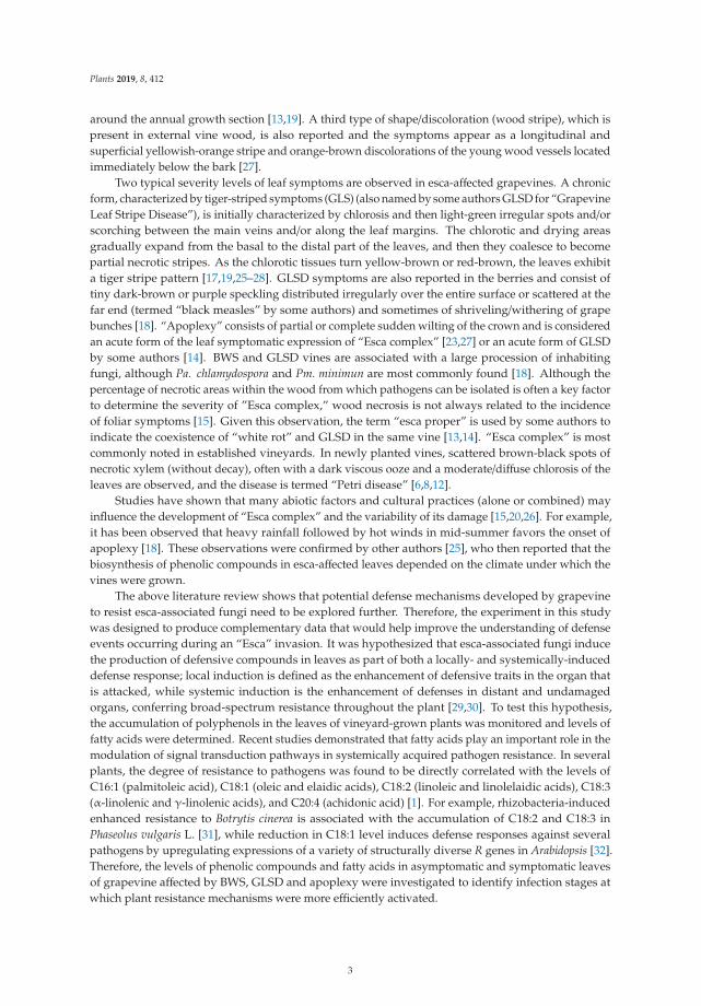
The major compounds (average content ≥ 1.00 mg· kg−1; Table S1) were first analyzed usingANOVA. This analysis allowed two main categories of compounds to be delineated, on the basis ofsimilar trends in the contents observed comparing control, asymptomatic and symptomatic leaves.
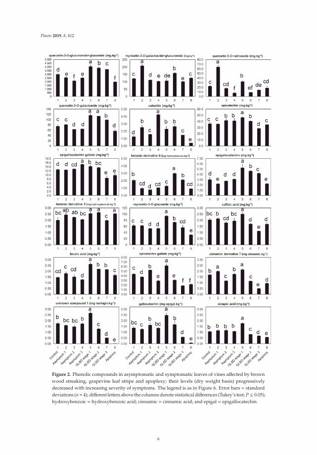
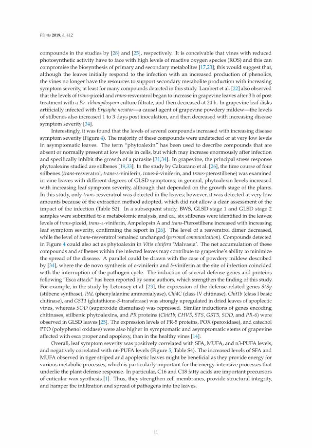
The first category (Figure 2) consisted of 20 compounds that showed three characteristics.(i) The levels of these compounds were particularly high in symptomatic leaves exhibiting theinitial foliar symptoms of GLSD (GLSD stage 1) compared with those in control and asymptomaticleaves. The percentage increase between control and GLSD stage 1 leaves ranged from 13%(epigallocatechin gallate) to 81% (catechin). However, there were some exceptions: the levels ofmyricetin-3-O-galactoside+myricetin-3-O-glucuronide and quercetin-3-O-rutinoside were the highestin asymptomatic leaves of BWS vines (asymptomatic 1), and the levels of epigallocatechin gallate andcatechin were the highest in asymptomatic leaves of GLSD vines with berry symptoms (asymptomatic3). (ii) The levels of these compounds progressively decreased with the increasing severity of theleaf symptom, with the lowest values usually being measured in apoplectic leaves. However, forsome compounds a slight level increase was observed in apoplectic leaves as compared to tigerstriped leaves (GLSD stage 3); this suggests that apoplexy might not only be a severe form of GLSD.These compounds included epicatechin, benzoic acid derivative 5, caffeic acid, hydroxycinnamic acidderivative 7, myricetin-3-O-galactoside+myricetin-3-O-glucuronide, and quercetin-3-O-rutinoside.(iii) The levels of these compounds were generally similar in asymptomatic leaves or lowerin asymptomatic leaves of BWS and GLSD vines than those in control leaves. For example,the levels of quercetin-3-O-glucuronide+quercetin-3-O-glucoside decreased by 14, 29, and 16% inasymptomatic leaves of BWS (asymptomatic 1), GLSD foliar-symptomatic (asymptomatic 2), and GLSDberry-symptomatic (asymptomatic 3) vines, respectively. The levels of only a few compounds increasedin asymptomatic leaves and that included an 18, 71, 188, 70, 99, 20, and 17 increase for hydroxycinnamicacid derivative 7, myricetin-3-O-galactoside+myricetin-3-O-glucuronide, quercetin-3-O-rutinosidein asymptomatic leaves of BWS vines, catechin in asymptomatic leaves of BWS vines, catechin inasymptomatic leaves of GLSD berry-symptomatic vines, epicatechin gallate in asymptomatic leavesof GLSD foliar-symptomatic vines, and epigallocatechin gallate in asymptomatic leaves of GLSDberry-symptomatic vines, respectively.
5
Plants 2019, 8, 412
Figure 2. Phenolic compounds in asymptomatic and symptomatic leaves of vines affected by brownwood streaking, grapevine leaf stripe and apoplexy; their levels (dry weight basis) progressivelydecreased with increasing severity of symptoms. The legend is as in Figure 6. Error bars = standarddeviations (n= 4); different letters above the columns denote statistical differences (Tukey’s test; P ≤ 0.05);hydroxybenzoic = hydroxybenzoic acid; cinnamic = cinnamic acid; and epigal = epigallocatechin.
6
Plants 2019, 8, 412
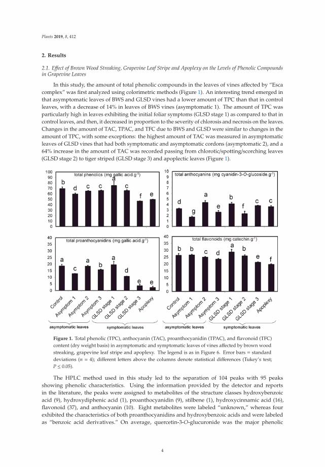
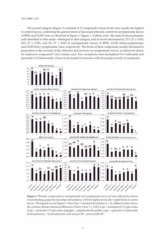
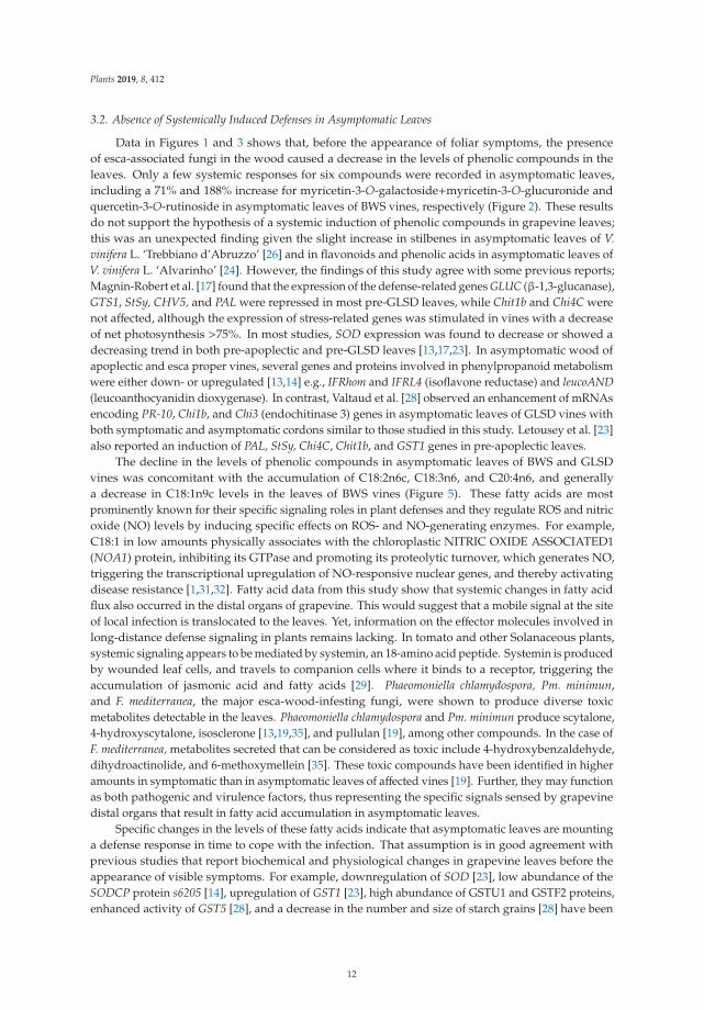
The second category (Figure 3) consisted of 13 compounds whose levels were usually the highestin control leaves, confirming the general trend of decreased phenolic content in asymptomatic leavesof BWS and GLSD vines as observed in Figure 1; Figure 2. Caftaric acid—the main hydroxycinnamicacid identified in this study—belonged to that category and its levels decreased by 27% (P ≤ 0.05),20% (P ≤ 0.05), and 8% (P > 0.05) in asymptomatic leaves of BWS, GLSD foliar-symptomatic,and GLSD berry-symptomatic vines, respectively. The levels of these compounds usually decreased inproportion to the severity of the chlorosis and necrosis on symptomatic leaves, as observed clearlyfor unknown compound 7 and coutaric acid. Few exceptions were kaempferol-3-O-rutinoside andquercetin-3-O-rhamnoside, whose levels tended to increase with increasing severity of symptoms.
Figure 3. Phenolic compounds in asymptomatic and symptomatic leaves of vines affected by brownwood streaking, grapevine leaf stripe and apoplexy, with the highest levels (dry weight basis) in controlleaves. The legend is as in Figure 6. Error bars = standard deviations (n = 4); different letters abovethe columns denote statistical differences (Tukey’s test; P ≤ 0.05); k-glc = kaempferol-3-O-glucoside;m-glc =myricetin-3-O-glucoside; epigalgal = epigallocatechin gallate; q-glc = quercetin-3-O-glucoside;hydroxybenzoic = hydroxybenzoic acid; and pro B1 = procyanidin B1.
7
Plants 2019, 8, 412
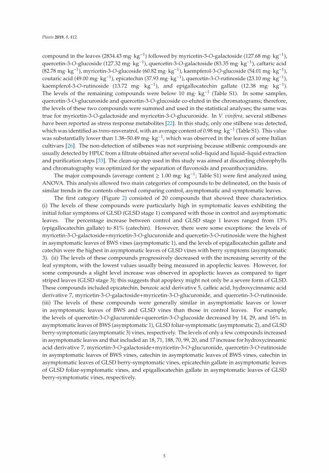
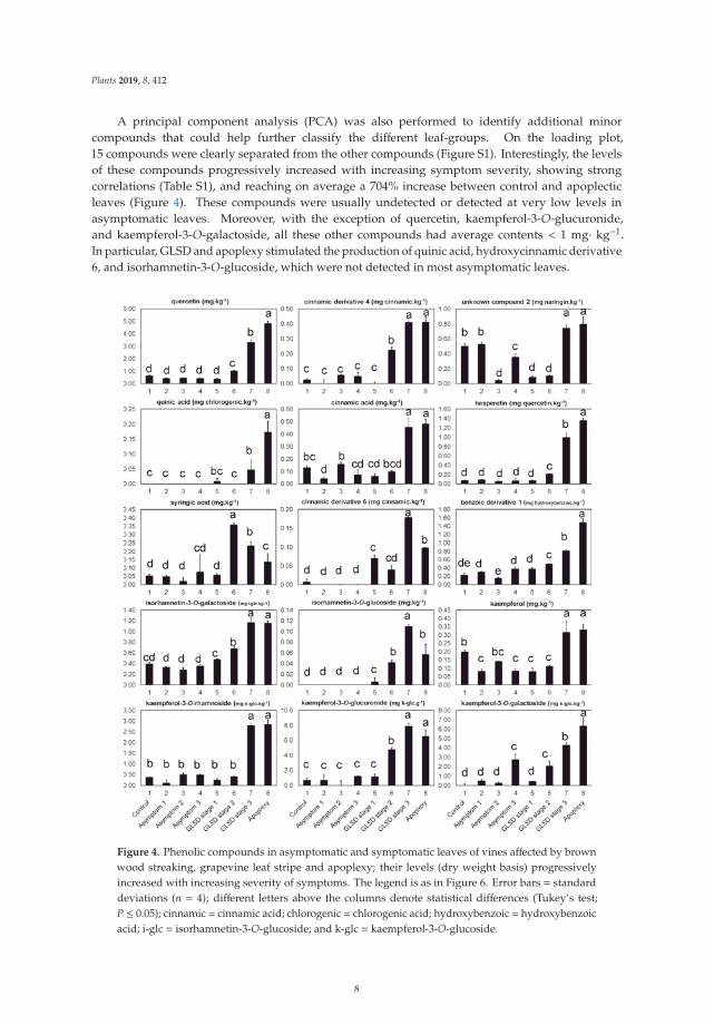
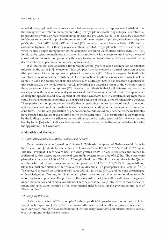
A principal component analysis (PCA) was also performed to identify additional minorcompounds that could help further classify the different leaf-groups. On the loading plot,15 compounds were clearly separated from the other compounds (Figure S1). Interestingly, the levelsof these compounds progressively increased with increasing symptom severity, showing strongcorrelations (Table S1), and reaching on average a 704% increase between control and apoplecticleaves (Figure 4). These compounds were usually undetected or detected at very low levels inasymptomatic leaves. Moreover, with the exception of quercetin, kaempferol-3-O-glucuronide,and kaempferol-3-O-galactoside, all these other compounds had average contents < 1 mg· kg−1.In particular, GLSD and apoplexy stimulated the production of quinic acid, hydroxycinnamic derivative6, and isorhamnetin-3-O-glucoside, which were not detected in most asymptomatic leaves.
Figure 4. Phenolic compounds in asymptomatic and symptomatic leaves of vines affected by brownwood streaking, grapevine leaf stripe and apoplexy; their levels (dry weight basis) progressivelyincreased with increasing severity of symptoms. The legend is as in Figure 6. Error bars = standarddeviations (n = 4); different letters above the columns denote statistical differences (Tukey’s test;P ≤ 0.05); cinnamic = cinnamic acid; chlorogenic = chlorogenic acid; hydroxybenzoic = hydroxybenzoicacid; i-glc = isorhamnetin-3-O-glucoside; and k-glc = kaempferol-3-O-glucoside.
8
Plants 2019, 8, 412
Overall, the levels of the remaining minor compounds were not affected in asymptomatic leaves,with the exception of four compounds that were detected primarily in these leaves (hydroxycinnamicacid derivative 2, unknown compound 4, unidentified flavonol 8, and p-hydroxybenzoic acid).For symptomatic leaves, the levels of some remaining minor compounds increased, while those of theothers decreased with the increasing severity of symptoms (Table S2).
2.2. Effect of Brown Wood Streaking, Grapevine Leaf Stripe and Apoplexy on the Levels of Fatty Acids inGrapevine Leaves
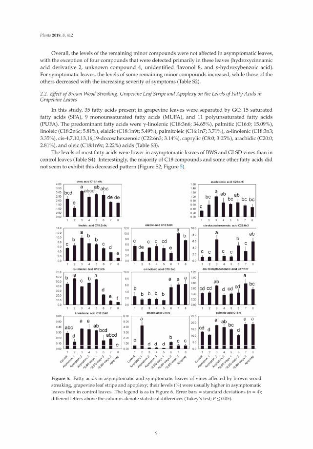
In this study, 35 fatty acids present in grapevine leaves were separated by GC: 15 saturatedfatty acids (SFA), 9 monounsaturated fatty acids (MUFA), and 11 polyunsaturated fatty acids(PUFA). The predominant fatty acids were γ-linolenic (C18:3n6; 34.65%), palmitic (C16:0; 15.09%),linoleic (C18:2n6c; 5.81%), elaidic (C18:1n9t; 5.49%), palmitoleic (C16:1n7; 3.71%), α-linolenic (C18:3n3;3.35%), cis-4,7,10,13,16,19-docosahexaenoic (C22:6n3; 3.14%), caprylic (C8:0; 3.05%), arachidic (C20:0;2.81%), and oleic (C18:1n9c; 2.22%) acids (Table S3).
The levels of most fatty acids were lower in asymptomatic leaves of BWS and GLSD vines than incontrol leaves (Table S4). Interestingly, the majority of C18 compounds and some other fatty acids didnot seem to exhibit this decreased pattern (Figure S2; Figure 5).
Figure 5. Fatty acids in asymptomatic and symptomatic leaves of vines affected by brown woodstreaking, grapevine leaf stripe and apoplexy; their levels (%) were usually higher in asymptomaticleaves than in control leaves. The legend is as in Figure 6. Error bars = standard deviations (n = 4);different letters above the columns denote statistical differences (Tukey’s test; P ≤ 0.05).
9
Plants 2019, 8, 412
In general, asymptomatic leaves of BWS vines (asymptomatic 1) had lower levels of C18:1n9c,C18:1n9t, C18:2n6t (linolelaidic acid) (P ≤ 0.05), and C18:3n3 (P > 0.05) than control leaves; however,the levels of C18:2n6c, C18:0 (stearic acid), and C18:3n6 were higher in asymptomatic leaves of BWSvines than in control leaves. In particular, a 600% increase was observed for C18:0. Compared tocontrol leaves, asymptomatic leaves of GLSD vines with both asymptomatic and symptomatic cordons(asymptomatic 2) had higher levels of C18:1n9c, C18:2n6c, and C18:2n6t, and lower levels of C18:0 andC18:3n6, while no change was recorded for C18:1n9t and C18:3n3. C18 levels in asymptomatic leavesof GLSD berry-symptomatic vines (asymptomatic 3) responded similarly to “Esca” attack as those inasymptomatic leaves of GLSD foliar-symptomatic vines, with the exception of C18:3n6, whose levelremained unchanged. Substantial differences between control and asymptomatic leaves were alsoobserved with regards to the levels of C16:0, C17:1n7 (cis-10-heptadecenoic acid), C20:4n6 (arachidonicacid), and C22:6n3. In all asymptomatic leaves, there was a strong increase in C20:4n6 levels. The levelsof C17:1n7, C22:6n3, and C16:0 increased in asymptomatic leaves of GLSD foliar-symptomatic vines,while the level of C16:0 increased in asymptomatic leaves of GLSD berry-symptomatic vines (Figure 5).
In symptomatic leaves, a distinct correlation between disease symptom severity and fatty acid levelswas observed (Table S3). Overall, leaf symptom severity was positively correlated with the levels of SFA(with the exception of tricosanoic acid C23:0 and heptadecanoic acid C17:0), MUFA (with the exceptionof C18:1n9c), and n3-PUFA (with the exception of cis-5,8,11,14,17-eicosapentaenoic acid C20:5n3),and negatively correlated with the levels of n6-PUFA (with the exception of cis-11,14-eicosadienoicacid C20:2n6) (Figure 5; Tables S3 and S4).
3. Discussion
In this study, great variability was observed in the accumulation of phenolic compounds and fattyacids in grapevine as a response to infection by esca-associated fungi, which indicated that dynamicand transient metabolic changes occur when symptoms spread from the trunk to the leaves.
3.1. Exhibition of Locally Induced Defenses in Symptomatic Leaves
It was clear from the data in Figures 1 and 2 that the levels of phenolic compounds increasedin symptomatic leaves of GLSD vines exhibiting the first symptoms of the disease. The precocity ofpathogen recognition and the velocity of the activation of defense responses are keys to enhancingthe resistance of plants to infections [1,22,34]. The recognition of esca-related pathogens by grapevineplants and the formation of foliar symptoms are debated topics because propagules of Pa. chlamydospora,Pm. minimun, and F. mediterranea have never been detected on the leaves [18]. The most acceptedinterpretation is that toxic metabolites secreted by esca-associated fungi or resulting from reactionproducts of the infected wood are translocated from the xylem to the leaves via the transpiration/sapstream, which thus incites foliar symptom development [18]. This assumption suggests that the fungiinduced local defense responses in grapevine when their metabolites reached the host leaf cells. In fact,foliar administration of calcium and subsequent accumulation of calmodulin, that mitigate the effect ofthe plant response, reduced GLSD leaf symptom expression [16]. The increase in the levels of phenoliccompounds was the greatest when GLSD symptoms started appearing on the leaves. However,with increasing symptom severity, the levels of these compounds decreased. It is reported that theresistance of plants to infections depends partly on the balance between production/degradation ofdefensive compounds [19]. Phaeomoniella chlamydospora and Pm. minimun produce several enzymesthat are known to travel in the plant and could reach the leaves [19]. However, the hypothesis of aphenolic decrease caused by enzymatic activities of esca-associated fungi is not tenable because thesefungi lack enzymes such as ligninases, which would enable them to degrade specific phenolic bonds [9].These decreases were also unlikely to be caused merely by chlorosis and necrosis. It is known that thedevelopment of GLSD necrotic areas in leaves leads to a decrease in photosynthetic assimilation [23].Similarly, the expression of photosynthesis-related genes is strongly repressed in apoplectic leaves [28].However, the reduced photosynthesis did coincide with the accumulation of hexoses and phenolic
10
Plants 2019, 8, 412
compounds in the studies by [28] and [25], respectively. It is conceivable that vines with reducedphotosynthetic activity have to face with high levels of reactive oxygen species (ROS) and this cancompromise the biosynthesis of primary and secondary metabolites [17,23]; this would suggest that,although the leaves initially respond to the infection with an increased production of phenolics,the vines no longer have the resources to support secondary metabolite production with increasingsymptom severity, at least for many compounds detected in this study. Lambert et al. [22] also observedthat the levels of trans-piceid and trans-resveratrol began to increase in grapevine leaves after 3 h of posttreatment with a Pa. chlamydospora culture filtrate, and then decreased at 24 h. In grapevine leaf disksartificially infected with Erysiphe necator—a causal agent of grapevine powdery mildew—the levelsof stilbenes also increased 1 to 3 days post inoculation, and then decreased with increasing diseasesymptom severity [34].
Interestingly, it was found that the levels of several compounds increased with increasing diseasesymptom severity (Figure 4). The majority of these compounds were undetected or at very low levelsin asymptomatic leaves. The term “phytoalexin” has been used to describe compounds that areabsent or normally present at low levels in cells, but which may increase enormously after infectionand specifically inhibit the growth of a parasite [31,34]. In grapevine, the principal stress responsephytoalexins studied are stilbenes [19,33]. In the study by Calzarano et al. [26], the time course of fourstilbenes (trans-resveratrol, trans-ε-viniferin, trans-δ-viniferin, and trans-pterostilbene) was examinedin vine leaves with different degrees of GLSD symptoms; in general, phytoalexin levels increasedwith increasing leaf symptom severity, although that depended on the growth stage of the plants.In this study, only trans-resveratrol was detected in the leaves; however, it was detected at very lowamounts because of the extraction method adopted, which did not allow a clear assessment of theimpact of the infection (Table S2). In a subsequent study, BWS, GLSD stage 1 and GLSD stage 2samples were submitted to a metabolomic analysis, and ca., six stilbenes were identified in the leaves;levels of trans-piceid, trans-ε-viniferin, Ampelopsin A and trans-Pterostilbene increased with increasingleaf symptom severity, confirming the report in [26]. The level of a resveratrol dimer decreased,while the level of trans-resveratrol remained unchanged (personal communication). Compounds detectedin Figure 4 could also act as phytoalexin in Vitis vinifera ‘Malvasia’. The net accumulation of thesecompounds and stilbenes within the infected leaves may contribute to grapevine’s ability to minimizethe spread of the disease. A parallel could be drawn with the case of powdery mildew describedby [34], where the de novo synthesis of ε-viniferin and δ-viniferin at the site of infection coincidedwith the interruption of the pathogen cycle. The induction of several defense genes and proteinsfollowing “Esca attack” has been reported by some authors, which strengthen the finding of this study.For example, in the study by Letousey et al. [23], the expression of the defense-related genes StSy(stilbene synthase), PAL (phenylalanine ammonialyase), Chi4C (class IV chitinase), Chit1b (class I basicchitinase), and GST1 (glutathione-S-transferase) was strongly upregulated in dried leaves of apoplecticvines, whereas SOD (superoxide dismutase) was repressed. Similar inductions of genes encodingchitinases, stilbenic phytoalexins, and PR proteins (Chit1b; CHV5, STS, GST5, SOD, and PR-6) wereobserved in GLSD leaves [25]. The expression levels of PR-5 proteins, POX (peroxidase), and catecholPPO (polyphenol oxidase) were also higher in symptomatic and asymptomatic stems of grapevineaffected with esca proper and apoplexy, than in the healthy vines [14].
Overall, leaf symptom severity was positively correlated with SFA, MUFA, and n3-PUFA levels,and negatively correlated with n6-PUFA levels (Figure 5; Table S4). The increased levels of SFA andMUFA observed in tiger striped and apoplectic leaves might be beneficial as they provide energy forvarious metabolic processes, which is particularly important for the energy-intensive processes thatunderlie the plant defense response. In particular, C16 and C18 fatty acids are important precursorsof cuticular wax synthesis [1]. Thus, they strengthen cell membranes, provide structural integrity,and hamper the infiltration and spread of pathogens into the leaves.
11
Plants 2019, 8, 412
3.2. Absence of Systemically Induced Defenses in Asymptomatic Leaves
Data in Figures 1 and 3 shows that, before the appearance of foliar symptoms, the presenceof esca-associated fungi in the wood caused a decrease in the levels of phenolic compounds in theleaves. Only a few systemic responses for six compounds were recorded in asymptomatic leaves,including a 71% and 188% increase for myricetin-3-O-galactoside+myricetin-3-O-glucuronide andquercetin-3-O-rutinoside in asymptomatic leaves of BWS vines, respectively (Figure 2). These resultsdo not support the hypothesis of a systemic induction of phenolic compounds in grapevine leaves;this was an unexpected finding given the slight increase in stilbenes in asymptomatic leaves of V.vinifera L. ‘Trebbiano d’Abruzzo’ [26] and in flavonoids and phenolic acids in asymptomatic leaves ofV. vinifera L. ‘Alvarinho’ [24]. However, the findings of this study agree with some previous reports;Magnin-Robert et al. [17] found that the expression of the defense-related genes GLUC (β-1,3-glucanase),GTS1, StSy, CHV5, and PAL were repressed in most pre-GLSD leaves, while Chit1b and Chi4C werenot affected, although the expression of stress-related genes was stimulated in vines with a decreaseof net photosynthesis >75%. In most studies, SOD expression was found to decrease or showed adecreasing trend in both pre-apoplectic and pre-GLSD leaves [13,17,23]. In asymptomatic wood ofapoplectic and esca proper vines, several genes and proteins involved in phenylpropanoid metabolismwere either down- or upregulated [13,14] e.g., IFRhom and IFRL4 (isoflavone reductase) and leucoAND(leucoanthocyanidin dioxygenase). In contrast, Valtaud et al. [28] observed an enhancement of mRNAsencoding PR-10, Chi1b, and Chi3 (endochitinase 3) genes in asymptomatic leaves of GLSD vines withboth symptomatic and asymptomatic cordons similar to those studied in this study. Letousey et al. [23]also reported an induction of PAL, StSy, Chi4C, Chit1b, and GST1 genes in pre-apoplectic leaves.
The decline in the levels of phenolic compounds in asymptomatic leaves of BWS and GLSDvines was concomitant with the accumulation of C18:2n6c, C18:3n6, and C20:4n6, and generallya decrease in C18:1n9c levels in the leaves of BWS vines (Figure 5). These fatty acids are mostprominently known for their specific signaling roles in plant defenses and they regulate ROS and nitricoxide (NO) levels by inducing specific effects on ROS- and NO-generating enzymes. For example,C18:1 in low amounts physically associates with the chloroplastic NITRIC OXIDE ASSOCIATED1(NOA1) protein, inhibiting its GTPase and promoting its proteolytic turnover, which generates NO,triggering the transcriptional upregulation of NO-responsive nuclear genes, and thereby activatingdisease resistance [1,31,32]. Fatty acid data from this study show that systemic changes in fatty acidflux also occurred in the distal organs of grapevine. This would suggest that a mobile signal at the siteof local infection is translocated to the leaves. Yet, information on the effector molecules involved inlong-distance defense signaling in plants remains lacking. In tomato and other Solanaceous plants,systemic signaling appears to be mediated by systemin, an 18-amino acid peptide. Systemin is producedby wounded leaf cells, and travels to companion cells where it binds to a receptor, triggering theaccumulation of jasmonic acid and fatty acids [29]. Phaeomoniella chlamydospora, Pm. minimun,and F. mediterranea, the major esca-wood-infesting fungi, were shown to produce diverse toxicmetabolites detectable in the leaves. Phaeomoniella chlamydospora and Pm. minimun produce scytalone,4-hydroxyscytalone, isosclerone [13,19,35], and pullulan [19], among other compounds. In the case ofF. mediterranea, metabolites secreted that can be considered as toxic include 4-hydroxybenzaldehyde,dihydroactinolide, and 6-methoxymellein [35]. These toxic compounds have been identified in higheramounts in symptomatic than in asymptomatic leaves of affected vines [19]. Further, they may functionas both pathogenic and virulence factors, thus representing the specific signals sensed by grapevinedistal organs that result in fatty acid accumulation in asymptomatic leaves.
Specific changes in the levels of these fatty acids indicate that asymptomatic leaves are mountinga defense response in time to cope with the infection. That assumption is in good agreement withprevious studies that report biochemical and physiological changes in grapevine leaves before theappearance of visible symptoms. For example, downregulation of SOD [23], low abundance of theSODCP protein s6205 [14], upregulation of GST1 [23], high abundance of GSTU1 and GSTF2 proteins,enhanced activity of GST5 [28], and a decrease in the number and size of starch grains [28] have been
12
Plants 2019, 8, 412
reported in asymptomatic leaves of esca-affected grapevine as an early response of cells distant fromthe damaged wood. Within the week preceding leaf symptoms, drastic physiological alterations ofphotosynthesis were also registered in pre-apoplectic and pre-GLSD leaves, as revealed by a decreasein CO2 assimilation, chlorophyll a fluorescence, and the repression of photosynthesis-related genespsbP1, rbcL, rbcS, SBP [17,23], PRK, and Lhca3 [17] probably due to a lower activity of Rubsico orcarbonic anhydrase [18]. Other metabolic alterations detected in asymptomatic leaves of esca-affectedvines include a slight upregulation of the aquaporin-encoding water-stress-related gene TIP1 [23].In this study, resistance mechanisms activated in asymptomatic leaves seem to first involve the use ofconserved antimicrobial compounds by the vines to respond to infection rapidly, as revealed by thedecreased levels of phenolic compounds (Figures 1 and 3).
It is known that esca-associated fungi require several years of wood colonization to establishinfection in the leaves [19]. Moreover, ”Esca complex” is characterized by partial remission or totaldisappearance of foliar symptoms on plants in some years [12]. The year-to-year fluctuation insymptom expression has been attributed to the combination of optimal circumstances which includerainfall [5], and the occurrence of abiotic stresses such as drought [20]. It has also been hypothesizedthat each season, the newly formed vessels redefining the vascular system of the vine may affectthe appearance of foliar symptoms [27]. Another hypothesis is that local defense reaction is theconsequence of the development of drying zones and discolorations after a sudden sap disruption afteror along the apparition and development of leaf stripe symptoms [27]. A complementary hypothesiscould be that symptoms sometimes do not appear because of the activation of constitutive defenses.These pre-formed compounds could be effective in restraining the propagation of fungi in the woodand the translocation of their metabolites to the leaves, depending on the years and environmentalconditions. The induced production of phenolic compounds would only occur after the metaboliteshave reached the leaves at doses sufficient to incite symptoms. This assumption is strengthenedby the finding that ex vivo, stilbenes do not influence the damaging effects of Pa. chlamydospora onhealthy leaves [26], which indicates that phytoalexins are synthesized in the leaves not before, but after,the apparition GLSD symptoms.
4. Materials and Methods
4.1. Site Characterization: Cultivar, Location, and Weather
Experiments were performed on V. vinifera L. ‘Malvasia’ composed of 21–24-year-old plants inthe vineyard of Quinta de Nossa Senhora de Loures (465 m, 41◦ 17.12’ 31” N, 7◦ 44.07’ 22” W) inVila Real, Portugal. The vineyard has 1247 vines grafted on 196-17-Castel rootstock and trained toa bilateral cordon according to the royal-type trellis system, on an area of 0.27 ha. The vines wereplanted at a distance of 1.80 × 1.20 m in 22 longitudinal rows. The climatic conditions in the Quintaare characterized by an average annual air temperature of 14.35 ◦C (2.04/29.23 ◦C day/night) and814 mm annual precipitation, with 75% relative humidity and a 16-h photoperiod (1350 μmol·m−2·s−1).The vineyard is located on Anthrosol (62% sand, 25% silt, 13% clay; pH 4.2) and the vines are managedwithout irrigation. Pruning, fertilization, and plant protection practices are undertaken annuallyaccording to local practices. The position of the vineyard in the Quinta allows all vines to be grownunder the same soil and climatic conditions. The vineyard is naturally infected with esca-associatedfungi, and since 2010, research at the experimental field focused on the prevention and cure of“Esca complex.”
4.2. Sampling Procedure
A characteristic trait of “Esca complex” is the unpredictable year-to-year discontinuity in foliarsymptomatic expression [5,15,19,25]. Thus, to assess the incidence of the affliction, vines were inspectedover four years through visual observations of leaf and berry symptoms and internal observations ofwood symptoms by destructive means.
13
Plants 2019, 8, 412
Several vines that did not show external symptoms since 2010 when work started at theexperimental site were inspected during a four-year study period for the presence of discolorationsassociated with “Esca complex;” these vines were characterized as “apparently healthy” by severalauthors [5,11,13,14,17,23,26,28]. In this study, it was decided that an internal inspection of the woodwas necessary before selecting “apparently healthy” vines. Therefore, wood cores were retrievedwith a sterilized Pressler increment borer at 30 and 110 cm above the ground from the trunk of thevines, as described in [19]. Based on the analysis of wood cores, the vines were categorized into twogroups. The first group consisted of vines that did not exhibit symptoms either in the trunk or inthe leaves; these vines were presumed healthy and considered as “controls,” as suggested in severalpapers [7,10,19,25]. Woods cores were subsequently subjected to fungal isolation and identification asdescribed in [6]; Pa. chlamydospora, Pm. minimun, and F. mediterranea were usually not identified in thesewood cores. The second group consisted of vines with brown necrosis and dark streaking of the xylemvessels, or BWS vines. These vines did not exhibit visible leaf or berry symptoms during the four-yearsurvey. Phaeomoniella chlamydospora and Pm. minimun were identified in these wood cores, along withsome Phaeoacremonium, Botryospaeriaceae, and other species (data not shown). The wood deteriorationcharacteristic of “white rot” was not observed. GLSD was the prevalent form of “Esca complex”in the vineyard. Some GLSD vines had both symptomatic and asymptomatic shoots (one cordonsymptomatic and one cordon asymptomatic), and they were selected for the study; such vines werealso studied by several authors [4,14,17,24,28]. Other vines that showed GLSD leaf symptoms in a oneor more inspection years and in some years only berry symptoms were also studied; however, this wasa rare observation in the vineyard.
GLSD leaf symptoms at different degrees of severity were easily identifiable in the field.Leaf symptoms appeared between late June and early August, and although they usually increased inseverity with plant growth, this increase was highly variable. In order to understand the biosynthesisof phenolic compounds by symptomatic expression, rather than selecting leaves with different degreesof symptom severity, vines with the majority of their leaves showing the same degree of symptomseverity at harvest were targeted. In some vines, small chloroses characteristic of GLSD appeared,but did not evolve rapidly into spotting/scorching or tiger stripes. At the time of berry harvest,the surface of most leaves on these vines was still covered with discolorations, although some leavesstarted producing spotting/scorching or assuming the “tiger stripes” pattern (GLSD severity stage 1).At harvest, GLSD symptoms appeared in some vines as mainly chlorotic/spotting/scorching zonesscattered over the leaf lamina (GLSD severity stage 2) or mainly tiger striped leaves (GLSD severitystage 3). An attempt was made to group vines exhibiting apoplectic symptoms; these symptomsappeared in a highly discontinuous manner in time (usually between early August and early September)and space in the vineyard. All selected vines were numbered and marked according to their place inthe lines and rows.
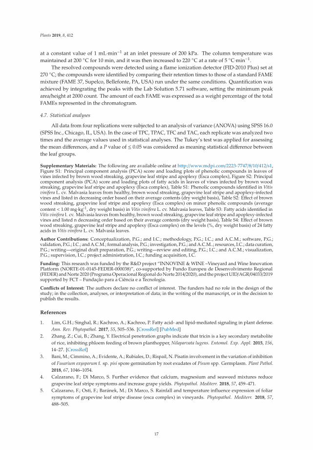
4.3. Sample Collection
The occurrence of symptoms in the vineyard allowed the collection of different sets of leaves,which were divided into eight groups (Figure 6): (1) Asymptomatic leaves from apparently healthyvines (control); (2) asymptomatic leaves from BWS vines (asymptomatic 1), to analyze the systemiceffects of trunk-localized fungi attack; (3) asymptomatic leaves from asymptomatic cordons on GLSDvines (asymptomatic 2), to assess whether the biosynthesis of defensive compounds was similar insymptomatic and asymptomatic parts of the same vine; (4) asymptomatic leaves from GLSD vineswith berry symptoms (asymptomatic 3), to analyze the systemic effects of berry-localized infection;(5) symptomatic leaves from vines with initial symptoms of GLSD i.e., chlorotic leaves (GLSD stage 1);(6) symptomatic leaves from vines with moderate symptoms of GLSD i.e., chlorotic/spotting/scorchingleaves (GLSD stage 2); (7) symptomatic leaves from vines with advanced symptoms of GLSDi.e., tiger striped leaves (GLSD stage 3); (8) symptomatic leaves from apoplectic vines (apoplexy). In thefield, apoplexy appeared quickly, affecting the entire vine with total wilt and immediate drying caused
14
Plants 2019, 8, 412
by the hot weather (an average of 32 ◦C day temperature during apoplexy expression); thus, apoplecticleaves were harvested and studied already dried as in [23]. All symptomatic leaves were collected tostudy locally induced defenses.
All samples were collected mid-September, one day prior to berry harvesting. This ensuredthat the leaves were at the same stage of maturity. For each leaf-group, four vines were used forsampling and were considered as replicates. Six to twelve leaves of the same size from different partsof a vine were selected. Only two vines exhibited berry symptoms at harvest; hence, two sets ofleaves were harvested from each vine to make four replicates, allowing for statistical comparisons.Leaves were immediately frozen in the field with liquid nitrogen to halt enzymatic activities and storedat −80 ◦C. Prior to use, the leaves were lyophilized, finely powdered with a hand blender, and sieved(0.2-mm mesh).
Figure 6. Description of the sampling procedure: A view of the foliar morphology of asymptomaticand symptomatic leaves of Vitis vinifera L. ‘Malvasia’ affected by brown wood streaking, grapevine leafstripe (GLSD) and apoplexy.
4.4. Determination of Total Amounts of Phenolic Compounds
Phenolic compounds were extracted using an optimized laboratory protocol. After defattingwith 1 mL hexane for 16 h, 0.2 g samples were extracted using 1 mL 70% methanol added with10 μL naringin as an internal standard, during ultrasonication in ice water for 20 min. The extractwas centrifuged at 13,000× g for 15 min (25 ◦C), and the extraction was repeated using the pellet.The combined supernatants were pre-purified on a Sep-Pak C18 cartridge (Waters, Milford, MA,USA) to remove chlorophylls, and then filtered through a Spartan 13/0.2 RC filter (Whatman, Dassel,Germany). The filtrate was used for the determination of total phenolic content (TPC) in mg gallic acidequivalent [GAE]·g−1 using the Folin–Ciocalteu method as described in [36]; total flavonoid content(TFC) in mg catechin equivalent [CAE]·g−1 using aluminum chloride as described in [36]; and totalproanthocyanidin content (TPAC) in mg [GAE]·g−1 using polyvinylpyrrolidone, as described in [37].The total anthocyanin content (TAC) was estimated using the pH differential assay [38], and the resultswere expressed in mg cyanidin 3-O-glucoside [CGE]·g−1.
15
Plants 2019, 8, 412
4.5. Chromatographic Separation and Identification of Phenolic Compounds
The quantitative analysis of individual phenolic compounds was carried out on a Gilson(Villers-le-bel, France) high-performance liquid chromatography (HPLC) instrument consisting of anautosampler, binary pump, column compartment, and a Finnigan photodiode array detector (DAD81401; Thermo Electron, San Jose, CA, USA). Chromatography was performed on 10 μL samples ofthe phenolic filtrate injected into the HPLC onto a C18 column (5 μm, 250 × 4.5 mm i.d.) suppliedfrom Sigma/Aldrich (Steinheim, Germany), and maintained at 25 ◦C. The solvent system consisted of0.1% trifluoroacetic acid in water (mobile phase A) and 0.1% trifluoroacetic acid in acetonitrile (mobilephase B). Elution was performed at a constant flow rate of 1 mL.min−1 using a linear gradient programstarting with 100% mobile phase A for 5 min, decreasing to 80% at 15 min, 50% at 30 min, 0% at 45 min,and then reverting to 100% at 55 until reaching 60 min.
The detection of compounds by DAD was conducted by scanning between 210–520 nm, with aresolution of 1.2 nm. Eluting peaks were monitored at 280, 320, 360, and 520 nm for hydroxybenzoicacids and other low molecular weight compounds, hydroxycinnamic acids and stilbenes, flavonoids,and anthocyanins, respectively, using the software Excalibur 2.0, which generated a three-dimensionaldataset (absorbance, retention time, and wavelength). Eluting peaks at 450 nm were also monitoredbecause two peaks were consistently observed with large areas at that wavelength. The peaks wereselected using both the Gensis and the ICIS detection algorithms of Xcalibur. The threshold forquantification by peak areas was 5000 μAU·min−1, and compounds whose peak areas were below thisvalue were considered “non-detected.”
For identification, 38 reference compounds previously reported in grapevineleaves [10,12,13,19,22,25,26,33], and representatives of the chemical classes under study werepurchased (Table S1); they were also separated by HPLC. Peaks were identified with “some certainty”to compounds by matching UV/vis spectra and retention times with those of the reference compounds.The remaining peaks were putatively identified by comparison with UV/vis bibliographic data.Some peaks could not match to any compounds or phenolic group and were labeled as “unknown.”Compounds were quantified by dividing their peak areas with that of the internal standard (naringin)and the results were converted to mg· kg−1 after correction by the peak area of the reference,its response factor, and the amount of biomass extracted. For compounds identified putatively,quantification was carried out using reference compounds with similar chemical characteristics asshown in Table S1.
4.6. Extraction, Separation, and Identification of Fatty Acids
The extraction of lipids was based on the method presented in [39]. Leaf samples (5 mg) wereadded with 0.8 mL water and 2 mL methanol in a DSR-2800V rotary shaker (Digisystem LaboratoryInstruments Inc, Taipei, Taiwan) at room temperature; after continuous shaking for 5 min, 1 mLchloroform was added, and it was followed by agitation for 5 min. The mixture was centrifuged for5 min at 2000× g (25 ◦C). The supernatant was collected and 2 mL chloroform/water (1/1, v/v) and fivedrops of 100 mM KCl were added. After vortexing, the mixture was centrifuged for 5 min at 2000× g(25 ◦C). The lipid fraction in the bottom layer was collected and the chloroform phase was evaporatedto dryness under nitrogen. The dried extract was then transesterified with 5 mL 14% boron trifluoridein methanol under nitrogen at 70 ◦C for 60 min. Transesterified lipids were extracted by adding 5 mLhexane, followed by 3 min of vortexing. The upper phase, constituting fatty acid methyl esters (FAME),was collected and 1 g Na2SO4 was added to remove water.
FAME were separated via capillary gas chromatography (CG) using Shimadzu GC-2010 Plus(Shimadzu, Kyoto, Japan) equipped with an autosampler and an automatic split/splitless injector.Exactly 1 μL of FAME extract was injected into the GC at an inlet temperature of 270 ◦C and a splitratio of 5:1; compounds were separated on a 30 m long, 0.25-μm-thick-film DB-225MS column with a0.25 mm i.d. (Agilent, Wilmington, DE, USA). The flow rate of the carrier gas (helium) was maintained
16
Plants 2019, 8, 412
at a constant value of 1 mL·min−1 at an inlet pressure of 200 kPa. The column temperature wasmaintained at 200 ◦C for 10 min, and it was then increased to 220 ◦C at a rate of 5 ◦C·min−1.
The resolved compounds were detected using a flame ionization detector (FID-2010 Plus) set at270 ◦C; the compounds were identified by comparing their retention times to those of a standard FAMEmixture (FAME 37, Supelco, Bellefonte, PA, USA) run under the same conditions. Quantification wasachieved by integrating the peaks with the Lab Solution 5.71 software, setting the minimum peakarea/height at 2000 count. The amount of each FAME was expressed as a weight percentage of the totalFAMEs represented in the chromatogram.
4.7. Statistical analyses
All data from four replications were subjected to an analysis of variance (ANOVA) using SPSS 16.0(SPSS Inc., Chicago, IL, USA). In the case of TPC, TPAC, TFC and TAC, each replicate was analyzed twotimes and the average values used in statistical analyses. The Tukey’s test was applied for assessingthe mean differences, and a P value of ≤ 0.05 was considered as meaning statistical difference betweenthe leaf groups.
Supplementary Materials: The following are available online at http://www.mdpi.com/2223-7747/8/10/412/s1,Figure S1: Principal component analysis (PCA) score and loading plots of phenolic compounds in leaves ofvines infected by brown wood streaking, grapevine leaf stripe and apoplexy (Esca complex), Figure S2: Principalcomponent analysis (PCA) score and loading plots of fatty acids in leaves of vines infected by brown woodstreaking, grapevine leaf stripe and apoplexy (Esca complex), Table S1: Phenolic compounds identified in Vitisvinifera L. cv. Malvasia leaves from healthy, brown wood streaking, grapevine leaf stripe and apoplexy-infectedvines and listed in decreasing order based on their average contents (dry weight basis), Table S2: Effect of brownwood streaking, grapevine leaf stripe and apoplexy (Esca complex) on minor phenolic compounds (averagecontent < 1.00 mg·kg-1, dry weight basis) in Vitis vinifera L. cv. Malvasia leaves, Table S3: Fatty acids identified inVitis vinifera l. cv. Malvasia leaves from healthy, brown wood streaking, grapevine leaf stripe and apoplexy-infectedvines and listed n decreasing order based on their average contents (dry weight basis), Table S4: Effect of brownwood streaking, grapevine leaf stripe and apoplexy (Esca complex) on the levels (%, dry weight basis) of 24 fattyacids in Vitis vinifera L. cv. Malvasia leaves.
Author Contributions: Conceptualization, P.G.; and I.C.; methodology, P.G.; I.C.; and A.C.M.; software, P.G.;validation, P.G.; I.C.; and A.C.M.; formal analysis, P.G.; investigation, P.G.; and A.C.M..; resources, I.C.; data curation,P.G.; writing—original draft preparation, P.G.; writing—review and editing, P.G.; I.C.; and A.C.M.; visualization,P.G.; supervision, I.C.; project administration, I.C.; funding acquisition, I.C.
Funding: This research was funded by the R&D project “INNOVINE & WINE –Vineyard and Wine InnovationPlatform (NORTE-01-0145-FEDER-000038)”, co-supported by Fundo Europeu de Desenvolvimento Regional(FEDER) and Norte 2020 (Programa Operacional Regional do Norte 2014/2020), and the project UID/AGR/04033/2019supported by FCT – Fundação para a Ciência e a Tecnologia.
Conflicts of Interest: The authors declare no conflict of interest. The funders had no role in the design of thestudy; in the collection, analyses, or interpretation of data; in the writing of the manuscript, or in the decision topublish the results.
References
1. Lim, G.H.; Singhal, R.; Kachroo, A.; Kachroo, P. Fatty acid- and lipid-mediated signaling in plant defense.Ann. Rev. Phytopathol. 2017, 55, 505–536. [CrossRef] [PubMed]
2. Zhang, Z.; Cui, B.; Zhang, Y. Electrical penetration graphs indicate that tricin is a key secondary metaboliteof rice, inhibiting phloem feeding of brown planthopper, Nilaparvata lugens. Entomol. Exp. Appl. 2015, 156,14–27. [CrossRef]
3. Bani, M.; Cimmino, A.; Evidente, A.; Rubiales, D.; Rispail, N. Pisatin involvement in the variation of inhibitionof Fusarium oxysporum f. sp. pisi spore germination by root exudates of Pisum spp. Germplasm. Plant Pathol.2018, 67, 1046–1054.
4. Calzarano, F.; Di Marco, S. Further evidence that calcium, magnesium and seaweed mixtures reducegrapevine leaf stripe symptoms and increase grape yields. Phytopathol. Mediterr. 2018, 57, 459–471.
5. Calzarano, F.; Osti, F.; Baránek, M.; Di Marco, S. Rainfall and temperature influence expression of foliarsymptoms of grapevine leaf stripe disease (esca complex) in vineyards. Phytopathol. Mediterr. 2018, 57,488–505.
17
Plants 2019, 8, 412
6. Aroca, A.; Raposo, R. PCR-based strategy to detect and identify species of Phaeoacremonium causing grapevinediseases. Appl. Environ. Microbiol. 2007, 73, 2911–2918. [CrossRef] [PubMed]
7. Travadon, R.; Lecomte, P.; Diarra, B.; Lawrence, P.D.; Renault, D.; Ojeda, H.; Rey, P.; Baumgartner, K.Grapevine pruning systems and cultivars influence the diversity of wood-colonizing fungi. Fungal Ecol.2016, 24, 82–93. [CrossRef]
8. Surico, G. Towards a redefinition of the diseases within the esca complex of grapevine. Phytopathol. Mediterr.2009, 48, 5–10.
9. Valtaud, C.; Larignon, P.; Roblin, G.; Fleurat-Lessard, P. Developmental and ultrastructural features ofPhaeomoniella chlamydospora and Phaeoacremonium aleophilum in relation to xylem degradation in esca diseaseof the grapevine. J. Plant Pathol. 2009, 91, 37–51.
10. Rusjan, D.; Persic, M.; Likar, M.; Biniari, K.; Mikulic-Petkovsek, M. Phenolic responses to esca-associatedfungi in differently decayed grapevine woods from different trunk parts of ‘Cabernet Sauvignon’. J. Agric.Food Chem. 2017, 65, 6615–6624. [CrossRef]
11. Bruez, E.; Vallance, J.; Gerbore, J.; Lecomte, P.; Da Costa, J.P.; Guerin-Dubrana, L.; Rey, P. Analyses of thetemporal dynamics of fungal communities colonizing the healthy wood tissues of esca leaf-symptomatic andasymptomatic vines. PLoS ONE 2014, 9, e95928. [CrossRef] [PubMed]
12. Martin, N.; Vesentini, D.; Rego, C.; Monteiro, S.; Oliveira, H.; Ferreira, B.R. Phaeomoniella chlamydosporainfection induces changes in phenolic compounds content in Vitis vinifera. Phytopathol. Mediterr. 2009, 48,101–116.
13. Magnin-Robert, M.; Spagnolo, A.; Boulanger, A.; Joyeux, C.; Clément, C.; Abou-Mansour, E.; Fontaine, F.Changes in plant metabolism and accumulation of fungal metabolites in response to Esca proper andapoplexy expression in the whole grapevine. Phytopathology 2016, 106, 541–553. [CrossRef] [PubMed]
14. Spagnolo, A.; Magnin-Robert, M.; Alayi, D.T.; Cilindre, C.; Mercier, L.; Schaeffer-Reiss, C.; Dorsselaer, V.A.;Clement, C.; Fontaine, F. Physiological changes in green stems of Vitis vinifera L. cv. chardonnay in responseto esca proper and apoplexy revealed by proteomic and transcriptomic analyses. J. Proteome Res. 2012, 11,461–475. [CrossRef] [PubMed]
15. Lecomte, P.; Diarra, B.; Carbonneau, A.; Rey, P.; Chevrier, C. Esca of grapevine and training practices inFrance: Results of a 10-year survey. Phytopathol. Mediterr. 2018, 57, 472–487.
16. Calzarano, F.; Osti, F.; D’agostino, V.; Pepe, A.; Di Marco, S. Mixture of calcium, magnesium and seaweedaffects leaf phytoalexin contents and grape ripening on vines with grapevine leaf stripe disease. Phytopathol.Mediterr. 2017, 56, 445–457.
17. Magnin-Robert, M.; Letousey, P.; Spagnolo, A.; Rabenoelina, F.; Jacquens, L.; Mercier, L.; Clément, C.;Fontaine, F. Leaf stripe form of esca induces alteration of photosynthesis and defense reactions inpresymptomatic leaves. Funct. Plant Biol. 2011, 38, 856–866. [CrossRef]
18. Fontaine, F.; Pinto, C.; Vallet, J.; Clément, C.; Gomes, C.A.; Spagnolo, A. The effects of grapevine trunkdiseases (GTDs) on vine physiology. Eur. J. Plant Patholol. 2016, 144, 707–721. [CrossRef]
19. Bruno, G.; Sparapano, L. Effects of three esca-associated fungi on Vitis vinifera L.: II. Characterization ofbiomolecules in xylem sap and leaves of healthy and diseased vines. Physiol. Mol. Plant Pathol. 2006, 69,195–208. [CrossRef]
20. Lima, R.M.M.; Machado, F.A.; Gubler, D.W. Metabolomic study of chardonnay grapevines double stressedwith esca-associated fungi and drought. Phytopathology 2017, 107, 669–680. [CrossRef]
21. Calzarano, F.; D’agostino, V.; Pepe, A.; Osti, F.; Pelle, D.F.; De Rosso, M.; Flamini, R.; Di Marco, S. Patternsof phytoalexins in the grapevine leaf stripe disease (esca complex)/grapevine pathosystem. Phytopathol.Mediterr. 2016, 55, 410–426.
22. Lambert, C.; Khiook, I.L.K.; Lucas, S.; Télef-Micouleau, N.; Mérillon, J.-M.; Cluzet, S. A faster and a strongerdefense response: One of the key elements in grapevine explaining its lower level of susceptibility to Esca?Phytopathology 2013, 103, 1028–1034. [CrossRef] [PubMed]
23. Letousey, P.; Baillieul, F.; Perrot, G.; Rabenoelina, F.; Boulay, M.; Vaillant-Gaveau, N.; Clément, C.; Fontaine, F.Early events prior to visual symptoms in the apoplectic form of grapevine esca disease. Phytopathology 2010,100, 424–431. [CrossRef] [PubMed]
24. Lima, R.M.M.; Felgueiras, M.L.; Cunha, A.; Chicau, G.; Ferreres, F.; Dias, C.P.A. Differential phenolicproduction in leaves of Vitis vinifera cv. Alvarinho affected with esca disease. Plant Physiol. Biochem. 2017,112, 45–52. [CrossRef] [PubMed]
18
Plants 2019, 8, 412
25. Martín, L.; Fontaine, F.; Castañoa, J.F.; Songy, A.; Roda, R.; Vallet, J.; Ferrer-Gallego, R. Specific profile ofTempranillo grapevines related to Esca-leaf symptoms and climate conditions. Plant Physiol. Biochem. 2019,135, 575–587. [CrossRef]
26. Calzarano, F.; Osti, F.; D’agostino, V.; Pepe, A.; Della Pelle, F.; De rosso, M.; Flamini, R.; Di Marco, S. Levels ofphytoalexins in vine leaves with different degrees of grapevine leaf stripe disease symptoms (Esca complexof diseases). Phytopathol. Mediterr. 2017, 56, 494–501.
27. Lecomte, P.; Darrieutort, G.; Limiñana, J.M.; Comont, A.; Muruamendiaraz, A.; Legorburu, F.-J.; Choueiri, E.;Jreijiri, F.; El Amil, R.; Fermaud, M. New insights into Esca of grapevine: The development of foliar symptomsand their association with xylem discoloration. Plant Dis. 2012, 96, 924–934. [CrossRef]
28. Valtaud, C.; Foyer, H.C.; Fleurat-Lessard, P.; Bourbouloux, A. Systemic effects on leaf glutathione metabolismand defence protein expression caused by esca infection in grapevines. Funct. Plant Biol. 2009, 36, 260–279.[CrossRef]
29. Pearce, G. Systemin, hydroxyproline-rich systemin, and the induction of protease inhibitors. Curr. Prot Pep.Sci. 2011, 12, 399–408. [CrossRef]
30. Tsunoda, T.; Grosser, K.; van Dam, M.N. Locally and systemically induced glucosinolates follow optimaldefence allocation theory upon root herbivory. Funct. Ecol. 2018, 32, 2127–2137. [CrossRef]
31. Ongena, M.; Duby, F.; Rossignol, F.; Fauconnier, M.L.; Dommes, J.; Thonart, P. Stimulation of the lipoxygenasepathway is associated with systemic resistance induced in bean by a nonpathogenic Pseudomonas strain. Mol.Plant-Microbe Interact. 2004, 17, 1009–1018. [CrossRef] [PubMed]
32. Mandal, M.K.; Chandra-Shekara, A.C.; Jeong, R.D.; Yu, K.; Zhu, S.; Chanda, B.; Navarre, D.; Kachroo, A.;Kachroo, P. Oleic acid–dependent modulation of NITRIC OXIDE ASSOCIATED1 protein levels regulatesnitric oxide–mediated defense signaling in Arabidopsis. Plant Cell 2012, 24, 1654–1674. [CrossRef] [PubMed]
33. Gabaston, J.; Cantos-Villar, E.; Biais, B.; Waffo-Teguo, P.; Renouf, E.; Corio-Costet, M.; Richard, T.; Mérillon, J.Stilbenes from Vitis vinifera L. waste: A sustainable tool for controlling Plasmopara Viticola. J. Agric. FoodChem. 2017, 65, 2711–2718. [CrossRef] [PubMed]
34. Schnee, S.; Viret, O.; Gindro, K. Role of stilbenes in the resistance of grapevine to powdery mildew. Physiol.Mol. Plant Pathol. 2008, 72, 128–133. [CrossRef]
35. Tabacchi, R.; Fkeyrat, A.; Poliart, C.; Dubin, G.-M. Phytotoxins from fungi of esca of grapevine. Phytopathol.Mediterr. 2000, 39, 156–161.
36. Goufo, P.; Pereira, J.; Figueiredo, N.; Oliveira, M.B.P.P.; Carranca, C.; Rosa, E.A.S.; Trindade, H. Effect ofelevated carbon dioxide (CO2) on phenolic acids, flavonoids, tocopherols, tocotrienols, γ-oryzanol andantioxidant capacities of rice (Oryza sativa L.). J. Cereal Sci. 2014, 59, 15–24. [CrossRef]
37. Queiroz, C.R.A.A.; Morais, S.A.L.; Nascimento, E.A. Characterization of aroeira-preta (Myracrodruonurundeuva) wood tannins. Rev. Árvore 2002, 26, 485–492. [CrossRef]
38. Giusti, M.M.; Wrolstad, E.R. Characterization and measurement of anthocyanins by UV-visible spectroscopy.Curr. Prot. Food Anal. Chem. 2001, F1.2.1–F1.2.13. [CrossRef]
39. Bligh, E.G.; Dyer, W.J. A lipid method of total lipid extraction and purification. Can. J. Biochem. 1959, 37,911–917.
© 2019 by the authors. Licensee MDPI, Basel, Switzerland. This article is an open accessarticle distributed under the terms and conditions of the Creative Commons Attribution(CC BY) license (http://creativecommons.org/licenses/by/4.0/).
19

plants
Article
Macrophomina Crown and Root Rot of Pistachioin California
Mohamed T. Nouri 1,2, Daniel P. Lawrence 2, Craig E. Kallsen 3 and Florent P. Trouillas 1,2,*
1 Kearney Agricultural Research and Extension Center, Parlier, CA 93648, USA; [email protected] Department of Plant Pathology, University of California, Davis, CA 95616, USA; [email protected] University of California Cooperative Extension Kern County, Bakersfield, CA 93307, USA;
[email protected]* Correspondence: [email protected]; Tel.: +1-559-646-6566
Received: 3 January 2020; Accepted: 20 January 2020; Published: 21 January 2020
Abstract: In this study, declining pistachio rootstocks were detected in newly planted commercialpistachio orchards in Kern County, California. Symptoms were characterized by wilted foliagecombined with crown rot in the rootstock. From diseased trees, 42 isolates were obtained, and all hadsimilar cultural and morphological characteristics of Macrophomina phaseolina. Analyses of nucleotidesequences of three gene fragments, the internal transcribed spacer region (ITS1–5.8S–ITS2), partialsequences of β-tubulin, and translation elongation factor 1-α (TEF1) confirmed this identification, and20 representative isolates are presented in the phylogenetic study. Testing of Koch’s postulates showedthat M. phaseolina, when inoculated to stems and roots of the pistachio rootstocks using mycelial plugsor a microsclerotial suspension, is indeed pathogenic to this host. The widely used clonal Universityof California Berkeley I (UCBI) rootstock appeared highly susceptible to M. phaseolina, suggesting thatthis pathogen is an emerging threat to the production of pistachio in California. This study confirmedthe association of M. phaseolina with the decline of pistachio trees and represents the first descriptionof this fungus as a crown rot-causing agent of pistachio in California.
Keywords: pistachio; crown rot; root rot; Macrophomina phaseolina; pathogenicity
1. Introduction
Pistachio (Pistacia vera L.) is one of California’s many important, high-value nut crops. In 2017,California accounted for more than 99.1% of the United States pistachio crop, with approximately101,327 hectares of bearing trees with a value of approximately $3.6 billion (http://www.acpistachios.org).Although pistachio cultivation in California is relatively new, this industry has had a record of steadyexpansion. As pistachio demand and acreage continues to grow, fungal pathogens pose a continuallyevolving challenge for the industry.
Panicle and shoot blight of pistachio has been one of the main fungal diseases affecting pistachioin California, with early reports dating back to the 1980s [1]. In California, at least eight species ofBotryosphaeriaceae fungi have been associated with this disease. Botrytis blossom and shoot blightcaused by Botrytis cinerea Pers. [2] and Alternaria late blight caused by multiple species of AlternariaNees [3] are additional important diseases of pistachio in California. Management of these variousdiseases has mainly relied upon the use of synthetic fungicides. Additional above-ground diseaseof pistachio includes Cytospora canker caused by several Cytospora spp. and Botryosphaeria cankercaused by Neofusicoccum mediterraneum Crous, M.J. Wingf. and A.J.L. Phillips [4,5].
Verticillium wilt caused by Verticillium dahliae Kleb. has been the most important soil-bornedisease affecting pistachio trees in California [6]. Verticillium dahliae is found worldwide in all types ofsoils and has an extremely wide host range, affecting more than 400 different plant species, includingvegetables, flowers, fruit crops, ornamentals, and perennial agronomic crops [7–9]. Verticillium wilt
Plants 2020, 9, 134; doi:10.3390/plants9020134 www.mdpi.com/journal/plants
21
Plants 2020, 9, 134
of pistachio mostly occurred in orchards planted after crops such as cotton and tomato, which arehighly susceptible to V. dahliae, or in orchards established adjacent to fields with susceptible crops [10].The fungus can survive in the soil for many years due to extremely persistent resting structures,the microsclerotia. Accordingly, control of Verticillium wilt begins before the trees are planted andthe site location is an important consideration. Former cotton, tomato, and alfalfa fields should beavoided, as they increase the risk of Verticillium wilt. The most effective control measures to combatVerticillium wilt is the use of resistant rootstocks. In California, commercial pistachio nut productionbegan in the 1970s with Pistacia atlantica Desf. as the main pistachio rootstock [11]. However, P. atlanticais highly susceptible to infection by V. dahliae, and the fungus is widespread in soils of the San JoaquinValley. The development and widespread planting of resistant University of California Berkeley I(UCBI) rootstocks has diminished the Verticillium wilt problem of pistachio in California. Symptomsof Verticillium wilt are now only observed sporadically in the southern half of the San Joaquin Valley,where the P. atlantica rootstock was initially planted.
California pistachio is also subject to root and crown rots caused by oomycetes such as Phytophthoraand Phytopythium [12–14]. Disease development with oomycetes is generally enhanced in poorlydrained soils, where orchards receive long durations of flood irrigation, or in trees in lower areas oralong creeks and natural drainage creeks in the orchard [6]. When the soil remains saturated for a longtime, these pathogens are able to infect susceptible roots.
Crespo et al. (2019) recently revealed species of Fusarium and Neocosmospora from decliningpistachio rootstocks and stem cankers in the southern San Joaquin Valley of California that werepathogenic to this host [15]. Three Fusarium (Fusarium equiseti (Corda) Sacc., F. oxysporum Schltdl.,and F. proliferatum (Matsush.) Nirenberg), and two Neocosmospora species (Neocosmospora falciformis(Carrión) L. Lombard and Crous (syn: Fusarium falciforme (Carrión) Summerb. and Schroers) andN. solani (Mart.) L. Lombard and Crous (syn: Fusarium solani (Mart.) Sacc.)) were found associated withcrown rot symptoms and vascular discoloration in stems of clonal UCBI rootstocks in California [15].Necrotic root lesions and black discoloration of the root cortex, epidermis, and vascular tissues associatedwith cylindrocarpon-like fungi included species in Dactylonectria, Neonectria, and Thelonectria, whichwere also detected in multiple counties in California [16].
Other disease symptoms observed in recent surveys of pistachio orchards have included youngpistachio trees that were collapsing, showing crown rot with black discolorations in the rootstocks.Isolation from these young declining pistachio rootstocks revealed the occurrence of a single putativepathogen tentatively identified as a member of the Botryosphaeriaceae. The aims of this study were to(1) identify and characterize the putative pathogen associated with pistachio rootstock decline basedon molecular and morphological methods; (2) test the pathogenicity of the putative pathogen on thecommonly planted UCBI pistachio rootstock in California.
2. Results
2.1. Field Surveys and Collection of Fungal Isolates
Forty-two fungal isolates resembling a member of the Botryosphaeriaceae were isolated fromthe rootstock of young declining pistachio trees showing root crown and lower trunk rot symptoms(Figure 1). Affected rootstocks mainly included the clonal UCBI rootstocks. All pistachio isolates wereobtained from orchards in Kern County, California. Orchards characteristics included heavy clay soilsand history of vegetable crops or cotton. In addition to pistachio, 21 isolates with typical characteristicsof the putative pathogen were also collected: 8 from sweet cherry in Fresno and San Joaquin Countiesand 13 from grapevine in Fresno County.
22
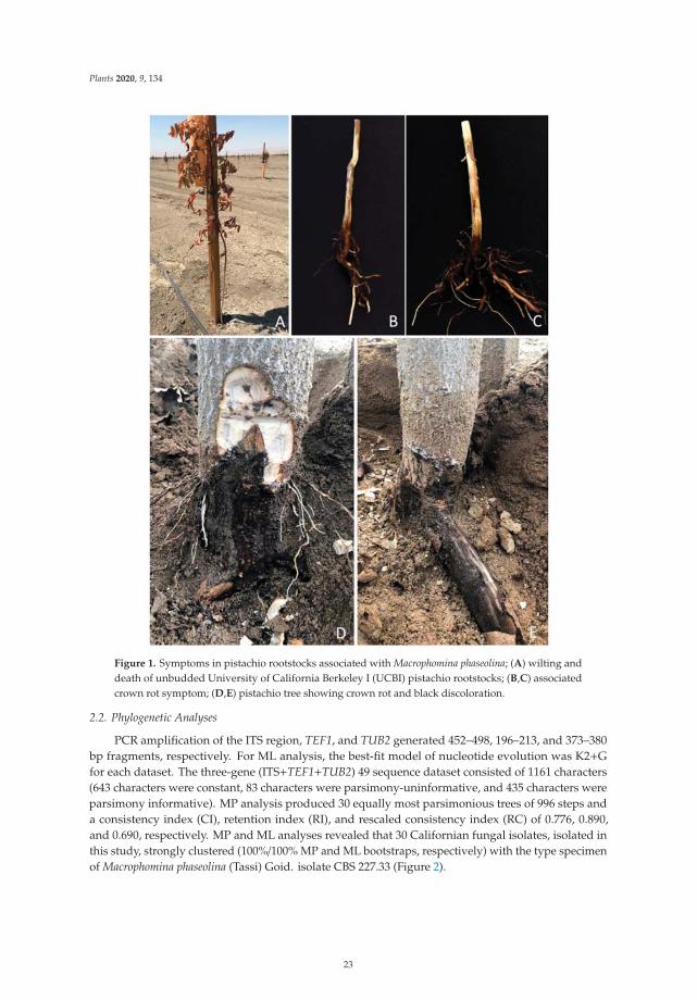
Plants 2020, 9, 134
Figure 1. Symptoms in pistachio rootstocks associated with Macrophomina phaseolina; (A) wilting anddeath of unbudded University of California Berkeley I (UCBI) pistachio rootstocks; (B,C) associatedcrown rot symptom; (D,E) pistachio tree showing crown rot and black discoloration.
2.2. Phylogenetic Analyses
PCR amplification of the ITS region, TEF1, and TUB2 generated 452–498, 196–213, and 373–380bp fragments, respectively. For ML analysis, the best-fit model of nucleotide evolution was K2+Gfor each dataset. The three-gene (ITS+TEF1+TUB2) 49 sequence dataset consisted of 1161 characters(643 characters were constant, 83 characters were parsimony-uninformative, and 435 characters wereparsimony informative). MP analysis produced 30 equally most parsimonious trees of 996 steps anda consistency index (CI), retention index (RI), and rescaled consistency index (RC) of 0.776, 0.890,and 0.690, respectively. MP and ML analyses revealed that 30 Californian fungal isolates, isolated inthis study, strongly clustered (100%/100% MP and ML bootstraps, respectively) with the type specimenof Macrophomina phaseolina (Tassi) Goid. isolate CBS 227.33 (Figure 2).
23
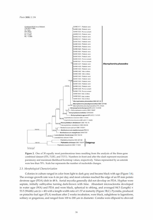
Plants 2020, 9, 134
Figure 2. One of 30 equally most parsimonious trees resulting from the analysis of the three-genecombined dataset (ITS, TUB2, and TEF1). Numbers in front and after the slash represent maximumparsimony and maximum likelihood bootstrap values, respectively. Values represented by an asteriskwere less than 70%. Scale bar represents the number of nucleotide changes.
2.3. Morphological Characterization
Colonies in culture ranged in color from light to dark gray and became black with age (Figure 3A).The average growth rate was 4 cm per day, and most colonies reached the edge of an 85 mm potatodextrose agar (PDA) dish in 48 h. Aerial mycelia generally did not develop on PDA. Hyphae wereseptate, initially subhyaline turning dark-brown with time. Abundant microsclerotia developedin water agar (WA) and PDA and were black, spherical to oblong, and averaged 84.5 (Length) ×53.5 (Width) μm (n = 40) with a length-width ratio of 1.57 at maturity (Figure 3B,C). Pycnidia, producedon pistachio leaf agar (PLA) medium after 2 weeks incubation, were black, subglobose to lageniform,solitary or gregarious, and ranged from 100 to 200 μm in diameter. Conidia were ellipsoid to obovoid
24
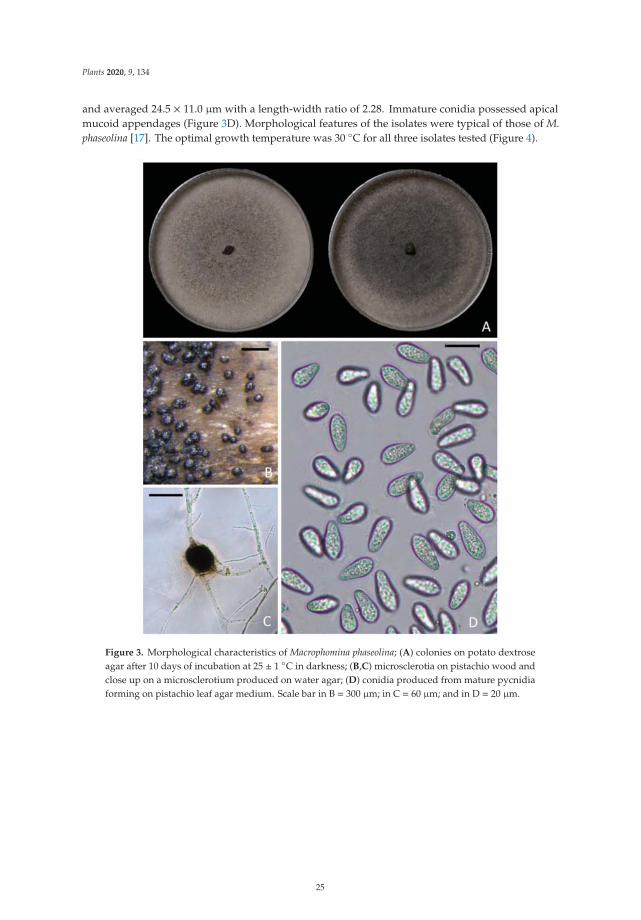
Plants 2020, 9, 134
and averaged 24.5 × 11.0 μm with a length-width ratio of 2.28. Immature conidia possessed apicalmucoid appendages (Figure 3D). Morphological features of the isolates were typical of those of M.phaseolina [17]. The optimal growth temperature was 30 ◦C for all three isolates tested (Figure 4).
Figure 3. Morphological characteristics of Macrophomina phaseolina; (A) colonies on potato dextroseagar after 10 days of incubation at 25 ± 1 ◦C in darkness; (B,C) microsclerotia on pistachio wood andclose up on a microsclerotium produced on water agar; (D) conidia produced from mature pycnidiaforming on pistachio leaf agar medium. Scale bar in B = 300 μm; in C = 60 μm; and in D = 20 μm.
25
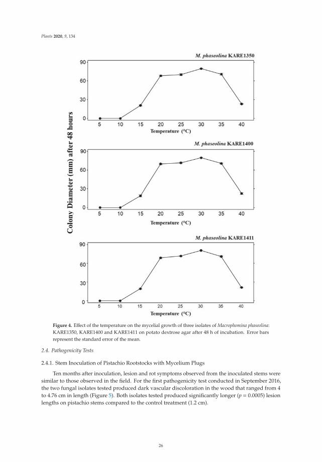
Plants 2020, 9, 134
Figure 4. Effect of the temperature on the mycelial growth of three isolates of Macrophomina phaseolina:KARE1350, KARE1400 and KARE1411 on potato dextrose agar after 48 h of incubation. Error barsrepresent the standard error of the mean.
2.4. Pathogenicity Tests
2.4.1. Stem Inoculation of Pistachio Rootstocks with Mycelium Plugs
Ten months after inoculation, lesion and rot symptoms observed from the inoculated stems weresimilar to those observed in the field. For the first pathogenicity test conducted in September 2016,the two fungal isolates tested produced dark vascular discoloration in the wood that ranged from 4to 4.76 cm in length (Figure 5). Both isolates tested produced significantly longer (p = 0.0005) lesionlengths on pistachio stems compared to the control treatment (1.2 cm).
26
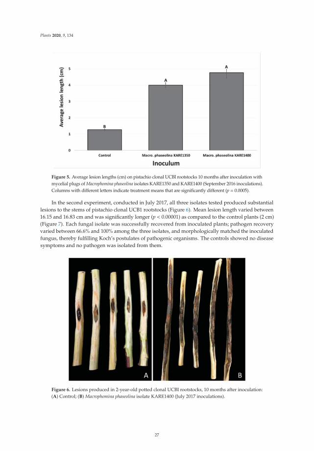
Plants 2020, 9, 134
Figure 5. Average lesion lengths (cm) on pistachio clonal UCBI rootstocks 10 months after inoculation withmycelial plugs of Macrophomina phaseolina isolates KARE1350 and KARE1400 (September 2016 inoculations).Columns with different letters indicate treatment means that are significantly different (p = 0.0005).
In the second experiment, conducted in July 2017, all three isolates tested produced substantiallesions to the stems of pistachio clonal UCB1 rootstocks (Figure 6). Mean lesion length varied between16.15 and 16.83 cm and was significantly longer (p < 0.00001) as compared to the control plants (2 cm)(Figure 7). Each fungal isolate was successfully recovered from inoculated plants; pathogen recoveryvaried between 66.6% and 100% among the three isolates, and morphologically matched the inoculatedfungus, thereby fulfilling Koch’s postulates of pathogenic organisms. The controls showed no diseasesymptoms and no pathogen was isolated from them.
Figure 6. Lesions produced in 2-year-old potted clonal UCBI rootstocks, 10 months after inoculation:(A) Control; (B) Macrophomina phaseolina isolate KARE1400 (July 2017 inoculations).
27
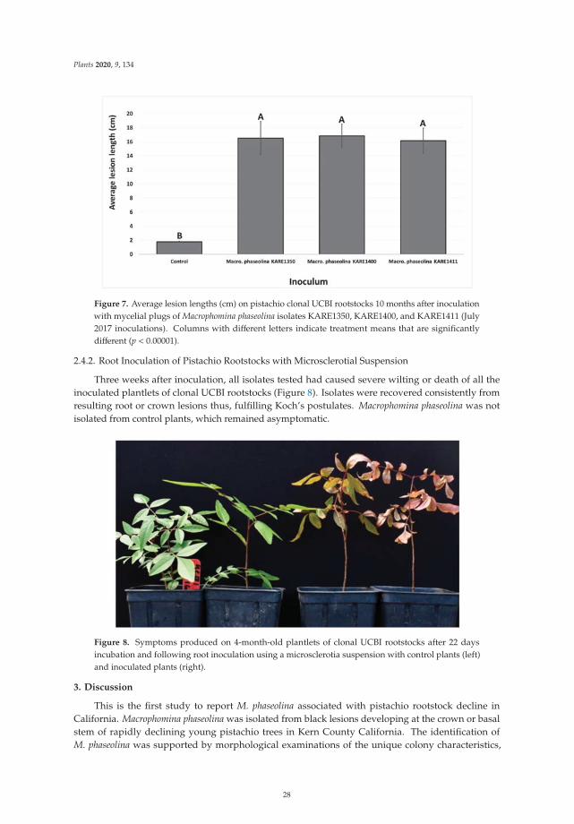
Plants 2020, 9, 134
Figure 7. Average lesion lengths (cm) on pistachio clonal UCBI rootstocks 10 months after inoculationwith mycelial plugs of Macrophomina phaseolina isolates KARE1350, KARE1400, and KARE1411 (July2017 inoculations). Columns with different letters indicate treatment means that are significantlydifferent (p < 0.00001).
2.4.2. Root Inoculation of Pistachio Rootstocks with Microsclerotial Suspension
Three weeks after inoculation, all isolates tested had caused severe wilting or death of all theinoculated plantlets of clonal UCBI rootstocks (Figure 8). Isolates were recovered consistently fromresulting root or crown lesions thus, fulfilling Koch’s postulates. Macrophomina phaseolina was notisolated from control plants, which remained asymptomatic.
Figure 8. Symptoms produced on 4-month-old plantlets of clonal UCBI rootstocks after 22 daysincubation and following root inoculation using a microsclerotia suspension with control plants (left)and inoculated plants (right).
3. Discussion
This is the first study to report M. phaseolina associated with pistachio rootstock decline inCalifornia. Macrophomina phaseolina was isolated from black lesions developing at the crown or basalstem of rapidly declining young pistachio trees in Kern County California. The identification ofM. phaseolina was supported by morphological examinations of the unique colony characteristics,
28
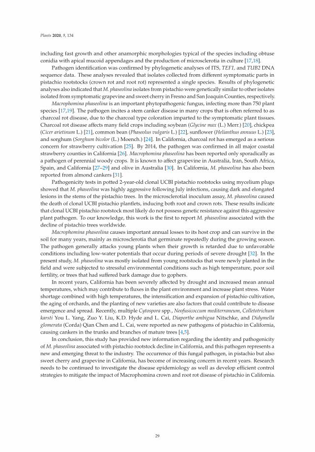
Plants 2020, 9, 134
including fast growth and other anamorphic morphologies typical of the species including obtuseconidia with apical mucoid appendages and the production of microsclerotia in culture [17,18].
Pathogen identification was confirmed by phylogenetic analyses of ITS, TEF1, and TUB2 DNAsequence data. These analyses revealed that isolates collected from different symptomatic parts inpistachio rootstocks (crown rot and root rot) represented a single species. Results of phylogeneticanalyses also indicated that M. phaseolina isolates from pistachio were genetically similar to other isolatesisolated from symptomatic grapevine and sweet cherry in Fresno and San Joaquin Counties, respectively.
Macrophomina phaseolina is an important phytopathogenic fungus, infecting more than 750 plantspecies [17,19]. The pathogen incites a stem canker disease in many crops that is often referred to ascharcoal rot disease, due to the charcoal type coloration imparted to the symptomatic plant tissues.Charcoal rot disease affects many field crops including soybean (Glycine max (L.) Merr.) [20], chickpea(Cicer arietinum L.) [21], common bean (Phaseolus vulgaris L.) [22], sunflower (Helianthus annuus L.) [23],and sorghum (Sorghum bicolor (L.) Moench.) [24]. In California, charcoal rot has emerged as a seriousconcern for strawberry cultivation [25]. By 2014, the pathogen was confirmed in all major coastalstrawberry counties in California [26]. Macrophomina phaseolina has been reported only sporadically asa pathogen of perennial woody crops. It is known to affect grapevine in Australia, Iran, South Africa,Spain, and California [27–29] and olive in Australia [30]. In California, M. phaseolina has also beenreported from almond cankers [31].
Pathogenicity tests in potted 2-year-old clonal UCBI pistachio rootstocks using mycelium plugsshowed that M. phaseolina was highly aggressive following July infections, causing dark and elongatedlesions in the stems of the pistachio trees. In the microsclerotial inoculum assay, M. phaseolina causedthe death of clonal UCBI pistachio plantlets, inducing both root and crown rots. These results indicatethat clonal UCBI pistachio rootstock most likely do not possess genetic resistance against this aggressiveplant pathogen. To our knowledge, this work is the first to report M. phaseolina associated with thedecline of pistachio trees worldwide.
Macrophomina phaseolina causes important annual losses to its host crop and can survive in thesoil for many years, mainly as microsclerotia that germinate repeatedly during the growing season.The pathogen generally attacks young plants when their growth is retarded due to unfavorableconditions including low-water potentials that occur during periods of severe drought [32]. In thepresent study, M. phaseolina was mostly isolated from young rootstocks that were newly planted in thefield and were subjected to stressful environmental conditions such as high temperature, poor soilfertility, or trees that had suffered bark damage due to gophers.
In recent years, California has been severely affected by drought and increased mean annualtemperatures, which may contribute to fluxes in the plant environment and increase plant stress. Watershortage combined with high temperatures, the intensification and expansion of pistachio cultivation,the aging of orchards, and the planting of new varieties are also factors that could contribute to diseaseemergence and spread. Recently, multiple Cytospora spp., Neofusicoccum mediterraneum, Colletotrichumkarsti You L. Yang, Zuo Y. Liu, K.D. Hyde and L. Cai, Diaporthe ambigua Nitschke, and Didymellaglomerata (Corda) Qian Chen and L. Cai, were reported as new pathogens of pistachio in California,causing cankers in the trunks and branches of mature trees [4,5].
In conclusion, this study has provided new information regarding the identity and pathogenicityof M. phaseolina associated with pistachio rootstock decline in California, and this pathogen represents anew and emerging threat to the industry. The occurrence of this fungal pathogen, in pistachio but alsosweet cherry and grapevine in California, has become of increasing concern in recent years. Researchneeds to be continued to investigate the disease epidemiology as well as develop efficient controlstrategies to mitigate the impact of Macrophomina crown and root rot disease of pistachio in California.
29
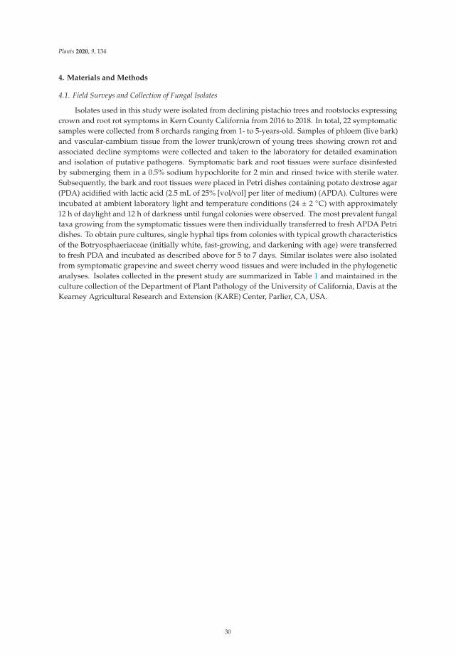
Plants 2020, 9, 134
4. Materials and Methods
4.1. Field Surveys and Collection of Fungal Isolates
Isolates used in this study were isolated from declining pistachio trees and rootstocks expressingcrown and root rot symptoms in Kern County California from 2016 to 2018. In total, 22 symptomaticsamples were collected from 8 orchards ranging from 1- to 5-years-old. Samples of phloem (live bark)and vascular-cambium tissue from the lower trunk/crown of young trees showing crown rot andassociated decline symptoms were collected and taken to the laboratory for detailed examinationand isolation of putative pathogens. Symptomatic bark and root tissues were surface disinfestedby submerging them in a 0.5% sodium hypochlorite for 2 min and rinsed twice with sterile water.Subsequently, the bark and root tissues were placed in Petri dishes containing potato dextrose agar(PDA) acidified with lactic acid (2.5 mL of 25% [vol/vol] per liter of medium) (APDA). Cultures wereincubated at ambient laboratory light and temperature conditions (24 ± 2 ◦C) with approximately12 h of daylight and 12 h of darkness until fungal colonies were observed. The most prevalent fungaltaxa growing from the symptomatic tissues were then individually transferred to fresh APDA Petridishes. To obtain pure cultures, single hyphal tips from colonies with typical growth characteristicsof the Botryosphaeriaceae (initially white, fast-growing, and darkening with age) were transferredto fresh PDA and incubated as described above for 5 to 7 days. Similar isolates were also isolatedfrom symptomatic grapevine and sweet cherry wood tissues and were included in the phylogeneticanalyses. Isolates collected in the present study are summarized in Table 1 and maintained in theculture collection of the Department of Plant Pathology of the University of California, Davis at theKearney Agricultural Research and Extension (KARE) Center, Parlier, CA, USA.
30
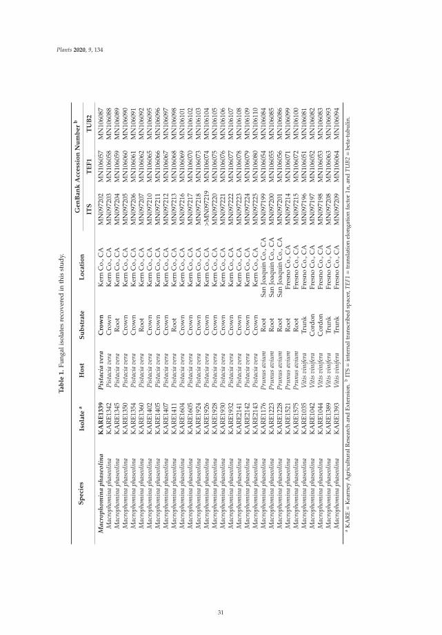
Plants 2020, 9, 134
Ta
ble
1.
Fung
alis
olat
esre
cove
red
inth
isst
udy.
Sp
eci
es
Iso
late
aH
ost
Su
bst
rate
Lo
cati
on
Ge
nB
an
kA
cce
ssio
nN
um
be
rb
ITS
TE
F1
TU
B2
Mac
roph
omin
aph
aseo
lina
KA
RE
13
39
Pis
taci
ave
raC
row
nK
ern
Co.
,CA
MN
0972
02M
N10
6057
MN
1060
87M
acro
phom
ina
phas
eolin
aK
AR
E134
2Pi
stac
iave
raC
row
nK
ern
Co.
,CA
MN
0972
03M
N10
6058
MN
1060
88M
acro
phom
ina
phas
eolin
aK
AR
E134
5Pi
stac
iave
raR
oot
Ker
nC
o.,C
AM
N09
7204
MN
1060
59M
N10
6089
Mac
roph
omin
aph
aseo
lina
KA
RE1
350
Pist
acia
vera
Cro
wn
Ker
nC
o.,C
AM
N09
7205
MN
1060
60M
N10
6090
Mac
roph
omin
aph
aseo
lina
KA
RE1
354
Pist
acia
vera
Cro
wn
Ker
nC
o.,C
AM
N09
7206
MN
1060
61M
N10
6091
Mac
roph
omin
aph
aseo
lina
KA
RE1
360
Pist
acia
vera
Roo
tK
ern
Co.
,CA
MN
0972
07M
N10
6062
MN
1060
92M
acro
phom
ina
phas
eolin
aK
AR
E140
2Pi
stac
iave
raC
row
nK
ern
Co.
,CA
MN
0972
10M
N10
6065
MN
1060
95M
acro
phom
ina
phas
eolin
aK
AR
E140
5Pi
stac
iave
raC
row
nK
ern
Co.
,CA
MN
0972
11M
N10
6066
MN
1060
96M
acro
phom
ina
phas
eolin
aK
AR
E140
7Pi
stac
iave
raC
row
nK
ern
Co.
,CA
MN
0972
12M
N10
6067
MN
1060
97M
acro
phom
ina
phas
eolin
aK
AR
E141
1Pi
stac
iave
raR
oot
Ker
nC
o.,C
AM
N09
7213
MN
1060
68M
N10
6098
Mac
roph
omin
aph
aseo
lina
KA
RE1
604
Pist
acia
vera
Cro
wn
Ker
nC
o.,C
AM
N09
7216
MN
1060
69M
N10
6101
Mac
roph
omin
aph
aseo
lina
KA
RE1
605
Pist
acia
vera
Cro
wn
Ker
nC
o.,C
AM
N09
7217
MN
1060
70M
N10
6102
Mac
roph
omin
aph
aseo
lina
KA
RE1
924
Pist
acia
vera
Cro
wn
Ker
nC
o.,C
AM
N09
7218
MN
1060
73M
N10
6103
Mac
roph
omin
aph
aseo
lina
KA
RE1
926
Pist
acia
vera
Cro
wn
Ker
nC
o.,C
A>
MN
0972
19M
N10
6074
MN
1061
04M
acro
phom
ina
phas
eolin
aK
AR
E192
8Pi
stac
iave
raC
row
nK
ern
Co.
,CA
MN
0972
20M
N10
6075
MN
1061
05M
acro
phom
ina
phas
eolin
aK
AR
E193
0Pi
stac
iave
raC
row
nK
ern
Co.
,CA
MN
0972
21M
N10
6076
MN
1061
06M
acro
phom
ina
phas
eolin
aK
AR
E193
2Pi
stac
iave
raC
row
nK
ern
Co.
,CA
MN
0972
22M
N10
6077
MN
1061
07M
acro
phom
ina
phas
eolin
aK
AR
E214
1Pi
stac
iave
raC
row
nK
ern
Co.
,CA
MN
0972
23M
N10
6078
MN
1061
08M
acro
phom
ina
phas
eolin
aK
AR
E214
2Pi
stac
iave
raC
row
nK
ern
Co.
,CA
MN
0972
24M
N10
6079
MN
1061
09M
acro
phom
ina
phas
eolin
aK
AR
E214
3Pi
stac
iave
raC
row
nK
ern
Co.
,CA
MN
0972
25M
N10
6080
MN
1061
10M
acro
phom
ina
phas
eolin
aK
AR
E117
6Pr
unus
aviu
mR
oot
San
Joaq
uin
Co.
,CA
MN
0971
99M
N10
6054
MN
1060
84M
acro
phom
ina
phas
eolin
aK
AR
E122
3Pr
unus
aviu
mR
oot
San
Joaq
uin
Co.
,CA
MN
0972
00M
N10
6055
MN
1060
85M
acro
phom
ina
phas
eolin
aK
AR
E122
8Pr
unus
aviu
mR
oot
San
Joaq
uin
Co.
,CA
MN
0972
01M
N10
6056
MN
1060
86M
acro
phom
ina
phas
eolin
aK
AR
E152
1Pr
unus
aviu
mR
oot
Fres
noC
o.,C
AM
N09
7214
MN
1060
71M
N10
6099
Mac
roph
omin
aph
aseo
lina
KA
RE1
575
Prun
usav
ium
Roo
tFr
esno
Co.
,CA
MN
0972
15M
N10
6072
MN
1061
00M
acro
phom
ina
phas
eolin
aK
AR
E103
5V
itis
vini
fera
Trun
kFr
esno
Co.
,CA
MN
0971
96M
N10
6051
MN
1060
81M
acro
phom
ina
phas
eolin
aK
AR
E104
2V
itis
vini
fera
Cor
don
Fres
noC
o.,C
AM
N09
7197
MN
1060
52M
N10
6082
Mac
roph
omin
aph
aseo
lina
KA
RE1
044
Viti
svi
nife
raC
ordo
nFr
esno
Co.
,CA
MN
0971
98M
N10
6053
MN
1060
83M
acro
phom
ina
phas
eolin
aK
AR
E138
9V
itis
vini
fera
Trun
kFr
esno
Co.
,CA
MN
0972
08M
N10
6063
MN
1060
93M
acro
phom
ina
phas
eolin
aK
AR
E139
3V
itis
vini
fera
Trun
kFr
esno
Co.
,CA
MN
0972
09M
N10
6064
MN
1060
94a
KA
RE=
Kea
rney
Agr
icul
tura
lRes
earc
han
dEx
tens
ion.
bIT
S=
inte
rnal
tran
scri
bed
spac
er,T
EF1=
tran
slat
ion
elon
gati
onfa
ctor
1α,a
ndTU
B2=
beta
-tub
ulin
.
31
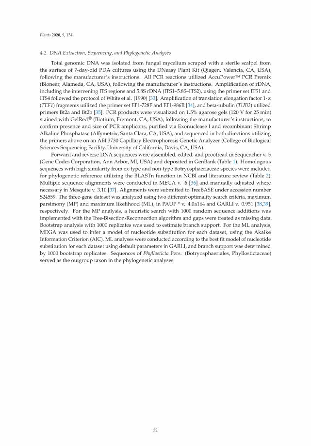
Plants 2020, 9, 134
4.2. DNA Extraction, Sequencing, and Phylogenetic Analyses
Total genomic DNA was isolated from fungal mycelium scraped with a sterile scalpel fromthe surface of 7-day-old PDA cultures using the DNeasy Plant Kit (Qiagen, Valencia, CA, USA),following the manufacturer’s instructions. All PCR reactions utilized AccuPower™ PCR Premix(Bioneer, Alameda, CA, USA), following the manufacturer’s instructions. Amplification of rDNA,including the intervening ITS regions and 5.8S rDNA (ITS1–5.8S–ITS2), using the primer set ITS1 andITS4 followed the protocol of White et al. (1990) [33]. Amplification of translation elongation factor 1-α(TEF1) fragments utilized the primer set EF1-728F and EF1-986R [34], and beta-tubulin (TUB2) utilizedprimers Bt2a and Bt2b [35]. PCR products were visualized on 1.5% agarose gels (120 V for 25 min)stained with GelRed® (Biotium, Fremont, CA, USA), following the manufacturer’s instructions, toconfirm presence and size of PCR amplicons, purified via Exonuclease I and recombinant ShrimpAlkaline Phosphatase (Affymetrix, Santa Clara, CA, USA), and sequenced in both directions utilizingthe primers above on an ABI 3730 Capillary Electrophoresis Genetic Analyzer (College of BiologicalSciences Sequencing Facility, University of California, Davis, CA, USA).
Forward and reverse DNA sequences were assembled, edited, and proofread in Sequencher v. 5(Gene Codes Corporation, Ann Arbor, MI, USA) and deposited in GenBank (Table 1). Homologoussequences with high similarity from ex-type and non-type Botryosphaeriaceae species were includedfor phylogenetic reference utilizing the BLASTn function in NCBI and literature review (Table 2).Multiple sequence alignments were conducted in MEGA v. 6 [36] and manually adjusted wherenecessary in Mesquite v. 3.10 [37]. Alignments were submitted to TreeBASE under accession numberS24559. The three-gene dataset was analyzed using two different optimality search criteria, maximumparsimony (MP) and maximum likelihood (ML), in PAUP * v. 4.0a164 and GARLI v. 0.951 [38,39],respectively. For the MP analysis, a heuristic search with 1000 random sequence additions wasimplemented with the Tree-Bisection-Reconnection algorithm and gaps were treated as missing data.Bootstrap analysis with 1000 replicates was used to estimate branch support. For the ML analysis,MEGA was used to infer a model of nucleotide substitution for each dataset, using the AkaikeInformation Criterion (AIC). ML analyses were conducted according to the best fit model of nucleotidesubstitution for each dataset using default parameters in GARLI, and branch support was determinedby 1000 bootstrap replicates. Sequences of Phyllosticta Pers. (Botryosphaeriales, Phyllostictaceae)served as the outgroup taxon in the phylogenetic analyses.
32
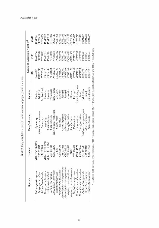
Plants 2020, 9, 134
Ta
ble
2.
Fung
alis
olat
esre
trie
ved
from
Gen
bank
for
phyl
ogen
etic
refe
renc
e.
Sp
eci
es
Iso
late
aH
ost/S
ub
stra
teL
oca
tio
nG
en
Ba
nk
Acc
ess
ion
Nu
mb
er
b
ITS
TEF1
TUB
2
Bot
ryos
pher
iaag
aves
MF
LU
CC
11
-01
25
Aga
ves
sp.
Thai
land
JX64
6791
JX64
6856
JX64
6841
Botr
yosp
heri
aco
rtic
isC
BS
11
90
47
Vac
cini
umco
rym
bosu
mN
J,U
SAD
Q29
9245
EU01
7539
EU67
3107
Botr
yosp
heri
ado
thid
eaC
MW
80
00
Prun
ussp
.Sw
itze
rlan
dA
Y23
6949
AY
2368
98A
Y23
6927
Botr
yosp
heri
afu
sisp
ora
MF
LU
CC
10
-00
98
Car
yota
sp.
Thai
land
JX64
6789
JX64
6854
JX64
6839
Cop
hinf
orm
aeu
caly
pti
MFL
UC
C11
-042
5Eu
caly
ptus
sp.
Thai
land
JX64
6800
JX64
6865
JX64
6848
Cop
hinf
orm
am
aman
eC
BS11
7444
Euca
lypt
ussp
.Ve
nezu
ela
KF5
3182
2K
F531
801
KF5
3180
2La
siod
iplo
dia
theo
brom
aeC
BS
16
4.9
6Fr
uito
nco
ralr
eefc
oast
New
Gui
nea
AY
6402
55A
Y64
0258
EU67
3110
Lasi
odip
lodi
aci
tric
ola
7-E8
0Ju
glan
sre
gia
CA
,USA
KC
3573
00K
C35
7312
KC
3573
06M
acro
phom
ina
phas
eolin
aC
BS
22
7.3
3Z
eam
ays
Pale
stin
eK
F531
825
KF5
3180
4K
F531
806
Mac
roph
omin
aps
eudo
phas
eolin
aC
BS
13
71
65
Ara
chis
hypo
gaea
Sene
gal
KF9
5179
1K
F952
153
KF9
5223
3M
acro
phom
ina
pseu
doph
aseo
lina
CPC
2152
4H
ibis
cus
sabd
arifa
Sene
gal
KF9
5179
9K
F952
161
KF9
5224
0N
eofu
sico
ccum
man
gife
rae
CBS
1185
32M
angi
fera
indi
caA
ustr
alia
AY
6151
86D
Q09
3220
AY
6151
73N
eofu
sico
ccum
med
iterr
aneu
mP
D3
12
Euca
lypt
ussp
.G
reec
eG
U25
1176
GU
2513
08G
U25
1836
Neo
fusi
cocc
umpa
rvum
CBS
1103
01V
itis
vini
fera
Port
ugal
AY
2590
98A
Y57
3221
EU67
3095
Neo
scyt
alid
ium
dim
idia
tum
CB
S1
45
.78
Hom
osa
pien
sU
nite
dK
ingd
omK
F531
816
KF5
3179
5K
F531
796
Neo
scyt
alid
ium
dim
idia
tum
CBS
499.
66M
angi
fera
indi
caM
ali
KF5
3182
0K
F531
798
KF5
3180
0Ph
yllo
stic
taam
pelic
ida
CBS
1116
45Pa
rthe
noci
ssus
quin
quef
olia
MO
,USA
FJ82
4766
FJ82
4772
FJ82
4777
Phyl
lost
icta
citr
icar
paC
BS
10
23
74
Citr
usau
rant
ium
Braz
ilFJ
8247
67FJ
5383
71FJ
8247
78Ph
yllo
stic
taph
ilopr
ina
CBS
447.
68Ta
xus
bacc
ata
Uni
ted
Stat
esFJ
8247
68FJ
8247
73FJ
8247
79a
Isol
ates
inbo
ldre
pres
entt
ype
spec
imen
s.b
ITS=
inte
rnal
tran
scri
bed
spac
er,T
EF1=
tran
slat
ion
elon
gati
onfa
ctor
1α,a
ndTU
B2=
beta
-tub
ulin
.
33
Plants 2020, 9, 134
4.3. Morphological Characterization
Three representative isolates (KARE1045, KARE1350, and KARE1400) were selected to study theircultural and conidial features. For each selected isolate, 5 mm mycelial plugs from developing colonieswere removed and transferred to the center of new 85 mm diameter PDA Petri dishes. Cultureswere incubated in the dark at temperatures ranging from 5 to 40 ◦C at 5-degree intervals. At eachtemperature (5◦, 10◦, 15◦, 20◦, 25◦, 30◦, 35◦, and 40 ◦C), 3 replicate dishes of each isolate were used.Measurements of colony diameter were taken each 24 h until the fastest-growing colony had reachedthe edge of the dish. For each of the 8 temperatures, the average colony diameter per 48 h of growthwas calculated. Colony characters and pigment production were noted after 48 h of incubation onPDA at 30 ◦C in darkness. The experiment was repeated once.
The representative isolates, as mentioned above, were further used to study conidial morphology.Pycnidia were induced to form by incubating colonies on pistachio leaf agar (PLA) medium [40].Pistachio leaves were autoclaved twice at 120 ◦C for 20 min and then placed into Petri dishes containingPDA (5 g agar, 5 g potato dextrose broth and 500 mL water). Cultures were then incubated undernear UV irradiation (12 h light/12 h dark) at 25 ± 1 ◦C for 2 weeks. Pycnidia containing conidiawere mounted in sterile water on glass slides, and conidial dimensions included length and width of40 conidia for each isolate were measured at ×400 magnification using a Leica compound microscope(Leica DM2000 LED Microscope, Wetzlar, Germany). Microsclerotia were also induced to form byincubating isolates on WA and characterized as described above.
4.4. Pathogenicity Tests
4.4.1. Stem Inoculation of Clonal UCBI Pistachio Rootstocks with Mycelium Plugs
Potted 2-year-old clonal UCBI pistachio rootstocks showing no apparent decline symptoms, orweak growth, were used for the pathogenicity tests. Two experiments were conducted using the samemethodology. The first pathogenicity test was conducted in September 2016. Four tree replicateswere inoculated per combination of 3 inoculation treatments (isolates KARE1350, KARE1400, and thecontrol). Plants were arranged in a completely randomized design and maintained in a greenhouse witha natural photoperiod and temperatures ranging from 22 to 30 ◦C for 10 months. Plants were wateredonce weekly during the course of the experiment. The second pathogenicity test was conducted inJuly 2017. Eight replicate trees were inoculated per combination of 4 inoculation treatments (isolatesKARE1350, KARE1400, KARE1411, and the control) and arranged in a completely randomized designin an outdoor gravel bed for 10 months. Inoculations were conducted by using a 5 mm-diametermycelium plug from a 7-day-old culture. The bark surface was disinfected at the point of inoculationwith 70% ethanol. Stems of the potted clonal UCBI rootstocks were inoculated 4 cm above the soil line,wounds to the stem/crown were produced with a flame-sterilized 5 mm cork borer followed by placinga 5 mm agar plug bearing aerial mycelia, upper surface facing inward, into the fresh wound, whichwas then sealed with petroleum jelly, and wrapped with Parafilm. Control plants were treated withsterile PDA agar plugs followed by sealing and wrapping as above. Plants were watered twice weekly.
Infection data were recorded 10 months after inoculation. The bark was removed, and the lengthof wood discoloration (LWD) upwards and downwards from the point of inoculation was measuredusing a digital caliper. In an attempt to fulfill Koch’s postulates, small pieces of necrotic tissue from theedge of each lesion were surface disinfested as described above and plated on APDA filled Petri dishesto recover the inoculated fungus. Emerging fungal colonies were recorded and identified based onmorphology as described above. Data for lesion length were tested for normality and homogeneity ofvariance using Shapiro-Wilk’s and Bartlett’s tests, respectively. ANOVAs were done in Statistix 10(Analytical Software, Tallahassee, FL, USA) to evaluate differences in the LWD between the controland fungal treatments. Differences in the means between fungal treatments and the mock-inoculatedcontrols were assessed using Tukey’s honestly significant difference (HSD) test at p = 0.05.
34
Plants 2020, 9, 134
4.4.2. Root Inoculation of UCBI Pistachio Rootstocks with Microsclerotial Suspensions
Clonal UCBI pistachio saplings were root-inoculated using a microsclerotial suspension.The experiment was conducted using 3 isolates (KARE1350, KARE1400, and KARE1411) of M. phaseolinainoculated to 4-month-old plantlets of clonal UCBI rootstocks. The inoculum, consisting of myceliumand microsclerotia (1 × 10⁵ propagules per mL), was prepared by scraping the surface of 12-day-oldPDA cultures containing microsclerotia of the pathogen with a sterile scalpel blade and homogenizingin sterile water using a kitchen blender on high speed for 30 s. Inoculations were performed by dippingthe bare root system of each rootstock into the inoculum slurry for 5 min, and subsequently mixingthe microsclerotia suspension homogeneously with potting soil in 10 × 24 cm tree pots. In total, 20clonal UCBI plantlet rootstocks were used. These included 5 replicates for each fungal isolate andthe control. The 5 mock-inoculated controls were subjected to the same process as described abovebut were treated with a mixture of distilled sterile water and potted and incubated as above. Diseasedevelopment and symptoms were assessed every 4 days for 1 month.
Author Contributions: Conceptualization, F.P.T. and M.T.N.; methodology, M.T.N., D.P.L., and F.P.T.; software,D.P.L. and M.T.N.; formal analysis, D.P.L. and M.T.N.; surveys, F.P.T., C.E.K. and M.T.N.; writing—original draftpreparation, M.T.N.; writing—review and editing, F.P.T. and D.P.L.; supervision, F.P.T.; funding acquisition, F.P.T.All authors have read and agreed to the published version of the manuscript.
Funding: This research was funded by the California Pistachio Research Board (CPRB).
Conflicts of Interest: The authors declare no conflict of interest.
References
1. Michailides, T.J. Pathogenicity, distribution, sources of inoculum, and infection courts of Botryosphaeriadothidea on pistachio. Phytopathology 1991, 81, 566–573. [CrossRef]
2. Michailides, T.J. Pest, Disease, and Physiological Disorders Management: Above Ground Fungal Diseases.2005. Available online: http://fruitsandnuts.ucdavis.edu/files/73707.pdf (accessed on 14 August 2019).
3. Pryor, B.M.; Michailides, T.J. Morphological, Pathogenic, and Molecular Characterization of Alternaria isolatesassociated with Alternaria late blight of pistachio. Phytopathology 2002, 92, 406–416. [CrossRef]
4. Lawrence, D.P.; Holland, L.A.; Nouri, M.T.; Travadon, R.; Abramians, A.; Michailides, T.J.; Trouillas, F.P.Molecular phylogeny of Cytospora species associated with canker diseases of fruit and nut crops in California,with the descriptions of ten new species and one new combination. IMA Fungus 2018, 9, 333–370. [CrossRef][PubMed]
5. Nouri, M.T.; Lawrence, D.P.; Holland, L.A.; Doll, D.A.; Kallsen, C.E.; Culumber, C.M.; Trouillas, F.P.Identification and pathogenicity of fungal species associated with canker diseases of pistachio in California.Plant Dis. 2019, 103, 2397–2411. [CrossRef] [PubMed]
6. Holtz, B.A.; Teviotdale, B.L. Soil-borne diseases. In Pistachio Production Manual; Ferguson, L., Haviland, D.,Eds.; University of California Agriculture and Natural Resources: Oakland, CA, USA, 2016; pp. 257–264.
7. Fradin, E.F.; Thomma, B.P.H.J. Physiology and molecular aspects of Verticillium wilt diseases caused by V.dahliae and V. albo-atrum. Mol. Plant Pathol. 2006, 7, 71–86. [CrossRef] [PubMed]
8. Pegg, G.F.; Brady, B.L. Verticillium Wilts; CAB International: Oxon, UK, 2002.9. Klosterman, S.J.; Atallah, Z.K.; Vallad, G.E.; Subbarao, K.V. Diversity, pathogenicity and management of
Verticillium species. Annu. Rev. Phytopathol. 2009, 47, 39–62. [CrossRef]10. Epstein, L.; Beede, R.; Kaur, S.; Ferguson, L. Rootstock effects on pistachio trees grown in Verticillium
dahliae-infested soil. Phytopathology 2004, 94, 388–395. [CrossRef]11. Ogawa, J.M.; English, H. Diseases of Temperate Zone Tree Fruit and Nut Crops; UC ANR Publications: Oakland,
CA, USA, 1991.12. Fichtner, E.J.; Kallsen, C.E.; Blomquist, C.L. First report of crown rot caused by Phytophthora parsiana on
pistachio in the Southern San Joaquin Valley, California. Plant Dis. 2016, 100, 1795. [CrossRef]13. Fichtner, E.J.; Browne, G.T.; Mortaz, M.; Ferguson, L.; Blomquist, C.L. First report of root rot caused by
Phytopythium helicoides on pistachio rootstock in California. Plant Dis. 2016, 100, 2337. [CrossRef]
35
Plants 2020, 9, 134
14. MacDonald, J.D.; Banihashemi, Z.; Mircetich, S.M.; Browne, G.; Bolkan, L. Trunk and branch canker ofpistachio caused by Phytophthora spp. Phytopathology 1992, 82, 1089.
15. Crespo, M.; Lawrence, D.P.; Nouri, M.T.; Doll, D.A.; Trouillas, F.P. Characterization of Fusarium andNeocosmospora species associated with crown rot and stem canker of pistachio rootstocks in California.Plant Dis. 2019, 103, 1931–1939. [CrossRef] [PubMed]
16. Lawrence, D.P.; Nouri, M.T.; Trouillas, F.P. Taxonomy and multi-locus phylogeny of cylindrocarpon-likespecies associated with diseased roots of grapevine and other fruit and nut crops in California. FUSE 2019,4, 59–75. [CrossRef]
17. Sarr, M.P.; Ndiaye, M.B.; Groenewald, J.Z.; Crous, P.W. Genetic diversity in Macrophomina phaseolina, thecausal agent of charcoal rot. Phytopathol. Mediterr. 2014, 53, 250–268.
18. Crous, P.W.; Slippers, B.; Wingfield, M.J.; Rheeder, J.; Marasas, W.F.O.; Philips, A.J.L.; Alves, A.; Burgess, T.;Barber, P.; Groenewald, J.Z. Phylogenetic lineages in the Botryosphaeriaceae. Stud. Mycol. 2006, 55, 235–253.[CrossRef]
19. Baird, R.E.; Watson, C.E.; Scruggs, M. Relative longevity of Macrophomina phaseolina and associated mycobiotaon residual soybean roots in soil. Plant Dis. 2003, 87, 563–566. [CrossRef]
20. Mengistu, A.; Wrather, A.; Rupe, J.C. Charcoal rot. In Compendium of Soybean Diseases and Pests, 5th ed.;Hartman, G.L., Rupe, J.C., Sikora, E.J., Domier, L.L., Davies, J.A., Steffey, L.K., Eds.; APS Press: Saint Paul,MN, USA, 2015; pp. 67–69.
21. Srivastava, A.K.; Singh, T.; Jana, T.K.; Arora, D.K. Microbial colonization of Macrophomina phaseolina andsuppression of charcoal rot of chickpea. In Microbes and Plants; Sinha, A., Ed.; Vedamse Books (P) Ltd.:New Delhi, India, 2001; pp. 269–319.
22. Mayék-Pérez, N.; Garcia-Espinosa, R.; López-Castañeda, C.; Acosta-Gallegos, J.A.; Simpson, J. Water relations,histopathology and growth of common bean (Phaseolus vulgaris L.) during pathogenesis of Macrophominaphaseolina under drought stress. Physiol. Mol. Plant Pathol. 2002, 60, 185–195. [CrossRef]
23. Bokor, P. Macrophomina phaseolina causing a charcoal rot of sunflower through Slovakia. Biologia 2007,62, 136–138. [CrossRef]
24. Sharma, I.; Kumari, N.; Sharma, V. Defense gene expression in Sorghum bicolor against Macrophominaphaseolina in leaves and roots of susceptible and resistant cultivars. J. Plant Interact. 2014, 9, 315–323.[CrossRef]
25. Koike, S.T. Crown rot of strawberry caused by Macrophomina phaseolina in California. Plant Dis. 2008, 92, 1253.[CrossRef]
26. Koike, S.T.; Arias, R.S.; Hogan, C.S.; Martin, F.N.; Gordond, T.R. Status of Macrophomina phaseolina onstrawberry in California and preliminary characterization of the pathogen. Int. J. Fruit Sci. 2016, 16, 148–159.[CrossRef]
27. Abed-Ashtiani, F.; Narmani, A.; Arzanlou, M. Macrophomina phaseolina associated with grapevine decline inIran. Phytopathol. Mediterr. 2018, 57, 107–111. [CrossRef]
28. Gonzalez, V.; Tello, M.L. The endophytic mycota associated with Vitis vinifera in central Spain. Fungal Divers.2011, 47, 29–42. [CrossRef]
29. Nouri, M.T.; Zhuang, G.; Culumber, C.M.; Trouillas, F.P. First Report of Macrophomina phaseolina Causingtrunk and cordon canker disease of grapevine in the United States. Plant Dis. 2018, 103, 579. [CrossRef]
30. Sergeeva, V.; Tesoriero, L.; Spooner-Hart, R.; Nair, N. First report of Macrophomina phaseolina on olives (Oleaeuropaea) in Australia. Australas. Plant Pathol. 2005, 34, 273–274. [CrossRef]
31. Inderbitzin, P.; Bostock, R.M.; Trouillas, F.P.; Michailides, T.J. A six locus phylogeny reveals high speciesdiversity in Botryosphaeriaceae from California almond. Mycologia 2010, 102, 1350–1368. [CrossRef]
32. Diourte, M.; Starr, J.L.; Jeger, M.J.; Stack, J.P.; Rosenow, D.T. 1995. Charcoal rot (Macrophomina phaseolina)resistance and the effects of water stress on disease development in sorghum. Plant Pathol. 1995, 44, 196–202.[CrossRef]
33. White, T.J.; Bruns, T.; Lee, S.; Taylor, J. Amplification and direct sequencing of fungal ribosomal RNA genesfor phylogenetics. In PCR Protocols: A Guide to Methods and Applications; Innis, M.A., Gelfand, D.H., Snisky, J.J.,White, T.J., Eds.; Academic: San Diego, CA, USA, 1990; pp. 315–322.
34. Carbone, I.; Kohn, L.M. A method for designing primer sets for speciation studies in filamentous ascomycetes.Mycologia 1999, 3, 553–556. [CrossRef]
36

Plants 2020, 9, 134
35. Glass, N.L.; Donaldson, G.C. Development of primer sets designed for use with the PCR to amplify conservedgenes from filamentous Ascomycetes. Appl. Environ. Microbiol. 1995, 61, 1323–1330. [CrossRef]
36. Tamura, K.; Stecher, G.; Peterson, D.; Filipski, A.; Kumar, S. MEGA6: Molecular evolutionary geneticsanalysis version 6.0. Mol. Biol. Evol. 2013, 30, 2725–2729. [CrossRef]
37. Maddison, W.P.; Maddison, D.R. Mesquite: A Modular System for Evolutionary Analysis. Version 3.10. 2016.Available online: http://mesquiteproject.org (accessed on 19 July 2019).
38. Swofford, D. PAUP 4: Phylogenetic Analysis Using Parsimony (*and Other Methods); Sinauer Associates:Sunderland, MA, USA, 2002.
39. Zwickl, D.J. Genetic Algorithm Approaches for the Phylogenetic Analysis of Large Biological Datasets underthe Maximum Likelihood Criterion. Ph.D. Thesis, Department of Integrative Biology, University of Texas atAustin, Austin, TX, USA, 2006.
40. Chen, S.F.; Morgan, D.P.; Hasey, J.K.; Anderson, K.; Michailides, T.J. Phylogeny, morphology, distribution,and pathogenicity of Botryosphaeriaceae and Diaporthaceae from English walnut in California. Plant Dis. 2014,98, 636–652. [CrossRef]
© 2020 by the authors. Licensee MDPI, Basel, Switzerland. This article is an open accessarticle distributed under the terms and conditions of the Creative Commons Attribution(CC BY) license (http://creativecommons.org/licenses/by/4.0/).
37

plants
Article
A Survey of Trunk Disease Pathogens within CitrusTrees in Iran
Nahid Espargham 1, Hamid Mohammadi 1,* and David Gramaje 2,*
1 Department of Plant Protection, Faculty of Agriculture, Shahid Bahonar University of Kerman,Kerman 7616914111, Iran; [email protected]
2 Instituto de Ciencias de la Vid y del Vino (ICVV), Consejo Superior de Investigaciones Científicas,Universidad de la Rioja, Gobierno de La Rioja, 26007 Logroño, Spain
* Correspondence: [email protected] (H.M.); [email protected] (D.G.);Tel.: +98-34-3132-2682 (H.M.); +34-94-1899-4980 (D.G.)
Received: 4 May 2020; Accepted: 12 June 2020; Published: 16 June 2020
Abstract: Citrus trees with cankers and dieback symptoms were observed in Bushehr (Bushehrprovince, Iran). Isolations were made from diseased cankers and branches. Recovered fungalisolates were identified using cultural and morphological characteristics, as well as comparisons ofDNA sequence data of the nuclear ribosomal DNA-internal transcribed spacer region, translationelongation factor 1α, β-tubulin, and actin gene regions. Dothiorella viticola, Lasiodiplodia theobromae,Neoscytalidium hyalinum, Phaeoacremonium (P.) parasiticum, P. italicum, P. iranianum, P. rubrigenum,P. minimum, P. croatiense, P. fraxinopensylvanicum, Phaeoacremonium sp., Cadophora luteo-olivacea,Biscogniauxia (B.) mediterranea, Colletotrichum gloeosporioides, C. boninense, Peyronellaea (Pa.) pinodella,Stilbocrea (S.) walteri, and several isolates of Phoma, Pestalotiopsis, and Fusarium species were obtainedfrom diseased trees. The pathogenicity tests were conducted by artificial inoculation of excised shootsof healthy acid lime trees (Citrus aurantifolia) under controlled conditions. Lasiodiplodia theobromaewas the most virulent and caused the longest lesions within 40 days of inoculation. According toliterature reviews, this is the first report of L. theobromae and N. hyalinum on citrus in Iran. Additionally,we report several Phaeoacremonium species, S. walteri, Pa. pinodella and C. luteo-olivacea on citrus treesfor the first time in the world.
Keywords: bscogniauxia; botryosphaeriaceae; cadophora; citrus dieback; colletotrichum;phaeoacremonium
1. Introduction
Iran is the sixth largest Citrus producer, accounting for 3.3% of the world’s Citrus production,which yielded 4.1 million tons in 2016 [1]. A total of 276,000 ha of various Citrus species arecultivated in Iran, including sweet orange (Citrus sinensis L.), acid lime (C. aurantifolia (Christm.)Swingle), sour orange (C. aurantium L.), mandarin (C. reticulata Blanco), lemon (C. limon (L.) Osbeck),and grapefruit (C. paradisi Macfad). The most important producing regions in Iran are Mazandaran,Fars, Hormozgan, Giroft, and Kahnouj.
Fungal trunk diseases have been studied in detail in grapevine, which are the main biotic factorlimiting vineyard productivity and longevity [2]. However, recent findings of high incidence in stoneand pome fruits, small fruits, nut crops, citrus, and olive worldwide highlight the need for a focus onthis novel group of hosts [3]. Trunk diseases are caused by a broad range of taxonomically unrelatedfungi that primarily infect wood hosts through winter pruning wounds, thus colonizing the vasculartissues. Members of the families Botryosphaeriaceae, Togninaceae, Diatrypaceae, Diaporthaceae,as well as several basidiomycetes are included in this group of fungi. Members of Diatrypaceae(Xylariales) can often be observed on dead wood and bark of a wide range of plant species around the
Plants 2020, 9, 754; doi:10.3390/plants9060754 www.mdpi.com/journal/plants
39
Plants 2020, 9, 754
world. Nevertheless, some species of this family are reported as putative plant pathogens on fruit,ornamental, and forest trees [4–8]. Some species of Eutypella have been previously isolated from citrusspecies, including Citrus limon, C. paradisi, C. maxima, and C. aurantium in Australia, Argentina, Brazil,Coted’Ivoire, Philippines, and USA [9–14]. Diatrypaceae spp. were also isolated from citrus trees inAustralia [15].
Species of Botryosphaeriaceae have a cosmopolitan distribution and have been associated withnumerous plant species worldwide [16–18]. Many species of the genera Lasiodiplodia [19–23], Diplodia,Dothiorella, Neofusicoccum [20], and Neoscytalidium [14,20] have been previously reported to affectcitrus trees. Togniniaceae (Togniniales), with the well-known asexual morph genus Phaeoacremonium,is another family of fungi traditionally associated with dieback, canker, and yellowing of variousfruit, forest, and ornamental trees, worldwide [24–31]. To date, 56 Phaeoacremonium species have beenidentified from woody hosts [32]. The most prevalent Phaeoacremonium species isolated from woodyhosts are P. minimum, followed by P. parasiticum [33]. Dieback and related disease symptoms have beenachieved by inoculating Phaeoacremonium species onto several hosts such as Prunus spp., kiwifruit, oak,and grapevine [33,34]. Phaeoacremonium species isolated from grapevine have been intensively studiedbecause of their involvement in two trunk diseases, Petri disease in young vines and esca in maturevines [32,35]. To our knowledge, there are no reports of Phaeoacremonium species affecting Citrus spp.
In spring 2014, a severe decline of citrus trees was noticed in some orchards in Bushehr (Bushehrprovince, Iran). External disease symptoms included chlorosis of leaves, defoliation, branch and shootcankers, and dieback. Internal wood symptoms ranged from brown to black wood streaking andblack spots to wedge-shaped necrosis, irregular wood discoloration, central necrosis, and arch-shapednecrosis. Many fungi associated with trunk diseases have been isolated from several woody hostsin Iran, including grapevine [36,37], pome and stone fruit trees [28,38], and ornamental and foresttrees [29,31,39,40]. However, little information is presently available on the causal agents of the severedecline of citrus trees in Iran. Therefore, the aim of this study was to investigate the etiology of fungaltrunk diseases associated with wood necrosis of citrus trees in Iran and to determine their pathogenicity.
2. Results
2.1. Field Survey and Diversity of Disease Symptoms
In this study, wood samples were collected from lime (46 trees), sweet lemon (23 trees), sweet orange(22 trees), mandarin (eight trees), sour orange (four trees) and lemon (three trees). Citrus trees showedvarious external disease symptoms, including yellowing, canker, defoliation, dieback, cracking ofthe bark associated with gumming, and sooty cankers (form a black powder underneath the bark).Examination of infected branches from symptomatic trees revealed different types of wood discolorationin cross-sections, black to brown streaking in the wood, wedge-shaped necrosis, black spots, irregularwood discoloration, central necrosis, and arch-shaped necrosis (Figure 1).
40
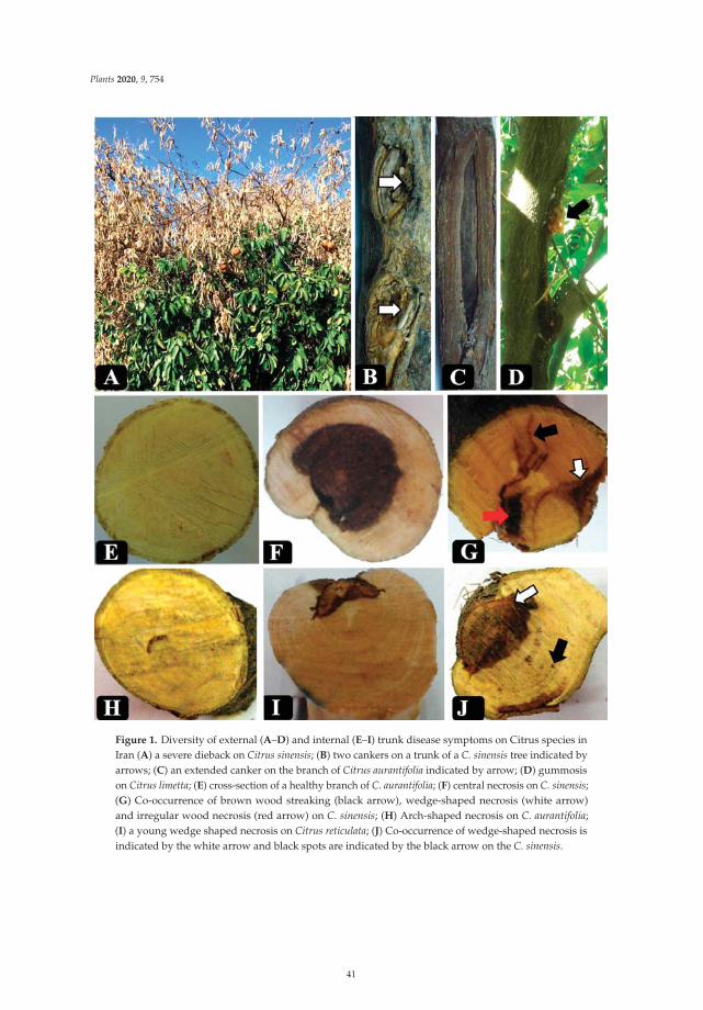
Plants 2020, 9, 754
Figure 1. Diversity of external (A–D) and internal (E–I) trunk disease symptoms on Citrus species inIran (A) a severe dieback on Citrus sinensis; (B) two cankers on a trunk of a C. sinensis tree indicated byarrows; (C) an extended canker on the branch of Citrus aurantifolia indicated by arrow; (D) gummosison Citrus limetta; (E) cross-section of a healthy branch of C. aurantifolia; (F) central necrosis on C. sinensis;(G) Co-occurrence of brown wood streaking (black arrow), wedge-shaped necrosis (white arrow)and irregular wood necrosis (red arrow) on C. sinensis; (H) Arch-shaped necrosis on C. aurantifolia;(I) a young wedge shaped necrosis on Citrus reticulata; (J) Co-occurrence of wedge-shaped necrosis isindicated by the white arrow and black spots are indicated by the black arrow on the C. sinensis.
41
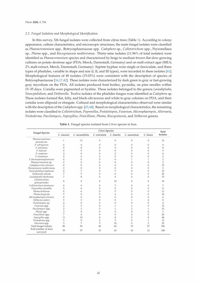
Plants 2020, 9, 754
2.2. Fungal Isolation and Morphological Identification
In this survey, 326 fungal isolates were collected from citrus trees (Table 1). According to colonyappearance, culture characteristics, and microscopic structures, the main fungal isolates were classifiedas Phaeoacremonium spp., Botryosphaeriaceae spp. Cadophora sp., Colletotrichum spp., Peyronellaeasp., Phoma spp., and Biscogniauxia mediterranea. Thirty-nine isolates (11.96% of total isolates) wereidentified as Phaeoacremonium species and characterized by beige to medium brown flat slow-growingcultures on potato dextrose agar (PDA; Merck, Darmstadt, Germany) and on malt extract agar (MEA;2% malt extract, Merck, Darmstadt, Germany). Septate hyphae were single or fasciculate, and threetypes of phialides, variable in shape and size (I, II, and III types), were recorded in these isolates [41].Morphological features of 49 isolates (15.03%) were consistent with the description of species ofBotryosphaeriaceae [16,17,42]. These isolates were characterized by dark green to gray or fast-growinggray mycelium on the PDA. All isolates produced fruit bodies, pycnidia, on pine needles within15–35 days. Conidia were pigmented or hyaline. These isolates belonged to the genera Lasiodiplodia,Neoscytalidium, and Dothiorella. Twelve isolates of the phialides fungus were identified as Cadophora sp.These isolates formed flat, felty, and black-olivaceous and white to gray colonies on PDA, and theirconidia were ellipsoid or elongate. Cultural and morphological characteristics observed were similarwith the description of the Cadophora spp. [43,44]. Based on morphological characteristics, the remainingisolates were classified to Colletotrichum, Peyronellea, Pestalotiopsis, Fusarium, Microsphaeropsis, Alternaria,Trichoderma, Paecilomyces, Aspergillus, Penicillium, Phoma, Biscogniauxia, and Stilbocrea genera.
Table 1. Fungal species isolated from Citrus species in Iran.
Fungal SpeciesCitrus Species Total
IsolatesC. sinensis C. aurantifolia C. reticulata C. limetta C. aurantium C. limon
Phaeoacremoniumparasiticum 1 11 0 0 0 0 12
P. rubrigenum 0 4 0 0 0 0 4P. minimum 0 8 0 0 0 0 8P. italicum 0 5 0 0 0 0 5
P. croatiense 2 0 0 2 0 0 4P. iranianum 0 0 2 0 0 0 2
P. fraxinopennsylvanicum 0 0 0 0 1 1 2Phaeoacremonium sp. 0 2 0 0 0 0 2
Cadophora luteo-olivacea 0 0 6 6 0 0 12Biscogniauxia mediterranea 2 0 0 0 0 0 2Neoscytalidium hyalinum 0 7 0 7 0 0 14
Dothiorella viticola 7 7 0 0 6 0 20Lasiodiplodia theobromae 7 8 0 0 0 0 15
Colletotrichumgleoesporioides 8 0 0 10 0 0 18
Colletotrichum boninense 0 0 0 7 0 0 7Peyronellea pinodella 5 6 0 0 0 0 11
Phoma herbarum 0 6 0 5 0 0 11Phoma fungicola 5 0 5 4 0 0 14
Microsphaeropsis olivacea 6 0 0 0 0 0 6Stilbocrea walteri 0 3 0 0 1 4 8Pestalotiopsis sp. 4 0 0 0 0 0 4Fusarium spp. 2 3 0 1 1 5 12
Paecilomyces spp. 2 3 5 1 3 1 15Phoma spp. 3 0 0 0 0 0 3
Penicillium spp. 7 4 9 0 4 5 29Aspergillus spp. 11 10 8 8 3 8 48Trichoderma spp. 5 0 2 5 0 1 13Alternaria spp. 3 7 3 10 0 2 25
Total fungal isolates 80 94 40 66 19 27 326Total number of trees
surveyed 39 57 19 43 18 12 188
42
Plants 2020, 9, 754
No association was found between wood symptoms and fungal species. Dual infections by trunkdisease fungi in a single tree occurred. Phaeoacremonium parasiticum and P. italicum were isolatedfrom one tree of C. aurantifolia; P. parasiticum, P. croatiense, and Do. viticola from one tree of C. sinensis,L. theobromae and Neoscytalidium hyalinum from one tree of C. aurantifolia, and C. luteo-olivacea andP. croatiense from one tree of C. limetta. In addition, some fungal species grew from an individual woodsegment, such as Stilbocrea walteri and P. fraxinopennsylvanicum from C. limon.
2.3. Molecular Characterization and Phylogenetic Analyses
BLASTn searches in GenBank showed that the nuclear ribosomal DNA-internal transcribed spacerregion (ITS) and translation elongation factor 1α (tef1-α) sequences of Botryosphaeriaceae isolates had99–100% identity with isolates of Lasiodiplodia theobromae (strain CBS559.70), Neoscytalidium hyalinum(strain CBS 145.78), and Dothiorella viticola (strain CBS 117006). The ITS sequences of the Cadophoraisolates had 99–100 % identity with isolates previously identified as Cadophora luteo-olivacea in GenBank(strain CBS 855.69). ITS and β-tubulin (BT) sequences of Colletotrichum isolates were identical to isolatespreviously reported as Colletotrichum gloeosporioides (ITS: strain CBS 132465; BT: strain CBS 100471)and Colletotrichum boninense (ITS and BT: strain CBS:123755) in GenBank. ITS and BT sequences ofPeyronellea isolates in our study showed 99–100 % identity with those isolates previously submitted asDidymella pinodella (strain CBS 531.66) in GenBank. ITS and tef1-α sequences of Stilbocrea isolates fromcitrus trees had 99–100% identity with Stilbocrea walteri (strain NQI). Regarding the Biscogniauxia isolates,ITS of our isolates had 99–100% identity with isolates previously identified as Biscogniauxia mediterranea(strain CBS 129072).
Datasets of the BT and actin (ACT) alignments of Phaeoacremonium were congruent and couldbe combined (p = 0.225). The Hasegawa–Kishino–Yano model (HKY) with gamma distributed withinvariant sites rates (G+I) was identified as the BIC best-fit nucleotide substitution model by thejModelTest for the Phaeoacremonium multi-locus analysis. Maximum likelihood (ML) of the combinedACT-BT regions provided a phylogeny with 98 to 100% ML bootstrap support for all species-level clades,with the exception of P. alvesii (paraphyletic, 87% bootstrap support), P. griseorubrum (paraphyletic,66% bootstrap support), P. roseum (89% bootstrap support), and P. viticola (paraphyletic with regard toP. roseum and P. angustius) (Figure 2). The 39 strains from Iran clustered in eight clades (P. italicum,Phaeoacremonium sp., P. rubrigenum, P. parasiticum, P. minimum, P. iranianum, P. fraxynopennsylvanicum,and P. croatiense). The isolates of the clade 2 grouped together in a polyphyletic clade with 100%bootstrap support with the P. italicum as a closely related species. The BT and ACT sequences ofthe second clade of Phaeoacremonium isolates were 98% (BT) and 98.77% (ACT) identical to those ofP. italicum CBS 137763 (GenBank KJ534074, KJ534046). Three nucleotides varied in the ACT regionand ten nucleotides in the BT region between the second clade of Phaeoacremonium isolates and theP. italicum CBS 137763 sequences.
43
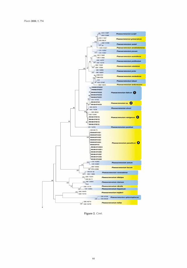
Plants 2020, 9, 754
Figure 2. Cont.
44
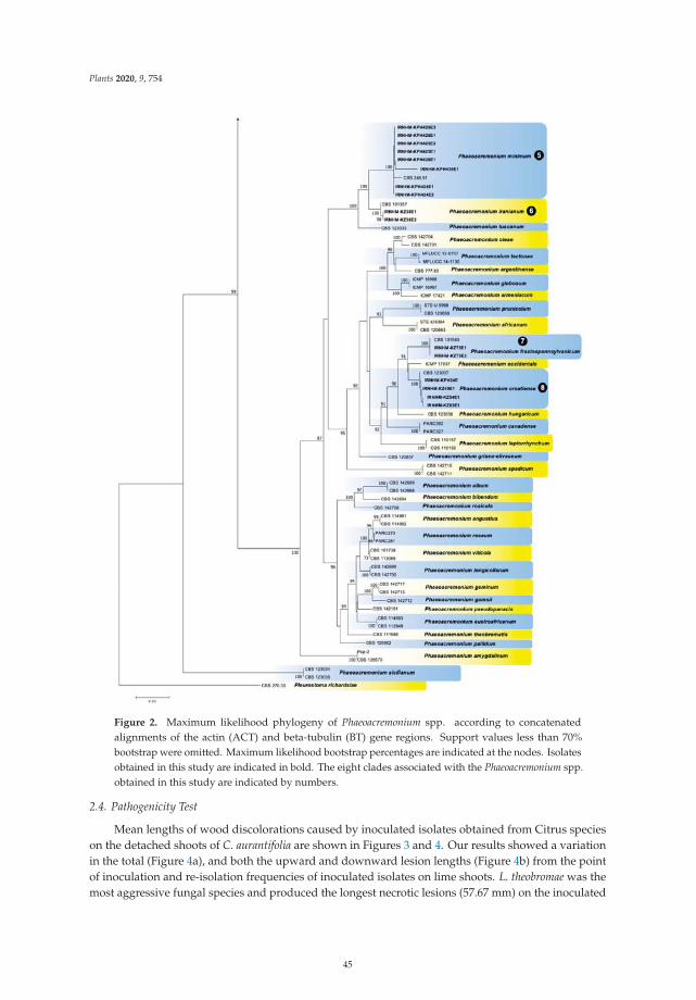
Plants 2020, 9, 754
Figure 2. Maximum likelihood phylogeny of Phaeoacremonium spp. according to concatenatedalignments of the actin (ACT) and beta-tubulin (BT) gene regions. Support values less than 70%bootstrap were omitted. Maximum likelihood bootstrap percentages are indicated at the nodes. Isolatesobtained in this study are indicated in bold. The eight clades associated with the Phaeoacremonium spp.obtained in this study are indicated by numbers.
2.4. Pathogenicity Test
Mean lengths of wood discolorations caused by inoculated isolates obtained from Citrus specieson the detached shoots of C. aurantifolia are shown in Figures 3 and 4. Our results showed a variationin the total (Figure 4a), and both the upward and downward lesion lengths (Figure 4b) from the pointof inoculation and re-isolation frequencies of inoculated isolates on lime shoots. L. theobromae was themost aggressive fungal species and produced the longest necrotic lesions (57.67 mm) on the inoculated
45
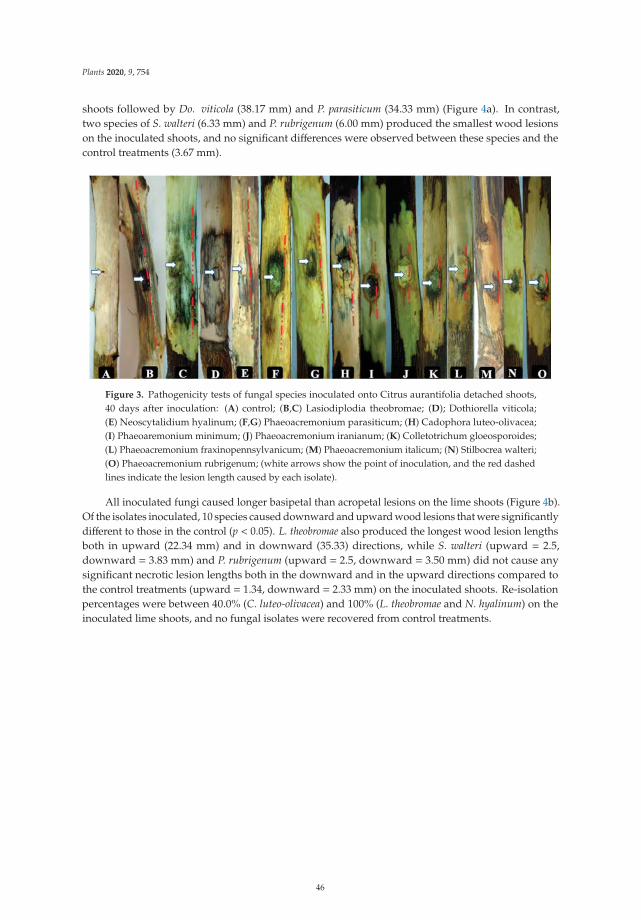
Plants 2020, 9, 754
shoots followed by Do. viticola (38.17 mm) and P. parasiticum (34.33 mm) (Figure 4a). In contrast,two species of S. walteri (6.33 mm) and P. rubrigenum (6.00 mm) produced the smallest wood lesionson the inoculated shoots, and no significant differences were observed between these species and thecontrol treatments (3.67 mm).
Figure 3. Pathogenicity tests of fungal species inoculated onto Citrus aurantifolia detached shoots,40 days after inoculation: (A) control; (B,C) Lasiodiplodia theobromae; (D); Dothiorella viticola;(E) Neoscytalidium hyalinum; (F,G) Phaeoacremonium parasiticum; (H) Cadophora luteo-olivacea;(I) Phaeoaremonium minimum; (J) Phaeoacremonium iranianum; (K) Colletotrichum gloeosporoides;(L) Phaeoacremonium fraxinopennsylvanicum; (M) Phaeoacremonium italicum; (N) Stilbocrea walteri;(O) Phaeoacremonium rubrigenum; (white arrows show the point of inoculation, and the red dashedlines indicate the lesion length caused by each isolate).
All inoculated fungi caused longer basipetal than acropetal lesions on the lime shoots (Figure 4b).Of the isolates inoculated, 10 species caused downward and upward wood lesions that were significantlydifferent to those in the control (p < 0.05). L. theobromae also produced the longest wood lesion lengthsboth in upward (22.34 mm) and in downward (35.33) directions, while S. walteri (upward = 2.5,downward = 3.83 mm) and P. rubrigenum (upward = 2.5, downward = 3.50 mm) did not cause anysignificant necrotic lesion lengths both in the downward and in the upward directions compared tothe control treatments (upward = 1.34, downward = 2.33 mm) on the inoculated shoots. Re-isolationpercentages were between 40.0% (C. luteo-olivacea) and 100% (L. theobromae and N. hyalinum) on theinoculated lime shoots, and no fungal isolates were recovered from control treatments.
46
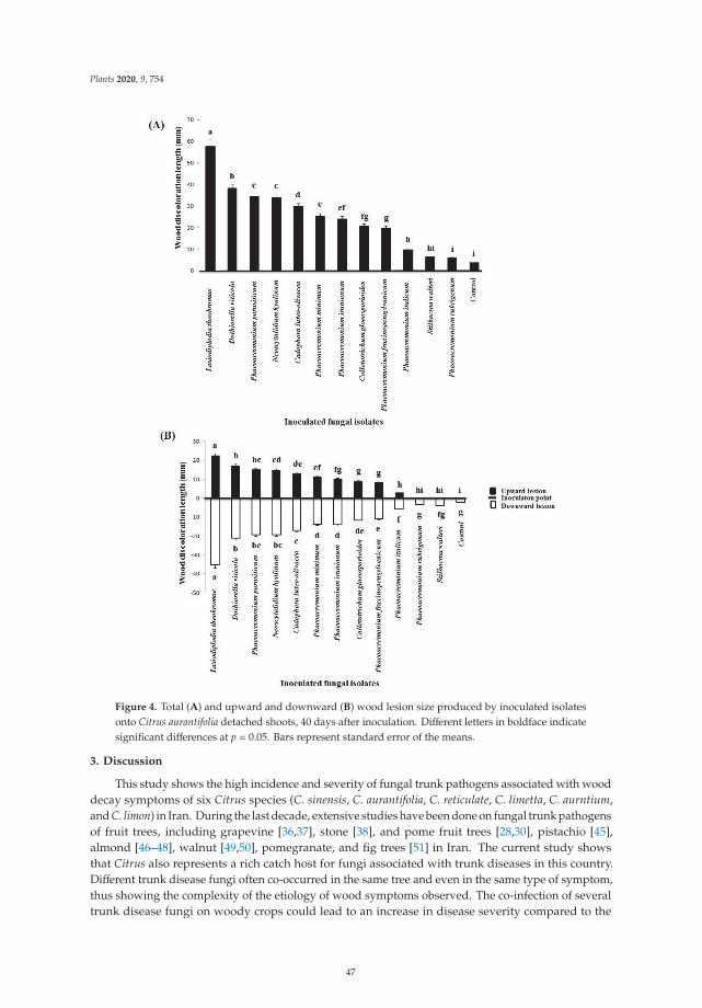
Plants 2020, 9, 754
Figure 4. Total (A) and upward and downward (B) wood lesion size produced by inoculated isolatesonto Citrus aurantifolia detached shoots, 40 days after inoculation. Different letters in boldface indicatesignificant differences at p = 0.05. Bars represent standard error of the means.
3. Discussion
This study shows the high incidence and severity of fungal trunk pathogens associated with wooddecay symptoms of six Citrus species (C. sinensis, C. aurantifolia, C. reticulate, C. limetta, C. aurntium,and C. limon) in Iran. During the last decade, extensive studies have been done on fungal trunk pathogensof fruit trees, including grapevine [36,37], stone [38], and pome fruit trees [28,30], pistachio [45],almond [46–48], walnut [49,50], pomegranate, and fig trees [51] in Iran. The current study showsthat Citrus also represents a rich catch host for fungi associated with trunk diseases in this country.Different trunk disease fungi often co-occurred in the same tree and even in the same type of symptom,thus showing the complexity of the etiology of wood symptoms observed. The co-infection of severaltrunk disease fungi on woody crops could lead to an increase in disease severity compared to the
47
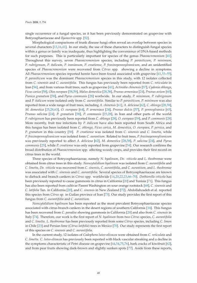
Plants 2020, 9, 754
single occurrence of a fungal species, as it has been previously demonstrated on grapevine withBotryosphaeriaceae and Ilyonectria spp. [52].
Morphological comparisons of trunk disease fungi often reveal an overlap between species inseveral characters [13,16,41]. In our study, the use of these characters to distinguish fungal specieswithin a genus or family was inadequate, thus highlighting the convenience of DNA-based methodsfor such purposes. This is particularly important for species of the genus Phaeoacremonium [41].Throughout this survey, seven Phaeoacremonium species, including P. parasiticum, P. minimum,P. rubrigenum, P. italicum, P. iranianum, P. croatiense, P. fraxinopennsylvanicum, and an unidentifiedspecies of Phaeoacremonium were recovered from Citrus spp. showing a decline in symptoms.All Phaeoacremonium species reported herein have been found associated with grapevine [41,53–55].P. parasiticum was the dominant Phaeoacremonium species in this study, with 12 isolates collectedfrom C. sinensis and C. aurantifolia. This fungus has previously been reported from C. reticulata inIran [56], and from various fruit trees, such as grapevine [41], Actinidia chinensis [57], Cydonia oblonga,Ficus carica [58], Olea europaea [58,59], Malus domestica [28,58], Prunus armeniaca [24], Prunus avium [60],Punica granatum [58], and Pyrus communis [28] worlwide. In our study, P. minimum, P. rubrigenumand P. italicum were isolated only from C. aurantifolia. Similar to P. parasiticum, P. minimum was alsoreported from a wide range of fruit trees, including A. chinensis [61], A. deliciosa [62], C. oblonga [28,58],M. domestica [25,28,63], O. europaea [64], P. armeniaca [24], Prunus dulcis [57], P. pennsylvanica [65],Prunus salicina [24], P. granatum [58], P. communis [25,28], in Iran and other parts of the world.P. rubrigenum has previously been reported from C. oblonga [28], O. europaea [59], and P. communis [28].More recently, fruit tree infections by P. italicum have also been reported from South Africa andthis fungus has been isolated from C. oblonga, Ficus carica, M. domestica, O. europaea, P. persica, andP. granatum in this country [58]. P. croatiense was isolated from C. sinensis and C. limetta, whileP. fraxinopennsylvanicum was isolated form C. aurantium. Related to fruit trees, P. fraxinopennsylvanicumwas previously reported to affect A. deliciosa [62], M. domestica [28,58], P. salicina [24], and Pyruscommunis [25], while P. croatiense was only reported from grapevine [54]. Our research confirms thebroad distribution of Phaeoacremonium spp. affecting woody crops, and provides their first record oncitrus trees in the world.
Three species of Botryosphaeriaceae, namely N. hyalinum, Do. viticola and L. theobromae wereobtained from citrus trees in this study. Neoscytalidium hyalinum was isolated from C. aurantifolia andC. limetta, Do. viticola was recovered from C. sinensis, C. aurantifolia, and C. aurantium, and L. theobromaewas associated with C. sinensis and C. aurantifolia. Several species of Botryosphaeriaceae are knownto dieback and branch cankers in Citrus spp. worldwide [14,20,22,23,66–70]. Dothiorella viticola hasbeen previously reported to cause gummosis in citrus in California [20] and Tunisia [71]. This fungushas also been reported from cultivar Parent Washington on sour orange rootstock [68], C. sinensis andC. latifolia Tan. in California [20], and C. sinensis in New Zealand [72]. Abdollahzadeh et al. reportedthis species from Citrus sp. in Guilan province of Iran [73]. Our study provides the first report of thisfungus from C. aurantifolia and C. aurantium.
Neoscytalidium hyalinum has been reported as the most prevalent Botryosphaeriaceae speciesassociated with citrus branch cankers in the desert regions of southern California [14]. This fungushas been recovered from C. paradise showing gummosis in California [20] and also from C. sinensis inItaly [74]. Therefore, our work is the first report of N. hyalinum from two Citrus species, C. aurantifoliaand C. limetta. L. theobromae has been previously reported from some Citrus species, including C. limonin Chile [23] and Persian lime (Citrus latifolia) trees in Mexico [70]. Our study represents the first reportof this species on C. sinensis and C. aurantifolia.
In the current study, 12 isolates of Cadophora luteo-olivacea were obtained from C. reticulata andC. limetta. C. luteo-olivacea has previously been reported with black vascular streaking and a decline inthe symptoms characteristic of Petri disease on grapevine [44,54,75,76], bark cracks of kiwifruit [62],and from pear fruits showing dark-brown and slightly sunken spots [77]. Aside from these reports,
48
Plants 2020, 9, 754
little is known regarding the role of Cadophora species involved in trunk diseases of trees. This is thefirst time that C. luteo-olivacea has been found on Citrus spp.
Most species of the genus Biscogniauxia are reported from forest trees, mainly from Quercusspp. [78–80]. Some species of this genus have also been found associated with fruit trees such asB. pruni and B. granmoi on Prunus padus [81,82], B. marginata on M. communis [83], B. rosacearumon P. communis, C. oblonga and Prunus domestica [84], and B. capnodes on Averrhoa carambola [85].Biscogniauxia mediterranea is known to be the causal agent of charcoal cankers on a wide range of treesworldwide, in particular Quercus spp. [79,80,86]. In Iran, this pathogen was already reported fromC. sinensis [87], along with other woody hosts, such as Quercus castaneifolia [88], Zelkova carpinifolia [89],Q. brantii [89], and Amygdalus scoparia [90].
In the current study, eight isolates of Stilbocrea walteri were isolated from C. aurantifolia, C. aurntium,and C. limon. This species was originally reported from dead corticated branches of Quercus ilex inPortugal [91], and to our knowledge, it has not been reported from necrotic wood tissues of trees.Therefore, this study is the first report of this species in Iran and on Citrus species worldwide.
Peyronellea pinodella (Didymellaceae) is a destructive necrotrophic pathogen on some plant families,including Fabaceae, Amaranthaceae, Asteraceae, Amaryllidaceae, Appiaceae Rubiaceae, Malvaceae, Poaceae,and Polemoniaceae [92]. To date, there is no report on the occurrence of P. pinodella on Citrus species andthis is the first data on the occurrence of this species on C. sinensis and C. aurantifolia.
Two species of Colletotrichum were found to be associated with trunk diseases of citrus trees inthis work, C. gleoesporioides on C. sinensis and C. limetta and C. boninense on C. limetta. Several speciesof Colletotrichum are associated with fruit and leaf anthracnose diseases of Citrus species; however,other diseases such as twig and shoot dieback caused by Colletotrichum spp. have been documented oncitrus trees [14,93]. Colletotrichum gloeosporioides has been reported from a wide range of fruit treessuch as strawberry, olive, almond, mango, apple, avocado, and citrus [94]. This fungus was foundto be associated with twig dieback of lemon trees in Portugal [93]. C. boninense has been associatedwith fruit and leaf anthracnose on citrus trees [95,96]. According to a recent study, some Colletotrichumspecies have been isolated and reported from stems of citrus trees in Iran. These included C. karstiifrom C. aurantifolia and C. sinensis and four species, C. gloeosporioides, C. novae–zelandiae, C. siamense,and C. fructicola from C. sinensis [97]. Therefore, our study represents the first report of C. gleoesporioidesand C. boninense from branches of C. limetta.
In our work, six isolates of M. olivacea were obtained from sweet orange. This fungus hasbeen reported from various plant species worldwide. This taxon has previously been isolated as anendophytic species from P. persica [98], from xylem and stems of Pinus sylvestris [99] and Chileangymnosperms [100]. Carlucci et al. isolated this species from internal wood discoloration of olive treesin Italy [101]. Microsphaeropsis olivacea has also been isolated and reported from some woody plants,such as Prunus cerasus, P. avium [102], and Persian oak (Quercus brantii) [103] in Iran. To our knowledge,this is the first report of M. olivacea on citrus trees. Several isolates of Fusarium, Pestalotiopsis, Phoma,Penicillium, Aspergillus, Trichoderma, and Alternaria species were also obtained from Citrus species inthis study. Therefore, more studies are needed on these taxa in order to elucidate their potential impacton citrus trunk diseases.
Pathogenicity of selected fungal species in detached shoots of lime tree were confirmed in thecurrent study. Results revealed that L. theobromae was more virulent on lime shoots than other species.In contrast to our results, Bautista-Cruz et al. reported that L. theobromae was the least virulent specieswhen inoculated in Persian lime branches [70]. Several factors differed from the study carried out byBautista-Cruz et al. and might have contributed to the discrepancy between the experiments, includingthe type of planting material inoculated, the environmental conditions for disease development, thetime for virulence assessment, and the fungal strain used in the pathogenicity test. L. theobromae hasbeen considered the most aggressive species on Eucalyptus [104,105], grapevine [42,106], and pistachiotrees [107]. Lasiodiplodia theobromae was considered an important pathogen on greengage, sour cherry,peach, apricot, cherry [38], and willow trees [29] in Iran. Our study improved the knowledge on
49
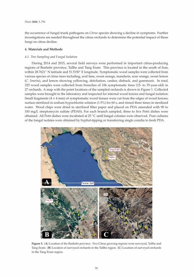
Plants 2020, 9, 754
the occurrence of fungal trunk pathogens on Citrus species showing a decline in symptoms. Furtherinvestigations are needed throughout the citrus orchards to determine the potential impact of thesefungi on citrus decline.
4. Materials and Methods
4.1. Tree Sampling and Fungal Isolation
During 2014 and 2015, several field surveys were performed in important citrus-producingregions of Bushehr province, Tallhe and Tang Eram. This province is located in the south of Iran,within 28.7621◦ N latitude and 51.5150◦ E longitude. Symptomatic wood samples were collected fromvarious species of citrus trees including, acid lime, sweet orange, mandarin, sour orange, sweet lemon(C. limetta), and lemon showing yellowing, defoliation, canker, dieback, and gummosis. In total,325 wood samples were collected from branches of 106 symptomatic trees (15- to 35-year-old) in27 orchards. A map with the point locations of the sampled orchards is shown in Figure 5. Collectedsamples were brought to the laboratory and inspected for internal wood lesions and fungal isolation.Small fragments (4 × 4 mm) of symptomatic wood tissues were cut from the edges of wood lesions,surface-sterilized in sodium hypochlorite solution (1.5%) for 60 s, and rinsed three times in sterilizedwater. Wood chips were dried in sterilized filter paper and placed on PDA amended with 90 to100 mg/L streptomycin sulfate (PDAS). For each branch sampled, three to five Petri dishes wereobtained. All Petri dishes were incubated at 25 ◦C until fungal colonies were observed. Pure culturesof the fungal isolates were obtained by hyphal-tipping or transferring single conidia to fresh PDA.
Figure 5. (A) Location of the Bushehr province. Two Citrus growing regions were surveyed, Tallhe andTang Eram. (B) Location of surveyed orchards in the Tallhe region. (C) Location of surveyed orchardsin the Tang Eram region.
50
Plants 2020, 9, 754
4.2. Morphological Identification
All fungal isolates were identified initially to the genus level based on colony morphology andmain microscopic structures using published articles and descriptions. Botryosphaeriaceae isolateswere identified based on colony appearance and conidial morphology [16,108]. To induce sporulation,three to five mycelial plugs from each isolate were placed on 2% water agar (WA; Biokar-Diagnostics)plates amended with sterilized pine needles and incubated at 25 ◦C under near-ultraviolet light for15–45 days [42]. Conidial characteristics (size, shape, color, and presence or absence of septa) wererecorded for all isolates. Phaeoacremonium isolates were grouped based on colony appearance, pigmentproduction on MEA, PDA and oatmeal agar (OA; 60 g oatmeal; 12.5 g agar; Difco, France) and themain microscopic structures (phialide shape and type, conidiophore morphology, size of hyphalwarts, and conidial shape and size) [41,57,109]. Identification of Cadophora isolates was based on thecolony and micro-morphological structures, such as conidiogenous cell size and shape, and conidia.The remaining fungal isolates were identified based on available identification keys and publishedpapers [91,110–114].
4.3. DNA Extraction, Amplification, and Sequencing
Identities of representative isolates were confirmed using molecular data. Fungi selected formolecular studies were grown on PDA for 10 to 15 days at 25 ◦C in the dark. DNA was extractedusing an AccuPrep®Genomic DNA Extraction Kit (Bioneer, South Korea) following the instructionsof the manufacturer. Four primer sets, ITS1/ITS4 [115], EF1-728F/EF1-986R [116], T1/Bt2b [117,118],and ACT-512F/ACT-783R [116] were used to amplify the ITS region ITS1-5.8S-ITS2, portions of thetef1-α, BT and ACT genes, respectively. The identification of Botryosphaeriaceae isolates was confirmedby the sequencing of ITS and a partial sequence of tef-1a. For Phaeoacremonium isolates, a partialsequence of BT and ACT genes were amplified and sequenced. Molecular identifications of otherisolates were confirmed by sequence analysis of ITS (Cadophora, Colletotrichum, Peyronellea, Stilbocrea,and Biscogniauxia isolates), BT (Colletotrichum and Peyronellea isolates), or tef1-α (Stilbocrea isolates).The polymerase chain reaction (PCR) was performed in a Techne TC-312 Thermal Cycler (Techne,Cambridge, UK), as described by Hashemi and Mohammadi [29]. For each isolate, 3–4 μL of PCRproduct was separated by electrophoresis on a 1% agarose gel (UltraPureTM Agarose, Invitrogen)containing ethidium bromide and visualized under UV illumination. The size of the products wasevaluated using a 100 bp ladder (Gene Ruler, TMDNA Ladder Mix, Fermentas). PCR products weresubmitted to Bioneer Corporation (Daejeon, South Korea) for sequencing. MegaBLAST approach ofthe NCBI database (https://www.ncbi.nlm.nih.gov/) was initially used to identify fungal species.
4.4. Phylogenetic Analysis
Due to the broad range of Phaeoacremonium spp. obtained in this study, a phylogenetic analysiswas carried out for the Phaeoacremonium spp. isolates. Sequences from citrus in Iran were alignedwith sequences available in GenBank/NCBI. These were compared using MAFFT sequence alignmentprogram v. 6 [119] with ex-type specimens from different hosts. Alignments were inspected inSequence Alignment Editor v. 2.0a11 [120]. PAUP version 4.0 b 10 [121] was used to perform a partitionhomogeneity test. The congruence between the ACT and BT datasets was tested at 1000 replicates,and the maximum likelihood (ML) was carried out on the concatenated alignment. The MEGA version7 software [122] was used for ML analysis. Bayesian information criterion in jModelTest 2.1.10 [123] wasused to estimate the best fit model. Single and concatenated datasets were tested for branch support(1000 bootstrap replicates). We included sequences published by Spies et al. as reference sequences [58].Pleurostoma richardsiae CBS 270.33 was included as an outgroup. Phaeoacremonium sequences obtainedin this study were submitted to GenBank/NCBI (Table 2) and the sequence alignments were depositedin TreeBASE under study number 26006 (http://treebase.org).
51
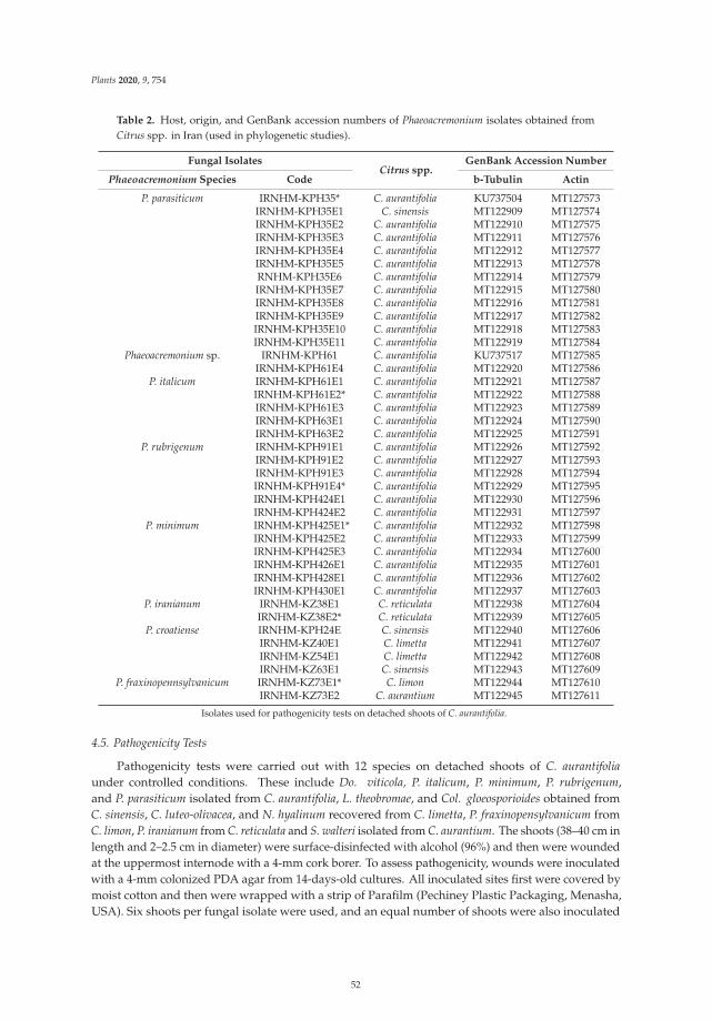
Plants 2020, 9, 754
Table 2. Host, origin, and GenBank accession numbers of Phaeoacremonium isolates obtained fromCitrus spp. in Iran (used in phylogenetic studies).
Fungal IsolatesCitrus spp.
GenBank Accession Number
Phaeoacremonium Species Code b-Tubulin Actin
P. parasiticum IRNHM-KPH35* C. aurantifolia KU737504 MT127573IRNHM-KPH35E1 C. sinensis MT122909 MT127574IRNHM-KPH35E2 C. aurantifolia MT122910 MT127575IRNHM-KPH35E3 C. aurantifolia MT122911 MT127576IRNHM-KPH35E4 C. aurantifolia MT122912 MT127577IRNHM-KPH35E5 C. aurantifolia MT122913 MT127578RNHM-KPH35E6 C. aurantifolia MT122914 MT127579IRNHM-KPH35E7 C. aurantifolia MT122915 MT127580IRNHM-KPH35E8 C. aurantifolia MT122916 MT127581IRNHM-KPH35E9 C. aurantifolia MT122917 MT127582IRNHM-KPH35E10 C. aurantifolia MT122918 MT127583IRNHM-KPH35E11 C. aurantifolia MT122919 MT127584
Phaeoacremonium sp. IRNHM-KPH61 C. aurantifolia KU737517 MT127585IRNHM-KPH61E4 C. aurantifolia MT122920 MT127586
P. italicum IRNHM-KPH61E1 C. aurantifolia MT122921 MT127587IRNHM-KPH61E2* C. aurantifolia MT122922 MT127588IRNHM-KPH61E3 C. aurantifolia MT122923 MT127589IRNHM-KPH63E1 C. aurantifolia MT122924 MT127590IRNHM-KPH63E2 C. aurantifolia MT122925 MT127591
P. rubrigenum IRNHM-KPH91E1 C. aurantifolia MT122926 MT127592IRNHM-KPH91E2 C. aurantifolia MT122927 MT127593IRNHM-KPH91E3 C. aurantifolia MT122928 MT127594IRNHM-KPH91E4* C. aurantifolia MT122929 MT127595IRNHM-KPH424E1 C. aurantifolia MT122930 MT127596IRNHM-KPH424E2 C. aurantifolia MT122931 MT127597
P. minimum IRNHM-KPH425E1* C. aurantifolia MT122932 MT127598IRNHM-KPH425E2 C. aurantifolia MT122933 MT127599IRNHM-KPH425E3 C. aurantifolia MT122934 MT127600IRNHM-KPH426E1 C. aurantifolia MT122935 MT127601IRNHM-KPH428E1 C. aurantifolia MT122936 MT127602IRNHM-KPH430E1 C. aurantifolia MT122937 MT127603
P. iranianum IRNHM-KZ38E1 C. reticulata MT122938 MT127604IRNHM-KZ38E2* C. reticulata MT122939 MT127605
P. croatiense IRNHM-KPH24E C. sinensis MT122940 MT127606IRNHM-KZ40E1 C. limetta MT122941 MT127607IRNHM-KZ54E1 C. limetta MT122942 MT127608IRNHM-KZ63E1 C. sinensis MT122943 MT127609
P. fraxinopennsylvanicum IRNHM-KZ73E1* C. limon MT122944 MT127610IRNHM-KZ73E2 C. aurantium MT122945 MT127611
Isolates used for pathogenicity tests on detached shoots of C. aurantifolia.
4.5. Pathogenicity Tests
Pathogenicity tests were carried out with 12 species on detached shoots of C. aurantifoliaunder controlled conditions. These include Do. viticola, P. italicum, P. minimum, P. rubrigenum,and P. parasiticum isolated from C. aurantifolia, L. theobromae, and Col. gloeosporioides obtained fromC. sinensis, C. luteo-olivacea, and N. hyalinum recovered from C. limetta, P. fraxinopensylvanicum fromC. limon, P. iranianum from C. reticulata and S. walteri isolated from C. aurantium. The shoots (38–40 cm inlength and 2–2.5 cm in diameter) were surface-disinfected with alcohol (96%) and then were woundedat the uppermost internode with a 4-mm cork borer. To assess pathogenicity, wounds were inoculatedwith a 4-mm colonized PDA agar from 14-days-old cultures. All inoculated sites first were covered bymoist cotton and then were wrapped with a strip of Parafilm (Pechiney Plastic Packaging, Menasha,USA). Six shoots per fungal isolate were used, and an equal number of shoots were also inoculated
52
Plants 2020, 9, 754
with 4-mm non-colonized PDA agar plugs for negative controls. Inoculated shoots were arrangedat random, including the six inoculated shoots per isolate. Inoculated shoots were placed in moistchambers and incubated at 25 ºC. The total, upward, and downward lesion length data were evaluatedindividually, 40 days after inoculation. Recorded data were checked for normality of distribution bymeans of the Shapiro–Wilk and Kolmogorov–Smirnov tests. The data were subjected to analysis ofvariance (one-way ANOVA) using SAS v 9.1 (SAS Institute, Cary, NC, USA) (Dataset S1; Dataset S2).The least significant difference (LSD) test was used for comparison of treatment means at p < 0.05.Fungal re-isolations were made from the edges of the lesions on the test and control shoots and placedon PDA. The identity of the re-isolated fungi was confirmed based on morphological characteristicsand molecular analysis in order to complete Koch’s postulates. The pathogenicity of other species wasnot tested in this work because they were identified after the pathogenicity trials had begun on thedetached shoots of C. aurantifolia.
Supplementary Materials: The following are available online at http://www.mdpi.com/2223-7747/9/6/754/s1,Dataset S1: SAS code, Dataset S2: Lesion length data.
Author Contributions: Conceived the idea of the research, designed the scientific experiments and methodology,H.M.; field surveys, sample preparation, laboratory works, sequencing of fungal isolates, pathogenicity tests, N.E.;software, H.M. and D.G.; writing—original draft preparation, H.M.; writing—review and editing, H.M. and D.G.;supervision, H.M.; phylogenetic analysis, D.G. All authors have read and agreed to the published version ofthe manuscript.
Funding: This research received no external funding.
Conflicts of Interest: The authors declare no conflict of interest.
References
1. FAO. Citrus Fruit Fresh and Processed. Statistical Bulletin. 2016. Available online: http://fao.org/3/a-i8092e.pdf (accessed on 15 May 2020).
2. Gramaje, D.; Úrbez-Torres, J.R.; Sosnowski, M.R. Managing grapevine trunk diseases with respect to etiologyand epidemiology: Current strategies and future prospects. Plant Dis. 2018, 102, 12–39. [CrossRef] [PubMed]
3. Gramaje, D.; Baumgartner, K.; Halleen, F.; Mostert, L.; Sosnowski, M.R.; Úrbez-Torres, J.R.; Armengol, J.Fungal trunk diseases: A problem beyond grapevines? Plant Pathol. 2016, 65, 355–356. [CrossRef]
4. Carter, M.V. The Status of Eutypa Lata as a Pathogen. Monograph, Phytopathological Paper No 32; CommonwealthAgricultural Bureau, International Mycological Institute: Surrey, UK, 1991.
5. Munkvold, G.P.; Marois, J.J. Eutypa dieback of sweet cherry and occurrence of Eutypa lata perithecia in thecentral valley of California. Plant Dis. 1994, 78, 200–207. [CrossRef]
6. Sinclair, W.A.; Lyon, H.H. Diseases of Trees and Shrubs, 2nd ed.; Cornell University Press: Ithaca, NY, USA,2005; p. 659.
7. Jurc, D.; Ogris, N.; Slippers, B.; Stenlid, J. First report of Eutypella canker of Acer pseudoplatanus in Europe.Plant Pathol. 2006, 55, 577. [CrossRef]
8. Úrbez-Torres, J.R.; Adams, P.; Kama, J.; Gubler, W.D. Identification, incidence and pathogenicity of fungalspecies associated with grapevine dieback in Texas. Am. J. Enol. Vitic. 2009, 60, 497–507.
9. Reinking, O.A. Philippine economic plant diseases. Philipp. J. Sci. 1918, 13, 165–274.10. Rappaz, F. Taxonomie et nomenclature des Diatrypacees a asques octosporees. Mycol. Helv. 1987, 2, 285–648.11. Resplandy, R.; Chevaugeon, J.; Delassus, M.; Luc, M. Premiere liste annotee de champignons parasites de
plantes cultivees en Cote d’Ivoire. Ann. Epiphyt. 1954, 1, 1–61.12. Hanlin, R.T. Index to genera and species of ascomycetes described by A. P. Viegas. Mycotaxon 1992, 43,
207–230.13. Trouillas, F.P.; Pitt, W.M.; Sosnowski, M.R.; Huang, R.; Peduto, F.; Loschiavo, A.; Savocchia, S.; Scott, E.S.;
Gubler, W.D. Taxonomy and DNA phylogeny of Diatrypaceae associated with Vitis vinifera and other woodyplants in Australia. Fungal Divers. 2011, 49, 203–223. [CrossRef]
14. Mayorquin, J.S.; Wang, D.H.; Twizeyimana, M.; Eskalen, A. Identification, distribution, and pathogenicity ofDiatrypaceae and Botryosphaeriaceae associated with citrus branch canker in the southern California desert.Plant Dis. 2016, 2016. 100, 2402–2413. [CrossRef]
53
Plants 2020, 9, 754
15. Trouillas, F.P.; Urbez-Torres, J.R.; Gubler, W.D. Diversity of Diatrypaceous fungi associated with grapevinecanker diseases in California. Mycologia 2010, 102, 319–336. [CrossRef] [PubMed]
16. Phillips, A.J.L.; Alves, A.; Abdollahzadeh, J.; Slippers, B.; Wingfield, M.J.; Groenewald, J.Z.; Crous, P.W.The Botryosphaeriaceae: Genera and species known from culture. Stud. Mycol. 2013, 76, 51–167. [CrossRef][PubMed]
17. Slippers, B.; Boissin, E.; Phillips, A.J.; Groenewald, J.Z.; Lombard, L.; Wingfield, M.J.; Postma, A.; Burgess, T.;Crous, P.W. Phylogenetic lineages in the Botryosphaeriales: A systematic and evolutionary framework.Stud. Mycol. 2013, 76, 31–49. [CrossRef] [PubMed]
18. Dissanayake, A.J.; Phillips, A.J.L.; Li, X.H.; Hyde, K.D. Botryosphaeriaceae: Current status of genera andspecies. Mycosphere 2016, 7, 1001–1073. [CrossRef]
19. Abdollahzadeh, J.; Javadi, A.; Mohammadi Goltapeh, E.; Zare, R.; Phillips, A.J.L. Phylogeny and morphologyof four new species of Lasiodiplodia from Iran. Persoonia 2010, 25, 1–10. [CrossRef]
20. Adesemoye, A.O.; Mayorquin, J.S.; Wang, D.H.; Twizeyimana, M.; Lynch, S.C.; Eskalen, A. Identificationof species of Botryosphaeriaceae causing bot gummosis in citrus in California. Plant Dis. 2014, 98, 55–61.[CrossRef]
21. Linaldeddu, B.T.; Dedda, A.; Scanu, B.; Franceschini, A.; Serra, S.; Berraf-Tebbal, A.; Zouaoui Boutiti, M.;Ben Jamâa, M.L.; Phillips, A.J.L. Diversity of Botryosphaeriaceae species associated with grapevine and otherwoody hosts in Italy, Algeria and Tunisia, with descriptions of Lasiodiplodia exigua and Lasiodiplodia mediterraneasp. nov. Fungal Divers. 2015, 71, 201–214. [CrossRef]
22. Coutinho, I.B.L.; Freire, F.C.O.; Lima, C.S.; Lima, J.S.; Gonçalves, F.J.T.; Machado, A.R.; Silva, A.M.S.;Cardoso, J.E. Diversity of genus Lasiodiplodia associated with perennial tropical fruit plants in northeasternBrazil. Plant Pathol. 2017, 66, 90–104. [CrossRef]
23. Guajardo, J.; Riquelme, N.; Tapia, L.; Larach, A.; Torres, C.; Camps, R.; Besoain, X. First report ofLasiodiplodia theobromae causing bot gummosis in Citrus limon in Chile. Plant Dis. 2018, 102, 818. [CrossRef]
24. Damm, U.; Mostert, L.; Crous, P.W.; Fourie, P.H. Novel Phaeoacremonium species associated with necroticwood of Prunus trees. Persoonia 2008, 20, 87–102. [CrossRef]
25. Cloete, M.; Fourie, P.H.; Damm, U.; Crous, P.W.; Mostert, L. Fungi associated with die-back symptoms ofapple and pear trees, a possible inoculum source of grapevine trunk disease pathogens. Phytopathol. Mediterr.2011, 50, S176–S190.
26. Gramaje, D.; Agustí-Brisach, C.; Pérez-Sierra, A.; Moralejo, E.; Olmo, D.; Mostert, L.; Damm, U.; Armengol, J.Fungal trunk pathogens associated with wood decay of almond trees on Mallorca (Spain). Persoonia 2012, 28,1–13. [CrossRef] [PubMed]
27. Olmo, D.; Gramaje, D.; Agustí-Brisach, C.; Leon, M.; Armengol, J. First report of Phaeoacremonium venezuelenseassociated with decay of apricot trees in Spain. Plant Dis. 2014, 98, 1001. [CrossRef] [PubMed]
28. Sami, S.; Mohammadi, H.; Heydarnejad, J. Phaeoacremonium species associated with necrotic wood of pomefruit trees in Iran. J. Plant Pathol. 2014, 96, 487–495.
29. Hashemi, H.; Mohammadi, H. Identification and characterization of fungi associated with internal woodlesions and decline disease of willow and poplar trees in Iran. For. Pathol. 2016, 46, 341–352. [CrossRef]
30. Mohammadi, H.; Sharifi, S. Association of Botryosphaeriaceae and Phaeoacremonium species withinsect-damaged quince shoots. J. Plant Pathol. 2016, 98, 35–42.
31. Kazemzadeh Chakusary, M.; Mohammadi, H.; Khodaparast, A. Decline-associated Phaeoacremonium spp.occurring on forest trees in the north of Iran. For. Pathol. 2017, 47, e12368. [CrossRef]
32. Marin-Felix, Y.; Hernández-Restrepo, M.; Wingfield, M.J.; Akulov, A.; Carnegie, A.J.; Cheewangkoon, R.;Gramaje, D.; Groenewald, J.Z.; Guarnaccia, V.; Halleen, F.; et al. Genera of phytopathogenic fungi: GOPHY 2.Stud. Mycol. 2018, 92, 47–133. [CrossRef]
33. Gramaje, D.; Mostert, L.; Groenewald, J.Z.; Crous, P.W. Phaeoacremonium: From esca disease tophaeohyphomycosis. Fungal Biol. 2015, 119, 759–783. [CrossRef]
34. Baloyi, M.A.; Mostert, L.; Halleen, F. Pathogenicity of ten Phaeoacremonium species associated with esca andPetri disease of grapevine. Phytopathol. Mediterr. 2018, 57, 538–546.
35. Mugnai, L.; Graniti, A.; Surico, G. Esca (black measles) and Brown wood-streaking: Two old and elusivediseases of grapevines. Plant Dis. 1999, 83, 404–416. [CrossRef] [PubMed]
36. Mohammadi, H.; Banihashemi, Z.; Gramaje, D.; Armengol, J. Fungal pathogens associated with grapevinetrunk diseases in Iran. J. Agric. Sci. Technol. 2013, 15, 137–150.
54
Plants 2020, 9, 754
37. Mohammadi, H.; Banihashemi, Z.; Gramaje, D.; Armengol, J. Characterization of Diplodia seriata andNeofusicoccum parvum associated with grapevine decline in Iran. J. Agric. Sci. Technol. 2013, 15, 603–616.
38. Soltaninejad, N.; Mohammadi, H.; Massumi, H. Isolation, identification and pathogenicity ofBotryosphaeriaceae and Phaeoacremonium species associated with decline of prunus species in Iran.J. Plant Pathol. 2017, 99, 571–581. [CrossRef]
39. Hashemi, H.; Mohammadi, H.; Abdollahzadeh, J. Symptoms and fungi associated with elm trees decline inIran. Eur. J. For. Res. 2017, 136, 857–879. [CrossRef]
40. Panahandeh, S.; Mohammadi, H.; Gramaje, D. Trunk disease fungi associated with Syzygium cumini in Iran.Plant Dis. 2019, 103, 711–720. [CrossRef]
41. Mostert, L.; Groenewald, J.Z.; Summerbell, R.C.; Gams, W.; Crous, P.W. Taxonomy and pathology of Togninia(Diaporthales) and its Phaeoacremonium anamorphs. Stud. Mycol. 2006, 54, 1–115. [CrossRef]
42. Van Niekerk, J.M.; Crous, P.W.; Groenewald, J.Z.; Fourie, P.H.; Halleen, F. DNA phylogeny, morphology andpathogenicity of Botryosphaeria species on grapevines. Mycologia 2004, 96, 781–798. [CrossRef]
43. Harrington, T.C.; McNew, D.L. Phylogenetic analysis places the Phialophora-like anamorph genus Cadophorain the Helotiales. Mycotaxon 2003, 87, e151.
44. Gramaje, D.; Mostert, L.; Armengol, J. Characterization of Cadophora luteo-olivacea and C. melinii isolatesobtained from grapevines and environmental samples from grapevine nurseries in Spain. Phytopathol. Mediterr.2011, 50, S112–S126.
45. Mohammadi, H.; Sarcheshmehpour, M.; Mafi, E. Fungal trunk pathogens associated with wood decay ofpistachio trees in Iran. Span. J. Agric. Res. 2015, 2, 6. [CrossRef]
46. Mousavi, M.; Mohammadi, H.; Soltaninejad, N. First report of Phaeoacremonium aleophilum (Ascomycota,Calosphaeriales) on almond and cherry trees in Kerman province. In Proceedings of the 21th Iranian PlantProtection Congress, Urmia, Iran, 23–26 August 2014; p. 28.
47. Sohrabi, M.; Mohammadi, H. Phaeoacremonium spp. and Pleurostoma richardsiae associated with trunk diseasesof almond trees in Iran. In Proceedings of the 3rd Iranian Mycological Congress, University of Kurdistan,Sanandaj, Iran, 26–28 August 2017; p. 51.
48. Sohrabi, M.; Mohammadi, H. First report of Lasiodiplodia theobromae from almond trees in Iran. In Proceedingsof the International Conference on Agricultural Science Medical Plants Traditional Medicine, Payam NoorUniversity, Mashhad, Iran, 14–15 February 2018; pp. 1–11.
49. Sohrabi, M.; Mohammadi, H. Report on two species of Phaeoacremonium from walnut trees (Juglans regia L.)in Iran. In Proceedings of the 3rd Iranian Mycological Congress, University of Kurdistan, Sanandaj, Iran,26–28 August 2017; p. 50.
50. Sohrabi, M.; Mohammadi, H.; Leon, M.; Armengol, J.; Banihashemi, Z. Detection of Phaeoacremonium spp.associated with decline of walnut trees (Juglanconis regia) in Kerman and Fars provinces. In Proceedings ofthe 4th Iranian Mycological Congress, Sari Agricultural Sciences and Nutural Resources University, Sari, Iran,26–28 August 2019; p. 61.
51. Kadkhodahemmatabadi, S. Study of Disease Symptoms and Fungi Associated with Decline of Pomegranateand Fig Trees in Fars and Kerman Provinces. Master’s Thesis, Shahid Bahonar University of Kerman,Kerman, Iran, 2018.
52. Whitelaw-Weckert, M.A.; Rahman, L.; Appleby, L.M.; Hall, A.; Clark, A.C.; Waite, H.; Hardie, W.J. Co-infectionby Botryosphaeriaceae and Ilyonectria spp. fungi during propagation causes decline of young graftedgrapevines. Plant Pathol. 2013, 62, 1226–1237. [CrossRef]
53. Essakhi, S.; Mugnai, L.; Crous, P.W.; Groenewald, J.Z.; Surico, G. Molecular and phenotypic characterizationof novel Phaeoacremonium species associated with Petri disease and esca of grapevine. Persoonia 2008, 21,119–134. [CrossRef] [PubMed]
54. Raimondo, M.L.; Lops, F.; Carlucci, A. Phaeoacremonium italicum sp nov., a new species associated with escaof grapevine in southern Italy. Mycologia 2014, 106, 1119–1126. [CrossRef]
55. Úrbez-Torres, J.R.; Haag, P.; Bowen, P.; O’Gorman, D.T. Grapevine trunk diseases in British Columbia:Incidence and characterization of the fungal pathogens associated with esca and Petri diseases of grapevine.Plant Dis. 2014, 98, 469–482. [CrossRef] [PubMed]
56. Sadeghi, F.; Samsampour, D.; Seyahooei, M.A.; Bagheri, A.; Soltani, J. Diversity and spatiotemporaldistribution of fungal endophytes associated with Citrus reticulata cv. Siyahoo. Curr. Microbiol. 2019, 76,279–289. [CrossRef] [PubMed]
55
Plants 2020, 9, 754
57. Di Marco, S.; Calzarano, F.; Osti, F.; Mazzullo, A. Pathogenicity of fungi associated with a decay of kiwifruit.Australas. Plant Pathol. 2004, 33, 337–342. [CrossRef]
58. Spies, C.F.J.; Moyo, P.; Halleen, F.; Mostert, L. Phaeoacremonium species diversity on woody hosts in theWestern Cape Province of South Africa. Persoonia 2018, 40, 26–62. [CrossRef]
59. Nigro, F.; Boscia, D.; Antelmi, I.; Ippolito, A. Fungal species associated with a severe decline of olive insouthern Italy. J. Plant Pathol. 2013, 95, 668.
60. Rumbos, I. Phialophora parasitica, causal agent of cherry dieback. J. Phytopathol. 1986, 117, 283–287. [CrossRef]61. Crous, P.W.; Gams, W. Phaeomoniella chlamydospora gen. et comb. nov., a causal organism of Petri grapevine
decline and esca. Phytopathol. Mediterr. 2000, 39, 112–188.62. Prodi, A.; Sandalo, S.; Tonti, S.; Nipoti, P.; Pisi, A. Phialophora-like fungi associated with kiwifruit
elephantiasis. J. Plant Pathol. 2008, 90, 487–494.63. Arzanlou, M.; Narmani, A.; Khodaei, S.; Moshari, S. Pome and stone fruit trees as possible reservoir hosts for
Phaeoacremonium spp., the causal agents of grapevine esca disease, in Iran. Arch. Phytopathol. Plant Protect.2014, 47, 717–727. [CrossRef]
64. Úrbez-Torres, J.R.; Peduto, F.; Vossen, P.M.; Krueger, W.H.; Gubler, W.D. Olive twig and branch dieback:Etiology, incidence, and distribution in California. Plant Dis. 2013, 97, 231–244. [CrossRef]
65. Hausner, G.; Eyjólfsdóttir, G.; Reid, J. Two additional species of the genus Togninia. Can. J. Bot. 1992, 70,724–734. [CrossRef]
66. Calavan, E.C.; Wallace, J.M. Hendersonula toruloidea Nattrass on citrus in California. Phytopathology 1954, 44,635–639.
67. Punithalingham, E. Plant diseases attributed to Botryodiplodia theobromae Pat. Bibl. Mycol. 1980, 71, 21–26.68. Whiteside, J.O. Dothiorella Gummosis. In Compendium of Citrus Diseases, 1st ed.; Whiteside, J.O., Garnsey, S.M.,
Timmer, L.W., Eds.; American Phytopathological Society: St. Paul, MN, USA, 1980; p. 14.69. Adesemoye, A.O.; Eskalen, A. First report of Spencermartinsia viticola, Neofusicoccum australe, and N. parvum
causing branch canker of citrus in California. Plant Dis. 2011, 95, 770. [CrossRef]70. Bautista-Cruz, M.A.; Almaguer-Vargas, G.; Leyva-Mir, S.G.; Colinas-León, M.T.; Correia, C.K.;
Camacho-Tapia, M.; Robles-Yerena, L.; Michereff, S.J.; Tovar-Pedraza, J.M. Phylogeny, distribution,and pathogenicity of Lasiodiplodia species associated withcankers and dieback symptoms of Persian lime inMexico. Plant Dis. 2018, 103. [CrossRef]
71. Hamrouni, N.; Nouri, M.T.; Trouillas, F.P.; Said, A.; Sadfi-Zouaoui, N.; Hajlaoui, M.R. Dothiorella gummosiscaused by Dothiorella viticola, first record from citrus in Tunisia. New Dis. Rep. 2018, 38, 10. [CrossRef]
72. Abdollahzadeh, J.; Javadi, A.; Zare, R.; Phillips, A.J.L. A phylogenetic study of Dothiorella and Spencermatinsiaspecies associated with woody plants in Iran, New Zealand, Portugal and Spain. Persoonia 2014, 32, 1–12.
73. Abdollahzadeh, J.; Hosseini, F.; Javadi, A. New records from Botryosphaeriaceae (Ascomycota) for mycobiotaof Iran. Mycol. Iran. 2014, 1, 43–51.
74. Polizzi, G.; Aiello, D.; Vitale, A.; Giuffrida, F.; Groenewald, J.Z.; Crous, P.W. First report of shoot blight, canker,and gummosis caused by Neoscytalidium dimidiatum on citrus in Italy. Plant Dis. 2009, 93, 1215. [CrossRef]
75. Overton, E.B.; Steward, E.L.; Wenner, N.G. Molecular phylogenetics of grapevine decline fungi fromPennsylvania and New York. Phytopathol. Mediterr. 2005, 44, 90–91.
76. Travadon, R.; Lawrence, D.P.; Rooney-Latham, S.; Gubler, W.D.; Wilcox, W.F.; Rolshausen, P.E.; Baumgartner, K.Cadophora species associated with wood-decay of grapevine in North America. Fungal Biol. 2015, 119, 53–66.[CrossRef] [PubMed]
77. Spadaro, D.; Pellegrino, C.; Garibaldi, A.; Gullino, M.L. Development of SCAR primers for thedetection of Cadophora luteo-olivacea on kiwifruit and pome fruit and of Cadophora malorum on pomefruit. Phytopathol. Mediterr. 2011, 50, 430–441.
78. De Sousa Santos, M.N. Contribuicão para conhecimento das relacoes Quercus suber–Biscogniauxia mediterranea(syn. Hypoxylon mediterraneum). Silva Lusit. 2003, 11, 21–29.
79. Vannini, A.; Scarascia Mugnozza, G. Water stress: A predisposing factor in the pathogenesis of Hypoxylonmediterraneum on Quercus cerris. Eur. J. For. Pathol. 1991, 21, 193–201. [CrossRef]
80. Vannini, A.; Biocca, M.; Paparatti, B. Contributo alla conoscenza del ciclo biologico di Hypoxylon mediterraneum(De Not.) Mill. su Quercus cerris. Inf. Fitopatol. 1996, 56, 53–55.
81. Læssøe, T.; Scheuer, C.; Granmo, A. Biscogniauxia granmoi (Xylariaceae) in Europe. Österr. Z. Pilzkd. 1999, 8,139–147.
56
Plants 2020, 9, 754
82. Vasilyeva, L.N. The taxonomic position of Camarops polysperma (Mont.) J. H. Miller and Biscogniauxia Kuntzein the Far East. Mikol. Fitopatol. 1988, 22, 388–396.
83. Pouzar, Z. Notes on taxonomy and nomenclature of Nummularia (Pyrenomycetes). Ceska Mykol. 1979, 33,207–219.
84. Raimondo, M.L.; Lops, F.; Carlucci, A. Charcoal canker of pear, plum, and quince trees caused byBiscogniauxia rosacearum sp. nov. in Southern Italy. Plant Dis. 2016, 100, 1813–1822. [CrossRef]
85. Sritharan, T.; Savitri, K.N.; Jayasinghe, L.; Araya, H.; Fujimoto, Y. Isocoumarins and Dihydroisocoumarinsfrom the endophytic fungus Biscogniauxia capnodes isolated from the fruits of Averrhoa carambola.Nat. Prod. Commun. 2019, 14. [CrossRef]
86. Henriques, J.; Nóbrega, F.; Sousa, E.; Lima, A. Analysis of the genetic diversity and phylogenetic relationshipsof Biscogniauxia mediterranea isolates associated with cork oak. Phytoparasitica 2016, 44, 19–34. [CrossRef]
87. Juybari, H.Z.; Tajick Ghanbary, M.A.; Rahimian, H.; Karimi, K.; Arzanlou, M. Seasonal, tissue and ageinfluences on frequency and biodiversity of endophytis fungi of Citrus sinensis in Iran. For. Pathol. 2019,e12559. [CrossRef]
88. Mirabolfathy, M.; Groenewald, J.Z.; Crous, P.W. The occurrence of charcoal disease caused byBiscogniauxia mediterranea on chestnut-leaved oak (Quercus castaneifolia) in the Golestan Forests of Iran.Plant Dis. 2011, 95, 876. [CrossRef]
89. Mirabolfathy, M.; Ju, Y.-M.; Hsieh, H.-M.; Rogers, J.D. Obolarina persica sp. nov., associated with dyingQuercus in Iran. Mycoscience 2013, 54, 315–320. [CrossRef]
90. Rostamian, M.; Kavosi, M.R.; Bazgir, E.; Babanezhad, M. First report of Biscogniauxia mediterranea causingcanker on wild almond (Amygdalus scoparia). Australas. Plant Dis. Notes 2016, 11, 30. [CrossRef]
91. Voglmayr, H.; Jaklitsch, W.M. Stilbocrea walteri sp. nov., an unusual species of Bionectriaceae. Mycol. Prog.2019, 18, 91–105. [CrossRef] [PubMed]
92. Kinsey, G.C. Phoma pinodella. [Descriptions of Fungi and Bacteria]. IMI Descr. Fungi Bact. 2002, 151, 1505.93. Ramos, A.P.; Talhinhas, P.; Sreenivasaprasad, S.; Oliveira, H. Characterization of Colletotrichum gloeosporioides,
as the main causal agent of citrus anthracnose, and C. karstii as species preferentially associated with lemontwig dieback in Portugal. Phytoparasitica 2016, 44, 549–561. [CrossRef]
94. Sutton, B.C. The genus Glomerella and its anamorph Colletotrichum. In Colletotrichum: Biology, Pathology andControl; Bailey, J.A., Jeger, M.J., Eds.; CAB Int.: Wallingford, UK, 1992; pp. 1–26.
95. Damm, U.; Cannon, P.F.; Woudenberg, J.H.C.; Johnston, P.R.; Weir, B.S.; Tan, Y.P.; Shivas, R.G.; Crous, P.W.The Colletotrichum boninense species complex. Stud. Mycol. 2012, 73, 1–36. [CrossRef] [PubMed]
96. Lijuan, P.; Youlian, Y.; Kevin, D.H.; Bahkali, A.H.; Zuoyi, L. Colletotrichum species on Citrus leaves in Guizhouand Yunnan provinces, China. Cryptogam. Mycol. 2012, 33, 267–283. [CrossRef]
97. Taheri, H.; Javan–Nikkhah, M.; Elahinia, S.A.; Khodaparast, S.A.; Golmohammadi, G. Species of Colletotrichumassociated with citrus trees in Iran. Mycol. Iran. 2016, 3, 1–14.
98. Sutton, B.C. The Coelomycetes. Fungi Imperfecti with Pycnidia, Acervuli and Stromata; CommonwealthMycological Institute: Kew, UK, 1980.
99. Petrini, O.; Fisher, P.J. A comparative study of fungal endophytes in xylem and whole stem of Pinus sylvestrisand Fagus sylvatica. Trans. Br. Mycol. Soc. 1988, 91, 233–238. [CrossRef]
100. Hormazabal, E.; Piontelli, E. Endophytic fungi from Chilean native gymnosperms: Antimicrobial activityagainst human and phytopathogenic fungi. World J. Microbiol. Biotechnol. 2009, 25, 813–819. [CrossRef]
101. Carlucci, A.; Lops, F.; Cibelli, F.; Raimondo, M.L. Phaeoacremonium species associated with olive wilt anddecline in southern Italy. Eur. J. Plant Pathol. 2015, 141, 717–729. [CrossRef]
102. Abdollahi Aghdam, S.; Fotouhifar, K.-B. New reports of endophytic fungi associated with cherry(Prunus avium) and sour cherry (Prunus cerasus) trees in Iran. Mycol. Iran. 2016, 3, 75–85.
103. Ghobad-Nejhad, M.; Meyn, R.; Langer, E. Endophytic fungi isolated from healthy and declining Persian oak(Quercus brantii) in western Iran. Nova Hedwig. 2018, 107, 273–290. [CrossRef]
104. Pavlic, D.; Slippers, B.; Coutinho, T.A.; Wingfield, M.J. Botryosphaeriaceae occurring on nativeSyzygium cordatum in South Africa and their potential threat to Eucalyptus. Plant Pathol. 2007, 56, 624–636.[CrossRef]
105. Mohali, S.; Slippers, B.; Wingfield, M.J. Pathogenicity of seven species of the Botryosphaeriaceae on Eucalyptusclones in Venezuela. Australas. Plant Pathol. 2009, 38, 135–140. [CrossRef]
57
Plants 2020, 9, 754
106. Úrbez-Torres, J.R.; Leavitt, G.M.; Guerrero, J.C.; Guevara, J.; Gubler, W.D. Identification and pathogenicity ofLasiodiplodia theobromae and Diplodia seriata, the causal agents of bot canker disease of grapevines in Mexico.Plant Dis. 2008, 92, 519–529. [CrossRef]
107. Michailides, T.J.; Morgan, D.P.; Felts, D. First report of Botryosphaeria rhodina causing shoot blight of pistachioin California. Plant Dis. 2002, 86, 1273. [CrossRef] [PubMed]
108. Phillips, A.J.L. The Botryosphaeria Site. 2006. Available online: http://www.crem.fct.unl.pt/botryosphaeria_site (accessed on 29 November 2016).
109. Crous, P.W.; Gams, W.; Wingfield, M.J.; Van Wyk, P.S. Phaeoacremonium gen. nov. associated with wilt anddecline diseases of woody hosts and human infections. Mycologia 1996, 88, 786–796. [CrossRef]
110. Boerema, G.H.; Gruyter, J.D.; Noordeloos, M.E.; Hamers, M.E.C. Phoma Identification Manual; CABI Publishing:Wallingford, UK, 2004.
111. Cai, L.; Hyde, K.D.; Taylor, P.W.J.; Weir, B.S.; Waller, J.; Abang, M.M.; Zhang, J.Z.; Yang, Y.L.; Phoulivong, S.;Liu, Z.Y.; et al. A polyphasic approach for studying Colletotrichum. Fungal Divers. 2009, 39, 183–204.
112. Aveskamp, M.M.; Verkley, G.J.M.; Gruyter, J.D.; Murace, M.A.; Perelló, A.; Woudenberg, H.C.;Groenewald, J.Z.; Crous, P.W. DNA phylogeny reveals polyphyly of Phoma section Peyronellaea and multipletaxonomic novelties. Mycologia 2009, 101, 363–382. [CrossRef]
113. Aveskamp, M.M.; de Gruyter, J.; Woudenberg, J.H.C.; Verkley, G.J.M.; Crous, P.W. Highlights of theDidymellaceae: A polyphasic approach to characterise Phoma and related pleosporalean genera. Stud. Mycol.2010, 65, 1–60. [CrossRef]
114. Weir, B.S.; Johnston, P.R.; Damm, U. The Colletotrichum gloeosporioides species complex. Stud. Mycol. 2012, 73,115–180. [CrossRef]
115. White, T.J.; Bruns, T.; Lee, S.; Taylor, J. Amplification and direct sequencing of fungal ribosomal RNA genesfor phylogenetics. In PCR Protocols: A Guide to Methods and Applications; Innis, M.A., Gelfand, D.H., Snisky, J.J.,White, T.J., Eds.; Academic: San Diego, CA, USA, 1990; pp. 315–322.
116. Carbone, I.; Kohn, L.M. A method for designing primer sets for speciation studies in filamentous ascomycetes.Mycologia 1999, 91, 553–556. [CrossRef]
117. Glass, N.L.; Donaldson, G.C. Development of primer sets designed for use with the PCR to amplify conservedgenes from filamentous ascomycetes. Appl. Environ. Microbiol. 1995, 61, 1323–1330. [CrossRef] [PubMed]
118. O’Donnell, K.; Cigelnik, E. Two divergent intragenomic rDNA ITS2 types within a monophyletic lineage ofthe fungus Fusarium are nonorthologous. Mol. Phylogenet. Evol. 1997, 7, 103–116. [CrossRef]
119. Katoh, K.; Toh, H. Parallelization of the MAFFT multiple sequence alignment program. Bioinformatics 2010,26, 1899–1900. [CrossRef] [PubMed]
120. Rambaut, A. Sequence Alignment Editor. Version 2.0; University of Oxford: Oxford, UK, 2002.121. Swofford, D.L. PAUP* 4.0: Phylogenetic Analysis Using Parsimony (* and Other Methods); Sinauer Associates:
Sunderland, MA, USA, 2003.122. Kumar, S.; Stecher, G.; Tamura, K. MEGA7: Molecular Evolutionary Genetics Analysis version 7.0 for bigger
datasets. Mol. Biol. Evol. 2016, 33, 1870–1874. [CrossRef] [PubMed]123. Darriba, D.; Taboada, G.L.; Doallo, R.; Posada, D. jModelTest 2: More models, new heuristics and parallel
computing. Nat. Methods 2012, 9, 772. [CrossRef]
© 2020 by the authors. Licensee MDPI, Basel, Switzerland. This article is an open accessarticle distributed under the terms and conditions of the Creative Commons Attribution(CC BY) license (http://creativecommons.org/licenses/by/4.0/).
58
plants
Article
Water Stress Enhances the Progression of BranchDieback and Almond Decline under Field Conditions
Carlos Agustí-Brisach 1,*, David Moldero 2, María del Carmen Raya 1, Ignacio J. Lorite 3,
Francisco Orgaz 2 and Antonio Trapero 1
1 Departamento de Agronomía, ETSIAM, Universidad de Córdoba, Campus de Rabanales, Edif. C4,14071 Córdoba, Spain; [email protected] (M.d.C.R.); [email protected] (A.T.)
2 Instituto de Agricultura Sostenible (IAS), Consejo Superior de Investigaciones Científicas (CSIC),Campus Alameda del Obispo, Avda Menéndez Pidal s/n, 14004 Córdoba, Spain;[email protected] (D.M.); [email protected] (F.O.)
3 IFAPA-Centro Alameda del Obispo, Junta de Andalucía, Avda Menéndez Pidal s/n, 14004 Córdoba, Spain;[email protected]
* Correspondence: [email protected]; Tel.: +34-957-218530
Received: 23 July 2020; Accepted: 13 September 2020; Published: 16 September 2020
Abstract: Branch dieback and tree decline have been described as a common complex diseaseworldwide in woody crops, with Botryosphaeriaceae and Diaporthaceae being considered the mostfrequent fungi associated with the disease symptoms. Their behaviour is still uncertain, since theyare considered endophytes becoming pathogenic in weakened hosts when stress conditions, such aswater deficiency occur. Therefore, the main goal of this study was to determine if water stressenhances general decline on weakened almond trees subjected to different irrigation treatments undernatural field conditions. In parallel, the occurrence of fungal species associated with almond declinewas also determined in relation to disease progression by fungal isolation, and morphological andmolecular based-methods. The symptoms of branch dieback and general decline were observed overtime, mainly in the experimental plots subjected to high water deficiency. Botryosphaeriaceae werethe most consistently isolated fungi, and Botryosphaeria dothidea was the most frequent. Collophorinahispanica was the second most frequent species and Diaporthe and Cytospora species were isolatedin a low frequency. Most of them were recovered from both asymptomatic and symptomatic trees,with their consistency of isolation increasing with the disease severity. This work reveals the needto elucidate the role of biotic and abiotic factors which increase the rate of infection of fungal trunkpathogens, in order to generate important knowledge on their life cycle.
Keywords: fungal trunk pathogens; occurrence; Prunus dulcis; water deficiency; weakened hosts
1. Introduction
Almond [Prunus dulcis (Mill.) D.A. Webb] represents the second highest woody crop by acreagein Spain after cultivated olive (Olea europaea subsp. europaea L.). To date, Spain currently leads theworld in almond cultivation, with 657,768 hectares of cultivated almond (34% of the global surface)and 339.033 tonnes of production [1,2]. In this country, Andalusia region (southern Iberian Peninsula)represents the 30% of the Spanish cultivated almond surface [2].
Until recently, almond crop has been associated with traditional dry farming systems in marginalareas of southern Spain with unfavorable conditions to produce high yields. However, due to theglobal economic impact of almond kernels, as well as the necessity to find extensive alternativecrops in Andalusia, almond plantings are increasingly being established in regions with betterfavorable conditions (i.e., moderate-warm temperatures, high humidity, irrigation-water resources,etc.). This change also involves different cultural practices than those used in traditional systems
Plants 2020, 9, 1213; doi:10.3390/plants9091213 www.mdpi.com/journal/plants
59
Plants 2020, 9, 1213
which are more likely to increase yields. They include dense planting, high levels of irrigation-waterand fertilization, pruning- and harvest-mechanization, and a high number of pesticide treatmentspreventing pest and plant diseases [3]. As a consequence of this new scenario, the occurrence ofsecondary almond diseases, as well as emerging ones, have been reported recently in the new almondgrowing regions across the Guadalquivir Valley in Andalusia region [3–5].
Among the emerging diseases already described in this geographic area [3–5], a new treedecline syndrome stands out in the new intensive almond plantings. It includes a broad diversity ofsymptoms, such as gummosis, shoot blight, defoliation, branch dieback, canker formation, internalwood discoloration and tree death. The first studies determining its etiology suggest that it is a complexdisease probably associated with Botryosphaeriaceae Theiss. and Syd. fungi, among other secondarypathogens [5]. On the other hand, other syndromes, such as a branch dieback and cankers associatedwith Diaporthe amygdali (Delacr.) Udayanga, Crous and K.D. Hyde, or the foamy canker, have alsobeen observed in the new almond plantings in Andalusia. In particular, foamy canker always occurswhen the vigorous hybrid Garnem is used as rootstock [5], but the causal agent of these syndromehas not yet been described anywhere, due to the impossibility of reproducing the symptoms with themicroorganisms isolated from the diseased trees [5,6]. However, the etiology of all these syndromes inthe environmental conditions of Andalusia is still uncertain, since little attention has been given to theirlow occurrence, until recently. The only previous studies describing symptoms of almond decline inSpain were conducted in Mallorca (Balearic Islands, western Mediterranean Sea) [7–9]. These authorsindicated that fungi belonging to Botryosphaeriaceae were the main causal agents associated withthe disease, among other secondary fungal species belonging to the genera Collophorina (=Collophora)Damm and Crous, Diaporthe Nitschke, Eutypa Tul. and C. Tul. or Phaeoacremonium W. Gams, Crous andM.J. Wingf. Branch dieback and tree decline has been described as a common syndrome worldwide ina broad diversity of woody crops including grapevine [10–12], olive [13–15] and tree nuts [6,15–17].In any cases, the main fungal species associated with tree decline belongs to Botryosphaeriaceae and/orDiaporthaceae Höhn. ex Wehm., with the first ones being the most aggressive [6,9,15–18]. However,the role of this wide diversity of fungi that has been causing tree decline is still uncertain. Most of thefungi are characterized by remaining latent in the infected tissues for a long period of time (endophyticphase), but they become pathogenic in weakened hosts when stress conditions occur [19,20]. In fact,it is rare to find weakened or stressed trees that are not infected by dieback and canker fungi, while theiroccurrence and aggressiveness is low in healthy plants [19].
In this sense, previous studies evaluating the effect of water stress on development of canker diseaseshave been conducted under semi-controlled conditions. Crist and Schoeneweiss [21] demonstrated thatcanker formation and colonization of bark and wood on birch tree (Betula alba L.) occurred when seedlingsinoculated with Botryosphaeria dothidea (Moug.) Ces. and De Not. were subjected to defoliation stress,increasing in severity with time of exposure to stress. Later studies with this same pathogen alsodemonstrated that the lesions developed on inoculated plants of peach [Prunus persica (L.) Batsch]were larger on water-stressed plants in comparison with those on non-stressed ones [22]. Similarstudies have also been conducted to determine the effect of water stress on the aggressiveness ofother pathogens, which are different to those described previously. For example, Maxwell et al. [23]evaluated the influence of water stress on Septoria canker, caused by Septoria musiva Peck in Populusstems. This study showed that cankers on inoculated water-stressed trees were significantly largerthan those on non-stressed ones.
However, to date, there is no scientific evidence on the question of whether water stress couldenhance the progression of branch dieback and general decline on weakened almond trees under fieldconditions. Since the occurrence of decline syndromes is growing in the new almond plantings insouthern Spain along the last few years [5], determining whether water stress enhances the incidenceand severity of almond decline is essential. The current scenario that we face to in the new almondplantings is subjected to two-limiting conditions, which could favor the disease development, including;(1) the typical environmental conditions in southern Spain are characterized by scarce rains and warm
60
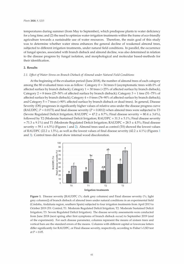
Plants 2020, 9, 1213
temperatures during summer (from May to September), which predispose plants to water deficiencyfor a long time; and (2) the need to optimize water-irrigation treatments within the frame of eco-friendlyagriculture towards a sustainable use of water resources. Therefore, the main goal of this studywas to determine whether water stress enhances the general decline of weakened almond trees,subjected to different irrigation treatments, under natural field conditions. In parallel, the occurrenceof fungal species, associated with branch dieback and almond decline, was also determined in relationto the disease progress by fungal isolation, and morphological and molecular based-methods fortheir identification.
2. Results
2.1. Effect of Water Stress on Branch Dieback of Almond under Natural Field Conditions
At the beginning of the evaluation period (June 2018), the number of almond trees of each categoryamong the 80 evaluated trees was as follow: Category 0 = 34 trees 0 (asymptomatic trees with 0% ofaffected surface by branch dieback), Category 1 = 30 trees (<25% of affected surface by branch dieback),Category 2 = 8 trees (25–50% of affected surface by branch dieback); Category 3 = 1 tree (51–75% ofaffected surface by branch dieback); Category 4 = 0 trees (76–90% of affected surface by branch dieback);and Category 5 = 7 trees (>90% affected surface by branch dieback or dead trees). In general, DiseaseSeverity (DS) progresses in significantly higher values of relative area under the disease progress curve(RAUDPC; P = 0.0173) and final disease severity (P = 0.0012) when almond trees were subjected to T3(Severe Regulated Deficit Irrigation; RAUDPC = 47.2 ± 8.7%; Final disease severity = 80.4 ± 3.6%),followed by T2 (Moderate Sustained Deficit Irrigation; RAUDPC = 31.3 ± 5.1%; Final disease severity= 71.3 ± 9.1%) and T1 (Moderate Regulated Deficit Irrigation; RAUDPC = 28.5 ± 4.5%; Final diseaseseverity = 59.1 ± 6.5%) (Figures 1 and 2). Almond trees used as control (T0) showed the lowest valuesof RAUDPC (22.2 ± 1.5%), as well as the lowest values of final disease severity (42.2 ± 4.1%) (Figures 1and 3). Control trees did not show internal wood discoloration.
RA
UD
PC (%
)
Fina
l dis
ease
sev
erity
(%)
Irrigation treatments
B B AB
A c
ab a
bc
Figure 1. Disease severity [RAUDPC (%; dark grey columns) and Final disease severity (%; lightgrey columns)] of branch dieback of almond trees under natural conditions in an experimental field(Córdoba, Andalusia region, southern Spain) subjected to four irrigation treatments from April 2013 toOctober 2019 (T0: Control; T1: Moderate Regulated Deficit Irrigation; T2: Moderate Sustained DeficitIrrigation; T3: Severe Regulated Deficit Irrigation). The disease severity assessments were conductedfrom June 2018 (next spring after first symptoms of branch dieback occur) to September 2019 (endof the experiment). For each disease parameter, columns represent the means of sixteen trees andvertical bars are the standard errors of the means. Columns with different capital or lowercase lettersdiffer significantly for RAUDPC, or Final disease severity, respectively, according to Fisher’s LSD testat P = 0.05.
61
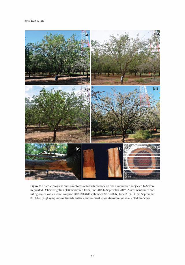
Plants 2020, 9, 1213
(a) (b)
(c) (d)
(e) (f) (g)
Figure 2. Disease progress and symptoms of branch dieback on one almond tree subjected to SevereRegulated Deficit Irrigation (T3) monitored from June 2018 to September 2019. Assessment times andrating-scales values were: (a) June 2018-2.0; (b) September 2018-3.0; (c) June 2019-3.0; (d) September2019-4.0; (e–g) symptoms of branch dieback and internal wood discoloration in affected branches.
62
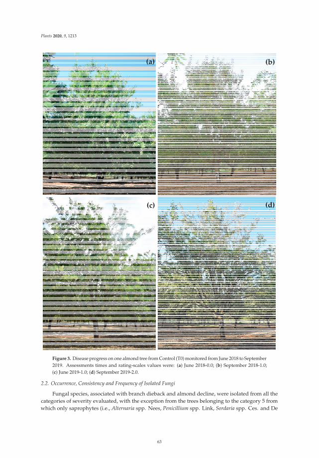
Plants 2020, 9, 1213
(a) (b)
(c) (d)
Figure 3. Disease progress on one almond tree from Control (T0) monitored from June 2018 to September2019. Assessments times and rating-scales values were: (a) June 2018-0.0; (b) September 2018-1.0;(c) June 2019-1.0; (d) September 2019-2.0.
2.2. Occurrence, Consistency and Frequency of Isolated Fungi
Fungal species, associated with branch dieback and almond decline, were isolated from all thecategories of severity evaluated, with the exception from the trees belonging to the category 5 fromwhich only saprophytes (i.e., Alternaria spp. Nees, Penicillium spp. Link, Sordaria spp. Ces. and De
63
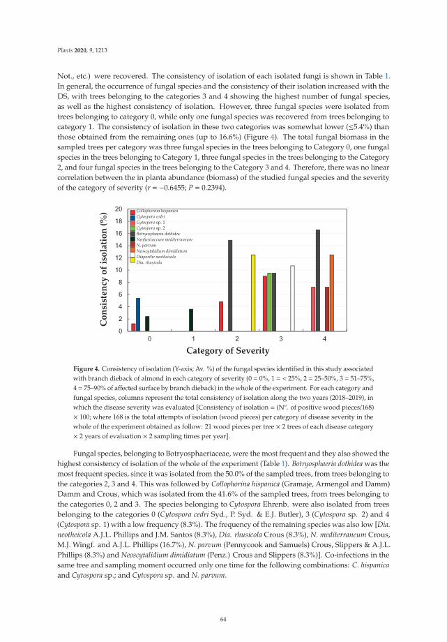
Plants 2020, 9, 1213
Not., etc.) were recovered. The consistency of isolation of each isolated fungi is shown in Table 1.In general, the occurrence of fungal species and the consistency of their isolation increased with theDS, with trees belonging to the categories 3 and 4 showing the highest number of fungal species,as well as the highest consistency of isolation. However, three fungal species were isolated fromtrees belonging to category 0, while only one fungal species was recovered from trees belonging tocategory 1. The consistency of isolation in these two categories was somewhat lower (≤5.4%) thanthose obtained from the remaining ones (up to 16.6%) (Figure 4). The total fungal biomass in thesampled trees per category was three fungal species in the trees belonging to Category 0, one fungalspecies in the trees belonging to Category 1, three fungal species in the trees belonging to the Category2, and four fungal species in the trees belonging to the Category 3 and 4. Therefore, there was no linearcorrelation between the in planta abundance (biomass) of the studied fungal species and the severityof the category of severity (r = −0.6455; P = 0.2394).
Con
sist
ency
of i
sola
tion
(%)
Category of Severity
Collophorina hispanica Cytospora cedri Cytospora sp. 1 Cytospora sp. 2 Botryosphaeria dothidea Neofusicoccum mediterraneum N. parvum Neoscytalidium dimidiatum Diaporthe neotheicola Dia. rhusicola
Figure 4. Consistency of isolation (Y-axis; Av. %) of the fungal species identified in this study associatedwith branch dieback of almond in each category of severity (0 = 0%, 1 = < 25%, 2 = 25–50%, 3 = 51–75%,4 = 75–90% of affected surface by branch dieback) in the whole of the experiment. For each category andfungal species, columns represent the total consistency of isolation along the two years (2018–2019), inwhich the disease severity was evaluated [Consistency of isolation = (Nº. of positive wood pieces/168)× 100; where 168 is the total attempts of isolation (wood pieces) per category of disease severity in thewhole of the experiment obtained as follow: 21 wood pieces per tree × 2 trees of each disease category× 2 years of evaluation × 2 sampling times per year].
Fungal species, belonging to Botryosphaeriaceae, were the most frequent and they also showed thehighest consistency of isolation of the whole of the experiment (Table 1). Botryosphaeria dothidea was themost frequent species, since it was isolated from the 50.0% of the sampled trees, from trees belonging tothe categories 2, 3 and 4. This was followed by Collophorina hispanica (Gramaje, Armengol and Damm)Damm and Crous, which was isolated from the 41.6% of the sampled trees, from trees belonging tothe categories 0, 2 and 3. The species belonging to Cytospora Ehrenb. were also isolated from treesbelonging to the categories 0 (Cytospora cedri Syd., P. Syd. & E.J. Butler), 3 (Cytospora sp. 2) and 4(Cytospora sp. 1) with a low frequency (8.3%). The frequency of the remaining species was also low [Dia.neotheicola A.J.L. Phillips and J.M. Santos (8.3%), Dia. rhusicola Crous (8.3%), N. mediterraneum Crous,M.J. Wingf. and A.J.L. Phillips (16.7%), N. parvum (Pennycook and Samuels) Crous, Slippers & A.J.L.Phillips (8.3%) and Neoscytalidium dimidiatum (Penz.) Crous and Slippers (8.3%)]. Co-infections in thesame tree and sampling moment occurred only one time for the following combinations: C. hispanicaand Cytospora sp.; and Cytospora sp. and N. parvum.
64
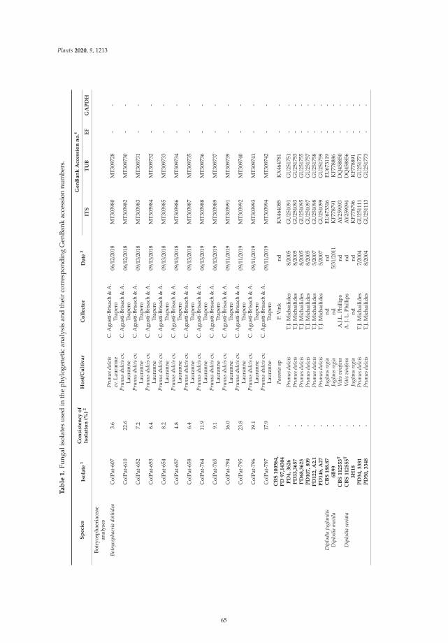
Plants 2020, 9, 1213
Ta
ble
1.
Fung
alis
olat
esus
edin
the
phyl
ogen
etic
anal
ysis
and
thei
rco
rres
pond
ing
Gen
Bank
acce
ssio
nnu
mbe
rs.
Sp
eci
es
Iso
late
1C
on
sist
en
cyo
fIs
ola
tio
n(%
)2
Ho
st/C
ult
ivar
Co
llect
or
Date
3G
en
Ban
kA
ccess
ion
no
.4
ITS
TU
BE
FG
AP
DH
Botr
yosp
haer
iace
aean
alys
es
Botr
yosp
haer
iado
thid
eaC
olPa
t-60
73.
6Pr
unus
dulc
iscv
.Lau
rann
eC
.Agu
stí-
Bris
ach
&A
.Tr
aper
o06/1
2/20
18M
T30
3980
MT
3097
28-
-
Col
Pat-
610
22.6
Prun
usdu
lcis
cv.
Laur
anne
C.A
gust
í-Br
isac
h&
A.
Trap
ero
06/1
2/20
18M
T30
3982
MT
3097
30-
-
Col
Pat-
652
7.2
Prun
usdu
lcis
cv.
Laur
anne
C.A
gust
í-Br
isac
h&
A.
Trap
ero
09/1
3/20
18M
T30
3983
MT
3097
31-
-
Col
Pat-
653
6.4
Prun
usdu
lcis
cv.
Laur
anne
C.A
gust
í-Br
isac
h&
A.
Trap
ero
09/1
3/20
18M
T30
3984
MT
3097
32-
-
Col
Pat-
654
8.2
Prun
usdu
lcis
cv.
Laur
anne
C.A
gust
í-Br
isac
h&
A.
Trap
ero
09/1
3/20
18M
T30
3985
MT
3097
33-
-
Col
Pat-
657
4.8
Prun
usdu
lcis
cv.
Laur
anne
C.A
gust
í-Br
isac
h&
A.
Trap
ero
09/1
3/20
18M
T30
3986
MT
3097
34-
-
Col
Pat-
658
6.4
Prun
usdu
lcis
cv.
Laur
anne
C.A
gust
í-Br
isac
h&
A.
Trap
ero
09/1
3/20
18M
T30
3987
MT
3097
35-
-
Col
Pat-
764
11.9
Prun
usdu
lcis
cv.
Laur
anne
C.A
gust
í-Br
isac
h&
A.
Trap
ero
06/1
3/20
19M
T30
3988
MT
3097
36-
-
Col
Pat-
765
9.1
Prun
usdu
lcis
cv.
Laur
anne
C.A
gust
í-Br
isac
h&
A.
Trap
ero
06/1
3/20
19M
T30
3989
MT
3097
37-
-
Col
Pat-
794
16.0
Prun
usdu
lcis
cv.
Laur
anne
C.A
gust
í-Br
isac
h&
A.
Trap
ero
09/1
1/20
19M
T30
3991
MT
3097
39-
-
Col
Pat-
795
23.8
Prun
usdu
lcis
cv.
Laur
anne
C.A
gust
í-Br
isac
h&
A.
Trap
ero
09/1
1/20
19M
T30
3992
MT
3097
40-
-
Col
Pat-
796
19.1
Prun
usdu
lcis
cv.
Laur
anne
C.A
gust
í-Br
isac
h&
A.
Trap
ero
09/1
1/20
19M
T30
3993
MT
3097
41-
-
Col
Pat-
797
17.9
Prun
usdu
lcis
cv.
Laur
anne
C.A
gust
í-Br
isac
h&
A.
Trap
ero
09/1
1/20
19M
T30
3994
MT
3097
42-
-
CB
S100564,
PD
97,1
4304
-Pa
eoni
asp
P.V
ink
ndK
X46
4085
KX
4647
81-
-
PD
4,3626
-Pr
unus
dulc
isT.
J.M
icha
ilide
s8/
2005
GU
2510
91G
U25
1751
--
PD
33,3
657
-Pr
unus
dulc
isT.
J.M
icha
ilide
s8/
2005
GU
2510
93G
U25
1753
--
PD
68,3
623
-Pr
unus
dulc
isT.
J.M
icha
ilide
s8/
2005
GU
2510
95G
U25
1755
--
PD
107,809
-Pr
unus
dulc
isT.
J.M
icha
ilide
s8/
2005
GU
2510
97G
U25
1757
--
PD
122,A
2.1
-Pr
unus
dulc
isT.
J.M
icha
ilide
s5/
2007
GU
2510
98G
U25
1758
--
PD
146,A
27
-Pr
unus
dulc
isT.
J.M
icha
ilide
s5/
2007
GU
2510
99G
U25
1759
--
Dip
lodi
aju
glan
dis
CB
S188.8
7-
Jugl
ans
regi
and
ndEU
6733
16EU
6731
19-
-D
iplo
dia
mut
ila6B
99
-Ju
glan
sre
gia
nd5/
31/2
011
KF7
7879
1K
F778
886
--
CB
S112553
T-
Viti
svi
nife
raA
.J.L.
Phill
ips
ndA
Y25
9093
DQ
4588
50-
-D
iplo
dia
seri
ata
CB
S112555
T-
Viti
svi
nife
raA
.J.L
.Phi
llips
ndA
Y25
9094
DQ
4588
56-
-3H
18
-Ju
glan
sre
gia
ndnd
KF7
7879
6K
F778
891
--
PD
34,3381
-Pr
unus
dulc
isT.
J.M
icha
ilide
s7/
2004
GU
2511
11G
U25
1771
--
PD
50,3348
-Pr
unus
dulc
isT.
J.M
icha
ilide
s8/
2004
GU
2511
13G
U25
1773
--
65
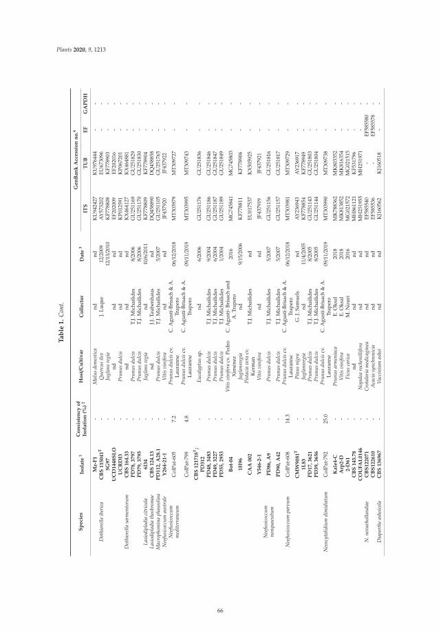
Plants 2020, 9, 1213
Ta
ble
1.
Con
t.
Sp
eci
es
Iso
late
1C
on
sist
en
cyo
fIs
ola
tio
n(%
)2
Ho
st/C
ult
ivar
Co
llect
or
Date
3G
en
Ban
kA
ccess
ion
no
.4
ITS
TU
BE
FG
AP
DH
Mz-F
1-
Mal
usdo
mes
tica
ndnd
KU
9424
27K
U97
6444
--
Dot
hior
ella
iber
ica
CB
S115041
T-
Que
rcus
ilex
J.Lu
que
12/2
009
AY
5732
02EU
6730
96-
-5G
97
-Ju
glan
sre
gia
12/1
3/20
10K
F778
808
KF7
7890
3-
-U
CD
1448S
LO
-nd
ndnd
EF20
2009
EF20
2016
--
UC
RD
I3-
Prun
usdu
lcis
ndnd
KP0
1259
1K
P067
201
--
Dot
hior
ella
sarm
ento
rum
CB
S164.3
3-
ndnd
ndK
X46
4127
KX
4648
81-
-P
D78,3797
-Pr
unus
dulc
isT.
J.M
icha
ilide
s8/
2006
GU
2511
69G
U25
1829
--
PD
79,3795
-Pr
unus
dulc
isT.
J.M
icha
ilide
s8/
2006
GU
2511
70G
U25
1830
--
Lasi
odip
lodi
aci
tric
ola
6I3
4-
Jugl
ans
regi
and
10/6/2
011
KF7
7880
9K
F778
904
--
Lasi
odip
lodi
ath
eobr
omae
CB
S124.1
3-
ndJ.J
.Tau
benh
aus
ndD
Q45
8890
DQ
4588
58-
-M
acro
phom
ina
phas
eolin
aP
D112,A
28.1
-Pr
unus
dulc
isT.
J.M
icha
ilide
s5/
2007
GU
2511
05G
U25
1765
--
Neo
fusi
cocc
umau
stra
leY
264-2
1-1
-V
itis
vini
fera
ndnd
JF43
7920
JF43
7922
--
Neo
fusi
cocc
umm
edite
rran
eum
Col
Pat-
605
7.2
Prun
usdu
lcis
cv.
Laur
anne
C.A
gust
í-Br
isac
h&
A.
Trap
ero
06/1
2/20
18M
T30
3979
MT
3097
27-
-
Col
Pat-
799
4.8
Prun
usdu
lcis
cv.
Laur
anne
C.A
gust
í-Br
isac
h&
A.
Trap
ero
09/1
1/20
19M
T30
3995
MT
3097
43-
-
CB
S121718
T;
PD
312
-Eu
caly
ptus
sp.
nd6/
2006
GU
2511
76G
U25
1836
--
PD
48,3483
-Pr
unus
dulc
isT.
J.M
icha
ilide
s9/
2004
GU
2511
86G
U25
1846
--
PD
49,3227
-Pr
unus
dulc
isT.
J.M
icha
ilide
s6/
2004
GU
2511
87G
U25
1847
--
PD
55,2953
-Pr
unus
dulc
isT.
J.M
icha
ilide
s1/
2004
GU
2511
89G
U25
1849
--
Bo
t-04
-V
itis
vini
fera
cv.P
edro
Xim
énez
C.A
gust
í-Br
isac
han
dA
.Tra
pero
2016
MG
7458
41M
G74
5803
--
1H
96
-Ju
glan
sreg
iand
9/15/2
006
KF7
7881
1K
F778
906
--
CA
A002
-Pi
stac
iave
racv
.K
erm
anT.
J.M
icha
ilide
snd
EU01
7537
KX
5059
25-
-
Y546-2
-1-
Viti
svi
nife
rand
ndJF
4379
19JF
4379
21-
-N
eofu
sico
ccum
nonq
uaes
itum
PD
86,A
9-
Prun
usdu
lcis
T.J.
Mic
haili
des
5/20
07G
U25
1156
GU
2518
16-
-
PD
90,A
42
-Pr
unus
dulc
isT.
J.M
icha
ilide
s5/
2007
GU
2511
57G
U25
1817
--
Neo
fusi
cocc
umpa
rvum
Col
Pat-
608
14.3
Prun
usdu
lcis
cv.
Laur
anne
C.A
gust
í-Br
isac
h&
A.
Trap
ero
06/1
2/20
18M
T30
3981
MT
3097
29-
-
CM
W9081
T-
Pinu
sni
gra
G.J
.Sam
uels
ndA
Y23
6943
AY
2369
17-
-1L
83
-Ju
glan
sreg
iand
11/4/2
005
KF7
7885
4K
F778
949
--
PD
17,3621
-Pr
unus
dulc
isT.
J.M
icha
ilide
s8/
2005
GU
2511
43G
U25
1803
--
PD
39,3656
-Pr
unus
dulc
isT.
J.M
icha
ilide
s8/
2005
GU
2511
44G
U25
1804
--
Neo
scyt
alid
ium
dim
idia
tum
Col
Pat-
792
25.0
Prun
usdu
lcis
cv.
Laur
anne
C.A
gust
í-Br
isac
h&
A.
Trap
ero
09/1
1/20
19M
T30
3990
MT
3097
38-
-
Kale
4-C
-Pr
unus
arm
enia
caE.
Oks
al20
18M
K78
8362
MK
8033
52-
-A
rp2-D
-V
itis
vini
fera
E.O
ksal
2018
MK
8138
52M
K81
6354
--
2-D
61
-Fi
cus
cari
caM
.Nou
ri20
16M
G02
1572
MG
0215
15-
-C
BS
145.7
8-
ndnd
ndM
H86
1121
KF5
3179
6-
-C
OU
FA
L0146
-N
opal
earo
chen
illife
rand
ndM
H25
1955
MH
2519
71-
-N
.nov
aeho
lland
iae
CB
S122071
-C
rota
lari
am
edic
agin
eand
ndEF
5855
40-
EF58
5580
-C
BS
122610
-A
caci
asy
nchr
onic
iand
ndEF
5855
36-
EF58
5578
-D
iapo
rthe
ashe
icol
aC
BS
136967
-V
acci
nium
ashe
ind
ndK
J160
562
KJ1
6051
8-
-
66
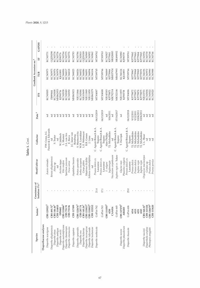
Plants 2020, 9, 1213
Ta
ble
1.
Con
t.
Sp
eci
es
Iso
late
1C
on
sist
en
cyo
fIs
ola
tio
n(%
)2
Ho
st/C
ult
ivar
Co
llect
or
Date
3G
en
Ban
kA
ccess
ion
no
.4
ITS
TU
BE
FG
AP
DH
Dia
po
rth
ace
ae
an
aly
ses
Dia
port
heac
aciig
ena
CB
S129521
T-
Aca
cia
retin
odes
P.W
.Cro
us,I
.G.
Pasc
oe&
J.Ed
war
dsnd
KC
3430
05K
C34
3973
KC
3437
31-
Dia
port
heal
legh
anie
nsis
CB
S495.7
2T
-Be
tuta
alle
ghan
iens
isnd
ndFJ
8894
44K
C34
3975
GQ
2502
98-
Dia
port
heal
nea
CB
S146.4
6T
-A
lnus
sp.
S.Tr
uter
ndK
C34
3008
KC
3439
76K
C34
3734
-D
iapo
rthe
ambi
gua
CB
S114015
-nd
ndnd
MH
8629
53K
C34
3978
KC
3437
36D
iapo
rthe
arct
iiD
P0482
-A
rctiu
mla
ppa
W.J
aklit
sch
ndK
J590
736
KJ6
1089
1K
J590
776
-D
iapo
rthe
aust
rala
fric
ana
CB
S111886
T-
Viti
svi
nife
raL.
Mos
tert
ndK
C34
3038
KC
3440
06K
C34
3764
-D
iapo
rthe
cham
aero
pis
CB
S753.7
0-
Spar
tium
junc
eum
J.A.v
onA
rxnd
KC
3430
49K
C34
4017
KC
3437
75-
Dia
port
heci
nera
scen
sC
BS
719.9
6-
Ficu
sca
rica
E.Il
ieva
ndK
C34
3050
KC
3440
18K
C34
3776
-
Dia
port
hecu
ppat
eaC
BS
117499
-A
spal
athu
slin
eari
sJ.C
.Jan
seva
nR
ensb
urg
ndM
H86
3021
KC
3440
25K
C34
3783
-
Dia
port
hecy
naro
idis
CB
S122676
T-
Prot
eacy
naro
ides
S.M
arin
cow
itz
ndN
R11
1846
KC
3440
26K
C34
3784
-D
iapo
rthe
eres
CB
S287.7
4-
Sorb
usau
cupa
ria
W.M
.Loe
rakk
ernd
KC
3430
84K
C34
4052
KC
3438
10-
Dia
port
hehi
ckor
iae
CB
S145.2
6T
-C
arya
glab
raL.
E.W
ehm
eyer
ndN
R10
3699
KC
3440
86G
Q25
0309
-D
iapo
rthe
inco
nspi
cua
CB
S133813
T-
May
tenu
sili
cifo
liaR
.R.G
omes
ndN
R11
1849
KC
3440
91K
C34
3849
-D
iapo
rthe
infe
cund
aC
BS
133812
T-
Schi
nus
tere
bint
hifo
lius
J.Li
ma
ndN
R11
1850
KC
3440
94K
C34
3852
-D
iapo
rthe
lusi
tani
cae
CB
S123212
-nd
ndnd
MH
8632
79K
C34
4104
KC
3438
62-
Dia
port
hene
othe
icol
aC
olPa
t-76
221
.4Pr
unus
dulc
iscv
.La
uran
neC
.Agu
stí-
Bris
ach
&A
.Tr
aper
o06/1
3/20
19M
T30
4007
MT
3097
45M
T309
762
-
Col
Pat-
763
17.1
Prun
usdu
lcis
cv.
Laur
anne
C.A
gust
í-Br
isac
h&
A.
Trap
ero
06/1
3/20
19M
T30
4008
MT
3097
46M
T309
763
-
CB
S111553
T-
F.vu
lgar
eA
lan
Phill
ips
ndN
R14
5303
KC
3440
69K
C34
3827
-6I3
0-
Jugl
ansr
egia
T.J.
Mic
haili
des
10/6/2
011
KF7
7887
1K
F778
966
KF7
7906
1-
CA
A816
-V
acci
nium
cory
mbo
sum
ndnd
MK
7923
14M
K83
7934
MK
8280
83
Col
Pat-
445
-Ju
glan
sre
gia
cv.T
ular
eC
.Agu
stí-
Bris
ach
&A
.Tr
aper
o07/1
4/20
17M
K52
2106
MK
4479
93M
K49
0932
Dia
port
heno
vem
CB
S127270
T-
Gly
cine
max
T.D
uvnj
aknd
NR
1118
55K
C34
4124
KC
3438
82D
iapo
rthe
phas
eolo
rum
AR
4203
-Ph
aseo
lus
vulg
aris
KJ5
9073
8K
J610
893
KJ5
9073
9-
Dia
port
herh
usic
ola
Col
Pat-
606
25.0
Prun
usdu
lcis
cv.
Laur
anne
C.A
gust
í-Br
isac
h&
A.
Trap
ero
06/1
2/20
18M
T30
4006
MT
3097
44M
T309
761
-
6I1
4-
Prun
usdu
lcis
T.J.
Mic
haili
des
9/12/2
011
KF7
7887
2K
F778
967
KF7
7906
26I1
5-
Prun
usdu
lcis
T.J.
Mic
haili
des
9/12/2
011
KF7
7887
3K
F778
968
KF7
7906
3-
6I3
1-
Jugl
ans
regi
aT.
J.M
icha
ilide
s10/0
6/20
11K
F778
874
KF7
7896
9K
F779
064
-6I4
3-
Jugl
ans
regi
aT.
J.M
icha
ilide
s10/0
6/20
11K
F778
875
KF7
7897
0K
F779
065
-D
iapo
rthe
vacc
inii
CB
S160.3
2T
-O
xyco
ccus
mac
roca
rpos
C.L
.She
arnd
NR
1037
01K
C34
4196
KC
3439
54D
iapo
rthe
llaco
rylin
aC
BS
121124
-C
oryl
ussp
.nd
ndK
C34
3004
KC
3439
72K
C34
3730
-Ph
omop
sis
amyg
dali
CB
S126679
T-
Prun
usdu
lcis
ndnd
KC
3430
22K
C34
3990
KC
3437
48-
CB
S115620
-Pr
unus
pers
ica
ndnd
KC
3430
20K
C34
3988
KC
3437
46-
67
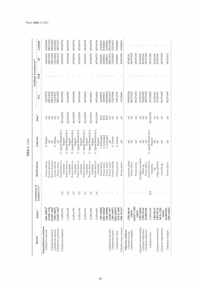
Plants 2020, 9, 1213
Ta
ble
1.
Con
t.
Sp
eci
es
Iso
late
1C
on
sist
en
cyo
fIs
ola
tio
n(%
)2
Ho
st/C
ult
ivar
Co
llect
or
Date
3G
en
Ban
kA
ccess
ion
no
.4
ITS
TU
BE
FG
AP
DH
Ty
mp
an
idace
ae
an
aly
ses
Col
loph
orin
aaf
rica
naC
BS
120872
TPr
unus
salic
ina
U.D
amm
ndG
Q15
4570
-G
Q15
4643
GQ
1546
48G
LM
C1736
Prun
usdo
mes
tica
ndnd
MK
3145
42-
MK
3145
07M
K31
4474
Col
loph
orin
aba
dens
isG
LM
C1684
TPr
unus
dom
esic
and
ndM
K31
4546
-M
K31
4503
MK
3144
82C
ollo
phor
ina
cape
nsis
CB
S120879
Prun
ussa
licin
aU
.Dam
mnd
GQ
1545
71-
GQ
1546
44G
Q15
4649
Col
loph
orin
age
rman
ica
GL
MC
1445
TPr
unus
aviu
mnd
ndM
K31
4550
-M
K31
4515
MK
3144
77
Col
loph
orin
ahi
span
ica
Col
Pat-
651
2.4
Prun
usdu
lcis
cv.
Laur
anne
C.A
gust
í-Br
isac
h&
A.
Trap
ero
09/1
3/20
18M
T30
3996
-M
T309
747
MT3
0975
4
Col
Pat-
655
9.5
Prun
usdu
lcis
cv.
Laur
anne
C.A
gust
í-Br
isac
h&
A.
Trap
ero
09/1
3/20
18M
T30
3997
-M
T309
748
MT3
0975
5
Col
Pat-
759
9.5
Prun
usdu
lcis
cv.
Laur
anne
C.A
gust
í-Br
isac
h&
A.
Trap
ero
06/1
3/20
19M
T30
3998
-M
T309
749
MT3
0975
6
Col
Pat-
760
9.5
Prun
usdu
lcis
cv.
Laur
anne
C.A
gust
í-Br
isac
h&
A.
Trap
ero
06/1
3/20
19M
T30
3999
-M
T309
750
MT3
0975
7
Col
Pat-
761
8.3
Prun
usdu
lcis
cv.
Laur
anne
C.A
gust
í-Br
isac
h&
A.
Trap
ero
06/1
3/20
19M
T30
4000
-M
T309
751
MT3
0975
8
Col
Pat-
800
2.4
Prun
usdu
lcis
cv.
Laur
anne
C.A
gust
í-Br
isac
h&
A.
Trap
ero
09/1
1/20
19M
T30
4001
-M
T309
752
MT3
0975
9
Col
Pat-
801
3.6
Prun
usdu
lcis
cv.
Laur
anne
C.A
gust
í-Br
isac
h&
A.
Trap
ero
09/1
1/20
19M
T30
4002
-M
T309
753
MT3
0976
0
CB
S128566
-Pr
unus
dulc
isJ.
Arm
engo
l20
10JN
8088
39-
JN80
8850
JN80
8843
CB
S128568
T-
Prun
usdu
lcis
J.A
rmen
gol
2010
JN80
8841
-JN
8088
52JN
8088
45C
BS
128569
-Pr
unus
dulc
isJ.
Arm
engo
l20
10M
H86
4962
-JN
8088
53JN
8088
46C
ollo
phor
ina
neor
ubra
GL
MC
929
T-
Prun
usav
ium
ndnd
MK
3145
33-
MK
3145
11M
K31
4485
Col
loph
orin
apa
arla
CB
S120877
-Pr
unus
salic
ina
U.D
amm
ndG
Q15
4586
-G
Q15
4645
GQ
1546
51
Col
loph
orin
aru
bra
CB
S120873
T,
ST
E-U
6109
-Pr
unus
pers
ica
U.D
amm
ndN
R11
9747
JN80
8855
JN80
8848
Cad
opho
ralu
teo-
oliv
acea
CB
S141.4
1T
-Pr
unus
dulc
isnd
ndA
Y24
9066
-K
M49
7089
JN80
8849
Vals
ace
ae
an
aly
ses
Cyt
ospo
raac
acia
eC
BS
468.6
9-
Cer
aton
iasi
liqua
ndnd
MH
8593
54-
KX
9651
81-
Cyt
ospo
raam
ygda
liL
H356
-Pr
unus
dulc
isnd
ndM
G97
1852
-M
G97
1658
-L
H357
T,C
BS
144233
-Pr
unus
dulc
isnd
ndM
G97
1853
-M
G97
1659
-
Cyt
ospo
raau
stro
mon
tana
CB
S116821
-Eu
caly
ptus
pauc
iflor
and
ndK
Y05
1796
-K
X96
5068
-C
ytos
pora
cabo
rnac
eaC
BS
219.5
4-
Ulm
ussp
.nd
ndD
Q24
3805
-K
X96
5164
-C
ytos
pora
calif
orni
caK
AR
E1105
-Pr
unus
dulc
isnd
ndM
G97
1947
-M
G97
1663
-
Cyt
ospo
race
dri
Col
Pat-
604
10.8
Prun
usdu
lcis
cv.
Laur
anne
C.A
gust
í-Br
isac
h&
A.
Trap
ero
06/1
2/20
18M
T30
4003
-M
T311
983
-
CB
S196.9
7-
ndnd
ndK
Y05
1906
-K
X96
5154
-C
ytos
pora
cera
tosp
erm
aC
BS
512.7
6-
Fagu
ssy
lvat
ica
ndnd
KY
0519
41-
KX
9651
84-
Cyt
ospo
rach
ryso
sper
ma
9E
-33,C
BS
144242
-C
amel
liasp
.nd
ndM
G97
1892
-M
G97
1602
-
Cyt
ospo
raeu
caly
pti
KA
RE
1585,
CB
S144241
-Pr
unus
dulc
isnd
ndM
G97
1907
-M
G97
1617
-
68
Plants 2020, 9, 1213
Ta
ble
1.
Con
t.
Sp
eci
es
Iso
late
1C
on
sist
en
cyo
fIs
ola
tio
n(%
)2
Ho
st/C
ult
ivar
Co
llect
or
Date
3G
en
Ban
kA
ccess
ion
no
.4
ITS
TU
BE
FG
AP
DH
Cyt
ospo
ragr
anat
i6F
-45
T,C
BS
144237
-Pu
nica
gran
atum
ndnd
MG
9717
99-
MG
9715
14-
Cyt
ospo
rajo
aqui
nens
isK
AR
E975
T,
CB
S144235
-Po
pulu
sde
ltoid
esnd
ndM
G97
1895
-M
G97
1605
-
Cyt
ospo
ralo
ngis
pora
10F
-57
T,C
BS
144236
-Pr
unus
dom
estic
and
ndM
G97
1905
-M
G97
1615
-
Cyt
ospo
ram
ali
CB
S109499
-M
alus
sp.
ndnd
KY
0517
69-
KX
9650
48-
Cyt
ospp
ora
olei
cola
KA
RE
1021
T,
CB
S144248
-O
lea
euro
paea
ndnd
MG
9719
44-
MG
9716
60-
Cyt
ospo
rapa
raka
ntsc
have
liiK
AR
E974,C
BS
144243
-Po
pulu
sde
ltoid
esnd
ndM
G97
1898
-M
G97
1608
-
Cyt
ospo
rapa
rapi
stac
iae
KA
RE
232
-Pi
stac
iave
rand
ndM
G97
1807
-M
G97
1522
-K
AR
E270
T,
CB
S144506
-Pi
stac
iave
rand
ndM
G97
1804
-M
G97
1519
-
Cyt
ospo
rapa
rasi
tica
CF
CC
53173
-Be
rber
issp
.nd
ndM
K67
3070
-M
K67
2957
-C
ytos
pora
pave
ttae
CB
S145562
-Pa
vett
are
volu
taM
.J.W
ingfi
eld
ndM
K87
6386
-M
K87
6497
-
Cyt
ospo
rapi
stac
iae
KA
RE
443
T,
CB
S144238
-Pi
stac
iave
rand
ndM
G97
1802
-M
G97
1517
-
Cyt
ospo
rapl
uriv
ora
KA
RE
1452
T,
CB
S144239
-O
lea
euro
paea
ndnd
MG
9718
61-
MG
9715
72-
Cyt
ospo
rapo
pulic
ola
KA
RE
973
T,
CB
S144240
-Po
pulu
sde
ltoid
esnd
ndM
G97
1891
-M
G97
1601
-
Cyt
ospo
rapu
nica
e7C
-09
-Pu
nica
gran
atum
ndnd
MG
9719
39-
MG
9716
50-
Cyt
ospo
rarh
odop
hila
CB
S190.4
2-
Syri
nga
sp.
ndnd
KY
0519
01-
KX
9651
47-
Cyt
ospo
rari
bis
CB
S187.3
6-
Rib
esru
brum
ndnd
DQ
2438
10-
KX
9651
44-
CF
CC
50039
-Pl
atyc
ladu
sor
ient
alis
Xin
leiF
annd
KR
0456
42-
KU
7109
31-
Cyt
ospo
rasa
ccul
usC
FC
C89626
-Ju
glan
sre
gia
Xin
leiF
annd
KR
0456
47-
KU
7109
34-
CB
S116855
-Q
uerc
usal
band
ndK
Y05
1824
-K
X96
5091
-C
ytos
pora
sorb
icol
a4L
-58
-Pr
unus
dom
estic
and
ndM
G97
1839
-M
G97
1553
-
Cyt
ospo
rasp
.1C
olPa
t-60
914
.3Pr
unus
dulc
iscv
.La
uran
neC
.Agu
stí-
Bris
ach
&A
.Tr
aper
o06/1
2/20
18M
T30
4004
-M
T311
984
-
Cyt
ospo
rasp
.2C
olPa
t-65
62.
0Pr
unus
dulc
iscv
.La
uran
neC
.Agu
stí-
Bris
ach
&A
.Tr
aper
o09/1
3/20
18M
T30
4005
-M
T311
985
-
Dia
port
hella
cory
lina
CB
S121124
-C
oryl
ussp
.L.
N.V
assi
ljeva
ndK
C34
3004
-K
C34
3730
-V
alsa
germ
anic
aC
BS
195.4
2-
ndnd
ndK
Y05
1902
-K
X96
5151
-1
Sequ
ence
sfr
omG
enBa
nkus
edin
the
phyl
ogen
etic
anal
ysis
indi
cate
din
bold
type
.T=
Ex-t
ype
isol
ates
;AR
,DP:
Isol
ates
incu
lture
colle
ctio
nof
Syst
emat
icM
ycol
ogy
and
Mic
robi
olog
yLa
bora
tory
,USD
A-A
RS,
Belt
svill
e,M
aryl
and,
USA
;CA
A=
A.A
lves
,Uni
vers
idad
ede
Ave
iro,
Port
ugal
;CBS
:Cul
ture
colle
ctio
nof
the
Cen
traa
lbur
eau
voor
Schi
mm
elcu
ltur
es,F
unga
lBi
odiv
ersi
tyC
entr
e,U
trec
ht,T
heN
ethe
rlan
ds;C
FCC=
Chi
naFo
rest
ryC
ultu
reC
olle
ctio
nC
ente
r;C
MW=
Cul
ture
colle
ctio
nof
the
Fore
stry
and
Agr
icul
tura
lBio
tech
nolo
gyIn
stit
ute
(FA
BI),
Uni
vers
ityof
Pret
oria
,Pre
tori
a,So
uth
Afr
ica;
Col
Pat=
‘Col
ecci
ónPa
tolo
gía’
,Dep
artm
ento
fAgr
onom
y,U
nive
rsity
ofC
ordo
ba,S
pain
;KA
RE=
Col
lect
ions
ofth
eD
epar
tmen
tof
Plan
tPat
holo
gyat
the
Kea
rney
Agr
icul
tura
lRes
earc
han
dE
xten
sion
Cen
tre
ofth
eU
nive
rsit
yof
Cal
ifor
nia,
Parl
ier,
CA
.;PD
:Pla
ntPr
otec
tion
Serv
ice,
Wag
enin
gen,
The
Net
herl
and
s;ST
E-U=
Uni
vers
ityof
Stel
lenb
osch
,Sou
thA
fric
a.2
The
cons
iste
ncy
ofis
olat
ion
(%)o
feac
his
olat
ew
asca
lcul
ated
asth
enu
mbe
rof
posi
tive
atte
mpt
sof
isol
atio
n(w
ood
piec
es)o
fagi
ven
fung
usdi
vide
dby
the
tota
latt
empt
sof
isol
atio
nin
the
who
leof
the
expe
rim
ent[
Con
sist
ency
ofis
olat
ion=
(Nº.
ofpo
siti
vew
ood
piec
es/1
68)×
100;
whe
re16
8is
the
tota
latt
empt
sof
isol
atio
n(w
ood
pie
ces)
per
cate
gory
ofd
isea
sese
veri
tyin
the
who
leof
the
exp
erim
ento
btai
ned
asfo
llow
:21
woo
dp
iece
sp
ertr
ee×2
tree
sof
each
dis
ease
cate
gory×2
year
sof
eval
uatio
n×2
sam
plin
gtim
espe
rye
ar].
3C
olle
ctio
nda
te:m
onth/d
ay/y
ear;
n/d:
non-
dete
rmin
ed.4
ITS=
inte
rnal
tran
scri
bed
spac
er,T
UB=β
-tub
ulin
-2ge
nere
gion
s,EF=
tran
slat
ion
elon
gati
on1-α
,GA
PDH=
200-
bpin
tron
ofth
egl
ycer
alde
hyde
-3-p
hosp
hate
dehy
drog
enas
e.
69
Plants 2020, 9, 1213
2.3. Molecular Identification of Isolated Fungi
For all Datasets, the topology obtained by Maximum Parsimony (MP) was confirmed with thoseobtained by BI analysis. The model used in BI analysis, and the gene boundaries, the number of totalcharacters (T), parsimony-informative characters (PI), parsimony-uninformative characters (PNI) andconserved sites (C) processed in each maximum parsimony analysis, as well as TL, consistency index(CI), retention index (RI), homoplasy index (HI) and rescaled consistency index (RC) values obtainedfrom the one most parsimonies trees in each Dataset are shown in Table 2.
Botryosphaeriaceae analyses (Dataset I-A). Most of the Botryosphaeriaceae isolates (13 out of 17isolates) were grouped in a well-supported clade with GenBank reference sequences of B. dothidea[bootstrap support (BS; %)/Bayesian posterior probability (PP):100/1.00]. The remaining isolateswere identified as N. mediterraneum (ColPat-605 and ColPat-799; BS/PP:77–83/0.99–0.88), N. parvum(ColPat-608; BS/PP:99/1.00), and Neoscytalidium dimidiatum (Penz.) Crous and Slippers (ColPat-792)(BS/PP:100/1.00) (Figure 5a). To confirm the identification of this last isolate, an additional phylogenywas conducted by means the combined alignment of ITS and EF loci, including reference isolatesof Neoscytalidium novaehollandiae Pavlic, T.I. Burgess and M.J. Wingf. (Dataset I-B). The MP analysesshowed nine most parsimonious, and one of those is shown in Figure 5b.
Diaporthaceae analyses (Dataset II). Our isolates clustered in two well-supported clades withreference sequences of Dia. neotheicola (ColPat-762 and ColPat-763; BS/PP:98/1.00) and Dia. rhusicola(ColPat-606; BS/PP:99/1.00) (Figure 6).
Tympanidaceae analysis (Dataset III). All the isolates belonging to Tympanidaceae clusteredtogether in a well-supported clade with GenBank reference sequence of Collophorina hispanica(=Collophora hispanica; BS/PP:100/1.00) (Figure 7).
Valsaceae analysis (Dataset IV). Among the three isolates belonging to Valsaceae included inthis study, only one (ColPat-604) was grouped in a well-supported clade with a GenBank referencesequence of Cytospora cedri Syd., P. Syd. and E.J. Butler (BS/PP:100/1.00). However, it was not possibleto distinguish the remaining two isolates (ColPat-609 and ColPat-656) at the species level into the genusCytospora, and they were identified as Cytospora sp. 1 (ColPat-609) and Cytospora sp. 2 (ColPat-656)(Figure 8).
70
Plants 2020, 9, 1213
Ta
ble
2.
Num
ber
ofta
xa,g
enes
and
stat
istic
alin
form
atio
nof
the
diff
eren
tana
lyse
spe
rfor
med
inth
isst
udy:
Baye
sian
(IB)
and
Max
imum
Pars
imon
yA
naly
ses
(MP)
.
Data
set/
Ph
ylo
gen
eti
cA
naly
ses
Nu
mb
er
of
Taxa
Gen
eB
ay
esi
an
An
aly
ses
Maxim
um
Pars
imo
ny
An
aly
ses
Ch
ara
cters
2M
PT
ree
3
InS
tud
yG
en
Ban
k(i
ncl
.ou
tgro
up
s)C
om
bin
ati
on
Bo
un
dari
es
Best
Fit
Mo
del
1T
PI
PN
IC
Nº.
Tre
es
TL
CI
RI
HI
RC
I-A/B
otry
osph
aeri
acea
e17
43IT
S/TU
B1-
503/
504-
921
K2+
G/
T92+
G73
311
913
348
14
375
0.67
70.
940
0.32
30.
636
I_B/
Neo
scyt
alid
ium
dim
idia
tum
18
ITS/
EF1-
490/
491-
711
T92/
K2
684
924
443
19
258
0.85
70.
833
0.14
30.
714
II/D
iapo
rtha
ceae
330
EF/T
UB/
ITS
1-25
4/25
5-73
9/74
0-12
27
K2+
G/
T92+
I/K
2+G+
I99
330
112
456
81
1046
0.55
10.
769
0.44
90.
424
III/
Tym
pani
dace
ae7
12IT
S/EF/
GA
PDH
1-48
2/48
3-66
7/66
8-80
5
K2+
G/
K2+
I/K
2+I
719
100
143
476
1033
40.
850
0.91
90.
150
0.78
2
IV/V
alsa
ceae
332
ITS/
EF/
1-55
7/55
8-82
1K
2+G+
I/H
K4+
G61
317
173
369
174
30.
469
0.74
20.
531
0.34
8
1B
estfi
tnuc
leot
ide
subs
titu
tion
mod
els
det
erm
ined
byM
EG
Av.
7.0,
used
for
each
gene
part
itio
nto
perf
orm
Bay
esia
nIn
fere
nce
anal
yses
usin
gM
rBay
esv.
3.2.
6.2
Num
bers
ofto
tal
char
acte
rs(p
osit
ions
)in
the
final
data
set(
T),p
arsi
mon
y-in
form
ativ
ech
arac
ters
(PI)
,par
sim
ony-
unin
form
ativ
ech
arac
ters
(PN
I)an
dco
nser
ved
site
s(C
),pr
oces
sed
inea
chan
alys
is;a
llpo
siti
ons
cont
aini
ngga
psan
dm
issi
ngda
taw
ere
elim
inat
ed.3
Tota
lofe
qual
lym
ostp
arsi
mon
ious
tree
sob
tain
edfo
rea
chM
Pan
alys
es:N
ºofT
ree,
tree
leng
th(T
L),c
onsi
sten
cyin
dex
(CI)
,ret
enti
onin
dex
(RI)
,hom
opla
syin
dex
(HI)
and
resc
aled
cons
iste
ncy
inde
x(R
C).
71
Plants 2020, 9, 1213
Neofusicoccum mediterraneumNeofusicoccum mediterraneumNeofusicoccum mediterraneumNeofusicoccum mediterraneumNeofusicoccum mediterraneum
Neofusicoccum mediterraneumNeofusicoccum mediterraneum
Neofusicoccum mediterraneum
Neofusicoccum nonquaesitumNeofusicoccum nonquaesitumNeofusicoccum parvumNeofusicoccum parvaNeofusicoccum parvum
Neofusicoccum parvumNeofusicoccum australe
Diplodia juglandisDothiorella ibericaDothiorella ibericaDothiorella ibericaDothiorella ibericaDothiorella sarmentorumDothiorella sarmentorumDothiorella sarmentorum
Lasiodiplodia theobromaeLasiodiplodia citricolaDiplodia mutilaDiplodia mutila Diplodia seriataDiplodia seriataDiplodia seriata
Diplodia seriataDiplodia seriata
Macrophomina phaseolinaBotryosphaeria dothideaBotryosphaeria dothidea
Botryosphaeria dothidea
Botryosphaeria dothideaBotryosphaeria dothideaBotryosphaeria dothidea
Botryosphaeria dothidea
Neoscytalidium dimidiatum Neoscytalidium dimidiatum
Diaporthe asheicola
Botryosphaeria dothidea
Neofusicoccum mediterraneum
Neofusicoccum parvum
Neoscytalidiumdimidiatum
Figure 5. Cont.
72
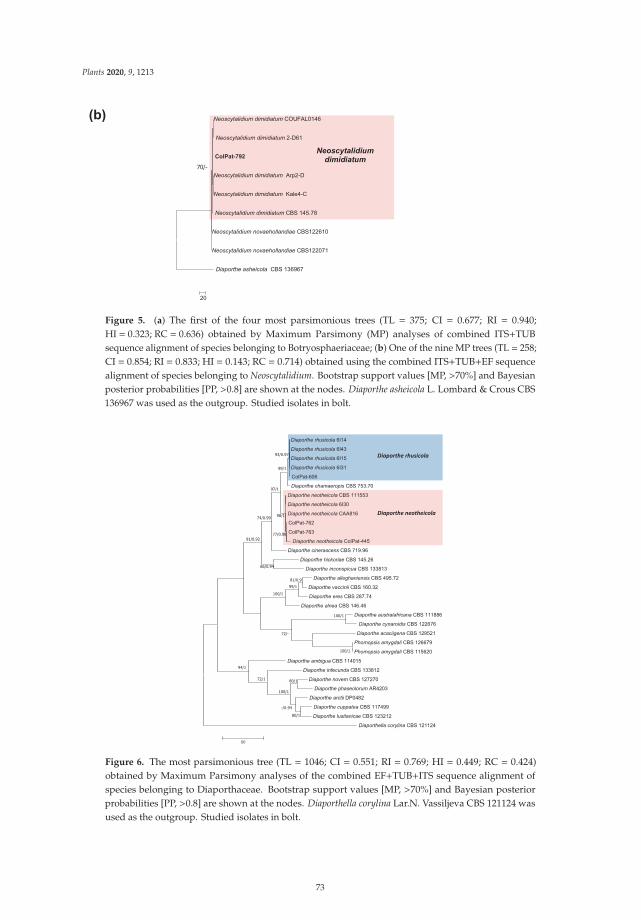
Plants 2020, 9, 1213
Neoscytalidium dimidiatum
Neoscytalidium dimidiatum
Neoscytalidium dimidiatum
Neoscytalidium dimidiatum
Neoscytalidium dimidiatum
Neoscytalidium novaehollandiae
Neoscytalidium novaehollandiae
Diaporthe asheicola
Neoscytalidiumdimidiatum
Figure 5. (a) The first of the four most parsimonious trees (TL = 375; CI = 0.677; RI = 0.940;HI = 0.323; RC = 0.636) obtained by Maximum Parsimony (MP) analyses of combined ITS+TUBsequence alignment of species belonging to Botryosphaeriaceae; (b) One of the nine MP trees (TL = 258;CI = 0.854; RI = 0.833; HI = 0.143; RC = 0.714) obtained using the combined ITS+TUB+EF sequencealignment of species belonging to Neoscytalidium. Bootstrap support values [MP, >70%] and Bayesianposterior probabilities [PP, >0.8] are shown at the nodes. Diaporthe asheicola L. Lombard & Crous CBS136967 was used as the outgroup. Studied isolates in bolt.
Diaporthe australafricana
Diaporthe rhusicola
Diaporthe rhusicola
Diaporthe rhusicola
Diaporthe rhusicola
Diaporthe chamaeropis
Diaporthe neotheicola
Diaporthe neotheicola
Diaporthe neotheicola
Diaporthe neotheicola
Diaporthe cinerascens
Diaporthe hickoriae
Diaporthe inconspicua
Diaporthe alleghaniensis
Diaporthe vaccinii
Diaporthe eres
Diaporthe alnea
Diaporthe cynaroidis
Diaporthe acaciigena
Phomopsis amygdali
Phomopsis amygdali
Diaporthe ambigua
Diaporthe infecunda
Diaporthe novem
Diaporthe phaseolorum
Diaporthe arctii
Diaporthe cuppatea
Diaporthe lusitanicae
Diaporthella corylina
Figure 6. The most parsimonious tree (TL = 1046; CI = 0.551; RI = 0.769; HI = 0.449; RC = 0.424)obtained by Maximum Parsimony analyses of the combined EF+TUB+ITS sequence alignment ofspecies belonging to Diaporthaceae. Bootstrap support values [MP, >70%] and Bayesian posteriorprobabilities [PP, >0.8] are shown at the nodes. Diaporthella corylina Lar.N. Vassiljeva CBS 121124 wasused as the outgroup. Studied isolates in bolt.
73
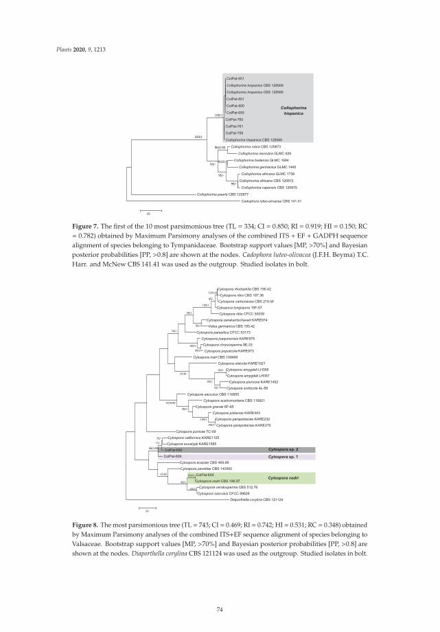
Plants 2020, 9, 1213
Collophorina hispanica
Collophorina hispanica
Collophorina hispanica
Collophorina rubra
Collophorina neorubra
Collophorina badensis
Collophorina germanica
Collophorina africana
Collophorina africana
Collophorina capensis
Collophorina paarla
Cadophora luteo-olivacea
Collophorina hispanica
Figure 7. The first of the 10 most parsimonious tree (TL = 334; CI = 0.850; RI = 0.919; HI = 0.150; RC= 0.782) obtained by Maximum Parsimony analyses of the combined ITS + EF + GADPH sequencealignment of species belonging to Tympanidaceae. Bootstrap support values [MP, >70%] and Bayesianposterior probabilities [PP, >0.8] are shown at the nodes. Cadophora luteo-olivacea (J.F.H. Beyma) T.C.Harr. and McNew CBS 141.41 was used as the outgroup. Studied isolates in bolt.
Cytospora rhodophila
Cytospora ribis
Cytospora carbonacea
Cytospora longispora
Cytospora ribis
Cytospora parakantschavelii
Valsa germanica
Cytospora parasitica
Cytospora joaquinensis
Cytospora chrysosperma
Cytospora populicola
Cytospora mali
Cytospora oleicola
Cytospora amygdali
Cytospora amygdali
Cytospora plurivora
Cytospora sorbicola
Cytospora sacculus
Cytospora austromontana
Cytospora granati
Cytospora pistaciae
Cytospora parapistaciae
Cytospora parapistaciae
Cytospora punicae
Cytospora californica
Cytospora eucalypti
Cytospora acaciae
Cytospora pavettae
Cytospora cedri
Cytospora ceratosperma
Cytospora sacculus
Diaporthella corylina
Cytospora cedri
Cytospora
Cytospora
Figure 8. The most parsimonious tree (TL = 743; CI = 0.469; RI = 0.742; HI = 0.531; RC = 0.348) obtainedby Maximum Parsimony analyses of the combined ITS+EF sequence alignment of species belonging toValsaceae. Bootstrap support values [MP, >70%] and Bayesian posterior probabilities [PP, >0.8] areshown at the nodes. Diaporthella corylina CBS 121124 was used as the outgroup. Studied isolates in bolt.
74
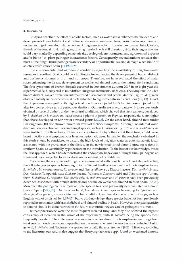
Plants 2020, 9, 1213
3. Discussion
Studying whether the effect of abiotic factors, such as water stress enhances the incidence anddevelopment of branch dieback and decline syndromes on weakened trees, is essential in improving ourunderstanding of the endophytic behaviour of fungi associated with this complex disease. In fact, to date,the role of the fungal trunk pathogens, causing tree decline, is still uncertain, since their aggressivenesscould vary markedly depending on abiotic (i.e., ecological, environmental and agronomical aspects)and/or biotic (i.e., plant-pathogen interactions) factors. Consequently, several authors consider thatmost of the fungal trunk pathogens are secondary or opportunistic, causing damage when biotic orabiotic circumstances occur [11,19,24,25].
The environmental and agronomic conditions regarding the availability of irrigation-waterresources in southern Spain could be a limiting factor, enhancing the development of branch diebackand decline syndromes on fruit and nut crops. Therefore, we have evaluated the effect of waterstress enhancing the disease development on weakened almond trees under natural field conditions.The first symptoms of branch dieback occurred in late-summer autumn 2017 in an eight-year oldexperimental field, subjected to four different irrigation treatments, since 2013. The symptoms includedbranch dieback, canker formation, internal wood discoloration and general decline (Figure 2d–g) wereobserved mainly in the experimental plots subjected to high water-stressed conditions (T2, T3). In fact,the DS progress was significantly higher in almond trees subjected to T3 than in those subjected to T0after two consecutive years of periodic evaluations. Our results are in accordance with those previouslyobtained by several authors under the control conditions, which showed that stem cankers, developedby B. dothidea or S. musiva on water-stressed plants of peach, or Populus, respectively, were higherthan those developed on non-water-stressed plants [22,23]. On the other hand, almond trees underfull irrigation (T0) also showed minimum levels of dieback symptoms. Although, no internal wooddiscoloration was observed, several fungal species, such as C. hispanica, Cy. cedri and N. mediterraneumwere isolated from those trees. These results reinforce the hypothesis that these fungi could causelatent infections in asymptomatic or lesser-symptomatic trees. In parallel, the conclusions obtained inthis study should be considered to discard the high levels of irrigation water as potential abiotic factorassociated with the prevalence of the disease in the newly established almond growing regions insouthern Spain, as we initially hypothesized in the introduction. To the best of our knowledge, this isthe first approach, which has demonstrated the endophytic behaviour of fungal trunk pathogens onweakened trees, subjected to water stress under natural field conditions.
Concerning the occurrence of fungal species associated with branch dieback and almond decline,the following seven species belonging to four different families were identified: Botryosphaeriaceae:B. dothidea, N. mediterraneum, N. parvum and Neoscytalidum sp.; Diaporthaceae: Dia. neotheicola andDia. rhusicola; Tympanidaceae: C. hispanica; and, Valsaceae: Cytospora cedri and Cytospora spp. Amongthem, B. dothidea, C. hispanica, Dia. neotheicola, N. mediterraneum and N. parvum have been previouslydescribed associated with branch dieback and decline on weakened almond trees in Spain [7,8,26].Moreover, the pathogenicity of most of these species has been previously demonstrated in almondtrees in Spain [5,8,9,26]. On the other hand, Dia. rhusicola and species belonging to Cytospora andNeoscytalidium genera, are associated with branch dieback and tree decline in other nut crops, such asEnglish walnut or pistachio [6,15–17], but to our knowledge, these species have not been previouslyreported in association with branch dieback and almond decline in Spain. However, their pathogenicityto almond should be demonstrated in the future to confirm they are canker pathogens of almond.
Botryosphaeriaceae were the most frequent isolated fungi and they also showed the highestconsistency of isolation in the whole of the experiment, with B. dothidea being the species mostfrequently isolated. The differences in consistency of isolation of Botryosphaeriaceae fungi fromweakened almonds can occur, depending on the scenario where the surveys are conducted, but ingeneral, B. dothidea and Neofusicoccum species are usually the most frequent [9,25]. Likewise, accordingto the literature, our results also suggest that Botryosphaeriaceae spp. found on weakened almond
75
Plants 2020, 9, 1213
trees are able to endanger the productivity and longevity of orchards in Spain, as well as in othercountries [9].
Among Botryosphaeriaceae fungi, notice that B. dothidea has been reported worldwide causingcanker diseases in a broad range of woody crops, including different Prunus spp. [6,7,27]. However,the role of this fungus as a trunk pathogen is still uncertain, given it has been reported as a latentpathogen of global importance for its endophytic behaviour in woody plant health [20]. In fact, studiesconducted recently in southern Spain, which compared the pathogenicity of B. dothidea on inoculateddetached and attached shoots of almond, the English walnut and pistachio, demonstrated that, inevery case, the fungus is significantly higher aggressive on detached shoots than on attached [5,16,17].It was confirmed that B. dothidea could remain latent in woody plants until trees become weakened asa consequence of different biotic and/or abiotic factors.
With respect to Diaporthaceae, Dia. neotheicola and Dia. rhusicola showed a low consistencyof isolation and their occurrence was also low. These two species have been previously reported,associated with branch dieback and shoot blight of English walnut in California [28] and southernSpain [16], and the first one was also isolated from pistachio in southern Spain [17] and recently reportedassociated with twig cankers and shoot blight of almond in Spain [26]. Usually, Diaporthaceae speciesoccur simultaneously with Botryosphaeriaceae in the same orchards, with Botryosphaeriaceae beingalways the most frequent [16,17,28]. In addition, studies conducted in California by Agustí-Brisachet al. [29] suggest that coinfections between Botryosphaeriaceae and Diaporthaceae species result inantagonistic interactions on infection and disease development on English walnut. But, Dia. amygdali,which is a common species associated with branch dieback and cankers of almond [5,7], was not foundin this experiment.
It is interesting to note that, in this study, C. hispanica was the second most frequent species isolatedfrom weakened almond trees after B. dothidea. Our results are in concordance with those found byOlmo et al. [8], who indicated that this slow-growing species is common in declined almond trees.However, it is usually excluded in the diagnosis process probably because its presence goes unnoticed,due to its slow growth [8].
Finally, Cytospora species were also isolated in low consistency from the trees of three differentcategories of severity. According to our results, Cytospora spp. have also been reported associated withcanker diseases in weakened tree nuts (English walnut and pistachio) in southern Spain showing lessfrequency and aggressiveness, and often simultaneously with Botryosphaeriaceae and/or Diaporthaceaefungi [16,17]. However, several Cytospora spp. have been already reported in California as cankerpathogens of several fruit and nut crops including Prunus spp. such as almond, apricot or peach [30].
The isolations made during this study suggest that most of these fungi can occur in bothasymptomatic and symptomatic trees, but their frequency of isolation increases with increasing DS.However, the consistency of isolation was low for all the species in the whole of the experiment,showing the highest values in weakened almond trees, belonging to categories 3 and 4 (51 to 90% offinal disease severity). This information reinforces the hypothesis that the fungal species associatedwith tree decline could have a major endophytic behaviour, and its aggressiveness is probably enhancedby abiotic factors, such as water stress on previously infected and weakened trees. In general, studieson the etiology of fungal trunk diseases describe a broad list of fungi associated with the disease, butdo not usually considering the strict pathogenic behaviour of each. Nevertheless, this work reveals theneed to go on elucidating the role of biotic and abiotic factors, enhancing the infection of fungal trunkpathogens and disease development on woody crops towards generating important knowledge on theirlife cycle. Therefore, focus the research on such relevant challenge will provide a better understandingof the biology of fungi associated with tree decline syndrome. It will build a strong foundation fordeveloping effective management approaches against the disease, by taking into consideration theoptimum water management.
76
Plants 2020, 9, 1213
4. Materials and Methods
4.1. Experimental Field, Irrigation Treatments and Experimental Design
The present study was conducted in a nine- to 10-years-old experimental field of almond cv.Guara grafted onto GF-677 rootstock (5.5 ha; 7 × 6 tree spacing; 238 trees/ha) belonging to theAndalusian Institute of Agricultural and Fisheries Research and Training (IFAPA in Spanish) Centre‘Alameda del Obispo’ located in Córdoba (Andalusia region, Spain; 37.8ºN, 4.8ºW), whose soil wasclassified as a Typic Xerofluvent of sandy loam texture and exceeds 1.5 m depth. The climate of thisregion is the typical Mediterranean climate, characterized by hot and dry summers (Tª Av. 27.0 ◦C;Tª min Av. = 19.3 ◦C; Tª max Av. = 36.7 ◦C), mild winters, with 600 mm of annual rainfall average,concentrated from October to April (Tª Av. 13.0 ◦C; Tª min Av. = 7.9 ◦C; Tª max Av. = 21.1 ◦C).
The experimental field used in this study was established in February 2009. Pruning for treeformation was done along the two first years, and then there never were pruning interventions.Control management strategies to prevent pest (Acetamiprid 20%; Deltametrine 2.5%) and diseases(Boscalid 26.7% + Pyraclostrobin 6.7%; Thiram 50%; Tebuconazole 50% + Tryfloxistrobin 25%) weredone according to a treatment-calendar based on the weather conditions which could favour the typicalalmond pest and diseases of this area. Weeds were controlled by mowing and herbicide applications(Glyphosate 36%; Oxifluorphen 24%). Mineral fertilization was calculated and applied following therecommendations of the California Fertilization Guidelines for Almonds (https://apps1.cdfa.ca.gov/FertilizerResearch/docs/Almonds.html). Therefore, this experimental field is representative of theedapho-climatic characteristics and the standard crop management of the new almond plantings inAndalusia [31].
The irrigation system was formed by two pressure compensating drip irrigation laterals, spaced 1 mfrom the tree rows, and all the trees were fully irrigated until the irrigation treatments began, as describedbelow. From April 2013 to October 2019 (before and along this present study), the experimental fieldwas subjected to four irrigation treatments: (i) Control (T0): The trees were irrigated to cover theirfull water requirements (ET), which was calculated using the relationship between ground cover(GC) and a transpiration coefficient, proposed by Espadafor et al. [32]. An additional 15% of thatquantity was supplied to account for the evaporation from emitter wet surfaces under the trees usingBonachela et al. [33] model; (ii) Moderate Regulated Deficit Irrigation (T1): seasonal irrigation was 65% ofT0, but the deficit was mainly concentrated during kernel filling stage, where almonds are less affectedby water stress. Specifically, the irrigation supplied was: 70% of T0 in spring; 40% in kernel fillingstage (from middle-July to harvest period in middle-August) and 100% in the postharvest period; (iii)Moderate Sustained Deficit Irrigation (T2): This treatment consisted of 65% of T0 steadily throughout theirrigation season. In total, a similar amount of irrigation water to that of the T1 was supplied; and (iv)Severe Regulated Deficit Irrigation (T3): This treatment received 30% of seasonal irrigation in relation toT0, following a similar water allocation strategy as in T1. Irrigation was 40% of T0 in spring and afterharvest, and only 15% during the kernel-filling stage.
A randomized complete block design, with four replicated blocks, each consisting of four irrigationtreatments, was used in this experiment. There were 16 trees per treatment plot, from which the fourcentral trees were used for experimental measurements and the remaining 12 ones served as guardline. Therefore, the experiment included a total of 256 trees (4 blocks × 4 irrigation treatments × 16trees per elementary plot), from which 80 trees were evaluated. Weather data were collected from anautomated weather station located at 300 m apart from the orchard.
4.2. Disease Severity Assessment and Data Analysis
In the summer-autumn of 2017, the experimental trees subjected to the different irrigationtreatments started to weaken, showing the first symptoms of branch dieback and general decline.Since then, the progress of this syndrome was monitored over time by periodic assessments from June2018 to September 2019. Two assessments per year were conducted, with a total of four assessments.
77
Plants 2020, 9, 1213
DS was assessed based on the estimation of the percentage of the affected surface of the tree canopyusing a 0–5 rating scale. Each scale value was referred as ‘category’ of severity (six categories intotal) for further purposes of this study (see Section 4.3. Sampling and fungal isolation). The valuesof this scale have a linear relationship with the percentage of affected tissues (leaves and shoots)in order to satisfy the homogeneity of variances and normality for suitable statistical analysis [34].The equivalences between the values of the scale and the percentage of affected surface of the treecanopy are: 0 = 0%, 1 = < 25%, 2 = 25–50%, 3 = 51–75%, 4 = 75–90%, 5 = ≥ 90%. The DS was assessedin June and September of each year (four evaluations in total), before, and after, harvest, respectively,and all the blocks of the whole of the experiment were evaluated each time. The relative area underthe disease progress curve (RAUDPC) was calculated by the trapezoidal integration method from thedisease severity values over time [35].
The dependent variables ‘final disease severity (%) and RAUDPC (%) were subjected to ANOVAto determine the differences in DS between irrigation treatments. Data were tested for normality andhomogeneity of variances, and logarithmically transformed where necessary. Treatment means forthe global analyses were compared using Fisher’s protected LSD test [36]. All the data were analysedusing Statistix 10 [37].
4.3. Sampling and Fungal Isolation
Two almond trees per each category of disease severity (12 trees in total) were selected totemporarily monitor the fungi isolated from affected tissues. From each tree, branches and shootsshowing dieback and cankers were collected in each disease assessment time. Samples were kept at4 ◦C until being processed in the laboratory.
For fungal isolation, the outer bark of affected wood samples was removed, and were subsequentlywashed under running tap water. Little wood pieces were collected from the margin of theaffected area of symptomatic samples or randomly selected across the wood section, in the case ofasymptomatic samples (Category 0). All the wood pieces were surface disinfected by dipping intoa 10% (vol/vol) solution of commercial bleach (Cl at 50 g l−1) for 2 min. Subsequently, they were airdried on sterile filter paper and plated onto malt extract agar (MEA) [20 g of MEA (Merck KGaA,Darrmstadt, Germany), 20 g of agar (Rokoagar AF LAB, ROKO Industries, Llanera, Asturias, Spain; 1 lof sterile distilled water (SDW)] supplemented with 0.5 g l−1 of streptomycin sulphate (Sigma-Aldrich,St. Louis, MO, USA) (MEAS). From each category of severity, a total of 168 wood pieces, obtainedfrom the margin of the affected tissues, were plated on Petri dishes for fungal isolation [attempts ofisolation (wood pieces) onto Petri dishes: 2 trees per disease category × 3 Petri dishes per tree × 7 woodpieces per Petri dish × 4 sampling moments]. Petri dishes were incubated for 5 to 14 days at 25 ◦C indarkness and they were examined daily removing the isolation attempts (wood pieces) contaminatedby saprophytes (i.e., Alternaria spp. Penicillium spp., etc.) by cutting the agar up to 1-cm-radio beyondthe colony margin using a sterile scalpel. It was necessary to prevent the contamination of the wholeagar surface of the Petri dishes before our target fungi developed onto MEAS, due to the low mycelialgrowth rate of some expected fungi for isolation.
When the colonies were large enough to be examined, hyphal tips from the margin of the fungalcolonies were transferred to the potato dextrose agar (PDA; Difco Laboratories®, Detroit) in orderto obtain pure cultures. They were all incubated as previously described, and were grouped intofour fungal groups (families) according to colony colour and mycelial growth development of each:Botryosphaeriaceae (light to dark grey, fast growing mycelium), Collophorina spp. (reddish to beige, veryslow growing mycelia), Cytospora (beige to olive grey, middle-slow growing mycelium), and Diaporthe(beige-white, middle-fast growing mycelium). These preliminary morphological observations werehelpful in selecting 30 representative isolates that were subsequently identified by molecular tools(Table 1). All the isolates were single-spored by a serial dilution method and they were registeredand maintained at 4 ◦C in darkness (Fungal collection of the Department of Agronomy, University ofCordoba, Spain).
78
Plants 2020, 9, 1213
4.4. Assessment of Consistency and Frequency of Isolated Fungi
The consistency of isolation (%) of each isolate was calculated as the number of positive attemptsof isolation (wood pieces) of a given fungus divided by the total attempts of isolation in the whole ofthe experiment [Consistency of isolation = (Nº. of positive wood pieces /168) × 100; where 168 is thetotal attempts of isolation (wood pieces) per category of disease severity in the whole of the experimentobtained as follow; 21 wood pieces per tree × 2 trees of each disease category × 2 years of evaluation ×2 sampling times per year]. The frequency of isolation (%) of each fungal species was estimated asthe ratio between the number of trees from which each species was isolated and the total of sampledtrees (12 trees). Additionally, the in planta abundance (biomass) of the studied fungal species and theseverity of the dieback symptoms (category) was also compared by Pearson’s linear correlation (n = 5;data from Category 5 was excluded since only saprophytes were isolates) using Statistix 10 [37].
4.5. Molecular Identification of Isolated Fungi
4.5.1. DNA Extraction
Mycelial tissues of the 30 isolated fungi (Table 1) previously grown on PDA were ground by meansthe FastPrep®-24 grinder machine (MP Biomedicals, Santa Ana, CA, USA). Subsequently, genomicDNA was extracted using the E.Z.N.A® Fungal DNA Kit (OMEGA BioTek, Norcross, GA, USA).A MaestroNano® spectrophotometer (MaestroGen, Taiwan) was used to determine the concentrationand purity of the extracted DNA.
4.5.2. PCR Analysis and Sequencing
The 5.8S nuclear ribosomal gene with two flanking internal transcribed spacers (ITS) was amplifiedfor all the 30 isolated fungi. Subsequently, part of the beta-tubulin (TUB) gene, part of the translationelongation factor 1-alpha (EF) and/or a 200-bp intron of the glyceraldehyde-3-phosphate dehydrogenase(GAPDH) were amplified for the different isolates according to the necessities to complete the furtherphylogenetic analysis. To this end, the protocols described in the literature for each family and genuswere followed to identify our fugal isolates (Tables 1 and 3). The PCRs were performed in a totalvolume of 25 μl [20 ng of genomic DNA, 5 μl of 5×My Taq Reaction Buffer and 0.13 μl of My Taq DNAPolymerase (Bioline)]. Additionally, 0.4 or 0.2 μM (each) primer was added for the ITS; or for the TUB,EF, and GAPDH PCRs, respectively. A negative control was included in all PCRs using ultrapure waterinstead of DNA. Primer pairs and PCR cycling programs used to amplify each locus are shown inTable 3. Ultrapure water was used instead of DNA as negative control. A MyCycler™ Thermal Cycler(BIO-RAD) was used to conduct the PCRs.
Electrophoresis of the amplification products from PCR was conducted on a 1.5% (w/v) agarose gelstained with RedSafeTM (Intron Biotechnology). A 100-bp DNA molecular weight marker (Ladder-GTP,gTPbio) was used, and the agarose gel was visualized under UV. Finally, the PCR products were purifiedby means the MEGAquick-spinTM Total Fragment DNA Purification kit (INTRON Biotechnology).The resulting amplicons were sequenced in both directions [Central Service Support Research (SCAI)of the UCO (Spain)].
4.5.3. Phylogenetic Analysis
Consensus sequences from DNA sequences generated with forward and reverse primers wereobtained with the SeqMan software (DNASTART Lasergen SeqMan® v. 7.0.0, Madison, WI, USA).They were compiled into a single FASTA file format. Subsequently, they were BLAST searchedin GenBank (http://www.ncbi.nlm.nih.gov/genbank/) to determine the close related species foreach sequence.
Firstly, a neighbor-joining (NJ) analysis was performed individually for each locus. It was usefulto determine whether the sequence datasets were congruent and combinable (data not shown). To thisend, the maximum composite likelihood method with 2000 bootstrap replications was used. Genetic
79
Plants 2020, 9, 1213
distances were calculated using the Kimura 2-parameter mode and tree topologies of 70% reciprocalbootstrap generated individually for each locus were compared visually. The data of different lociwere combined into single concatenated datasets when no supported nodes were in conflict.
Independent phylogenetic analyses were conducted for the isolates of each fungal group (family),previously established according to their main morphological characteristics and Blast analysis.The combined alignment of the ITS and TUB loci was analysed in order to infer the phylogeny ofisolates belonging to Botryosphaeriaceae (Dataset I-A). Additionally, a little phylogeny combiningITS and EF loci was also conducted into Botryosphaeriaceae group to confirm the identification ofNeoscytalidium sp. Isolate (Dataset I-B). In the case of Diaporthaceae, the combined alignment of the EF,TUB and ITS loci was conducted (Dataset II). Isolates belonging to Tympanidaceae were identified bymeans the combined alignment of the ITS, EF and GAPDH loci (Dataset III). Finally, the combinedalignment of the ITS and EF loci was performed to infer the phylogeny of the isolates belonging toValsaceae (Dataset IV). For each multilocus alignment, data of the reference taxa (including outgroup)downloaded from GenBank and the number of the taxa included in this study are shown in Table 1,and Table 2, respectively.
The reference Genbank taxa were selected based on their high similarity with our query sequencesusing MegaBLAST [38] and they were added and aligned with our sequences by Clustal W. Maximumparsimony (MP) analyses were conducted using MEGA version 7.0 software [38], and they wereperformed by means the Tree-Bisection-Regrafting (TBR) algorithm with search level one. The initialtrees were obtained by the random addition of sequences (10 replicates). The gaps and missingdata were treated as complete deletions. A total of 1,000 bootstrap replications were done to ensurethe robustness of the topology [39]. Tree length (TL), consistency index (CI), retention index (RI),homoplasy index (HI) and rescaled consistency index (RC) were calculated for each resulting MP tree.
Additionally, Markov chain Monte Carlo (MCMC) methods were used to perform Bayesianinference (BI) analyses by means the software MrBayes v.3.2.6 [40]. They were useful for estimatingthe posterior probability of trees. The best fit models of the evolution used for each gene partitionwere also determined by MEGA v. 7.0 [38]. Two analyses with four MCMC chains each were runsimultaneously for 1 × 10⁷ generations, starting from a random tree topology. The trees were sampledevery 100 generations, and the “temperature” parameter was set to 0.2. The first 25% of the savedtrees was discarded as the burn-in phase of the analysis. The sequences derived in this study wereuploaded at GenBank (Table 1).
80
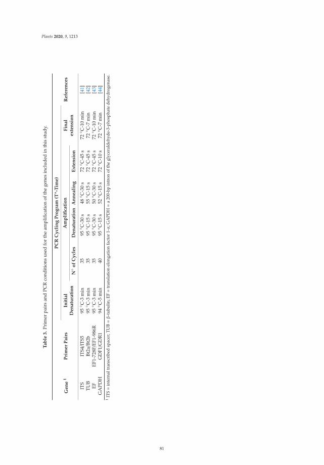
Plants 2020, 9, 1213
Ta
ble
3.
Prim
erpa
irs
and
PCR
cond
itio
nsus
edfo
rth
eam
plifi
cati
onof
the
gene
sin
clud
edin
this
stud
y.
Ge
ne
1P
rim
er
Pa
irs
PC
RC
ycl
ing
Pro
gra
m(T
ª-T
ime
)
Re
fere
nce
sIn
itia
lD
en
atu
rati
on
Am
pli
fica
tio
nF
ina
le
xte
nsi
on
N◦ o
fC
ycl
es
De
na
tura
tio
nA
nn
ea
lin
gE
xte
nsi
on
ITS
ITS4/I
TS5
95◦ C
-3m
in35
95◦ C
-30
s48◦ C
-30
s72◦ C
-45
s72◦ C
-10
min
[41]
TUB
Bt2a/B
t2b
95◦ C
-3m
in35
95◦ C
-15
s55◦ C
-15
s72◦ C
-45
s72◦ C
-7m
in[4
2]EF
EF1-
728F/E
F1-9
86R
95◦ C
-3m
in35
95◦ C
-30
s50◦ C
-30
s72◦ C
-45
s72◦ C
-10
min
[43]
GA
PDH
GD
F1/G
DR
194◦ C
-5m
in40
95◦ C
-15
s52◦ C
-15
s72◦ C
-10
s72◦ C
-7m
in[4
4]1
ITS=
inte
rnal
tran
scri
bed
spac
er;T
UB=β
-tub
ulin
;EF=
tran
slat
ion
elon
gati
onfa
ctor
1-α
;GA
PDH=
a20
0-bp
intr
onof
the
glyc
eral
dehy
de-3
-pho
spha
tede
hydr
ogen
ase.
81
Plants 2020, 9, 1213
Author Contributions: Field evaluations and sampling, fungal isolation and morphological identification, fungalcollection, writing-original draft preparation, editing, C.A.-B.; field evaluations, monitoring irrigation treatmentsover time, writing-review and editing, D.M.; phylogenetic investigation, formal analyses, and phylogeneticvisualization/data presentation, GenBank sequences submission, writing review and editing M.d.C.R.; experimentalfield management, funding acquisition, project administration, I.J.L.; conceived the idea of the research, designedthe scientific experiments and methodology, writing-review and editing, supervision, project administration, F.O.and A.T. All authors have read and agreed to the published version of the manuscript.
Funding: This research was partially funded by Spanish Ministry of Science, Innovation and Universities(MICINN), project AGL2015-66141-R, co-financed by the European Union FEDER Funds, and by EuropeanRegional Development Fund, project Innova-Clima (PR.AVA.AVA2019.051). C.A.-B. and D.M. are the holdersof a ‘Juan de la Cierva-Incorporación’ postdoctoral fellowship and a ‘Formación de Personal Investigador’pre-doctoral fellowship, respectively, both funded by MICINN.
Conflicts of Interest: The authors declare no conflict of interest.
References
1. Food and Agricultural Organisation of the United Nations (FAO) Website. 2017. Available online:http://www.fao.org/faostat/en/#data/QC (accessed on 21 April 2020).
2. Ministry of Agriculture, Fisheries and Food (MAPA; Spain). 2018. Available online: https://www.mapa.gob.es/es/estadistica/temas/estadisticas-agrarias/agricultura/superficies-producciones-anuales-cultivos/(accessed on 21 April 2020).
3. Ollero-Lara, A.; Agustí-Brisach, C.; Lovera, M.; Roca, L.F.; Arquero, O.; Trapero, A. Field susceptibility ofalmond cultivars to the four most common aerial fungal diseases in southern Spain. Crop Prot. 2019, 121,18–27. [CrossRef]
4. López-Moral, A.; Raya-Ortega, M.C.; Agustí-Brisach, C.; Roca, L.F.; Lovera, M.; Luque, F.; Arquero, O.;Trapero, A. Morphological, pathogenic and molecular characterization of Colletotrichum acutatum isolatescausing almond anthracnose in Spain. Plant Dis. 2017, 101, 2034–2045. [CrossRef]
5. Agustí-Brisach, C.; Roca, L.F.; Antón-Domínguez, B.I.; López-Moral, A.; Raya, M.C.; Lovera, M.; Arquero, O.;Trapero, A. Decaimiento del almendro en plantaciones jóvenes en Andalucía. Vida Rural 2020, 476, 36–43.
6. Teviotdale, B.L.; Michailides, T.J.; Pscheidt, J.W. Compendium of Nut Crop Diseases in Temperate Zones; APSPress: Saint Paul, MN, USA, 2002.
7. Gramaje, D.; Agustí-Brisach, C.; Pérez-Sierra, A.; Moralejo, E.; Olmo, D.; Mostert, L.; Damm, U.; Armengol, J.Fungal trunk pathogens associated with wood decay of almond trees on Mallorca (Spain). Persoonia 2012, 28,1–13. [CrossRef]
8. Olmo, D.; Armengol, J.; León, M.; Gramaje, D. Pathogenicity testing of lesser-known fungal trunk pathogensassociated with wood decay of almond trees. Eur. J. Plant Pathol. 2015, 143, 607–611. [CrossRef]
9. Olmo, D.; Armengol, J.; León, M.; Gramaje, D. Characterization and pathogenicity of Botryosphaeriaceaespecies isolated from almond trees on the island of Mallorca (Spain). Plant Dis. 2016, 100, 2483–2491.[CrossRef]
10. Úrbez-Torres, J.R. The status of Botryosphaeriaceae species infecting grapevines. Phytopathol. Mediterr. 2011,50, S5–S45.
11. Bertsch, C.; Ramírez-Suero, M.; Magnin-Robert, M.; Larignon, P.; Chong, J.; Abou-Mansour, E.; Spagnolo, A.;Clément, C.; Fontaine, F. Grapevine trunk disease: Complex and still poorly understood. Plant Pathol. 2013,62, 243–265. [CrossRef]
12. Gramaje, D.; Úrbez-Torres, J.R.; Sosnowski, M.R. Managing grapevine trunk diseases with respect to etiologyand epidemiology: Current strategy and future prospects. Plant Dis. 2018, 102, 12–39. [CrossRef]
13. Úrbez-Torres, J.R.; Peduto, F.; Vossen, P.M.; Krueger, W.H.; Gubler, W.D. Olive twig and branch dieback:Etiology, incidence, and distribution in California. Plant Dis. 2013, 97, 231–244. [CrossRef]
14. Moral, J.; Agustí-Brisach, C.; Pérez Rodríguez, M.; Xavíer, C.; Raya, M.C.; Rhouma, A.; Trapero, A.Identification of fungal species associated with branch dieback of olive and resistance of table cultivars toNeofusicoccum mediterraneum and Botryosphaeria dothidea. Plant Dis. 2017, 101, 306–316. [CrossRef]
15. Moral, J.; Morgan, D.; Trapero, A.; Michailides, T.J. Ecology and epidemiology of nut crops and olive diseasescaused by Botryosphaeriaceae fungi. Plant Dis. 2019, 103, 1809–1827. [CrossRef]
82
Plants 2020, 9, 1213
16. López-Moral, A.; Lovera, M.; Raya, M.C.; Cortés-Cosano, N.; Arquero, O.; Trapero, A.; Agustí-Brisach, C.Etiology of branch dieback and shoot blight of English walnut caused by Botryosphaeriaceae and Diaporthefungi in southern Spain. Plant Dis. 2020, 104, 533–550. [CrossRef]
17. López-Moral, A.; Raya, M.C.; Ruíz-Blancas, C.; Medialdea, I.; Lovera, M.; Arquero, O.; Trapero, A.;Agustí-Brisach, C. Aetiology of branch dieback, panicle and shoot blight of pistachio associated with fungaltrunk pathogens in southern Spain. Plant Pathol. 2020. [CrossRef]
18. Phillips, A.J.L.; Alves, A.; Abdollahzadeh, J.; Slippers, B.; Wingfield, M.J.; Groenewald, J.Z.; Crous, P.W.The Botryosphaeriaceae: Genera and species known from culture. Stud. Mycol. 2013, 76, 51–167. [CrossRef][PubMed]
19. Schoeneweiss, D.F. The role of environmental stress in diseases of woody plants. Plant Dis. 1981, 65, 308–314.[CrossRef]
20. Marsberg, A.; Kemler, M.; Jami, F.; Nagel, J.H.; Postma-Smidt, A.; Naidoo, S.; Wingfield, M.J.; Crous, P.W.;Spataforas, J.W.; Hesse, C.N.; et al. Botryosphaeria dothidea: A latent pathogen of global importance to woodyplant health. Mol. Plant Pathol. 2017, 18, 477–488. [CrossRef]
21. Crist, C.R.; Schoeneweiss, D.F. The influence of controlled stresses on susceptibility of European white birchstems to attack by Botryosphaeria dothidea. Phytopathology 1975, 65, 369–373.
22. Pusey, P.L. Influence of water stress on susceptibility of nonwounded peach bark to Botryosphaeria dothidea.Plant Dis. 1989, 73, 1000–1003. [CrossRef]
23. Maxwell, D.L.; Kruger, E.L.; Stanosz, G.R. Effects of water stress on colonization of poplar stems and excisedleaf disks by Septoria musiva. Phytopathology 1997, 87, 381–388. [CrossRef]
24. Hofstetter, V.; Buyck, B.; Croll, D.; Viret, O.; Couloux, A.; Gindro, K. What if Esca disease of grapevine werenot a fungal disease? Fungal Divers. 2012, 54, 51–67. [CrossRef]
25. Gramaje, D.; Baumgartner, K.; Halleen, F.; Mostert, L.; Sosnowski, M.R.; Úrbez-Torres, J.R.; Armengol, J.Fungal trunk diseases: A problem beyond grapevines? Plant Pathol. 2016, 65, 355–356. [CrossRef]
26. León, M.; Berbegal, M.; Rodríguez-Reina, J.M.; Elena, G.; Abad-Campos, P.; Ramón-Albalat, A.; Olmo, D.;Vicent, A.; Luque, J.; Miarnau, X.; et al. Identification and characterization of Diaporthe spp. associated withtwig cankers and shoot blight of almonds in Spain. Agronomy 2020, 10, 1062.
27. Sutton, B.C. The Coelomycetes. Fungi Imperfecti with Pycnidia, Acervuli and Stromata; CommonwealthMycological Institute: Kew, UK, 1980.
28. Chen, S.F.; Morgan, D.P.; Hasey, J.K.; Anderson, K.; Michailides, T.J. Phylogeny, morphology, distribution,and pathogenicity of Botryosphaeriaceae and Diaporthaceae from English walnut in California. Plant Dis.2014, 98, 636–652. [CrossRef]
29. Agustí-Brisach, C.; Moral, J.; Felts, D.; Trapero, A.; Michailides, T.J. Interaction between Diaporthe rhusicolaand Neofusicoccum mediterraneum causing branch dieback and fruit blight of English walnut in California,and effect of pruning wounds to the infection. Plant Dis. 2019, 103, 1196–1205. [CrossRef]
30. Lawrence, D.P.; Holland, L.A.; Nouri, M.T.; Travadon, R.; Abramians, A.; Michailides, T.J.; Trouillas, F.P.Molecular phylogeny of Cytospora species associated with canker diseases of fruit and nut crops in California,with the descriptions of ten new species and one new combination. IMA Fungus 2018, 9, 333–369. [CrossRef]
31. Arquero, O.; Belmonte, A.; Casado, B.; Cruz-Blanco, M.; Espadafor, M.; Fernández, J.L.; Gallego, J.C.;García, A.; Lorite, I.J.; Lovera, M.; et al. Manual del almendro. Consejería de Agricultura, Pesca y Desarrollo Rural;Ediciones Junta de Andalucía: Sevilla, Spain, 2013.
32. Espadafor, M.; Orgaz, F.; Testi, L.; Lorite, I.J.; Villalobos, F.J. Transpiration of young almond trees in relationto intercepted radiation. Irrig. Sci. 2001, 33, 265–275. [CrossRef]
33. Bonachela, S.; Orgaz, F.; Villalobos, F.J.; Fereres, E. Soil evaporation from drip-irrigated olive orchards.Irrig. Sci. 2001, 20, 65–71. [CrossRef]
34. Moral, J.; Trapero, A. Assessing the susceptibility of olive cultivars to anthracnose caused by Colletotrichumacutatum. Plant Dis. 2009, 93, 1028–1036. [CrossRef]
35. Campbell, C.L.; Madden, L.V. Introduction to Plant Disease Epidemiology; John Wiley & Sons, Ltd.: New York,NY, USA, 1990.
36. Steel, R.G.D.; Torrie, J.H. Bioestadística, 2nd ed.; McGraw-Hill: Bogotá, Colombia, 1985.37. Statistix10. User’s Manual; Analytical Software: Tallahassee, FL, USA, 2013.38. Kumar, S.; Stecher, G.; Tamura, K. MEGA7: Molecular Evolutionary Genetics Analysis version 7.0 for bigger
datasets. Mol. Biol. Evol. 2016, 33, 1870–1874. [CrossRef]
83

Plants 2020, 9, 1213
39. Felsenstein, J. Confidence limits on phylogenies: An approach using the bootstrap. Evolution 1985, 39,783–791. [CrossRef] [PubMed]
40. Ronquist, F.; Teslenko, M.; Van Der Mark, P.; Ayres, D.L.; Darling, A.; Höhna, S.; Larget, B.; Liu, L.;Suchard, M.A.; Huelsenbeck, J.P. MrBayes 3.2: Efficient Bayesian phylogenetic inference and model choiceacross a large model space. Syst. Biol. 2012, 61, 539–542. [CrossRef]
41. White, T.J.; Bruns, T.; Lee, S.; Taylor, J. Amplification and direct sequencing of fungal ribosomal RNA genesfor phylogenetics. In PCR Protocols: A Guide to Methods and Applications; Innis, M.A., Gelfand, D.H., Snisky, J.J.,White, T.J., Eds.; Academic: San Diego, CS, USA, 1990; pp. 315–322.
42. Glass, N.L.; Donaldson, G. Development of primer sets designed for use with PCR to amplify conservedgenes from filamentous ascomycetes. Appl. Environ. Microb. 1995, 61, 1323–1330. [CrossRef]
43. Carbone, I.; Kohn, L.M. A method for designing primer sets for speciation studies in filamentous ascomycetes.Mycologia 1999, 91, 553–556. [CrossRef]
44. Guerber, J.C.; Liu, B.; Correll, J.C.; Johnston, P.R. Characterization of diversity in Colletotrichum acutatumsensu lato by sequence analysis of two gene introns, mtDNA and intron RFLPs, and mating compatibility.Mycologia 2003, 95, 872–895. [CrossRef]
© 2020 by the authors. Licensee MDPI, Basel, Switzerland. This article is an open accessarticle distributed under the terms and conditions of the Creative Commons Attribution(CC BY) license (http://creativecommons.org/licenses/by/4.0/).
84
plants
Article
Early Season Symptoms on Stem, Inflorescences andFlowers of Grapevine Associated withBotryosphaeriaceae Species
Pedro Reis 1,*, Ana Gaspar 1, Artur Alves 2, Florence Fontaine 3, Inês Lourenço 4, José Saramago 4,
Mariana Mota 1 and Cecília Rego 1
1 LEAF—Linking Landscape, Environment, Agriculture and Food, School of Agriculture,University of Lisbon, 1349-017 Lisbon, Portugal; [email protected] (A.G.);[email protected] (M.M.); [email protected] (C.R.)
2 CESAM—Centre for Environmental and Marine Studies, Department of Biology, University of Aveiro,3810-193 Aveiro, Portugal; [email protected]
3 SFR Condorcet FR CNRS 3417, Université de Reims Champagne-Ardenne, Résistance Induite etBioprotection des Plantes EA 4707, BP 1039, 51687 Reims Cedex 2, France; [email protected]
4 BASF Portuguesa, S.A. Rua 25 de Abril, 1, 2689-538 Prior Velho, Portugal; [email protected] (I.L.);[email protected] (J.S.)
* Correspondence: [email protected]
Received: 21 September 2020; Accepted: 22 October 2020; Published: 24 October 2020
Abstract: Botryosphaeria dieback caused by several Botryosphaeriaceae species is one of the mostimportant grapevine trunk diseases affecting vineyards worldwide. These fungi cause wedge-shapedperennial cankers and black streaking of the wood and have also been associated with interveinleaf chlorosis, dried or mummified berries, and eventually, the death of the plant. Early seasonsymptoms may sometimes be disregarded by growers, being mistaken with symptoms from otherdiseases such as downy mildew or botrytis rot. Currently, few studies are available to determinewhat species may be causing these early season symptoms in grapevines. During the 2018 season,during the flowering period, grapevine samples showing necrosis on green shoots, dried inflorescences,and flowers, were collected in vineyards throughout the central regions of Portugal. Isolations wereperformed from symptomatic organs, and twenty-three isolates of Botryosphaeriaceae were selected.An analysis of the ITS and part of the translation elongation factor 1-α sequences was performed,revealing that the two main species apparently responsible for these symptoms were Diplodia seriataand Neofusicoccum parvum. In pathogenicity tests conducted on 1-year-old plants grown undercontrolled conditions in a greenhouse and on field-grown clusters, symptoms were reproduced,confirming the pathogenic behavior of the selection of isolates.
Keywords: grapevine; Diplodia seriata; Neofusicoccum parvum; pathogenicity; diagnostic
1. Introduction
Grapevine trunk diseases (GTDs) are one of the most critical problems affecting viticultureworldwide, causing yield reduction and increased production costs adding to the existing costof the annual vineyard operations. In fact, these diseases pose a significant threat to sustainableviticulture worldwide since the cost associated with GTD losses has been increasing during recentyears. For example, in Spain, there was an increase of 1.8% of incidence in vineyards in 2003 up to 10.5%in 2007 [1]. In France, it is estimated that the equivalent of 1 billion euros is lost every year due to thesediseases [2], while in Australia, the economic impact can reach up to 8.3 billion AUD [3]. GTDs includethree main fungal diseases—eutypa dieback, esca disease and botryosphaeria dieback—that involveone or several species of fungi [4–6]. In Portugal, botryosphaeria dieback and Esca are considered the
Plants 2020, 9, 1427; doi:10.3390/plants9111427 www.mdpi.com/journal/plants
85
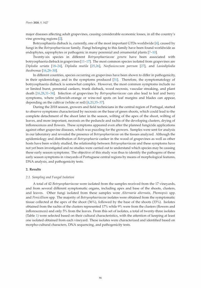
Plants 2020, 9, 1427
major diseases affecting adult grapevines, causing considerable economic losses, in all the country’svine growing regions [2].
Botryosphaeria dieback is, currently, one of the most important GTDs worldwide [4], caused byfungi in the Botryosphaeriaceae family. Fungi belonging to this family have been found worldwide asendophytes, saprophytes or pathogenic in many perennial and ornamental plants [7–10].
Twenty-six species in different Botryosphaeriaceae genera have been associated withbotryosphaeria dieback in grapevines [11–17]. The most common species isolated from grapevines areDiplodia seriata [18–24], Diplodia mutila [25,26], Neofusicoccum parvum [27], and Lasiodiplodiatheobromae [16,28–30].
In different countries, species occurring on grapevines have been shown to differ in pathogenicity,in their epidemiology, and in the symptoms produced [31]. Therefore, the symptomatology ofbotryosphaeria dieback is somewhat complex. However, the most common symptoms include noor limited burst, perennial cankers, trunk dieback, wood necrosis, vascular streaking, and plantdeath [16,28,31–34]. Infection of grapevines by Botryosphaeriaceae can also lead to leaf and berrysymptoms, where yellowish-orange or wine-red spots on leaf margins and blades can appear,depending on the cultivar (white or red) [4,20,35–37].
During the 2018 season, growers and field technicians in the central region of Portugal, startedto observe symptoms characterized by necrosis on the base of green shoots, which could lead to thecomplete detachment of the shoot later in the season, wilting of the apex of the shoot, wilting ofleaves, and more important, necrosis on the peduncle and rachis of the developing clusters, drying ofinflorescences and flowers. These symptoms appeared even after the planned fungicide applicationsagainst other grapevine diseases, which was puzzling for the growers. Samples were sent for analysisin our laboratory and revealed the presence of Botryosphaeriaceae on the tissues analyzed. Although theepidemiology and distribution of Botryosphaeria canker in the wood of grapevines as well as otherhosts have been widely studied, the relationship between Botryosphaeriaceae and these symptoms havenot yet been investigated and no studies were carried out to understand which species may be causingthese early season symptoms. The objective of this study was thus to identify the pathogens of theseearly season symptoms in vineyards of Portuguese central regions by means of morphological features,DNA analysis, and pathogenicity tests.
2. Results
2.1. Sampling and Fungal Isolation
A total of 42 Botryosphaeriaceae were isolated from the samples received from the 17 vineyards,and from several different symptomatic organs, including apex and base of the shoots, clusters,and leaves. Other fungi isolated from these samples were Alternaria alternata, Phomopsis spp.and Penicillium spp. The majority of Botryosphaeriaceae isolates were obtained from the symptomatictissue collected at the apex of the shoot (36%), followed by the base of the shoots (33%). Isolatesobtained from the rachis of the clusters represented 17% while 9% were from the clusters (flowers andinflorescences) and only 5% from the leaves. From this set of isolates, a total of twenty-three isolates(Table 1) were selected based on their cultural characteristics, with the attention of keeping at leastone isolated obtained from each vineyard. These isolates were characterized and identified based onmorpho-cultural characters, DNA sequencing, and pathogenicity tests.
86
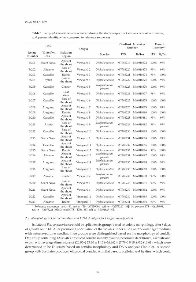
Plants 2020, 9, 1427
Table 1. Botryosphaeriaceae isolates obtained during the study, respective GenBank accession numbers,and percent identity when compared to reference sequences.
HostOrigin
GenBank AccessionNumber
PercentIdentity *
IsolateNumber
(V. viniferacvs.)
IsolationRegion
Species ITS Tef1-α ITS Tef1-α
Bt201 Seara Nova Apex ofthe shoot Vineyard 1 Diplodia seriata MT786219 MW018672 100% 99%
Bt202 Alicante Base ofthe shoot Vineyard 2 Diplodia seriata MT786220 MW018673 99% 99%
Bt203 Castelão Rachis Vineyard 3 Diplodia seriata MT786221 MW018674 99% 100%
Bt204 Syrah Base ofthe shoot Vineyard 4 Diplodia seriata MT786222 MW018675 100% 99%
Bt205 Castelão Cluster Vineyard 5 Neofusicoccumparvum MT786223 MW018676 100% 99%
Bt206 Castelão Leafstem Vineyard 5 Diplodia seriata MT786224 MW018677 98% 99%
Bt207 Castelão Base ofthe shoot Vineyard 6 Diplodia seriata MT786225 MW018678 100% 100%
Bt208 Aragonez Apex ofthe shoot Vineyard 7 Diplodia seriata MT786226 MW018679 100% 99%
Bt209 Aragonez Rachis Vineyard 8 Diplodia seriata MT786227 MW018680 100% 100%
Bt210 Castelão Apex ofthe shoot Vineyard 5 Diplodia seriata MT786228 MW018681 99% 99%
Bt211 Arinto Base ofthe shoot Vineyard 9 Neofusicoccum
parvum MT786229 MW018682 99% 99%
Bt212 Castelão Base ofthe shoot Vineyard 10 Diplodia seriata MT786230 MW018683 100% 100%
Bt213 Seara Nova Apex ofthe shoot Vineyard 1 Diplodia seriata MT786231 MW018684 100% 99%
Bt214 Castelão Apex ofthe shoot Vineyard 11 Diplodia seriata MT786232 MW018685 100% 100%
Bt215 Seara Nova Rachis Vineyard 12 Diplodia seriata MT786233 MW018686 98% 100%
Bt216 Alicante Apex ofthe shoot Vineyard 13 Neofusicoccum
parvum MT786234 MW018687 100% 98%
Bt217 Aragonez Apex ofthe shoot Vineyard 14 Neofusicoccum
parvum MT786235 MW018688 100% 98%
Bt218 Aragonez Base ofthe shoot Vineyard 15 Diplodia mutila MT786236 MW018689 100% 100%
Bt219 Alicante Cluster Vineyard 2 Neofusicoccumparvum MT786237 MW018690 99% 100%
Bt220 Seara Nova Base ofthe shoot Vineyard 1 Diplodia seriata MT786238 MW018691 99% 99%
Bt221 Seara Nova Apex ofthe shoot Vineyard 1 Diplodia seriata MT786239 MW018692 100% 99%
Bt222 Castelão Apex ofthe shoot Vineyard 16 Diplodia seriata MT786240 MW018693 100% 100%
Bt223 Alicante Rachis Vineyard 17 Diplodia seriata MT786241 MW018694 99% 99%
* Reference sequences used—D. seriata ITS—AY259094, tef1-α—AY573220 [38]; N. parvum ITS—AY259098,tef1-α—AY573221 [38]; D. mutila ITS—KJ361837, tef1-α—KJ361829 [39].
2.2. Morphological Characterization and DNA Analysis for Fungal Identification
Isolates of Botryosphaeriaceae could be split into six groups based on colony morphology, after 8 daysof growth on PDA. After promoting sporulation of the isolates under study on 2% water agar mediumwith autoclaved pine needles, three groups were distinguished based on the morphology of conidia.One group containing 12 isolates produced conidia initially hyaline, becoming dark brown, aseptate andovoid, with average dimensions of (20.95–) 23.60 ± 1.15 (–26.46) × (7.79–) 9.18 ± 0.5 (10.61), which weredetermined to be D. seriata based on conidia morphology and DNA analysis (Table 2). A secondgroup with 3 isolates produced ellipsoidal conidia, with flat base, unicellular and hyaline, which could
87
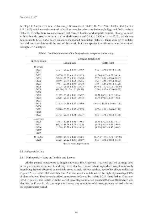
Plants 2020, 9, 1427
develop 1 to 2 septa over time, with average dimensions of (14.18–) 16.39 ± 1.95 (–19.46) × (4.50–) 5.19 ±0.13 (–6.02) which were determined to be N. parvum, based on conidial morphology and DNA analysis(Table 2). Finally, there was one isolate that formed hyaline and aseptate conidia, oblong to ovoidwith both ends broadly rounded and with dimensions of (20.80–) 23.50 ± 1.41 (–25.05), which wasdetermined to be D. mutila based on above-mentioned parameters (Table 2). There were seven isolatesthat did not sporulate until the end of this work, but their species identification was determinedthrough DNA analysis.
Table 2. Conidial dimension of the Botryosphaeriaceae species under study.
Species/IsolateConidial dimensions
Length (μm) Width (μm)
D. seriataBt201 (21.27–) 25.22 ± 1.89 (–28.69) (8.33–) 9.91 ± 0.88 (–11.75)Bt202 *Bt203 (20.75–) 22.50 ± 1.12 (–24.23) (6.71–) 8.17 ± 0.57 (–9.16)Bt204 (20.43–) 22.65 ± 1.56 (–26.20) (7.83–) 9.06 ± 0.76 (–10.93)Bt206 (20.99–) 23.84 ± 1.52 (–26.36) (7.71–) 9.23 ± 0.95 (–10.77)Bt207 (19.61–) 23.58 ± 1.95 (–27.30) (7.49–) 9.29 ± 0.81 (–11.26)Bt208 (21.72–) 25.26 ± 1.41 (–28.73) (9.53–) 11.13 ± 1.01 (–13.37)Bt209 (19.61–) 22.17 ± 1.52 (24.53) (7.20–) 9.07 ± 0.78 (–10.55)Bt210Bt212 (21.27–) 23.91 ± 1.36 (–26.22) (7.34–) 8.34 ± 0.60 (–9.36)Bt213 (23.28–) 25.99 ± 1.58 (–29.18) (7.73–) 9.03 ± 0.56 (–9.84)Bt214Bt215 (21.02–) 24.58 ± 1.47 (–26.99) (10.14–) 11.21 ± 0.66 (–12.83)Bt220Bt221 (20.80–) 23.28 ± 1.35 (–25,53) (8.59–) 9.99 ± 0.60 (–11.10)Bt222Bt223 (21.42–) 22.94 ± 1.34 (–26.37) (8.97–) 9.72 ± 0.66 (–11.40)
N. parvumBt205 (15.51–) 17.18 ± 1.02 (–19.92) (4.56–) 5.22 ± 0.43 (–6.11)Bt211 (15,.2–) 18.28 ± 1.75 (–22,.4) (4.73–) 5.33 ± 0.31 (–5.94)Bt216 (11.39–) 13.71 ± 1.28 (–16.13) (4.20–) 5.02 ± 0.45 (–6.02)Bt217Bt219
D. mutila (20.80–) 23.50 ± 1.41 (–25.05) (9.47–) 11.15 ± 1.57 (–16.25)Bt218 (21.27–) 25.22 ± 1.89 (–28.69) (8.33–) 9.91 ± 0.88 (–11.75)
* Isolate without sporulation.
2.3. Pathogenicity Tests
2.3.1. Pathogenicity Tests on Tendrils and Leaves
All the isolates tested were pathogenic towards the Aragonez 1-year-old grafted cuttings usedin the greenhouse experiment, and they were able to, in some extent, reproduce symptoms closelyresembling the ones observed on the field survey, namely necrotic tendrils, apex of the shoots and leaves(Figure 1A–C). Isolate Bt204 identified as D. seriata, was the isolate where the highest percentage (50%)of plants showed the above-described symptoms, followed by isolate Bt216 identified as N. parvum(40%) (Figure 2). The isolate with the lowest percentage of infected plants (20%) was Bt218 which wasidentified as D. mutila. No control plants showed any symptoms of disease, growing normally duringthe experimental period.
88
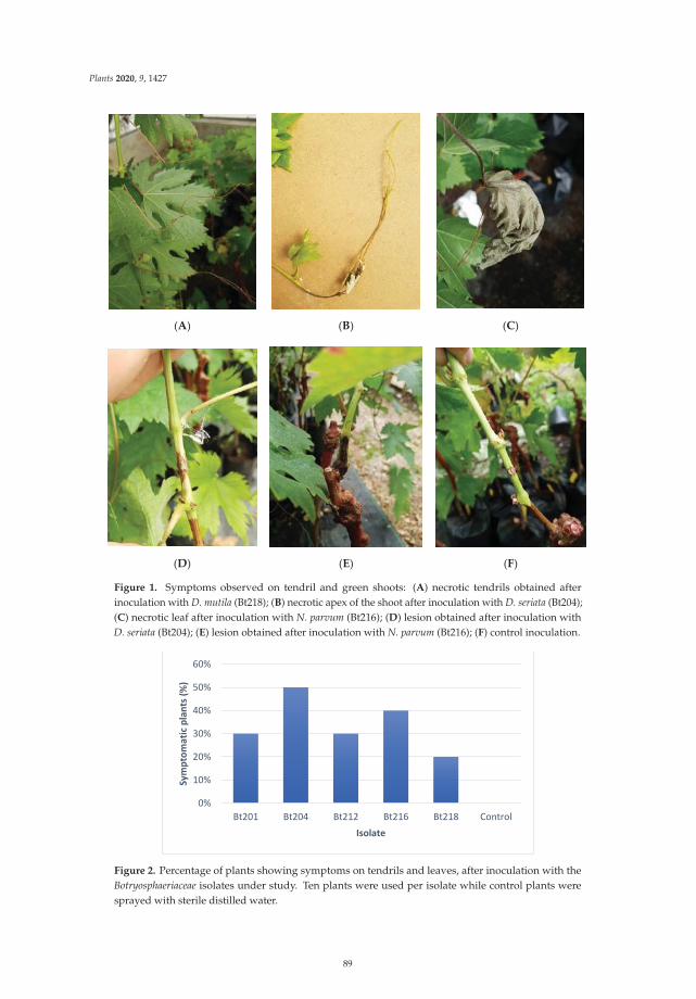
Plants 2020, 9, 1427
(A) (B) (C)
(D) (E) (F)
Figure 1. Symptoms observed on tendril and green shoots: (A) necrotic tendrils obtained afterinoculation with D. mutila (Bt218); (B) necrotic apex of the shoot after inoculation with D. seriata (Bt204);(C) necrotic leaf after inoculation with N. parvum (Bt216); (D) lesion obtained after inoculation withD. seriata (Bt204); (E) lesion obtained after inoculation with N. parvum (Bt216); (F) control inoculation.
0%
10%
20%
30%
40%
50%
60%
Bt201 Bt204 Bt212 Bt216 Bt218 Control
Sym
ptom
atic
plan
ts (%
)
Isolate
Figure 2. Percentage of plants showing symptoms on tendrils and leaves, after inoculation with theBotryosphaeriaceae isolates under study. Ten plants were used per isolate while control plants weresprayed with sterile distilled water.
89
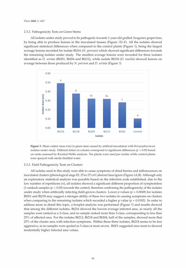
Plants 2020, 9, 1427
2.3.2. Pathogenicity Tests on Green Stems
All isolates under study proved to be pathogenic towards 1-year-old grafted Aragonez grapevines,by being able to produce lesions in the inoculated tissues (Figure 1D–F). All the isolates showedsignificant statistical differences when compared to the control plants (Figure 3), being the largestaverage lesions recorded for isolate Bt216 (N. parvum) which showed significant differences towardsthe remaining isolates under study. The smallest average lesions were recorded for three isolatesidentified as D. seriata (Bt201, Bt204 and Bt212), while isolate Bt218 (D. mutila) showed lesions onaverage between those produced by N. parvum and D. seriata (Figure 3)
a
b
bc bcc
d
0.00
0.10
0.20
0.30
0.40
0.50
0.60
Bt216 Bt218 Bt204 Bt212 Bt201 Control
Cank
er A
rea
(cm
)
Isolate
Figure 3. Mean canker areas (cm) in green stem caused by artificial inoculation with Botryosphaeriaceaeisolates under study. Different letters in column correspond to significant differences (p < 0.05) basedon ranks assessed by Kruskal-Wallis analysis. Ten plants were used per isolate while control plantswere sprayed with sterile distilled water.
2.3.3. Field Pathogenicity Tests on Clusters
All isolates used in this study were able to cause symptoms of dried berries and inflorescences oninoculated clusters (phenological stage EL 25 to 27) of Cabernet Sauvignon (Figure 4A,B). Although onlyan exploratory statistical analysis was possible based on the infection scale established, due to thelow number of repetitions (n), all isolates showed a significant different proportion of symptomless(1-ranked) samples (p < 0.05) towards the control, therefore confirming the pathogenicity of the isolatesunder study when artificially infecting field-grown clusters. Lower p-values (p = 0.0049) for isolatesBt201 and Bt218 may suggest a stronger ability of these two isolates in causing symptoms on clusterswhen comparing to the remaining isolates which recorded a higher p value (p = 0.0182). In order toaddress more in detail this topic, a boxplot analysis was performed (Figure 5) and results showedthat among the different isolates, Bt216 showed the lowest average infected area, as nearly all thesamples were ranked as a 2-class, and no sample ranked more than 3-class, corresponding to less than25% of affected area. For the isolates Bt212, Bt218 and Bt204, half of the samples, showed more that25% of the cluster area with infection symptoms. Within these three isolates, Bt218 seems to be lessaggressive, as no samples were quoted as 3-class or more severe. Bt201 suggested once more to showedtendentially higher infected area values.
90
Plants 2020, 9, 1427
Figure 4. (A) best cluster from each inoculation/isolate; (B) worst cluster from each inoculation/isolate.
Figure 5. Boxplot of the percentage of infected area on clusters artificially inoculated withBotryosphaeriaceae isolates under study. Scale of evaluation of area infected—1 = no symptoms; 2 = 1–5%;3 = 5–25%; 4 = 25–50%; 5 = >50%. The median is represented by the solid line. Top and bottom lines ofthe box correspond to the 25th and 75th percentiles of the data, respectively. Circles represent outliers.
For all the pathogenicity experiments, pathogens were recovered from symptomatic tissues of allinfected plants, while no Botryosphaeriaceae isolates were re-isolated from control plants.
3. Discussion
This is the first study aimed at describing grapevine early season symptoms associated withBotryosphaeriaceous fungi in Portugal, including necrosis and wilting of the apex of the green shoots,wilting of leaves, necrosis on the peduncle and rachis of the developing clusters and, drying ofinflorescences and flowers. Morphological studies and DNA sequence analysis allowed to identify thepresence of three different species of Botryosphaeriaceae: D. seriata, N. parvum and D. mutila. The most
91
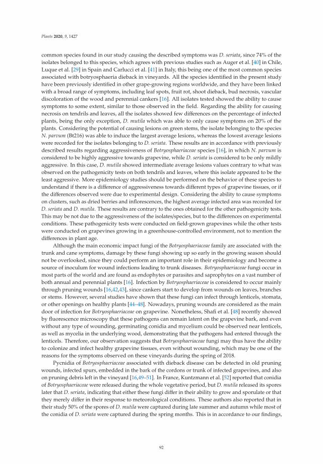
Plants 2020, 9, 1427
common species found in our study causing the described symptoms was D. seriata, since 74% of theisolates belonged to this species, which agrees with previous studies such as Auger et al. [40] in Chile,Luque et al. [29] in Spain and Carlucci et al. [41] in Italy, this being one of the most common speciesassociated with botryosphaeria dieback in vineyards. All the species identified in the present studyhave been previously identified in other grape-growing regions worldwide, and they have been linkedwith a broad range of symptoms, including leaf spots, fruit rot, shoot dieback, bud necrosis, vasculardiscoloration of the wood and perennial cankers [16]. All isolates tested showed the ability to causesymptoms to some extent, similar to those observed in the field. Regarding the ability for causingnecrosis on tendrils and leaves, all the isolates showed few differences on the percentage of infectedplants, being the only exception, D. mutila which was able to only cause symptoms on 20% of theplants. Considering the potential of causing lesions on green stems, the isolate belonging to the speciesN. parvum (Bt216) was able to induce the largest average lesions, whereas the lowest average lesionswere recorded for the isolates belonging to D. seriata. These results are in accordance with previouslydescribed results regarding aggressiveness of Botryosphaeriaceae species [16], in which N. parvum isconsidered to be highly aggressive towards grapevine, while D. seriata is considered to be only mildlyaggressive. In this case, D. mutila showed intermediate average lesions values contrary to what wasobserved on the pathogenicity tests on both tendrils and leaves, where this isolate appeared to be theleast aggressive. More epidemiology studies should be performed on the behavior of these species tounderstand if there is a difference of aggressiveness towards different types of grapevine tissues, or ifthe differences observed were due to experimental design. Considering the ability to cause symptomson clusters, such as dried berries and inflorescences, the highest average infected area was recorded forD. seriata and D. mutila. These results are contrary to the ones obtained for the other pathogenicity tests.This may be not due to the aggressiveness of the isolates/species, but to the differences on experimentalconditions. These pathogenicity tests were conducted on field-grown grapevines while the other testswere conducted on grapevines growing in a greenhouse-controlled environment, not to mention thedifferences in plant age.
Although the main economic impact fungi of the Botryosphaeriaceae family are associated with thetrunk and cane symptoms, damage by these fungi showing up so early in the growing season shouldnot be overlooked, since they could perform an important role in their epidemiology and become asource of inoculum for wound infections leading to trunk diseases. Botryosphaeriaceae fungi occur inmost parts of the world and are found as endophytes or parasites and saprophytes on a vast number ofboth annual and perennial plants [16]. Infection by Botryosphaeriaceae is considered to occur mainlythrough pruning wounds [16,42,43], since cankers start to develop from wounds on leaves, branchesor stems. However, several studies have shown that these fungi can infect through lenticels, stomata,or other openings on healthy plants [44–48]. Nowadays, pruning wounds are considered as the maindoor of infection for Botryosphaeriaceae on grapevine. Nonetheless, Shafi et al. [48] recently showedby fluorescence microscopy that these pathogens can remain latent on the grapevine bark, and evenwithout any type of wounding, germinating conidia and mycelium could be observed near lenticels,as well as mycelia in the underlying wood, demonstrating that the pathogens had entered through thelenticels. Therefore, our observation suggests that Botryosphaeriaceae fungi may thus have the abilityto colonize and infect healthy grapevine tissues, even without wounding, which may be one of thereasons for the symptoms observed on these vineyards during the spring of 2018.
Pycnidia of Botryosphaeriaceae associated with dieback disease can be detected in old pruningwounds, infected spurs, embedded in the bark of the cordons or trunk of infected grapevines, and alsoon pruning debris left in the vineyard [16,49–51]. In France, Kuntzmann et al. [52] reported that conidiaof Botryosphaeriaceae were released during the whole vegetative period, but D. mutila released its sporeslater that D. seriata, indicating that either these fungi differ in their ability to grow and sporulate or thatthey merely differ in their response to meteorological conditions. These authors also reported that intheir study 50% of the spores of D. mutila were captured during late summer and autumn while most ofthe conidia of D. seriata were captured during the spring months. This is in accordance to our findings,
92
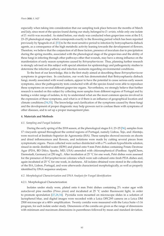
Plants 2020, 9, 1427
especially when taking into consideration that our sampling took place between the months of Marchand July, since most of the species found during our study, belonged to D. seriata, while only one isolateof D. mutila was recorded. As stated before, our study was conducted when grapevines were at the E-L23–25 phenological stage which corresponds exactly to the flowering period which has been reportedpreviously by Spagnolo et al. [53] to be the most sensitive period to infection by botryosphaeria diebackagents, as a consequence of the high metabolic activity leaning towards the development of flowers.Therefore, we believe that the conjunction of all these factors, presence of inoculum due to precipitationduring the spring months, associated with the phenological stage of the grapevines and the ability ofthese fungi in infecting through other pathways other that wounds, may have a strong influence on themanifestation of early season symptoms caused by Botryosphaeriaceae. Thus, planning further researchis strongly advised on this subject with special attention for epidemiology and pathogenicity studies todetermine the infection pathway and infection moments regarding environmental conditions.
To the best of our knowledge, this is the first study aimed at describing these Botryosphaeriaceaesymptoms in grapevines. In conclusion, our work has demonstrated that Botryosphaeria diebackfungi, mostly associated with wood cankers, appear to have the potential to cause serious early seasonsymptoms, since the pathogenicity tests conducted with all the species found were able to reproducethese symptoms on several different grapevine organs. Nevertheless, we strongly believe that furtherresearch is needed on this subject by collecting more samples from different regions of Portugal and bytesting a wider range of isolates, to try to understand what are the most common species involved inthe expression of these symptoms, and what is or if there is an influence of geographical location andclimate conditions [54,55]. The knowledge and clarification of the symptoms caused by these fungiand the development of proper diagnostic may help growers not to confuse them with symptoms ofother diseases, and to set up a proper management plan.
4. Materials and Methods
4.1. Sampling and Fungal Isolation
During the early spring of the 2018 season, at the phenological stages E-L 23–25 [56], samples from17 vineyards spread throughout the central regions of Portugal, namely Lisboa, Tejo, and Alentejo,were received at Instituto Superior de Agronomia (ISA). These samples showed necrosis on shootsand dried inflorescences and flowers, and isolations were made by cutting several pieces fromsymptomatic organs. Pieces collected were surface disinfected with a 7% sodium hypochlorite solution,rinsed in sterile distilled water (SDW) and plated onto 9 mm Petri dishes containing Potato DextroseAgar (PDA, BD Difco, Sparks, MD, USA) amended with chloramphenicol (PanReac AppliChem,Darmstadt, Germany) at 250 mg/L. After incubation at 25 ◦C for one week, Petri dishes were assessedfor the presence of Botryosphaeriaceae colonies which were sub-cultured onto fresh PDA dishes and,again incubated at 25 ◦C for one week, in darkness. All isolates obtained were stored in the collectionof the ISA, Lisbon, Portugal, and were afterwards characterized morphologically, as well as properlyidentified by DNA sequence analyses.
4.2. Morphological Characterization and DNA Analysis for Fungal Identification
4.2.1. Morphological Characterization
Isolates under study were, plated onto 6 mm Petri dishes containing 2% water agar withautoclaved pine needles (Pinus pinea) and incubated at 25 ◦C under fluorescent light, in orderto promote sporulation [27,28,34]. Pycnidia were mounted on microscope slides in a solution oflactophenol blue, and digital images were recorded with a Leica DFC295 camera on a Leica DM2500 microscope at a 400× amplification. Twenty conidia were measured with the Leica Suite v3.16program, for each isolate under study. Dimensions of the conidia are given as the range of dimensionswith minimum and maximum dimensions in parentheses followed by mean and standard deviation.
93
Plants 2020, 9, 1427
4.2.2. DNA Analysis
DNeasyTM Plant Mini Kit by Qiagen® (Venlo, The Netherlands) was used to extract genomicDNA from 8-day-old cultures grown in PDA and incubated at 25 ◦C, in the darkness. The ITS regionwas amplified using primers ITS5 and ITS4 [57], while the primers EF1-688F and EF1–1251R [58]were used to amplify part of the elongation factor 1α gene. The PCR mixtures contained 1 × PCRbuffer REDTaq Ready Mix (Sigma-Aldrich, Saint Louis, MO, USA), 3 mM MgCl2, 0.4 mM dNTP mix,12.5 pmol of each primer, 0.06 unit/μL of Taq Polymerase and 25–50 ng of template DNA. Each reactionvolume was made up to 25 μL with sterile ultrapure water. Negative controls with sterile ultrapurewater instead of the template DNA were used in every reaction. The amplification conditions for ITSwere as follows: initial denaturation of 5 min at 95 ◦C, followed by 40 cycles of 30 s at 94 ◦C, 30 s at58 ◦C, 1 min and 40 s at 72 ◦C and a final extension period of 10 min at 72 ◦C. For the amplificationof part of the tef1-α gene, the conditions were, an initial denaturation of 2 min at 94 ◦C, followed by35 cycles of 30 s at 94 ◦C, 45 s at 55 ◦C, 1 min at 72 ◦C and a final extension period of 10 min at 72 ◦C.Each amplicon was separated by electrophoresis at 120 V for 30 min in a 1% agarose gel in 0.5 × TBEbuffer. Gels were stained with 3 μL of GreenSafe Premium (Nzytech, Lisbon, Portugal), and werevisualized using with a UV transilluminator to assess PCR amplification.
The amplified PCR fragments were purified using an Illustra ExoProStar Enzymatic PCR andSequencing Clean-up Kit (GE Life Sciences, Buckinghamshire, UK) and both strands of the PCRproducts were sent for sequencing at STABVIDA (Lisbon, Portugal). Sequences obtained were editedand aligned using MEGA7 [59] to find a consensus sequence. These sequences were then comparedwith sequences from GenBank in BLAST searches, and species identification was obtained when atleast 98% of similarity was found.
4.3. Pathogenicity Tests
Five representative isolates were selected from the fungal collection under study for pathogenicitytests, with the attention of selecting at least one isolate from the three different species ofBotryosphaeriaceae identified three isolates of Diplodia seriata (Bt201, Bt204 and Bt212), one isolateof Neofusicoccum parvum (Bt216) and one isolate of Diplodia mutila (Bt218). To reproduce all thesymptoms found in the field, three different pathogenicity tests were designed, being two performedon grapevines kept in a greenhouse and one on grapevines established in the field. Greenhousepathogenicity tests were conducted on 1-year-old grafted Aragonez (=Tempranillo) plants, since is themost planted cultivar in Portugal and field tests were conducted on clusters of Cabernet Sauvignonwhich is currently the most planted cultivar worldwide.
4.3.1. Pathogenicity tests on Tendrils and Leaves
Isolates were plated in Petri dishes containing 2% water agar with autoclaved pine needles(Pinus pinea) and incubated at 25 ◦C under fluorescent light, to promote sporulation [27,28,34]. Conidiawere harvested from these plates by collecting the pycnidia formed on the pine needles into a1.5 mL Eppendorf tube containing sterile distilled water and crushing them with the help of a pestle.These spore suspensions were filtered through cheesecloth and the concentrations were adjusted to105 spores/mL. To ensure full coverage of the tissues to be inoculated, 2 mL of each spore suspension wassprayed on the green tissues (leaves and tendrils) of 1-year-old grafted cuttings of cultivar Aragonez(=Tempranillo), individually potted in 1 L free draining bags containing a sandy soil mixture kept in aventilated greenhouse at 24 ◦C under natural light. The aerial part of the plants was covered witha plastic bag for 3 days to promote infection, and the plants were assessed for the development ofsymptoms, namely necrosis on any of the inoculated organs or drying of leaves and tendrils, one weekafter inoculation. Ten plants were used for each isolate, while control plants were sprayed with steriledistilled water. The percentage of plants showing symptoms for each isolate was recorded.
94
Plants 2020, 9, 1427
4.3.2. Pathogenicity Tests on Green Stems
One-year old grafted cuttings of cultivar Aragonez (=Tempranillo), individually potted in 1 Lfree draining bags containing a sandy soil mixture, kept in a ventilated greenhouse at 24 ◦C undernatural light, were inoculated following the method described by Reis et al. [35]. Ten plants wereused for each isolate, and the assessment for symptoms development, including external lesionsor cankers, was performed one month after inoculation, by measuring the width and length of thelesions and calculating the elliptical area of the lesion. All statistical analysis was performed using theR program (www.r-project.org). Assumptions for variance analysis was assessed and when all theassumptions were not accomplished, the influence of distinct levels of one factor was assessed usingthe non-parametrical test of Kruskal-Wallis. In this case, when the significant differences were found (p< 0.05), the comparison between the distinct level was made using the ranks.
4.3.3. Field Pathogenicity Tests on Clusters
Clusters of cultivar Cabernet Sauvignon on EL −25 to 27 stages, were selected from severalfield-grown grapevines in a vineyard located at ISA, Lisbon, Portugal. Artificial inoculations wereperformed with spore suspensions obtained as referred earlier for the pathogenicity tests on greentissues. Again, to ensure full coverage of the tissues to be inoculated, 1 ml of each spore suspension wassprayed on each cluster, which were covered individually with plastic bags for 3 days. Ten replicateswere used for each isolate, and control cluster were sprayed with sterile distilled water. After oneweek, clusters were assessed for the development of symptoms such as dried or necrotic berries andinflorescences. These symptoms were quantified by using an adaptation of the EPPO protocol forevaluation of fungicides against Botryotinia fuckeliana on grapevine [60], where the percentage of thecluster area infected was assessed according to the following scale: 1 = no symptoms; 2 = 1–5%;3 = 5–25%; 4 = 25–50%; 5 = >50%. The total percentage of clusters showing symptoms for each isolatewas also recorded. The virulence of the different isolates was assessed through a multiple proportiontest, comparing the proportion of symptomless samples of each modality towards the control. Boxplotsfor the infection rank of each isolate were defined to compare aggressiveness of the different isolates.All statistical analysis was performed using the R program (www.r-project.org).
In order to fulfil Koch’s postulates, samples were collected from all the different types ofsymptoms/lesions observed, and placed on PDA (Difco, USA, BD) amended with chloramphenicol(PanReac AppliChem, Darmstadt, Germany) at 250 mg/L to recovered the inoculated fungi.
Author Contributions: P.R, C.R., A.A and F.F. designed the experiments; P.R. and A.G. implemented themethodology; I.L. and J.S. collected the samples on the field; P.R., M.M. and A.G. did the formal analysis; P.R.did the original draft preparation; A.A., F.F. and C.R. did the review and editing; A.A., F.F. and C.R. did thesupervision; A.A. and C.R. did the funding acquisition. All authors have read and agreed to the published versionof the manuscript.
Funding: This research was funded by FEDER funding through COMPETE program (POCI-01-0145-FEDER-016788)and Programa Operacional Regional de Lisboa-POR Lisboa (LISBOA-01-0145-FEDER-016788) and by nationalfunding through FCT within the research project ALIEN (PTDC/AGR-PRO/2183/2014). The authors are thankful toFCT/MCTES for financing CESAM (UIDB/50017/2020+UIDP/50017/2020), LEAF—Linking Landscape, Environment,Agriculture and Food (UID/AGR/04129/2013), and the PhD grant of Pedro Reis (SFRH/BD/131766/2017).
Conflicts of Interest: The authors declare no conflict of interest. The funders had no role in the design of thestudy; in the collection, analyses, or interpretation of data; in the writing of the manuscript, or in the decision topublish the results.
References
1. Rubio, J.J.; Garzón, E. Las enfermedades de madera de vid como amenaza del sector vitícola. Rev. Winetech2011, 11, 18–21.
2. De la Fuente, M.; Fontaine, F.; Gramaje, D.; Armengol, J.; Smart, R.; Nagy, Z.A.; Rego, C.; Corio-Costet, M.Grapevine Trunk Diseases. A Review, 1st ed.; OIV Publications: Paris, France, 2016.
95
Plants 2020, 9, 1427
3. Ridgway, H.J.; Baskarathevan, J.; Amponsah, N.; Jaspers, M.V.; Jones, E.E. The identity, distribution anddiversity of botryosphaeriaceous species in New Zealand vineyards—A national perspective. Phytopathol.Mediterr. 2014, 53, 565.
4. Bertsch, C.; Ramirez-Suero, M.; Magnin-Robert, M.; Larignon, P.; Chong, J.; Abou-Mansour, E.; Spagnolo, A.;Clément, C.; Fontaine, F. Trunk diseases of grapevine: Complex and still poorly understood syndromesdescribed as eutypa dieback, esca and black dead arm. Plant Pathol. 2012, 62, 243–265. [CrossRef]
5. Bruez, E.; Lecomte, P.; Grosman, J.; Doublet, B.; Bertsch, C.; Fontaine, F.; Da Costa, J.P.; Ugaglia, A.;Teissedre, P.L.; Guerin-Dubrana, L.; et al. Overview of grapevine trunk diseases in France in the early 2000s.Phytopathol. Mediterr. 2013, 52, 262–275.
6. Mondello, V.; Songy, A.; Battiston, E.; Pinto, C.; Coppin, C.; Trotel-Aziz, P.; Clément, C.; Mugnai, L.;Fontaine, F. Grapevine trunk diseases: A review of fifteen years of trials for their control with chemicals andbiocontrol agents. Plant Dis. 2018, 102, 1189–1217. [CrossRef]
7. Smith, H.; Wingfield, M.J.; Crous, P.W.; Coutinho, T.A. Sphaeropsis sapinea and Botryosphaeria dothideaendophytic in Pinus spp. in South Africa. S. Afr. J. Bot. 1996, 62, 86–88. [CrossRef]
8. Smith, H.; Wingfield, M.J.; Petrini, O. Botryosphaeria dothidea endophytic in Eucalyptus grandis and Eucalyptusnitens in South Africa. Forest. Ecol. Manag. 1996, 89, 189–195. [CrossRef]
9. Van Niekerk, J.M.; Fourie, P.H.; Halleen, F.; Crous, P.W. Botryosphaeria spp. as grapevine trunk diseasepathogens. Phytopathol. Mediterr. 2006, 45, S43–S54.
10. Bellé, A.; Comont, G.; Nivault, A.; Abou-Mansour, E.; Coppin, C.; Dufour, M.C.; Corio-Costet, M.F. Life traitsof four Botryosphaeriaceae species and molecular responses of different grapevine cultivars or hybrids. PlantPathol. 2017, 66, 763–776. [CrossRef]
11. Gramaje, D.; Úrbez-Torres, J.R.; Sosnowsky, M.R. Managing grapevine trunk diseases with respect to etiologyand epidemiology: Current strategies and future prospects. Plant Dis. 2018, 102, 12–39. [CrossRef]
12. Pitt, W.M.; Huang, R.; Steel, C.C.; Savocchia, S. Pathogenicity and epidemiology of Botryosphaeriaceae speciesisolated from grapevines in Australia. Australas. Plant Pathol. 2013, 42, 573–582. [CrossRef]
13. Pitt, D.G.; Lanteigne, L.; Hoepting, M.K.; Plamondon, J. Effects of precommercial thinning on the forest valuechain in north-western New Brunswick: Part 1—Roundwood production and stumpage value. For. Chron.2013, 89, 446–457. [CrossRef]
14. Pitt, W.M.; Úrbez-Torres, J.R.; Trouillas, F.P. Dothiorella vidmadera, a novel species from grapevines in Australiaand notes on Spencermartinsia. Fungal Divers. 2015, 61, 209–219. [CrossRef]
15. Rolshausen, P.E.; Akgül, D.S.; Perez, R.; Eskalen, A.; Gispert, C. First report of wood canker caused byNeoscytalidium dimidiatum on grapevine in California. Plant Dis. 2013, 97, 1511. [CrossRef]
16. Úrbez-Torres, J.R. The status of Botryosphaeriaceae species infecting grapevines. Phytopathol. Mediterr. 2011,50, S5–S45.
17. Yang, T.; Groenewald, J.Z.; Cheewangkoon, R.; Jami, F.; Abdollahzadeh, J.; Lombard, L.; Crous, P.W. Families,genera and species of Botryosphaeriales. Fungal Biol. 2017, 121, 322–346. [CrossRef]
18. Cristinzo, G. Gravil attachi di Botryosphaeria obtusa su vite provincial di Insernia. Inf. Fitopatol. 1978, 6,21–23.
19. Castillo-Pando, M.; Sommers, A.; Green, C.D.; Priest, M.; Sriskanthades, M. Fungi associated with diebackof Semillon grapevines in the Hunter Valley of New South Wales. Australas. Plant Pathol. 2001, 30, 59–63.[CrossRef]
20. Larignon, P.; Fulchic, R.; Laurent, C.; Dubos, B. Observation of black dead arm in French vineyards.Phytopathol. Mediterr. 2001, 40, S336–S342.
21. Phillips, A.J.L.; Crous, P.W.; Alves, A. Diplodia seriata, the anamorph of “Botryosphaeria” obtusa. Fungal Divers.2007, 25, 141–155.
22. Rovesti, L.; Montermini, A. Un deprimento della vitte causato de Sphaeropsis malorum in provincia di ReggioEmilia. Inf. Fitopatol. 1987, 1, 59–61.
23. Savocchia, S.; Steel, C.C.; Stodart, B.J.; Somers, A. Pathogenicity of Botryosphaeria species from declininggrapevines in sub-tropical of Eastern Australia. Vitis 2007, 46, 27–32.
24. Úrbez-Torres, J.R.; Leavitt, G.M.; Guerrero, J.C.; Guevara, J.; Gubler, W.D. Identification and pathogenicity ofLasiodiplodia theobromae and Diplodia seriata, the causal agents of bot canker disease of grapevines in Mexico.Plant Dis. 2008, 92, 519–529. [CrossRef] [PubMed]
96
Plants 2020, 9, 1427
25. Lehoczky, J. Black dead arm disease of grapevine caused by Botryosphaeria stevensii infection. Acta Phytopathol.Acad. Sci. Hung. 1974, 9, 319–327.
26. Taylor, A.; Hardy, G.E.; St, J.; Wood, P.; Burgess, T. Identification and pathogenicity of Botryosphaeria speciesassociated with grapevine decline in Western Australia. Australas. Plant Pathol. 2005, 34, 187–195. [CrossRef]
27. Crous, P.W.; Slippers, M.J.; Rheeder, J.; Marasas, W.F.O.; Phillips, A.J.L.; Alves, A.; Burguess, T.; Barber, P.;Groenewald, J.Z. Phylogenetic lineages in the Botryosphaeriaceae. Stud. Mycol. 2006, 55, 235–253. [CrossRef][PubMed]
28. Phillips, A.J.L. Botryosphaeria species associated with diseases of grapevines in Portugal. Phytopathol. Mediterr.2002, 41, 3–18.
29. Luque, J.; Martos, S.; Aroca, A.; Raposo, R.; Garcia-Figueres, F. Symptoms and fungi associated with maturegrapevine plants in Northeast Spain. J. Plant Pathol. 2009, 91, 381–390.
30. Rodríguez-Gálvez, E.; Maldonado, E.; Alves, A. Identification and pathogenicity of Lasiodiplodia theobromaecausing dieback of table grapes in Peru. Eur. J. Plant Pathol. 2015, 141, 477–489. [CrossRef]
31. Van Niekerk, J.M.; Crous, P.W.; Groenewald, J.Z.; Fourie, P.H.; Halleen, F. DNA phylogeny, morphology andpathogenicity of Botryosphaeria species on grapevines. Mycologia 2004, 96, 781–798. [CrossRef]
32. Larignon, P.; Dubos, B. The villainy of Black Dead Arm. Wines Vines 2001, 82, 86–89.33. Linaldeddu, B.T.; Deidda, A.; Scanu, B.; Francheschini, A.; Serra, S.; Berraf-Tebbal, A.; Zouaoui Boutiti, M.;
Ben Jamâa, M.L.; Phillips, A.J.L. Diversity of Botryosphaeriaceae species associated with grapevine andother woody hosts in Italy, Algeria and Tunisia, with descriptions of Lasiodiplodia exigua and Lasiodiplodiamediterranea sp. Nov. Fungal Divers. 2015, 71, 201–214. [CrossRef]
34. Úrbez-Torres, J.R.; Leavitt, G.M.; Voegel, T.; Gubler, W.D. Identification and distribution of Botryosphaeriaspecies associated with grapevine cankers in California. Plant Dis. 2006, 90, 1490–1503. [CrossRef] [PubMed]
35. Reis, P.; Magnin-Robert, M.; Nascimento, T.; Spagnolo, A.; Abou-Mansour, E.; Fioretti, C.; Clément, C.;Rego, C.; Fontaine, F. Reproducing Botryosphaeria Dieback Foliar Symptoms in a Simple Model System.Plant Dis. 2016, 100, 1071–1079. [CrossRef]
36. Reis, P.; Pierron, P.; Larignon, L.; Lecomte, P.; Abou-Mansour, E.; Farine, S.; Bertsch, C.; Jaques, A.;Trotel-Aziz, P.; Fontaine, F. Vitis methods to understand and develop strategies for diagnosis and sustainablecontrol of grapevine trunk diseases. Phytopathology 2019, 109, 916–931. [CrossRef]
37. Spagnolo, A.; Larignon, P.; Magnin-Robert, M.; Hovasse, A.; Cilindre, C.; Van Dorsselaer, A.; Clément, C.;Schaffer-Reiss, C.; Fontaine, F. Flowering as the most highly sensitive period of grapevine (Vitis vinifera cv.Mourvèdre) to the botryosphaeria dieback agents of Neofusicoccum parvum and Diplodia seriata infection. Int. J.Mol. Sci. 2014, 15, 9644–9669. [CrossRef]
38. Phillips, A.J.L.; Alves, A.; Abdollahzadeh, J.; Slippers, B.; Wingfield, M.J.; Groenwald, J.Z.; Crous, P.W.The Botryosphaeriaceae: Genera and species known from culture. Stud. Mycol. 2013, 76, 51–167. [CrossRef]
39. Alves, A.; Linaldeddu, B.T.; Deidda, A.; Scanu, B.; Phillips, A.J.L. The complex of Diplodia species associatedwith Fraxinus and some other woody hosts in Italy and Portugal. Fungal Divers. 2014, 67, 143–156. [CrossRef]
40. Auger, J.; Esterio, M.; Ricke, G.; Pérez, I. Black dead arm and basal canker of Vitis vinifera cv. Red Globecaused by Botryosphaeria obtusa in Chile. Plant Dis. 2004, 88, 1286. [CrossRef]
41. Carlucci, A.; Cibelli, F.; Lops, F.; Raimondo, M.L. Characterization of Botryosphaeriaceae species as casualagents of trunk diseases on grapevines. Plant Dis. 2015, 99, 1678–1688. [CrossRef]
42. Von Arx, J.A.; Muller, E. Die Gattungen der amerosporen Pyrenomyceten. Beriage Kryptogamenflora Schweiz1954, 11, 1–434.
43. Smith, H.; Kemp, G.H.J.; Wingfield, M.J. Canker and die-back of Eucalyptus in South Africa caused byBotryosphaeria dothidea. Plant Pathol. 1994, 43, 1031–1034. [CrossRef]
44. Kim, K.W.; Park, E.W.; Ahn, K.-K. Pre-penetration behaviour of Botryosphaeria dothidea on apple fruits.Plant Pathol. J. 1999, 15, 223–227.
45. Michailides, K. Pathogenicity, distribution, sources of inoculum, and infection courts of Botryosphaeria dothideaon pistachio. Phytopathology 1991, 81, 566–573. [CrossRef]
46. Smith, R.J.; Verdegaal, P.; Gubler, W.D. Botryosphaeriaceae species spore-trapping studies in Californiavineyards. Plant Dis. 2010, 94, 717–724.
47. Slippers, B.; Wingfield, M.J. Botryosphaeriaceae as endophytes and latent pathogens of woody plants: Diversity,ecology and impact. Fungal Biol. 2007, 21, 90–106. [CrossRef]
97
Plants 2020, 9, 1427
48. Shafi, A.; Ridgway, H.; Jaspers, M.; Jones, E. Saprophytic colonization of the bark by Neofusicoccum speciesmediates subsequent infection of grapevines through wounds. Phytopathol. Mediterr. 2019, 58, 395–449.
49. Elena, G.; Luque, J. Pruning debris of grapevine as a potential inoculum source of Diplodia seriata, causalagent of Botryospaheria dieback. Eur. J. Plant Pathol. 2016, 144, 803–810. [CrossRef]
50. Hewitt, W.B. Diplodia cane dieback and bunch rot. In Compendium of Grape Diseases; Pearson, R.C.,Goheen, A.C., Eds.; APS Press: St. Paul, MN, USA, 1988; pp. 25–26.
51. Van Niekerk, J.M.; Calitz, F.J.; Halleen, F.; Fourie, P.H. Temporal spore dispersal patterns of grapevine trunkpathogens in South Africa. Eur. J. Plant Pathol. 2010, 127, 375–390. [CrossRef]
52. Kuntzmann, P.; Villaume, S.; Bertsch, C. Conidial dispersal on Diplodia species in a French vineyard.Phytopathol. Mediterr. 2009, 48, 150–154.
53. Spagnolo, A.; Mondello, V.; Larignon, P.; Villaume, S.; Rabenoelina, F.; Clément, C.; Fontaine, F. Defenseresponses in grapevine (cv. Mourvèdre) after inoculation with the Botryosphaeria dieback pathogensNeofusicoccum parvum and Diplodia seriata and their relationship with flowering. Int. J. Mol. Sci. 2017, 18,1–12. [CrossRef] [PubMed]
54. Fischer, M.; Peighami Ashnaei, S. Grapevine, esca complex, and environment: The disease triangle.Phytopathol. Mediterr. 2019, 58, 17–37.
55. Songy, A.; Fernandez, O.; Clément, C.; Larignon, P.; Fontaine, F. Grapevine trunk diseases under thermaland water stresses. Planta 2019, 249, 1655–1679. [CrossRef]
56. Dry, P.; Coombe, B. Grapevine growth stages—The modified E-L system. In Viticulture 1—Resources, 2nd ed.;Winetitles: Broadview, Australia, 2004.
57. White, T.J.; Bruns, T.; Lee, S.; Taylor, J. Amplified and direct sequencing of fungal ribosomal RNA genes forphylogenetics. In PCR Protocols: A Guide to Methods and Applications; Innis, M.A., Gelfand, D.H., Sninsky, J.J.,White, T.J., Eds.; Academic: San Diego, CA, USA, 1990; pp. 315–322.
58. Alves, A.; Crous, P.W.; Correia, A.; Phillips, A.J.L. Morphological and molecular data reveal cryptic speciationin Lasiodiplodia theobromae. Fungal Divers. 2008, 28, 1–13.
59. Kumar, S.; Stecher, G.; Tamura, K. MEGA7: Molecular Evolutionary Genetics Analysis version 7.0 for biggerdatasets. Mol. Biol. Evol. 2016, 33, 1870–1874. [CrossRef]
60. OEPP/EPPO. EPPO Standards PP1, Efficacy Evaluation of Plant Protection Products, Vol. 2, Fungicides &Bactericides; OEPP/EPPO: Paris, France, 2004; pp. 22–24.
Publisher’s Note: MDPI stays neutral with regard to jurisdictional claims in published maps and institutionalaffiliations.
© 2020 by the authors. Licensee MDPI, Basel, Switzerland. This article is an open accessarticle distributed under the terms and conditions of the Creative Commons Attribution(CC BY) license (http://creativecommons.org/licenses/by/4.0/).
98
plants
Article
Production of Phytotoxic Metabolites by Botryosphaeriaceae inNaturally Infected and Artificially Inoculated Grapevines
Pierluigi Reveglia 1,2,3,*, Regina Billones-Baaijens 1, Jennifer Millera Niem 1,4, Marco Masi 2, Alessio Cimmino 2,
Antonio Evidente 2 and Sandra Savocchia 1
Citation: Reveglia, P.;
Billones-Baaijens, R.; Millera Niem, J.;
Masi, M.; Cimmino, A.; Evidente, A.;
Savocchia, S. Production of Phytotoxic
Metabolites by Botryosphaeriaceae in
Naturally Infected and Artificially
Inoculated Grapevines. Plants 2021,
10, 802. https://doi.org/10.3390/
plants10040802
Academic Editor:
Carlos Agustí-Brisach
Received: 20 March 2021
Accepted: 12 April 2021
Published: 19 April 2021
Publisher’s Note: MDPI stays neutral
with regard to jurisdictional claims in
published maps and institutional affil-
iations.
Copyright: © 2021 by the authors.
Licensee MDPI, Basel, Switzerland.
This article is an open access article
distributed under the terms and
conditions of the Creative Commons
Attribution (CC BY) license (https://
creativecommons.org/licenses/by/
4.0/).
1 National Wine and Grape Industry Centre, School of Agricultural and Wine Sciences,Charles Sturt University, Locked Bag 588, Wagga Wagga, NSW 2678, Australia;[email protected] (R.B.-B.); [email protected] (J.M.N.); [email protected] (S.S.)
2 Dipartimento di Scienze Chimiche, Universita’ di Napoli Federico II, Complesso Universitario MonteSant’Angelo, Via Cintia 4, 80126 Napoli, Italy; [email protected] (M.M.); [email protected] (A.C.);[email protected] (A.E.)
3 Department of Clinical and Experimental Medicine, University of Foggia, Viale Pinto 1, 71121 Foggia, Italy4 UPLB Museum of Natural History, University of the Philippines—Los Baños, College,
Laguna 4031, Philippines* Correspondence: [email protected]
Abstract: Grapevine trunk diseases (GTDs) are considered a serious problem to viticulture world-wide. Several GTD fungal pathogens produce phytotoxic metabolites (PMs) that were hypothesizedto migrate to the foliage where they cause distinct symptoms. The role of PMs in the expression ofBotryosphaeria dieback (BD) symptoms in naturally infected and artificially inoculated wood usingmolecular and analytical chemistry techniques was investigated. Wood samples from field vines natu-rally infected with BD and one-year-old vines inoculated with Diplodia seriata, Spencermartinsia viticolaand Dothiorella vidmadera were analysed by cultural isolations, quantitative PCR (qPCR) and targetedLC-MS/MS to detect three PMs: (R)-mellein, protocatechuic acid and spencertoxin. (R)-mellein wasdetected in symptomatic naturally infected wood and vines artificially inoculated with D. seriata butwas absent in all non-symptomatic wood. The amount of (R)-mellein detected was correlated withthe amount of pathogen DNA detected by qPCR. Protocatechuic acid and spencertoxin were absentin all inoculated wood samples. (R)-mellein may be produced by the pathogen during infectionto break down the wood, however it was not translocated into other parts of the vine. The foliarsymptoms previously reported in vineyards may be due to a combination of PMs produced andclimatic and physiological factors that require further investigation.
Keywords: Vitis vinifera; Botryosphaeria dieback; foliar symptoms; (R)-mellein; qPCR; LC-MS/MS
1. Introduction
Grapevine is one of the most economically important crops worldwide, with approxi-mately 71% of the world grape production being used for wine production [1]. A varietyof fungal diseases threaten viticultural regions all over the world, compromising the yieldand quality of the wine [2–5]. Among them, grapevine trunk diseases (GTDs), causedby one or several xylem-inhabiting fungi result in a progressive decline of vines, loss inproductivity and eventually death of the vines [6].
Over the past few decades considerable knowledge has been gained on identifyingGTDs, and therefore the frequency of symptoms reported due to these diseases has in-creased [6,7]. Recently, significant findings have been obtained on grapevine leaf stripedisease, a widely spread wood disease of Esca complex, regarding phytotoxic metabolites,symptom expression and their management [8,9]. However, the relationship of GTDswith biotic and abiotic stresses [10–13], the expression of symptoms and their effectivemanagement requires further investigation [6,14,15].
Plants 2021, 10, 802. https://doi.org/10.3390/plants10040802 https://www.mdpi.com/journal/plants99
Plants 2021, 10, 802
The main GTDs that threaten vineyards worldwide are Eutypa dieback (ED), Escacomplex diseases, and Botryosphaeria dieback (BD), all of them caused by pathogenicfungi belonging to different families. Vines infected with ED, Esca complex, and BD usuallypresent external symptoms such as necrotic buds, cane and trunk dieback, chlorotic leavesand leaf necrosis, and internal wood symptoms such as wedge-shaped necrotic lesions,arch-shaped necrotic lesions, brown streaking and a blackened cortex [6].
The interest in BD has increased substantially over the past decades due to increasedincidence in vineyards worldwide [4,6,7,15,16]. Internal symptoms of BD include brownstreaks and wedge-shaped discolorations in the wood, while external symptoms includedeath of the canes, shoots, and buds, stunting of shoots, delayed budburst, and foliarsymptoms [4,17,18]. The appearance of foliar symptoms in grapevines infected with Escacomplex and ED is frequently reported in the northern hemisphere [6,19–22]. Foliar symp-toms in vines infected with BD have also been reported in Europe [17,23,24]. Nevertheless,and to our knowledge, BD foliar symptoms have not been observed in Australian vineyardsto date [13].
Many foliar symptoms are usually associated with phytotoxic metabolites (PMs)produced by the causal fungi [25]. A general hypothesis suggests that PMs are translocatedfar from the inoculum without being detoxified, resulting in the development of foliarsymptoms [26]. However, no conclusive data has been reported to support this hypothesisto date [25,27–30]. Therefore, the role of PMs in the expression of foliar symptoms requiresfurther investigation. European isolates of Botryosphaeriaceae involved in BD have beenreported to produce phytotoxins in vitro [31–33] and PMs produced by Botryosphaeriaceaepathogens have also been detected in infected wood with BD and Esca symptoms [34].
Considering this background and the absence of foliar symptoms in Australianvineyards, the following questions arose: (i) are the Botryosphaeriaceae spp. that in-fect grapevines in Australia capable of producing PMs in vitro?; (ii) are the PMs producedin vitro further produced and translocated in planta when the pathogens attack the host?;and (iii) are the PMs involved in symptom expression?
In 2016, studies were undertaken to investigate the ability of the most prevalentAustralian Botryosphaeriaceae to produce PMs in vitro. These studies revealed all BDpathogens produced PMs in vitro. Some of the purified PMs were shown to cause phyto-toxicity in detached leaves [35–40].
Based on these previous results, the objective of this study was to investigate theproduction of PMs by Botryosphaeriaceae pathogens in naturally and artificially infectedgrapevines, and their likelihood of translocation in woody tissue. A multifaceted approachusing a combination of molecular (quantitative PCR) and analytical chemistry (LC/MS-MS)techniques were used for these investigations.
2. Results
2.1. Artificially Inoculated Vines2.1.1. Wood Symptoms
All inoculated vines did not exhibit foliar symptoms or external necrosis and showedhealthy development during the entire duration of the experiment. At 6 months post-inoculation, representative vines inoculated with the pathogens, exhibited vascular stainingand discoloration of the wood close to the inoculation point (Figure S1) when the bark wasremoved, and the trunk was cut into cross sections.
However, the lesion lengths were not significantly different between varieties orany of the inoculation treatments, including the non-inoculated negative control vines(p > 0.05; Figure 1). No significant differences for lesion length were observed betweenBotryosphaeriaceae species. All three Botryosphaeriaceae species were re-isolated fromtheir corresponding trunk sections with necrotic lesions, while negative control vines werefree of BD pathogens.
100
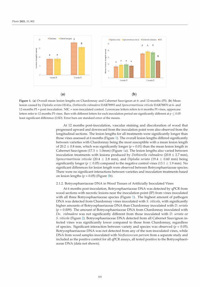
Plants 2021, 10, 802
Figure 1. (a) Overall mean lesion lengths on Chardonnay and Cabernet Sauvignon at 6- and 12-months (PI). (b) Meanlesion caused by Diplodia seriata H141a, Dothiorella vidmadera DAR78993 and Spencermartinsia viticola DAR78870 at 6- and12-months PI = post inoculation. NIC = non-inoculated control. Lowercase letters refers to 6 months PI vines, uppercaseletters refer to 12 months PI vines. Bars with different letters for each inoculation period are significantly different at p ≤ 0.05least significant difference (LSD). Error bars are standard error of the means.
At 12 months post-inoculation, vascular staining and discoloration of wood thatprogressed upward and downward from the inoculation point were also observed from thelongitudinal sections. The lesion lengths for all treatments were significantly longer thanthose vines assessed at 6 months (Figure 1). The overall lesion lengths differed significantlybetween varieties with Chardonnay being the most susceptible with a mean lesion lengthof 20.2 ± 0.8 mm, which was significantly longer (p = 0.01) than the mean lesion length inCabernet Sauvignon (17.3 ± 1.0mm) (Figure 1a). The lesion lengths also varied betweeninoculation treatments with lesions produced by Dothiorella vidmadera (20.8 ± 2.7 mm),Spencermartinsia viticola (20.4 ± 2.8 mm), and Diplodia seriata (19.4 ± 0.60 mm) beingsignificantly longer (p ≤ 0.05) compared to the negative control vines (13.1 ± 1.9 mm). Nosignificant differences for lesion length were observed between Botryosphaeriaceae species.There were no significant interactions between varieties and inoculation treatments basedon lesion lengths (p > 0.05) (Figure 1b).
2.1.2. Botryosphaeriaceae DNA in Wood Tissues of Artificially Inoculated Vines
At 6 months post-inoculation, Botryosphaeriaceae DNA was detected by qPCR fromwood sections with necrotic lesions near the inoculation point (IP) from vines inoculatedwith all three Botryosphaeriaceae species (Figure 1). The highest amount of pathogenDNA was detected from Chardonnay vines inoculated with S. viticola, with significantlyhigher amounts of Botryosphaeriaceae DNA than Chardonnay inoculated with D. seriata(p = 0.009). The amount of Botryosphaeriaceae DNA from Chardonnay inoculated withDo. vidmadera was not significantly different from those inoculated with D. seriata orS. viticola (Figure 2). Botryosphaeriaceae DNA detected from all Cabernet Sauvignon in-fected vines was significantly lower compared to those from Chardonnay, regardlessof species. Significant interaction between variety and species was observed (p < 0.05).Botryosphaeriaceae DNA was not detected from any of the non-inoculated vines, whileDNA from wood samples inoculated with Neofusicoccum parvum from a separate study andincluded as the positive control for all qPCR assays, all tested positive to the Botryosphaeri-aceae DNA (data not shown).
101
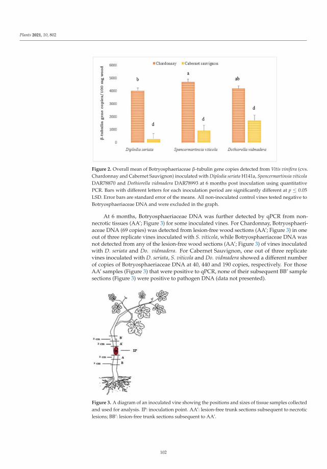
Plants 2021, 10, 802
Figure 2. Overall mean of Botryosphaeriaceae β-tubulin gene copies detected from Vitis vinifera (cvs.Chardonnay and Cabernet Sauvignon) inoculated with Diplodia seriata H141a, Spencermartinsia viticolaDAR78870 and Dothiorella vidmadera DAR78993 at 6 months post inoculation using quantitativePCR. Bars with different letters for each inoculation period are significantly different at p ≤ 0.05LSD. Error bars are standard error of the means. All non-inoculated control vines tested negative toBotryosphaeriaceae DNA and were excluded in the graph.
At 6 months, Botryosphaeriaceae DNA was further detected by qPCR from non-necrotic tissues (AA’; Figure 3) for some inoculated vines. For Chardonnay, Botryosphaeri-aceae DNA (69 copies) was detected from lesion-free wood sections (AA’; Figure 3) in oneout of three replicate vines inoculated with S. viticola, while Botryosphaeriaceae DNA wasnot detected from any of the lesion-free wood sections (AA’; Figure 3) of vines inoculatedwith D. seriata and Do. vidmadera. For Cabernet Sauvignon, one out of three replicatevines inoculated with D. seriata, S. viticola and Do. vidmadera showed a different numberof copies of Botryosphaeriaceae DNA at 40, 440 and 190 copies, respectively. For thoseAA’ samples (Figure 3) that were positive to qPCR, none of their subsequent BB’ samplesections (Figure 3) were positive to pathogen DNA (data not presented).
Figure 3. A diagram of an inoculated vine showing the positions and sizes of tissue samples collectedand used for analysis. IP: inoculation point. AA’: lesion-free trunk sections subsequent to necroticlesions; BB’: lesion-free trunk sections subsequent to AA’.
102
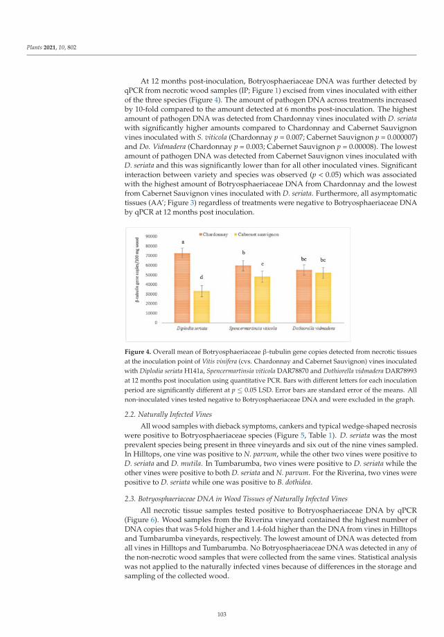
Plants 2021, 10, 802
At 12 months post-inoculation, Botryosphaeriaceae DNA was further detected byqPCR from necrotic wood samples (IP; Figure 1) excised from vines inoculated with eitherof the three species (Figure 4). The amount of pathogen DNA across treatments increasedby 10-fold compared to the amount detected at 6 months post-inoculation. The highestamount of pathogen DNA was detected from Chardonnay vines inoculated with D. seriatawith significantly higher amounts compared to Chardonnay and Cabernet Sauvignonvines inoculated with S. viticola (Chardonnay p = 0.007; Cabernet Sauvignon p = 0.000007)and Do. Vidmadera (Chardonnay p = 0.003; Cabernet Sauvignon p = 0.00008). The lowestamount of pathogen DNA was detected from Cabernet Sauvignon vines inoculated withD. seriata and this was significantly lower than for all other inoculated vines. Significantinteraction between variety and species was observed (p < 0.05) which was associatedwith the highest amount of Botryosphaeriaceae DNA from Chardonnay and the lowestfrom Cabernet Sauvignon vines inoculated with D. seriata. Furthermore, all asymptomatictissues (AA’; Figure 3) regardless of treatments were negative to Botryosphaeriaceae DNAby qPCR at 12 months post inoculation.
Figure 4. Overall mean of Botryosphaeriaceae β-tubulin gene copies detected from necrotic tissuesat the inoculation point of Vitis vinifera (cvs. Chardonnay and Cabernet Sauvignon) vines inoculatedwith Diplodia seriata H141a, Spencermartinsia viticola DAR78870 and Dothiorella vidmadera DAR78993at 12 months post inoculation using quantitative PCR. Bars with different letters for each inoculationperiod are significantly different at p ≤ 0.05 LSD. Error bars are standard error of the means. Allnon-inoculated vines tested negative to Botryosphaeriaceae DNA and were excluded in the graph.
2.2. Naturally Infected Vines
All wood samples with dieback symptoms, cankers and typical wedge-shaped necrosiswere positive to Botryosphaeriaceae species (Figure 5, Table 1). D. seriata was the mostprevalent species being present in three vineyards and six out of the nine vines sampled.In Hilltops, one vine was positive to N. parvum, while the other two vines were positive toD. seriata and D. mutila. In Tumbarumba, two vines were positive to D. seriata while theother vines were positive to both D. seriata and N. parvum. For the Riverina, two vines werepositive to D. seriata while one was positive to B. dothidea.
2.3. Botryosphaeriaceae DNA in Wood Tissues of Naturally Infected Vines
All necrotic tissue samples tested positive to Botryosphaeriaceae DNA by qPCR(Figure 6). Wood samples from the Riverina vineyard contained the highest number ofDNA copies that was 5-fold higher and 1.4-fold higher than the DNA from vines in Hilltopsand Tumbarumba vineyards, respectively. The lowest amount of DNA was detected fromall vines in Hilltops and Tumbarumba. No Botryosphaeriaceae DNA was detected in any ofthe non-necrotic wood samples that were collected from the same vines. Statistical analysiswas not applied to the naturally infected vines because of differences in the storage andsampling of the collected wood.
103
Plants 2021, 10, 802
Figure 5. (a) Vine with trunk canker (arrow); (b) cross-section of the trunk with centralnecrosis; (c) cross-section of the cordon with wedge-shape necrosis; (d) cross-section of the trunkwith wedge-shape necrosis.
Table 1. Botryosphaeriaceae species isolated from naturally infected vines from three vineyards inNew South Wales, Australia.
Location Variety Vine Sample Botryosphaeriaceae Species
Hilltops Chardonnay 1 Neofusicoccum parvum2 Diplodia seriata3 Diplodia mutila
Tumbarumba Chardonnay 1 D.seriata
2 D. seriata,N. parvum
3 D. seriata
Riverina Shiraz 1 D. seriata2 Botryosphaeria dothidea3 D. seriata
Figure 6. Number of copies of pathogen DNA quantified by qPCR in naturally infected vines fromthree vineyards in New South Wales, Australia. Error bars are standard deviation of the means.
104
Plants 2021, 10, 802
2.4. Selection of Protocol for Extraction of PMs from Wood
Two different protocols were tested for the extraction of PMs from naturally infectedwood material: (A) n-hexane/MeOH [34]; and (B) H2O/MeOH/CH3Cl [41]. Protocol Bwas less time consuming and resulted in a higher amount of organic compounds. Analysisof the organic extracts using LC-MS/MS to detect (R)-mellein also showed that protocolB yielded a higher amount of the target metabolite. The peak of (R)-mellein detected inthe extract obtained with Protocol B (Figure S2, red) was 12-fold higher than the organicextract obtained with protocol A (Figure S2, green). Identification of (R)-mellein fromextracts of symptomatic wood samples was accomplished according to its retention time,precursor ion 179.1 m/z [M + H]+ and fragment ions (Figure S2b). Protocol B was selectedfor extracting PMs from inoculated vines.
2.5. PMs in Wood Tissues of Naturally Infected Vines
Both symptomatic and asymptomatic wood materials were analysed by LC-MS/MSafter extraction, giving different results depending on the vines sampled and vineyardlocation. For symptomatic wood samples, only one out of three Chardonnay vines fromHilltops, two out of three Chardonnay vines from Tumbarumba and all Shiraz vines fromthe Riverina were positive to (R)-mellein. (R)-mellein was not detected in any of the lesion-free wood samples. A comparison of the chromatograms (Figure S3a) led to the detectionof a signal at a retention time of 27.44 min in the infected sample (Figure S3a, red, greenand purple), which was absent in the asymptomatic sample (Figure S3a, black). The signalin the chromatograms was due to the (R)-mellein fragment ions (Figure S3b). Furthermore,the area of (R)-mellein signals in the LC-MS/MS analysis was most significant for theRiverina vines with a high number of DNA copies of the pathogen detected in the trunk(Figure S3c), indicating production of (R)-mellein by BD pathogens in field vines.
2.6. PMs in Wood Tissues of Artificially Inoculated Vines
At six months post-inoculation, no spencertoxin, protocatechuic acid and (R)-melleinwere detected in any of the wood sections (IP, Figure 3) from non-inoculated vines. How-ever, (R)-mellein was detected from the extracts of necrotic wood samples (IP) of bothChardonnay and Cabernet Sauvignon vines infected with D. seriata at 12 months post-inoculation (Figure S4, red) indicating that this pathogen produced (R)-mellein duringfungal colonization. Furthermore, neither spencertoxin nor protocatechuic acid weredetected from necrotic wood samples (IP) infected with S. viticola and Do. vidmadera, re-spectively, at 12 months post-inoculation. (R)-mellein was further detected from extractsobtained from the Chardonnay vine infected with N. parvum (DAR78998) from a separateexperiment and included in the test as a positive control (Figure S4, green). The peak of(R)-mellein detected from this vine was 2.4-fold higher than the amount detected in vinesinfected with D. seriata 12 months post-inoculation. No target PMs were detected in thenegative control vines. Targeted PMs were also not detected in lesion-free wood samples(AA’ and BB’, Figure 3), thus, no further analyses were performed for tissues collectedfurther away from the necrotic wood (IP) including the leaves.
3. Discussion
To the best of our knowledge, this is the first study to investigate the productionand translocation of PMs by Botryosphaeriaceae species in BD naturally infected andartificially inoculated vines using a multifaceted approach. However, multidisciplinaryapproaches for the detection of target PMs in plant tissues have been reported in otherpathosystems [42–45].
This current study also represents the first study to use a combination of conventionalplant pathology and molecular techniques to detect and quantify BotryosphaeriaceaeDNA from artificially inoculated and naturally infected vines. The qPCR primers andprobe used in this study [46] are not species-specific, therefore they cannot distinguish theBotryosphaeriaceae pathogen at the species level. However, the isolation of pathogens from
105
Plants 2021, 10, 802
naturally infected wood allowed the identification of Botryosphaeriaceae spp. Similarly,re-isolations of the pathogens from artificially inoculated vines resulted in the recovery ofD. seriata, S. viticola and Do. vidmadera from previously inoculated vines. Lesion lengthby itself was insufficient to show differences in disease severity and pathogen virulenceat the early stage of infection (6 months post-inoculation). Furthermore, negative controlvines also exhibited necrosis near the inoculation point similar to those vines inoculatedwith pathogens. The qPCR analysis, on the other hand, was able to quantify the amountof pathogen DNA between treatments and assessment periods. Consequently, pathogenDNA was not detected in negative control vines. It is important to note that low levels ofpathogen DNA were detected from lesion-free tissues adjacent to the lesions from a fewinoculated vines at 6-month post-inoculation, particularly for S. viticola. This suggests thatthe pathogens were able to move endophytically beyond the lesions as latent pathogen,similar to the study reported by Billones-Baaijens et al. [47]. The absence of pathogen DNAin non-necrotic tissues adjacent to the lesions at 12 months post inoculation suggests thatas the infection advanced, these pathogens shifted from being latent to necrotrophic.
The qPCR analysis of inoculated vines showed a significant difference in susceptibilitybetween Chardonnay and Cabernet Sauvignon to different Botryosphaeriaceae species,with the former being more susceptible to D. seriata. The reduced susceptibility of CabernetSauvignon to some Botryosphaeriaceae species may be associated with the amount ofstilbene polyphenols that are usually higher in red vine varieties. These compounds havefungistatic activity [48,49], that may assist the plant in limiting infection by BD pathogens.Nevertheless, stilbene polyphenols antimicrobial activity depends on the pathogen infect-ing the plant [48,49]. For instance, Cabernet Sauvignon is one of the most susceptible culti-vars to Esca pathogens: Phaeomoniella chlamydospora and Phaeoacremonium minimum [10].
The qPCR analysis used in this study was also useful in quantifying pathogen DNAin symptomatic wood samples collected from naturally infected vines. Overall, theseresults confirm that molecular techniques could be applied to determine and quantify thepathogens in field material.
The LC-MS/MS analysis of the naturally infected and artificially inoculated vinesshowed that (R)-mellein can be detected in infected woody tissues with symptoms ofBD. A previous study that investigated the production of Botryosphaeriaceae PMs inplanta only used a limited number of naturally infected vines with both BD and Escasymptoms [34]. More recently (R)-mellein was also detected in grapevine tissues withsymptoms of Esca and Grapevine leaf stripe disease (GLSD) [50]. However, none of theprevious studies identified the pathogens or quantified the pathogen DNA in the analysedsamples. Furthermore, the application of a multifaceted approach suggests a probablecorrelation between the amount of pathogen DNA in the wood and the area of the peak ofthe (R)-mellein in the chromatograms. This correlation was more evident in the Riverinavines, which resulted in more intense peaks corresponding to (R)-mellein and the highestamount of pathogen DNA in the wood. Moreover, the amount of (R)-mellein detected in thewood infected with N. parvum, an aggressive Botryosphaeriaceae pathogen [4], 6 monthspost-inoculation was 2.4-fold higher than the amount detected in vines infected with thelesser aggressive D. seriata at 12 months post-inoculation. These data further support thosepreviously reported that the amount of (R)-mellein produced by N. parvum and D. seriataunder in vitro conditions is proportional to the aggressiveness of the pathogens [51].
BD pathogens colonize the woody parts of the plants, and during colonization, thefungi can produce and release a series of PMs. The translocation hypothesis suggests thatPMs produced by the pathogens can migrate far from the inoculum along the asymptomaticwood, reaching the leaves or the green shoots without being catabolized or without beingentirely detoxified [26]. The main goal of our experiment was to verify the movementof targeted PMs along the trunk. For this purpose, symptomatic and adjacent lesion-freefunctional woody tissues (trunk and branches) were sampled. To ensure that the detectionof the target PMs in asymptomatic wood was due to their migration, qPCR analysis wasperformed. Our results showed that (R)-mellein was detected only in the wood samples
106
Plants 2021, 10, 802
with necrotic lesions showing a high amount of pathogen DNA. The failure to detect (R)-mellein in all the lesion-free wood samples suggests that, at least under these experimentalconditions, the translocation of (R)-mellein in its native form did not occur along the trunkto the foliage. It is possible that D. seriata produces (R)-mellein as part of its strategy tobreakdown the host cells during infection. In a recent study using in vitro plantlets, Trotel-Aziz et al. showed that (R)-mellein strongly suppresses the expression of genes involved inplant defence and that (R)-mellein may be accumulated in planta in its native chemical form.The detection of this compound in symptomatic wood during our experiment supportsthis hypothesis [52].
However, the mere presence of PMs in the wood may not be enough to induce foliarsymptoms in grapevines with BD. The development of symptoms may be more compli-cated than previously thought, and they may arise from interactions between biotic andabiotic stresses (water stress, drought, heat stress), which require more in-depth studiesto understand. Fungi involved in GTDs can act as endophytes for several years beforebecoming pathogens, and many have hypothesised that the abiotic conditions, in particularthermal and water stress, can weaken the plant defence and therefore result in the develop-ment of GTD foliar symptom [9,10,22,53–56]. For instance, previous investigation, showedthat GLSD leaf symptoms increased when the rainfall was abundant in June–July [54].
More comprehensive field studies on the influence of climatic conditions on foliarsymptoms associated with GTD, including the role of PMs produced in planta, are funda-mental for elucidating the relationship between fungal PMs and physiological changes inthe vine which result in the expression of foliar symptoms.
Another result arising from this study was the failure to detect protocatechuic acidand spencertoxin in the LC-MS/MS analysis of vines inoculated with D. vidmadera andS. viticola, respectively. These two species were shown to produce these PMs in vitro [35,39].The lack of detection of these PMs may be due to various reasons: (i) they may not beproduced in planta; (ii) they could be detoxified by the plant; (iii) they may form toxinderivatives with other compounds; and/or (iv) they may be irreversibly bound to thewood contributing to lesion expression. The latter hypothesis was already suggested forother phytotoxins produced by the ED pathogen, E. lata [27–29]. Overall, the generalconclusion may be that not all the secondary metabolites produced in vitro can be detectedin planta since their fate mainly depends on the biological role played in the interactionof the pathogen with the host. This can be further validated by applying our approach todifferent experimental conditions. For instance, plant materials with reported BD foliarsymptoms could be analyzed to further investigate the role of these PMs in foliar symptomdevelopment. Comparing data obtained from various vine-growing regions can be valuableto validate our results and help to explain the lack of BD foliar symptoms observed inAustralian vineyards.
PMs are also known to interact with different cellular targets or can inhibit the activityof plant enzymes [57]. All these processes can result in the formation of toxin conjugatesor derivatives that could explain the failure to detect target PMs in their native form.Metabolomics approaches can be applied to investigate the formation of PM derivativesand their role in symptoms development or plant-pathogen interaction. Indeed, untargeted,and targeted metabolomics have become fundamental tools in plant science and chemicalecology [58,59]. Recently, metabolic changes in grapevine wood infected with N. parvumhave been reported [60]. This was accompanied by an accumulation of a number ofunknown metabolites in the infected wood samples. Their structural elucidation will becrucial to better understand the response of the plant to GTDs and to also identify if any ofthese unknown metabolites could be potential derivatives of PMs.
107
Plants 2021, 10, 802
4. Materials and Methods
4.1. Artificially Inoculated Vines4.1.1. Planting Materials
Vitis vinifera cvs. Chardonnay and Cabernet Sauvignon, two of the most commonlygrown varieties in Australian vineyards [61] were selected for a glasshouse experiment.Twenty- eight dormant, apparently healthy cuttings for each variety were collected in acommercial vineyard in Hilltops, New South Wales (NSW), Australia in Winter (June 2017)and stored at 4 ◦C for 4 weeks until rooting. All cuttings were surface-sterilised with 0.5%sodium hypochlorite for 1 min, rinsed twice with tap water and rooted in plastic trayscontaining perlite. The trays were placed on heat mats at 30 ◦C for 4 weeks to facilitaterooting. The rootlings were planted in 10 L pots containing commercial garden mix (60%compost, 20% wash sand, 20% screen loam). All vines were maintained in a glasshouse(17–27 ◦C) and watered every 12 h for 5 min (8 L/h) with an automatic dripper system for6–12 months until assessment.
4.1.2. Fungal Isolates
The three Botryosphaeriaceae species: (a) Diplodia seriata H141a; (b) Spencermartinsia viticolaDAR78870 and; (c) Dothiorella vidmadera DAR78993 from the National Wine and Grape In-dustry Centre (Charles Sturt University, Wagga Wagga, NSW, Australia) culture collection,which produced PMs in vitro and were previously characterised [33,34,37], were used forinoculating the glasshouse vines in Spring (November 2017). For inoculations, woundswere created in the middle internode of the trunk for each vine using a flame-sterilised4 mm sterile cork borer. Mycelial plugs (4 mm) cut from the margins of 4- day-old culturesof the selected fungal species grown on potato dextrose agar supplemented with Chloram-phenicol (100 mg/L) (PDA-C), were inserted into the wounds and sealed with Parafilm(Bemis, USA). Sterile non-colonised plugs of PDA-C were used as negative controls. Theinoculated vines for each variety were arranged in a randomised complete block design(RCBD) at 7 replications per inoculum per variety combination.
4.1.3. Sampling of Artificially Inoculated Vines
The trunks of randomly selected vines were cut at the base and shoots were trimmedoff. The bark surrounding the inoculation point was removed and lesions were measuredusing a digital caliper (Workzone, Australia). Tissue samples from each vine were collectedas shown in Figure 3. Three different samples were collected from each vine: (a) trunksections with visible lesions including the inoculation point (IP); (b) 2 cm lesion-free trunksections cut above and below the necrotic lesions (labelled as AA’); (c) 2 cm trunk sectionsabove and below section A and A’ (labelled as BB’). The samples were surface-sterilisedfor 2 min in 70% ethanol and rinsed three times with sterile deionised water (SDW) beforecutting longitudinally to obtain four quarters of each section. One quarter of the sectionwas used for isolation of the pathogen, while the remaining sections were stored at −80 ◦Cand used for DNA and toxin extractions.
4.1.4. Fungal Isolation from Artificially Inoculated Vines
One quarter of surface-sterilised trunk sections collected from the inoculated vines(Figure 1) were placed onto PDA-C. Plates were incubated at 25 ◦C in the dark and observedfor growth of Botryosphaeriaceae species for 4–7 days. The re-isolated pathogens wereidentified using morphological and molecular methods. Three vines per treatment wereassessed at 6 months post inoculation in Autumn (May 2018). Four vines per treatment werefurther assessed at 12 months post inoculation which occurred in Spring (November 2018).
4.2. Fungal Isolation from Naturally Infected Vines
Wood samples (cordons and trunks) exhibiting BD cankers were collected from20–24 years old grapevines from three wine regions in NSW, Australia in Winter (June–August 2017). The Tumbarumba region has a cool climate with an altitude of 700 m.
108
Plants 2021, 10, 802
The Hilltops region has a continental climate with an altitude of 450 m. The Riverinaregion has a semiarid climate with an altitude of 66–540 m [62]. Four to five woodpieces (10–20 cm) with necrotic lesions and non-necrotic tissues 20–30 cm away fromthe necrotic ones were cut from three different vines in Hilltops (cv. Shiraz), Riverina(cv. Shiraz) and Tumbarumba (cv. Chardonnay) and stored at 4 ◦C for 2 weeks until pro-cessed. Each sample were processed and analysed individually. For isolations, the barkwas removed to expose the necrotic lesions and the wood was cut into ~1 cm sections(20–24 pcs) with each section containing necrotic and healthy wood. All sections weresurface-sterilised following the methods described for the inoculated vines. Approximately10–12 pieces of the surface-sterilised tissues were stored at −80 ◦C for DNA and toxinextractions. The remaining sections were plated onto PDA-C with four sections per platefor a total of three plates per vine. Plates were incubated at 25 ◦C for 4–7 days and observedfor growth of Botryosphaeriaceae species. All Botryosphaeriaceae isolates were identifiedby morphological and molecular methods.
4.3. DNA Extractions from Fungal Mycelia
DNA samples were extracted from the mycelium for all the isolated fungi usingPrepMan Ultra (Applied Biosystems, UK) and following the manufacturer’s instructions.Mycelium (~100 mg) was scraped from the edge of the colony for each isolate using a sterilepipette tip and transferred into a sterile 1.5 mL tube containing 100 μL of the PrepManUltra (Applied Biosystems, UK) preparation reagent. Mycelial suspensions were vortexedfor 30 s and incubated at 95 ◦C in a heat block for 10 min. The tubes were centrifuged for2 min at 3220× g and 50 μL of the supernatant was transferred to a new sterile 1.5 mL tubeand stored at −20 ◦C until required for PCR.
4.4. DNA Extraction from Grapevine Wood
Wood samples stored at −80 ◦C were freeze-dried (Christ, John Morris Scientific,USA) for 24–36 h. Dried samples were homogenized at 20 Hz for 2 min 30 s using 10 mLgrinding jars attached to a TissueLyser II (Qiagen, Hilden, Germany). The ground wood(100 mg) was transferred into a sterile 2 mL tube for DNA extraction while the remainingground wood was stored in a separate tube for the extraction of toxins. DNA was extractedfrom wood samples using the methods described by Pouzoulet et al. [63] with somemodifications. The CTAB (Cetyl Trimethyl Ammonium Bromide) extraction buffer wasprepared according to Doyle and Doyle [64], and 1 ml was added to each tube containing100 mg of ground wood and gently mixed by pipetting. The mixture was incubated at 65 ◦Cfor 1 h using a heat block. After incubation, 500 μL of chloroform/isoamyl alcohol (24:1,Sigma Aldrich, St. Louis, MI, USA) was added and the tube was inverted 10×, incubatedon ice for 5 min and then centrifuged at 4 ◦C for 10 min at 2300× g. Approximately 420 μLof the lysate was pipetted into a QIAshredder spin column placed in a 2 mL collectiontube from the Qiagen DNeasy Plant DNA extraction kit (Qiagen, Hilden, Germany) andcentrifuged for 2 min at 20,000× g. The subsequent steps were performed using the buffers,materials and protocol from the DNeasy Plant DNA extraction kit. All DNA samples wereeluted to a final volume of 100 μL using the Qiagen AE buffer. All DNA samples werequantified using a Quantus™ Fluorometer (Promega, Madison, WI, USA) prior to qPCR.
DNA was further extracted from the necrotic tissues collected from potted vines (cv.Chardonnay) inoculated six months prior with N. parvum DAR78998 following the methodsdescribed above. This isolate was found to be highly virulent in a separate experiment [18]and is known to produce (R)-mellein in vitro [35]. The DNA extracted from these vineswas included in the qPCR analysis and toxin analyses.
4.5. Identification of Isolated Botryosphaeriaceae by PCR
All Botryosphaeriaceae recovered from the artificially inoculated and naturally in-fected vines were identified using PCR and DNA sequencing. To amplify the internaltranscribed spacer (ITS) region of the ribosomal DNA of the pathogens, PCR was per-
109
Plants 2021, 10, 802
formed using universal primers ITS1 and ITS4 [65]. Each 25 μL PCR reaction contained 1xPCR buffer (Bioline, Memphis, TN, USA), 0.4 μL for each of primer, 1.25 U of My TaqRedDNA polymerase (Bioline, Memphis, USA) and approximately 1–5 ng of DNA template.PCRs were performed using a thermal cycler (C100 Thermal cycler, Biorad Laboratories,Pty, Ltd., Hercules, CA, USA) under the following conditions: initial denaturation at95 ◦C for 5 min, 35 cycles of 30 s at 94 ◦C, 45 s at 55 ◦C, and 90 s at 72 ◦C, with a finalextension of 5 min at 72 ◦C. Following amplification, the PCR products were visualizedby gel electrophoresis. PCR products were purified with FavorPrep Gel/PCR purificationkit (Favorgen Biotech Corp, Taiwan) and sequenced at the Australian Genome ResearchFacility (AGRF; Sydney, NSW, Australia). All DNA sequences and chromatographs wereanalyzed using the DNAMAN 5.2 (Lynnon Biosoft©, San Ramon, CA, USA) and ChromasLite 2.1© (Technelysium PTY Ltd, Brisbane, Australia) software. All trimmed DNA se-quences were analysed using the Basic Local Alignment Search Tool (BLAST) in GenBank(https://www.ncbi.nlm.nih.gov/genbank/ (accessed on 18 April 2021)).
4.6. Quantification of Botryosphaeriaceae spp. from Wood Samples by qPCR
The qPCR assay using Botryosphaeriaceae multi-species primers and hydrolysis probedeveloped by Billones-Baaijens et al. [46] were used to detect and quantify Botryosphaeri-aceae spp. from artificially inoculated and naturally infected vines. All qPCR assays wereperformed with the RotorGene 6000 system (Corbett Life Science, Qiagen, Hilden, Germany)using Botryosphaeriaceae multi-species primers Bot-BtF1 (5’-GTATGGCAATCTTCTGAACG-3’) and Bot-BtR1 (5’-CAGTTGTTACCGGCRCCRGA-3’), and a hydrolysis probe, Taq-Botprobe 5’-/56-FAM/TCGAGCCCG/ZEN/GCACSATGGAT/3IBkFQ/-3’) [41]. For eachassay, three controls were included: (1) non-template control (H2O); (2) standard (500 pg)Bot-Btub gBlock [41]; (3) DNA from vine inoculated with N. parvum DAR78998 at fourtechnical replicates each.
For the artificially inoculated vines, all necrotic tissue samples collected from the IPwere first analysed followed by the tissues collected above and below the necrotic lesions(AA’). For AA’ tissues which tested positive to Botryosphaeriaceae DNA, their subsequentBB samples were further analysed for a total of 32 and 36 samples for 6- and 12- monthsPI, respectively. For naturally infected vines, wood samples containing necrotic tissueswere first analysed by qPCR. When the necrotic tissue sample was positive to qPCR, thehealthy wood samples away from the necrotic tissue were further analysed for a total of18 samples overall.
To determine the amount of pathogen DNA that was amplified by each qPCR assay,previously developed standard curves [46] were imported in the Rotor-Gene Q software(Version 2.3.1). The standard (Bot-Btub gBlock, 500 pg) that was included in each qPCRassay was used to calibrate the imported standard curve and the resulting regressionequations were used to quantify the number of Botryosphaeriaceae β-tubulin gene copiesin each reaction as previously described by Billones-Baaijens et al. [46] following the MIQEguidelines [66]. To calculate the number of copies of the Botryosphaeriaceae β-tubulingene in each wood sample, the following formula was used:
N = g (d × c)/t × c, (1)
where N: calculated number of β-tubulin gene copies in one wood sample, g: the meannumber of gene copies detected by qPCR; d: total gDNA extracted from 100 mg of wood(100 μL), c: DNA concentration (μL); T: the amount of DNA template (5 μL) in one reaction.
4.7. Statistical Analysis
Data arising from the glasshouse experiment were analysed using IBM SPSS 24software. All data were tested for homogeneity using Levene’s test at p ≤ 0.05. Forinoculated vines, univariate analysis of variance (ANOVA) was used to assess differencesin lesion lengths and pathogen copies between varieties, inoculated pathogens and theirinteractions (p ≤ 0.05). All means were separated by pairwise comparison using Fischer’s
110
Plants 2021, 10, 802
least significant differences (LSD) test at 5% significance level. Statistical analysis was notapplied to the naturally infected vines because of differences in the storage and samplingof the collected wood.
4.8. Chemicals and Standards for LC-MS/MS
CH3Cl, MeOH and n-hexane were analytical grade (Sigma-Aldrich, St. Louis, USA).H2O, 0.1% HCOOH and MeCN were LC-MS grade (Sigma-Aldrich, St. Louis, USA).The (R)-mellein [35], protocatechuic alcohol [36] and spencertoxin [39] that were used asstandards were isolated from in vitro cultures of D. seriata, Do. vidmadera and S. viticola aspreviously reported.
4.9. Testing of Protocols for Extraction of PMs from Wood
Two published extraction protocols were compared for their suitability to extracttoxins from wood samples [34,41]. Freeze-dried wood samples collected from the naturallyinfected vines (36 in total) were extracted using both protocols and were analysed byLC-MS/MS. Freeze-dried wood samples (100 mg) were used for each extraction. Theprotocol by Saviano et al. [41] was subsequently used to extract PMs from the woodsamples (100 mg) collected from inoculated vines (100 in total).
4.10. LC-MS/MS Analysis of Targeted PMs from Wood
Analyses were carried out using a 1290 Infinity II LC system (Agilent) hyphenatedto an Agilent 6470 triple quadrupole (QqQ). The UPLC system included a binary pumpand a cooled autosampler maintained at 15 ◦C. Mass Hunter software was used to controlthe instruments and to acquire the data which were then processed for analysis. The chro-matographic separation was performed using a reverse phase column Phenomenex Luna5.4 μm 250 × 4 mm i.d., protected by a security guard column Phenomenex maintainedat 30 ◦C. The mobile phase consisted of H2O 1% (v/v) HCOOH (Phase A) and MeCN(Phase B). The flow rate was 0.7 mL/min, the gradient system was initiated with 10% ofPhase B for 2 min and reached 30% at 15 min, 80% at 25 min, isocratic until 27 min and 95%at 40 min. Samples were injected into the column with an injection volume of 20 μL.
The Agilent 6470 triple quadrupole (QqQ) was used as the detector in MRM modewith electrospray ionization (ESI) in positive ionization mode. The source and desolvationtemperatures were respectively set at 350 ◦C, Nebulizer, 40 psi; N2 flow, 12 L min−1.Capillary voltage was set at 3.5 kV in positive mode. The MRM transitions (precursor ion→ daughter ions), fragmentor energy and collision energy for (R)-mellein, spencertoxinand protocatechuic acid were optimized using the Agilent Optimizer Software, and theoptimized parameters were shown in Table 2. For the parameter optimization, standardsof (R)-mellein, spencertoxin and protocatechuic acid were used.
Table 2. Toxins used as standards for the LC-MS/MS and their corresponding optimized parameters.
ToxinPrecursor Ion
m/zFragment Ion
m/zFragmentor
Voltage *CV **
Retention Time(Min)
(R)-mellein
179.1 [M + H]+161.0133.0105.0
90121624
27.44
111
Plants 2021, 10, 802
Table 2. Cont.
ToxinPrecursor Ion
m/zFragment Ion
m/zFragmentor
Voltage *CV **
Retention Time(Min)
Protocatechuicalcohol
123.1[M-H2O + H]+
67.155.151.1
90162440
6.94
Spencertoxin
283.1 [M + K]+ 177.8118 90 44
50 18.11
* Fragmentor voltage: controls the speed at which the ions pass through a medium pressure capillary between the electrospray chamberand the mass spectrometer. ** CV: collision energy voltage.
5. Conclusions
Detection of PMs in infected plant tissue may provide insights into the involvementof PMs in the pathogenicity of fungal isolates and symptom development of GTDs. Thismultidisciplinary approach was appropriate for investigating the production and translo-cation of (R)-mellein in vines showing BD symptoms. Our results highlighted a possiblecorrelation between the amount of (R)-mellein and the amount of pathogen DNA copies inthe wood samples. The development of a robust quantitative LC-MS/MS method for thedetection of (R)-mellein can assist further investigations regarding this correlation. Thesedata may provide valuable information on the infection strategies of the pathogens andmay clarify the role played by (R)-mellein in the development of the disease.
Our study did not find any evidence that PMs are translocated into healthy tissues ofthe vine. Thus, the targeted PMs we investigated in this study were most likely producedby BD pathogens for penetrating and invading the hosts. The migration of PMs in theirnative form into the woody tissues of the plant should not be considered a generalisedprocess for all pathosystems.
Supplementary Materials: The following are available online at https://www.mdpi.com/article/10.3390/plants10040802/s1, Figure S1: Representative photos of the inoculated vines showing internalstaining of the wood, Figure S2: Representative LC-MS/MS chromatograms of naturally infectedwood extract, Figure S3: LC-MS/MS analysis of Riverina vines, Figure S4: MRM Chromatograms ofChardonnay vine inoculated with D. seriata.
Author Contributions: Conceptualisation, S.S., R.B.-B., P.R. and A.E.; methodology, P.R., S.S. andR.B.-B.; validation, S.S. and R.B.-B.; formal analysis, S.S. and R.B.-B.; investigation, P.R. and J.M.N.;resources, S.S., R.B.-B. and A.E.; writing—original draft preparation, P.R.; writing—review andediting, P.R., R.B.-B., J.M.N., M.M., A.C., A.E. and S.S.; visualization, P.R., S.S. and R.B.-B.; supervision,S.S., R.B.-B. and A.E.; project administration, S.S., R.B.-B. and A.E.; funding acquisition, S.S. and A.E.All authors have read and agreed to the published version of the manuscript.
Funding: This work was supported by academic grants from the National Wine Grape IndustryCentre, Charles Sturt University, Australia; Dipartimento di Scienze Chimiche, Università di NapoliFederico II, Italy and a Wine Australia PhD Student Support Grant.
Institutional Review Board Statement: Not applicable.
112
Plants 2021, 10, 802
Informed Consent Statement: Not applicable.
Data Availability Statement: The data presented in this study are contained within the article andSupplementary Material.
Conflicts of Interest: The authors declare no conflict of interest.
References
1. Popa, E.O.; Rosca, I. Main trends of the pests management in agroecosystems of grapevine plantations. Sci. Pap. Ser. Manag. Econ.Eng. Agric. Rural. Dev. 2011, 11, 146–150.
2. Halleen, F.; Fourie, P.H.; Crous, P.W. A review of black foot disease of grapevine. Phytopathol. Med. 2006, 45, 55–67.3. Gessler, C.; Pertot, I.; Perazzolli, M. Plasmopara viticola: A review of knowledge on downy mildew of grapevine and effective
disease management. Phytopathol. Med. 2011, 50, 3–44.4. Úrbez-Torres, J.R. The status of Botryosphaeriaceae species infecting grapevines. Phytopathol. Med. 2011, 50, 5–45.5. Fontaine, F.; Pinto, C.; Vallet, J.; Clément, C.; Gomes, A.C.; Spagnolo, A. The effects of grapevine trunk diseases (GTDs) on vine
physiology. Eur. J. Plant Pathol. 2016, 144, 707–721. [CrossRef]6. Mondello, V.; Songy, A.; Battiston, E.; Pinto, C.; Coppin, C.; Trotel-Aziz, P.; Clément, C.; Mugnai, L.; Fontaine, F. Grapevine trunk
diseases: A review of fifteen years of trials for their control with chemicals and biocontrol agents. Plant Dis. 2018, 102, 1189–1217.[CrossRef]
7. Baránek, M.; Armengol, J.; Holleinová, V.; Pecenka, J.; Calzarano, F.; Penázová, E.; Vachun, M.; Eichmeier, A. Incidence ofsymptoms and fungal pathogens associated with grapevine trunk diseases in Czech vineyards: First example from a north-easternEuropean grape-growing region. Phytopathol. Mediterr. 2018, 57, 449–458.
8. Calzarano, F.; D’Agostino, V.; Pepe, A.; Osti, F.; Della Pelle, F.; De Rosso, M.; Flamini, R.; Di Marco, S. Patterns of phytoalexins inthe grapevine leaf stripe disease (esca complex)/grapevine pathosystem. Phytopathol. Mediterr. 2016, 55, 410–426.
9. Calzarano, F.; Di Marco, S. Further evidence that calcium, magnesium and seaweed mixtures reduce grapevine leaf stripesymptoms and increase grape yield. Phytopathol. Mediterr. 2018, 57, 459–471.
10. Claverie, M.; Notaro, M.; Fontaine, F.; Wéry, J. Current knowledge on grapevine trunk diseases with complex etiology: A systemicapproach. Phytopathol. Med. 2020, 59, 29–53.
11. Hrycan, J.; Hart, M.; Bowen, P.; Forge, T.; Úrbez-Torres, J.R. Grapevine trunk disease fungi: Their roles as latent pathogens andstress factors that favour disease development and symptom expression. Phytopathol. Med. 2020, 59, 395–424.
12. Bruez, E.; Vallance, J.; Gautier, A.; Laval, V.; Compant, S.; Maurer, W.; Sessitsch, A.; Lebrun, M.H.; Rey, P. Major changes ingrapevine wood microbiota are associated with the onset of esca, a devastating trunk disease. Environ. Microbiol. 2020, 22,5189–5206. [CrossRef]
13. Sosnowski, M.R.; Ayres, M.; Scott, E. The influence of water deficit stress on the grapevine trunk disease pathogens Eutypa lataand Diplodia seriata. Plant Dis. 2020. [CrossRef]
14. Bertsch, C.; Ramírez-Suero, M.; Magnin-Robert, M.; Larignon, P.; Chong, J.; Abou-Mansour, E.; Spagnolo, A.; Clément, C.;Fontaine, F. Grapevine trunk diseases: Complex and still poorly understood. Plant Pathol. 2013, 62, 243–265. [CrossRef]
15. Billones-Baaijens, R.; Savocchia, S. A review of Botryosphaeriaceae species associated with grapevine trunk diseases in Australiaand New Zealand. Austral. Plant Pathol. 2019, 48, 3–18. [CrossRef]
16. Guerin-Dubrana, L.; Fontaine, F.; Mugnai, L. Grapevine trunk disease in European and Mediterranean vineyards: Occurrence,distribution and associated disease-affecting cultural factors. Phytopathol. Med. 2019, 58, 49–71.
17. Larignon, P.; Dubos, B.; Cere, L.; Fulchic, R. Observation on black dead arm in French vineyards. Phytopathol. Med. 2001, 40,336–342.
18. Pitt, W.M.; Huang, R.; Steel, C.; Savocchia, S. Pathogenicity and epidemiology of Botryosphaeriaceae species isolated fromgrapevines in Australia. Austral. Plant Pathol. 2013, 42, 573–582. [CrossRef]
19. Carter, M. The Status of Eutypa lata as a Pathogen. In Monograph–Phytopathological Paper No. 32; International MycologicalInstitute: Surrey, UK, 1991.
20. Mugnai, L.; Graniti, A.; Surico, G. Esca (black measles) and brown wood-streaking: Two old and elusive diseases of grapevines.Plant Dis. 1999, 83, 404–418. [CrossRef]
21. Surico, G.; Mugnai, L.; Marchi, G. The esca disease complex. In Integrated Management of Diseases Caused by Fungi, Phytoplasma andBacteria; Springer: Berlin/Heidelberg, Germany, 2008; pp. 119–136.
22. Sosnowski, M.; Shtienberg, D.; Creaser, M.; Wicks, T.; Lardner, R.; Scott, E. The influence of climate on foliar symptoms of Eutypadieback in grapevines. Phytopathology 2007, 97, 1284–1289. [CrossRef]
23. Reis, P.; Magnin-Robert, M.; Nascimento, T.; Spagnolo, A.; Abou-Mansour, E.; Fioretti, C.; Clement, C.; Rego, C.; Fontaine, F.Reproducing Botryosphaeria dieback foliar symptoms in a simple model system. Plant Dis. 2016, 100, 1071–1079. [CrossRef][PubMed]
24. Larignon, P.; Dubos, B. The villainy of black dead arm. Wines Vines 2001, 82, 86–89.25. Masi, M.; Cimmino, A.; Reveglia, P.; Mugnai, L.; Surico, G.; Evidente, A. Advances on fungal phytotoxins and their role in
grapevine trunk diseases. J. Agric. Food Chem. 2018, 66, 5948–5958. [CrossRef]
113
Plants 2021, 10, 802
26. Tey-Rulh, P.; Philippe, I.; Renaud, J.-M.; Tsoupras, G.; de Angelis, P.; Fallot, J.; Tabacchi, R. Eutypine, a phytotoxin produced byEutypa lata the causal agent of dying-arm disease of grapevine. Phytochemistry 1991, 30, 471–473. [CrossRef]
27. Mahoney, N.; Molyneux, R.J.; Smith, L.R.; Schoch, T.K.; Rolshausen, P.E.; Gubler, W.D. Dying-arm disease in grapevines: Diagnosisof infection with Eutypa lata by metabolite analysis. J. Agric. Food Chem. 2005, 53, 8148–8155. [CrossRef] [PubMed]
28. Lardner, R.; Mahoney, N.; Zanker, T.P.; Molyneux, R.J.; Scott, E.S. Secondary metabolite production by the fungal pathogen Eutypalata: Analysis of extracts from grapevine cultures and detection of those metabolites in planta. Aust. J. Grape Wine Res. 2006, 12,107. [CrossRef]
29. Rolshausen, P.; Greve, L.; Labavitch, J.; Mahoney, N.; Molyneux, R.; Gubler, W. Pathogenesis of Eutypa lata in grapevine:Identification of virulence factors and biochemical characterization of cordon dieback. Phytopathology 2008, 98, 222–229. [CrossRef][PubMed]
30. Andolfi, A.; Mugnai, L.; Luque, J.; Surico, G.; Cimmino, A.; Evidente, A. Phytotoxins produced by fungi associated with grapevinetrunk diseases. Toxins 2011, 3, 1569–1605. [CrossRef]
31. Martos, S.; Andolfi, A.; Luque, J.; Mugnai, L.; Surico, G.; Evidente, A. Production of phytotoxic metabolites by five species ofBotryosphaeriaceae causing decline on grapevines, with special interest in the species Neofusicoccum luteum and N. parvum. Eur. J.Plant Pathol. 2008, 121, 451–461. [CrossRef]
32. Djoukeng, J.D.; Polli, S.; Larignon, P.; Abou-Mansour, E. Identification of phytotoxins from Botryosphaeria obtusa, a pathogen ofblack dead arm disease of grapevine. Eur. J. Plant Pathol. 2009, 124, 303–308. [CrossRef]
33. Evidente, A.; Punzo, B.; Andolfi, A.; Cimmino, A.; Melck, D.; Luque, J. Lipophilic phytotoxins produced by Neofusicoccum parvum,a grapevine canker agent. Phytopathol. Med. 2010, 49, 74–79.
34. Abou-Mansour, E.; Débieux, J.-L.; Ramírez-Suero, M.; Bénard-Gellon, M.; Magnin-Robert, M.; Spagnolo, A.; Chong, J.; Farine, S.;Bertsch, C.; L’Haridon, F. Phytotoxic metabolites from Neofusicoccum parvum, a pathogen of Botryosphaeria dieback of grapevine.Phytochemistry 2015, 115, 207–215. [CrossRef]
35. Reveglia, P.; Savocchia, S.; Billones-Baaijens, R.; Masi, M.; Cimmino, A.; Evidente, A. Phytotoxic metabolites by nine species ofBotryosphaeriaceae involved in grapevine dieback in Australia and identification of those produced by Diplodia mutila, Diplodiaseriata, Neofusicoccum australe and Neofusicoccum luteum. Nat. Prod. Res. 2019, 33, 2223–2229. [CrossRef]
36. Reveglia, P.; Savocchia, S.; Billones-Baaijens, R.; Cimmino, A.; Evidente, A. Isolation of phytotoxic phenols and characterizationof a new 5-hydroxymethyl-2-isopropoxyphenol from Dothiorella vidmadera, a causal agent of grapevine trunk disease. J. Agric.Food Chem. 2018, 66, 1760–1764. [CrossRef]
37. Masi, M.; Reveglia, P.; Baaijens-Billones, R.; Górecki, M.; Pescitelli, G.; Savocchia, S.; Evidente, A. Phytotoxic metabolites from threeNeofusicoccum species causal agents of Botryosphaeria dieback in Australia, luteopyroxin, neoanthraquinone, and luteoxepinone,a disubstituted furo-α-pyrone, a hexasubstituted anthraquinone, and a trisubstituted oxepi-2-one from Neofusicoccum luteum. J.Nat. Prod. 2020, 83, 453–460.
38. Masi, M.; Reveglia, P.; Femina, G.; Baaijens-Billones, R.; Savocchia, S.; Evidente, A. Luteoethanones A and B, two phytotoxic1-substituted ethanones produced by Neofusicoccum luteum, a causal agent of Botryosphaeria dieback on grapevine. Nat. Prod.Res. 2020, 1–8. [CrossRef]
39. Reveglia, P.; Savocchia, S.; Billones-Baaijens, R.; Masi, M.; Evidente, A. Spencertoxin and spencer acid, new phytotoxic derivativesof diacrylic acid and dipyridinbutan-1,4-diol produced by Spencermartinsia viticola, a causal agent of grapevine Botryosphaeriadieback in Australia. Arab. J. Chem. 2020, 13, 1803–1808. [CrossRef]
40. Reveglia, P.; Savocchia, S.; Billones-Baaijens, R.; Masi, M.; Cimmino, A.; Evidente, A. Diploquinones A and B, two new phytotoxictetrasubstituted 1, 4-naphthoquinones from Diplodia mutila, a causal agent of grapevine trunk disease. J. Agric. Food Chem. 2018,66, 11968–11973. [CrossRef]
41. Saviano, G.; Paris, D.; Melck, D.; Falasca, A.; Trupiano, D.; Iorizzi, M.; Scippa, G.S.; Motta, A. Monitoring spatial and temporalmetabolic dynamics of woody poplar root under mechanical stress conditions by NMR-based metabolomics. Metabolomics 2016,12, 65. [CrossRef]
42. Deighton, N.; Muckenschnabel, I.; Colmenares, A.J.; Collado, I.G.; Williamson, B. Botrydial is produced in plant tissues infectedby Botrytis cinerea. Phytochemistry 2001, 57, 689–692. [CrossRef]
43. Zhao, Y.; Jones, W.; Sutherland, P.; Palmer, D.; Mitchell, R.; Reynolds, P.; Damicone, J.; Bender, C. Detection of the phytotoxincoronatine by ELISA and localization in infected plant tissue. Physiol. Mol. Plant. Path. 2001, 58, 247–258. [CrossRef]
44. Ghosh, M.; Amudha, R.; Jayachandran, S.; Sakthivel, N. Detection and quantification of phytotoxic metabolites of Sarocladiumoryzae in sheath rot-infected grains of rice. Lett. Appl. Microbiol. 2002, 34, 398–401. [CrossRef] [PubMed]
45. Buckel, I.; Andernach, L.; Schüffler, A.; Piepenbring, M.; Opatz, T.; Thines, E. Phytotoxic dioxolanones are potential virulencefactors in the infection process of Guignardia bidwellii. Sci. Rep. 2017, 7, 1–7. [CrossRef] [PubMed]
46. Billones-Baaijens, R.; Úrbez-Torres, J.R.; Liu, M.; Ayres, M.; Sosnowski, M.; Savocchia, S. Molecular methods to detect andquantify Botryosphaeriaceae inocula associated with grapevine dieback in Australia. Plant Dis. 2018, 102, 1489–1499. [CrossRef]
47. Billones-Baaijens, R.; Jones, E.; Ridgway, H.; Jaspers, M. Virulence affected by assay parameters during grapevine pathogenicitystudies with Botryosphaeriaceae nursery isolates. Plant Pathol. 2013, 62, 1214–1225. [CrossRef]
48. Del Río, J.A.; Gómez, P.; Báidez, A.; Fuster, M.D.; Ortuño, A.; Frías, V. Phenolic compounds have a role in the defence mechanismprotecting grapevine against the fungi involved in Petri disease. Phytopathol. Med. 2004, 43, 87–94.
114
Plants 2021, 10, 802
49. Lambert, C.; Bisson, J.; Waffo-Téguo, P.; Papastamoulis, Y.; Richard, T.; Corio-Costet, M.-F.; Mérillon, J.-M.; Cluzet, S.P. Phenolicsand their antifungal role in grapevine wood decay: Focus on the Botryosphaeriaceae family. J. Agric. Food Chem. 2012, 60,11859–11868. [CrossRef]
50. Magnin-Robert, M.; Spagnolo, A.; Boulanger, A.; Joyeux, C.; Clément, C.; Abou-Mansour, E.; Fontaine, F. Changes in plantmetabolism and accumulation of fungal metabolites in response to esca proper and apoplexy expression in the whole grapevine.Phytopathology 2016, 106, 541–553. [CrossRef]
51. Ramírez-Suero, M.; Bénard-Gellon, M.; Chong, J.; Laloue, H.; Stempien, E.; Abou-Mansour, E.; Fontaine, F.; Larignon, P.; Mazet-Kieffer, F.; Farine, S. Extracellular compounds produced by fungi associated with Botryosphaeria dieback induce differentialdefence gene expression patterns and necrosis in Vitis vinifera cv. Chardonnay cells. Protoplasma 2014, 251, 1417–1426. [CrossRef]
52. Trotel-Aziz, P.; Abou-Mansour, E.; Courteaux, B.; Rabenoelina, F.; Clément, C.; Fontaine, F.; Aziz, A. Bacillus subtilis PTA-271counteracts Botryosphaeria dieback in grapevine, triggering immune responses and detoxification of fungal phytotoxins. Front.Plant. Sci. 2019, 10, 25. [CrossRef]
53. Larignon, P.; Fontaine, F.; Farine, S.; Clement, C.; Bertsch, C. Esca and black dead arm: Two major actors of grapevine trunkdiseases. Compt. Rendus Biol. 2009, 332, 765–783. [CrossRef]
54. Calzarano, F.; Osti, F.; Baranek, M.; Di Marco, S. Rainfall and temperature influence expression of foliar symptoms of grapevineleaf stripe disease (esca complex) in vineyards. Phytopathol. Med. 2018, 57, 488–505.
55. Serra, S.; Ligios, V.; Schianchi, N.; Prota, V.A.; Scanu, B. Expression of grapevine leaf stripe disease foliar symptoms in fourcultivars in relation to grapevine phenology and climatic conditions. Phytopathol. Med. 2019, 57, 557–568.
56. Songy, A.; Fernandez, O.; Clément, C.; Larignon, P.; Fontaine, F. Grapevine trunk diseases under thermal and water stresses.Planta 2019, 249, 1655–1679. [CrossRef]
57. Möbius, N.; Hertweck, C. Fungal phytotoxins as mediators of virulence. Curr. Opin. Plant Biol. 2009, 12, 390–398. [CrossRef]58. Ghatak, A.; Chaturvedi, P.; Weckwerth, W. Metabolomics in plant stress physiology. In Plant Genetics and Molecular Biology;
Springer: Berlin/Heidelberg, Germany, 2018; pp. 187–236.59. Poulin, R.X.; Pohnert, G. Simplifying the complex: Metabolomics approaches in chemical ecology. Anal. Bioanal. Chem. 2019, 411,
13–19. [CrossRef]60. Labois, C.; Wilhelm, K.; Laloue, H.; Tarnus, C.; Bertsch, C.; Goddard, M.-L.; Chong, J. Wood metabolomic responses of wild and
cultivated grapevine to infection with Neofusicoccum parvum, a trunk disease pathogen. Metabolites 2020, 10, 232. [CrossRef]61. WineAustralia. Wine Australia Annual Report; WineAustralia: Kent Town, Australia, 2018.62. WineAustralia.com. Available online: https://www.wineaustralia.com/growing-making/environment-and-climate (accessed
on 31 March 2020).63. Pouzoulet, J.; Mailhac, N.; Couderc, C.; Besson, X.; Daydé, J.; Lummerzheim, M.; Jacques, A. A method to detect and quantify
Phaeomoniella chlamydospora and Phaeoacremonium aleophilum DNA in grapevine-wood samples. Appl. Microbiol. Biotechnol. 2013,97, 10163–10175. [CrossRef]
64. Doyle, J.J.; Doyle, J.L. A rapid DNA isolation procedure for small quantities of fresh leaf tissue. Phytochem. Bul. 1987, 19, 11–15.65. White, T.J.; Bruns, T.; Lee, S.; Taylor, J. Amplification and direct sequencing of fungal ribosomal RNA genes for phylogenetics.
PCR Protoc. A Guide Methods Appl. 1990, 18, 315–322.66. Bustin, S.A.; Benes, V.; Garson, J.A.; Hellemans, J.; Huggett, J.; Kubista, M.; Mueller, R.; Nolan, T.; Pfaffl, M.W.; Shipley, G.L. The
MIQE guidelines: Minimum information for publication of quantitative real-time PCR experiments. Clin. Chem. 2009, 55, 611–622.[CrossRef] [PubMed]
115

plants
Article
Factors Involved on Tiger-Stripe Foliar Symptom Expression ofEsca of Grapevine
Francesco Calzarano 1,*, Giancarlo Pagnani 1, Michele Pisante 1, Mirella Bellocci 2, Giuseppe Cillo 3,
Elisa Giorgia Metruccio 4 and Stefano Di Marco 4
Citation: Calzarano, F.; Pagnani, G.;
Pisante, M.; Bellocci, M.; Cillo, G.;
Metruccio, E.G.; Di Marco, S. Factors
Involved on Tiger-Stripe Foliar
Symptom Expression of Esca of
Grapevine. Plants 2021, 10, 1041.
https://doi.org/10.3390/
plants10061041
Academic Editor:
Carlos Agustí-Brisach
Received: 25 March 2021
Accepted: 17 May 2021
Published: 21 May 2021
Publisher’s Note: MDPI stays neutral
with regard to jurisdictional claims in
published maps and institutional affil-
iations.
Copyright: © 2021 by the authors.
Licensee MDPI, Basel, Switzerland.
This article is an open access article
distributed under the terms and
conditions of the Creative Commons
Attribution (CC BY) license (https://
creativecommons.org/licenses/by/
4.0/).
1 Faculty of Bioscience and Technologies for Food, Agriculture and Environment, University of Teramo,Via Renato Balzarini, 1, 64100 Teramo, Italy; [email protected] (G.P.); [email protected] (M.P.)
2 Veterinary Public Health Institute of Abruzzo and Molise Regions (IZSAM), Campo Boario,64100 Teramo, Italy; [email protected]
3 Department of Agronomy, Food, Natural Resources, Animal and Environment, University of Padova,16-35020 Legnaro, Italy; [email protected]
4 CNR, IBE, Via Gobetti 101, 40129 Bologna, Italy; [email protected] (E.G.M.);[email protected] (S.D.M.)
* Correspondence: [email protected]
Abstract: Esca of grapevine causes yield losses correlated with incidence and severity symptomexpression. Factors associated with leaf symptom mechanisms are yet to be fully clarified. Therefore,in 2019 and 2020, macro and microelement analyses and leaf reflectance measurements were carriedout on leaves at different growth stages in a vineyard located in Abruzzo, central Italy. Surveys werecarried out on leaves of both never leaf-symptomatic vines and different categories of diseased vineshoots. Never leaf-symptomatic and diseased vines were also treated with a fertilizer mixture thatproved to be able to limit the symptom expression. Results showed that untreated asymptomaticdiseased vines had high calcium contents for most of the vegetative season. On the contrary, treatedasymptomatic diseased vines showed higher contents of calcium, magnesium, and sodium, at berriespea-sized, before the onset of symptoms. These vines had better physiological efficiency showinghigher water index (WI), normalized difference vegetation index (NDVI), and green normalizeddifference vegetation index (GNDVI) values, compared to untreated asymptomatic vines, at fruitset. Results confirmed the strong response of the plant to symptom expression development and thepossibility of limiting this response with calcium and magnesium applications carried out before thesymptom onset.
Keywords: grapevine; esca complex; wood disease control; leaf symptoms
1. Introduction
Esca of grapevine is a complex, destructive and widely spread disease. The diseaseincludes the involvement of several microorganisms producing different types of wooddeterioration at different age of the plant, from nursery to ageing vineyards. The disease hasbeen commonly associated with tracheomycotic pathogens as Phaeomoniella chlamydospora,Phaeoacremonium minimum (or another species of Phaeoacremonium), and with the basid-iomycete Fomitiporia mediterranea, or other species recently isolated in United States [1–5].The complexity of the disease, especially for the role of pathogens, i.e., of Botryosphaeri-aceae species [2], led to the proposal of a classification, although the fail to fulfill Koch’spostulates. In particular, Esca of grapevine was defined Esca complex and divided into fivesyndromes [6]. Three diseases, Brown wood streaking, Petri disease and grapevine leafstripe disease (GLSD) were grouped as “grapevine phaeo-tracheomycosis complex”. Whiterot, the fourth disease of Esca complex, was mainly caused by Fomitiporia mediterranea [7].The fifth disease, named as Esca proper, was considered the concomitant occurrence oftracheomycotic and white rot pathogens and relative wood alterations in the same plant,traditionally reported as “Esca of grapevine”.
Plants 2021, 10, 1041. https://doi.org/10.3390/plants10061041... https://www.mdpi.com/journal/plants117
Plants 2021, 10, 1041
Some studies where tiger-stripe symptom expression was associated with infectionsof tracheomycotic fungi, without the occurrence of white-rot necrosis, as for “young esca”in Australia, somehow supported this classification [8,9]. On the other hand, and morerecently, a correlation between the amount of white-rot necrotic tissues and leaf symptomswas recorded in different studies [10–13].
The leaf blade of diseased plants showed light green chlorosis that can expand andcoalesce in interveinal yellow red-brown stripes, displaying the so-called tiger-stripe symp-toms [1,3]. In any case, incidence and severity of the foliar symptom expression demon-strated to be correlated with qualitative–quantitative yield losses [14,15]. Moreover, thesymptoms occurrence and the consequent yield losses may vary from one season to another,regardless of the rate of woody tissue deterioration, and in relation to physiological, cultural,and environmental factors not completely cleared yet [9]. Meteorological factors can influ-ence the occurrence of foliar symptoms in a given area and season as demonstrated for Juneand July rainfall, which appeared to be correlated to the foliar symptom expression [16,17].Cultural factors also proved to play a role in foliar symptoms expression [18].
Studies hypothesized that foliar symptoms occurrence was based on the toxicmetabolites produced by fungi in the wood [19–21]. Toxins can reach the canopy throughthe transpiration stream and contribute to induce leaf responses that leads to the for-mation of interveinal necrosis as the results of a hypersensitivity reaction [2,22,23].However, further hypotheses on the occurrence of foliar symptoms were recently con-sidered [24]. A functional disorder of sap flow, as sap disruption caused by the woodaltered by pathogens [25], nongaseous embolisms or occlusions of vessels by gels and/ortyloses [26,27], and/or a role of annual infections on annual shoots [28] were postulated.These studies once again highlight the number and complex interaction of parametersassociated with the outburst of foliar symptoms.
Phytoalexins, mainly present in symptomatic leaves at pre-bunch closure, were syn-thesized in symptomatic leaves as a consequence of the appearance of necrosis, andtherefore did not seem effective in reducing symptom development [29]. This hypothesisseemed to be confirmed by the proportional increasing of phytoalexins with leaf symptomsincrease [30]. Therefore, plants might react to the toxic metabolites with a kind of hypersen-sitivity reaction, in agreement with studies on the formation of anti-microbial compoundsafter the occurrence of symptoms [31].
The response of plant to the occurrence of symptoms seemed also associated with thenutritional status of the infected vines, and in particular with the calcium content [32,33].Diseased vines treated with a fertilizer mixture based on calcium, magnesium, and sea-weed significantly reduced foliar symptoms, synthesizing as well higher amounts oftrans-resveratrol in the leaves earlier in the season, compared to untreated diseased vines.Therefore, an early increase of phytoalexins might contribute to the reduction of symp-toms [34,35].
Precision agriculture is a site-specific informed management system that analyzes thefactors that can vary over space and time in the production process, minimizing inputs (aswater, fertilizers, plant protection products). The aim is to reduce the impact of agriculturalpractices on the environment, to increase the quality of products and the profitability ofagriculture [36]. In this context, remote sensing techniques allow to detect physical orchemical characteristics of soil or plant organs either from proximal platforms (sensors) orremotely via satellite, drone, and aircraft [37].
Assessments are carried out in relation to the electromagnetic radiation emitted ina certain range of different wavelengths, called spectrum [38]. The bands of a spectrumin the optical domain make it possible to investigate phenomena connected with thephotosynthetic capacity of crops. The optical domain [39] includes the visible, also calledphotosynthetically active radiation (PAR), with wavelengths between 400 and 700 nm, theinfrared (IR) which in turn includes: near infrared (NIR, 700–1300 nm); short wave infrared(SWIR, 1300–2500 nm); midwave infrared (MWIR, 3000–8000 nm); thermal infrared (TIR,7000–20,000 nm).
118
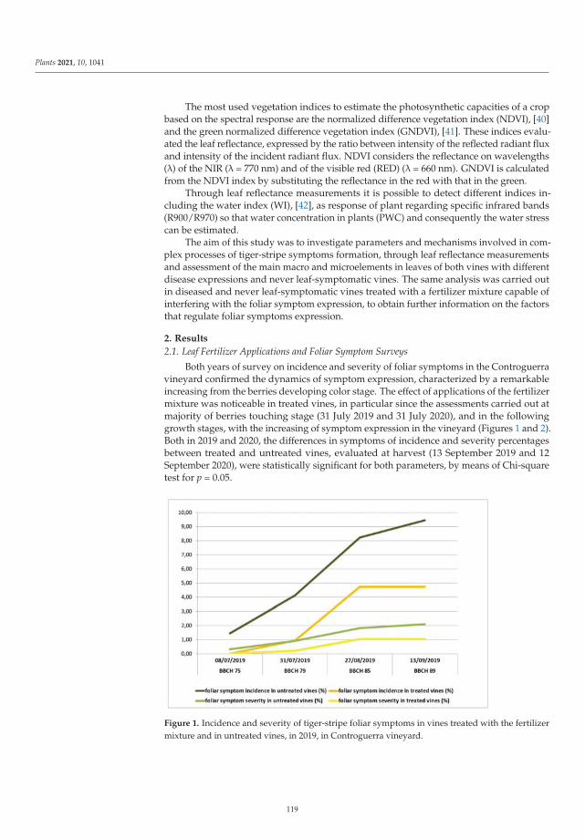
Plants 2021, 10, 1041
The most used vegetation indices to estimate the photosynthetic capacities of a cropbased on the spectral response are the normalized difference vegetation index (NDVI), [40]and the green normalized difference vegetation index (GNDVI), [41]. These indices evalu-ated the leaf reflectance, expressed by the ratio between intensity of the reflected radiant fluxand intensity of the incident radiant flux. NDVI considers the reflectance on wavelengths(λ) of the NIR (λ = 770 nm) and of the visible red (RED) (λ = 660 nm). GNDVI is calculatedfrom the NDVI index by substituting the reflectance in the red with that in the green.
Through leaf reflectance measurements it is possible to detect different indices in-cluding the water index (WI), [42], as response of plant regarding specific infrared bands(R900/R970) so that water concentration in plants (PWC) and consequently the water stresscan be estimated.
The aim of this study was to investigate parameters and mechanisms involved in com-plex processes of tiger-stripe symptoms formation, through leaf reflectance measurementsand assessment of the main macro and microelements in leaves of both vines with differentdisease expressions and never leaf-symptomatic vines. The same analysis was carried outin diseased and never leaf-symptomatic vines treated with a fertilizer mixture capable ofinterfering with the foliar symptom expression, to obtain further information on the factorsthat regulate foliar symptoms expression.
2. Results
2.1. Leaf Fertilizer Applications and Foliar Symptom Surveys
Both years of survey on incidence and severity of foliar symptoms in the Controguerravineyard confirmed the dynamics of symptom expression, characterized by a remarkableincreasing from the berries developing color stage. The effect of applications of the fertilizermixture was noticeable in treated vines, in particular since the assessments carried out atmajority of berries touching stage (31 July 2019 and 31 July 2020), and in the followinggrowth stages, with the increasing of symptom expression in the vineyard (Figures 1 and 2).Both in 2019 and 2020, the differences in symptoms of incidence and severity percentagesbetween treated and untreated vines, evaluated at harvest (13 September 2019 and 12September 2020), were statistically significant for both parameters, by means of Chi-squaretest for p = 0.05.
Figure 1. Incidence and severity of tiger-stripe foliar symptoms in vines treated with the fertilizermixture and in untreated vines, in 2019, in Controguerra vineyard.
119
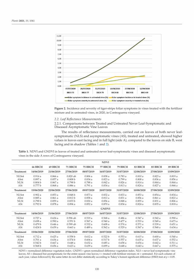
Plants 2021, 10, 1041
Figure 2. Incidence and severity of tiger-stripe foliar symptoms in vines treated with the fertilizermixture and in untreated vines, in 2020, in Controguerra vineyard.
2.2. Leaf Reflectance Measurements2.2.1. Comparisons between Treated and Untreated Never Leaf-Symptomatic andDiseased Asymptomatic Vine Leaves
The results of reflectance measurements, carried out on leaves of both never leaf-symptomatic (NLS) and asymptomatic vines (AS), treated and untreated, showed highervalues in leaves east facing and in full light (side A), compared to the leaves on side B, westfacing and in shadow (Tables 1 and 2).
Table 1. NDVI and GNDVI in leaves of treated and untreated never leaf-symptomatic vines and diseased asymptomaticvines in the side A rows of Controguerra vineyard.
NDVI
66 BBCH 69 BBCH 71 BBCH 75 BBCH 77 BBCH 79 BBCH 83 BBCH 85 BBCH 89 BBCH
Treatment 14/06/2019 21/06/2019 27/06/2019 08/07/2019 16/07/2019 31/07/2019 12/08/2019 27/08/2019 13/09/2019
NLSnt 0.914 a 0.866 a 0.820 ab 0.806 a 0.838 a 0.783 a 0.810 a 0.823 a 0.819 aASnt 0.897 a 0.857 a 0.808 b 0.834 a 0.823 a 0.799 a 0.808 a 0.836 a 0.856 aNLSt 0.804 b 0.867 a 0.798 b 0.788 a 0.842 a 0.826 a 0.814 a 0.844 a 0.844 aASt 0.777 b 0.868 a 0.886 a 0.791 a 0.834 a 0.813 a 0.824 a 0.827 a 0.844 a
Treatment 15/06/2020 22/06/2020 27/06/2020 09/07/2020 20/07/2020 31/07/2020 10/08/2020 27/08/2020 12/09/2020
NLSnt 0.902 a 0.853 a 0.848 b 0.873 a 0.832 a 0.833 a 0.853 a 0.854 a 0.834 aASnt 0.885 a 0.861 a 0.812 b 0.858 a 0.812 a 0.831 a 0.856 a 0.861 a 0.822 aNLSt 0.798 b 0.859 a 0.833 b 0.830 a 0.856 a 0.848 a 0.855 a 0.831 a 0.806 aASt 0.792 b 0.878 a 0.894 a 0.852 a 0.870 a 0.834 a 0.824 a 0.835 a 0.818 a
GNDVI
Treatment 14/06/2019 21/06/2019 27/06/2019 08/07/2019 16/07/2019 31/07/2019 12/08/2019 27/08/2019 13/09/2019
NLSnt 0.727 a 0.624 a 0.596 ab 0.519 a 0.568 a 0.486 a 0.547 a 0.542 a 0.590 aASnt 0.698 a 0.590 a 0.551 b 0.567 a 0.540 a 0.477 a 0.533 a 0.549 a 0.673 aNLSt 0.479 b 0.646 a 0.599 ab 0.532 a 0.608 a 0.574 a 0.571 a 0.539 a 0.651 aASt 0.424 b 0.639 a 0.663 a 0.489 a 0.562 a 0.529 a 0.567 a 0.568 a 0.634 a
Treatment 15/06/2020 22/06/2020 27/06/2020 09/07/2020 20/07/2020 31/07/2020 10/08/2020 27/08/2020 12/09/2020
NLSnt 0.712 a 0.625 a 0.558 b 0.612 a 0.522 b 0.553 a 0.615 a 0.600 a 0.540 aASnt 0.701 a 0.636 a 0.574 b 0.604 a 0.517 b 0.557 a 0.635 a 0.637 a 0.552 aNLSt 0.542 b 0.667 a 0.648 a 0.632 a 0.685 a 0.658 a 0.654 a 0.642 a 0.511 aASt 0.568 b 0.656 a 0.623 a 0.639 a 0.699 a 0.648 a 0.643 a 0.667 a 0.575 a
NDVI = normalized difference vegetation index. GNDVI = green normalized difference vegetation index. NLS = never leaf-symptomatic vineleaves; AS = diseased but asymptomatic for the entire season vine leaves; t = treated with fertilizer mixture; nt = untreated. For each column ofeach year, values followed by the same letter do not differ statistically according to Tukey’s honest significant difference (HSD) test at p = 0.05.
120
Plants 2021, 10, 1041
Table 2. NDVI and GNDVI in leaves of treated and untreated never leaf-symptomatic vines and diseased asymptomaticvines in the side B rows of Controguerra vineyard.
NDVI
66 BBCH 69 BBCH 71 BBCH 75 BBCH 77 BBCH 79 BBCH 83 BBCH 85 BBCH 89 BBCH
Treatment 14/06/2019 21/06/2019 27/06/2019 08/07/2019 16/07/2019 31/07/2019 12/08/2019 27/08/2019 13/09/2019
NLSnt 0.928 a 0.904 a 0.938 b 0.950 a 0.951 a 0.934 a 0.950 a 0.936 a 0.935 aASnt 0.907 a 0.915 a 0.958 b 0.971 a 0.957 a 0.947 a 0.957 a 0.946 a 0.959 aNLSt 0.810 c 0.934 a 0.978 a 0.974 a 0.967 a 0.927 a 0.913 a 0.944 a 0.950 aASt 0.867 b 0.936 a 0.981 a 0.971 a 0.975 a 0.933 a 0.943 a 0.927 a 0.940 a
Treatment 15/06/2020 22/06/2020 27/06/2020 09/07/2020 20/07/2020 31/07/2020 10/08/2020 27/08/2020 12/09/2020
NLSnt 0.981 a 0.913 a 0.946 b 0.979 a 0.979 a 0.971 a 0.964 a 0.971 a 0.938 aASnt 0.977 a 0.922 a 0.943 b 0.977 a 0.976 a 0.974 a 0.971 a 0.977 a 0.931 aNLSt 0.838 b 0.945 a 0.984 a 0.965 a 0.942 b 0.969 a 0.970 a 0.974 a 0.948 aASt 0.849 b 0.955 a 0.984 a 0.975 a 0.951 b 0.967 a 0.973 a 0.973 a 0.940 a
GNDVI
Treatment 14/06/2019 21/06/2019 27/06/2019 08/07/2019 16/07/2019 31/07/2019 12/08/2019 27/08/2019 13/09/2019
NLSnt 0.764 a 0.713 a 0.794 b 0.764 a 0.787 a 0.737 a 0.799 a 0.735 a 0.757 aASnt 0.730 a 0.745 a 0.831 b 0.850 a 0.814 a 0.766 a 0.829 a 0.768 a 0.847 aNLSt 0.504 b 0.790 a 0.864 b 0.880 a 0.847 a 0.770 a 0.752 a 0.839 a 0.833 aASt 0.546 b 0.781 a 0.877 a 0.864 a 0.859 a 0.789 a 0.809 a 0.768 a 0.808 a
Treatment 15/06/2020 22/06/2020 27/06/2020 09/07/2020 20/07/2020 31/07/2020 10/08/2020 27/08/2020 12/09/2020
NLSnt 0.812 a 0.823 a 0.865 ab 0.872 a 0.864 a 0.853 a 0.819 b 0.848 a 0.715 aASnt 0.807 a 0.845 a 0.833 b 0.864 a 0.859 a 0.855 a 0.845 ab 0.871 a 0.730 aNLSt 0.622 b 0.812 a 0.921 a 0.878 a 0.838 a 0.878 a 0.868 ab 0.886 a 0.800 aASt 0.676 b 0.833 a 0.915 a 0.895 a 0.842 a 0.875 a 0.883 a 0.892 a 0.765 a
NDVI = normalized difference vegetation index. GNDVI = green normalized difference vegetation index. NLS = never leaf-symptomatic vineleaves; AS = diseased but asymptomatic for the entire season vine leaves; t = treated with fertilizer mixture; nt = untreated. For each column ofeach year values followed by the same letter do not differ statistically according to Tukey’s honest significant difference (HSD) test at p = 0.05.
Conversely, the trend of reflectance measurements carried out from growth stage 66to 89, were similar in the two sides (A and B) in both years of survey (Tables 1 and 2).
The NDVI and GNDVI values showed very similar trends and were always signif-icantly lower in leaves of treated compared to untreated NLS and AS vines, in the firstsurvey, at growth stage 66 (Tables 1 and 2). On the contrary, in the third survey, at growthstage 71, the NDVI and GNDVI values of treated AS leaves appeared significantly higherthan those of AS untreated leaves. In the same survey, the treated NLS leaves showedhigher or similar values than those of untreated NLS leaves (Tables 1 and 2). From thegrowth stage 75 to the last survey, growth stage 89, no differences were found between NLSand AS leaves. Few exceptions were observed only in 2020, at growth stage 77, for GNDVI(side A), and for NDVI (side B), with values of treated NLS and AS leaves significantlyhigher and lower than untreated NLS and AS leaves, respectively (Tables 1 and 2).
In the first three surveys (growth stages 66, 69, and 71), values of the WI were higherin leaves of treated NLS and AS vines, compared to those of the corresponding untreatedvines, both in side A and in side B. From the survey at growth stage 75 to the survey atgrowth stage 83 any difference between treated and untreated NLS and AS values wasdetected (Table 3). In growth stages 85 and 89 surveys, WI values were lower, in most ofthe cases significantly, in treated compared to untreated AS leaves (Table 3).
121
Plants 2021, 10, 1041
Table 3. Water Index in leaves of treated and untreated never leaf-symptomatic vines and diseased asymptomatic vines inthe Controguerra vineyard.
Side A WI
66 BBCH 69 BBCH 71 BBCH 75 BBCH 77 BBCH 79 BBCH 83 BBCH 85 BBCH 89 BBCH
Treatment 14/06/2019 21/06/2019 27/06/2019 08/07/2019 16/07/2019 31/07/2019 12/08/2019 27/08/2019 13/09/2019
NLSnt 1.042 b 1.056 b 1.039 c 1.069 ab 1.046 a 1.020 a 1.041 a 1.013 a 1.018 abASnt 1.086 b 1.072 b 1.064 bc 1.091 a 1.049 a 1.036 a 1.065 a 1.028 a 1.037 aNLSt 1.114 a 1.100 a 1.078 ab 1.048 b 1.011 a 1.027 a 1.041 a 1.000 a 0.995 abASt 1.110 a 1.103 a 1.107 a 1.068 ab 1.086 a 1.007 a 1.046 a 0.995 a 0.987 b
Treatment 15/06/2020 22/06/2020 27/06/2020 09/07/2020 20/07/2020 31/07/2020 10/08/2020 27/08/2020 12/09/2020
NLSnt 1.036 b 1.023 b 1.043 b 1.059 a 1.050 a 1.029 b 1.039 a 1.038 a 1.032 abASnt 1.056 b 1.044 b 1.062 b 1.082 a 1.056 a 1.051 ab 1.047 a 1.063 a 1.051 aNLSt 1.112 a 1.114 a 1.081 ab 1.060 a 1.058 a 1.081 a 1.048 a 1.029 a 1.017 abASt 1.116 a 1.110 a 1.106 a 1.058 a 1.058 a 1.047 b 1.056 a 1.044 a 1.013 b
Side B WI
Treatment 14/06/2019 21/06/2019 27/06/2019 08/07/2019 16/07/2019 31/07/2019 12/08/2019 27/08/2019 13/09/2019
NLSnt 1.054 c 1.084 b 1.102 c 1.153 a 1.116 a 1.053 a 1.112 a 1.043 ab 1.070 abASnt 1.082 bc 1.116 b 1.130 bc 1.199 a 1.131 a 1.080 a 1.175 a 1.091 a 1.107 aNLSt 1.123 ab 1.166 a 1.257 ab 1.179 a 1.180 a 1.056 a 1.098 a 0.965 b 1.020 abASt 1.140 a 1.180 a 1.286 a 1.140 a 1.193 a 1.033 a 1.096 a 0.969 b 1.007 b
Treatment 15/06/2020 22/06/2020 27/06/2020 09/07/2020 20/07/2020 31/07/2020 10/08/2020 27/08/2020 12/09/2020
NLSnt 1.088 b 1.101 b 1.111 b 1.076 a 1.105 a 1.066 b 1.067 b 1.087 b 1.078 aASnt 1.098 b 1.086 b 1.157 b 1.129 a 1.111 a 1.142 a 1.129 a 1.186 a 1.129 aNLSt 1.122 a 1.143 a 1.203 a 1.106 a 1.094 a 1.119 ab 1.104 ab 1.157 ab 1.040 aASt 1.151 a 1.148 a 1.208 a 1.106 a 1.089 a 1.144 a 1.127 a 1.102 ab 1.052 a
WI = water index. NLS = never leaf-symptomatic vine leaves; AS = diseased but asymptomatic for the entire season vine leaves; t = treatedwith fertilizer mixture; nt = untreated. For each column of each year values followed by the same letter do not differ statistically accordingto Tukey’s honest significant difference (HSD) test at p = 0.05.
2.2.2. Comparisons among Untreated Never Leaf-Symptomatic, Diseased Asymptomatic,and Diseased Symptomatic Vine Leaves
The leaf reflectance measurements carried out from the first leaf symptom appearance(8 July 2019 and 9 July 2020, growth stage 75) to the harvest (growth stage 89) on leaves ofuntreated never leaf-symptomatic (NLS) and asymptomatic (AS) vines, and on leaves ofthe different shoots of untreated symptomatic diseased vines (PRE-S, ASTIGR, and TIGR),did not show differences for both NDVI and GNDVI, except for significantly lower valuesin symptomatic leaves (TIGR) (Table 4).
Table 4. NDVI and GNDVI in never leaf-symptomatic vine leaves and in different categories of leaves of diseased vines inthe side A rows of Controguerra vineyard.
NDVI
75 BBCH 77 BBCH 79 BBCH 83 BBCH 85 BBCH 89 BBCH
Treatment 08/07/2019 16/07/2019 31/07/2019 12/08/2019 27/08/2019 13/09/2019
NLS 0.806 a 0.838 a 0.783 a 0.810 a 0.823 a 0.819 aAS 0.834 a 0.823 a 0.799 a 0.808 a 0.836 a 0.856 a
PRE-S 0.806 a 0.844 a 0.812 a 0.786 a 0.830 a 0.821 aASTIGR 0.870 a 0.880 a 0.778 a 0.778 a 0.829 a 0.810 a
TIGR 0.723 b 0.622 b 0.570 b 0.759 a 0.636 b 0.635 b
Treatment 09/07/2020 20/07/2020 31/07/2020 10/08/2020 27/08/2020 12/09/2020
NLS 0.873 a 0.832 a 0.833 a 0.853 a 0.854 a 0.834 aAS 0.858 a 0.812 a 0.831 a 0.856 a 0.861 a 0.822 a
PRE-S 0.852 a 0.798 a 0.820 a 0.859 a 0.875 a 0.784 aASTIGR 0.875 a 0.806 a 0.825 a 0.831 a 0.833 a 0.816 a
TIGR 0.743 b 0.658 b 0.678 b 0.634 b 0.600 b 0.542 b
122
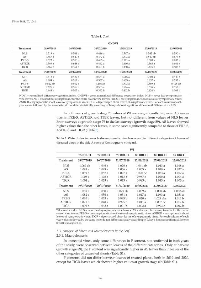
Plants 2021, 10, 1041
Table 4. Cont.
GNDVI
Treatment 08/07/2019 16/07/2019 31/07/2019 12/08/2019 27/08/2019 13/09/2019
NLS 0.519 a 0.568 a 0.486 a 0.547 a 0.542 ab 0.590 aAS 0.567 a 0.540 a 0.477 a 0.533 a 0.549 ab 0.673 a
PRE-S 0.523 a 0.550 a 0.485 a 0.521 a 0.608 a 0.633 aASTIGR 0.569 a 0.648 a 0.462 a 0.496 a 0.563 a 0.601 a
TIGR 0.465 b 0.451 b 0.303 b 0.448 a 0.415 b 0.487 b
Treatment 09/07/2020 20/07/2020 31/07/2020 10/08/2020 27/08/2020 12/09/2020
NLS 0.612 a 0.522 a 0.553 a 0.615 a 0.600 a 0.540 aAS 0.604 a 0.517 a 0.557 a 0.635 a 0.637 a 0.552 a
PRE-S 0.522 ab 0.502 a 0.466 ab 0.573 a 0.589 a 0.425 abASTIGR 0.625 a 0.559 a 0.553 a 0.564 a 0.635 a 0.552 a
TIGR 0.448 b 0.505 a 0.392 b 0.402 b 0.424 b 0.365 b
NDVI = normalized difference vegetation index. GNDVI = green normalized difference vegetation index. NLS = never leaf-symptomaticvine leaves; AS = diseased but asymptomatic for the entire season vine leaves; PRE-S = pre-symptomatic shoot leaves of symptomatic vines;ASTIGR = asymptomatic shoot leaves of symptomatic vines; TIGR = tiger-striped shoot leaves of symptomatic vines. For each column of eachyear values followed by the same letter do not differ statistically according to Tukey’s honest significant difference (HSD) test at p = 0.05.
In both years at growth stage 75 values of WI were significantly higher in AS leavesthan in PRE-S, ASTIGR and TIGR leaves, but not different from values of NLS leaves.From surveys at growth stage 79 to the last surveys (growth stage 89), AS leaves showedhigher values than the other leaves, in some cases significantly compared to those of PRE-S,ASTIGR, and TIGR (Table 5).
Table 5. Water Index in never leaf-symptomatic vine leaves and in different categories of leaves ofdiseased vines in the side A rows of Controguerra vineyard.
WI
75 BBCH 77 BBCH 79 BBCH 83 BBCH 85 BBCH 89 BBCH
Treatment 08/07/2019 16/07/2019 31/07/2019 12/08/2019 27/08/2019 13/09/2019
NLS 1.069 ab 1.046 a 1.020 a 1.041 ab 1.013 a 1.018 aAS 1.091 a 1.049 a 1.036 a 1.065 a 1.028 a 1.037 a
PRE-S 1.059 b 1.057 a 1.027 a 1.020 bc 1.023 a 1.017 aASTIGR 1.008 c 1.108 a 1.013 a 0.987 c 1.020 a 1.004 a
TIGR 1.001 c 1.033 a 1.013 a 0.983 c 1.013 a 1.003 a
Treatment 09/07/2020 20/07/2020 31/07/2020 10/08/2020 27/08/2020 12/09/2020
NLS 1.059 a 1.050 a 1.029 ab 1.039 a 1.038 ab 1.032 abAS 1.082 a 1.056 a 1.051 a 1.047 a 1.063 a 1.051 a
PRE-S 1.010 b 1.033 a 0.995 b 1.020 a 1.028 abc 1.011 bASTIGR 1.021 b 1.048 a 0.995 b 1.011 a 1.007 bc 1.012 b
TIGR 1.009 b 1.042 a 1.001 b 1.002 a 0.993 c 1.002 bWI = water index. NLS = never leaf-symptomatic vine leaves; AS = diseased but asymptomatic for the entireseason vine leaves; PRE-S = pre-symptomatic shoot leaves of symptomatic vines; ASTIGR = asymptomatic shootleaves of symptomatic vines; TIGR = tiger-striped shoot leaves of symptomatic vines. For each column of eachyear values followed by the same letter do not differ statistically according to Tukey’s honest significant difference(HSD) test at p = 0.05.
2.3. Analysis of Macro and Microelements in the Leaf2.3.1. Macroelements
In untreated vines, only some differences in P content, not confirmed in both yearsof the study, were observed between leaves of the different categories. Only at harvest(growth stage 89), the P content was significantly higher in AS leaves than in leaves of theother categories of untreated shoots (Table S1).
P contents did not differ between leaves of treated plants, both in 2019 and 2020,except for TIGR leaves which showed higher values at growth stage 89 (Table S1).
123
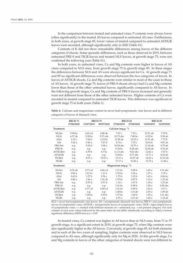
Plants 2021, 10, 1041
In the comparison between treated and untreated vines, P contents were always lower(often significantly) in the treated AS leaves compared to untreated AS ones. Furthermore,in both years, at growth stage 85, lower values of treated compared to untreated ASTIGRleaves were recorded, although significantly only in 2020 (Table S1).
Contents of K did not show remarkable differences among leaves of the differentcategories of shoots. Some sporadic differences, such as those observed in 2019, betweenuntreated PRE-S and TIGR leaves and treated NLS leaves, at growth stage 75, were notconfirmed the following year (Table S1).
In both years, in untreated vines, Ca and Mg contents were higher in leaves of ASvines compared to NLS ones, from growth stage 75 to growth stage 85. In these stagesthe differences between NLS and AS were always significant for Ca. At growth stage 71and 89 no significant differences were observed between the two categories of leaves. Inleaves of ASTIGR shoots, Ca and Mg contents were similar in most of the cases to thoseof AS leaves. At growth stage 75, leaves of PRE-S shoots always had Ca and Mg contentslower than those of the other untreated leaves, significantly compared to AS leaves. Inthe following growth stages, Ca and Mg contents of PRE-S leaves increased and generallywere not different from those of the other untreated leaves. Higher contents of Ca wererecorded in treated compared to untreated TIGR leaves. This difference was significant atgrowth stage 75 in both years (Table 6).
Table 6. Calcium and magnesium content in never leaf-symptomatic vine leaves and in differentcategories of leaves of diseased vines.
BBCH 71 BBCH 75 BBCH 85 BBCH 89
27/06/2020 11/07/2019 09/07/2020 27/08/2019 27/08/2020 12/09/2019 12/09/2020
Treatment Calcium (mg g−1)
NLSnt 5.98 bc 4.62 cd 4.80 de 7.67 c 7.23 c 12.21 ab 7.93 bNLSt 6.37 ab 5.38 bc 5.27 cde 10.73 abc 7.80 bc 6.92 bc 9.58 abASnt 5.15 c 7.06 b 6.23 bc 12.58 ab 10.53 a 12.51 ab 7.73 bASt 7.02 a 9.49 a 7.67 d 8.74 c 7.55 c 8.03 bc 9.87 ab
PRE-Snt n.p. 3.22 d 3.98 e 10.28 abc 10.57 a 11.94 ab 9.75 abPRE-St n.p. n.p. n.p. 9.18 bc 8.28 abc 12.00 ab 9.53 ab
ASTIGRnt n.p. 6.59 b 6.7 bc 11.13 abc 9.63 abc 12.82 a 7.07 bASTIGRt n.p. n.p. n.p. 9.38 bc 8.77 abc 13.71 a 8.42 abTIGRnt n.p. 8.91 a 10.03 a 13.17 a 10.07 ab 14.03 a 10.10 abTIGRt n.p. n.p. n.p. 13.17 a 10.42 a 13.73 a 11.48 a
Treatment Magnesium (mg g−1)
NLSnt 0.76 ab 0.73 cd 0.66 cd 1.21 bc 0.90 b 1.65 a 1.01 abcNLSt 0.85 a 1.01 bc 1.13 a 1.52 bc 1.56 a 1.57 a 1.35 aASnt 0.63 b 1.27 b 0.76 c 1.73 b 1.03 b 1.62 a 0.86 bcASt 0.86 a 1.66 a 1.01 ab 1.35 bc 0.87 b 1.14 a 1.22 ab
PRE-Snt n.p. 0.55 d 0.57 d 1.10 c 1.07 b 1.39 a 1.23 abPRE-St n.p. n.p. n.p. 1.41 bc 0.98 b 1.92 a 0.99 abc
ASTIGRnt n.p. 0.77 cd 0.68 cd 1.41 bc 0.96 b 1.63 a 0.71 cASTIGRt n.p. n.p. n.p. 1.48 bc 1.16 ab 2.05 a 1.09 abTIGRnt n.p. 1.00 bc 0.93 b 1.57 bc 1.00 b 1.55 a 1.01 abcTIGRt n.p. n.p. n.p. 2.35 a 1.23 ab 1.45 a 1.18 ab
NLS = never leaf-symptomatic vine leaves; AS = asymptomatic diseased vine leaves; PRE-S = pre-symptomaticleaves of symptomatic vines; ASTIGR = asymptomatic leaves of symptomatic vines; TIGR = tiger-striped leavesof symptomatic vines. t = treated with fertilizer mixture; nt = untreated; n.p. = not present category. For eachcolumn of each year values followed by the same letter do not differ statistically according to Tukey’s honestsignificant difference (HSD) test at p = 0.05.
In treated vines, Ca content was higher in AS leaves than in NLS ones, from 71 to 75growth stage, to a significant extent in 2019, at growth stage 75, when Mg contents werealso significantly higher in the AS leaves. Conversely, at growth stage 85, for both elementsand in each of the two years of sampling, higher contents were observed in NLS leavescompared to AS ones, although significantly only for Mg in 2020. At this growth stage, Caand Mg contents in leaves of the other categories of treated shoots were not different to
124
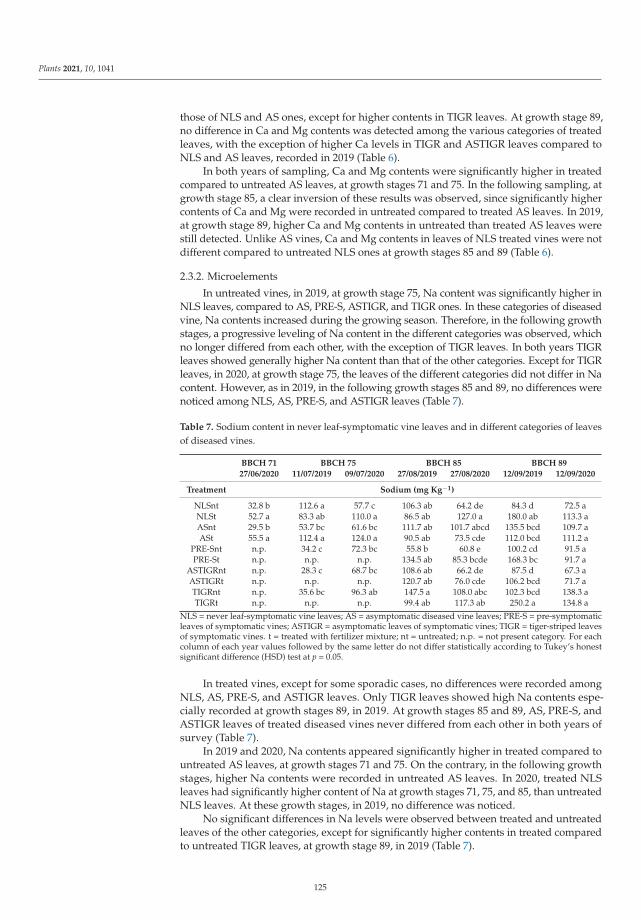
Plants 2021, 10, 1041
those of NLS and AS ones, except for higher contents in TIGR leaves. At growth stage 89,no difference in Ca and Mg contents was detected among the various categories of treatedleaves, with the exception of higher Ca levels in TIGR and ASTIGR leaves compared toNLS and AS leaves, recorded in 2019 (Table 6).
In both years of sampling, Ca and Mg contents were significantly higher in treatedcompared to untreated AS leaves, at growth stages 71 and 75. In the following sampling, atgrowth stage 85, a clear inversion of these results was observed, since significantly highercontents of Ca and Mg were recorded in untreated compared to treated AS leaves. In 2019,at growth stage 89, higher Ca and Mg contents in untreated than treated AS leaves werestill detected. Unlike AS vines, Ca and Mg contents in leaves of NLS treated vines were notdifferent compared to untreated NLS ones at growth stages 85 and 89 (Table 6).
2.3.2. Microelements
In untreated vines, in 2019, at growth stage 75, Na content was significantly higher inNLS leaves, compared to AS, PRE-S, ASTIGR, and TIGR ones. In these categories of diseasedvine, Na contents increased during the growing season. Therefore, in the following growthstages, a progressive leveling of Na content in the different categories was observed, whichno longer differed from each other, with the exception of TIGR leaves. In both years TIGRleaves showed generally higher Na content than that of the other categories. Except for TIGRleaves, in 2020, at growth stage 75, the leaves of the different categories did not differ in Nacontent. However, as in 2019, in the following growth stages 85 and 89, no differences werenoticed among NLS, AS, PRE-S, and ASTIGR leaves (Table 7).
Table 7. Sodium content in never leaf-symptomatic vine leaves and in different categories of leavesof diseased vines.
BBCH 71 BBCH 75 BBCH 85 BBCH 89
27/06/2020 11/07/2019 09/07/2020 27/08/2019 27/08/2020 12/09/2019 12/09/2020
Treatment Sodium (mg Kg−1)
NLSnt 32.8 b 112.6 a 57.7 c 106.3 ab 64.2 de 84.3 d 72.5 aNLSt 52.7 a 83.3 ab 110.0 a 86.5 ab 127.0 a 180.0 ab 113.3 aASnt 29.5 b 53.7 bc 61.6 bc 111.7 ab 101.7 abcd 135.5 bcd 109.7 aASt 55.5 a 112.4 a 124.0 a 90.5 ab 73.5 cde 112.0 bcd 111.2 a
PRE-Snt n.p. 34.2 c 72.3 bc 55.8 b 60.8 e 100.2 cd 91.5 aPRE-St n.p. n.p. n.p. 134.5 ab 85.3 bcde 168.3 bc 91.7 a
ASTIGRnt n.p. 28.3 c 68.7 bc 108.6 ab 66.2 de 87.5 d 67.3 aASTIGRt n.p. n.p. n.p. 120.7 ab 76.0 cde 106.2 bcd 71.7 aTIGRnt n.p. 35.6 bc 96.3 ab 147.5 a 108.0 abc 102.3 bcd 138.3 aTIGRt n.p. n.p. n.p. 99.4 ab 117.3 ab 250.2 a 134.8 a
NLS = never leaf-symptomatic vine leaves; AS = asymptomatic diseased vine leaves; PRE-S = pre-symptomaticleaves of symptomatic vines; ASTIGR = asymptomatic leaves of symptomatic vines; TIGR = tiger-striped leavesof symptomatic vines. t = treated with fertilizer mixture; nt = untreated; n.p. = not present category. For eachcolumn of each year values followed by the same letter do not differ statistically according to Tukey’s honestsignificant difference (HSD) test at p = 0.05.
In treated vines, except for some sporadic cases, no differences were recorded amongNLS, AS, PRE-S, and ASTIGR leaves. Only TIGR leaves showed high Na contents espe-cially recorded at growth stages 89, in 2019. At growth stages 85 and 89, AS, PRE-S, andASTIGR leaves of treated diseased vines never differed from each other in both years ofsurvey (Table 7).
In 2019 and 2020, Na contents appeared significantly higher in treated compared tountreated AS leaves, at growth stages 71 and 75. On the contrary, in the following growthstages, higher Na contents were recorded in untreated AS leaves. In 2020, treated NLSleaves had significantly higher content of Na at growth stages 71, 75, and 85, than untreatedNLS leaves. At these growth stages, in 2019, no difference was noticed.
No significant differences in Na levels were observed between treated and untreatedleaves of the other categories, except for significantly higher contents in treated comparedto untreated TIGR leaves, at growth stage 89, in 2019 (Table 7).
125
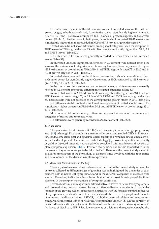
Plants 2021, 10, 1041
Fe contents were similar in the different categories of untreated leaves at the first twogrowth stages, in both years of study. Later in the season, significantly higher contents inAS, ASTIGR, and TIGR leaves compared to NLS ones, at growth stage 85, in 2020, werenoticed (Table S2). Furthermore, in both years, Fe contents of untreated TIGR leaves weresignificantly higher than that recorded in NLS and AS leaves, at growth stages 89.
Treated vines did not show differences among shoot categories, with the exception ofTIGR leaves in 2019 at growth stage 85, with Fe content significantly higher than NLS, AS,and PRE-S leaves (Table S2).
No difference in Fe levels was generally recorded between treated and untreatedleaves (Table S2).
In untreated vines, no significant differences in Cu content were noticed among theleaves of the various shoot categories, apart from very few exceptions only related to higherNLS leaf content at growth stage 75 in 2019, in ASTIGR at growth stage 75 in 2020 and inAS at growth stage 85 in 2020 (Table S2).
In treated vines, leaves from the different categories of shoots never differed fromeach other, except for significantly higher Cu contents in TIGR compared to NLS leaves, atgrowth stage 85, in 2019 (Table S2).
In the comparison between treated and untreated vines, no differences were generallynoticed in Cu content among the different investigated categories (Table S2).
In untreated vines, in 2020, Mn contents were significantly higher: in ASTIGR thanPRE-S leaves, at growth stage 75; in AS than NLS, PRE-S and TIGR leaves, at growth stage85. These results were not observed at the corresponding growth stages in 2019 (Table S3).
No differences in Mn content were found among leaves of treated shoots, except forsignificantly higher contents in PRE-S than NLS and ASTIGR leaves, at growth stage 85 of2019 (Table S3).
Mn contents did not show any difference between the leaves of the same shootcategories of treated and untreated vines.
No differences were generally recorded in Zn leaf content (Table S3).
3. Discussion
The grapevine trunk diseases (GTDs) are increasing in almost all grape growingareas [43]. Although Esca complex is the most widespread and studied GTDs in Europeanvineyards, some etiological and epidemiological aspects still remained unexplained as wellas for the development of an effective control strategy [3]. Losses in quantity and qualityof yield in diseased vineyards appeared to be correlated with incidence and severity ofplant symptom expression [14,15]. However, mechanisms and factors associated with theoccurrence of symptoms are yet to be fully clarified. Therefore, the present study aimed toevaluate some aspects of the physiology of diseased vines involved with the appearanceand development of the disease symptom expression.
3.1. Macro and Microelements in the Leaf
The analysis of macro and microelements carried out in the present study on samplesof leaves collected at different stages of growing season highlighted the dynamics of eachelement both in never leaf-symptomatic and in the different categories of diseased vineshoots. Therefore, indications have been obtained on a possible role played by theseelements in the complex mechanisms of symptom expression.
Levels of calcium and magnesium differed between leaves of never leaf-symptomaticand diseased vines, but also between leaves of different diseased vine shoots. In particular,for most of the growing season, in the parcel not treated with the fertilizer mixture, the leavesof asymptomatic vines, AS, and, at berries pea-sized, the leaves of asymptomatic shootsof symptomatic diseased vines, ASTIGR, had higher levels of calcium and magnesiumcompared to untreated leaves of never leaf-symptomatic vines, NLS. On the contrary, atpea-sized berries, still green leaves at the base of shoots that began to show symptoms inthe leaves of distal part, PRE-S, had lower contents of calcium and magnesium, maybe also
126
Plants 2021, 10, 1041
due to a shoot malfunction, compared to the leaves of the other shoots, and in particularcompared to AS leaves. As observed in a previous study [32], accumulations of calcium,magnesium, sodium, and microelements were recorded in symptomatic leaves, TIGR,probably because of their impaired functionality. Other remarkable differences were in lowsodium contents in PRE-S shoot leaves compared to AS leaves, at softening of berries. Theseresults provided a first evidence on the role of calcium and magnesium in the expressionof leaf symptoms, notable in both AS and ASTIGR leaves, which remained asymptomaticthroughout the season [34,44]. The involvement of calcium and magnesium in the symptomoccurrence could be confirmed by low levels of both elements in leaves of PRE-S, comparedto AS shoots, which then showed a progressive development of symptoms. Furthermore,the lower sodium contents in PRE-S compared to AS leaves, indicated a possible function ofthis element in the dynamics of foliar symptom expression. The correlation between sodiumand the expression of leaf symptoms is in agreement with what was found in the decay ofkiwifruit, a disease similar for many aspects to Esca of grapevine [45].
Fertilizer mixture applications confirmed to be effective in reducing foliar symptomexpression [34,44], but also contributed to obtain further information on leaf symptomexpression dynamics. Leaves of treated AS vines had higher contents of calcium and mag-nesium, compared to NLS treated leaves, at berries pea-sized, while at softening of berries,higher calcium and magnesium contents in NLS compared to AS leaves were recorded.AS vines probably responded better to the treatment than NLS vines, because most likelyNLS vines did not require calcium and magnesium. Therefore, it may not be excludedthat AS vines, due to infection, could have a better physiological attitude to calcium andmagnesium supply. Unlike what was observed in untreated AS leaves, treated AS leavesshowed sodium dynamics similar to that found for calcium and magnesium, providingfurther indications of a possible involvement of sodium in foliar symptom expression.Also treated TIGR leaves, as for the untreated, accumulated calcium, magnesium, sodium,and microelements.
The comparison of elements content between treated and untreated leaves gavefurther information on their dynamics in leaf symptom development. The same dynamicsobserved for calcium, magnesium, and sodium in treated AS and NLS leaves appearedclearer in the comparison between treated and untreated AS leaves. In fact, contents of thethree elements were higher in treated compared to untreated AS leaves, at fruit set andberries pea-sized stages, while at softening of berries contents reversed and were higher inuntreated compared to treated AS leaves.
Calcium and magnesium contents were not different between treated and untreatedNLS leaves, indicating how the applications of calcium and magnesium, at fruit set andberries pea-sized stages, before the onset of symptoms in the vineyard, were useful in ASvines particularly, by reducing the symptom expression.
Comparisons between leaves of treated and untreated shoots of the same category didgenerally not show any difference for the other investigated elements.
3.2. Role of Calcium in Leaf Symptom Expression
The overall analysis of results on macro and microelements content in leaves ofthe different categories confirmed the involvement of calcium in the expression of foliarsymptoms, hypothesizing a possible role in reducing the symptom expression, as shown ina previous study [34].
The role of calcium was evident both in untreated AS vines, which showed leaf calciumincreases for most of the season, and in treated AS vines, whose leaves accumulated calciumup to pea-sized berries, at the onset of symptoms in the vineyard. In treated vines, includingthe ones who most likely would show symptoms if not treated, calcium accumulationsassessed in the first part of the season and due to fertilizer applications, indicated a possiblerole of the element in the modulation of the plant response, which was hypothesizedfor leaf symptom expression [46]. This plant response can be associated with phytotoxicsubstances produced by fungi in infected woody tissues and translocated to the leaf via
127
Plants 2021, 10, 1041
transpiration current [20,21,47]. Therefore, the development of necrosis in symptomaticleaves was associated with a strong response of the plant to such phytotoxic substances,comparable to a hypersensitive response [48].
To support the hypothesis mentioned, previous studies showed that at pre-bunchclosure, tiger-stripe leaves, compared to never leaf-symptomatic and asymptomatic vineleaves, had higher contents of trans-resveratrol and other phytoalexins, which are synthe-sized after the hypersensitive response of the plant [29,31].
Furthermore, previous studies demonstrated that vines without foliar symptoms asa consequence of fertilizer mixture applications had higher contents of trans-resveratrol,trans-ε-viniferin, trans-δ-viniferin, and flavonoids, as well as calcium oxalate druse, at pre-bunch closure, with respect to both untreated never leaf-symptomatic and asymptomaticdiseased vines [34,35].
In the subsequent growth stages phytoalexins contents progressively decreased intreated vines and increased in untreated vines which showed higher contents than treatedvines [35], reproducing the same dynamics of calcium, magnesium, and sodium observedin the present study.
These results strengthened the hypothesis of a defense plant response involved inthe foliar symptom expression, indicating as well the role of the fertilizer mixture in thisresponse, by reducing symptoms.
Moreover, what assessed on both untreated and treated asymptomatic plants sug-gested that calcium activity could be linked to the reduction of plant oxidative response [48].The penetration of calcium into leaves, facilitated by the seaweed, can increase the synthesisof calmodulin. Calmodulin regulates salicylic acid and, consequently, plant response tofungal toxic metabolites, reducing effects of the hypersensitive response, associated withleaf symptom expression [49].
Moreover, the reduction of symptom expression could also be due to the ability ofcalcium to strengthen cell walls, as calcium oxalate crystal accumulation, assessed in leavesof treated asymptomatic vines [34,50,51]. Furthermore, the increasing of calcium content inextracellular spaces can increase phytoalexin synthesis [52,53].
3.3. Role of Magnesium and Sodium in Leaf Symptom Expression
Applications of the fertilizer mixture were particularly effective in diseased plants,likely for a plant health improvement stimulated by magnesium [54]. It therefore might notbe excluded an involvement of magnesium as fundamental constituent of chlorophyll, inreducing or delaying its degradation, limiting as well leaf symptom expression in synergywith calcium [34,44].
The role of magnesium associated with disease control proved to be much morecontroversial, as it can reduce, but also favor, disease severity [55]. Magnesium can haveindirect beneficial effects on diseases with the possibility to withstand disease outbreak,because of the involvement of this element in several physiological processes associatedwith plant health [54].
In GTDs, studies demonstrated that Mg++ and Mn++ detoxified eutypin, the toxinproduced by Eutypa lata, causal agent of Eutypa dieback [56]. It might not be excluded sim-ilar effects on toxic metabolites produced by the pathogens involved in Esca of grapevine.Although the reduction of symptom expression was demonstrated in the present studymainly as effect of calcium and magnesium, further studies are needed to give a specificrole to magnesium.
The significant sodium increasing at berries pea-sized in treated compared to un-treated AS vine leaves could be related to phytoalexin increasing observed in a previousstudy at the same growth stage. Regulatory mechanisms associated with second messengermetabolites and sodium uptake have been studied; in particular, fast and temporary in-creases of reactive oxygen species (ROS) involved in hypersensitive response and secondarymetabolites synthesis, soon after increases of Na+, were recorded [57].
128
Plants 2021, 10, 1041
Given the structural similarity of sodium and potassium ions [58], sodium can replacepotassium but only in conditions of its deficiency [59,60]. Therefore, a peculiar function ofsodium in the leaf symptom expression might even be possible, since the uptake of thiselement did not seem linked to a leaf potassium deficiency never found in the presentstudy in AS leaves.
3.4. Leaf Reflectance Measurements
At berries pea-sized, leaves of never leaf-symptomatic and asymptomatic diseasedvines had WI values higher than leaves of the different categories of symptomatic shoots,indicating a higher plant water concentration (PWC). In the first three surveys carriedout at full flowering, end of flowering, and fruit set stages, never leaf-symptomatic andasymptomatic diseased vines, treated with the fertilizer mixture, showed WI leaves valueshigher than those of leaves of corresponding untreated vines. The higher WI values inuntreated AS vine leaves, recorded at berries pea-sized, highlighted better physiologicalefficiency of AS vines, further improved by fertilizer applications.
On the contrary, at full flowering, NDVI and GNDVI values were lower in treated NLSand AS leaves, compared to the untreated ones, while at fruit set, both indices were higherin AS treated compared to untreated, but similar between treated and untreated NLS leaves.These results demonstrated the activity of fertilizer mixture applications carried out untilthe fruit set. Higher WI and lower NDVI and GNDVI values, observed at full flowering intreated compared to the untreated AS leaves, might be related to the ‘energy’ required forplant defense response modulated by the components of fertilizer mixture.
However, decreasing NDVI and GNDVI values in treated leaves was transitory; infact, at fruit set, near the beginning of first symptom appearance, these values increased intreated compared to untreated AS leaves, because of an increased photosynthetic activityprobably due to fertilizer mixture applications.
At fruit set, higher contents of calcium and magnesium were also recorded on treatedcompared to untreated AS leaves. In a previous study, at the first days of symptomappearance, trans-resveratrol, was higher in treated compared to untreated AS leaves [26].Thus, the mixture applications seemed to act with a further mechanism of action, byincreasing photosynthetic activity, probably favoring increases of the two elements andtrans-resveratrol.
On the other hand, at berries pea-sized low contents of calcium and magnesium, aswell lower values of WI, were detected in untreated PRE-S leaves, confirming the roleplayed by the two elements in limiting foliar symptoms.
4. Materials and Methods
4.1. Vineyard
Field trials were carried out in a 42-year-old vineyard, cv. Trebbiano d’Abruzzo on 420Arootstock, trained as Geneva Double Courtain (GDC). The vineyard, located in Controguerra(TE), Abruzzo, a central Italy Region, consisted of 700 vines, planted on an area of 5984 m2
on a clayey-limestone soil, with a 2 × 4 m planting. The average yield per vine ranged from13 to 16.5 kg. This vineyard is being surveyed since 1994 for the incidence and severity oftiger-stripe foliar symptoms.
4.2. Leaf Applications
Two parcels were identified in the vineyard; one of them was treated with thefertilizer mixture (Algescar ®, Natural Development Group, Castelmaggiore, Bologna,Italy) based on CaCl2 (33.7%), Mg(NO3)2 (29.1%), and Fucales seaweed extract (6.1%),the second parcel represented the untreated control. Each parcel included three plots of90 vines, each consisting of a replicate. In 2019 and 2020, vines of a parcel were subjectedto six foliar applications with the fertilizer mixture. The mixture was applied with an airblast sprayer at a dose of 5 L, in a water volume of 800 L ha−1, at 10–15 days interval,from 53 “inflorescences clearly visible” to 77 “berries beginning to touch” BBCH growth
129
Plants 2021, 10, 1041
stages [61]. In 2019, applications were performed on 14 May; 3, 12, 21 and 26 June; and15 July; in 2020 on 13 May; 3, 13, 22 and 26 June; and 15 July.
4.3. Leaf Symptom Surveys
The foliar symptom incidence and severity were recorded every year for 25 consecutiveseasons. Thus, during the survey of the present study carried out in 2019 and 2020, itwas possible to identify the asymptomatic infected vines, which did not show symptomsin the years of survey but have shown symptoms in at least one of the previous years ofsurvey. These asymptomatic vines were distinguished from the never leaf-symptomaticvines, which did not show symptoms in any of the 25 years of survey.
In 2019, four surveys on foliar symptoms were carried out on 8 and 31 July, 27 Augustand 13 September; in 2020, six surveys were carried out on 7, 20, and 31 July, 10 and 27August; and 12 September, both in treated and untreated parcels. Surveys in July wereperformed from 75 “berries pea sized” to 79 “majority of berries touching” BBCH growthstages; in August at 83 “berries developing color” and 85 “softening of berries” stages; inSeptember at 89 “berries ripe for harvest” stage [61].
In each survey, both the disease incidence and severity were recorded on each singleplant. The incidence was calculated by dividing the number of plants with symptoms onthe total number of diseased vines (that are plants that showed symptoms in at least oneof the 25 years of survey) and multiplying by 100. The severity was calculated using theformula SN × 100/(Y × Z), where SN = sum of symptom severity values; Y = number of themonitored plants; Z = maximum value of the symptom scale [62]. The symptom severitywas calculated using an arbitrary 0–5 scale, where 0 = absence of symptom; 1 = 1–10%;2 = 11–30%; 3 = 31–50%; 4 = 51–70%; 5 = 71–100% of foliar symptoms on the canopy.
4.4. Leaf Reflectance Measurements
For each plant category (never leaf-symptomatic, diseased but asymptomatic forthe entire season and symptomatic diseased) leaf reflectance measurements were carriedout in both years of the study on leaves of shoots classified as follows: never leaf-symptomatic (NLS) from never leaf-symptomatic vines and asymptomatic (AS) fromdiseased but asymptomatic for the entire season vines. In the symptomatic diseased vinegroup measurements were carried out on: i) pre-symptomatic shoots, namely shoots thatshowed leaves with early symptoms at the bottom of the shoot, and leaves still withoutsymptoms in the remaining part (PRE-S); ii) shoots with only asymptomatic leaves forthe entire season (ASTIGR); and iii) shoots with only symptomatic leaves (TIGR). Foreach category of shoot, measurements, performed with a portable spectroradiometermod. Fieldspec Pro®, Malvern Panalytical Ltd. (Malvern, UK), were carried out onleaves located in the median part of the primary shoots, opposite to a cluster, in order toavoid variability along the shoot.
The spectroradiometer sensor is equipped with high sensitivity detector array, lowstraylight, built-in shutter, background current compensation system, and second-orderfilter that allows detections with a high signal-to-noise ratio in less than a second. Spectrumdetection consists in the acquisition of discrete measurements, recorded by the instrumentthrough the internal software and shown on the integrated display. It is then possible toconnect the instrument to a computer for processing and exporting the detected spectra,using a special program supplied with the instrument.
For each of categories never leaf-symptomatic vines (NLS) and diseased but asymp-tomatic for the entire season vines (AS), six plants were identified. In each plant, two shootswere considered, one facing east (side A) and one facing west (side B). In each of the twoshoots, five measurements were made on three leaves, finally obtaining an average. Also inthe symptomatic diseased vines, six vines were chosen, but for each vine the measurementswere performed on three shoots, one for each PRE-S, ASTIGR, and TIGR treatments; inthis case, measurements were carried out only on the shoots of side A, given the lack ofsymptomatic shoots in the part of the canopy facing west (side B).
130
Plants 2021, 10, 1041
In NLS and AS vines data of both treated and control parcels were recorded. Onthe contrary, measurements on the five types of shoot (NLS, AS, PRE-S, ASTIGR, andTIGR) were performed only in the untreated parcel, due to the low expression of foliarsymptoms, probably because of the combination of the fertilizer mixture activity andseasonal meteorological conditions.
Reflectance measurements of NLS and AS shoots were carried out in each of the9 surveys, from 66 “full flowering” to 89 “berries ripe for harvest” BBCH growth stages,whereas measurements of the categories of shoot in the untreated parcel were carried outin six surveys, from 75 “berries pea-sized”, at the onset of symptoms, to 89 “berries ripe forharvest” BBCH growth stages.
NDVI, GNDVI, and WI values were recorded at each reflectance measurement.
4.5. Analysis of Macro and Microelements in the Leaf4.5.1. Leaf Sampling
In order to assess the content of macro and microelements in leaves of the different cat-egories of plants, in 2019 leaf samples were taken on 11 July, 27 August, and 12 September,corresponding respectively to the following BBCH growth stages: 77 “berries beginning totouch”; 85 “softening of berries”; and 89 “berries ripe for harvest”. In 2020, samples weretaken on July 9, August 27, and September 12, with an extra sampling on June 27, at 71“fruit set” [61].
For each plant category (never leaf-symptomatic, diseased but asymptomatic for theentire season and symptomatic diseased) samples were taken on six vines. Eight leaveswere taken from each never leaf-symptomatic vine (NLS) and all season asymptomaticdiseased vine (AS). From each symptomatic diseased vine, eight leaves were taken for eachof the three types of shoot—PRE-S, ASTIGR, and TIGR. For each type of shoot, six sampleswere collected, each consisting of eight leaves. Leaves were always collected from themedian portion of primary shoots and in the opposite position to a cluster.
4.5.2. Macro and Microelements AnalysisReagents and Standards
Mix elements stock standard solution of calcium (Ca), magnesium (Mg), potassium(K), sodium (Na) at 2000, 400, 200 e 1000 mg L−1 respectively and single element stocksolution of yttrium (Y) at 1000 mg L−1 were purchased from Sigma-Aldrich (St. Louis,MO, USA). Mix elements stock standard solution of manganese (Mn), copper (Cu), iron(Fe), zinc (Zn), and single element stock solution of phosphorus (P) at 100 mg L−1 wereprovided by Panreac Química SLU (Castellar del Vallès, Barcelona, Spain) and CPAchem(Stara Zagora, Bulgaria) respectively.
Solutions were prepared with high-purity water of 18.2 MΩ·cm resistivity obtainedfrom a PURELAB® (ELGA LabWater, High Wycombe, United Kingdom). Reagents used forthe sample digestion were nitric acid at concentration ≥67%, (CHEM-LAB NV, Zedelgem,Belgium), and hydrogen peroxide at 30% (Merck, Darmstadt, Germany). Argon gas of99.9995% purity was supplied by Sapio (Monza, Italy).
The reference material BCR 1573a, tomato leaves (National Institute of Standard &Technology, Gaithersburg, Maryland, USA) has certified values of concentration of allinvestigated elements.
Analytical Method—Sample Preparation
Each sample was analyzed to determine levels of calcium, magnesium, potassium,sodium, manganese, copper, iron, zinc, and phosphorus.
The sample preparative step was made applying two official methods UNI EN 13804:2013(sample homogenization) and UNI EN 13805:2014 (sample microwave digestion).
The instrumental analysis of all investigated elements was conducted by inductivelycoupled plasma–atomic emission spectrometry (ICP-AES) applying official method UNIEN 15621:2017.
131
Plants 2021, 10, 1041
The method provides for a complete destruction of organic matter with nitric acidand hydrogen peroxide at high temperatures and pressure, in a closed vessel, applyingmicrowave assisted heating, prior to ICP-AES analysis.
At each session, a certified reference material (BCR 1573a) as quality control for allinvestigated elements was analyzed.
Prior to analysis, all the apparatus intended to come into direct contact with thesample and glassware were treated with nitric acid solution (1 ÷ 2%) and then rinsed withhigh-purity water.
Leaf samples, with own foil and petiole, after sampling in the field, were stored at thetemperature of −20 ◦C.
At the time of analysis, samples were homogenized with cutting mill (GrindomixGM-200, Retsch, Germany) at 9000 rpm for (30 ÷ 60) seconds.
Homogenized sample (500 ± 50) mg L−1 was weighed into PTFE vessels and dissolvedin 5 mL of concentrated nitric acid and 1 mL of hydrogen peroxide at 30%. Mineralizationwas performed in a Multiwave 3000 microwave digestion system (Anton Paar, Graz,Austria) according to the program shown in Table 8.
Table 8. Instrumental parameters of microwave digestion oven.
Step Power (watt) Ramp (◦C/min) Duration (min)
1 450 1 4
2 800 5 8
3 1000 5 15
4 0 - 15
After cooling, the resulting clear solutions (samples, blank and reference material)were quantitatively transferred and diluted exactly to a volume of 15 mL with high-purity water.
Analytical Method—Instrumental AnalysisAnalysis of investigated elements was carried out with an inductively coupled plasma
atomic emission spectrometer Optima 7000 (PerkinElmer, Waltham, MA, USA). Measurementswere performed applying the instrumental conditions mentioned below (Tables 9 and 10).
Table 9. Instrumental conditions of ICP-AES.
Parameter Value
Power radiofrequency (W) 1300 ÷ 1500
Nebulizer gas flow (L min−1) 0.6 ÷ 0.7
Plasma gas flow (L min−1) 15
Auxiliary gas flow (L min−1) 0.2
Nebulizer Mira Mist (in peek)
Spray chamber Cyclonic
Quantitative determination was performed by an external calibration with Y as inter-nal standard. The linearity range of all calibration curves are reported in Table 11.
At each analytical session an aliquot of certified reference material BCR 1573a wasanalyzed. In Table 12 certified values and achieved recovery average were reported.
132
Plants 2021, 10, 1041
Table 10. Wavelength and mode view.
Element Wavelenght (nm) Plasma Mode View
Fe 238.204
AxialCu 327.393
Zn 213.857
Mn 257.610 Attenuated Axial
P 213.617
Radial
Na 589.592
Ca 317.933
Mg 285.213
K 766.490
Y 361.104–371.029 -
Table 11. Linearity range of calibration curve of elements.
Element Linearity Range (mg L−1)
Fe
0.0050 ÷ 1.0Cu
Zn
Mn
P 0.25 ÷ 10
Na 0.13 ÷ 2.0
Ca 0.25 ÷ 10
Mg 0.050 ÷ 2.0
K 0.025 ÷ 1.0
Table 12. Certified values of BCR1573a and achieved recovery average.
Element Certified Values (mg Kg−1) Recovery (%) (n = 5)
Fe 368 ± 7 100
Cu 4.70 ± 0.14 109
Zn 30.9 ± 0.7 97
Mn 246 ± 8 98
P * 0.216 ± 0.004 102
Na 136 ± 4 95
Ca * 5.05 ± 0.09 101
Mg * 1.2 ** 93
K * 2.70 ± 0.05 91* measurement unit (%); ** information value; n = number of analytical session.
4.6. Statistical Analysis
In both years of the study, the incidence and severity of foliar symptoms recorded inthe parcel treated with the fertilizer mixture, were compared with values of the untreatedparcel. The comparison was carried out at harvest (13 September 2019 and 12 September2020) using Chi-square tests at p = 0.05, following what reported in our previous study [25].A one-way analysis of variance (ANOVA) was applied for each survey or sampling values,respectively of NDVI, GNDVI, WI or macro and microelements, recorded in leaves of NLS
133
Plants 2021, 10, 1041
and AS vines and in leaves of shoots of symptomatic diseased vines ASTIGR, PRE-S, andTIGR. When significant differences emerged, means separation was performed by Tukey’shonest significant difference (HSD) test at p = 0.05. Statistical analysis was performed usingXLSTAT 2016 (Addinsoft, Paris, France).
5. Conclusions
Dynamics of macro and microelements and vegetation indices highlighted in thisstudy, as for the dynamics of phytoalexins discussed in our recent studies, suggested therole of calcium and magnesium in tiger-stripe foliar symptoms expression. These resultsalso reinforced the hypothesis on the triggering of a complex response of the plant to theoccurrence and development of leaf symptoms induced by toxic substances promotedby pathogens.
Calcium could play a role in modulating the plant’s response to toxic fungal metabo-lites, reducing the effects of an uncontrolled reaction associated with the expression offoliar symptoms in diseased vines.
The activity of calcium was indicated both in untreated vines and in the vines treatedwith the fertilizer mixture, which significantly reduced the symptom expression, althoughwith different dynamics.
Calcium, magnesium, and sodium contents were particularly high in leaves of treatedasymptomatic diseased vines at berries pea-sized, when foliar symptoms began to appearin the vineyard. In these leaves, the high content of the three elements was accompaniedby high values of WI, preceded by high NDVI and GNDVI values recorded at fruit set, andfollowed by an early synthesis of phytoalexins, demonstrated in previous studies.
Therefore, increased availability of calcium and magnesium up to pea-sized berriesreduced foliar symptom expression just when they begin to appear.
Further evidence carried out in different grape growing areas and infection conditionsare needed to strengthen the hypothesis discussed in the present study.
Supplementary Materials: The following are available online at https://www.mdpi.com/article/10.3390/plants10061041.../s1, Table S1: Phosphorus and potassium content in never leaf-symptomaticvine leaves and in different categories of leaves of diseased vines, Table S2: Iron and Copper contentin never leaf-symptomatic vine leaves and in different categories of leaves of diseased vines, Table S3:Manganese and Zinc content in never leaf-symptomatic vine leaves and in different categories ofleaves of diseased vines.
Author Contributions: Conceptualization, F.C. and S.D.M.; methodology, F.C., M.B. and G.C.;software, G.P.; validation, F.C., S.D.M. and M.P.; formal analysis, G.P.; investigation, F.C. and E.G.M.;data curation, F.C. and S.D.M.; writing—original draft preparation, F.C.; writing—review andediting, F.C. and S.D.M. All authors have read and agreed to the published version of the manuscript.
Funding: This research received no external funding.
Institutional Review Board Statement: Not applicable.
Informed Consent Statement: Not applicable.
Data Availability Statement: Data are reported within the Article and Supplementary materials.
Acknowledgments: The authors thank Francesco Pitocco for much help given during this study.
Conflicts of Interest: The authors declare no conflict of interest.
References
1. Mugnai, L.; Graniti, A.; Surico, G. Esca (black measles) and brown wood streaking: Two old and elusive diseases of grapevines.Plant Dis. 1999, 83, 404–418. [CrossRef] [PubMed]
2. Bertsch, C.; Ramírez-Suero, M.; Magnin-Robert, M.; Larignon, P.; Chong, J.; Abou-Mansour, E.; Spagnolo, A.; Clèment, C.;Fontaine, F. Grapevine trunk diseases: Complex and still poorly understood. Plant Pathol. 2013, 62, 243–265. [CrossRef]
3. Gramaje, D.; Urbez-Torres, J.R.; Sosnowski, M.R. Managing grapevine trunk diseases with respect to etiology and epidemiology:Current strategies and future prospects. Plant Dis. 2018, 102, 12–39. [CrossRef]
134
Plants 2021, 10, 1041
4. Brown, A.A.; Lawrence, D.P.; Baumgartner, K. Role of basidiomycete fungi in the grapevine trunk disease esca. Plant Pathol. 2020,69, 205–220. [CrossRef]
5. Baránek, M.; Armengol, J.; Holleinová, V.; Pecenka, J.; Calzarano, F.; Penázová, E.; Vachun, M.; Eichmeier, A. Incidence ofsymptoms and fungal pathogens associated with grapevine trunk diseases in Czech vineyards: First example from a north-easternEuropean grape-growing region. Phytopathol. Mediterr. 2018, 57, 449–458. [CrossRef]
6. Surico, G. Towards a redefinition of the diseases within the esca complex. Phytopathol. Mediterr. 2009, 48, 5–10.7. Fischer, M. Biodiversity and geographic distribution of basidiomycetes causing esca-associated white rot in grapevine: A world-
wide perspective. Phytopathol. Mediterr. 2006, 45, S30–S42.8. Edwards, J.; Marchi, G.; Pascoe, I. Young esca in Australia. Phytopathol. Mediterr. 2001, 40, S303–S310.9. Calzarano, F.; Di Marco, S. Wood discoloration and decay in grapevines with esca proper and their relationship with foliar
symptoms. Phytopathol. Mediterr. 2007, 46, 96–101.10. Lecomte, P.; Darrieutort, G.; Liminana, J.M.; Louvet, G.; Tandonnet, J.P. Eutypiose et esca: I: Eléments de réflexion pour mieux
appréhender ces phénomènes de dépérissement. Phytoma 2008, 615, 42–48.11. Ouadi, L.; Bruez, E.; Bastien, S.; Vallance, J.; Lecomte, P.; Domec, J.C.; Rey, P. Ecophysiological impacts of Esca, a devastating
grapevine trunk disease, on Vitis vinifera L. PloS ONE 2019, 14, e0222586. [CrossRef] [PubMed]12. Maher, N.; Piot, J.; Bastien, S.; Vallance, J.; Rey, P.; Guérin-Dubrana, L. Wood necrosis in esca-affected vines: Types, relationships
and possible links with foliar symptom expression. J. Int. Sci. Vigne Vin. 2012, 46, 15–27. [CrossRef]13. Bruez, E.; Vallance, J.; Gerbore, J.; Lecomte, P.; Da Costa, J.P.; Guerin-Dubrana, L.; Rey, P. Analyses of the temporal dynamics of
fungal communities colonizing the healthy wood tissues of esca leaf-symptomatic and asymptomatic vines. PLoS ONE 2014, 9,e95928. [CrossRef] [PubMed]
14. Calzarano, F.; Seghetti, L.; Del Carlo, M.; Cichelli, A. Effect of esca on the quality of berries, musts and wines. Phytopathol. Mediterr.2004, 43, 125–135.
15. Lorrain, B.; Ky, I.; Pasquier, G.; Jourdes, M.; Guerin Dubrana, L.; Gény, L.; Rey, P.; Donèche, B.; Teissedre, P.L. Effect of Escadisease on the phenolic and sensory attributes of Cabernet Sauvignon grapes, musts and wines. Aust. J. Grape Wine R. 2012, 18,64–72. [CrossRef]
16. Marchi, G.; Peduto, F.; Mugnai, L.; Di Marco, S.; Calzarano, F.; Surico, G. Some observations on the relationship on manifest andhidden esca to rainfall. Phytopathol. Mediterr. 2006, 45, 117–126.
17. Calzarano, F.; Osti, F.; Baránek, M.; Di Marco, S. Rainfall and temperature influence expression of foliar symptoms of grapevineleaf stripe disease (esca complex) in vineyards. Phytopathol. Mediterr. 2018, 57, 488–505. [CrossRef]
18. Lecomte, P.; Diarra, B.; Carbonneau, A.; Rey, P.; Chevrier, C. Esca of grapevine and training practices in France: Results of a10-year survey. Phytopathol. Mediterr. 2018, 57, 472–487.
19. Sparapano, L.; Bruno, G.; Graniti, A. Effects on plants of metabolites produced in culture by Phaeoacremonium chlamydosporum,P. aleophilum and Fomitiporia punctata. Phytopathol. Mediterr. 2000, 39, 169–177.
20. Evidente, A.; Sparapano, L.; Andolfi, A.; Bruno, G. Two naphthalenone pentaketides from liquid cultures of Phaeoacremoniumaleophilum, a fungus associated with esca of grapevine. Phytopathol. Mediterr. 2000, 39, 162–168.
21. Tabacchi, R.; Fkyerat, A.; Poliart, C.; Dubin, G. Phytotoxins from fungi of esca of grapevine. Phytopathol. Mediterr. 2000, 39, 156–161.22. Andolfi, A.; Mugnai, L.; Luque, J.; Surico, G.; Cimmino, A.; Evidente, A. Phytotoxins produced by fungi associated with grapevine
trunk diseases. Toxins 2011, 3, 1569–1605. [CrossRef]23. Magnin-Robert, M.; Letousey, P.; Spagnolo, A.; Rabenoelina, F.; Jacquens, L.; Mercier, L.; Clément, C.; Fontaine, F. Leaf stripe form
of esca induces alteration of photosynthesis and defence reactions in presymptomatic leaves. Funct. Plant Biol. 2011, 38, 856–866.[CrossRef] [PubMed]
24. Claverie, M.; Notaro, M.; Fontaine, F.; Wery, J. Current knowledge on Grapevine Trunk Diseases with complex etiology: A systemicapproach. Phytopathol. Mediterr. 2020, 59, 29–53.
25. Lecomte, P.; Darrieutort, G.; Liminana, J.M.; Comont, G.; Muruamendiaraz, A.; Legorburu, F.J.; Choueiri, E.; Jreijiri, F.; El Amil, R.;Fermaud, M. New insights into esca of grapevine: The development of foliar symptoms and their association with xylemdiscoloration. Plant Dis. 2012, 96, 924–934. [CrossRef] [PubMed]
26. Bortolami, G.; Gambetta, G.A.; Delzon, S.; Lamarque, L.J.; Pouzoulet, J.; Badel, E.; Burlett, R.; Charrier, G.; Cochard, H.;Dayer, S.; et al. Exploring the hydraulic failure hypothesis of esca leaf symptom formation. Plant Physiol. 2019, 181, 1163–1174.[CrossRef]
27. Pouzoulet, J.; Scudiero, E.; Schiavon, M.; Santiago, L.S.; Rolshausen, P.E. Modeling of xylem vessel occlusion in grapevine. TreePhysiol. 2019, 39, 1438–1445. [CrossRef]
28. Larignon, P. Effect of sodium arsenite on the life cycles of the pathogenic agents involved in wood grapevine diseases. Phytopathol.Mediterr. 2017, 56, 537.
29. Calzarano, F.; D’Agostino, V.; Pepe, A.; Osti, F.; Della Pelle, F.; de Rosso, M.; Flamini, R.; Di Marco, S. Patterns of phytoalexins inthe grapevine leaf stripe disease (esca complex)/grapevine pathosystem. Phytopathol. Mediterr. 2016, 55, 410–426. [CrossRef]
30. Calzarano, F.; Osti, F.; D’Agostino, V.; Pepe, A.; Della Pelle, F.; de Rosso, M.; Flamini, R.; Di Marco, S. Levels of phytoalexins invine leaves with different degrees of grapevine leaf stripe disease symptoms (Esca complex of diseases). Phytopathol. Mediterr.2017, 56, 494–501. [CrossRef]
31. Heath, M.C. Hypersensitive response-related death. Plant Mol. Biol. 2000, 44, 321–334. [CrossRef] [PubMed]
135
Plants 2021, 10, 1041
32. Calzarano, F.; Amalfitano, C.; Seghetti, L.; Cozzolino, V. Nutritional status of vines affected with esca proper. Phytopathol. Mediterr.2009, 48, 20–31.
33. Di Marco, S.; Osti, F. Effect of biostimulant sprays on Phaeomoniella chlamydospora and esca proper infected vines under greenhouseand field conditions. Phytopathol. Mediterr. 2009, 48, 47–58.
34. Calzarano, F.; Di Marco, S.; D’Agostino, V.; Schiff, S.; Mugnai, L. Grapevine leaf stripe disease (esca complex) are reduced by anutrients and seaweed mixture. Phytopathol. Mediterr. 2014, 53, 543–558.
35. Calzarano, F.; Osti, F.; D’Agostino, V.; Pepe, A.; Di Marco, S. Mixture of calcium, magnesium and seaweed affects leaf phytoalexincontents and grape ripening on vines with grapevine leaf stripe disease. Phytopathol. Mediterr. 2017, 56, 445–457. [CrossRef]
36. Bhakta, I.; Phadikar, S.; Majumder, K. State-of-the-art technologies in precision agriculture: A systematic review. J. Sci. Food Agric.2019, 99, 4878–4888. [CrossRef]
37. Vincini, M.; Calegari, F.; Casa, R. Sensitivity of leaf chlorophyll empirical estimators obtained at Sentinel-2 spectral resolution fordifferent canopy structures. Precis. Agric. 2015, 17, 313–331. [CrossRef]
38. Wójtowicz, M.; Wójtowicz, A.; Piekarczyk, J. Application of remote sensing methods in agriculture. Commun. Biom. Crop. Sci.2016, 11, 31–50.
39. Daughtry, C. Estimating corn leaf chlorophyll concentration from leaf and canopy reflectance. Remote Sens. Environ. 2000, 74,229–239. [CrossRef]
40. Rouse, J.W.; Haas, R.H.; Schell, J.A.; Deering, D.W. Monitoring Vegetation Systems in the Great Plains with ERTS. In Third EarthResources Technology Satellite–1 Symposium; Freden, S.C., Mercanti, E.P., Becker, M., Eds.; NASA Special Publication-351; Nasa:Washington, DC, USA, 1974; Volume I: Technical Presentations, pp. 309–317.
41. Gitelson, A.A.; Kaufman, Y.; Merzlyak, M.N. Use of a green channel in remote sensing of global vegetation from EOS-MODIS.Remote Sens. Environ. 1996, 58, 289–298. [CrossRef]
42. Penuelas, J.; Pinol, J.; Ogaya, R.; Filella, I. Estimation of plant water concentration by the reflectance Water Index WI (R900/R970).Int. J. Remote Sens. 1997, 18, 2869–2875. [CrossRef]
43. Mondello, V.; Songy, A.; Battiston, E.; Pinto, C.; Coppin, C.; Trotel-Aziz, P.; Clément, C.; Mugnai, L.; Fontaine, F. Grapevine trunkdiseases: A review of fifteen years of trials for their control with chemicals and biocontrol agents. Plant Dis. 2018, 102, 1189–1217.[CrossRef]
44. Calzarano, F.; Di Marco, S. Further evidence that calcium, magnesium and seaweed mixtures reduce grapevine leaf stripesymptoms and increase grape yield. Phytopathol. Mediterr. 2018, 57, 459–471. [CrossRef]
45. Di Marco, S.; Osti, F. Foliar symptom expression of wood decay in Actinidia deliciosa in relation to environmental factors. PlantDis. 2008, 92, 1150–1157. [CrossRef]
46. Lecourieux, D.; Ranjeva, R.; Pugin, A. Calcium in plant defence-signalling pathways. New Phytol. 2006, 171, 249–269. [CrossRef]47. Andolfi, A.; Cimmino, A.; Evidente, A.; Iannaccone, M.; Capparelli, R.; Mugnai, L.; Surico, G. A new flow cytometry technique to
identify Phaeomoniella chlamydospora exopolysaccharides and study mechanisms of esca grapevine foliar symptoms. Plant Dis.2009, 93, 680–684. [CrossRef] [PubMed]
48. Lima, M.R.M.; Ferreres, F.; Dias, A.C.P. Response of Vitis vinifera cell cultures to Phaeomoniella chlamydospora: Changes in phenolicproduction, oxidative state and expression of defence-related genes. Eur. J. Plant Pathol. 2012, 132, 133–146. [CrossRef]
49. Du, L.; Ali, G.S.; Simons, K.A.; Hou, J.; Yang, T.; Reddy, A.S.N.; Poovaiah, B.W. Ca2+/ calmodulin regulate salicylic-acid-mediatedplant immunity. Nature 2009, 457, 1154–1158. [CrossRef]
50. Conway, W.S.; Sams, C.E.; Abbott, J.A.; Bruton, B.D. Postharvest calcium treatment of apple fruit to provide broad-spectrumprotection against postharvest pathogens. Plant Dis. 1991, 75, 620–622. [CrossRef]
51. Kratzke, M.G. Study of Mechanism of Calcium Uptake by Potato Tubers and of Cellular Properties Affecting Soft Rot. Ph.D.Thesis, Department of Horticulture, University of Wisconsin, Wisconsin, MD, USA, 1988; p. 321.
52. Stäb, M.R.; Ebel, J. Effects of Ca2+ on phytoalexin induction by fungal elicitor in soybean cells. Arch. Biochem. Biophys. 1987, 257,416–423. [CrossRef]
53. Tavernier, E.; Wendehenne, D.; Blein, J.P.; Pugin, A. Involvement of free calcium in action of cryptogein, a proteinaceous elicitorof hypersensitive reaction in tobacco cells. Plant Physiol. 1995, 109, 1025–1031. [CrossRef]
54. Datnoff, L.E.; Elmer, W.H.; Huber, D.M. Mineral Nutrition and Plant Disease; The American Phytopathological Society: St. Paul,MN, USA, 2007; p. 278.
55. Huber, D.M.; Jones, J.B. The role of magnesium in plant disease. Plant Soil 2013, 368, 73–85. [CrossRef]56. Colrat, S.; Deswarte, C.; Latché, A.; Klaébe, K.; Bouzayen, M.; Fallot, J.; Roustan, J.P. Enzymatic detoxification of eutypine, a toxin
from Eutypa lata, by Vitis vinifera cells: Partial purification of an NADPH-dependent aldehyde reductase. Planta 1999, 207, 544–550.[CrossRef]
57. Maathuis, F.J. Sodium in plants: Perception, signalling, and regulation of sodium fluxes. J. Exp. Bot. 2014, 65, 849–858. [CrossRef]58. Amtmann, A.; Sanders, D. Mechanisms of Na (+) uptake by plant cells. Adv. Bot. Res. 1999, 29, 75–112.59. Fageria, V.D. Nutrient interactions in crop plants. J. Plant Nutr. 2001, 24, 1269–1290. [CrossRef]60. Garciadeblas, B.; Senn, M.E.; Banuelos, M.A.; Rodriguez-Navarro, A. Sodium transport and HKT transporters: The rice model.
Plant J. 2003, 34, 788–801. [CrossRef] [PubMed]
136

Plants 2021, 10, 1041
61. Lorenz, D.H.; Eichhorn, K.W.; Bleiholder, H.; Close, R.; Meier, U.; Weber, E. Phenological growth stages of the grapevine(Vitis vinifera L. ssp. vinifera). Encoding and description of the phenological stages of the grapevine according to the extendedBBCH scheme. Aust. J. Grape Wine Res. 1995, 1, 100–103.
62. McKinney, H.H. Influence of soil temperature and moisture on infection of wheat seedlings by Helminthosporium sativum. J. Agr.Res. 1923, 26, 195–218.
137

plants
Article
Cultivar Susceptibility to Natural Infections Caused by FungalGrapevine Trunk Pathogens in La Mancha Designation ofOrigin (Spain)
Juan L. Chacón-Vozmediano 1,*, David Gramaje 2, Maela León 3, Josep Armengol 3, Juan Moral 4,
Pedro M. Izquierdo-Cañas 1 and Jesús Martínez-Gascueña 1
Citation: Chacón-Vozmediano, J.L.;
Gramaje, D.; León, M.; Armengol, J.;
Moral, J.; Izquierdo-Cañas, P.M.;
Martínez-Gascueña, J. Cultivar
Susceptibility to Natural Infections
Caused by Fungal Grapevine Trunk
Pathogens in La Mancha Designation
of Origin (Spain). Plants 2021, 10,
1171. https://doi.org/10.3390/
plants10061171
Academic Editor: Gabriella Cirvilleri
Received: 12 April 2021
Accepted: 4 June 2021
Published: 9 June 2021
Publisher’s Note: MDPI stays neutral
with regard to jurisdictional claims in
published maps and institutional affil-
iations.
Copyright: © 2021 by the authors.
Licensee MDPI, Basel, Switzerland.
This article is an open access article
distributed under the terms and
conditions of the Creative Commons
Attribution (CC BY) license (https://
creativecommons.org/licenses/by/
4.0/).
1 Institute for Agri-food and Forestry Research and Development of Castilla-La Mancha (IRIAF), Tomelloso,13700 Ciudad Real, Spain; [email protected] (P.M.I.-C.); [email protected] (J.M.-G.)
2 Institute of Grapevine and Wine Sciences (ICVV), Spanish National Research Council (CSIC),University of La Rioja and Government of La Rioja, 26007 Logroño, Spain; [email protected]
3 Instituto Agroforestal Mediterráneo, Universitat Politècnica de València, 46022 Valencia, Spain;[email protected] (M.L.); [email protected] (J.A.)
4 Department of Agronomy, María de Maeztu Unit of Excellence, Campus of Rabanales, University of Córdoba,14071 Córdoba, Spain; [email protected]
* Correspondence: [email protected]
Abstract: Grapevine trunk diseases (GTDs) are one of the main biotic stress factors affecting thiscrop. The use of tolerant grapevine cultivars would be an interesting and sustainable alternativestrategy to control GTDs. To date, most studies about cultivar susceptibility have been conductedunder controlled conditions, and little information is available about tolerance to natural infectionscaused by GTD fungi. The objectives of this study were: (i) to identify tolerant cultivars to GTD fungiwithin a Spanish germplasm collection, based on external symptoms observed in the vineyard; and(ii) to characterize the pathogenic mycoflora associated with symptomatic vines. For this purpose,a grapevine germplasm collection including 22 white and 25 red cultivars was monitored alongthree growing seasons, and their susceptibility for esca foliar symptoms was assessed. Fungi wereidentified by using morphological and molecular methods. Cultivars such as, ‘Monastrell’, ‘Graciano’,‘Cabernet Franc’, ‘Cabernet Sauvignon’, ‘Syrah’, ‘Moscatel de Alejandría’, ‘Sauvignon Blanc’, and‘Airén’ displayed high susceptibility to GTDs, whereas others such as ‘Petit Verdot’, ‘Pinot Noir’,‘Chardonnay’, and ‘Riesling’ were considered as tolerant. The prevalent fungal species isolatedfrom symptomatic vines were Phaeomoniella chlamydospora (27.9% of the fungal isolates), Cryptovalsaampelina (24.6%), and Dothiorella sarmentorum (21.3%).
Keywords: fungal pathogens; grapevine; grapevine trunk diseases; natural infections; pathogenicity
1. Introduction
Grapevine trunk diseases (GTDs) are currently considered one of the main typesof biotic stress of this crop due to reducing both yield and lifespan of vineyards, whichresults in substantial economic losses to the grape and wine industry worldwide [1]. GTDsare characterized by presenting a broad diversity of internal wood and foliar symptoms,resulting in an overall decline and eventual death of the affected plants [2]. These diseasesare as old as vine cultivation; however, their impact and significance have only beenrecognized in the early 1990s, when wine growers and the wine industry began to worryabout the economic losses that they caused [1]. This emergence is thought to be correlatedwith several factors, including changes in viticulture practices and vineyard management,and the prohibition of effective fungicides against GTD fungi [1,3,4]. The increasingincidence of GTDs over recent decades is probably related to a sum of pathogen, hostplant,environmental (i.e., abiotic stresses), and cultural factors [5].
Plants 2021, 10, 1171. https://doi.org/10.3390/plants10061171 https://www.mdpi.com/journal/plants139
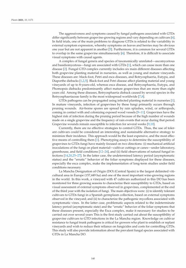
Plants 2021, 10, 1171
The aggressiveness and symptoms caused by fungal pathogens associated with GTDsdiffer significantly between grapevine-growing regions and vary depending on cultivars [6].In field trials, one of the main problems to diagnose GTDs is related to the variability inexternal symptom expression, whereby symptoms on leaves and berries may be obviousone year but are not apparent in another [5]. Furthermore, it is common for several GTDsto overlap in the same grapevine simultaneously [6]. Therefore, it is difficult to associatevisual symptoms with causal agents.
A complex of fungal genera and species of taxonomically unrelated—ascomycetousand basidiomycetous—fungi are associated with GTDs [1], which can cause more than onedisease [2]. Fungal GTD complex currently includes six main different diseases affectingboth grapevine planting material in nurseries, as well as young and mature vineyards.These diseases are: black-foot, Petri and esca diseases, and Botryosphaeria, Eutypa, andDiaporthe diebacks [1,2,5]. Black-foot and Petri disease affect planting material and youngvineyards of up to 8-years-old, whereas esca disease, and Botryosphaeria, Eutypa, andPhomopsis diebacks predominantly affect mature grapevines that are more than eightyears old. Among these diseases, Botryosphaeria dieback caused by several species in theBotryosphaeriaceae family is the most widespread worldwide [7,8].
GTDs pathogens can be propagated using infected planting material in nurseries [1].In mature vineyards, infection of grapevines by these fungi primarily occurs throughpruning wounds. Air-borne spores are spread by rain splashes, wind, or arthropods,coming in contact with and colonizing exposed wood vessels [9–11]. Grapevines have thehighest risk of infection during the pruning period because of the high number of woundsmade on a single grapevine and the frequency of rain events that occur during that period.Grapevine wounds remain susceptible to infection for several weeks [9,10].
Currently, there are no effective strategies to control GTDs. Thus, the use of toler-ant cultivars could be considered an interesting and sustainable alternative strategy tominimize their incidence. This approach would be the least expensive, and the most effec-tive means of controlling them [1]. Phenotyping assays to determine the susceptibility ofgrapevines to GTDs fungi have mainly focused on two directions: (i) mechanical artificialinoculations of the fungi on plant material—cultivar cuttings or canes—under laboratory,greenhouse, and field conditions [12–24], and (ii) field observations of natural fungal in-fections [14,20,25–27]. In the latter case, the undetermined latency period (asymptomaticstatus) and the “erratic” behavior of the foliar symptoms displayed for these diseases,especially the esca complex, make the implementation of long-term studies under fieldconditions necessary.
La Mancha Designation of Origin (DO) (Central Spain) is the largest delimited viti-cultural area in Europe (157,449 ha) and one of the most important wine-growing regionsin the world. In this work, a vineyard with 47 cultivars authorized in this DO has beenmonitored for three growing seasons to characterize their susceptibility to GTDs, based onvisual assessment of external symptoms observed in grapevines, complemented at the endof the third year with the isolation of fungi. The main objectives were: (i) to identify tolerantcultivars to GTDs fungi in a Spanish germplasm collection, based on external symptomsobserved in the vineyard; and (ii) to characterize the pathogenic mycoflora associated withsymptomatic vines. In the latter case, problematic aspects related to the indeterminatelatency period (asymptomatic state) and the “erratic” behavior of the foliar symptoms thatthese diseases present, especially the Esca complex, make it necessary for studies to becarried out over several years This is the first study carried out about the susceptibility ofgrapevine cultivars to GTD infections in the La Mancha region. Knowledge on cultivarresistance to fungal trunk pathogens is critical for growers who plant to establish or replantvineyards and wish to reduce their reliance on fungicides and costs for controlling GTDs.This study will also provide information about the prevalent fungal species associated withGTDs in La Mancha DO.
140
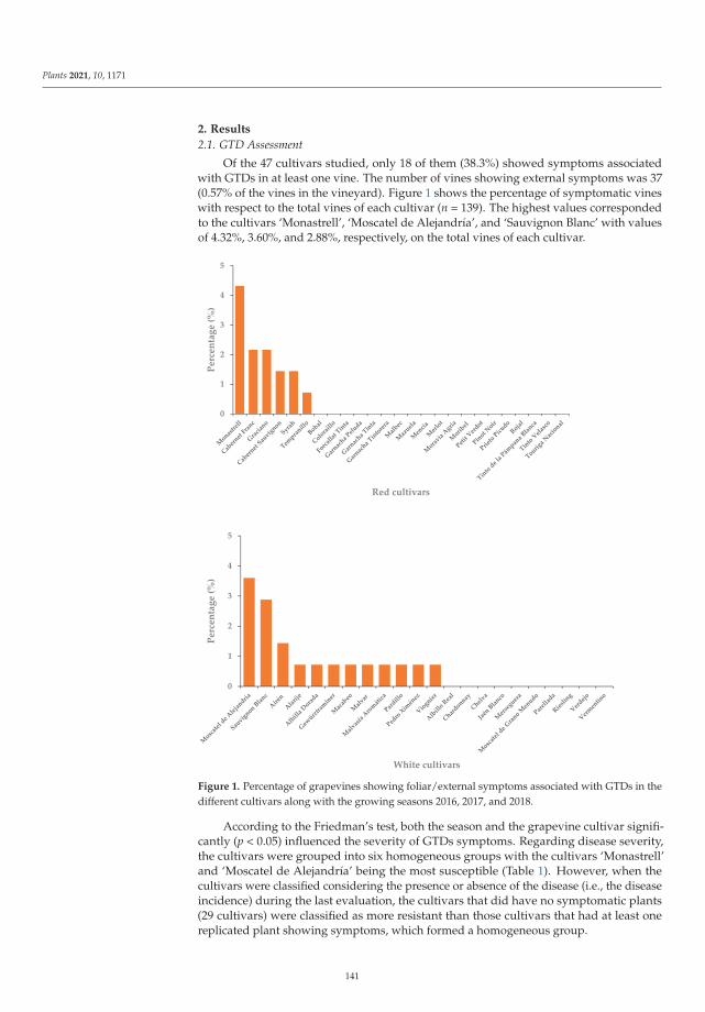
Plants 2021, 10, 1171
2. Results
2.1. GTD Assessment
Of the 47 cultivars studied, only 18 of them (38.3%) showed symptoms associatedwith GTDs in at least one vine. The number of vines showing external symptoms was 37(0.57% of the vines in the vineyard). Figure 1 shows the percentage of symptomatic vineswith respect to the total vines of each cultivar (n = 139). The highest values correspondedto the cultivars ‘Monastrell’, ‘Moscatel de Alejandría’, and ‘Sauvignon Blanc’ with valuesof 4.32%, 3.60%, and 2.88%, respectively, on the total vines of each cultivar.
0
1
2
3
4
5
Perc
enta
ge (%
)
Red cultivars
0
1
2
3
4
5
Perc
enta
ge (%
)
White cultivars
Figure 1. Percentage of grapevines showing foliar/external symptoms associated with GTDs in thedifferent cultivars along with the growing seasons 2016, 2017, and 2018.
According to the Friedman’s test, both the season and the grapevine cultivar signifi-cantly (p < 0.05) influenced the severity of GTDs symptoms. Regarding disease severity,the cultivars were grouped into six homogeneous groups with the cultivars ‘Monastrell’and ‘Moscatel de Alejandría’ being the most susceptible (Table 1). However, when thecultivars were classified considering the presence or absence of the disease (i.e., the diseaseincidence) during the last evaluation, the cultivars that did have no symptomatic plants(29 cultivars) were classified as more resistant than those cultivars that had at least onereplicated plant showing symptoms, which formed a homogeneous group.
141
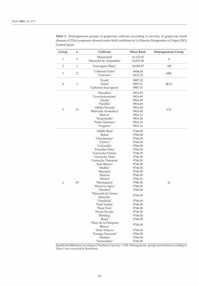
Plants 2021, 10, 1171
Table 1. Homogeneous groups of grapevine cultivars according to severity of grapevine trunkdiseases (GTDs) symptoms showed under field conditions in La Mancha Designation of Origin (DO),Central Spain.
Group n Cultivars Mean Rank Homogeneous Group
1 2‘Monastrell’ 10,122.03
10,097.48 A‘Moscatel de Alejandría’
2 1 ‘Sauvignon Blanc’ 10,028.47 AB
3 2‘Cabernet Franc’ 9958.05
9933.76ABC‘Graciano’
‘Syrah’ 9887.52BCD4 3 ‘Airén’ 9887.01
‘Cabernet Sauvignon’ 9887.01
5
‘Macabeo’ 9816.83
CD
‘Gewürztraminer’ 9816.69‘Alarije’ 9816.69
‘Pardillo’ 9816.69
10‘Albilla Dorada’ 9816.44
‘Malvasía Aromática’ 9816.44‘Malvar’ 9816.31
‘Tempranillo’ 9816.26‘Pedro Ximénez’ 9816.16
‘Viognier’ 9816.16
6
‘Albillo Real’ 9746.00
D
‘Bobal’ 9746.00‘Chardonnay’ 9746.00
‘Chelva’ 9746.00‘Coloraillo’ 9746.00
‘Forcallat Tinta’ 9746.00‘Garnacha Peluda’ 9746.00‘Garnacha Tinta’ 9746.00
‘Garnacha Tintorera’ 9746.00‘Jaén Blanco’ 9746.00
‘Malbec’ 9746.00’Mazuela’ 9746.00‘Mencía’ 9746.00’Merlot’ 9746.00
29 ‘Merseguera’ 9746.00‘Moravia Agria’ 9746.00
‘Moribel’ 9746.00‘Moscatel de Grano
Menudo’ 9746.00
‘Parellada’ 9746.00‘Petit Verdot’ 9746.00‘Pinot Noir’ 9746.00
‘Prieto Picudo’ 9746.00‘Riesling’ 9746.00
‘Rojal’ 9746.00‘Tinto de la Pámpana
Blanca’ 9746.00
‘Tinto Velasco’ 9746.00‘Touriga Nacional’ 9746.00
‘Verdejo’ 9746.00‘Vermentino’ 9746.00
Significant differences according to Friedman’s test at p = 0.05. Homogeneous. groups were formed according toDunn’s test corrected by Bonferroni.
142
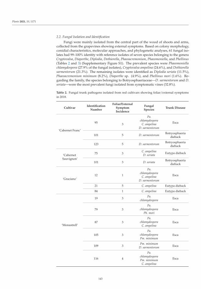
Plants 2021, 10, 1171
2.2. Fungal Isolation and Identification
Fungi were mainly isolated from the central part of the wood of shoots and arms,collected from the grapevines showing external symptoms. Based on colony morphology,conidial characteristics, molecular approaches, and phylogenetic analyses, 61 fungal iso-lates had 99–100% identity with reference isolates of seven species belonging to the generaCryptovalsa, Diaporthe, Diplodia, Dothiorella, Phaeoacremonium, Phaeomoniella, and Phellinus(Tables 2 and 3) (Supplementary Figure S1). The prevalent species were Phaeomoniellachlamydospora (27.9% of the fungal isolates), Cryptovalsa ampelina (24.6%), and Dothiorellasarmentorum (21.3%). The remaining isolates were identified as Diplodia seriata (11.5%),Phaeoacremonium minimum (8.2%), Diaporthe sp. (4.9%), and Phellinus mori (1.6%). Re-garding the family, the species belonging to Botryosphaeriaceae—D. sarmentorum and D.seriata—were the most prevalent fungi isolated from symptomatic vines (32.8%).
Table 2. Fungal trunk pathogens isolated from red cultivars showing foliar/external symptomsin 2018.
CultivarIdentification
Number
Foliar/ExternalSymptomIncidence
FungalSpecies
Trunk Disease
‘Cabernet Franc’
95
Pa.chlamydospora
Esca3 C. ampelinaD. sarmentorum
101 5 D. sarmentorum Botryosphaeriadieback
123 5 D. sarmentorum Botryosphaeriadieback
‘CabernetSauvignon’
75 3C. ampelina Eutypa diebackD. seriata
101 3 D. seriata Botryosphaeriadieback
‘Graciano’12 1
Pa.chlamydospora
EscaC. ampelinaD. sarmentorum
21 5 C. ampelina Eutypa dieback
84 1 C. ampelina Eutypa dieback
‘Monastrell’
19 3 Pa.chlamydospora Esca
79 3Pa.
chlamydospora EscaPh. mori
87 3Pa.
chlamydospora EscaC. ampelina
105 3Pa.
chlamydospora EscaPm. minimum
109 3Pm. minimum
EscaD. sarmentorum
116 4
Pa.chlamydospora
EscaPm. minimumC. ampelina
143
Plants 2021, 10, 1171
Table 2. Cont.
CultivarIdentification
Number
Foliar/ExternalSymptomIncidence
FungalSpecies
Trunk Disease
‘Syrah’
39 4Pa.
chlamydospora EscaC. ampelina
51 5
Pa.chlamydospora
EscaD. sarmentorumD. seriata
‘Tempranillo’ 35 2 None Not detected
Table 3. Fungal trunk pathogens isolated from white cultivars showing foliar/external symptomsin 2018.
CultivarIdentification
Number
Foliar/ExternalSymptomIncidence
FungalSpecies
Trunk Disease
‘Airén’16 3
Pa.chlamydospora
EscaC. ampelinaD. sarmentorum
79 3 D. sarmentorum Botryosphaeriadieback
‘Alarije’ 126 4Pa.
chlamydospora EscaC. ampelina
‘Albilla Dorada’ 36 3C. ampelina Eutypa diebackD. seriata
‘Gewürztraminer’ 22 4 Diaporthe sp. Phomopsisdieback
‘Macabeo’ 42 5D. sarmentorum Botryosphaeria
diebackD. seriata
‘Malvar’ 101 2Pa.
chlamydospora EscaD. sarmentorum
‘MalvasíaAromática’ 13 3 D. seriata Botryosphaeria
dieback
‘Moscatel deAlejandría’
45 2 Pa.chlamydospora Esca
46 2 Pa.chlamydospora Esca
47 2Pa.
chlamydospora EscaC. ampelina
48 2Pa.
chlamydospora EscaD. sarmentorum
50 2Pm. minimum
EscaD. sarmentorum
‘Pardillo’ 96 4 C. ampelina Eutypa dieback
‘Pedro Ximénez’ 91 1 Pm. minimum Esca
144
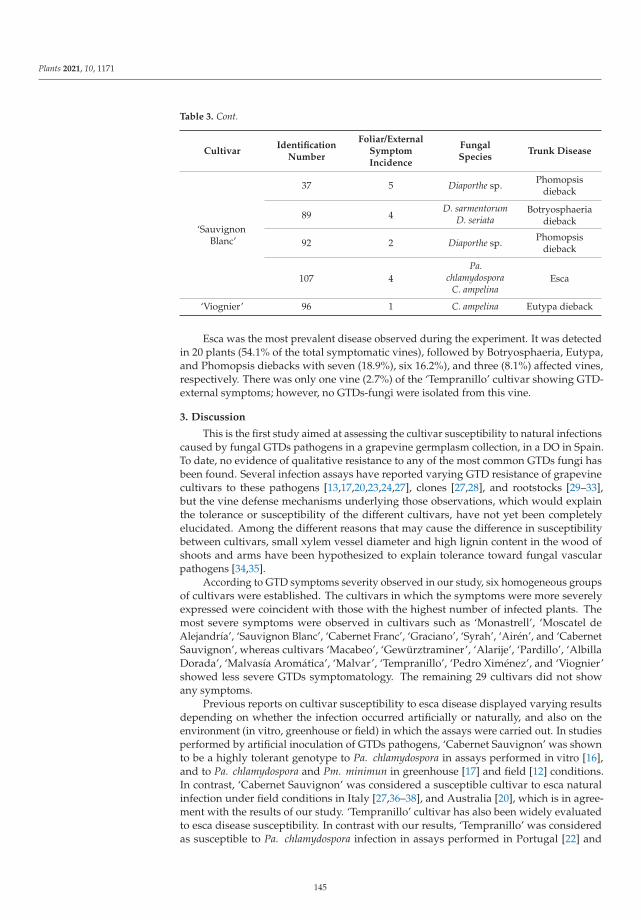
Plants 2021, 10, 1171
Table 3. Cont.
CultivarIdentification
Number
Foliar/ExternalSymptomIncidence
FungalSpecies
Trunk Disease
‘SauvignonBlanc’
37 5 Diaporthe sp. Phomopsisdieback
89 4D. sarmentorum Botryosphaeria
diebackD. seriata
92 2 Diaporthe sp. Phomopsisdieback
107 4Pa.
chlamydospora EscaC. ampelina
‘Viognier’ 96 1 C. ampelina Eutypa dieback
Esca was the most prevalent disease observed during the experiment. It was detectedin 20 plants (54.1% of the total symptomatic vines), followed by Botryosphaeria, Eutypa,and Phomopsis diebacks with seven (18.9%), six 16.2%), and three (8.1%) affected vines,respectively. There was only one vine (2.7%) of the ‘Tempranillo’ cultivar showing GTD-external symptoms; however, no GTDs-fungi were isolated from this vine.
3. Discussion
This is the first study aimed at assessing the cultivar susceptibility to natural infectionscaused by fungal GTDs pathogens in a grapevine germplasm collection, in a DO in Spain.To date, no evidence of qualitative resistance to any of the most common GTDs fungi hasbeen found. Several infection assays have reported varying GTD resistance of grapevinecultivars to these pathogens [13,17,20,23,24,27], clones [27,28], and rootstocks [29–33],but the vine defense mechanisms underlying those observations, which would explainthe tolerance or susceptibility of the different cultivars, have not yet been completelyelucidated. Among the different reasons that may cause the difference in susceptibilitybetween cultivars, small xylem vessel diameter and high lignin content in the wood ofshoots and arms have been hypothesized to explain tolerance toward fungal vascularpathogens [34,35].
According to GTD symptoms severity observed in our study, six homogeneous groupsof cultivars were established. The cultivars in which the symptoms were more severelyexpressed were coincident with those with the highest number of infected plants. Themost severe symptoms were observed in cultivars such as ‘Monastrell’, ‘Moscatel deAlejandría’, ‘Sauvignon Blanc’, ‘Cabernet Franc’, ‘Graciano’, ‘Syrah’, ‘Airén’, and ‘CabernetSauvignon’, whereas cultivars ‘Macabeo’, ‘Gewürztraminer’, ‘Alarije’, ‘Pardillo’, ‘AlbillaDorada’, ‘Malvasía Aromática’, ‘Malvar’, ‘Tempranillo’, ‘Pedro Ximénez’, and ‘Viognier’showed less severe GTDs symptomatology. The remaining 29 cultivars did not showany symptoms.
Previous reports on cultivar susceptibility to esca disease displayed varying resultsdepending on whether the infection occurred artificially or naturally, and also on theenvironment (in vitro, greenhouse or field) in which the assays were carried out. In studiesperformed by artificial inoculation of GTDs pathogens, ‘Cabernet Sauvignon’ was shownto be a highly tolerant genotype to Pa. chlamydospora in assays performed in vitro [16],and to Pa. chlamydospora and Pm. minimun in greenhouse [17] and field [12] conditions.In contrast, ‘Cabernet Sauvignon’ was considered a susceptible cultivar to esca naturalinfection under field conditions in Italy [27,36–38], and Australia [20], which is in agree-ment with the results of our study. ‘Tempranillo’ cultivar has also been widely evaluatedto esca disease susceptibility. In contrast with our results, ‘Tempranillo’ was consideredas susceptible to Pa. chlamydospora infection in assays performed in Portugal [22] and
145
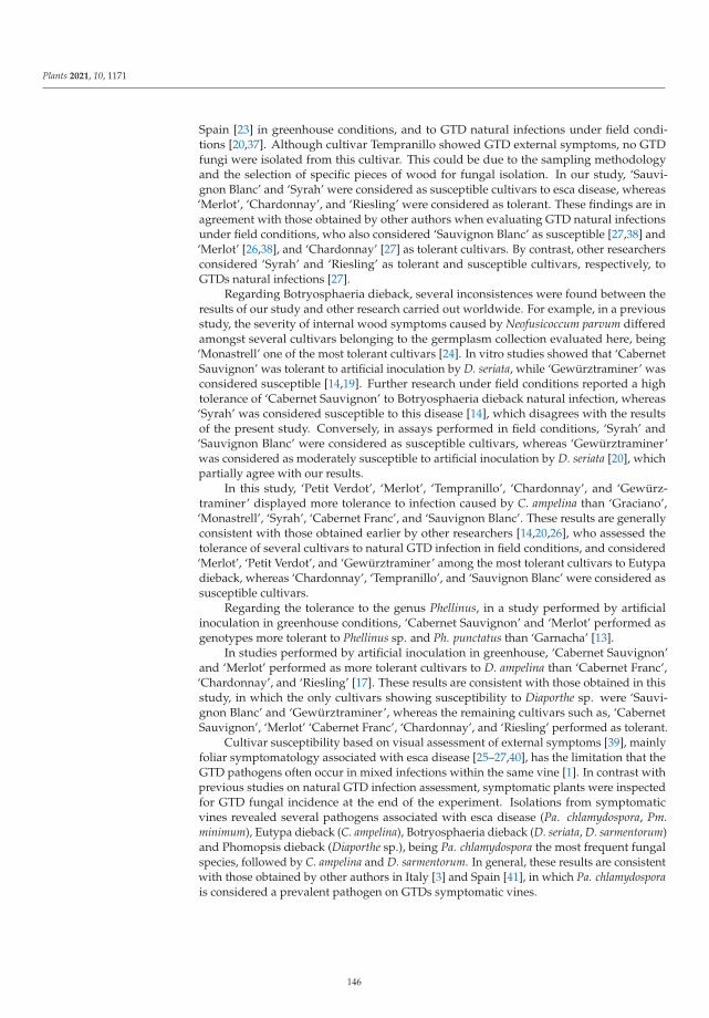
Plants 2021, 10, 1171
Spain [23] in greenhouse conditions, and to GTD natural infections under field condi-tions [20,37]. Although cultivar Tempranillo showed GTD external symptoms, no GTDfungi were isolated from this cultivar. This could be due to the sampling methodologyand the selection of specific pieces of wood for fungal isolation. In our study, ‘Sauvi-gnon Blanc’ and ‘Syrah’ were considered as susceptible cultivars to esca disease, whereas‘Merlot’, ‘Chardonnay’, and ‘Riesling’ were considered as tolerant. These findings are inagreement with those obtained by other authors when evaluating GTD natural infectionsunder field conditions, who also considered ‘Sauvignon Blanc’ as susceptible [27,38] and‘Merlot’ [26,38], and ‘Chardonnay’ [27] as tolerant cultivars. By contrast, other researchersconsidered ‘Syrah’ and ‘Riesling’ as tolerant and susceptible cultivars, respectively, toGTDs natural infections [27].
Regarding Botryosphaeria dieback, several inconsistences were found between theresults of our study and other research carried out worldwide. For example, in a previousstudy, the severity of internal wood symptoms caused by Neofusicoccum parvum differedamongst several cultivars belonging to the germplasm collection evaluated here, being‘Monastrell’ one of the most tolerant cultivars [24]. In vitro studies showed that ‘CabernetSauvignon’ was tolerant to artificial inoculation by D. seriata, while ‘Gewürztraminer’ wasconsidered susceptible [14,19]. Further research under field conditions reported a hightolerance of ‘Cabernet Sauvignon’ to Botryosphaeria dieback natural infection, whereas‘Syrah’ was considered susceptible to this disease [14], which disagrees with the resultsof the present study. Conversely, in assays performed in field conditions, ‘Syrah’ and‘Sauvignon Blanc’ were considered as susceptible cultivars, whereas ‘Gewürztraminer’was considered as moderately susceptible to artificial inoculation by D. seriata [20], whichpartially agree with our results.
In this study, ‘Petit Verdot’, ‘Merlot’, ‘Tempranillo’, ‘Chardonnay’, and ‘Gewürz-traminer’ displayed more tolerance to infection caused by C. ampelina than ‘Graciano’,‘Monastrell’, ‘Syrah’, ‘Cabernet Franc’, and ‘Sauvignon Blanc’. These results are generallyconsistent with those obtained earlier by other researchers [14,20,26], who assessed thetolerance of several cultivars to natural GTD infection in field conditions, and considered‘Merlot’, ‘Petit Verdot’, and ‘Gewürztraminer’ among the most tolerant cultivars to Eutypadieback, whereas ‘Chardonnay’, ‘Tempranillo’, and ‘Sauvignon Blanc’ were considered assusceptible cultivars.
Regarding the tolerance to the genus Phellinus, in a study performed by artificialinoculation in greenhouse conditions, ‘Cabernet Sauvignon’ and ‘Merlot’ performed asgenotypes more tolerant to Phellinus sp. and Ph. punctatus than ‘Garnacha’ [13].
In studies performed by artificial inoculation in greenhouse, ‘Cabernet Sauvignon’and ‘Merlot’ performed as more tolerant cultivars to D. ampelina than ‘Cabernet Franc’,‘Chardonnay’, and ‘Riesling’ [17]. These results are consistent with those obtained in thisstudy, in which the only cultivars showing susceptibility to Diaporthe sp. were ‘Sauvi-gnon Blanc’ and ‘Gewürztraminer’, whereas the remaining cultivars such as, ‘CabernetSauvignon’, ‘Merlot’ ‘Cabernet Franc’, ‘Chardonnay’, and ‘Riesling’ performed as tolerant.
Cultivar susceptibility based on visual assessment of external symptoms [39], mainlyfoliar symptomatology associated with esca disease [25–27,40], has the limitation that theGTD pathogens often occur in mixed infections within the same vine [1]. In contrast withprevious studies on natural GTD infection assessment, symptomatic plants were inspectedfor GTD fungal incidence at the end of the experiment. Isolations from symptomaticvines revealed several pathogens associated with esca disease (Pa. chlamydospora, Pm.minimum), Eutypa dieback (C. ampelina), Botryosphaeria dieback (D. seriata, D. sarmentorum)and Phomopsis dieback (Diaporthe sp.), being Pa. chlamydospora the most frequent fungalspecies, followed by C. ampelina and D. sarmentorum. In general, these results are consistentwith those obtained by other authors in Italy [3] and Spain [41], in which Pa. chlamydosporais considered a prevalent pathogen on GTDs symptomatic vines.
146
Plants 2021, 10, 1171
Cryptovalsa ampelina was previously reported in several regions of Spain [42,43]. Thispathogen is mainly found on pruning debris and rarely on standing vines showing symp-toms of trunk diseases [42].
The basidiomycete species Ph. mori was only isolated in one vine of the ‘Monastrell’cultivar together with the esca pathogen Pa. chlamydospora. This species has not beenreported so far as a pathogen associated with GTDs worldwide. The genus Phellinusappears to be associated with a secondary stage of the esca disease, colonizing grapevinesinitially infected by Pa. chlamydospora and Pm. minimum, which are more prevalent andvirulent species [5].
The Diaporthe species have been associated with several major diseases of grapevines,such as Phomopsis cane and leaf spot and Diaporthe dieback [44–47]. The most frequentspecies isolated of this genus in Europe are D. eres and D. ampelina (syn. Phomopsis viti-cola) [48]. Both species are shown to be pathogenic on grapevine [48]. Recently, two newDiaporthe species were isolated from symptomatic vines collected in Spain, namely Diaporthehispaniae and Diaporthe hungariae. These species were closely related, but clearly separatedbased on morphological and molecular characteristics from D. ampelina, historically knownas the most virulent Diaporthe species of grapevine [47].
The use of tolerant cultivars would be an interesting and sustainable alternativestrategy to control GTD infections. This study allowed for classifying several grapevinecultivars according to external symptoms associated with natural infections caused by fun-gal grapevine trunk pathogens in La Mancha DO, as well as to characterize the pathogenicmicroflora associated with symptomatic vines in this area. Knowledge of tolerant cultivarsto fungal trunk pathogens may help growers to reduce their reliance on fungicides andcosts for controlling GTDs. Further research is needed to evaluate the correlation betweenfoliar symptoms and wood deterioration, and to explore the mycoflora associated withasymptomatic vines.
4. Material and Methods
4.1. Study Area
A plot of the Instituto Regional de Investigación y Desarrollo Agroalimentario y Fore-stal (IRIAF) was planted with a grapevine germplasm collection and located at 663 m.a.s.l.(latitude: 39.176753N, longitude: −3.000247W) was used. This plot includes a broad rangeof V. vinifera cultivars authorized in the Castilla–La Mancha wine region, which can beconsidered representative of the vineyards in La Mancha DO. The soil is classified asCalcixerept petric. Its main features are: shallow (<40 cm depth), well-drained, with about40% of coarse elements and loam to sandy-clay-loam textures (46.6% sand; 32.2% silt, 2.12%clay) and 3.2% of organic matter content [49]. The region has a temperate climate with highdifferences in temperature between winter and summer. According to the Winkler index,this region is classified as Region IV, and it records scarce rainfall during the year (about350 mm), with less than 50% occurring in the vine growing season (between vine sproutingand harvesting). The reference evapotranspiration value (ET0) is about 1300 mm/year,exceeding 1000 mm during the active vegetation period.
4.2. Plant Material
The germplasm collection consists of 22 white (‘Airén’, ‘Alarije’, ‘Albilla Dorada’,‘Albillo Real’, ‘Chardonnay’, ‘Chelva’, ‘Gewürztraminer’, ‘Jaén Blanco’, ‘Macabeo’, ‘Mal-var’, ‘Malvasía Aromática’, ‘Merseguera’, ‘Moscatel de Alejandría’, ‘Moscatel de GranoMenudo’, ‘Pardillo’, ‘Parellada’, ‘Pedro Ximénez’, ‘Riesling’, ‘Sauvignon Blanc’, ‘Verdejo’,‘Vermentino’, and ‘Viognier’), and 25 red (‘Bobal’, ‘Cabernet Franc’, ‘Cabernet Sauvignon’,‘Coloraillo’, ‘Forcallat Tinta’, ‘Garnacha Peluda’, ‘Garnacha Tinta’, ‘Garnacha Tintorera’,‘Graciano’, ‘Malbec’, ‘Mazuela’, ‘Mencía’, ‘Merlot’, ‘Monastrell’, ‘Moravia Agria’, ‘Mori-bel’, ‘Petit Verdot’, ‘Pinot Noir’, ‘Prieto Picudo’, ‘Rojal’, ‘Syrah’, ‘Tempranillo’, ‘Tinto dela Pámpana Blanca’, ‘Tinto Velasco’, and ‘Touriga Nacional’) grapevine cultivars graftedonto 110 Richter rootstock and planted in 2002 with one panel of 139 vines per cultivar.
147
Plants 2021, 10, 1171
The information used in this research referred to 47 cultivars with 139 grapevines each(6533 vines in total). The planting pattern was 3 m between rows and 1.5 m betweenplants (density of 2222 grapevines/ha). All vines were double cordon trained and spurpruned, with no specific strategies to control GTDs. Vines were cultivated under irrigatedconditions by a drip system with two drippers per grapevine. Irrigation was appliedconsidering about 25% of crop evapotranspiration, and it represented 120–150 mm peryear, on average. The rows were positioned 120◦E-SE/300◦W-NW.
4.3. GTD Assessment and Fungal Isolations
All cultivars were inspected four to five times per season during the vegetative periodduring the growing seasons 2016, 2017, and 2018, between flowering and maturity, which isthe time when GTDs symptoms are most evident. The cultivar susceptibility was assessedfor esca foliar symptoms according to a scale ranging from 0 to 5, depending on theaffectation level and GTD-associated cordon dieback (partially or totally dry) [50] (Table 4).
Table 4. Scale of foliar/external symptoms incidence according to percentage of affected vegetation.
Level Foliar/External Symptom Incidence (%)
0 01 0–102 11–253 26–504 51–805 >80
Vines showing either esca foliar or cordon dieback symptoms were marked andrecorded. GTD fungal isolations were performed from these vines at the end of the study(2018) to correlate foliar symptoms and fungal incidence. In total, 37 samples were collectedfrom 18 different cultivars.
Fungal isolations were carried out according to the methodology described by [51].Pieces of wood from symptomatic canes and arms were debarked and cut into transverseslices approximately 1 mm thick. These slices were then surface disinfected by immersionin 70% alcohol for 1 or 2 min, depending on thickness, and air dried on sterile filterpaper. Later, they were placed in plates of malt extract agar supplemented with 0.5 g/L ofstreptomycin sulfate (MEAS) and incubated at 25 ◦C in darkness for 10 days. The plateswere observed daily to check the growth of the mycelium. Fungal colonies were transferredto new Petri dishes with Potato Dextrose Agar (PDA) and incubated at 25 ◦C in darkness, toobtain pure cultures. From these primary isolations, single spore or hyphal tipped isolateswere obtained previously to their identification.
4.4. Morphological and Molecular Identification of Fungal Cultures
Preliminary morphological identification of the isolates at different taxonomical levelswas carried out by observing the cultural and microscope characters of the colonies understereoscope and microscope, respectively [52]. Colonies were then tentatively grouped asBasidiomycetes, or fungi belonging to the families Botryosphaeriaceae, and Diatrypaceae,the genus Phaeoacremonium, and the species Pa. chlamydospora.
For species identity confirmation, total fungal DNA was extracted from fungal culturesgrown on PDA medium, using the E.Z.N.A. Plant DNA Kit (Omega Bio-tek, Norcross,GA, USA), following the manufacturer’s instructions. Diatrypaceae, Basidiomycetes,and Pa. chlamydospora isolates were identified based on the sequence of the ITS region,Botryosphaeriaceae isolates were identified based on the sequences of ITS and a portion oftranslation elongation factor 1-alpha (tef-1α) region, while, for Phaeoacremonium isolates,part of the β-tubulin gene (tub) was used. The primer pairs used for amplification andsequencing of each region were as follows: ITS1-F [53] and ITS4 [54] for ITS, EF1-688F andEF1-1251R for tef-1α [55] and BtCadF and BtCadR for tub [56]. Amplification by polymerase
148
Plants 2021, 10, 1171
chain reaction (PCR) was performed in a total volume of 25 μL using HotBegan™ TaqDNA Polymerase (Canvax Biotech SL, Córdoba, Spain), according to the manufacturer’sinstructions on a Peltier Thermal Cycler-200 (MJ Research). One reaction was composedof 1× PCR Buffer B, 2.5 mM of MgCl2, 0.8 mM of dNTPs, 0.4 μM of each primer, 1 U ofHotBegan Taq DNA Polymerase, and 1 μL of purified template DNA. The PCR cyclingconditions consisted of an initial step of 3 min at 94 ◦C, followed by 35 cycles of denaturationat 94 ◦C for 30 s, annealing at 55 ◦C for 30 s and elongation at 72 ◦C for 45 s. A final extensionwas performed at 72 ◦C for 5 min. PCR products were confirmed by 1.2% agarose gelelectrophoresis and were purified and sequenced by Macrogen Inc. (Madrid, Spain) usingboth PCR primers. Sequences were assembled and edited using Sequencher software 5.0(Gene Codes Corp., Ann Arbor, MI, USA). The isolate identities were based mainly onBLASTn searches in NCBI, but, for Botryosphaeriaceae isolates, the multiple sequencealignments and maximum likelihood phylogenetic analyses were conducted in MEGAX [57], using closely related ex-type or representative species as phylogenetic reference.
4.5. Data Analysis
Mean, standard deviation, and sum were calculated using the descriptive processof the software Statistix 10 (Analytical Software; Tallahassee, FL, USA). The effect ofthe evaluation season and the grapevine cultivar on the disease severity were examinedusing Friedman’s test. For that, the back-transformation of the rating scale was used.Friedman’s test was used because the dependent variable does not satisfy the requirementsof parametric tests. The means were compared using Dunn’s test with a Bonferroniadjustment after a Kruskall–Wallis test at p = 0.05 [58]. A Zar’s test of multiple comparisonsof proportions was performed to study the effect of the cultivar on disease presence (1) orabsence (0) in the last studied season (2018) [59]. Data were analyzed using the softwareStatistix 10 (Analytical Software; Tallahassee, FL, USA) and SPSS (version 19; SPSS Inc.,Chicago, IL, USA).
Supplementary Materials: The following are available online at https://www.mdpi.com/article/10.3390/plants10061171/s1, Figure S1: Maximum likelihood phylogeny inferred from the alignmentof combined sequences of a portion of translation elongation factor 1-alpha (tef-1α) region andinternal transcribed spacers (ITS). Support values higher than 70% are given at the nodes. The treewas rooted using Neofusicoccum luteum (CBS110299 and CBS140738) as outgroup sequences. Scale barshows expected changes per site. Species isolated in this study are indicated in bold. This analysiswas conducted in MEGAX, and involved 26 nucleotide sequences. There were a total of 825 positionsin the final dataset (EF:1-313 and ITS:314-825).
Author Contributions: Conceptualization, D.G.; methodology, D.G., M.L., J.A., J.M., and J.L.C.-V.;investigation, J.L.C.-V., J.A., P.M.I.-C., J.M., and J.M.-G.; resources, D.G., J.A., and J.L.C.-V.; writing—original draft preparation, J.L.C.-V.; writing—review and editing, J.L.C.-V., D.G., J.A., and J.M.; projectadministration, D.G., and J.L.C.-V.; funding acquisition, D.G. All authors have read and agreed to thepublished version of the manuscript.
Funding: This research was founded by FEDER funding through a State Program of I+D+i orientedto the Challenges of Society (RTA2015-00015-C02-00), supported by The National Institute for Agri-cultural and Food Research and Technology (INIA). D.G. and J.M. were supported by the Ramón yCajal program, Spanish Government (RyC-2017-23098 and RyC-028404).
Institutional Review Board Statement: Not applicable.
Informed Consent Statement: Not applicable.
Conflicts of Interest: The authors declare no conflict of interest. The funders had no role in the designof the study; in the collection, analyses, or interpretation of data; in the writing of the manuscript, orin the decision to publish the results.
149
Plants 2021, 10, 1171
References
1. Gramaje, D.; Úrbez-Torres, J.R.; Sosnowski, M.R. Management trunk diseases with respect to etiology and epidemiology: Currentstrategies and future prospects. Plant Dis. 2018, 102, 12–39. [CrossRef]
2. Bertsch, C.; Ramírez-Suero, M.; Magnin-Robert, M.; Larignon, P.; Chong, J.; Abou-Mansour, E.; Fontaine, F. Grapevine trunkdiseases: Complex and still poorly understood. Plant Pathol. 2013, 62, 243–265. [CrossRef]
3. Mugnai, L.; Graniti, A.; Surico, G. Esca (black measles) and brown wood-streaking: Two old and elusive diseases of grapevines.Plant Dis. 1999, 83, 404–416. [CrossRef]
4. Chiarappa, L. Esca (black measles) of grapevine. An overview. Phytopathol. Mediterr. 2000, 39, 11–15.5. Mondello, V.; Larignon, P.; Armengol, J.; Kortekamp, A.; Váczy, K.Z.; Prezman, F.; Serrano, E.; Rego, C.; Mugnai, L.; Fontaine, F.
Management of grapevine trunk diseases: Knowledge transfer, current strategies and innovative strategies adopted in Europe.Phytopathol. Mediterr. 2018, 57, 369–383. [CrossRef]
6. Yan, J.Y.; Xie, Y.; Zhang, W.; Wang, Y.; Liu, J.K.; Hyde, K.D.; Li, X.H. Species of Botryosphaeriaceae involved in grapevine diebackin China. Fungal Divers. 2013, 61, 221–236. [CrossRef]
7. Úrbez-Torres, J.R.; Leavitt, G.M.; Voegel, T.M.; Gubler, W.D. Identification and distribution of Botryosphaeria spp. associated withgrapevine cankers in California. Plant Dis. 2006, 90, 1490–1503. [CrossRef] [PubMed]
8. Úrbez-Torres, J.R. The status of Botryosphaeriaceae species infecting grapevines. Phytopathol. Mediterr. 2011, 50, 5–45.9. Eskalen, A.; Feliciano, A.J.; Gubler, W.D. Susceptibility of grapevine pruning wounds and symptom development in response to
infection by Phaeoacremonium aleophilum and Phaeomoniella chlamydospora. Plant Dis. 2007, 91, 1100–1104. [CrossRef]10. Úrbez-Torres, J.R.; Gubler, W.D. Double pruning, a potential method to control Bot canker disease of grapes, and susceptibility of
grapevine pruning wounds to infection by Botryosphaeriaceae. Phytopathol. Mediterr. 2008, 48, 185.11. Moyo, P.; Allsopp, E.; Roets, F.; Mostert, L.; Halleen, F. Arthropods vector grapevine trunk disease pathogens. Phytopathology
2014, 104, 1063–1069. [CrossRef]12. Feliciano, A.J.; Eskalen, A.; Gubler, W.D. Differential susceptibility of three grapevine cultivars to Phaeoacremoium aleophilum and
Phaeomoniella chlamydospora in California. Phytopathol. Mediterr. 2004, 43, 66–69.13. Sosnowski, M.R.; Wicks, T.J.; Lardner, R.; Scott, E.S. The influence of grapevine cultivar and isolate of Eutypa lata on wood and
foliar symptoms. Plant Dis. 2007, 91, 924–931. [CrossRef]14. Savocchia, S.; Steel, C.C.; Stodart, B.J.; Somers, A. Pathogenicity of Botryosphaeria species isolated from declining grapevines in
sub-tropical regions of eastern Australia. Vitis 2007, 46, 27–32.15. Zanzotto, A.; Gardiman, M.; Lovat, L. Effect of Phaeomoniella chlamydospora and Phaeoacremonium sp. on in vitro grapevine plants.
Sci. Hortic. 2008, 116, 404–408. [CrossRef]16. Landi, L.; Murolo, S.; Romanazzi, G. Colonization of Vitis spp. wood by sGFP-transformed Phaeomoniella chlamydospora, a
tracheomycotic fungus involved in esca disease. Phytopathology 2012, 102, 290–297. [CrossRef]17. Travadon, R.; Rolshausen, P.E.; Gubler, W.D.; Cadle-Davidson, L.; Baumgartner, K. Susceptibility of cultivated and wild Vitis spp.
to wood infection by fungal trunk pathogens. Plant Dis. 2013, 97, 1529–1536. [CrossRef] [PubMed]18. Billones-Baaijens, R.; Jones, E.E.; Ridgway, H.J.; Jaspers, M.V. Susceptibility of common rootstock and scion varieties of grapevines
to Bottryosphaeriaceae species. Australas. Plant Pathol. 2014, 43, 25–31. [CrossRef]19. Guan, X.; Essakhi, S.; Laloue, H.; Nick, P.; Bertsch, C.; Chong, J. Mining new resources for grape resistance against Botryosphaeri-
aceae: A focus on Vitis vinifera subsp. sylvestris. Plant Pathol. 2016, 65, 273–284. [CrossRef]20. Sosnowski, M.; Ayres, M.; Wicks, T.; McCarthy, M.; Scott, E. Investigating potential for resistance to grapevine trunk diseases.
Wine Vitic. J. 2016, 31, 41–45.21. Markakis, E.A.; Koubouris, G.C.; Sergentani, C.K.; Ligoxigakis, E.K. Evaluation of Greek grapevine cultivars for resistance to
Phaeomoniella chlamydospora. Eur. J. Plant Pathol. 2017, 149, 277–283. [CrossRef]22. Sofia, J.; Mota, M.; Gonçalves, M.T.; Rego, C. Response of four Portuguese grapevine cultivars to infection by Phaeomoniella
chlamydospora. Phytopathol. Mediterr. 2018, 57, 506–518.23. Martínez-Diz, M.P.; Díaz-Losada, E.; Barajas, E.; Ruano-Rosa, D.; Andrés-Sodupe, M.; Gramaje, D. Screening of Spanish Vitis
vinifera germplasm for resistance to Phaeomoniella chlamydospora. Sci. Hortic. 2019, 246, 104–109. [CrossRef]24. Chacón, J.L.; Gramaje, D.; Izquierdo, P.M.; Martínez, J.; Mena, A. Evaluation of six red grapevine cultivars inoculated with
Neofusicoccum parvum. Eur. J. Plant Pathol. 2020, 158, 811–815. [CrossRef]25. Marchi, G. Susceptibility to esca of various grapevine (Vitis vinifera) cultivars grafted on different rootstocks in a vineyard in the
province of Siena (Italy). Phytopathol. Mediterr. 2001, 40, 27–36.26. Fussler, L.; Kobes, N.; Bertrand, F.; Maumy, M.; Grosman, J.; Savary, S. Characterization of grapevine trunk diseases in France
from data generated by National Grapevine Wood Diseases Survey. Phytopathology 2008, 98, 571–579. [CrossRef]27. Murolo, S.; Romanazzi, G. Effects of grapevine cultivar, rootstock and clone on esca disease. Australas. Plant Pathol. 2014, 43,
215–221. [CrossRef]28. Berlanas, C.; Songy, A.; Clément, C.; Fontaine, F.; Gramaje, D. Variation amongst ‘Tempranillo’ clones in susceptibility to
Neofusicoccum parvum. Phytopathol. Mediterr. 2017, 56, 545.29. Eskalen, A.; Gubler, W.D.; Khan, A. Rootstock susceptibility to Phaeomoniella chlamydospora and Phaeoacremonium spp. Phytopathol.
Mediterr. 2001, 40, S433–S438.
150
Plants 2021, 10, 1171
30. Gubler, W.D.; Baumgartner, K.; Browne, G.T.; Eskalen, A.; Rooney-Latham, S.; Petit, E.; Bayramian, L.A. Root diseases ofgrapevines in California and their control. Australas. Plant Pathol. 2004, 33, 157–165. [CrossRef]
31. Jaspers, M.V.; Bleach, C.M.; Harvey, I.C. Susceptibility of grapevine rootstocks to Cylindrocarpon disease. Phytopathol. Mediterr.2007, 46, 114.
32. Alaniz, S.; García-Jiménez, J.; Abad-Campos, P.; Armengol, J. Susceptibility of grapevine rootstocks to Cylindrocarpon liriodendriand C. macrodidymum. Sci. Hortic. 2010, 125, 305–308. [CrossRef]
33. Gramaje, D.; García-Jiménez, J.; Armengol, J. Field evaluation of grapevine rootstocks inoculated with fungi associated with Petridisease and esca. Am. J. Enol. Vitic. 2010, 61, 512–520. [CrossRef]
34. Rolshausen, P.E.; Greve, L.C.; Labavitch, J.M.; Mahoney, N.E.; Molyneux, R.J.; Gubler, W.D. Pathogenesis of Eutypa lata ingrapevine: Identification of virulence factors and biochemical characterization of cordon dieback. Phytopathology 2008, 98,222–229. [CrossRef]
35. Pouzoulet, J.; Pivovaroff, A.L.; Santiago, L.S.; Rolshausen, P.E. Can vessel dimension explain tolerance toward fungal vascularwilt diseases in woody plants? Lessons from Dutch elm disease and esca disease in grapevine. Front. Plant Sci. 2014, 5, 1–11.[CrossRef]
36. Andreini, L.; Caruso, G.; Bertolla, C.; Scalabrelli, G.; Viti, R.; Gucci, R. Gas exchange, stem water potential and xylem flux onsome grapevine cultivars affected by esca disease. S. Afr. J. Enol. Vitic. 2009, 30, 142–147. [CrossRef]
37. Andreini, L.; Guarino, L.; Viti, R.; Scalabrelli, G. Evaluation of the effect of esca disease on bud break in Vitis vinifera L.: Possiblerelationship between cultivars and rootstocks. Vitis 2013, 52, 33–40.
38. Serra, S.; Ligios, V.; Schianchi, N.; Prota, V.A.; Deidda, A.; Scanu, B. Incidence of grapevine trunk diseases on four cultivars inSardinia, Southern Italy. Vitis 2021, 60, 35–42.
39. Sosnowski, M.; Ayres, M.; Wicks, T.; McCarthy, M. In search of resistance to grapevine trunk diseases. Wine Vitic. J. 2013, 28,55–58.
40. Bruez, E.; Lecomte, P.; Grosman, J.; Doublet, B.; Bertsch, C.; Guerin-Dubrana, L. Overview of grapevine trunk diseases in Francein the 2000s. Phytopathol. Mediterr. 2013, 52, 262–275.
41. Luque, J.; Martos, S.; Aroca, A.; Raposo, R.; García-Figueres, F. Symptoms and fungi associated with declining mature grapevineplants in northeast Spain. J. Plant Pathol. 2009, 91, 381–390.
42. Luque, J.; Sierra, D.; Torres, E.; García, F. Cryptovalsa ampelina on grapevines in N.E. Spain: Identification and pathogenicity.Phytopathol. Mediterr. 2006, 45, S101–S109.
43. Martín, M.T.; Martín, L.; de-Francisco, M.T.; Cobos, R. First report of Lasidiplodia theobromae and Cryptovalsa ampelina associatedwith grapevine decline from Castilla y Leon, Spain. Plant Dis. 2009, 93, 545. [CrossRef]
44. Mostert, L.; Crous, P.W.; Kang, J.C.; Phillips, A.J. Species of Phomopsis and a Libertella sp. occurring on grapevines with specificreference to South Africa: Morphological, cultural, molecular and pathological characterization. Mycologia 2001, 93, 146–167.[CrossRef]
45. van Niekerk, J.M.; Groenewald, J.Z.; Farr, D.F.; Fourie, P.H.; Halleer, F.; Crous, P.W. Reassessment of Phomopsis species ongrapevines. Australas. Plant Pathol. 2005, 34, 27–39. [CrossRef]
46. Rawnsley, B.; Wicks, T.J.; Scott, E.S.; Stummer, B.E. Diaporthe perjuncta does not cause Phomopsis cane and leaf spot disease ofgrapevine in Australia. Plant Dis. 2004, 88, 1005–1010. [CrossRef]
47. Úrbez-Torres, J.R.; Peduto, F.; Smith, R.J.; Gubler, W.D. Phomopsis dieback: A grapevine trunk disease caused by Phomopsisviticola in California. Plant Dis. 2013, 97, 1571–1579. [CrossRef]
48. Guarnaccia, V.; Groenewald, J.Z.; Woodhall, J.; Armengol, J.; Cinelli, T.; Eichmeier, A.; Crous, P.W. Diaporthe diversity andpathogenicity revealed from a broad survey of grapevine diseases in Europe. Pers. Mol. Phylogenet. Evol. 2018, 40, 135–153.[CrossRef]
49. Amorós-Ortíz-Villajos, J.A.; Bravo-Martín-Consuegra, S.; García-Navarro, F.J.; Pérez-de-los-Reyes, C.; Chacón-Vozmediano,J.L.; Martínez-Gascueña, J.; Jiménez-Ballesta, R. Atlas de Suelos Vitícolas de Castilla-La Mancha, 1st ed.; Martínez, F.J., Ed.; Arte yComunicación Calatrava: Almagro, Spain, 2015; pp. 152–153. (In Spanish)
50. Lecomte, P.; Darrieutort, G.; Liminana, J.M.; Comont, G.; Muruamendiaraz, A.; Fermaud, M. New insights into esca of grapevine:The development of foliar symptoms and their association with xylem discoloration. Plant Dis. 2012, 96, 924–934. [CrossRef]
51. Larignon, P.; Dubos, B. Fungi associated with esca disease in grapevine. Eur. J. Plant Pathol. 1997, 103, 147–157. [CrossRef]52. Armengol, J.; Vicent, A.; Torné, L.; García-Figueres, F.; García-Jiménez, J. Fungi associated with esca and grapevine declines in
Spain: A three-year survey. Phytopathol. Mediterr. 2001, 40, S325–S329.53. Gardes, M.; Bruns, T.D. ITS primers with enhanced specificity for basiodiomycetes-applications to the identification of mycorrhizae
and rusts. Mol. Ecol. 1993, 2, 113–118. [CrossRef]54. White, T.J.; Bruns, T.D.; Lee, S.B.; Taylor, J.W. Amplification and direct sequencing of fungal ribosomal RNA genes for phylogenet-
ics. In PCR Protocols: A Guide to Methods and Applications; Innis, M.A., Gelfand, D.H., Sninsky, J.J., White, T.J., Eds.; AcademicPress: New York, NY, USA, 1990; Volume 18, pp. 315–322.
55. Alves, A.; Crous, P.W.; Correia, A.; Phillips, A.J.L. Morphological and molecular data reveal cryptic speciation in Lasiodiplodiatheobromae. Fungal Divers. 2008, 28, 1–13.
56. Travadon, R.; Lawrence, D.P.; Rooney-Latham, S.; Gubler, W.D.; Wilcox, W.F.; Rolshausen, P.E.; Baumgartner, K. Cadophora speciesassociated with wood-decay of grapevine in North America. Fungal Biol. 2015, 119, 53–66. [CrossRef] [PubMed]
151

Plants 2021, 10, 1171
57. Kumar, S.; Stecher, G.; Li, M.; Knyaz, C.; Tamura, K. MEGA X: Molecular evolutionary genetics analysis across computingplatforms. Mol. Biol. Evol. 2018, 35, 1547–1549. [CrossRef]
58. Demšar, J. Statistical comparisons of classifiers over multiple data sets. J. Mach. Learn. Res. 2006, 7, 1–30.59. Zar, J.H. Biostatistical Analysis, 5th ed.; Pearson Education India: New Delhi, India, 2010; pp. 724–725.
152
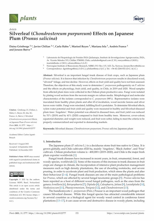
plants
Article
Silverleaf (Chondrostereum purpureum) Effects on JapanesePlum (Prunus salicina)
Daina Grinbergs 1,*, Javier Chilian 1,*, Carla Hahn 1, Marisol Reyes 1, Mariana Isla 1, Andrés France 1
and Jorunn Børve 2
Citation: Grinbergs, D.; Chilian, J.;
Hahn, C.; Reyes, M.; Isla, M.;
France, A.; Børve, J. Silverleaf
(Chondrostereum purpureum) Effects
on Japanese Plum (Prunus salicina).
Plants 2021, 10, 2777. https://
doi.org/10.3390/plants10122777
Academic Editor: Carlos Agustí-
Brisach
Received: 5 August 2021
Accepted: 14 September 2021
Published: 16 December 2021
Publisher’s Note: MDPI stays neutral
with regard to jurisdictional claims in
published maps and institutional affil-
iations.
Copyright: © 2021 by the authors.
Licensee MDPI, Basel, Switzerland.
This article is an open access article
distributed under the terms and
conditions of the Creative Commons
Attribution (CC BY) license (https://
creativecommons.org/licenses/by/
4.0/).
1 Laboratorio de Fitopatología de Frutales INIA Quilamapu, Instituto de Investigaciones Agropecuarias, INIA,Av. Vicente Méndez 515, Chillán 3780000, Chile; [email protected] (C.H.); [email protected] (M.R.);[email protected] (M.I.); [email protected] (A.F.)
2 Norwegian Institute of Bioeconomy Research, NIBIO, P.O. Box 115, 1431 Ås, Norway; [email protected]* Correspondence: [email protected] (D.G.); [email protected] (J.C.); Tel.: +56-42-2206-832 (D.G. & J.C.)
Abstract: Silverleaf is an important fungal trunk disease of fruit crops, such as Japanese plum(Prunus salicina). It is known that infection by Chondrostereum purpureum results in discolored wood,“silvered” foliage, and tree decline. However, effects on fruit yield and quality have not been assessed.Therefore, the objectives of this study were to determine C. purpureum pathogenicity on P. salicinaand the effects on physiology, fruit yield, and quality, in Chile, in 2019 and 2020. Wood samplesfrom affected plum trees were collected in the Chilean plum productive area. Fungi were isolatedby plating wood sections from the necrosis margin on culture media. Morphological and molecularcharacteristics of the isolates corresponded to C. purpureum (98%). Representative isolates wereinoculated from healthy plum plants and after 65-d incubation, wood necrotic lesions and silverleaves were visible. Fungi were reisolated, fulfilling Koch’s postulates. To determine Silverleaf effects,xylem water potential and fruit yield and quality were measured in healthy and Silverleaf-diseasedplum trees ‘Angeleno’. Water potential was altered in diseased trees, and fruit yield was reducedby 51% (2019) and by 41% (2020) compared to fruit from healthy trees. Moreover, cover-colour,equatorial-diameter, and weight were reduced, and fruit were softer, failing to meet the criteria to beproperly commercialized and exported to demanding markets.
Keywords: Silverleaf disease; Chondrostereum purpureum; Prunus salicina; Japanese plum
1. Introduction
The Japanese plum (P. salicina L.) is a deciduous stone fruit tree native to China. It isgrown globally, and Chile cultivates 4520 ha, mainly ‘Angeleno’, ‘Black-Amber’, and ‘Friar’cultivars. Chilean production volume is ~400,000 t (2019–2020), and Chile is the major freshplum exporter worldwide [1].
Fungal trunk diseases have increased in recent years, in fruit, ornamental, forest, andwoody species, worldwide [2]. Some of the reasons of this increase in trunk diseases in fruitcrops are changes in climate, the incorporation of intensive plant production managementmeasures such as high density plantations, the use of dwarfing rootstocks, and severepruning, in order to accelerate plant and fruit production, which stress the plants and altertheir behaviour [2–4]. Fungal trunk diseases are one of the main pathological problemsfor Prunus, which are affected by several fungal taxonomic groups [5]. Prunus salicina hasbeen reported as a host of different wood fungal pathogens, such as the ones from thegenera Armillaria [6], Botryosphaeria [7], Diplodia [8], Calosphaeria, Jattaea [9], Lasiodiplodia [10],Neofusicoccum [11], Phaeoacremonium, Tonignia [12], and Chondrostereum [13].
The basidiomycete C. purpureum (Pers.) Pouzar is an important wood pathogen thatcauses Silverleaf disease. While this fungal species has attracted considerable interestin several countries as a biological agent for woody weed control in coniferous forestplantations [13–17], it can cause severe and destructive disease in woody plants, including
Plants 2021, 10, 2777. https://doi.org/10.3390/plants10122777 https://www.mdpi.com/journal/plants153
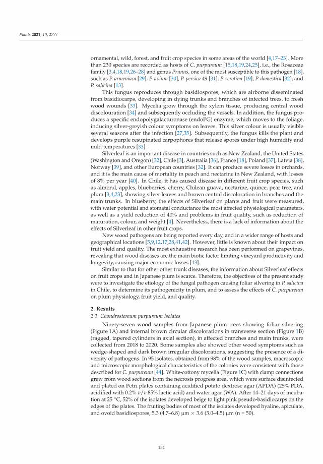
Plants 2021, 10, 2777
ornamental, wild, forest, and fruit crop species in some areas of the world [4,17–23]. Morethan 230 species are recorded as hosts of C. purpureum [15,18,19,24,25], i.e., the Rosaceaefamily [3,4,18,19,26–28] and genus Prunus, one of the most susceptible to this pathogen [18],such as P. armeniaca [29], P. avium [30], P. persica 49 [31], P. serotina [19], P. domestica [32], andP. salicina [13].
This fungus reproduces through basidiospores, which are airborne disseminatedfrom basidiocarps, developing in dying trunks and branches of infected trees, to freshwood wounds [33]. Mycelia grow through the xylem tissue, producing central wooddiscolouration [34] and subsequently occluding the vessels. In addition, the fungus pro-duces a specific endopolygalacturonase (endoPG) enzyme, which moves to the foliage,inducing silver-greyish colour symptoms on leaves. This silver colour is usually visibleseveral seasons after the infection [27,35]. Subsequently, the fungus kills the plant anddevelops purple resupinated carpophores that release spores under high humidity andmild temperatures [33].
Silverleaf is an important disease in countries such as New Zealand, the United States(Washington and Oregon) [32], Chile [3], Australia [36], France [18], Poland [37], Latvia [38],Norway [39], and other European countries [32]. It can produce severe losses in orchards,and it is the main cause of mortality in peach and nectarine in New Zealand, with lossesof 8% per year [40]. In Chile, it has caused disease in different fruit crop species, suchas almond, apples, blueberries, cherry, Chilean guava, nectarine, quince, pear tree, andplum [3,4,23], showing silver leaves and brown central discoloration in branches and themain trunks. In blueberry, the effects of Silverleaf on plants and fruit were measured,with water potential and stomatal conductance the most affected physiological parameters,as well as a yield reduction of 40% and problems in fruit quality, such as reduction ofmaturation, colour, and weight [4]. Nevertheless, there is a lack of information about theeffects of Silverleaf in other fruit crops.
New wood pathogens are being reported every day, and in a wider range of hosts andgeographical locations [5,9,12,17,28,41,42]. However, little is known about their impact onfruit yield and quality. The most exhaustive research has been performed on grapevines,revealing that wood diseases are the main biotic factor limiting vineyard productivity andlongevity, causing major economic losses [43].
Similar to that for other other trunk diseases, the information about Silverleaf effectson fruit crops and in Japanese plum is scarce. Therefore, the objectives of the present studywere to investigate the etiology of the fungal pathogen causing foliar silvering in P. salicinain Chile, to determine its pathogenicity in plum, and to assess the effects of C. purpureumon plum physiology, fruit yield, and quality.
2. Results
2.1. Chondrostereum purpureum Isolates
Ninety-seven wood samples from Japanese plum trees showing foliar silvering(Figure 1A) and internal brown circular discolorations in transverse section (Figure 1B)(ragged, tapered cylinders in axial section), in affected branches and main trunks, werecollected from 2018 to 2020. Some samples also showed other wood symptoms such aswedge-shaped and dark brown irregular discolorations, suggesting the presence of a di-versity of pathogens. In 95 isolates, obtained from 98% of the wood samples, macroscopicand microscopic morphological characteristics of the colonies were consistent with thosedescribed for C. purpureum [44]. White-cottony mycelia (Figure 1C) with clamp connectionsgrew from wood sections from the necrosis progress area, which were surface disinfectedand plated on Petri plates containing acidified potato dextrose agar (APDA) (25% PDA,acidified with 0.2% v/v 85% lactic acid) and water agar (WA). After 14–21 days of incuba-tion at 25 ◦C, 52% of the isolates developed beige to light pink pseudo-basidiocarps on theedges of the plates. The fruiting bodies of most of the isolates developed hyaline, apiculate,and ovoid basidiospores, 5.3 (4.7–6.8) μm × 3.6 (3.0–4.5) μm (n = 50).
154
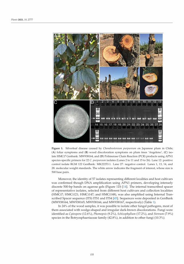
Plants 2021, 10, 2777
Figure 1. Silverleaf disease caused by Chondrostereum purpureum on Japanese plum in Chile;(A) foliar symptoms and (B) wood discoloration symptoms on plum trees ‘Angeleno’, (C) iso-late HMCi7 Genbank: MW938164, and (D) Polimerase Chain Reaction (PCR) products using APN1species-specific primers for 22 C. purpureum isolates (Lanes 2 to 11 and 15 to 26). Lane 12: positivecontrol isolate RGM 122 GenBank: MK22253.1. Lane 27: negative control. Lanes 1, 13, 14, and28: molecular weight standards. The white arrow indicates the fragment of interest, whose size is500 base pairs.
Moreover, the identity of 57 isolates representing different localities and host cultivarswas confirmed though DNA amplification using APN1 primers, developing intenselydiscrete 500-bp bands on agarose gels (Figure 1D) [14]. The internal transcribed spacerof representative isolates, selected from different host cultivars and collection localities(HMCi7; HMCi121; HMCi147; and HMCi148), was also amplified using Internal Tran-scribed Spacer sequence (ITS) ITS1 and ITS4 [45]. Sequences were deposited in GenBank(MW938164, MW938165; MW938166; and MW938167, respectively) (Table 1).
In 24% of the wood samples, it was possible to isolate other fungal pathogens, most ofthem associated with wedge-shaped and irregular dark-brown discolorations. Fungi wereidentified as Cytospora (12.6%), Phomopsis (9.2%), Schizophyllum (17.2%), and Stereum (7.9%)species in the Botryosphaeriaceae family (42.8%), in addition to other fungi (10.3%).
155
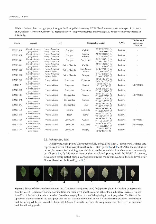
Plants 2021, 10, 2777
Table 1. Isolate, plant host, geographic origin, DNA amplification using APN1 Chondrostereum purpureum-specific primers,and GenBank Accession number of 17 representative C. purpureum isolates, morphologically and molecularly identified inthis study.
Isolate Species Host Geographic Origin APN1ITS GenBank
AccessionNumber
HMCi 314 Chondrostereumpurpureum
Prunus domesticasubsp. domestica D’Agen Colbún 35◦45′01.1592′′ S,
71◦25′46.4889′′ W Positive -
HMCi 325 Chondrostereumpurpureum
Prunus domesticasubsp. domestica D’Agen Sagrada
Familia34◦59′50.0820′′ S,71◦21′48.2976′′ W Positive -
HMCi 331 Chondrostereumpurpureum
Prunus domesticasubsp. domestica D’Agen San Javier 35◦38′54.7944′′ S,
71◦36′47.9340′′ W Positive -
HMCi 341 Chondrostereumpurpureum
Prunus domesticasubsp. italica Reina Claudia Chillán 34◦58′10.7536′′ S,
71◦21′14.8464′′ W Positive -
HMCi 308 Chondrostereumpurpureum
Prunus domesticasubsp. italica Reina Claudia San Rafael,
Maule35◦18′37.0044′′ S,71◦29′06.5832′′ W Positive -
HMCi 290 Chondrostereumpurpureum
Prunus domesticasubsp. italica Reina Claudia Yungay 37◦07′21.6127′′ S,
72◦00′02.1028′′ W Positive -
HMCi 249 Chondrostereumpurpureum Prunus salicina Angeleno Codegua 34◦01′12.3420′′ S,
70◦41′50.0352′′ W Positive -
HMCi 7 Chondrostereumpurpureum Prunus salicina Angeleno Curicó 34◦58′58.21′′ S,
71◦16′37.01′′ W Positive MW938164
HMCi 340 Chondrostereumpurpureum Prunus salicina Angeleno Portezuelo 36◦34′43.9356′′ S,
72◦33′19.7424′′ W Positive -
HMCi 121 Chondrostereumpurpureum Prunus salicina Black amber Curicó 36◦37′27.1128′′ S,
72◦00′27.8532′′ W Positive MW938165
HMCi 272 Chondrostereumpurpureum Prunus salicina Black amber Romeral 34◦57′17.2836′′ S,
71◦08′11.2560′′ W Positive -
HMCi 276 Chondrostereumpurpureum Prunus salicina Black amber Teno 34◦52′39.4149′′ S,
71◦05′19.0032′′ W Positive -
HMCi 168 Chondrostereumpurpureum Prunus salicina Fortune Melipilla 33◦41′09.3696′′ S,
71◦06′23.7960′′ W Positive -
HMCi 253 Chondrostereumpurpureum Prunus salicina Friar Paine 33◦52′11.7156′′ S,
70◦44′21.5700′′ W Positive -
HMCi 148 Chondrostereumpurpureum Prunus salicina Larry Ann Curicó 34◦58′56.0352′′ S,
71◦16′34.0896′′ W Positive MW938167
HMCi 147 Chondrostereumpurpureum Prunus salicina Larry Ann Rio Claro 35◦12′0.18936′′ S,
71◦14′36.1140′′ W Positive MW938166
HMCi 157 Chondrostereumpurpureum Prunus salicina Larry Ann Yungay 37◦08′50.452′′ S,
71◦52′20.673′′ W Positive -
2.2. Pathogenicity Tests
Healthy nursery plants were successfully inoculated with C. purpureum isolates andreproduced silver foliar symptoms (Grade 3–8) (Figures 2 and 3A,B). After the incubationperiod, brown central staining was visible when the inoculated branches were transversallycut (Figure 3A,C). Moreover, one of the inoculated plants, with the HMCi121 isolate,developed resupinated purple carpophores in the main trunk, above the soil level, after20 months of incubation (Figure 3E).
Figure 2. Silverleaf disease foliar symptom visual severity scale (one to nine) for Japanese plum. 1 = healthy or apparentlyhealthy leaf, 3 = epidermis starts detaching from the mesophyll and the color is lighter than in healthy leaves, 5 = morethan 75% of the leaf epidermis is detached from the mesophyll and the leaf is beginning to look gray–silver, 7 = 100% of theepidermis is detached from the mesophyll and the leaf is completely white–silver, 9 = the epidermis peels off from the leafand the mesophyll begins to oxidize. Grades 2, 4, 6, and 8 indicate intermediate symptom severity between the previousand the following grade.
156
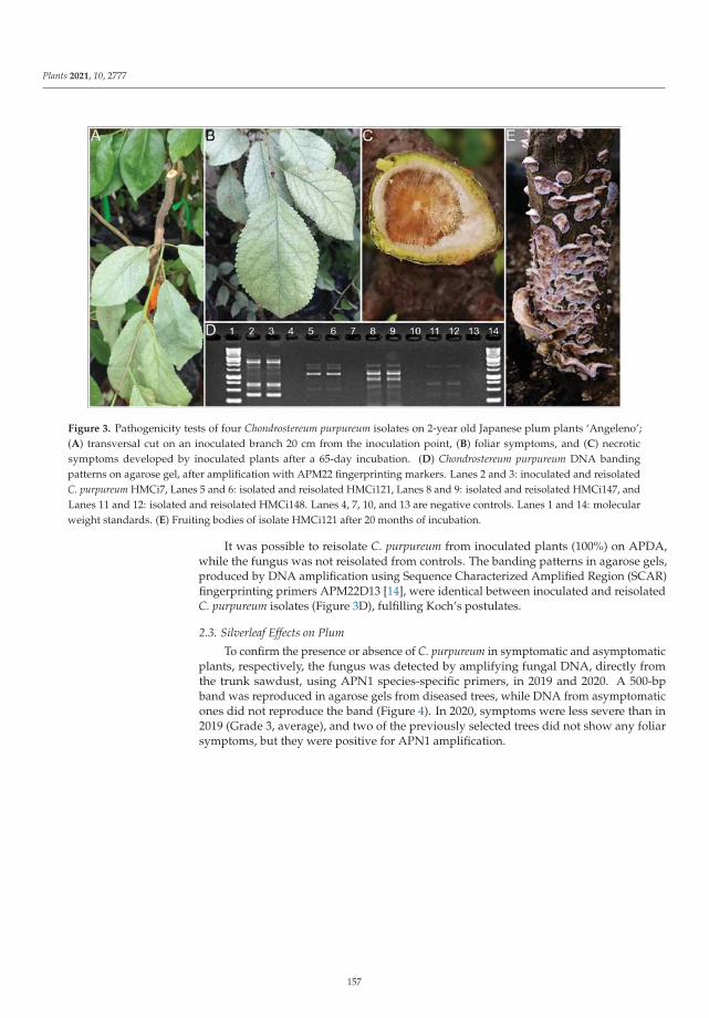
Plants 2021, 10, 2777
Figure 3. Pathogenicity tests of four Chondrostereum purpureum isolates on 2-year old Japanese plum plants ‘Angeleno’;(A) transversal cut on an inoculated branch 20 cm from the inoculation point, (B) foliar symptoms, and (C) necroticsymptoms developed by inoculated plants after a 65-day incubation. (D) Chondrostereum purpureum DNA bandingpatterns on agarose gel, after amplification with APM22 fingerprinting markers. Lanes 2 and 3: inoculated and reisolatedC. purpureum HMCi7, Lanes 5 and 6: isolated and reisolated HMCi121, Lanes 8 and 9: isolated and reisolated HMCi147, andLanes 11 and 12: isolated and reisolated HMCi148. Lanes 4, 7, 10, and 13 are negative controls. Lanes 1 and 14: molecularweight standards. (E) Fruiting bodies of isolate HMCi121 after 20 months of incubation.
It was possible to reisolate C. purpureum from inoculated plants (100%) on APDA,while the fungus was not reisolated from controls. The banding patterns in agarose gels,produced by DNA amplification using Sequence Characterized Amplified Region (SCAR)fingerprinting primers APM22D13 [14], were identical between inoculated and reisolatedC. purpureum isolates (Figure 3D), fulfilling Koch’s postulates.
2.3. Silverleaf Effects on Plum
To confirm the presence or absence of C. purpureum in symptomatic and asymptomaticplants, respectively, the fungus was detected by amplifying fungal DNA, directly fromthe trunk sawdust, using APN1 species-specific primers, in 2019 and 2020. A 500-bpband was reproduced in agarose gels from diseased trees, while DNA from asymptomaticones did not reproduce the band (Figure 4). In 2020, symptoms were less severe than in2019 (Grade 3, average), and two of the previously selected trees did not show any foliarsymptoms, but they were positive for APN1 amplification.
157
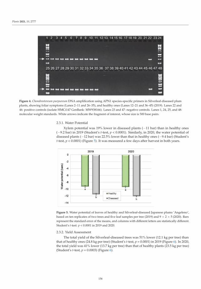
Plants 2021, 10, 2777
Figure 4. Chondrostereum purpureum DNA amplification using APN1 species-specific primers in Silverleaf-diseased plumplants, showing foliar symptoms (Lanes 2–11 and 26–35), and healthy ones (Lanes 12–21 and 36–45) (2019). Lanes 22 and46: positive controls (isolate HMCi147 GenBank: MW938166). Lanes 23 and 47: negative controls. Lanes 1, 24, 25, and 48:molecular weight standards. White arrows indicate the fragment of interest, whose size is 500 base pairs.
2.3.1. Water Potential
Xylem potential was 19% lower in diseased plants (−11 bar) than in healthy ones(−9.2 bar) in 2019 (Student’s t-test, p < 0.0001). Similarly, in 2020, the water potential ofdiseased plants (−12 bar) was 22.5% lower than that in healthy ones (−9.4 bar) (Student’st-test, p < 0.0001) (Figure 5). It was measured a few days after harvest in both years.
Figure 5. Water potential of leaves of healthy and Silverleaf-diseased Japanese plums ’Angeleno’,based on ten replicates of two trees and five leaf samples per tree (2019) and 9 × 2 × 5 (2020). Barsrepresent the standard error of the means, and columns with different letters are statistically different.Student’s t-test: p < 0.0001 in 2019 and 2020.
2.3.2. Yield Assessment
The total yield of the Silverleaf-diseased trees was 51% lower (12.1 kg per tree) thanthat of healthy ones (24.8 kg per tree) (Student’s t-test, p = 0.0001) in 2019 (Figure 6). In 2020,the total yield was 41% lower (13.7 kg per tree) than that of healthy plants (23.5 kg per tree)(Student’s t-test, p = 0.0003) (Figure 6).
158
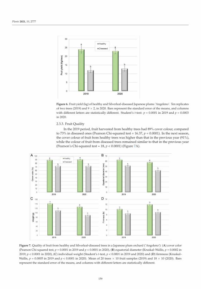
Plants 2021, 10, 2777
Figure 6. Fruit yield (kg) of healthy and Silverleaf-diseased Japanese plums ’Angeleno’. Ten replicatesof two trees (2019) and 9 × 2, in 2020. Bars represent the standard error of the means, and columnswith different letters are statistically different. Student’s t-test: p = 0.0001 in 2019 and p = 0.0003in 2020.
2.3.3. Fruit Quality
In the 2019 period, fruit harvested from healthy trees had 89% cover colour, comparedto 73% in diseased ones (Pearson Chi-squared test = 16.37, p = 0.0001). In the next season,the cover colour of fruit from healthy trees was higher than that in the previous year (91%),while the colour of fruit from diseased trees remained similar to that in the previous year(Pearson’s Chi-squared test = 18, p < 0.0001) (Figure 7A).
Figure 7. Quality of fruit from healthy and Silverleaf-diseased trees in a Japanese plum orchard (’Angeleno’): (A) cover color(Pearson Chi-squared test, p = 0.0001 in 2019 and p < 0.0001 in 2020), (B) equatorial diameter (Kruskal–Wallis, p = 0.0002 in2019; p < 0.0001 in 2020), (C) individual weight (Student’s t-test, p < 0.0001 in 2019 and 2020) and (D) firmness (Kruskal–Wallis, p = 0.0005 in 2019 and p < 0.0001 in 2020). Mean of 20 trees × 10 fruit samples (2019) and 18 × 10 (2020). Barsrepresent the standard error of the means, and columns with different letters are statistically different.
159
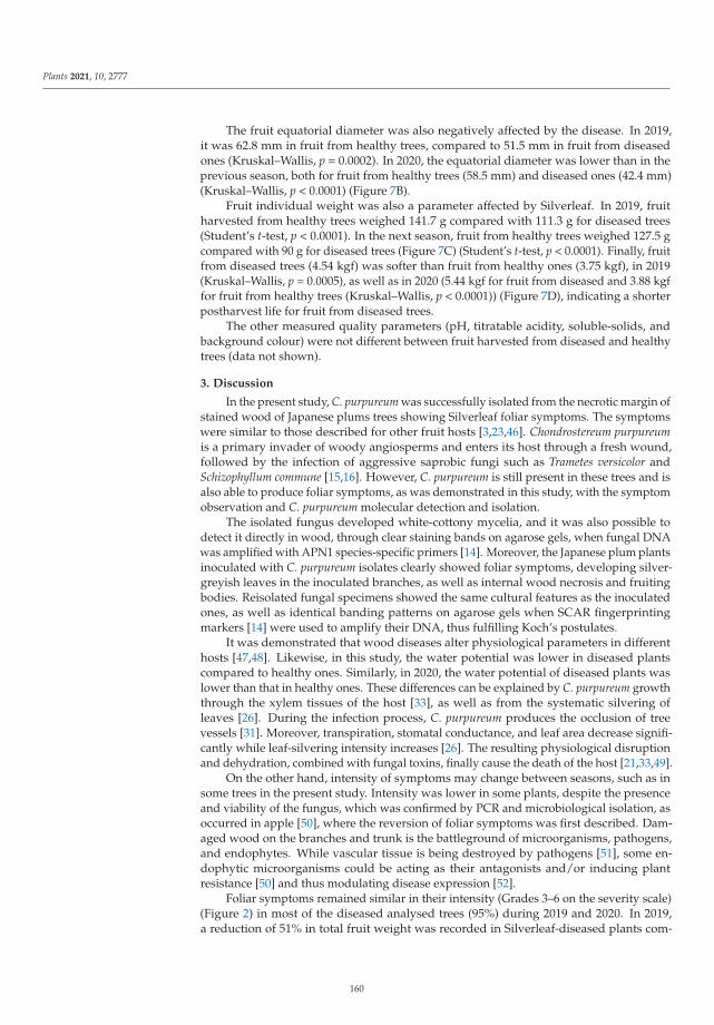
Plants 2021, 10, 2777
The fruit equatorial diameter was also negatively affected by the disease. In 2019,it was 62.8 mm in fruit from healthy trees, compared to 51.5 mm in fruit from diseasedones (Kruskal–Wallis, p = 0.0002). In 2020, the equatorial diameter was lower than in theprevious season, both for fruit from healthy trees (58.5 mm) and diseased ones (42.4 mm)(Kruskal–Wallis, p < 0.0001) (Figure 7B).
Fruit individual weight was also a parameter affected by Silverleaf. In 2019, fruitharvested from healthy trees weighed 141.7 g compared with 111.3 g for diseased trees(Student’s t-test, p < 0.0001). In the next season, fruit from healthy trees weighed 127.5 gcompared with 90 g for diseased trees (Figure 7C) (Student’s t-test, p < 0.0001). Finally, fruitfrom diseased trees (4.54 kgf) was softer than fruit from healthy ones (3.75 kgf), in 2019(Kruskal–Wallis, p = 0.0005), as well as in 2020 (5.44 kgf for fruit from diseased and 3.88 kgffor fruit from healthy trees (Kruskal–Wallis, p < 0.0001)) (Figure 7D), indicating a shorterpostharvest life for fruit from diseased trees.
The other measured quality parameters (pH, titratable acidity, soluble-solids, andbackground colour) were not different between fruit harvested from diseased and healthytrees (data not shown).
3. Discussion
In the present study, C. purpureum was successfully isolated from the necrotic margin ofstained wood of Japanese plums trees showing Silverleaf foliar symptoms. The symptomswere similar to those described for other fruit hosts [3,23,46]. Chondrostereum purpureumis a primary invader of woody angiosperms and enters its host through a fresh wound,followed by the infection of aggressive saprobic fungi such as Trametes versicolor andSchizophyllum commune [15,16]. However, C. purpureum is still present in these trees and isalso able to produce foliar symptoms, as was demonstrated in this study, with the symptomobservation and C. purpureum molecular detection and isolation.
The isolated fungus developed white-cottony mycelia, and it was also possible todetect it directly in wood, through clear staining bands on agarose gels, when fungal DNAwas amplified with APN1 species-specific primers [14]. Moreover, the Japanese plum plantsinoculated with C. purpureum isolates clearly showed foliar symptoms, developing silver-greyish leaves in the inoculated branches, as well as internal wood necrosis and fruitingbodies. Reisolated fungal specimens showed the same cultural features as the inoculatedones, as well as identical banding patterns on agarose gels when SCAR fingerprintingmarkers [14] were used to amplify their DNA, thus fulfilling Koch’s postulates.
It was demonstrated that wood diseases alter physiological parameters in differenthosts [47,48]. Likewise, in this study, the water potential was lower in diseased plantscompared to healthy ones. Similarly, in 2020, the water potential of diseased plants waslower than that in healthy ones. These differences can be explained by C. purpureum growththrough the xylem tissues of the host [33], as well as from the systematic silvering ofleaves [26]. During the infection process, C. purpureum produces the occlusion of treevessels [31]. Moreover, transpiration, stomatal conductance, and leaf area decrease signifi-cantly while leaf-silvering intensity increases [26]. The resulting physiological disruptionand dehydration, combined with fungal toxins, finally cause the death of the host [21,33,49].
On the other hand, intensity of symptoms may change between seasons, such as insome trees in the present study. Intensity was lower in some plants, despite the presenceand viability of the fungus, which was confirmed by PCR and microbiological isolation, asoccurred in apple [50], where the reversion of foliar symptoms was first described. Dam-aged wood on the branches and trunk is the battleground of microorganisms, pathogens,and endophytes. While vascular tissue is being destroyed by pathogens [51], some en-dophytic microorganisms could be acting as their antagonists and/or inducing plantresistance [50] and thus modulating disease expression [52].
Foliar symptoms remained similar in their intensity (Grades 3–6 on the severity scale)(Figure 2) in most of the diseased analysed trees (95%) during 2019 and 2020. In 2019,a reduction of 51% in total fruit weight was recorded in Silverleaf-diseased plants com-
160
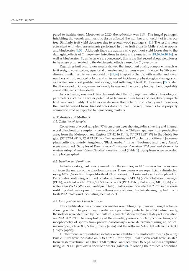
Plants 2021, 10, 2777
pared to healthy ones. Moreover, in 2020, the reduction was 41%. The fungal pathogeninhabiting the vessels and necrotic tissue affected the number and weight of fruits pertree. Similarly, fruit yield decreases due to several wood pathogens [41]. The results wereconsistent with yield assessments performed in other fruit crops in Chile, such as applesand blueberries [4,53]. Although there are authors who point out yield losses due to thedamaging effects of C. purpureum infections in stone and pome fruits [18,26,36,40,44], aswell as blueberries [4], as far as we are concerned, this is the first record about yield lossesin Japanese plum related to the detrimental effects caused by C. purpureum.
Regarding fruit quality, our results showed that important quality components such asfruit weight, cover colour, equatorial diameter, and firmness were influenced by Silverleafdisease. Similar results were reported by [25,26] in apple orchards, with smaller and lowernumbers of fruit, reduced colour, and an increased incidence of physiological damage suchas a water core, short post-harvest storage, and softening of fruit. Furthermore, [27] statedthat the spread of C. purpureum in woody tissues and the loss of photosynthetic capabilityeventually leads to tree death.
In conclusion, our work has demonstrated that C. purpureum alters physiologicalparameters such as the water potential of Japanese plum trees and negatively impactsfruit yield and quality. The latter can decrease the orchard productivity and, moreover,the fruit harvested from diseased trees does not meet the requirements to be properlycommercialized or exported to demanding markets.
4. Materials and Methods
4.1. Collection of Samples
Collections of wood samples (97) from plum trees showing foliar silvering and internalwood discoloration symptoms were conducted in the Chilean Japanese plum productivearea, from the Metropolitana Region (33◦42′16.11′′ S, 70◦59′11.82′′ W) to the Ñuble Re-gion (36◦37′24.98′′ S, 72◦0′23.39′′ W). Two nurseries and 27 orchards of different Japaneseplum cultivars, mainly ‘Angeleno’, ‘Black Amber’, ‘Friar’, ‘Fortune’, and ‘Larry Anne’,were examined. Samples of Prunus domestica subsp. domestica ‘D’Agen’ and Prunus do-mestica subsp. italica ‘Reina Claudia’ were included (Table 1). Symptoms were recordedand photographed.
4.2. Isolation and Purification
In the laboratory, bark was removed from the samples, and 0.5 cm wooden pieces werecut from the margin of the discoloration area. These pieces were superficially disinfectedusing 10% v/v sodium hypochlorite (4.9% chlorine) for 4 min and aseptically plated onPetri plates containing acidified potato dextrose agar (APDA) (25% potato dextrose agar(PDA), acidified with 0.2% v/v 85% lactic acid) (PDA Difco, Baltimore, MD, USA) andwater agar (WA) (Winkler, Santiago, Chile). Plates were incubated at 25 ◦C in darknessuntil mycelial development. Pure cultures were obtained by transferring hyphal tips tofresh PDA plates and incubating them at 25 ◦C.
4.3. Identification and Characterization
The identification was focused on isolates resembling C. purpureum. Fungal coloniesshowing white to beige cottony mycelia were preliminary selected (n = 95). Subsequently,the isolates were identified by their cultural characteristics after 7 and 14 days of incubationon PDA at 25 ◦C. The morphology of the mycelia, presence of clamp connections, andmorphometry of spores from pseudo-basidiocarps were determined using an opticalmicroscope (Eclipse 80i, Nikon, Tokyo, Japan) and the software Nikon NIS-elements D2.30(Tokyo, Japan).
Furthermore, representative isolates were identified by molecular means (n = 57).Pure cultures were incubated on PDA at 25 ◦C for 7 days. Total nucleic acids were isolatedfrom fresh mycelium using the CTAB method, and genomic DNA (20 ng) was amplifiedusing APN 1 C. purpureum-specific primers (Table 2), following the protocols described
161
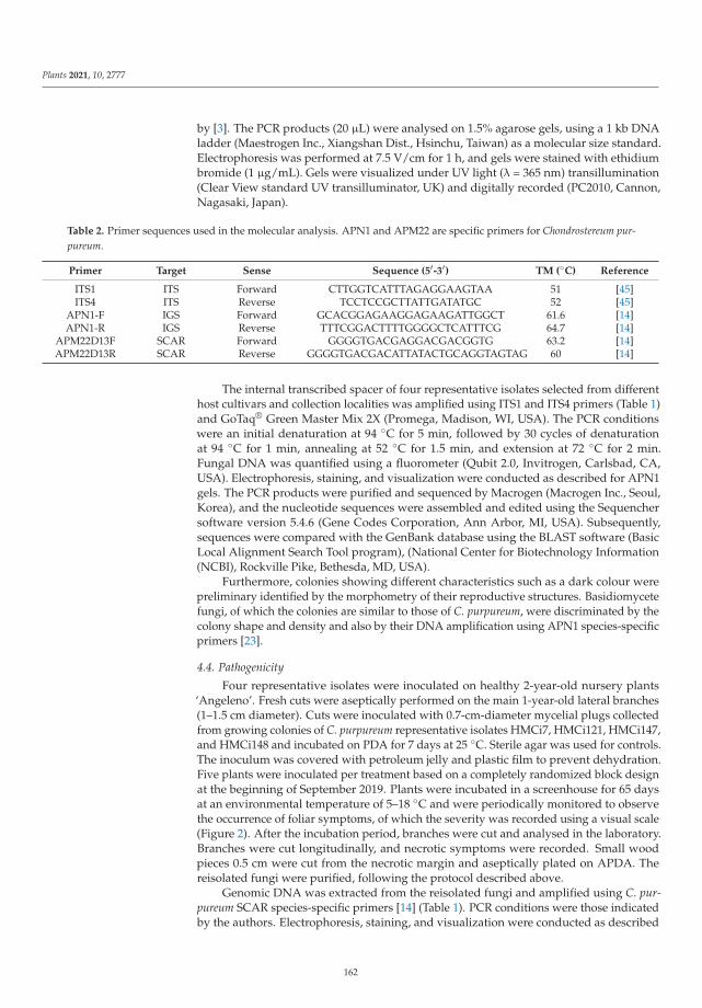
Plants 2021, 10, 2777
by [3]. The PCR products (20 μL) were analysed on 1.5% agarose gels, using a 1 kb DNAladder (Maestrogen Inc., Xiangshan Dist., Hsinchu, Taiwan) as a molecular size standard.Electrophoresis was performed at 7.5 V/cm for 1 h, and gels were stained with ethidiumbromide (1 μg/mL). Gels were visualized under UV light (λ = 365 nm) transillumination(Clear View standard UV transilluminator, UK) and digitally recorded (PC2010, Cannon,Nagasaki, Japan).
Table 2. Primer sequences used in the molecular analysis. APN1 and APM22 are specific primers for Chondrostereum pur-pureum.
Primer Target Sense Sequence (5′-3′) TM (◦C) Reference
ITS1 ITS Forward CTTGGTCATTTAGAGGAAGTAA 51 [45]ITS4 ITS Reverse TCCTCCGCTTATTGATATGC 52 [45]
APN1-F IGS Forward GCACGGAGAAGGAGAAGATTGGCT 61.6 [14]APN1-R IGS Reverse TTTCGGACTTTTGGGGCTCATTTCG 64.7 [14]
APM22D13F SCAR Forward GGGGTGACGAGGACGACGGTG 63.2 [14]APM22D13R SCAR Reverse GGGGTGACGACATTATACTGCAGGTAGTAG 60 [14]
The internal transcribed spacer of four representative isolates selected from differenthost cultivars and collection localities was amplified using ITS1 and ITS4 primers (Table 1)and GoTaq® Green Master Mix 2X (Promega, Madison, WI, USA). The PCR conditionswere an initial denaturation at 94 ◦C for 5 min, followed by 30 cycles of denaturationat 94 ◦C for 1 min, annealing at 52 ◦C for 1.5 min, and extension at 72 ◦C for 2 min.Fungal DNA was quantified using a fluorometer (Qubit 2.0, Invitrogen, Carlsbad, CA,USA). Electrophoresis, staining, and visualization were conducted as described for APN1gels. The PCR products were purified and sequenced by Macrogen (Macrogen Inc., Seoul,Korea), and the nucleotide sequences were assembled and edited using the Sequenchersoftware version 5.4.6 (Gene Codes Corporation, Ann Arbor, MI, USA). Subsequently,sequences were compared with the GenBank database using the BLAST software (BasicLocal Alignment Search Tool program), (National Center for Biotechnology Information(NCBI), Rockville Pike, Bethesda, MD, USA).
Furthermore, colonies showing different characteristics such as a dark colour werepreliminary identified by the morphometry of their reproductive structures. Basidiomycetefungi, of which the colonies are similar to those of C. purpureum, were discriminated by thecolony shape and density and also by their DNA amplification using APN1 species-specificprimers [23].
4.4. Pathogenicity
Four representative isolates were inoculated on healthy 2-year-old nursery plants‘Angeleno’. Fresh cuts were aseptically performed on the main 1-year-old lateral branches(1–1.5 cm diameter). Cuts were inoculated with 0.7-cm-diameter mycelial plugs collectedfrom growing colonies of C. purpureum representative isolates HMCi7, HMCi121, HMCi147,and HMCi148 and incubated on PDA for 7 days at 25 ◦C. Sterile agar was used for controls.The inoculum was covered with petroleum jelly and plastic film to prevent dehydration.Five plants were inoculated per treatment based on a completely randomized block designat the beginning of September 2019. Plants were incubated in a screenhouse for 65 daysat an environmental temperature of 5–18 ◦C and were periodically monitored to observethe occurrence of foliar symptoms, of which the severity was recorded using a visual scale(Figure 2). After the incubation period, branches were cut and analysed in the laboratory.Branches were cut longitudinally, and necrotic symptoms were recorded. Small woodpieces 0.5 cm were cut from the necrotic margin and aseptically plated on APDA. Thereisolated fungi were purified, following the protocol described above.
Genomic DNA was extracted from the reisolated fungi and amplified using C. pur-pureum SCAR species-specific primers [14] (Table 1). PCR conditions were those indicatedby the authors. Electrophoresis, staining, and visualization were conducted as described
162
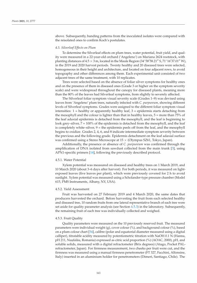
Plants 2021, 10, 2777
above. Subsequently, banding patterns from the inoculated isolates were compared withthe reisolated ones to confirm Koch’s postulates.
4.5. Silverleaf Effects on Plum
To determine the Silverleaf effects on plum trees, water potential, fruit yield, and qual-ity were measured in a 22-year-old orchard (‘Angeleno’) on Mariana 2624 rootstock, withplanting distances of 4.5 × 3 m, located in the Maule Region (34◦58′58.21′′ S, 71◦16′37.01′′ W),in the 2019 and 2020 harvest periods. Twenty healthy and 20 diseased trees were selected,homogeneous in their height and architecture, and located on four adjacent rows, to avoidtopography and other differences among them. Each experimental unit consisted of twoadjacent trees of the same treatment, with 10 replicates.
Trees were selected based on the absence of foliar silver symptoms for healthy onesand on the presence of them in diseased ones (Grade 3 or higher on the symptom severityscale) and were widespread throughout the canopy for diseased plants, meaning morethan the 80% of the leaves had Silverleaf symptoms, from slightly to severely affected.
The Silverleaf foliar symptom visual severity scale (Grades 1–9) was devised usingleaves from ‘Angeleno’ plum trees, naturally infected with C. purpureum, showing differentlevels of Silverleaf symptoms. Grades were assigned to the different foliar symptom visualintensities: 1 = healthy or apparently healthy leaf, 3 = epidermis starts detaching fromthe mesophyll and the colour is lighter than that in healthy leaves, 5 = more than 75% ofthe leaf adaxial epidermis is detached from the mesophyll, and the leaf is beginning tolook grey–silver, 7 = 100% of the epidermis is detached from the mesophyll, and the leafis completely white–silver, 9 = the epidermis peels off from the leaf, and the mesophyllbegins to oxidize. Grades 2, 4, 6, and 8 indicate intermediate symptom severity betweenthe previous and the following grade. Epidermis detachment on the leaf adaxial surfacewas confirmed using a Stereo Microscope at 15 × (Olympus SZ61, Tokyo, Japan).
Additionally, the presence or absence of C. purpureum was confirmed through theamplification of DNA isolated from sawdust collected from the main trunk [3], usingAPN1-specific primers [14], following the previously described protocol.
4.5.1. Water Potential
Xylem potential was measured on diseased and healthy trees on 1 March 2019, and10 March 2020 (about 3–6 days after harvest). For both periods, it was measured on light-exposed leaves (five leaves per plant), which were previously covered for 2 h to avoidsunlight. Xylem potential was measured using a Scholander-type pressure chamber (Model615, PMS Instruments, Albany, NY, USA).
4.5.2. Yield Assessment
Fruit was harvested on 27 February 2019 and 4 March 2020, the same dates thatproducers harvested the orchard. Before harvesting the fruit from each selected healthyand diseased tree, 10 random fruits from one lateral representative branch of each tree wereset aside for quality parameter analysis (see Section 4.5.3) in the laboratory. Subsequently,the remaining fruit of each tree was individually collected and weighed.
4.5.3. Fruit Quality
Quality parameters were measured on the 10 previously reserved fruit. The measuredparameters were individual weight (g), cover colour (%), and background colour (%), basedon a plum colour chart [54], calibre (polar and equatorial diameter measured using a digitalcalliper), titratable acidity measured by potentiometric titration with NaOH 0.1 N (Hanna,pH 211, Nus, falău, Romania) expressed as citric acid proportion (%) (AOAC, 2000), pH, andsoluble solids, measured with a digital refractometer (Brix degrees) (Atago, Pocket PAL-refractometer, Japan). For firmness measurement, two cheeks per fruit were cut, and thefirmness was measured using a manual firmness penetrometer (FT 327, Facchini, Alfonsine,Italy) inserted in an aluminium holder for penetrometers (Dimeri, Santiago, Chile). The
163
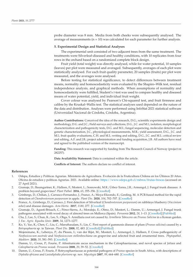
Plants 2021, 10, 2777
probe diameter was 8 mm. Media from both cheeks were subsequently analysed. Theaverage of measurements (n = 10) was calculated for each parameter for further analysis.
5. Experimental Design and Statistical Analyses
The experimental unit consisted of two adjacent trees from the same treatment. Thetreatments were Silverleaf-diseased and healthy conditions, with 10 replicates from fourrows in the orchard based on a randomized complete block design.
Fruit yield (total weight) was directly analysed, while for water potential, 10 samples(leaves) per plot were measured and averaged. Subsequently, averages of each plot werestatistically analysed. For each fruit quality parameter, 20 samples (fruits) per plot weremeasured, and the averages were analysed.
Before testing for statistical significance, to detect differences between treatmentmeans, normality and homoscedasticity were evaluated by the Shapiro–Wilk test, residualindependence analysis, and graphical methods. When assumptions of normality andhomoscedasticity were fulfilled, Student’s t-test was used to compare healthy and diseasedmeans of water potential, yield, and individual fruit weight.
Cover colour was analysed by Pearson’s Chi-squared test, and fruit firmness andcalibre by the Kruskal–Wallis test. The statistical analyses used depended on the nature ofthe data and distribution. Analyses were performed using InfoStat 2021 statistical software(Universidad Nacional de Córdoba, Córdoba, Argentina).
Author Contributions: Conceived the idea of the research, D.G.; scientific experiments design andmethodology, D.G. and J.C.; Field surveys and collections, D.G., J.C. and M.I.; isolation, morphologicalcharacterization and pathogenicity tests, D.G. and M.I.; fungal sequencing, molecular detection andgenetic characterization, J.C., physiological measurements, M.R.; yield assessment, D.G., J.C. andM.I.; fruit quality evaluations, C.H. and M.I.; writing and editing, D.G., J.C. and M.I.; critical reviewand editing, A.F. and J.B.; project administration and funding acquisition, J.B. All authors have readand agreed to the published version of the manuscript.
Funding: This research was supported by funding from The Research Council of Norway (project nr.282257).
Data Availability Statement: Data is contained within the article.
Conflicts of Interest: The authors declare no conflict of interest.
References
1. Odepa, Estudios y Políticas Agrarias. Ministerio de Agricultura. Evolución de la Fruticultura Chilena en los Últimos 20 Años.Oficina de estudios y políticas Agrarias. 2021. Available online: https://www.odepa.gob.cl/rubros/frutas-frescas (accessed on27 April 2021).
2. Gramaje, D.; Baumgartner, K.; Halleen, F.; Mostert, L.; Sosnowski, M.R.; Úrbez-Torres, J.R.; Armengol, J. Fungal trunk diseases: Aproblem beyond grapevines? Plant Pathol. 2016, 65, 355–356. [CrossRef]
3. Grinbergs, D.; Chilian, J.; Carrasco-Fernandez, J.; France, A.; Moya-Elizondo, E.; Gerding, M. A PCR-based method for the rapiddetection of Chondrostereum purpureum in apple. Plant Dis. 2020, 104, 702–707. [CrossRef]
4. France, A.; Grinbergs, D.; Carrasco, J. First detection of Silverleaf (Chondrostereum purpureum) on rabbiteye blueberry (Vacciniumashei) and disease damages. Acta Hortic. 2017, 1180, 277. [CrossRef]
5. Gramaje, D.; Agustí-Brisach, C.; Pérez-Sierra, A.; Moralejo, E.; Olmo, D.; Mostert, L.; Damm, U.; Armengol, J. Fungal trunkpathogens associated with wood decay of almond trees on Mallorca (Spain). Persoonia 2012, 28, 1–13. [CrossRef] [PubMed]
6. Cha, J.; Lee, S.; Chun, K.; Lee, S.; Ohga, S. Armillaria root rot caused by Armillaria Tabescens on Prunus Salicina in a Korean garden.J. Fac. Agric. Kyushu Univ. 2009, 54, 1273–1277.
7. Ko, Y.; Yao, K.; Chen, C.; Liu, C.; Maruthasalam, S.; Lin, C. First report of gummosis disease of plum (Prunus salicina) caused by aBotryosphaeria sp. in Taiwan. Plant Dis. 2008, 92, 483. [CrossRef] [PubMed]
8. Mojeremane, K.; Lebenya, P.; du Plessis, I.; van der Rijst, M.; Mostert, L.; Armengol, J.; Halleen, F. Cross pathogenicity ofNeofusicoccum australe and Neofusicoccum stellenboschiana on grapevine and selected fruit and ornamental trees. Phytopathol.Mediterr. 2020, 59, 581–593. [CrossRef]
9. Damm, U.; Crous, P.; Fourie, P. Afissitunicate ascus mechanism in the Calosphaeriaceae, and novel species of Jattaea andCalosphaeria on Prunus wood. Persoonia 2008, 20, 39–52. [CrossRef]
10. Damm, U.; Crous, P.; Fourie, P. Botryosphaeriaceae as potential pathogens of Prunus species in South Africa, with descriptions ofDiplodia africana and Lasiodiplodia plurivora sp. nov. Mycologia 2007, 99, 664–680. [CrossRef]
164
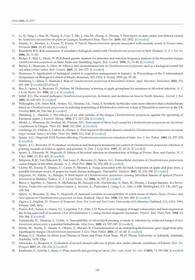
Plants 2021, 10, 2777
11. Li, Q.; Tang, L.; Sun, W.; Huang, S.; Guo, T.; Mo, J.; Fan, M.; Zhang, A.; Hsiang, T. First report of stem canker and dieback causedby Neofusicoccum parvum on plum in Guangxi, Southern China. Plant Dis. 2019, 103, 2952. [CrossRef]
12. Damm, U.; Mostert, L.; Crous, P.; Fourie, P. Novel Phaeoacremonium species associated with necrotic wood of Prunus trees.Persoonia 2008, 20, 87–102. [CrossRef]
13. Ramsfield, R.D. Risk assessment of inundative biological control with Chondrostereum purpureum in New Zealand. N. Z. J. For. Sci.2006, 36, 11–20.
14. Becker, E.; Ball, L.; Hintz, W. PCR Based genetic markers for detection and infection frequency analysis of the biocontrol fungusChondrostereum purpureum on Stika Alder and Trembling Aspen. Biol. Control. 1999, 15, 71–80. [CrossRef]
15. Becker, E.; Shamoun, S.; Hinz, W. Efficacy and environmental fate of Chondrostereum purpureum used as a biological control forred alder (Alnus rubra). Biol. Control. 2005, 33, 269–277. [CrossRef]
16. Shamoun, S. Application of biological control to vegetation management in forestry. In Proceedings of the X InternationalSymposium on Biological Control of Weeds, Bozeman, MT, USA, 4–14 July 1999; pp. 87–96.
17. Hamberg, L.; Saksa, T.; Hantula, J. Role of Chondrostereum purpureum in biocontrol of trees. Appl. Microbiol. Biotechnol. 2021, 105,431–440. [CrossRef] [PubMed]
18. Bus, V.; Spiers, A.; Brewster, D.; Hofstee, M. Preliminary screening of apple germplasm for resistance to Silverleaf infection. N. Z.J. Crop Hortic. Sci. 1996, 24, 1–6. [CrossRef]
19. Setliff, E.C. The wound pathogen Chondrostereum purpureum, its history and incidence on trees in North America. Austral. J. Bot.2002, 50, 645–651. [CrossRef]
20. Willoughby, I.H.; Seier, M.K.; Stokes, V.J.; Thomas, S.E.; Varia, S. Synthetic herbicides were more effective than a bioherbicidebased on Chondrostereum purpureum in reducing resprouting of Rhododendron ponticum, a host of Phytophthora ramorum in the UK.Forestry 2015, 88, 336–344. [CrossRef]
21. Hamberg, L.; Hantula, J. The efficacy of six elite isolates of the fungus Chondrostereum purpureum against the sprouting ofEuropean aspen. J. Environ. Manag. 2016, 171, 217–224. [CrossRef]
22. Merlet, L.; Wiseman, M.; Serdani, M.; Putnam, M.L. First report of Silver Leaf caused by Chondrostereum purpureum on Vacciniumcorymbosum in Oregon. Plant Dis. 2018, 102, 2041. [CrossRef]
23. Grinbergs, D.; Chilian, J.; Lisboa, K.; France, A. First report of Silverleaf disease caused by Chondrostereum purpureum on murta(Ugni molinae Turcz.) in Chile. Plant Dis. 2019, 103, 2140. [CrossRef]
24. Spiers, A.G.; Hopcroft, D.H. Factors affecting Chondrostereum purpureum infection of Salix. Eur. J. For. Pathol. 1988, 18, 257–278.[CrossRef]
25. Spiers, A.G.; Brewster, D. Evaluation of chemical and biological treatments for control of Chondrostereum purpureum infection ofpruning wounds in willows, apples, and peaches. N. Zeal. J. Crop Hort. 1997, 25, 19–31. [CrossRef]
26. Spiers, A.; Edwards, W.; Hopcroft, D. Effects of Silverleaf infection on ultrastructure of foliage of Prunus, Rosa, and Populus. N. Z.J. Bot. 1987, 25, 411–423. [CrossRef]
27. Simpson, R.M.; Van Hekezen, R.; Van Lune, F.; Brewster, D.; Spiers, A.G. Extracellular enzymes of Chondrostereum purpureum,causal fungus of Silverleaf disease. N. Z. Plant Prot. 2001, 54, 202–208. [CrossRef]
28. Cloete, M.; Fourie, P.; Damm, U.; Crous, P.; Mostert, L. Fungi associated with die-back symptoms of apple and pear trees, apossible inoculum source of grapevine trunk disease pathogens. Phytopathol. Mediterr. 2011, 50, 176–190. [CrossRef]
29. Ozgonen, H.; Erkilic, A.; Baloglu, S. First report of Chondrostereum purpureum causing Silverleaf disease of apricot (Prunusarmeniaca) in Malatya, Turkey. N. Z. J. Crop Hortic. Sci. 2006, 34, 357. [CrossRef]
30. Børve, J.; Ippolito, A.; Tanovic, B.; Michalecka, M.; Sanzani, S.M.; Ponitowska, A.; Mari, M.; Hrustic, J. Fungal diseases. In Cherries.Botany, Production and Uses; Quero-García, J., Iezzoni, A., Pulawska, J., Lang, G.A., Eds.; CABI: Wallingford, CT, UK, 2017; pp.338–364.
31. Spiers, A.; Brewster, D.; Bus, V.; Hopcroft, H. Seasonal variation in susceptibility of xylem tissue of Malus, Pyrus, Prunus, andSalix species to Chondrostereum purpureum in New Zealand. Mycol. Res. 1998, 102, 881–890. [CrossRef]
32. Ogawa, J.; English, H. Diseases of Temperate Zone Tree Fruit and Nut Crops; University of California: Oakland, CA, USA, 1991;Volume 3345, 461p.
33. Pearce, R.B.; Sumer, S.; Doran, S.J.; Carpenter, T.A.; Hali, L.D. Non-invasive imaging of fungal colonization and host response inthe living sapwood of sycamore (Acer pseudoplatanus L.) using nuclear magnetic resonance. Physiol. Mol. Plant Path. 1994, 45,359–384. [CrossRef]
34. Vartiamäki, H.; Hantula, J.; Uotila, A. Susceptibility of silver birch pruning wounds to infection by white-rot fungus (Chon-drostereum purpureum), a potential bioherbicide. Silva Fenn. 2009, 43, 537–547. [CrossRef]
35. Senda, M.; Narita, T.; Akada, S.; Okuno, T.; Miyairi, K. Characterization of an endopolygalacturonase gene cppgl from phy-topathogenic fungus Chondrostereum purpureum. J. Gen. Plant Pathol. 2001, 67, 41–44. [CrossRef]
36. Bishop, G.C. Studies on Silver Leaf Disease of Stone and Pome Fruit Trees. Ph.D. Thesis, University of Adelaide, Adelaide,Australia, 1978; 128p.
37. Głowacka, A.; Rozpara, E. Evaluation of several dessert cultivars of plum, new under climatic conditions of Poland. Hort. Sci.(Prague) 2017, 44, 126–132. [CrossRef]
38. Kaufmane, E.; Grâvîte, I.; Ikase, L. Plum research and growing in Latvia. Proc. Latv. Acad. Sci. Sect. B 2019, 73, 195–206. [CrossRef]
165
Plants 2021, 10, 2777
39. Børve, J.; Talgø, V.; Stensvand, A. Silver Leaf in Norwegian Prunus domestica Plum Orchards. In Proceedings of the IOBC/WPRSWorking Groups: Pheromones and Other Semiochemicals in IP & Integrated Protection of Fruit Crops, PheroFIP 2019, Lisbon,Portugal, 20–25 January 2019; p. 219.
40. Atkinson, J.D. Diseases of Tree Fruits in New Zealand; Bulletin 81; Department of Scientific and Industrial Research: Auckland, NewZealand, 1971; 406p.
41. Bien, S.; Damm, U. Prunus trees in Germany—A hideout of unknown fungi? Mycol. Prog. 2020, 19, 667–690. [CrossRef]42. Espargham, N.; Mohammadi, H.; Gramaje, D. A survey of trunk disease pathogens within Citrus trees in Iran. Plants 2020, 9, 754.
[CrossRef]43. Bertsch, C.; Ramirez-Suero, M.; Magnin-Robert, M.; Larignon, P.; Chong, J.; Abou-Mansour, E.; Spagnolo, A.; Clément, C.;
Fontaine, F. Grapevine trunk diseases: Complex and still poorly understood. Plant Pathol. 2013, 62, 243–265. [CrossRef]44. Spiers, A.; Brewster, D.; Slade, A.; Gardiner, S. Characterization of New Zealand isolates of Chondrostereum purpureum with regard
to morphology, growth, pathogenicity and RAPD bandings patterns. Mycol. Res. 2000, 104, 395–402. [CrossRef]45. White, T.J.; Bruns, T.; Lee, S.; Taylor, J. Amplification and direct sequencing of fungi ribosomal RNA genes for phylogenetics. In
PCR Protocols. A Guide to Methods and Applications: 315–322; Academic Press: San Diego, CA, USA, 1990.46. France, A.; Santelices, C.; Buddie, A.; Kirk, P. Silverleaf: First worlwide report of a new and harmful disease on blueberry. Acta
Hortic. (ISHS) 2009, 810, 341–344. [CrossRef]47. Petit, A.N.; Vaillant, N.; Boulay, M.; Clément, C.; Fontaine, F. Alteration of photosynthesis in grapevines affected by esca.
Phytopathology 2006, 96, 1060–1066. [CrossRef] [PubMed]48. Fontaine, F.; Pinto, C.; Vallet, J.; Clément, C.; Gomes, A.; Spagnolo, A. The effects of grapevine trunk diseases (GTDs) on vine
physiology. Eur. J. Plant Pathol. 2015. [CrossRef]49. Hamberg, L.; Vartiamäki, H.; Hantula, J. Breeding Increases the Efficacy of Chondrostereum purpureum in the Sprout Control of
Birch. PLoS ONE 2015, 10, e0117381. [CrossRef] [PubMed]50. Grinbergs, D.; Chilian, J.; Padilla, N.; Reyes, M.; France, A.; Gerding, M.; Ernesto, A. Moya-Elizondo.. Endophytic microorganisms
associated with reversion of Silverleaf disease symptoms in apple. Phytopathology 2021. [CrossRef] [PubMed]51. Xing, J.; Li, P.; Zhang, Y.; Li, J.; Liu, Y.; Lachenbruch, B.; Su, X.; Zhao, J. Fungal pathogens of canker disease trigger canopy dieback
in poplar saplings by inducing functional failure of the phloem and cambium and carbon starvation in the xylem. Physiol. Mol.Plant Path. 2020, 112, 101523. [CrossRef]
52. Bailey, B.; Bae, H.; Strem, M.; Crozier, J.; Thomas, S.; Samuels, G.; Vinyard, B.; Holmes, K. Antibiosis, mycoparasitism, andcolonization success for endophytic Trichoderma isolates with biological control potential in Theobroma cacao. Biol. Control 2008, 46,24–35. [CrossRef]
53. Grinbergs, D.; France, A.; Chilian, J. Silverleaf disease in apple orchards: Understanding and preventing an increasing problem.In Proceedings of the 11th International IOBC-WPRS Workshop on Pome Fruit Diseases, Jurmala, Latvia, 26–30 June 2017;Stensvand, A., Lennox, C., Wenneker, M., Morocko-Bicevska, I., Rancane, R., Eds.; International Organization for Biological andIntegrated Control of Noxious Animals and Plants, West Palearctic Regional Section (IOBC-WPRS): Darmstadt, Germany, 2018;Volume 138, p. 100.
54. Rivero, M.; Quiroga, M.; Gonzalez, O.; Moraga, L. Plum postharvest manual. INTA, Instituto de Investigaciones Agropecuarias.2013. Available online: https://inta.gob.ar/sites/default/files/script-tmp-ficha_n_1_-_cosecha.pdf (accessed on 27 April 2021).
166
Citation: Mang, S.M.; Marcone, C.;
Maxim, A.; Camele, I. Investigations
on Fungi Isolated from Apple Trees
with Die-Back Symptoms from
Basilicata Region (Southern Italy).
Plants 2022, 11, 1374. https://
doi.org/10.3390/plants11101374
Academic Editors: Paula Baptista
and Carlos Agustí-Brisach
Received: 2 March 2022
Accepted: 18 May 2022
Published: 21 May 2022
Publisher’s Note: MDPI stays neutral
with regard to jurisdictional claims in
published maps and institutional affil-
iations.
Copyright: © 2022 by the authors.
Licensee MDPI, Basel, Switzerland.
This article is an open access article
distributed under the terms and
conditions of the Creative Commons
Attribution (CC BY) license (https://
creativecommons.org/licenses/by/
4.0/).
plants
Article
Investigations on Fungi Isolated from Apple Trees withDie-Back Symptoms from Basilicata Region (Southern Italy)
Stefania Mirela Mang 1,*, Carmine Marcone 2, Aurel Maxim 3 and Ippolito Camele 1,*
1 School of Agricultural, Forestry, Food and Environmental Sciences (SAFE), University of Basilicata,Viale dell’Ateneo Lucano 10, 85100 Potenza, Italy
2 Department of Pharmacy, University of Salerno, Via Giovanni Paolo II 132, 84084 Salerno, Italy;[email protected]
3 Department of Engineering and Environmental Protection, Faculty of Agriculture, University of AgriculturalSciences and Veterinary Medicine, No. 3-5, Calea Manastur Street, 400372 Cluj-Napoca, Romania;[email protected]
* Correspondence: [email protected] (S.M.M.); [email protected] (I.C.);Tel.: +39-0971205519 (S.M.M.); +39-0971205544 (I.C.)
Abstract: Val d’Agri is an important orchard area located in the Basilicata Region (Southern Italy).A phenomenon affecting cv. “Golden Delicious” apples which lead to tree death has been observedin the past several years in this area. This phenomenon has already been detected in about 20 hectaresand is rapidly expanding. The symptoms observed were “scaly bark” and extensive cankers, mainlylocated in the lower part of the trunk, associated with wood decay. Dead plants ranged from 20%to 80% and, in many cases, trees were removed by farmers. In order to identify the causes of thisphenomenon, investigations were started in autumn/winter 2019. In order to determine the possiblecausal agents, fungal and bacterial isolations, from symptomatic tissues, were performed in laboratory.Bacterial isolations gave negative results, whereas pure fungal cultures (PFCs) were obtained after3–4 passages on potato dextrose agar (PDA) media. Genetic material was extracted from each PFCand amplified by PCR using three pairs of primers: ITS5/4, Bt2a/Bt2b and ACT-512F/ACT-783R.The amplicons were directly sequenced, and nucleotide sequences were compared with those alreadypresent in the NCBI GenBank nucleotide database. All isolated fungi were identified based onmorphological features and multilocus molecular analyses. Neofusicoccum parvum, Diaporthe eresand Trametes versicolor were most frequently isolated, while Pestalotiopsis funerea, Phomopsis spp. andDiaporthe foeniculina were less frequently isolated. All nucleotide sequences obtained in this studyhave been deposited into the EMBL database. Pathogenicity tests showed that N. parvum was the mostpathogenic and aggressive fungus, while Phomopsis sp. was demonstrated to be the less virulent one.All the investigated fungi were repeatedly reisolated from artificially inoculated twigs of 2-year-oldapple trees, cv. “Golden Delicious”, and subsequently morphologically and molecularly identified.The role played by the above-mentioned fungi in the alterations observed in field is also discussed.
Keywords: apple die-back; canker; fungi; multi-loci phylogeny; wood decay
1. Introduction
Apple die-back syndrome is a complex disease initially characterized by a stuntedappearance of the plants and the presence of chlorosis symptoms on leaves. As the diseasedevelops, cracks and necrotic lesions of the cortex appear mostly at the base of the stemand at the grafting point. Subsequently, “scaly bark” and extensive cankers, generallylocated in the lower part of the trunk develop, which are also associated with wooddecay, and the progressive death of the trees is registered. Many authors worldwideinvestigated the apple die-back syndrome, attributing it to different causal agents, such asvarious phytopathogens or other possible physiological causes [1–5]. Furthermore, in 2011,Cloete et al. [6] reported the presence of the die-back syndrome and cankers on apples and
Plants 2022, 11, 1374. https://doi.org/10.3390/plants11101374 https://www.mdpi.com/journal/plants167
Plants 2022, 11, 1374
pears in South Africa, from which they isolated several fungal pathogens belonging to theDiplodia, Phaeoacremonium, Phomopsis, Neofusicoccum and Eutypa genera. A very recent studyby Di Francesco et al. [5], which characterized, in Brazil, apple cultivars’ susceptibility toNeofusicoccum parvum (Pennycook & Samuels) P.W. Crous, Slippers & A.J.L. Phillips, statedthat due to climate change, this fungus is emerging as a new pathogen on species of theRosaceae plant family. An apple tree die-back syndrome causing severe tree losses was alsoobserved in the main apple producing regions in Tunisia, as described by Souli et al. [7].The authors identified, both morphologically and molecularly, Phytophthora and Pythiumspecies as being the causal agents and the factors that promoted apple tree die-back. Theyalso showed that soil salinity contributed to increase the disease severity [7].
In Italy, apple cultivation is mainly concentrated in the northeast part of the country,specifically in Trentino Alto Adige/Südtirol region. This area comprises about half ofthe harvested hectares and has an intensive growing system, producing almost 70% ofthe Italian apples. However, some other Italian regions such as Veneto, Emilia-Romagna,Piedmont, Campania and Basilicata also successfully cultivate apple trees with quiterelevant productions. Overall, in 2019 in Italy, the surface cultivated with apple was about58,000 hectares and the apple production reported in the same year was about 23 millionquintals [8].
Due to the economic importance of this crop in Italy, many studies regarding itscultivation and phytosanitary status have been performed. The phytosanitary surveys onapples cultivated in Bolzano area, was investigated by Lindner [9]. The author reportedthe cortical damage of apple cvs. “Gala”, “Golden Delicious” and “Red Delicious”, inspring 2007, and alterations very much resembling the “blister bark” and “paper bark”symptoms often associated the with withering and drying of the branches were alsodescribed. Nevertheless, the author reported that they did not isolate any bacterial orfungal pathogen from the plants, concluding that the cause of the observed symptoms wasprobably to be found among particularly unfavorable climatic conditions registered duringthe winter and spring periods. The same author also observed damages at the graftingpoint level and identified two fungi, known as canker agents, namely Phomopsis mali andDiplodia malorum.
The apple tree die-back syndrome was reported during 2008–2009 in many appleorchards in the north of Italy and since that period many other trees have become infected,especially young plants.
The phytosanitary status of the apples from Trentino region (Northern Italy) wasinvestigated by Prodorutti et al. [10]. The authors reported an increase in the die-backsymptoms on apple trees, showing that plants were usually stunted with cracking andnecrosis in the lower part of the trunk and on the graft union site. The trees died during thegrowing season. Furthermore, the same authors reported that the incidence of the diseasewas, in some cases, very high (reaching almost 80%), and that the most affected trees werethe youngest ones of about 2–5 years old that had been subjected to various types of stress.One bacterium, Pseudomonas syringae pv. syringae, and a few fungi, such as Phomopsis spp.,Neonectria spp. And some Botryosphaeriaceae, were isolated from trunk tissues, taken fromsymptomatic trees. Their role in the syndrome expression was also demonstrated, even ifthey appeared not to cause the death of the artificially inoculated branches [10].
The Val d’Agri area, located in the Basilicata Region (Southern Italy), has geographicaland climatic conditions that favor apple cultivation. In particular, “Fuji” and “GoldenDelicious” apple cultivars are mostly grown in the area. Apple orchards belong to privatefarmers and are kept as small-scale cultivation systems. During the autumn/winter 2019,in the Val d’Agri area, on about 20 hectares cultivated with apples, symptoms characterizedby “scaly bark” and extensive cankers, mainly located in the lower part of the trunk andassociated with wood decay, were observed. Additionally, dead plants ranged from 20 to80%, and in many cases, trees were removed by farmers. In order to identify the causes ofthis phenomenon, investigations were started in 2019. It is to be mentioned that despitegood prevention and control measures employed so far against the key diseases on fruit
168
Plants 2022, 11, 1374
trees in the south of Italy, including the Basilicata region, knowledge about the distributionand the pathogens involved in apple die-back syndrome is still missing. These data arevery important since the presence of the die-back syndrome could economically affectthe growers in the region. The identification and characterization of various fungal andbacterial pathogens attacking fruit trees, including apples, were initially based on only themorphological features of the pure cultures obtained in vitro [11,12]. However, over time,despite the ease of application, the morphological features proved to be inefficient to furtherclassify fungal and bacterial pathogens. Therefore, other solutions, including molecularapproaches, were investigated in order to identify and characterize the phytopathogensassociated with the die-back symptoms [6,7,13,14]. Nowadays, several gene regions orgenes, such as the Internal Transcribed Spacer (ITS) of the ribosomal DNA (rDNA), β-tubulin (TUB-2) and actin (ACT) protein-coding genes, are extensively utilized to identifyand characterize phytopathogens [15–23].
The aim of the present study was to investigate the die-back syndrome on apple or-chards from the Val d’Agri area (Basilicata region, Southern Italy). More precisely, the mainobjectives of the present study were to: (1) identify fungi or bacteria eventually associatedwith the die-back symptoms observed on apple trees; and (2) perform pathogenicity testson apple trees in order to verify the involvement of the identified pathogens in the appledie-back disease observed in the Val d’Agri area.
2. Results
2.1. Pathogens Isolations
Pure culture fungal isolates on PDA media from die-back symptomatic materialobtained in this study were selected for further characterization through morphologicaland cultural characteristics, DNA sequencing and phylogenetic analysis (Table 1).
From apple die-back symptomatic samples, the above-described fungi were isolatedwith different frequencies. Among the most frequently isolated fungi were the N. parvum,with a 55% isolation frequency (IF%), followed by D. eres, with a 15% IF, and T. versicolor,with a 14% IF. All the other fungi were less frequently isolated with an IF ranging from10–12%, except for Phomopssis spp., which was very rarely isolated (<5% IF).
Despite repeated trials to isolate bacteria from symptomatic apple wood, no bacterialcolonies were ever obtained. During the investigation for the identification of the apple die-back disease cause no symptom or damage of the root system were noticed. Furthermore,all isolation attempts, performed from roots taken from the symptomatic apple trees, gavenegative results.
2.2. Morphological Identification
Based on their cultural and morphological features, pure fungal isolates were classifiedin five distinct genera: Neofusicoccum [24], Diaporthe [25–28], Trametes [29–33], Pestalotiop-sis [34–36] and Phomopsis [6,37–40] (Table 1 and Figure 1). In particular, in the case ofDiaporthe grayish or white colonies on PDA and alpha and beta conidia were observed;in the case of Neofusicoccum, grey-black colonies and fusiform conidia, nonseptate whenyoung and biseptate ellipsoidal (partially light brown with a darker middle center) whenold, were detected. In the case of Phomopsis, white colonies and alpha and beta conidia wereobserved. For Pestalotiopsis, reddish colonies and 4-septate conidia, fusiform to ellipsoidand straight to slightly curved, were noticed. Trametes genus was identified based on whitecolonies and the presence of clavate basidia with an inflated epibasidial segment, 4-spored,clamped at the base and basidiospore cylindrical in large spores with slightly inflated top,ellipsoid to ovoid.
169
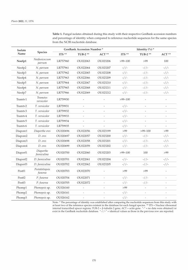
Plants 2022, 11, 1374
Table 1. Fungal isolates obtained during this study with their respective GenBank accession numbersand percentage of identity when compared to reference nucleotide sequences for the same speciesfrom the NCBI nucleotide database.
IsolateName
SpeciesGenBank Accession Number * Identity (%) *
ITS ** TUB-2 ** ACT ** ITS ** TUB-2 ** ACT **
Noefp1Neofusicoccum
parvum LR757960 OU022063 OU023206 >99–100 >99 100
Neofp2 N. parvum LR757961 OU022064 OU023207 -//- -//- -//-
Neofp3 N. parvum LR757962 OU022065 OU023208 -//- -//- -//-
Neofp4 N. parvum LR757963 OU022066 OU023209 -//- -//- -//-
Neofp5 N. parvum LR757964 OU022067 OU023210 -//- -//- -//-
Neofp6 N. parvum LR757965 OU022068 OU023211 -//- -//- -//-
Neofp7 N. parvum LR757966 OU022069 OU023212 -//- -//- -//-
Tramtv1 Trametesversicolor LR759930 - - >99–100 - -
Tramtv2 T. versicolor LR759931 - - -//- - -
Tramtv3 T. versicolor LR759932 - - -//- - -
Tramtv4 T. versicolor LR759933 - - -//- - -
Tramtv5 T. versicolor LR759934 - - -//- - -
Tramtv6 T. versicolor LR759935 - - -//- - -
Diapore1 Diaporthe eres OU020696 OU022056 OU023199 >99 >99–100 >99
Diapore2 D. eres OU020697 OU022057 OU023200 -//- -//- -//-
Diapore3 D. eres OU020698 OU022058 OU023201 -//- -//- -//-
Diapore4 D. eres OU020699 OU022059 OU023202 -//- -//- -//-
Diaporf1 Diaporthefoeniculina OU020700 OU022060 OU023203 >99–100 100 >99
Diaporf2 D. foeniculina OU020701 OU022061 OU023204 -//- -//- -//-
Diaporf3 D. foeniculina OU020702 OU022062 OU023205 -//- -//- -//-
Pestf1 Pestalotiopsisfunerea OU020703 OU022070 - >99 >99 -
Pestf2 P. funerea OU020704 OU022071 - -//- -//- -
Pestf3 P. funerea OU020705 OU022072 - -//- -//- -
Phomp1 Phomopsis sp. OU026160 - - >99 - -
Phomp2 Phomopsis sp. OU026161 - - -//- - -
Phomp3 Phomopsis sp. OU026162 - - -//- - -
Note: * The percentage of identity was established after comparing the nucleotide sequences from this study withat least two of the reference species existent in the database for each fungal species. ** ITS = Nuclear ribosomalinternal transcribed spacer regions; TUB-2 = β-tubulin 2 gene; ACT = actin gene. “-” = no data were obtained/orexist in the GenBank nucleotide database. “-//-” = identical values as those in the previous row are reported.
170
Plants 2022, 11, 1374
Figure 1. Pure fungal cultures on PDA obtained from samples of apples with dieback symptoms.(a,g) = Trametes versicolor; (b,h) = Diaporthe eres; (c,i) = Diaporthe feoniculina; (d,j) = Pestalotiopsis funerea;(e,k) = Phomopsis spp.; (f,l) = Neofusicoccum parvum.
2.3. Molecular Characterization
The PCR amplifications for each gene investigated yielded amplicons of expected sizes:ITS5/ITS4 (~700 bp), tub-2 (~500 bp) and ACT (~300 bp), which, after direct sequencingin both directions, using the same primers as for the amplification, led to 26 nucleotidesequences (Table 1). A megablast search, excluding “uncultured/environmental samplesequences”, performed in the NCBI’s nucleotide database (www.ncbi.org, accessed on 12January 2022) for all nucleotide sequences obtained in this study, identified at Genus levelall fungal isolates (Table 1).
2.4. Phylogenetic Analysis
Single locus analysis gave consistent results for all three loci (ITS, tub-2 and ACT), andthe topology of trees was congruent in terms of species grouping. All sequences obtained inthis study have been deposited in the European Molecular Biology Laboratory (EMBL-EBI)nucleotide database (www.ebi.ac.uk, accessed on 12 January 2022) and their GenBankaccession numbers are presented in Table 1.
The final alignment dataset, for the ITS region, was composed of a total number of658 characters. It contained 58 nucleotide sequences, including five outgroup species,namely: Diaporthella corylina (acc. no. KC343004) utilized for Diaporthe fungi; Valsa japonica(acc. no AF191185) for Phomopsis sp. fungi; Sordaria alcina (acc. no. AY681198) for thePestaliopsis sp. Fungal group; Grifola frondosa (acc. No. AY049140) utilized for Trametessp. Fungi; and for the Neofusicoccum parvum group, Diplodia seriata (acc. no. MH221102).Phylogenetic analysis based on the ITS region variation showed that fungal isolates fromthe same species clustered together into the same clade and, as expected, outgroup specieswere placed separately from the other groups (Figure 2).
171
Plants 2022, 11, 1374
Figure 2. Molecular phylogenetic tree obtained through the neighbor-joining (NJ) method, based onthe 58 ITS region sequences data (658 bp) from fungal isolates in the present study and publishedsequences. Five fungal species (Diaporthella corylina, Valsa japonica, Sordaria alcina, Grifola fondosaand Diplodia seriata) were used as outgroups in the analysis. The optimal tree with the sum ofbranch length = 1.38532669 is shown. The confidence probability estimated using the bootstrap test(1000 replicates) is shown next to the branches. The tree is drawn to scale, with branch lengths in thesame units as those of the evolutionary distances used to infer the phylogenetic tree. The evolutionarydistances were computed using the Tajima–Nei method and are in the units of the number of basesubstitutions/site. Scientific names of the fungi along with collection place, isolate abbreviation andGenBank AC number are shown in the trees.
172
Plants 2022, 11, 1374
Overall, the 53 nucleotide sequences obtained in this study, based on the ITS regionsequence analysis, clustered in two clades which contained all six fungal species. Two ofthese species belonged to Diaporthe genus, namely, Diaporthe eres Nitschke and D. foeniculina(Sacc.) Udayanga & Castl., and others have been identified as N. parvum, Pestalotiopsisfunerea (Desm.) Steyaert, Phomopsis sp. Sacc. & Roum. and T. versicolor (L.) Lloyd (Figure 2).Within the phylogenetic tree, the first clade grouped together five of the species previouslymentioned, while the second clade contained only one species, Trametes versicolor, clearlyseparated from the others. Additionally, the fungal species isolated and identified in thisstudy were positioned close to similar reference species downloaded from the GenBankfor each fungus, and their location was well supported by very high (97–100%) bootstrapvalues (Figure 2). The ITS data confirmed the previous preliminary fungal identificationbased on morphological features.
Since ITS alone does not provide sufficient resolution to exactly classify fungi at specieslevel, other loci were considered for the phylogeny-based identification of the taxa investi-gated in this study. In particular, the β-tubulin (tub-2) gene, a very well-known molecularlocus extensively used in phylogenetic studies of phytopathogenic fungi [35,38–40], was ex-amined. A total number of 39 nucleotide sequences of the tub-2 partial gene were obtainedand employed along with the species D. corylina, B. dothidea and S. alcina, used as outgroupsin phylogenetic analysis, which was carried out using the NJ method, as performed for theITS (Figure 3).
Results from the phylogenetic analysis of the fungal species from this study, basedon the tub-2 gene, have shown that they grouped together with similar species from theGenBank database. A better separation within the clades and subclades compared to whatobtained from the ITS was also observed (Figure 3). Two separate clades were obtainedfrom the nucleotide sequences investigated in this study. In one, Neofusicoccum isolatesgrouped together into the same subgroup, which was very well supported (98% bootstrapsupport value), and were separated from the Pestalotiopsis subclade, which was also highlysupported (99% bootstrap support value), whereas the Diaporthe isolates were all placed ina separate clade and were clearly distinguished in two subclades as species, e.g., D. eres andD. foeniculina, both sharing their vicinity with the same outgroup, D. corylina (Figure 3). Allfungal species based the tub-2 gene variation were grouped together with the same speciesfrom the GenBank with an elevated bootstrap support (98–99%). The separation of thefungal species within each clade or subclade, strongly supported by high bootstrap values,was 99% for D. eres, D. feoniculina and N. parvum and 98% in the case of P. funerea (Figure 3).
Regarding the third gene, namely actin, despite our repeated PCR trials, ampliconscould not be obtained for all fungal species (Figure 4). Therefore, the alignment for theACT gene used in phylogenetic analysis contained 298 characters and involved only24 nucleotide sequences. Moreover, ACT gene analysis showed that the two Diaporthespecies identified in this study along with their reference species from the GenBank werewell separated from the Neofusicoccum sp. isolates and, thus, clustered in two differ-ent clades also supported by very high bootstrap values of 94% and 99%, respectively.In addition, Neofusicoccum isolates were grouped together with their reference specieswith a 99% bootstrap support value (Figure 4). The phylogenetic reconstruction basedon the ACT gene reconfirmed the molecular identification based on other loci at specieslevel for each fungal species analyzed and also was in concordance with the preliminarymorphological characterization.
173
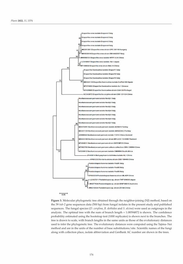
Plants 2022, 11, 1374
Figure 3. Molecular phylogenetic tree obtained through the neighbor-joining (NJ) method, based onthe 39 tub-2 gene sequences data (500 bp) from fungal isolates in the present study and publishedsequences. The fungal species (D. corylina, B. dothidea and S. alcina) were used as outgroups in theanalysis. The optimal tree with the sum of branch length = 1.08594872 is shown. The confidenceprobability estimated using the bootstrap test (1000 replicates) is shown next to the branches. Thetree is drawn to scale, with branch lengths in the same units as those of the evolutionary distancesused to infer the phylogenetic tree. The evolutionary distances were computed using the Tajima–Neimethod and are in the units of the number of base substitutions/site. Scientific names of the fungialong with collection place, isolate abbreviation and GenBank AC number are shown in the trees.
174
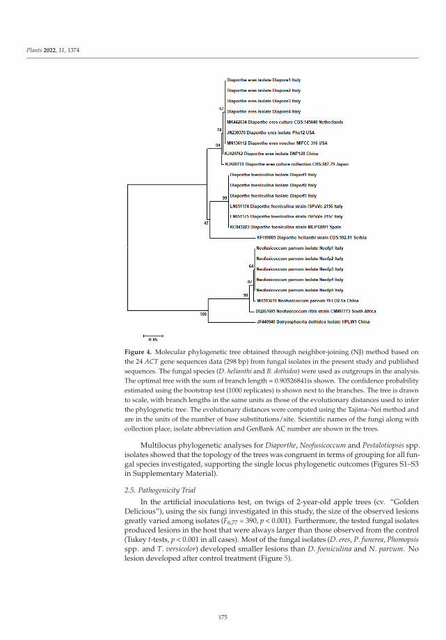
Plants 2022, 11, 1374
Figure 4. Molecular phylogenetic tree obtained through neighbor-joining (NJ) method based onthe 24 ACT gene sequences data (298 bp) from fungal isolates in the present study and publishedsequences. The fungal species (D. helianthi and B. dothidea) were used as outgroups in the analysis.The optimal tree with the sum of branch length = 0.90526841is shown. The confidence probabilityestimated using the bootstrap test (1000 replicates) is shown next to the branches. The tree is drawnto scale, with branch lengths in the same units as those of the evolutionary distances used to inferthe phylogenetic tree. The evolutionary distances were computed using the Tajima–Nei method andare in the units of the number of base substitutions/site. Scientific names of the fungi along withcollection place, isolate abbreviation and GenBank AC number are shown in the trees.
Multilocus phylogenetic analyses for Diaporthe, Neofusicoccum and Pestalotiopsis spp.isolates showed that the topology of the trees was congruent in terms of grouping for all fun-gal species investigated, supporting the single locus phylogenetic outcomes (Figures S1–S3in Supplementary Material).
2.5. Pathogenicity Trial
In the artificial inoculations test, on twigs of 2-year-old apple trees (cv. “GoldenDelicious”), using the six fungi investigated in this study, the size of the observed lesionsgreatly varied among isolates (F6,77 = 390, p < 0.001). Furthermore, the tested fungal isolatesproduced lesions in the host that were always larger than those observed from the control(Tukey t-tests, p < 0.001 in all cases). Most of the fungal isolates (D. eres, P. funerea, Phomopsisspp. and T. versicolor) developed smaller lesions than D. foeniculina and N. parvum. Nolesion developed after control treatment (Figure 5).
175
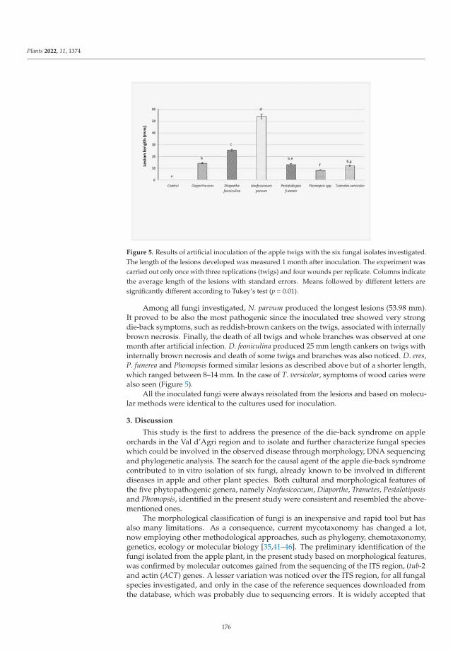
Plants 2022, 11, 1374
Figure 5. Results of artificial inoculation of the apple twigs with the six fungal isolates investigated.The length of the lesions developed was measured 1 month after inoculation. The experiment wascarried out only once with three replications (twigs) and four wounds per replicate. Columns indicatethe average length of the lesions with standard errors. Means followed by different letters aresignificantly different according to Tukey’s test (p = 0.01).
Among all fungi investigated, N. parvum produced the longest lesions (53.98 mm).It proved to be also the most pathogenic since the inoculated tree showed very strongdie-back symptoms, such as reddish-brown cankers on the twigs, associated with internallybrown necrosis. Finally, the death of all twigs and whole branches was observed at onemonth after artificial infection. D. feoniculina produced 25 mm length cankers on twigs withinternally brown necrosis and death of some twigs and branches was also noticed. D. eres,P. funerea and Phomopsis formed similar lesions as described above but of a shorter length,which ranged between 8–14 mm. In the case of T. versicolor, symptoms of wood caries werealso seen (Figure 5).
All the inoculated fungi were always reisolated from the lesions and based on molecu-lar methods were identical to the cultures used for inoculation.
3. Discussion
This study is the first to address the presence of the die-back syndrome on appleorchards in the Val d’Agri region and to isolate and further characterize fungal specieswhich could be involved in the observed disease through morphology, DNA sequencingand phylogenetic analysis. The search for the causal agent of the apple die-back syndromecontributed to in vitro isolation of six fungi, already known to be involved in differentdiseases in apple and other plant species. Both cultural and morphological features ofthe five phytopathogenic genera, namely Neofusicoccum, Diaporthe, Trametes, Pestalotiposisand Phomopsis, identified in the present study were consistent and resembled the above-mentioned ones.
The morphological classification of fungi is an inexpensive and rapid tool but hasalso many limitations. As a consequence, current mycotaxonomy has changed a lot,now employing other methodological approaches, such as phylogeny, chemotaxonomy,genetics, ecology or molecular biology [35,41–46]. The preliminary identification of thefungi isolated from the apple plant, in the present study based on morphological features,was confirmed by molecular outcomes gained from the sequencing of the ITS region, (tub-2and actin (ACT) genes. A lesser variation was noticed over the ITS region, for all fungalspecies investigated, and only in the case of the reference sequences downloaded fromthe database, which was probably due to sequencing errors. It is widely accepted that
176
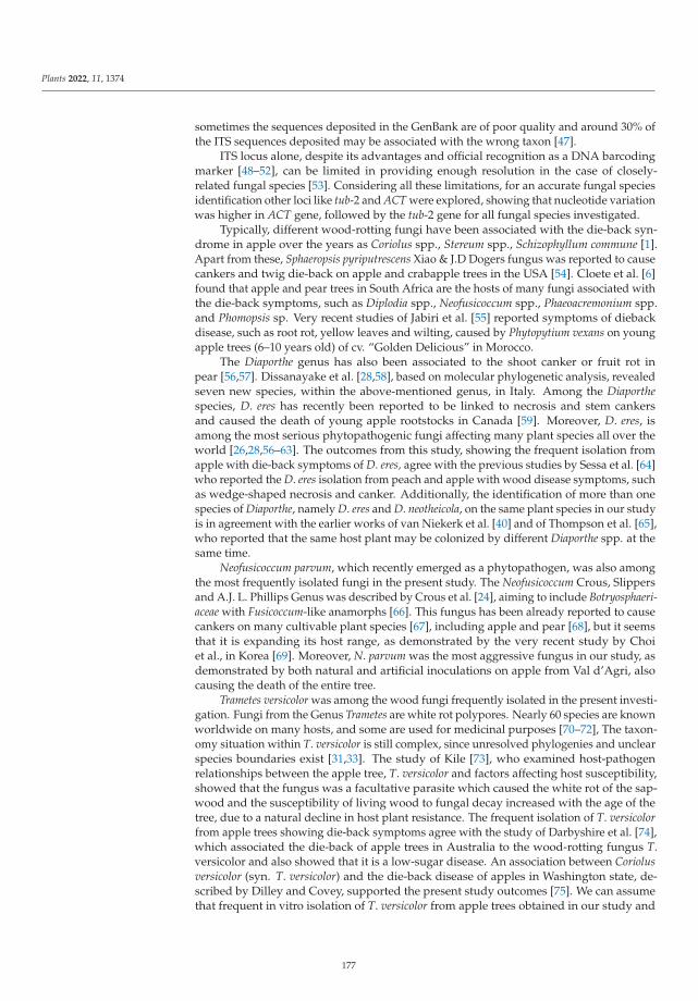
Plants 2022, 11, 1374
sometimes the sequences deposited in the GenBank are of poor quality and around 30% ofthe ITS sequences deposited may be associated with the wrong taxon [47].
ITS locus alone, despite its advantages and official recognition as a DNA barcodingmarker [48–52], can be limited in providing enough resolution in the case of closely-related fungal species [53]. Considering all these limitations, for an accurate fungal speciesidentification other loci like tub-2 and ACT were explored, showing that nucleotide variationwas higher in ACT gene, followed by the tub-2 gene for all fungal species investigated.
Typically, different wood-rotting fungi have been associated with the die-back syn-drome in apple over the years as Coriolus spp., Stereum spp., Schizophyllum commune [1].Apart from these, Sphaeropsis pyriputrescens Xiao & J.D Dogers fungus was reported to causecankers and twig die-back on apple and crabapple trees in the USA [54]. Cloete et al. [6]found that apple and pear trees in South Africa are the hosts of many fungi associated withthe die-back symptoms, such as Diplodia spp., Neofusicoccum spp., Phaeoacremonium spp.and Phomopsis sp. Very recent studies of Jabiri et al. [55] reported symptoms of diebackdisease, such as root rot, yellow leaves and wilting, caused by Phytopytium vexans on youngapple trees (6–10 years old) of cv. “Golden Delicious” in Morocco.
The Diaporthe genus has also been associated to the shoot canker or fruit rot inpear [56,57]. Dissanayake et al. [28,58], based on molecular phylogenetic analysis, revealedseven new species, within the above-mentioned genus, in Italy. Among the Diaporthespecies, D. eres has recently been reported to be linked to necrosis and stem cankersand caused the death of young apple rootstocks in Canada [59]. Moreover, D. eres, isamong the most serious phytopathogenic fungi affecting many plant species all over theworld [26,28,56–63]. The outcomes from this study, showing the frequent isolation fromapple with die-back symptoms of D. eres, agree with the previous studies by Sessa et al. [64]who reported the D. eres isolation from peach and apple with wood disease symptoms, suchas wedge-shaped necrosis and canker. Additionally, the identification of more than onespecies of Diaporthe, namely D. eres and D. neotheicola, on the same plant species in our studyis in agreement with the earlier works of van Niekerk et al. [40] and of Thompson et al. [65],who reported that the same host plant may be colonized by different Diaporthe spp. at thesame time.
Neofusicoccum parvum, which recently emerged as a phytopathogen, was also amongthe most frequently isolated fungi in the present study. The Neofusicoccum Crous, Slippersand A.J. L. Phillips Genus was described by Crous et al. [24], aiming to include Botryosphaeri-aceae with Fusicoccum-like anamorphs [66]. This fungus has been already reported to causecankers on many cultivable plant species [67], including apple and pear [68], but it seemsthat it is expanding its host range, as demonstrated by the very recent study by Choiet al., in Korea [69]. Moreover, N. parvum was the most aggressive fungus in our study, asdemonstrated by both natural and artificial inoculations on apple from Val d’Agri, alsocausing the death of the entire tree.
Trametes versicolor was among the wood fungi frequently isolated in the present investi-gation. Fungi from the Genus Trametes are white rot polypores. Nearly 60 species are knownworldwide on many hosts, and some are used for medicinal purposes [70–72], The taxon-omy situation within T. versicolor is still complex, since unresolved phylogenies and unclearspecies boundaries exist [31,33]. The study of Kile [73], who examined host-pathogenrelationships between the apple tree, T. versicolor and factors affecting host susceptibility,showed that the fungus was a facultative parasite which caused the white rot of the sap-wood and the susceptibility of living wood to fungal decay increased with the age of thetree, due to a natural decline in host plant resistance. The frequent isolation of T. versicolorfrom apple trees showing die-back symptoms agree with the study of Darbyshire et al. [74],which associated the die-back of apple trees in Australia to the wood-rotting fungus T.versicolor and also showed that it is a low-sugar disease. An association between Coriolusversicolor (syn. T. versicolor) and the die-back disease of apples in Washington state, de-scribed by Dilley and Covey, supported the present study outcomes [75]. We can assumethat frequent in vitro isolation of T. versicolor from apple trees obtained in our study and
177
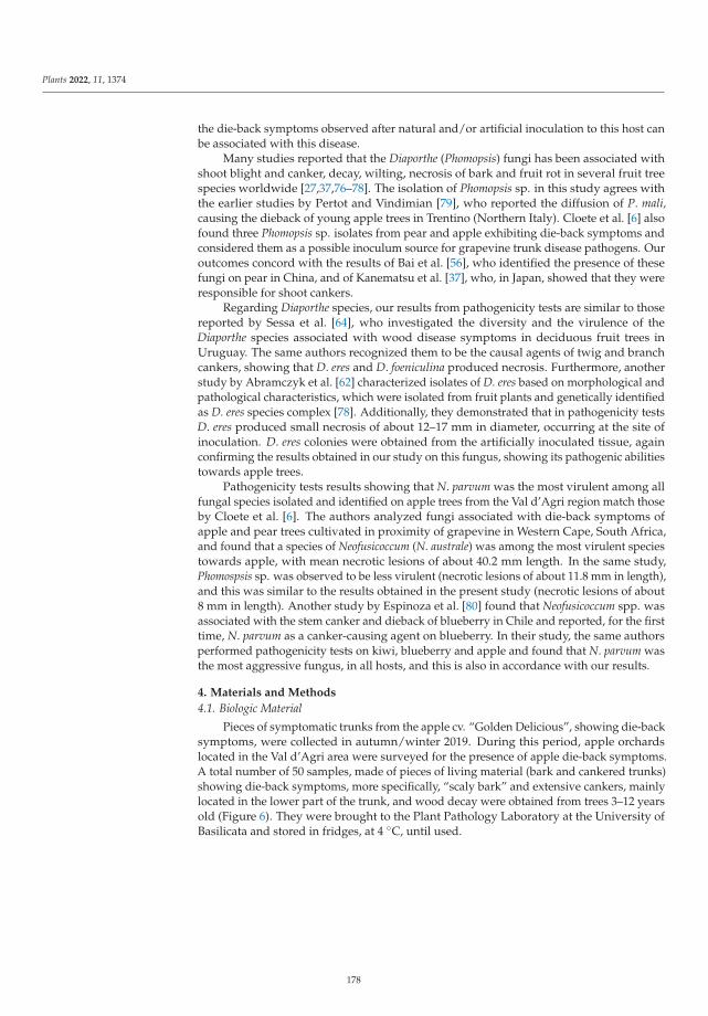
Plants 2022, 11, 1374
the die-back symptoms observed after natural and/or artificial inoculation to this host canbe associated with this disease.
Many studies reported that the Diaporthe (Phomopsis) fungi has been associated withshoot blight and canker, decay, wilting, necrosis of bark and fruit rot in several fruit treespecies worldwide [27,37,76–78]. The isolation of Phomopsis sp. in this study agrees withthe earlier studies by Pertot and Vindimian [79], who reported the diffusion of P. mali,causing the dieback of young apple trees in Trentino (Northern Italy). Cloete et al. [6] alsofound three Phomopsis sp. isolates from pear and apple exhibiting die-back symptoms andconsidered them as a possible inoculum source for grapevine trunk disease pathogens. Ouroutcomes concord with the results of Bai et al. [56], who identified the presence of thesefungi on pear in China, and of Kanematsu et al. [37], who, in Japan, showed that they wereresponsible for shoot cankers.
Regarding Diaporthe species, our results from pathogenicity tests are similar to thosereported by Sessa et al. [64], who investigated the diversity and the virulence of theDiaporthe species associated with wood disease symptoms in deciduous fruit trees inUruguay. The same authors recognized them to be the causal agents of twig and branchcankers, showing that D. eres and D. foeniculina produced necrosis. Furthermore, anotherstudy by Abramczyk et al. [62] characterized isolates of D. eres based on morphological andpathological characteristics, which were isolated from fruit plants and genetically identifiedas D. eres species complex [78]. Additionally, they demonstrated that in pathogenicity testsD. eres produced small necrosis of about 12–17 mm in diameter, occurring at the site ofinoculation. D. eres colonies were obtained from the artificially inoculated tissue, againconfirming the results obtained in our study on this fungus, showing its pathogenic abilitiestowards apple trees.
Pathogenicity tests results showing that N. parvum was the most virulent among allfungal species isolated and identified on apple trees from the Val d’Agri region match thoseby Cloete et al. [6]. The authors analyzed fungi associated with die-back symptoms ofapple and pear trees cultivated in proximity of grapevine in Western Cape, South Africa,and found that a species of Neofusicoccum (N. australe) was among the most virulent speciestowards apple, with mean necrotic lesions of about 40.2 mm length. In the same study,Phomospsis sp. was observed to be less virulent (necrotic lesions of about 11.8 mm in length),and this was similar to the results obtained in the present study (necrotic lesions of about8 mm in length). Another study by Espinoza et al. [80] found that Neofusicoccum spp. wasassociated with the stem canker and dieback of blueberry in Chile and reported, for the firsttime, N. parvum as a canker-causing agent on blueberry. In their study, the same authorsperformed pathogenicity tests on kiwi, blueberry and apple and found that N. parvum wasthe most aggressive fungus, in all hosts, and this is also in accordance with our results.
4. Materials and Methods
4.1. Biologic Material
Pieces of symptomatic trunks from the apple cv. “Golden Delicious”, showing die-backsymptoms, were collected in autumn/winter 2019. During this period, apple orchardslocated in the Val d’Agri area were surveyed for the presence of apple die-back symptoms.A total number of 50 samples, made of pieces of living material (bark and cankered trunks)showing die-back symptoms, more specifically, “scaly bark” and extensive cankers, mainlylocated in the lower part of the trunk, and wood decay were obtained from trees 3–12 yearsold (Figure 6). They were brought to the Plant Pathology Laboratory at the University ofBasilicata and stored in fridges, at 4 ◦C, until used.
178
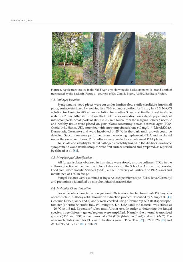
Plants 2022, 11, 1374
Figure 6. Apple trees located in the Val d’Agri area showing die-back symptoms (a–c) and death oftree caused by die-back (d). Figure a—courtesy of Dr. Camilla Nigro, ALSIA, Basilicata Region.
4.2. Pathogen Isolation
Symptomatic wood pieces were cut under laminar flow sterile conditions into smallparts, surface-sterilized by soaking in a 70% ethanol solution for 1 min, in a 1% NaOClsolution for 1 min, in 70% ethanol solution for another 30 sec and finally rinsed in sterilewater for 2 min. After sterilization, the trunk pieces were dried on a sterile paper and cutinto small parts. Small parts of about 2 × 2 mm taken from the margins between necroticand healthy tissue were placed on petri plates containing potato dextrose agar (PDA,Oxoid Ltd., Hants, UK), amended with streptomycin sulphate (40 mg L−1, MerckKGaA,Darmstadt, Germany) and were incubated at 25 ◦C in the dark until growth could bedetected. Subcultures were performed from the growing hyphae onto PDA and incubatedunder the same conditions. Pure cultures were created for all obtained PDA plates.
To isolate and identify bacterial pathogens probably linked to the die-back syndromesymptomatic wood trunk, samples were first surface sterilized and prepared, as reportedby Schaad et al. [81].
4.3. Morphological Identification
All fungal isolates obtained in this study were stored, as pure cultures (PFC), in theculture collection of the Plant Pathology Laboratory of the School of Agriculture, Forestry,Food and Environmental Sciences (SAFE) at the University of Basilicata on PDA slants andmaintained at 4 ◦C in fridge.
Fungal isolates were examined using a Axioscope microscope (Zeiss, Jena, Germany)and preliminary identified by morphological characteristics.
4.4. Molecular Characterization
For molecular characterization, genomic DNA was extracted from fresh PFC myceliaof each isolate, 7–10 days old, through an extraction protocol described by Mang et al. [43].Genomic DNA quality and quantity were checked using a Nanodrop ND-1000 spectropho-tometer (Thermo Scientific Inc., Willmington, DE, USA) and the material was stored at−20 ◦C in 1.5 mL Eppendorf tubes until further use. In order to determine the fungalspecies, three different genes/regions were amplified. Namely, the internal transcribedspacers (ITS1 and ITS2) of the ribosomal RNA (ITS); β-tubulin (tub-2) and actin (ACT). Theoligonucleotides used for PCR amplifications were: ITS5/ITS4 [82], Bt2a/Bt2b [83] andACT512F/ACT783R [84] (Table 2).
179
Plants 2022, 11, 1374
Table 2. Details of primers pairs used in this study for the amplification and sequencing offungal DNA.
Locus * Primer Sequences 5′→3′ Reference
ITSITS5 5′-GGA AGT AAA AGT CGT AAC AAG G-3′
White et al., 1990ITS4 5′-TCC TCC GCT TAT TGA TAT GC-3′
TUB-2 Bt2a 5′-GGT AAC CAA ATC GGT GCT GCT TTC-3′ Glass and Donaldson, 1995Bt2b 5′-ACC CTC AGT GTA GTG ACC CTT GGC-3′
ACT ACT-512F 5′-ATG TGC AAG GCC GGT TTC GC-3′ Carbone and Kohn, 1999ACT-783R 5′-TAC GAG TCC TTC TGG CCC AT-3′
* ITS: internal transcribed spacer regions and intervening 5.8S rRNA gene; TUB-2: partial beta tubulin gene; ACT:actin gene.
PCR amplifications were performed under the conditions explained in Manget al. [43,85] for ITS only. For the other two genes the Phire Direct PCR Master mix(Thermo Scientific Inc., USA) was used, following manufacturer’s instructions with somemodifications. PCR mixtures were composed of 10 μL of 2X Phire Plant PCR Buffer (includ-ing 1.5 mM MgCl2 and 20 μM of dNTPs), Primers 0.5 μM each; 0.4 μL of Phire Hot Start IIDNA polymerase enzyme, 5 μL of template DNA (20 ng/μL) and double distilled water upto 20 μL. The PCR cycling protocol consisted of: an initial denaturation at 98 ◦C for 5 minfor 1 cycle; then 40 cycles of denaturation at 98 ◦C for 5 s; annealing at 60 ◦C for ITS and at62 ◦C for tub-2 and ACT genes for 5 s; extension at 72 ◦C for 20 s, followed by a final exten-sion at 72 ◦C for 1 min for 1 cycle. All PCR products were separated in 1.5% agarose gels inTris-Acetic acid-EDTA (TAE) buffer and visualized under the UV after staining with SYBRSafe DNA Gel Stain (ThermoFisher Scientific™, Carlsbad, CA, USA). A 100-bp GeneRulerExpress DNA Ladder (ThermoFisher Scientific™ Baltics UAB, Vilnius, Lithuania) wasused as a molecular weight marker. Direct sequencing of all PCR products was performedby BMR Genomics [Padua, Italy], using a 3130xl automatic sequencer in both directionsand using the same primers as for the PCR. Subsequently, the sequence information wasanalyzed by the local alignment search tool using BLASTn [86,87] in the National Centerfor Biotechnology Information (NCBI) database (http://www.ncbi.nlm.nih.gov/BLAST,accessed on 12 January 2022). Annotations were based on BLAST searches with a minimumof 99–100% identity over at least 80% of the length of the nucleotide sequence, which arethe commonly used thresholds for reliable sequence annotation [88]. Nucleotide sequencesprimary identification was carried out through the BLASTn search tool program [86,87] ofthe NCBI by comparing all sequences obtained in this study with those already present inthe database.
4.5. Sequences Alignments and Phylogenetic Analysis
All nucleotide sequences produced by this study and identified based on high sequenceidentity (>99–100%) to similar species already present in nucleotide databases, along withfew additional reference sequences downloaded from GenBank (http://www.ncbi.nlm.nih.gov/GenBank, accessed on 12 January, 2022), were used for the phylogenetic analysis.Subsequently, they were manually edited and aligned with the ClustalX version 2.0 [88]program, using the MEGA X (Molecular Evolutionary Genetic Analysis) [89] phylogenypackage to build representative alignments (Table 3). As reported in previous studies bySlippers et al. [66] and by Crous et al. [24], N. parvum and N. ribis are closely related crypticspecies within the recently described Genus Neofusicoccum (Botryosphaeriaceae, Ascomycetes).Therefore, in case of the ACT gene, when no other reference species were available in theGenBank nucleotide database, this fungal species was also used, allowing us to performthe phylogenetic investigation (Table 3).
180
Plants 2022, 11, 1374
Ta
ble
3.
List
ofta
xa,f
unga
liso
late
san
dG
enBa
nkac
cess
ion
num
bers
ofth
ege
nes
anal
yzed
inth
isst
udy
and
used
for
phyl
ogen
etic
anal
ysis
.
Ta
xa
Iso
late
Na
me
Cu
ltu
reN
o.
Ge
ne
Ge
nB
an
kA
cce
ssio
nN
um
be
r*
Re
fere
nce
ITS
TU
B-2
AC
TIT
ST
UB
-2A
CT
Dia
port
heer
esC
BS18
6.37
CPC
3011
6C
BS:1
4504
0M
H85
5881
MG
2812
61M
K44
2634
Wu
etal
.,20
19
D.e
res
SS48
NEF
F3-
23-4
Pho1
2K
P903
620
MW
2085
55JN
2303
70G
enBa
nk
D.e
res
STEU
8322
DB1
4AG
O27
MIF
CC
316
KY
3126
45M
H06
3919
MN
1361
12G
enBa
nk
D.e
res
STEU
8323
Nc1
CBS
:587
.79
KY
3126
46LC
3166
67K
J420
770
Gen
Bank
D.e
res
Fi23
33M
JL13
DN
P128
KR
0236
23M
T109
632
KJ4
2076
2G
enBa
nk
Dia
port
hefo
enic
ulin
aIS
PaVe
2156
Col
Pat-
560
ISPa
Ve21
56LN
6511
72M
K52
2116
LN65
1174
Gen
Bank
aLo
pez-
Mor
alet
al.,
2020
D.f
oeni
culin
aA
1907
BA
v-1
ISPa
Ve21
57M
T230
444
MT3
7409
3LN
6511
75G
enBa
nkc
Uda
yang
aet
al.,
2014
D.f
oeni
culin
aP1
01b
CA
A13
3M
EP12
891
MT7
3564
6K
Y43
5665
KC
8432
83G
enBa
nka
Mat
hiou
daki
set
al.,
2020
cU
daya
nga
etal
.,20
14
**D
iapo
rthe
llaco
rylin
aC
BS12
1124
CBS
1211
24C
BS:5
92.8
1K
C34
3004
KC
3439
72N
/AG
omes
etal
.,20
13
**D
iapo
rthe
helia
nthi
N/A
N/A
AR
4131
N/A
N/A
KF1
9988
5c
Gen
Bank
Neo
fusi
cocc
umpa
rvum
B32
AC
BA15
YEL
O-2
1aK
J499
738
MG
9702
91M
H39
3619
Gen
Bank
N.p
arvu
mB6
5A
KK
A30
8.2
CM
W77
73K
J499
740
MH
2211
23-e
Gen
Bank
cH
unte
ret
al.,2
006
dN
.rib
is-e
-eC
MW
7773
-e-e
DQ
2676
05G
enBa
nk
N.p
arvu
mB1
3511
215_
3N
/AK
J499
742
JX39
8944
N/A
Gen
Bank
N.p
arvu
mB1
46M
FLU
CC
_12-
0380
N/A
KJ4
9974
3M
N64
3160
N/A
Gen
Bank
N.p
arvu
mFi
2326
GD
TCM
F23
N/A
KR
0028
30M
T424
811
N/A
Gen
Bank
N.p
arvu
mJR
ad16
CBS
:130
994
N/A
KY
6802
81a
MT5
2970
9N
/AG
enBa
nka
Zha
nget
al.,
2021
N.p
arvu
mC
MW
9081
;IC
MP
8003
CM
W99
4N
/AN
R11
9487
aA
Y23
6912
N/A
Gen
Bank
aSl
ippe
rset
al.,
2004
**D
iplo
dia
seri
ata
ASJ
297
HL1
N/A
MH
2211
02JF
4040
814
N/A
Gen
Bank
bBo
tryo
spha
eria
doth
idea
N/A
N/A
HPL
W1
N/A
N/A
JF44
0940
aTa
nget
al.,
2012
Pest
alot
iops
isfu
nere
aM
L4D
YM
L4D
YN
/AEF
0551
97EF
0552
34N
/AG
enBa
nk
Pest
alot
iops
issp
.SG
SGf1
6TA
P18N
030
N/A
EU71
5650
LC42
7211
N/A
Gen
Bank
Pest
alot
iops
issp
.Z
C-W
-1-1
BRIP
6661
5N
/AK
R82
2153
MK
9776
34N
/AG
enBa
nk
Pest
alot
iops
issp
.N
W-F
WA
2867
ZX
18A
N/A
MG
0983
25M
W21
8534
N/A
Gen
Bank
**So
rdar
iaal
cina
CBS
1094
60C
BS10
9460
N/A
AY
6811
98a
AY
6812
32N
/ALi
uet
al.,
2010
aC
aiet
al.,
2006
181
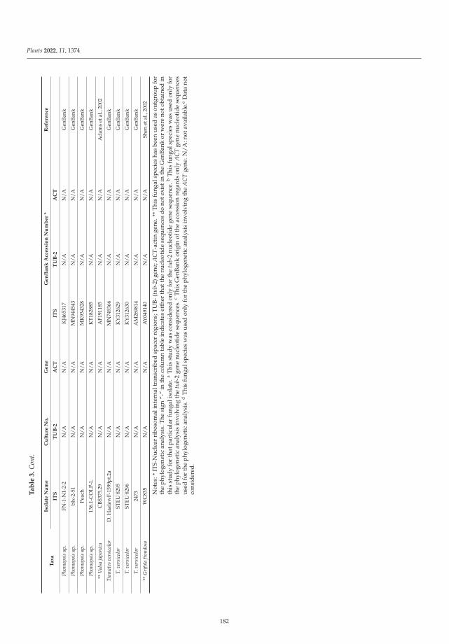
Plants 2022, 11, 1374
Ta
ble
3.
Con
t.
Ta
xa
Iso
late
Na
me
Cu
ltu
reN
o.
Ge
ne
Ge
nB
an
kA
cce
ssio
nN
um
be
r*
Re
fere
nce
ITS
TU
B-2
AC
TIT
ST
UB
-2A
CT
Phom
opsi
ssp
.FN
-1-N
1-2-
2N
/AN
/AK
J465
317
N/A
N/A
Gen
Bank
Phom
opsi
ssp
.bl
x-2-
51N
/AN
/AM
N94
4543
N/A
N/A
Gen
Bank
Phom
opsi
ssp
.Pe
ach
N/A
N/A
MK
9343
28N
/AN
/AG
enBa
nk
Phom
opsi
ssp
.13
6.1-
CO
LP-L
N/A
N/A
KT1
8288
5N
/AN
/AG
enBa
nk
**V
alsa
japo
nica
CBS
375.
29N
/AN
/AA
F191
185
N/A
N/A
Ada
ms
etal
.,20
02
Tram
etes
vers
icol
orD
.Hae
lew
F-15
99pt
.2a
N/A
N/A
MN
7493
66N
/AN
/AG
enBa
nk
T.ve
rsic
olor
STEU
8295
N/A
N/A
KY
3126
29N
/AN
/AG
enBa
nk
T.ve
rsic
olor
STEU
8296
N/A
N/A
KY
3126
30N
/AN
/AG
enBa
nk
T.ve
rsic
olor
2473
N/A
N/A
AM
2698
14N
/AN
/AG
enBa
nk
**G
rifo
lafr
ondo
saW
C83
5N
/AN
/AA
Y04
9140
N/A
N/A
Shen
etal
.,20
02
Not
es:*
ITS-
Nu
clea
rri
boso
mal
inte
rnal
tran
scri
bed
spac
erre
gion
s;T
UB
-(tu
b-2)
gene
;AC
T-a
ctin
gene
.**
Thi
sfu
ngal
spec
ies
has
been
use
das
outg
rou
pfo
rth
eph
ylog
enet
ican
alys
is.T
hesi
gn“-
”in
the
colu
mn
tabl
ein
dic
ates
eith
erth
atth
enu
cleo
tid
ese
quen
ces
do
note
xist
inth
eG
enB
ank
orw
ere
noto
btai
ned
inth
isst
udy
for
that
part
icul
arfu
ngal
isol
ate.
aT
his
stud
yw
asco
nsid
ered
only
for
the
tub-
2nu
cleo
tid
ege
nese
quen
ce.b
Thi
sfu
ngal
spec
ies
was
used
only
for
the
phyl
ogen
etic
anal
ysis
invo
lvin
gth
etu
b-2
gene
nucl
eoti
de
sequ
ence
s.c
Thi
sG
enB
ank
orig
inof
the
acce
ssio
nre
gard
son
lyA
CT
gene
nucl
eoti
de
sequ
ence
sus
edfo
rth
eph
ylog
enet
ican
alys
is.d
Thi
sfu
ngal
spec
ies
was
used
only
for
the
phyl
ogen
etic
anal
ysis
invo
lvin
gth
eA
CT
gene
.N/
A:n
otav
aila
ble.
eD
ata
not
cons
ider
ed.
182
Plants 2022, 11, 1374
Phylogeny reconstructions were performed with MEGAX [89] for each gene using theneighbor-joining (NJ) statistical method [90] with an interior branch test and 1000 bootstrapreplications [91,92], nucleotide substitution type and the Tajima–Nei substitution model [93]with uniform rates among sites. A deletion was used as treatment for gaps and missing dataand the codon positions included were 1st, 2nd, 3rd and noncoding sites. The evolutionarydistances computed using the Tajima–Nei method [93] are in the units of the number of basesubstitutions/site. The same procedure described previously was used for the tub-2 andACT genes. An unequal number of nucleotide sequences were involved in the phylogeneticanalyses for each gene investigated, which was caused by the lack of positive PCR andsequencing results for some of the genes and also the nonexistence of nucleotide sequencesin the GenBank database. Therefore, only single gene phylogenies could be performed,each one containing all nucleotide sequences obtained in this study for the examined geneplus reference species downloaded from the GenBank.
In particular, the reference sequences representing the relevant species used to buildalignments for species identification were: for D. eres and D. foeniculina (D. corylina strainCBS121124 and only for ACT gene D. helianthi strain AR4131), N. parvum (Diplodia seriatastrain ASJ297; or Botryosphaeria dothidea strains HL1 and HPLW1 for the tub-2 and ACTgenes, respectively), Pestalotiopsis sp. (Sordaria alcina strain CBS 109460), Phomopsis sp.(Valsa japonica isolate CBS375.29) and Trametes versicolor (Grifola frondosa isolate WC835)(Table 3).
A different number of nucleotide sequences were obtained for each gene and fungalspecies in this study; therefore, a multilocus phylogeny with three genes (ITS + tub-2 + ACT)was possible only for Diaporthe and Neofusicoccum spp., while for Pestalotiopsis spp. a two-gene phylogeny (ITS + tub-2) was performed using the Seaview5 program, as presented inTable S1 and in Figures S1–S3. (Supplementary material).
4.6. Pathogenicity Trials
A trial was conducted under field conditions to examine the formation of lesions ontwigs of 2-year-old apple trees (cv. “Golden Delicious”), using a common protocol. Inparticular, the pathogenicity tests were performed using 4 mm diameter mycelial plugstaken from the margins of 7-day-old cultures on PDA amended with antibiotic streptomycinsulphate (40 mg L–1, MerckKGaA, Darmstadt, Germany). An equal number of young appleshoots were equally treated but using only sterile agar plugs, which were left as controls.One fungal isolate was used for each apple tree, according to the fungal species identifiedand characterized in this study, and each treatment was replicated four times. A wound ofthe about the size of the agar plug was made on each woody shoot, in the phloem and cortextissue, with a sterile scalpel. Immediately after wounding, the plug was positioned in thecenter of the wound and covered by a sterile water wetted cotton piece. In order to avoid arapid dehydration, lesion sites were wrapped with parafilm (Pechiney Plastic Packaging,Menasha, WI, USA). For each fungal pathogen the trial layout was a randomized blockdesign with four repetitions using twigs as experimental units. The whole pathogenicitytrial consisted of six fungal pathogens, isolated in this study from apple trees in Val d’Agri,and an agar plug only. Following inoculation, all young apple trees were placed in agreenhouse, where they were kept under natural light conditions at 22 ◦C and at about70% relative humidity. After 30 days of inoculation, apple twigs were inspected for lesiondevelopment and after 45 days post-inoculation, when their necrosis was evident, thetwigs were removed and brought to the laboratory for immediate analysis. The numberof twigs with necrosis was recorded and, after the removal of the bark, the length of thedeveloped canker lesions was measured. In order to reisolate the causal agent, small pieces(approx. 5 mm length) of diseased wooden tissue were cut from the edge of the necroticlesions from the inoculated twigs and, after surface disinfection, were placed in petri dishescontaining PDA and antibiotic streptomycin sulphate. Plates were incubated for 7 days at20 ◦C in an incubator, under dark conditions, until growth was detected. Subsequently, theidentification of the reisolated fungi was carried out by both morphological features and
183
Plants 2022, 11, 1374
molecular analysis, using the protocols for morphological identification, DNA extractionand PCR conditions described above.
4.7. Statistical Analysis
Since the data obtained from the lesion measurements were normally distributed(Shapiro–Wilk tests [94], followed by a Holm–Bonfferoni [95] correction), a one-wayANOVA was used to test for mean differences among the investigated fungal isolates.Tukey post hoc tests for multiple comparisons of means were also performed to detect sig-nificant differences among the treatments. The statistical analyses performed in this studywere performed using the R version 3.6.2 software (R Core Team, Vienna, Austria) [96].
5. Conclusions
Fungal species investigated in this study are well known to be involved in the fruittree trunk diseases. The present study demonstrated that among all fungi investigated, N.parvum was the most aggressive and may be involved in the heavy decline of apple treesin the Val D’Agri area. In addition, other fungi, such as D. eres, D. foeniculina, P. funerea,T. versicolor and Phomopsis spp., could have contributed to the aggravation of the existingsymptoms. Our field observations allowed us to assume that fungi, and in particular N.parvum, could penetrate the trees through wounds created by cuttings. Therefore, to avoidthis, it is necessary to protect the wounds, in particular after cuttings.
Given the economic importance of apples worldwide, more investigations relatedto the role played by the phytopathogens discovered in this study, which are involvedin die-back disease on apple trees, seem necessary. Future outcomes will be expected toadd beneficial knowledge to better understand this complex disease in order to establishappropriate strategies to protect this regionally relevant and worldwide nutritionallyimportant crop.
Supplementary Materials: The following supporting information can be downloaded at: https://www.mdpi.com/article/10.3390/plants11101374/s1, Figure S1: PhyML tree (ITS + tub-2 + ACT)for Diaporthe spp.; Figure S2: PhyML tree (ITS + tub-2 + ACT) for Neofusicoccum spp.; Figure S3:PhyML tree (ITS + tub-2) for Pestalotiopsis spp.; Table S1: Multilocus phylogeny parameters used inthe study.
Author Contributions: Conceptualization, S.M.M. and I.C.; methodology, S.M.M.; software, S.M.M.and A.M.; validation, S.M.M., C.M., A.M. and I.C.; formal analysis, S.M.M.; investigation, S.M.M.;resources, I.C.; data curation, S.M.M., I.C., C.M. and A.M.; writing—original draft preparation,S.M.M.; writing—review and editing, S.M.M., I.C., C.M. and A.M.; visualization, S.M.M., I.C., C.M.and A.M.; supervision, I.C. and S.M.M.; project administration, I.C. and S.M.M.; funding acquisition,I.C. All authors have read and agreed to the published version of the manuscript.
Funding: This research was funded by the Basilicata Region-Phytosanitary Office (Matera, Italy),under the project entitled: “Epidemiological studies regarding the presence and spread in Basilicataof pathogens of agricultural and forestry plants with particular concern on those of quarantine.Molecular characterization of the pathogens and possibilities of fighting against them”.
Institutional Review Board Statement: Not applicable.
Informed Consent Statement: Not applicable.
Data Availability Statement: The authors declare that the data supporting the findings of this studyare available within the article and in the supplementary materials (Figures S1–S3 and Table S1).
Acknowledgments: The authors thank Giuseppe Malvasi and Vincenzo Pucciariello (PhytosanitaryOffice of Matera, Department of Agricultural and Forestry Politics, Basilicata Region, Italy) forfurnishing the information regarding the presence of the disease investigated in this study and fortheir valuable help with the field investigations.
Conflicts of Interest: The authors declare no conflict of interest.
184
Plants 2022, 11, 1374
References
1. Doepel, R.F. Wood-rotting fungi associated with dieback in apple trees: Progress report: Orchard surveys and laboratory studies.J. Dep. Agric. West. Aust. 1962, 3, 668–678. Available online: https://researchlibrary.agric.wa.gov.au/journal_agriculture4/vol3/iss9/2 (accessed on 15 January 2022).
2. Jeffers, S.N.; Aldwinckle, H.S.; Burr, T.J.; Arnerson, P.A. Phytophthora and Phytium species associated with crown rot in New Yorkapple orchards. Phytopathology 1982, 72, 533–538. [CrossRef]
3. Latorre, B.A.; Rioja, M.E.; Wilcox, W.F. Phytophthora species associated with crown rot of apple in Chile. Plant Dis. 2001, 85,603–605. [CrossRef] [PubMed]
4. Mazzola, M.; Andrews, P.K.; Reganold, J.P.; Lévesque, A. Frequency, virulence, and metalaxyl sensitivity of Phytium spp. isolatedfrom apple roots under conventional and organic production systems. Plant Dis. 2002, 86, 669–674. [CrossRef] [PubMed]
5. Di Francesco, A.; Rusin, C.; Di Foggia, M.; Marceddu, S.; Rombolà, A.; Botelho, R.V.; Baraldi, E. Characterization of applescultivars susceptibility to Neofusicoccum parvum Brazilian strains. Eur. J. Plant Pathol. 2020, 156, 939–951. [CrossRef]
6. Cloete, M.; Fourie, P.H.; Damm, U.; Crous, P.W.; Moster, L. Fungi associated with die-back symptoms of apple and pear trees, apossible inoculum source of grapevine trunk disease pathogens. Phytopathol. Mediterr. 2011, 50, S176–S190. [CrossRef]
7. Souli, M.; Paloma, A.-C.; Prez-Sierra, A.; Fattouch, S.; Armengol, J.; Boughalleb, N. Etiology of apple tree dieback in Tunisia andabiotic factors associated with the disease. Afric. J. Microbiol. Res. 2014, 8, 2272–2281. [CrossRef]
8. ISTAT 2019. Coltivazioni Legnose Fruttifere. Available online: http://dati.istat.it (accessed on 15 January 2022).9. Lindner, L. Analisi diagnostiche. In Diagnostica Fitopatologica. Relazioni D’attività anni 2014–2006. Centro di Sperimentazione
Laimburg, Bolzano (BZ), 77 Pag. (Diagnostic Analyses. Phytopathology Diagnostic. Activity Reports Years 2012–2006; ExperimentalCenter Laimburg: Bolzano, Italy, 2014; 77p.
10. Prodorutti, D.; Cainelli, C.; Gualandri, V.; Profaizer, D.; Dallago, G.; Branz, A.; Deliati, I.; Pertot, I.; Angeli, G. Moria e deperimentodel melo in Trentino. Atti Giornate Fitopatol. 2012, 2, 619–621.
11. Kienholz, J.R. Comparative study of the apple anthracnose and perennial canker fungi. J. Agric. Res. 1939, 59, 635–665.12. Pennycook, S.R.; Samuels, G.J. Botryosphaeria and Fusicoccum species associated with ripe fruit rot of Actinidia deliciosa (Kiwifruit)
in New Zealand. Mycotaxon 1985, 24, 445–458.13. Hantula, J.; Lilja, A.; Nuorteva, H.; Parikka, P.; Werres, S. Pathogenicity, morphology and genetic variation of Phytophthora
cactorum from strawberry, apple, rhododendron, and silver birch. Mycol. Res. 2000, 104, 1062–1068. [CrossRef]14. Petróczy, M.; Palkovics, L. First report of Monilia polystroma on apple in Hungary. Eur. J. Plant Pathol. 2009, 125, 343–347.
[CrossRef]15. Hibett, D.S. Ribosomal RNA and fungal systematics. Trans. Mycol. Soc. Jpn. 1992, 33, 533–556.16. Arzanlou, M.; Bakhshi, M. ITS-rDNA sequences differentiate a new lineage of Diplodia associated with canker disease of apple in
Iran. Plant Pathol. Quar. 2012, 2, 132–141. [CrossRef]17. Diaz, G.A.; Mostert, L.; Halleen, F.; Lolas, M.; Gutierrez, M.; Ferrada, E.; Latorre, B.A. Diplodia seriata Associated with Botrypshaeria
Canker and Dieback in Apples Trees in Chile. Plant Dis. 2019, 103, 1025. [CrossRef]18. McKean, P.G.; Vaughan, S.; Gull, K. The extended tubulin superfamily. J. Cell Sci. 2001, 114, 2723–2733. [CrossRef] [PubMed]19. Santos, L.; Phillips, A.J.L.; Crous, P.; Alves, A. Diaporthe species on Rosaceae with descriptions of D. pyracanthae sp. nov. and D.
malorum sp. nov. Mycosphere 2017, 8, 485–511. [CrossRef]20. Pan, T.-T.; Xue, D.-S.; Li, B.-H.; Lian, S.; Wang, C.-X. Characterization of the pathogen causing shoot dieback on apple. Acta
Phytopathol. Sinica 2020, 50, 107–111. [CrossRef]21. Li, H.; Zhang, Z.; Qin, G.; He, C.; Li, B.; Tian, S. Actin is required for cellular development and virulence of Botrytis cinerea via the
mediation of secretory proteins. Msystems 2020, 5, e00732-19. [CrossRef]22. Tang, W.; Ding, Z.; Zhou, Z.Q.; Wang, Y.Z.; Guo, L.Y. Phylogenetic and pathogenic analyses show that the causal agent of apple
ring rot in China is Botryosphaeria dothidea. Plant Dis. 2012, 96, 486–496. [CrossRef]23. Wennecker, M. Fungal Pathogens in Pome Fruit Orchards and Causal Agents of Postharvest Decay. Ph.D. Thesis, Wageningen
University, Wageningen, The Netherlands, 25 February 2019; p. 200.24. Crous, P.W.; Slippers, B.; Wingfield, M.J.; Rheeder, J.; Marasas, W.F.O.; Phillips, A.; Alves, A.; Burgess, T.I.; Barber, P.A.;
Groenewald, J.Z. Resolving phylogenetic lineages in the Botryosphaeriaceae. Stud. Mycol. 2006, 55, 235–253. [CrossRef]25. Udayanga, D.; Liu, X.Z.; Crous, P.W.; McKenzie, E.H.C.; Chukeatirote, E.; Hyde, K.D. A multi-locus phylogenetic evaluation of
Diaporthe (Phomopsis). Fungal Divers. 2012, 56, 157–171. [CrossRef]26. Udayanga, D.; Castlebury, L.A.; Rossman, A.Y.; Chukeatirote, E.; Hyde, K.D. Insights into the genus Diaporthe: Phylogenetic
species delimitation in the D. eres species complex. Fungal Divers. 2014, 67, 203–229. [CrossRef]27. Gomes, R.R.; Glienke, C.; Videira, S.I.R.; Lombard, L.; Groenewald, J.Z.; Crous, W. Diaporthe: A genus of endophitic, saprofitic
and plant pathogenic fungi. Persoonia 2013, 31, 1–41. [CrossRef] [PubMed]28. Dissanayake, A.J.; Phillips, A.J.L.; Hyde, K.D.; Yan, J.Y.; Li, X.H. The current status of species in Diaporthe. Mycosphere 2017, 8,
1106–1156. [CrossRef]29. Gilbertson, R.L.; Ryvarden, L. North American Polypores Vol. 2: Megasporoporia–Wrightoporia; Fungiflora: Oslo, Norway, 1987;
pp. 437–885.30. Ryvarden, L.; Gilbertson, R.L. European Polypores: Part 2: Meripilus–Tyromyces; Fungiflora: Oslo, Norway, 1994; pp. 394–743.
185
Plants 2022, 11, 1374
31. Justo, A.; Hibbett, D.S. Phylogenetic classification of Trametes (Basidiomycota, Polyporales) based on a five-marker dataset. Taxon2011, 60, 1567–1583. [CrossRef]
32. Welti, S.; Moreau, P.-A.; Favel, A.; Courtecuisse, R.; Haon, M.; Navarro, D.; Taussac, S.; Lesage-Meessen, L. Molecular phylogenyof Trametes and related genera, and description of a new genus Leiotrametes. Fungal Divers. 2012, 55, 47–64. [CrossRef]
33. Carlson, A.; Justo, A.; Hibbett, D.S. Species delimitation in Trametes: A comparison of ITS, RPB1, RPB2 and TEF1 Gene Phylogenies.Mycologia 2014, 106, 735–745. [CrossRef]
34. Crous, P.W.; Summerell, B.A.; Swart, L.; Denman, S.; Taylor, J.E.; Beziudenhout, C.M.; Palm, M.E.; Mariconowitz, S.; Groenewald,J.Z. Fungal pathogens of Proteaceae. Persoonia 2011, 27, 20–45. [CrossRef]
35. Maharachchikumbura, S.S.N.; Guo, L.-D.; Chukeatirote, E.; Bahkali, A.H.; Hyde, K.D. Pestalotiopsis—Morphology, phylogeny,biochemistry and diversity. Fungal Divers. 2011, 50, 167–187. [CrossRef]
36. Maharachchikumbura, S.S.N.; Hyde, K.D.; Groenewald, J.Z.; Xu, J.; Crous, P.W. Pestalotiopsis revisited. Stud. Mycol. 2014, 79,121–186. [CrossRef]
37. Kanematsu, S.; Kobayashi, T.; Kudo, A.; Ohtsu, Y. Conidial morphology, pathogenicity and culture characteristics of Phomopsisisolates from peach, Japanese pear and apple in Japan. Jpn. J. Phytopathol. 1999, 65, 264–273. [CrossRef]
38. Santos, J.M.; Phillips, A.J.L. Resolving the complex of Diaporthe (Phomopsis) species occurring on Feoniculum vulgare in Portugal.Fungal Divers. 2009, 34, 111–125.
39. Van Niekerk, J.M.; Crous, P.W.; Groenewald, J.Z.; Fourie, P.H.; Halleen, F. DNA phylogeny, morphology and pathogenicity ofBotryosphaeria species on grapevines. Mycologia 2004, 96, 781–798. [CrossRef] [PubMed]
40. Van Niekerk, J.M.; Groenewald, J.Z.; Farr, D.F.; Fourie, P.H.; Halleen, F.; Crous, P.W. Reassessment of Phomopsis species ongrapevine. Australas. Plant Pathol. 2005, 34, 27–39. [CrossRef]
41. Koenraadt, S.; Somerville, S.C.; Jones, A.L. Characterization of mutations in the beta-tubulin gene of benomyl-resistant fieldstrains of Venturia inequalis and other plant pathogenic fungi. Phytopathology 1992, 82, 1348–1354. [CrossRef]
42. Begerow, D.; Beate, J.; Oberwinkler, F. Evolutionary relationships among β-tubulin gene sequences of basidiomycetous fungi.Mycol. Res. 2004, 108, 1257–1263. [CrossRef]
43. Mang, S.M.; Scrano, L.; Camele, I. Preliminary studies on fungal contamination of two rupestrian churches from Matera (SouthernItaly). Sustainability 2020, 12, 6988. [CrossRef]
44. Hyde, K.D.; Dong, Y.; Phookamsak, R.; Jeewon, R.; Bhat, D.J.; Jones, E.B.; Liu, N.G.; Abeywickrama, P.D.; Mapook, A.; Wei, D.;et al. Fungal diversity notes 1151–1276: Taxonomic and phylogenetic contributions on genera and species of fungal taxa. FungalDiver. 2020, 100, 5–277. [CrossRef]
45. Mang, S.M.; Raciopi, R.; Camele, I.; Rana, G.L.; D’Auria, M. Use of metabolic profiles to distinguish three Monilinia species. PlantPtahol. 2015, 97, 55–59. [CrossRef]
46. Sofo, A.; Elshafie, H.S.; Scopa, A.; Mang, S.M.; Camele, I. Impact of airborne zinc polution on the antimicrobial activity of olive oiland teh microbial metabolic profiles of Z-contaminated soils in an Italian olive orchard. J. Trace Elem. Med. Biol. 2018, 49, 276–284.[CrossRef]
47. Hofstetter, V.; Buyck, B.; Eyssartier, G.; Schnee, S.; Gindro, K. The unabearable lightness of sequence-based identification. FungalDiver. 2019, 96, 243–284. [CrossRef]
48. Senanayake, I.C.; Jeewon, R.; Chomnunti, P.; Wanasinghe, D.N.; Norphanphoun, C.; Karunarathna, A.; Pem, D.; Perera, R.H.;Camporesi, E.; McKenzie, E.H.; et al. Taxonomic circumscription of Diaporthales based on multigene phylogeny and morphology.Fungal Divers. 2018, 93, 241–443. [CrossRef]
49. Senanayake, I.C.; Rathnayaka, A.R.; Marasinghe, D.S.; Calabon, M.S.; Gentekaki, E.; Lee, H.B.; Hurdeal, V.G.; Pem, D.; Dis-sanayake, L.S.; Wijesinghe, S.N.; et al. Morphological approaches in studying fungi: Collection, examination, isolation, sporulationand preservation. Mycosphere 2020, 11, 2678–2754. [CrossRef]
50. Wibberg, D.; Stadler, M.; Lambert, C.; Bunk, B.; Spröer, C.; Rückert, C.; Kalinowski, J.; Cox, R.J.; Kuhnert, E. High quality genomesequences of thirteen Hypoxylaceae (Ascomycota) strengthen the phylogenetic family backbone and enable the discovery of newtaxa. Fungal Divers. 2021, 106, 7–28. [CrossRef]
51. Vu, D.; Groenewald, M.; de Vries, M.; Gehrmann, T.; Stielow, B.; Eberhardt, U.; Al-Hatmi, A.; Groenewald, J.Z.; Cardinali, G.;Houbraken, J.; et al. Large-scale generation and analysis of filamentous fungal DNA barcodes boosts coverage for kingdom fungiand reveals thresholds for fungal species and higher taxon delimitation Stud. Mycol. 2019, 92, 135–154. [CrossRef]
52. Schoch, C.L.; Seifert, K.A.; Huhndorf, S.; Robert, V.; Spouge, J.L.; Levesque, C.A.; Chen, W.; Fungal Barcoding Consortium; FungalBarcoding Consortium Author List; Bolchacova, E. Nuclear ribosomal internal transcribed spacer (ITS) region as a universalDNA barcode marker for Fungi. Proc. Natl. Acad. Sci. USA 2012, 109, 6241–6246. [CrossRef]
53. Kiss, L. Limits of nuclear ribosomal DNA internal transcribed spacer (ITS) sequences as species barcode for fungi. Proc. Natl.Acad. Sci. USA 2012, 109, E1811. [CrossRef]
54. Xiao, C.L.; Boal, R.J. A New Canker and Twig Dieback Disease of Apple and Crabapple Trees Caused by Sphaeropsis pyriputrescensin Washington State. Plant Dis. 2005, 89, 10. [CrossRef]
55. Jabiri, S.; Lahlali, R.; Bahra, C.; Amraoui, M.B.; Tahiri, A.; Amiri, S. First report of Phytopythium vexans associated with diebackdisease of apple trees in Morocco. J. Plant Pathol. 2020, 102, 1319. [CrossRef]
186
Plants 2022, 11, 1374
56. Bai, Q.; Zhai, L.F.; Chen, X.R.; Hong, N.X.; Xu, W.X.; Wang, G.P. Biological and molecular characterization of five Phomopsisspecies associated with pear shoot canker in China. Plant Dis. 2015, 99, 1704–1712. Available online: http://apsjournals.apsnet.org/loi/pdis (accessed on 18 January 2021). [CrossRef]
57. Bertetti, D.; Guarnaccia, V.; Spadaro, D.; Gullino, M. First report of fruit rot caused by Diaporthe eres Nitschkle on Europeanpear cultivated in Italy. (Diaporthe eres Nitschkle, agente di marciume su frutti di pero in Italia). Prot. Delle Colt. 2018, 5, 10–13.[CrossRef]
58. Dissanayake, A.J.; Camporesi, E.; Hyde, K.D.; Zhang, W.; Yan, J.Y.; Li, X.H. Molecular phylogenetic analysis reveals seven newDiaporthe species from Italy. Mycosphere 2017, 8, 853–877. [CrossRef]
59. Ali, S.; Renderos, W.; Bevis, E.; Hebb, J.; Abassi, P.A. Diaporthe eres causes stem cankers and death of young apple rootstocks inCanada. Can. J. Plant Pathol. 2020, 42, 217–227. [CrossRef]
60. Florian, V.C.; Puia, C.; Groza, R.; Suciu, L.A.; Florian, T. Study of the major pathogens that lead to apple fruit decay duringstorage. Not. Bot. Horti Agrobot. Cluj-Napoca 2018, 46, 538–545. [CrossRef]
61. Sun, W.; Huang, S.; Xia, J.; Zhang, X.; Li, Z. Morphological and molecular identification of Diaporthe species in south-westernChina, with description of eight new species. MycoKeys 2021, 77, 65–95. [CrossRef]
62. Abramczyk, B.; Król, E.D.; Zalewska, E.D.; Zimowska, B. Morphological characteristics and pathogenicity of Diaporthe eres isolatesto the fruit tree shoots. Acta Sci. Pol. -Hortorum Cultus 2018, 17, 125–133. [CrossRef]
63. Chaisiri, C.; Liu, X.; Lin, Y.; Fu, Y.; Zhu, F.; Luo, C. Phylogenetic and Haplotype Network Analyses of Diaporthe eres Species inChina Based on Sequences of Multiple Loci. Biology 2021, 10, 179. [CrossRef]
64. Sessa, L.; Abreo, E.; Bettucci, L.; Lupo, S. Diversity and virulence of Diaporthe species associated with wood diseases symptoms indeciduous fruit trees in Uruguay. Phytopathol. Mediterr. 2017, 56, 431–444. [CrossRef]
65. Thompson, S.M.; Tan, Y.P.; Young, A.J.; Neate, S.M.; Aitken, E.A.B.; Shivas, R.G. Stem cankers on sunflower (Helianthus annuus)in Australia reveal a complex of pathogenic Diaporthe (Phomopsis) species. Pers. Molec. Phylogen. Evol. Fungi 2011, 27, 80–89.[CrossRef]
66. Slippers, B.; Smit, W.A.; Crous, P.W.; Countinho, T.A.; Wingfield, B.D.; Wingfield, M.J. Taxonomy, phylogeny and identification ofBotryosphaeriaceace associated with pome and stone fruit trees in South Africa and other regions of the world. Plant Pathol. 2007,56, 128–139. [CrossRef]
67. Yu, Z.D.; Tang, G.H.; Peng, S.B.; Chen, H.; Zhai, M.Z. Neofusicoccum parvum causing canker of seedlings of Juglans regia in China. J.For. Res. 2015, 26, 1019–1024. [CrossRef]
68. Shen, Y.-M.; Chao, C.-H.; Liu, H.L. First Report of Neofusicoccum parvum Associated with Stem Canker and Dieback of Asian PearTrees in Taiwan. Plant Dis. 2010, 94, 1062. [CrossRef] [PubMed]
69. Choi, S.; Paul, N.C.; Lee, K.-H.; Kim, H.-J.; Sang, H. Morphology, Molecular Phylogeny, and Pathogenicity of Neofusicoccumparvum, Associated with Leaf Spot Disease of a New Host, the Japanese Bay Tree (Machilus thunbergii). Forests 2021, 12, 440.[CrossRef]
70. Zmitrovich, I.V.; Ezhov, O.N.; Wasser, S.P. A survey of species of genus Trametes Fr. (higher basidiomycetes) with estimation oftheir medicinal source potential. Int. J. Med. Mushrooms 2012, 14, 307–319. [CrossRef]
71. Habtermariam, S. Trametes versicolor (Synn. Coriolus versicolor) Polysaccharides in Cancer Therapy: Targets and Efficiency.Biomedicines 2020, 8, 135. [CrossRef]
72. Pop, R.M.; Puia, I.C.; Puia, A.; Chedea, V.S.; Leopold, N.; Bocsan, I.C.; Buzoianu, A.D. Characterization of Trametes versicolor:Medicinal Mushroom with Important Health Benefits. Not. Bot. Horti Agrobot. Cluj-Napoca 2018, 46, 343–349. [CrossRef]
73. Kile, G. The Host-Pathogen Relationship between Apple and Trametes versicolor and Factors Affecting Host Susceptibility. Ph.D.Thesis, University of Tasmania, Hobart, Australia, 1971.
74. Darbyshire, B. Factors Influencing the Susceptibility of Apple Trees to Trametes (Polystictus) versicolor. Ph.D. Thesis, University ofTasmania, Hobart, Australia, 1968.
75. Dilley, M.A.; Covey, R.P., Jr. Association of Coriolus versicolor with a dieback disease of apples in Washington State. Plant Dis.1981, 85, 77–78. [CrossRef]
76. Udayanga, D.; Liu, X.Z.; McKenzie, E.H.C.; Chukeatirote, E.; Bahkali, A.H.A.; Hyde, L.D. The genus Phomopsis: Biology,applications, species concepts and names of common phytopathogens. Fungal Divers. 2011, 50, 189–225. [CrossRef]
77. Karaoglanidi, G.; Bardas, G. First Report of Phomopsis Fruit Decay on Apple Caused by Phomopsis mali in Greece. Plant Dis. 2006,90, 375. [CrossRef]
78. Abramczyk, B.; Król, E. Use of RAPD-PCR and its markers for identification of Diaporthe/Phomopsis from fruit trees in south-easternPoland. Acta Sci. Pol. -Hortorum Cultus 2016, 15, 161–175. Available online: http://ww.acta.media.pl/pl/action/getfullphp?id=5213 (accessed on 8 January 2020).
79. Pertot, I.; Vindimian, M.E. Diffusione di Phomopsis mali, agente di moria in giovani impianti di melo in Trentino. Inform. Fitopatol.1998, 48, 45–49. Available online: http://hdl.handle.net/10449/17658 (accessed on 8 January 2020).
80. Espinoza, J.G.; Briceño, E.X.; Chávez, E.R.; Úrbez-Torrez, J.R.; Latorre, B.A. Neofusicoccum spp. Associated with Stem Canker andDieback of Blueberry in Chile. Plant Dis. 2009, 93, 1187–1194. [CrossRef] [PubMed]
81. Schaad, N.W.; Jones, J.B.; Chun, W. Laboratory Guide for the Identification of Plant Pathogenic Bacteria, 3rd ed.; The AmericanPhytopathological Society: St. Paul, MN, USA, 2001; 373p.
187
Plants 2022, 11, 1374
82. White, T.J.; Bruns, T.; Lee, S.; Taylor, J. Amplified and direct sequencing of fungal ribosomal RNA genes for phylogenetics. In PCRProtocols: A Guide to Methods and Applications; Innis, M.A., Gelfand, D.H., Sninsky, J.J., White, T.J., Eds.; Academic: San Diego, CA,USA, 1990; pp. 315–322.
83. Glass, N.L.; Donaldson, G.C. Development of primer sets designed for use with the PCR to amplify conserved genes fromfilamentous ascomycetes. Appl. Environ. Microbiol. 1995, 61, 1323–1330. [CrossRef] [PubMed]
84. Cabone, I.; Kohn, L.M. A method for designing primer sets for speciation studies in filamentous ascomycetes. Mycologia 1999, 91,553–556. [CrossRef]
85. Mang, S.M.; Bonomo, M.G.; Camele, I.; Saturnino, C.; Capasso, A.; Salzano, G. Preliminary Investigation of Allergenic Fungi in anIndoor Environment. Pharmacologyonline 2020, 2, 314–320.
86. Altschul, S.F.; Gish, W.; Miller, W.; Myers, E.W.; Lipman, J.D. Basic Local Alignment Search Tool. J. Mol. Biol. 1990, 215, 403–410.[CrossRef]
87. Johnson, M.; Zaretskaya, I.; Raytselis, Y.; Merezhuk, Y.; Mcginnis, S.; Madden, T.L. NCBI BLAST: A better web interface. Nucl.Acids Res. 2008, 36 (Suppl. 2), W5–W9. [CrossRef] [PubMed]
88. Larkin, M.A.; Blackshields, G.; Brown, N.P.; Chenna, R.; McGettigan, P.A.; McWilliam, H.; Valentin, F.; Wallace, I.M.; Wilm, A.;Lopez, R.; et al. Clustal W and Clustal X version 2.0. Bioinformatics 2007, 23, 2947–2948. [CrossRef]
89. Kumar, S.; Stecher, G.; Li, M.; Knyaz, C.; Tamura, K. MEGA X: Molecular Evolutionary Genetic Analysis across computingplatforms. Molec. Biol. Evol. 2018, 35, 1547–1549. [CrossRef]
90. Saitou, N.; Nei, M. The neighbor-joining method: A new method for reconstructing phylogenetic trees. Mol. Biol. Evol. 1987, 4,406–425. [CrossRef]
91. Felsenstein, J. Confidence intervals on phylogenetics: An approach using bootstrap. Evolution 1985, 39, 783–791. [CrossRef]92. Dopazo, J. Estimating errors and confidence intervals for branch lengths in phylogenetic trees by a bootstrap approach. J. Mol.
Evol. 1994, 38, 300–304. [CrossRef] [PubMed]93. Tajima, F.; Nei, M. Estimation of evolutionary distance between nucleotide sequences. Mol. Biol. Evol. 1984, 1, 269–285. [PubMed]94. Shapiro, S.S.; Wilk, M.B. An analysis of variance test for normality (complete samples). Biometrika 1965, 52, 591–611. [CrossRef]95. Holm, S. A simple sequentially rejective multiple test procedure. Scand. J. Stat. 1979, 6, 65–70.96. R Core Team. R: A Language and Environment for Statistical Computing, 2019; R Foundation for Statistical Computing: Vienna,
Austria; Available online: https://www.R-project.org/ (accessed on 15 January 2022).
188

MDPISt. Alban-Anlage 66
4052 BaselSwitzerland
Tel. +41 61 683 77 34Fax +41 61 302 89 18
www.mdpi.com
Plants Editorial OfficeE-mail: [email protected]
www.mdpi.com/journal/plants


MDPI
St. Alban-Anlage 66
4052 Basel
Switzerland
Tel: +41 61 683 77 34
Fax: +41 61 302 89 18
www.mdpi.com ISBN 978-3-0365-4412-0
Related Documents


![#ASAP20 | #KeepFarmsSafe | #VisionOnAgSafety a priority on the farm. [Optional quote] ... • Decline in care of crops, animals or farm • Increasing life insurance • Increase in](https://static.cupdf.com/doc/110x72/6027c8fef610d968853d75cf/asap20-keepfarmssafe-visiononagsafety-a-priority-on-the-farm-optional-quote.jpg)
