Death Domain Assembly Mechanism Revealed by Crystal Structure of the Oligomeric PIDDosome Core Complex Hyun Ho Park, 1 Emmanuelle Logette, 2 Stefan Raunser, 3 Solange Cuenin, 2 Thomas Walz, 3 Jurg Tschopp, 2 and Hao Wu 1, * 1 Weill Medical College and Graduate School of Medical Sciences of Cornell University, New York, NY 10021, USA 2 Department of Biochemistry, University of Lausanne, CH-1066 Epalinges, Switzerland 3 Department of Cell Biology, Harvard Medical School, Boston, MA 02115, USA *Correspondence: [email protected] DOI 10.1016/j.cell.2007.01.019 SUMMARY Proteins of the death domain (DD) superfamily mediate assembly of oligomeric signaling com- plexes for the activation of caspases and kinases via unknown mechanisms. Here we report the crystal structure of the PIDD DD and RAIDD DD complex, which forms the core of the caspase-2-activating complex PIDDo- some. Although RAIDD DD and PIDD DD are monomers, they assemble into a complex that comprises seven RAIDD DDs and five PIDD DDs. Despite the use of an asymmetric assem- bly mechanism, all DDs in the complex are in quasi-equivalent environments. The structure provided eight unique asymmetric interfaces, which can be classified into three types. These three types of interactions together cover a ma- jority of the DD surface. Mutagenesis on almost all interfaces leads to disruption of the assem- bly, resulting in defective caspase-2 activation. The three types of interactions may represent most, if not all, modes of interactions in the DD superfamily for assembling complexes of different stoichiometry. INTRODUCTION The death domain (DD) superfamily comprises the death domain (DD) subfamily, the death effector domain (DED) subfamily, the caspase recruitment domain (CARD) sub- family, and the pyrin domain (PYD) subfamily. It is one of the largest protein domain superfamilies (Kohl and Grutter, 2004; Park et al., 2007; Reed et al., 2004). These domains mediate homotypic interactions within each sub- family and play critical roles in the formation of oligomeric signaling complexes, such as the death-inducing signal- ing complex (DISC) assembled by some members of the TNF receptor family for caspase-8 and caspase-10 activa- tion, the apoptosome for caspase-9 activation, the inflam- masome for caspase-1 activation, and the PIDDosome for caspase-2 activation (Kohl and Grutter, 2004; Park et al., 2007; Reed et al., 2004). These domains also participate in the assembly of signaling complexes for kinase and NF-kB activation in TNF signaling, T cell and B cell recep- tor signaling, intracellular pathogen sensing and defense, and response to DNA damage (Kohl and Grutter, 2004; Park et al., 2007; Reed et al., 2004). The DD superfamily domains appear to mediate two types of functions in these oligomeric signaling complexes for caspase and kinase activation. One function is to me- diate the assembly of oligomeric platforms for these com- plexes, and the other is to recruit downstream effectors. In a simplified view, these molecular complexes activate their effectors via proximity-induced autoactivation, such as dimerization, proteolytic processing and transphos- phorylation. For caspases, proximity-induced dimeriza- tion is sufficient for their activation (Baliga et al., 2004; Pop et al., 2006; Yin et al., 2006). The unifying feature of the DD superfamily is the six- helical bundle structural fold, as first revealed by NMR structures of Fas DD, FADD DED, RAIDD CARD, and NALP1 PYD (Kohl and Grutter, 2004; Park et al., 2007; Reed et al., 2004). There are currently two complex struc- tures in the DD superfamily that are involved in effector recruitment, the Pelle DD:Tube DD complex involved in Drosophila Toll signaling (Xiao et al., 1999) and the Apaf-1 CARD:procaspase-9 CARD complex involved in caspase-9 activation (Qin et al., 1999). Despite the funda- mental importance of the DD superfamily in apoptotic and immune signaling pathways, no structures of any oligo- meric DD superfamily complexes are currently available. Caspase-2 is an initiator caspase and the most evolu- tionarily conserved caspase (Lassus et al., 2002; Wang et al., 1994). Caspase-2-deficient germ cells and oocytes are resistant to cell death after treatment with chemother- apeutic agents (Bergeron et al., 1998). In response to DNA damage, caspase-2 acts upstream of the mitochon- dria by inducing Bid cleavage, Bax translocation, and Cell 128, 533–546, February 9, 2007 ª2007 Elsevier Inc. 533

Welcome message from author
This document is posted to help you gain knowledge. Please leave a comment to let me know what you think about it! Share it to your friends and learn new things together.
Transcript
Death Domain Assembly MechanismRevealed by Crystal Structure oftheOligomericPIDDosomeCoreComplexHyun Ho Park,1 Emmanuelle Logette,2 Stefan Raunser,3 Solange Cuenin,2 Thomas Walz,3
Jurg Tschopp,2 and Hao Wu1,*1Weill Medical College and Graduate School of Medical Sciences of Cornell University, New York, NY 10021, USA2Department of Biochemistry, University of Lausanne, CH-1066 Epalinges, Switzerland3Department of Cell Biology, Harvard Medical School, Boston, MA 02115, USA
*Correspondence: [email protected]
DOI 10.1016/j.cell.2007.01.019
SUMMARY
Proteins of the death domain (DD) superfamilymediate assembly of oligomeric signaling com-plexes for the activation of caspases andkinases via unknown mechanisms. Here wereport the crystal structure of the PIDD DDand RAIDD DD complex, which forms the coreof the caspase-2-activating complex PIDDo-some. Although RAIDD DD and PIDD DD aremonomers, they assemble into a complex thatcomprises seven RAIDD DDs and five PIDDDDs. Despite the use of an asymmetric assem-bly mechanism, all DDs in the complex are inquasi-equivalent environments. The structureprovided eight unique asymmetric interfaces,which can be classified into three types. Thesethree types of interactions together cover a ma-jority of the DD surface. Mutagenesis on almostall interfaces leads to disruption of the assem-bly, resulting in defective caspase-2 activation.The three types of interactions may representmost, if not all, modes of interactions in theDD superfamily for assembling complexes ofdifferent stoichiometry.
INTRODUCTION
The death domain (DD) superfamily comprises the death
domain (DD) subfamily, the death effector domain (DED)
subfamily, the caspase recruitment domain (CARD) sub-
family, and the pyrin domain (PYD) subfamily. It is one of
the largest protein domain superfamilies (Kohl and
Grutter, 2004; Park et al., 2007; Reed et al., 2004). These
domains mediate homotypic interactions within each sub-
family and play critical roles in the formation of oligomeric
signaling complexes, such as the death-inducing signal-
ing complex (DISC) assembled by some members of the
TNF receptor family for caspase-8 and caspase-10 activa-
tion, the apoptosome for caspase-9 activation, the inflam-
masome for caspase-1 activation, and the PIDDosome for
caspase-2 activation (Kohl and Grutter, 2004; Park et al.,
2007; Reed et al., 2004). These domains also participate
in the assembly of signaling complexes for kinase and
NF-kB activation in TNF signaling, T cell and B cell recep-
tor signaling, intracellular pathogen sensing and defense,
and response to DNA damage (Kohl and Grutter, 2004;
Park et al., 2007; Reed et al., 2004).
The DD superfamily domains appear to mediate two
types of functions in these oligomeric signaling complexes
for caspase and kinase activation. One function is to me-
diate the assembly of oligomeric platforms for these com-
plexes, and the other is to recruit downstream effectors. In
a simplified view, these molecular complexes activate
their effectors via proximity-induced autoactivation, such
as dimerization, proteolytic processing and transphos-
phorylation. For caspases, proximity-induced dimeriza-
tion is sufficient for their activation (Baliga et al., 2004;
Pop et al., 2006; Yin et al., 2006).
The unifying feature of the DD superfamily is the six-
helical bundle structural fold, as first revealed by NMR
structures of Fas DD, FADD DED, RAIDD CARD, and
NALP1 PYD (Kohl and Grutter, 2004; Park et al., 2007;
Reed et al., 2004). There are currently two complex struc-
tures in the DD superfamily that are involved in effector
recruitment, the Pelle DD:Tube DD complex involved in
Drosophila Toll signaling (Xiao et al., 1999) and the
Apaf-1 CARD:procaspase-9 CARD complex involved in
caspase-9 activation (Qin et al., 1999). Despite the funda-
mental importance of the DD superfamily in apoptotic and
immune signaling pathways, no structures of any oligo-
meric DD superfamily complexes are currently available.
Caspase-2 is an initiator caspase and the most evolu-
tionarily conserved caspase (Lassus et al., 2002; Wang
et al., 1994). Caspase-2-deficient germ cells and oocytes
are resistant to cell death after treatment with chemother-
apeutic agents (Bergeron et al., 1998). In response to
DNA damage, caspase-2 acts upstream of the mitochon-
dria by inducing Bid cleavage, Bax translocation, and
Cell 128, 533–546, February 9, 2007 ª2007 Elsevier Inc. 533
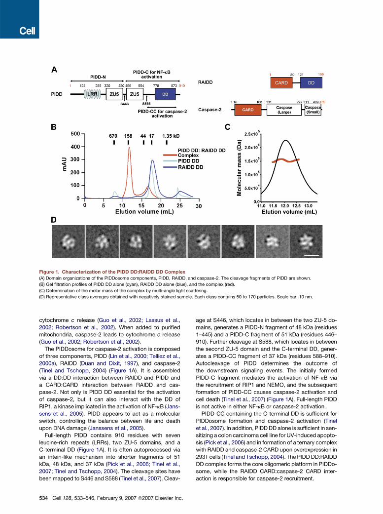
Figure 1. Characterization of the PIDD DD:RAIDD DD Complex(A) Domain organizations of the PIDDosome components, PIDD, RAIDD, and caspase-2. The cleavage fragments of PIDD are shown.
(B) Gel filtration profiles of PIDD DD alone (cyan), RAIDD DD alone (blue), and the complex (red).
(C) Determination of the molar mass of the complex by multi-angle light scattering.
(D) Representative class averages obtained with negatively stained sample. Each class contains 50 to 170 particles. Scale bar, 10 nm.
cytochrome c release (Guo et al., 2002; Lassus et al.,
2002; Robertson et al., 2002). When added to purified
mitochondria, caspase-2 leads to cytochrome c release
(Guo et al., 2002; Robertson et al., 2002).
The PIDDosome for caspase-2 activation is composed
of three components, PIDD (Lin et al., 2000; Telliez et al.,
2000a), RAIDD (Duan and Dixit, 1997), and caspase-2
(Tinel and Tschopp, 2004) (Figure 1A). It is assembled
via a DD:DD interaction between RAIDD and PIDD and
a CARD:CARD interaction between RAIDD and cas-
pase-2. Not only is PIDD DD essential for the activation
of caspase-2, but it can also interact with the DD of
RIP1, a kinase implicated in the activation of NF-kB (Jans-
sens et al., 2005). PIDD appears to act as a molecular
switch, controlling the balance between life and death
upon DNA damage (Janssens et al., 2005).
Full-length PIDD contains 910 residues with seven
leucine-rich repeats (LRRs), two ZU-5 domains, and a
C-terminal DD (Figure 1A). It is often autoprocessed via
an intein-like mechanism into shorter fragments of 51
kDa, 48 kDa, and 37 kDa (Pick et al., 2006; Tinel et al.,
2007; Tinel and Tschopp, 2004). The cleavage sites have
been mapped to S446 and S588 (Tinel et al., 2007). Cleav-
534 Cell 128, 533–546, February 9, 2007 ª2007 Elsevier Inc.
age at S446, which locates in between the two ZU-5 do-
mains, generates a PIDD-N fragment of 48 kDa (residues
1–445) and a PIDD-C fragment of 51 kDa (residues 446–
910). Further cleavage at S588, which locates in between
the second ZU-5 domain and the C-terminal DD, gener-
ates a PIDD-CC fragment of 37 kDa (residues 588–910).
Autocleavage of PIDD determines the outcome of
the downstream signaling events. The initially formed
PIDD-C fragment mediates the activation of NF-kB via
the recruitment of RIP1 and NEMO, and the subsequent
formation of PIDD-CC causes caspase-2 activation and
cell death (Tinel et al., 2007) (Figure 1A). Full-length PIDD
is not active in either NF-kB or caspase-2 activation.
PIDD-CC containing the C-terminal DD is sufficient for
PIDDosome formation and caspase-2 activation (Tinel
et al., 2007). In addition, PIDD DD alone is sufficient in sen-
sitizing a colon carcinoma cell line for UV-induced apopto-
sis (Pick et al., 2006) and in formation of a ternary complex
with RAIDD and caspase-2 CARD upon overexpression in
293T cells (Tinel and Tschopp, 2004). The PIDD DD:RAIDD
DD complex forms the core oligomeric platform in PIDDo-
some, while the RAIDD CARD:caspase-2 CARD inter-
action is responsible for caspase-2 recruitment.
To elucidate the molecular basis of caspase-2 activa-
tion and of the assembly mechanisms of the DD super-
family, we determined the crystal structure of the PIDD
DD:RAIDD DD complex, which comprises seven RAIDD
DD and five PIDD DD molecules. Despite the use of an
asymmetric assembly mechanism, all DDs in the complex
are in quasi-equivalent environments. The structure pro-
vided multiple observations of eight unique asymmetric
interfaces, which can be further classified into three types.
These interactions can coexist on a single DD and to-
gether cover a majority of the DD surface. Structure-
based mutagenesis on almost all interfaces leads to dis-
ruption of the assembly, resulting in defective caspase-2
activation. In contrast to the concept that DD superfamily
interactions may involve any available surfaces and may
be very diverse, we show here that the three types of inter-
actions in this complex may represent most, if not all,
modes of interactions in the DD superfamily and may be
used to assemble oligomeric complexes of different
stoichiometry.
RESULTS
Overall Structure of the PIDD DD:RAIDD DD
Complex, the Core Oligomerization
Platform of the PIDDosome
As a first step toward elucidating the molecular basis of
PIDDosome formation, we expressed and purified the
DDs of PIDD and RAIDD. Although PIDD DD and RAIDD
DD are both monomeric in solution, when mixed together,
the complex containing both DDs eluted at�150 kDa from
a Superdex 200 gel filtration column (Figure 1B). Because
both mass and shape affect gel filtration positions, we fur-
ther used multi-angle light scattering (MALS) with refrac-
tive index to accurately measure its molecular mass.
MALS measurement gave a molecular mass of 152.4
kDa (0.8% fitting error) for the complex, with a polydisper-
sity of 1.001 (Figure 1C). These data suggest that PIDD DD
and RAIDD DD assemble into an oligomeric complex.
Electron microscopy (EM) of the negatively stained
PIDD DD:RAIDD DD complex revealed a monodisperse
and homogeneous particle population (Figure S1 in the
Supplemental Data available with this article online). Clas-
sification of 3708 particle images into 25 groups produced
class averages that depicted molecules of similar size,
about 9 nm in diameter, but with varying structural fea-
tures (Figure 1D; Figure S1). The differences in the projec-
tions most likely arise from different orientations, in which
the complex had adsorbed to the carbon support film.
We crystallized the complex and determined its struc-
ture at 3.2A resolution using single-wavelength anoma-
lous diffraction of a mercury derivative (Table 1). The
structure revealed that the PIDD DD:RAIDD DD complex
contains five PIDD DD and seven RAIDD DD molecules.
It forms a compact globular structure of approximately
90 A in diameter (Figures 2A and 2B). The globular shape
of the structure is consistent with the normal elution be-
havior of the complex in gel filtration. This size agrees
well with the EM images. In addition, despite the strong
contrast between the individual domains in the EM projec-
tion averages due to stain accumulation, comparison of
the experimental class averages with projections calcu-
lated from the atomic model and resolution filtered to
30 A clearly showed that the class averages depict the
same complex. The differences between the class aver-
ages arise from different orientations in which the complex
had adsorbed to the carbon support film (Figure S1). As
the calculated molecular weights of monomeric PIDD
DD and RAIDD DD are 13,036 Da and 13,075 Da, respec-
tively, the calculated molecular mass of a 5:7 PIDD
DD:RAIDD DD complex is 156.7 kDa, which agrees well
with the molecular mass measured by MALS.
PIDDosome for caspase-2 activation contains the PIDD
autoprocessing fragment PIDD-CC, RAIDD, and caspase-
2 (Tinel et al., 2007; Tinel and Tschopp, 2004), with calcu-
lated molecular weights of 36,560 Da, 22,745 Da, and
50,685 Da, respectively. If the same PIDD DD:RAIDD DD
stoichiometry is present in the PIDDosome for caspase-
2 activation, the calculated molecular mass of the
PIDDosome with five PIDD-CC, seven RAIDD, and seven
caspase-2 molecules would be 696.8 kDa. This molecular
mass is in striking agreement with gel filtration analysis
of the PIDDosome, which showed a molecular mass of
�670 kDa (Read et al., 2002; Tinel and Tschopp, 2004).
The structure of the PIDD DD:RAIDD DD complex may
be divided into three layers viewing from the side of the
complex, two RAIDD DDs at the top layer (R6 and R7),
five RAIDD DDs in the middle layer (R1-R5), and five
PIDD DDs at the bottom layer (P1-P5) (Figure 2A). Viewing
from the top of the complex, the middle and the bottom
layers form two stacked closed rings (Figures 2B and
2C). The termini of the DDs point to the periphery of the
complex (Figures 2B and 2C). The peripheral locations of
the N termini allow PIDD DD to connect to the N-terminal
region of PIDD-CC and RAIDD DD to connect to its CARD
domain (Figure 2D). Therefore, one could envision that the
PIDD DD:RAIDD DD complex localizes in the center of
the PIDDosome to mediate oligomerization, while the
N-terminal region of PIDD-CC, RAIDD CARD, and cas-
pase-2 occupy the outer part of the PIDDosome. In this
scenario, the seven caspase-2 molecules in the complex
are brought into proximity for their dimerization and acti-
vation (Figure 2D).
The PIDD DD:RAIDD DD Complex Is Constructed
by Successive Screw Rotations
Strikingly, the core complex of five PIDD DDs and five
RAIDD DDs does not possess a recognizable symmetry.
Looking down from the top, the RAIDD DDs and the
PIDD DDs around the two stacked rings are related by
rotations around a common central axis (Figure 2C;
Figure S2). Pairwise superposition showed that the mole-
cules are related by two different rotation angles at differ-
ent locations (Figure 2C). In addition, viewing from the side
of the complex, the DDs in each layer are not localized on
Cell 128, 533–546, February 9, 2007 ª2007 Elsevier Inc. 535
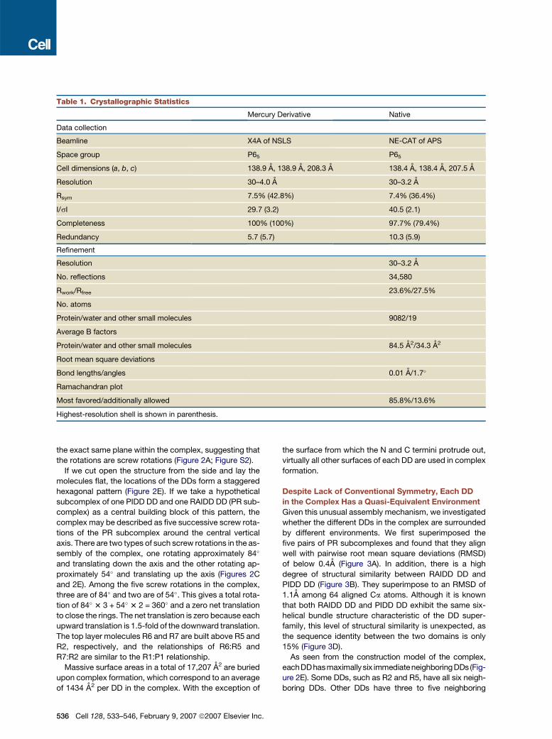
Table 1. Crystallographic Statistics
Mercury Derivative Native
Data collection
Beamline X4A of NSLS NE-CAT of APS
Space group P65 P65
Cell dimensions (a, b, c) 138.9 A, 138.9 A, 208.3 A 138.4 A, 138.4 A, 207.5 A
Resolution 30–4.0 A 30–3.2 A
Rsym 7.5% (42.8%) 7.4% (36.4%)
I/sI 29.7 (3.2) 40.5 (2.1)
Completeness 100% (100%) 97.7% (79.4%)
Redundancy 5.7 (5.7) 10.3 (5.9)
Refinement
Resolution 30–3.2 A
No. reflections 34,580
Rwork/Rfree 23.6%/27.5%
No. atoms
Protein/water and other small molecules 9082/19
Average B factors
Protein/water and other small molecules 84.5 A2/34.3 A2
Root mean square deviations
Bond lengths/angles 0.01 A/1.7�
Ramachandran plot
Most favored/additionally allowed 85.8%/13.6%
Highest-resolution shell is shown in parenthesis.
the exact same plane within the complex, suggesting that
the rotations are screw rotations (Figure 2A; Figure S2).
If we cut open the structure from the side and lay the
molecules flat, the locations of the DDs form a staggered
hexagonal pattern (Figure 2E). If we take a hypothetical
subcomplex of one PIDD DD and one RAIDD DD (PR sub-
complex) as a central building block of this pattern, the
complex may be described as five successive screw rota-
tions of the PR subcomplex around the central vertical
axis. There are two types of such screw rotations in the as-
sembly of the complex, one rotating approximately 84�
and translating down the axis and the other rotating ap-
proximately 54� and translating up the axis (Figures 2C
and 2E). Among the five screw rotations in the complex,
three are of 84� and two are of 54�. This gives a total rota-
tion of 84� 3 3 + 54� 3 2 = 360� and a zero net translation
to close the rings. The net translation is zero because each
upward translation is 1.5-fold of the downward translation.
The top layer molecules R6 and R7 are built above R5 and
R2, respectively, and the relationships of R6:R5 and
R7:R2 are similar to the R1:P1 relationship.
Massive surface areas in a total of 17,207 A2 are buried
upon complex formation, which correspond to an average
of 1434 A2 per DD in the complex. With the exception of
536 Cell 128, 533–546, February 9, 2007 ª2007 Elsevier Inc.
the surface from which the N and C termini protrude out,
virtually all other surfaces of each DD are used in complex
formation.
Despite Lack of Conventional Symmetry, Each DD
in the Complex Has a Quasi-Equivalent Environment
Given this unusual assembly mechanism, we investigated
whether the different DDs in the complex are surrounded
by different environments. We first superimposed the
five pairs of PR subcomplexes and found that they align
well with pairwise root mean square deviations (RMSD)
of below 0.4A (Figure 3A). In addition, there is a high
degree of structural similarity between RAIDD DD and
PIDD DD (Figure 3B). They superimpose to an RMSD of
1.1A among 64 aligned Ca atoms. Although it is known
that both RAIDD DD and PIDD DD exhibit the same six-
helical bundle structure characteristic of the DD super-
family, this level of structural similarity is unexpected, as
the sequence identity between the two domains is only
15% (Figure 3D).
As seen from the construction model of the complex,
eachDDhasmaximally six immediateneighboringDDs (Fig-
ure 2E). Some DDs, such as R2 and R5, have all six neigh-
boring DDs. Other DDs have three to five neighboring
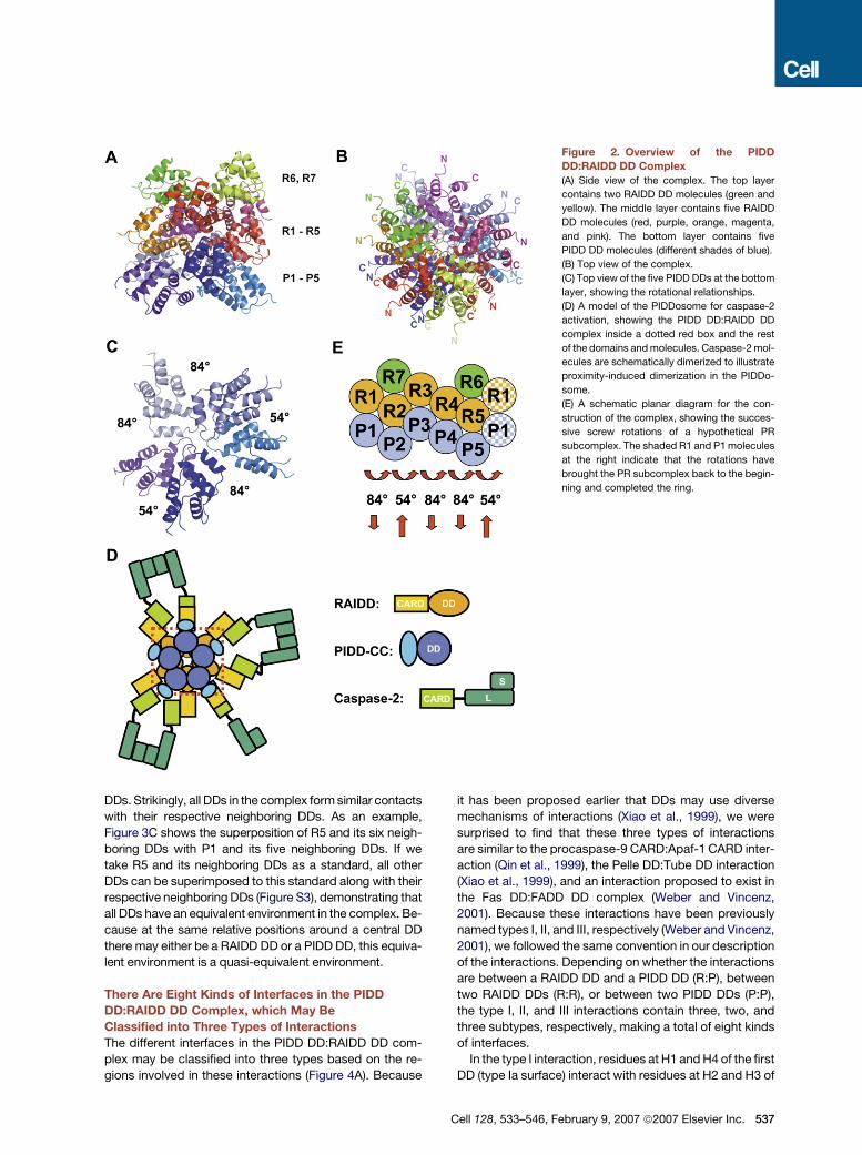
Figure 2. Overview of the PIDD
DD:RAIDD DD Complex
(A) Side view of the complex. The top layer
contains two RAIDD DD molecules (green and
yellow). The middle layer contains five RAIDD
DD molecules (red, purple, orange, magenta,
and pink). The bottom layer contains five
PIDD DD molecules (different shades of blue).
(B) Top view of the complex.
(C) Top view of the five PIDD DDs at the bottom
layer, showing the rotational relationships.
(D) A model of the PIDDosome for caspase-2
activation, showing the PIDD DD:RAIDD DD
complex inside a dotted red box and the rest
of the domains and molecules. Caspase-2 mol-
ecules are schematically dimerized to illustrate
proximity-induced dimerization in the PIDDo-
some.
(E) A schematic planar diagram for the con-
struction of the complex, showing the succes-
sive screw rotations of a hypothetical PR
subcomplex. The shaded R1 and P1 molecules
at the right indicate that the rotations have
brought the PR subcomplex back to the begin-
ning and completed the ring.
DDs. Strikingly, all DDs in the complex form similar contacts
with their respective neighboring DDs. As an example,
Figure 3C shows the superposition of R5 and its six neigh-
boring DDs with P1 and its five neighboring DDs. If we
take R5 and its neighboring DDs as a standard, all other
DDs can be superimposed to this standard along with their
respective neighboring DDs (Figure S3), demonstrating that
all DDs have an equivalent environment in the complex. Be-
cause at the same relative positions around a central DD
there may either be a RAIDD DD or a PIDD DD, this equiva-
lent environment is a quasi-equivalent environment.
There Are Eight Kinds of Interfaces in the PIDD
DD:RAIDD DD Complex, which May Be
Classified into Three Types of Interactions
The different interfaces in the PIDD DD:RAIDD DD com-
plex may be classified into three types based on the re-
gions involved in these interactions (Figure 4A). Because
it has been proposed earlier that DDs may use diverse
mechanisms of interactions (Xiao et al., 1999), we were
surprised to find that these three types of interactions
are similar to the procaspase-9 CARD:Apaf-1 CARD inter-
action (Qin et al., 1999), the Pelle DD:Tube DD interaction
(Xiao et al., 1999), and an interaction proposed to exist in
the Fas DD:FADD DD complex (Weber and Vincenz,
2001). Because these interactions have been previously
named types I, II, and III, respectively (Weber and Vincenz,
2001), we followed the same convention in our description
of the interactions. Depending on whether the interactions
are between a RAIDD DD and a PIDD DD (R:P), between
two RAIDD DDs (R:R), or between two PIDD DDs (P:P),
the type I, II, and III interactions contain three, two, and
three subtypes, respectively, making a total of eight kinds
of interfaces.
In the type I interaction, residues at H1 and H4 of the first
DD (type Ia surface) interact with residues at H2 and H3 of
Cell 128, 533–546, February 9, 2007 ª2007 Elsevier Inc. 537
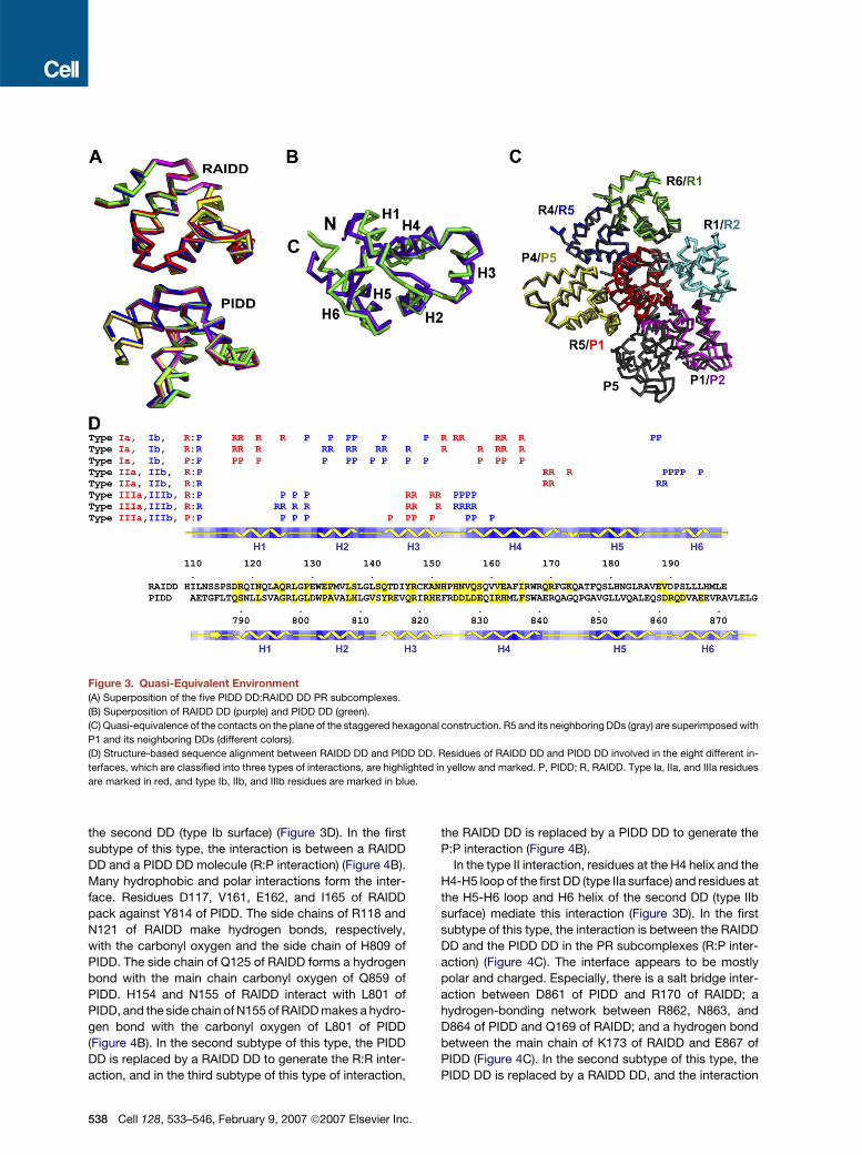
Figure 3. Quasi-Equivalent Environment
(A) Superposition of the five PIDD DD:RAIDD DD PR subcomplexes.
(B) Superposition of RAIDD DD (purple) and PIDD DD (green).
(C) Quasi-equivalence of the contacts on the plane of the staggered hexagonal construction. R5 and its neighboring DDs (gray) are superimposed with
P1 and its neighboring DDs (different colors).
(D) Structure-based sequence alignment between RAIDD DD and PIDD DD. Residues of RAIDD DD and PIDD DD involved in the eight different in-
terfaces, which are classified into three types of interactions, are highlighted in yellow and marked. P, PIDD; R, RAIDD. Type Ia, IIa, and IIIa residues
are marked in red, and type Ib, IIb, and IIIb residues are marked in blue.
the second DD (type Ib surface) (Figure 3D). In the first
subtype of this type, the interaction is between a RAIDD
DD and a PIDD DD molecule (R:P interaction) (Figure 4B).
Many hydrophobic and polar interactions form the inter-
face. Residues D117, V161, E162, and I165 of RAIDD
pack against Y814 of PIDD. The side chains of R118 and
N121 of RAIDD make hydrogen bonds, respectively,
with the carbonyl oxygen and the side chain of H809 of
PIDD. The side chain of Q125 of RAIDD forms a hydrogen
bond with the main chain carbonyl oxygen of Q859 of
PIDD. H154 and N155 of RAIDD interact with L801 of
PIDD, and the side chain of N155 of RAIDD makes a hydro-
gen bond with the carbonyl oxygen of L801 of PIDD
(Figure 4B). In the second subtype of this type, the PIDD
DD is replaced by a RAIDD DD to generate the R:R inter-
action, and in the third subtype of this type of interaction,
538 Cell 128, 533–546, February 9, 2007 ª2007 Elsevier Inc.
the RAIDD DD is replaced by a PIDD DD to generate the
P:P interaction (Figure 4B).
In the type II interaction, residues at the H4 helix and the
H4-H5 loop of the first DD (type IIa surface) and residues at
the H5-H6 loop and H6 helix of the second DD (type IIb
surface) mediate this interaction (Figure 3D). In the first
subtype of this type, the interaction is between the RAIDD
DD and the PIDD DD in the PR subcomplexes (R:P inter-
action) (Figure 4C). The interface appears to be mostly
polar and charged. Especially, there is a salt bridge inter-
action between D861 of PIDD and R170 of RAIDD; a
hydrogen-bonding network between R862, N863, and
D864 of PIDD and Q169 of RAIDD; and a hydrogen bond
between the main chain of K173 of RAIDD and E867 of
PIDD (Figure 4C). In the second subtype of this type, the
PIDD DD is replaced by a RAIDD DD, and the interaction
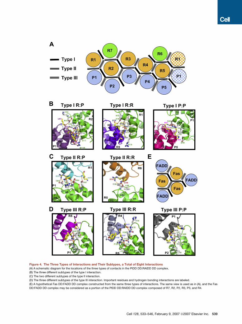
Figure 4. The Three Types of Interactions and Their Subtypes, a Total of Eight Interactions
(A) A schematic diagram for the locations of the three types of contacts in the PIDD DD:RAIDD DD complex.
(B) The three different subtypes of the type I interaction.
(C) The two different subtypes of the type II interaction.
(D) The three different subtypes of the type III interaction. Important residues and hydrogen bonding interactions are labeled.
(E) A hypothetical Fas DD:FADD DD complex constructed from the same three types of interactions. The same view is used as in (A), and the Fas
DD:FADD DD complex may be considered as a portion of the PIDD DD:RAIDD DD complex composed of R7, R2, P2, R3, P3, and R4.
Cell 128, 533–546, February 9, 2007 ª2007 Elsevier Inc. 539
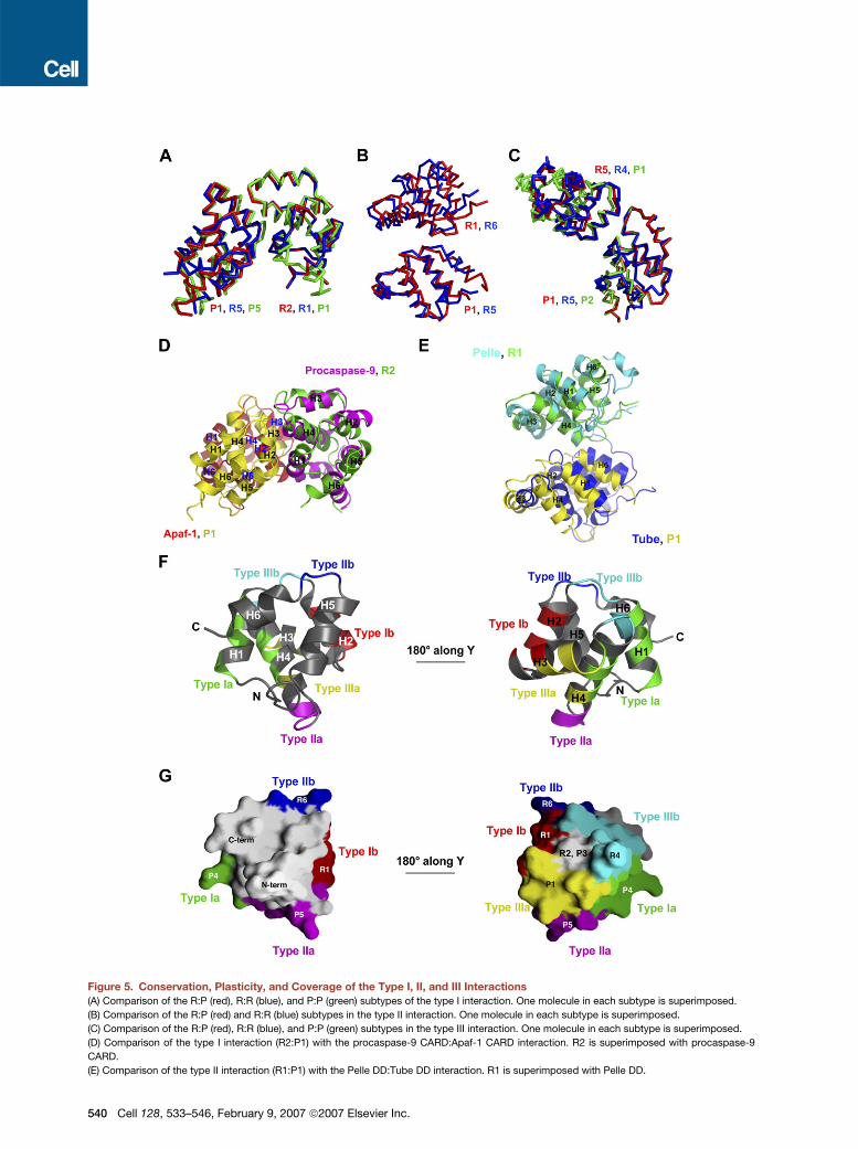
Figure 5. Conservation, Plasticity, and Coverage of the Type I, II, and III Interactions
(A) Comparison of the R:P (red), R:R (blue), and P:P (green) subtypes of the type I interaction. One molecule in each subtype is superimposed.
(B) Comparison of the R:P (red) and R:R (blue) subtypes in the type II interaction. One molecule in each subtype is superimposed.
(C) Comparison of the R:P (red), R:R (blue), and P:P (green) subtypes in the type III interaction. One molecule in each subtype is superimposed.
(D) Comparison of the type I interaction (R2:P1) with the procaspase-9 CARD:Apaf-1 CARD interaction. R2 is superimposed with procaspase-9
CARD.
(E) Comparison of the type II interaction (R1:P1) with the Pelle DD:Tube DD interaction. R1 is superimposed with Pelle DD.
540 Cell 128, 533–546, February 9, 2007 ª2007 Elsevier Inc.
is between two RAIDD DDs, such as those between R6
and R5 and between R7 and R2 (R:R interaction) (Fig-
ure 4C). This R:R interaction appears to be much less
extensive than the corresponding R:P interaction.
In the type III interaction, residues at H3 of the first DD
(type IIIa) interact with residues near the H1-H2 and the
H3-H4 loops of the second DD (type IIIb) (Figure 3D). In
the first subtype of this type, the interaction is between
a RAIDD DD and a PIDD DD (R:P interaction) (Figure 4D).
A mixture of hydrophobic, polar, and charged interactions
occur at this interface, including the hydrophobic interac-
tion between L801 of PIDD and Y146 of RAIDD, the salt
bridge between D829 of PIDD and R147 of RAIDD, and
a hydrogen bond between the main chain of L828 of
PIDD and the side chain of N151 of RAIDD (Figure 4D). In
the second subtype of this type, the PIDD DD is replaced
by a RAIDD DD molecule (R:R interaction) (Figure 4D). In
the third subtype of this type, the RAIDD DD is replaced
by a PIDD DD molecule (P:P interaction) (Figure 4D).
Conservation, Plasticity, and Coverage of the Type I,
II, and III Interactions in the DD Superfamily
Comparison among the different observations within each
type revealed conservation, variation, and plasticity in
these interactions. First, different observations within
each subtype of interactions in the PIDD DD:RAIDD DD
complex are completely conserved. In type I interactions,
there are three observations of the R:P subtype, four ob-
servations of the R:R subtype, and two observations of
the P:P subtype (Figure 4A). These are all conserved and
align well to within RMSD of 0.4A. Similar well-conserved
alignment statistics are also observed within the five ob-
servations of the type II interaction R:P subtype, the two
observations of the type II interaction R:R subtype, the
two observations of the type III interaction R:P subtype,
the five observations of the type III interaction R:R subtype,
and the three observations of the type III interaction P:P
subtype. These data suggest that each observed interac-
tion is specific to the particular partners in the interaction.
Second, among the different interactions within each
type, variations in orientations are observed. The different
subtypes within each type of interaction in the PIDD
DD:RAIDD DD complex show small adjustments in orien-
tation (Figures 5A–5C). More significant adjustments
are observed when the type I interaction in the PIDD
DD:RAIDD DD complex is compared with the procas-
pase-9 CARD:Apaf-1 CARD interaction (Figure 5D) and
when the type II interaction in the PIDD DD:RAIDD DD
complex is compared with the Pelle DD:Tube DD complex
(Figure 5E). Nonetheless, in all type I interactions, it is the
H1 and H4 region of the first molecule (type Ia) interacting
with the H2 and H3 region of the second molecule (type
Ib). In the type II interaction, however, the regions of con-
tact are somewhat different. In addition to the common in-
teraction between the H4-H5 region of the first DD and the
H5-H6 region of the second DD, in the Pelle DD:Tube DD
complex, the adjacent H2 region of the Pelle DD and the
adjacent H1-H2 region of the Tube DD also participate in
the interaction. In comparison with the type I interaction,
the type II interaction buries a smaller surface area. In the
Pelle DD:Tube DD complex, this interaction is strength-
ened by an additional interaction between a long tail of
Tube and the H2-H3 and H4-H5 region of the Pelle DD.
Therefore, depending on the exact partners in the com-
plex, there is adjustment in orientation within each type
of interactions. This structural plasticity may be important
for accommodating the different sequences at these inter-
faces and for achieving specificity of different interaction
pairs.
Not only are the regions of contacts relatively con-
served, but the surface shape complementarity also ap-
pears to be preserved within each type of interaction. In
type I interaction, the type Ia surface is concave and re-
ceives the convex surface of the type Ib surface. In both
type II and type III interactions, the IIa and IIIa surfaces
are convex, and the IIb and IIIb surfaces are concave.
However, the nature of contacts is not conserved within
each type of interaction. For example, in the procas-
pase-9 CARD:Apaf-1 CARD complex, the interacting sur-
faces are complementary in charge. In the analogous type
I interactions in the PIDD DD:RAIDD DD complex, a com-
plex network of hydrophobic contacts and hydrogen
bonds mediate the interfaces.
The three types of interactions can coexist on a single
DD, and each DD in the PIDD DD:RAIDD DD complex
uses all types of surfaces to interact with neighboring
DDs. Strikingly, when these interactions are mapped
onto a particular DD (e.g., R5), the DD surface is almost
all covered by these interactions, with the exception of
the surface from which the termini protrude out (Figures
5F and 5G). In the standard orientation we use in this re-
port, the DDs use type IIa and IIb surfaces to interact with
other DDs above or below and use other types of surfaces
for lateral interactions. The full coverage of these interac-
tions on a DD and their conservation suggest that these
three types of interaction may likely represent the major,
if not all, modes of interactions in the DD superfamily.
Mutations that Disrupt the PIDD DD:RAIDD
DD Interaction Prevent PIDDosome Formation
and Caspase-2 Activation
To correlate the PIDD DD:RAIDD DD structure with
PIDDosome function, we generated extensive structure-
based mutations on all eight potential interfaces of the
(F) The six types of regions of R5 in its interaction with neighboring DDs in the complex. Two views of R5 are shown. Green and red: type Ia and Ib
regions. Magenta and blue: type IIa and IIb regions. Yellow and cyan: type IIIa and IIIb regions.
(G) Surface representation of R5, showing the same six surfaces of contacts. Same color coding is used as in (F). The small gray area of surface at the
180� rotated view of R5 that does not contact any of the six immediate neighboring molecules interacts with R2 and P3 in the three-dimensional
assembly.
Cell 128, 533–546, February 9, 2007 ª2007 Elsevier Inc. 541
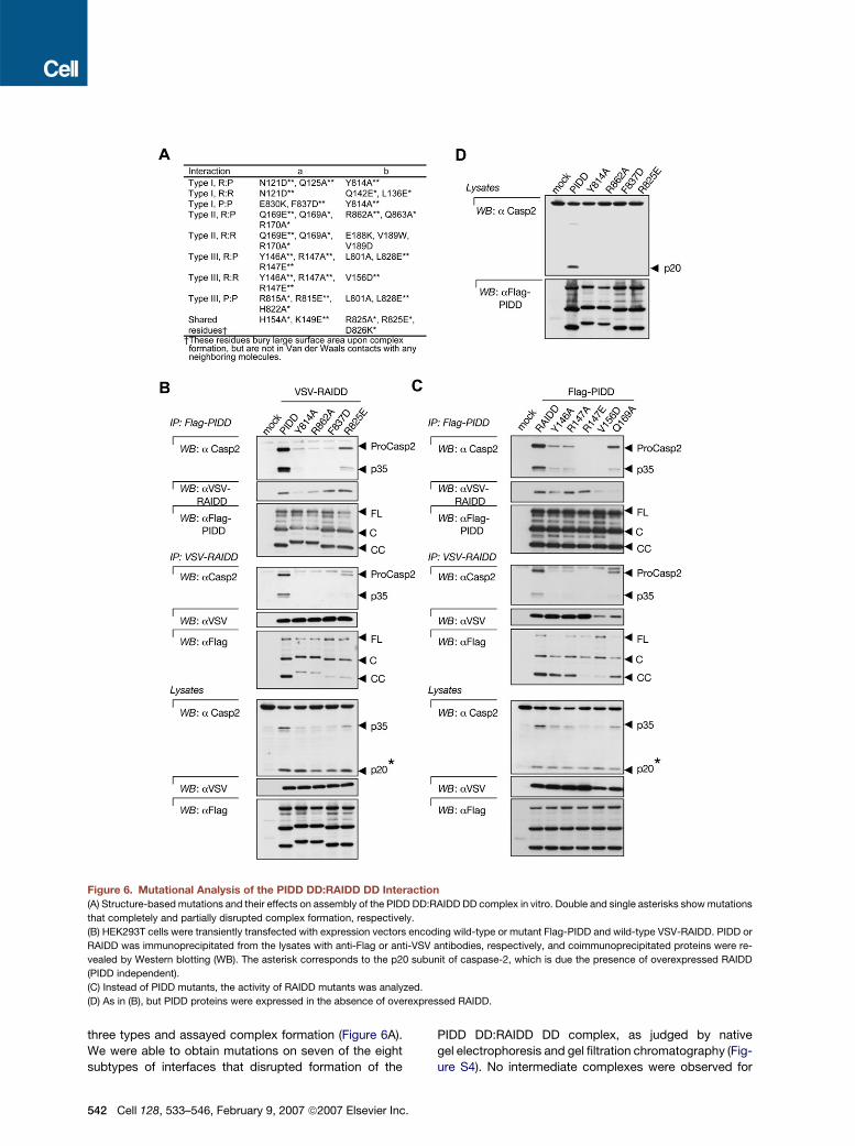
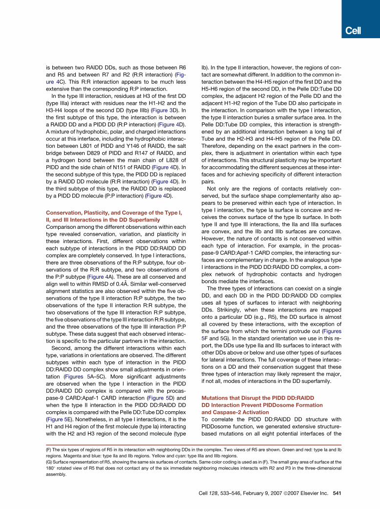
Figure 6. Mutational Analysis of the PIDD DD:RAIDD DD Interaction
(A) Structure-based mutations and their effects on assembly of the PIDD DD:RAIDD DD complex in vitro. Double and single asterisks show mutations
that completely and partially disrupted complex formation, respectively.
(B) HEK293T cells were transiently transfected with expression vectors encoding wild-type or mutant Flag-PIDD and wild-type VSV-RAIDD. PIDD or
RAIDD was immunoprecipitated from the lysates with anti-Flag or anti-VSV antibodies, respectively, and coimmunoprecipitated proteins were re-
vealed by Western blotting (WB). The asterisk corresponds to the p20 subunit of caspase-2, which is due the presence of overexpressed RAIDD
(PIDD independent).
(C) Instead of PIDD mutants, the activity of RAIDD mutants was analyzed.
(D) As in (B), but PIDD proteins were expressed in the absence of overexpressed RAIDD.
three types and assayed complex formation (Figure 6A).
We were able to obtain mutations on seven of the eight
subtypes of interfaces that disrupted formation of the
542 Cell 128, 533–546, February 9, 2007 ª2007 Elsevier Inc.
PIDD DD:RAIDD DD complex, as judged by native
gel electrophoresis and gel filtration chromatography (Fig-
ure S4). No intermediate complexes were observed for
any of the mutations. These data suggest that complex
assembly may require the simultaneous presence of
most, if not all, interfaces. We have so far not been able
to obtain disruptive mutations on the R:R subtype of the
type II interaction (Figure 6A), which mediates the assem-
bly of R6 and R7 on the top layer.
We next investigated whether mutations at the PIDD
DD:RAIDD DD interfaces would impact on PIDDosome
formation and caspase-2 activation. To this end, several
PIDD mutants were overexpressed in HEK293T cells
along with wild-type RAIDD, and complex formation and
caspase-2 activation were assessed by coimmunopreci-
pitation experiments after transient cotransfection (Fig-
ure 6B). While combined expression of the wild-type
version resulted in formation of a complex containing
PIDD (most likely the PIDD-CC form), RAIDD and active
caspase-2, complex formation, and caspase-2 activation
were either completely absent or attenuated with PIDD
mutants. Several RAIDD mutants were also examined for
their capacity to form the PIDDosome and shown to be
defective (Figure 6C). Partial complex formation and cas-
pase-2 activation were observed with several PIDD and
RAIDD mutants, most of which also exhibited less drastic
effects on PIDD DD:RAIDD DD interaction in vitro (Fig-
ure 6A). In addition, a complete absence of caspase-2
processing was seen with all PIDD mutants in the absence
of RAIDD overexpression (using endogenous RAIDD)
(Figure 6D). This indicates that caspase-2 recruitment
and activation in the PIDDosome are critically dependent
on the PIDD DD:RAIDD DD interaction we observe in the
structure.
DISCUSSION
Molecular Mechanism of Caspase-2 Activation
in the PIDDosome
Caspase activation is a hallmark of apoptotic cell death
(Riedl and Shi, 2004; Salvesen, 2002). According to their
sequence of activation, caspases may be divided into
two groups: initiator caspases such as caspase-2, -8,
-9, and -10, and effector caspases such as caspase-3
and -7 (Riedl and Shi, 2004; Salvesen, 2002). Unlike effec-
tor caspases, initiator caspases possess a domain of the
DD superfamily at their N-terminal region for recruitment
to oligomeric adaptor protein complexes upon apoptosis
induction. Caspases are synthesized as single-chain pro-
caspases, which undergo intrachain cleavage to generate
the large and small subunits.
Caspases need to form specific dimers to be active.
Because effector caspases are constitutive dimers, their
activation is strictly a consequence of intrachain cleavage
by initiator caspases. In contrast, intrachain cleavage
does not appear to be the crucial factor for initiator cas-
pase activation due to the relative longer lengths of the
intersubunit linker regions. A proximity-induced dimeriza-
tion model was proposed for initiator caspase activation
because initiator caspases such as caspase-2, -8, and -9
are not constitutive dimers in solution, and specific homo-
dimerization appears to be crucial for their activation
(Baliga et al., 2004; Pop et al., 2006; Yin et al., 2006).
In agreement with this analysis, the different caspase-
activating platforms are in different oligomerization states
and may recruit caspases with different stoichiometry.
While the mammalian apoptosome is a heptamer (Yu
et al., 2005a), the Drosophila apoptosome is octameric
(Yu et al., 2005b). CED4, the Apaf-1 homolog in C. ele-
gans, is a tetramer (Yan et al., 2005). The DISC contains
a trimer or likely multiple trimers, which may recruit three
or more caspase-8 or caspase-10 molecules (Yang
et al., 2005). Regardless of the stoichiometry, the key
common event is oligomerization, which allows neighbor-
ing caspases to form specific activating dimers. This ap-
pears to be the case even when the oligomeric platform
recruits odd numbers of caspases so that not all recruited
caspases have dimeric partners. In this scenario, under-
standing caspase activation is reduced to understanding
the oligomerization mechanisms of their activating
complexes.
Therefore, it is not surprising that the PIDDosome brings
seven caspase-2 molecules into proximity for their activa-
tion. One might ask why such an apparently unusual PIDD
DD:RAIDD DD oligomerization platform is used to induce
the proximity of caspase-2. Perhaps nature has evolved
many different oligomerization platforms for caspase acti-
vation, and the PIDD DD:RAIDD DD complex is simply one
of such examples. Upon recruitment to the PIDDosome,
caspase-2 is able to form dimers and be activated, even
in the absence of processing. The 50 residue linker region
between the CARD and the catalytic region of caspase-2
likely facilitates dimerization in the correct orientations for
caspase activation. Autoprocessing then proceeds, and
caspase-2 dimerization and activity are both enhanced
to induce mitochondrial events and cell death (Baliga
et al., 2004; Read et al., 2002).
General Mechanisms of Interactions
in the DD Superfamily
The DD superfamily is one of the largest and most widely
distributed domain superfamilies. Evolutionarily, it seems
that the ever-expanding DD superfamily may have
evolved by inserting its domains into various signal trans-
duction proteins such as caspases, kinases, and adaptor
proteins. In this regard, it is amazing that almost all oligo-
meric signaling complexes in apoptosis and inflammation
contain domains of the DD superfamily. For caspase-
activating complexes, the DD superfamily domains may
either be the major oligomerization platforms or the major
mediators in recruiting the caspases. The DDs in the
PIDDosome for caspase-2 activation and in the DISC for
caspase-8 and caspase-10 activation fall into the former
category, while the CARDs in the PIDDosome, DEDs in
the DISC, and CARDs in the inflammasome fall into the
latter category.
Our structure of the PIDD DD:RAIDD DD complex pro-
vides a glimpse of an oligomeric complex of the DD
Cell 128, 533–546, February 9, 2007 ª2007 Elsevier Inc. 543
superfamily and forms a template for other interactions in
this superfamily. Because the RIP1 kinase DD is homolo-
gous to RAIDD DD, it is likely that the PIDD DD:RIP1 DD
complex uses a similar assembly mechanism. In a much
broader scenario, in contrast to the concept that the DD
superfamily interactions may involve any available sur-
faces and may be very diverse, our study suggests that
the observed three types of asymmetric interactions
may represent preferred modes of interactions for DDs,
and likely for the entire DD superfamily. This may be
shown by the conservation between the type I interactions
in the PIDD DD:RAIDD DD complex and the procaspase-9
CARD:Apaf-1 CARD interaction. In addition, in the
DED1:DED2 interaction in the tandem DED-containing
viral FLIP MC159, the H1 and H4 of DED2 interacts with
H2 and H5 of DED1, which is somewhat similar to the
type I interaction as well (Li et al., 2006; Yang et al., 2005).
Curiously, all these known interactions in the DD super-
family are asymmetric despite what might have been
expected of homotypic interactions. This appears to be
true for both effector recruitment, as in the procaspase-
9 CARD:Apaf-1 CARD complex and the Pelle DD:Tube
DD complex, and for oligomerization, as in the PIDD
DD:RAIDD DD complex. This is not true, however, for sec-
ondary self-associations of DD superfamily domains that
are involved in effector recruitment and are linked to
oligomerization domains outside the DD superfamily. For
example, Apaf-1 CARD is linked to a nucleotide-binding
oligomerization domain (NOD). In the heptameric apopto-
some, Apaf-1 NOD confers a 7-fold symmetry, and the
Apaf-1 CARD also self-contacts with this symmetry in
the presence of the NOD (Yu et al., 2005a).
On the subject of asymmetric interactions, an asymmet-
ric trimeric model has also been proposed for the Fas
DD:FADD DD complex (Weber and Vincenz, 2001). This
model was generated from the structures of the Apaf-1
CARD:caspase-9 CARD complex (type I interaction) and
the Pelle DD:Tube DD complex (type II interaction).
When Pelle DD is superimposed with procaspase-9
CARD, the associated Tube DD and the Apaf-1 CARD
pack against Pelle DD (or procaspase-9 CARD) in a well-
organized trimer. The newly formed interaction between
Tube DD and Apaf-1 CARD forms a different interface,
which strikingly is very similar to the type III interaction ob-
served in the PIDD DD:RAIDD DD complex. By continuing
building interactions using these structures, a hexamer of
DDs can be formed, in which the three central DDs may ei-
ther be Fas DDs or FADD DDs (Figure 4E). Therefore, sim-
ilar to the PIDD DD:RAIDD DD complex, the Fas DD:FADD
DD complex may also be constructed from these three
types of interfaces. In an independent experiment, type
I, II, and III interactions were also found in docking models
between Fas DD and FADD DD (Thakar et al., 2006).
The involvement of all three types of interactions in the
assembly of various caspase-activating complexes is
consistent with and explains the existing mutagenesis
data on Fas (Huang et al., 1996; Martin et al., 1999), FADD
(Hill et al., 2004), TNFR1 (Tartaglia et al., 1993; Telliez
544 Cell 128, 533–546, February 9, 2007 ª2007 Elsevier Inc.
et al., 2000b), and TRADD (Park and Baichwal, 1996).
In each case, residues affecting the binding and/or func-
tion of the DD spread throughout its entire sequence.
Similarly, our structure-based mutagenesis also identified
important residues throughout the PIDD DD and RAIDD
DD sequences (Figure 6A).
Asymmetry and the apparent preference for the three
types of interactions may represent a unique feature of
the homotypic interactions in the DD superfamily. One
potentially strong rationale is that the conserved common
fold of the DD superfamily members determines, or at
least contributes to, the surface shape complementarity
seen in all three types of asymmetric interactions. How-
ever, the nature of contacts may not be conserved within
each type of interaction, as exemplified by the different
surface hydrophobicity, hydrophilicity, and charge fea-
tures of the different DDs (Park and Wu, 2006). In addition,
as the major function of these domains is homotypic inter-
action, the interactions may have evolved from several
primordial interaction pairs and be preserved through
coevolution. Therefore, the preservation of these pre-
ferred interactions may reflect both fold conservation
and evolutionary circumstances.
As the three types of interactions essentially cover a ma-
jority of the available surface of a DD (Figure 5G), it is likely
that these three types of interactions represent the major,
if not all, modes of interactions of the DD superfamily. For
effector recruitment, only one of the three types of interac-
tions is required. For oligomerization, it is likely that all
three types of interactions are needed. The oligomeriza-
tion stoichiometry is probably dictated by both the struc-
tural plasticity of the exact interaction pairs and how the
interactions could be terminated. For the PIDD DD:RAIDD
DD complex, the structure terminates within the layers as
it forms rings. The top layer has only two RAIDD DD mol-
ecules, likely because of the two available spaces for
interactions and the less extensive R:R subtype of the
type II interaction (Figures 2E and 4A). The lower surfaces
of the bottom layer of the PIDD DD molecules apparently
have no affinity for either RAIDD DDs or PIDD DDs, there-
fore terminating the buildup of further layers. In the Fas
DD:FADD DD complex, the inability of either FADD DD
or Fas DD to associate with itself in the complex may leave
the complex in a trimeric form. Given some plasticity at the
interfaces, it is likely that, by choosing among the three
types of interactions around each DD, a wide number of
oligomeric complexes with different stoichiometry may
be built. Selective usage of a certain type of interaction
may even switch the binding to alternative DD adapters
(Sandu et al., 2005). Therefore, these conserved asym-
metric interactions may underlie the unique but elegant
common assembly mechanism of the DD superfamily.
EXPERIMENTAL PROCEDURES
Protein Expression and Purification
The PIDD DD (residues 778–883) and RAIDD DD (residues 94–199)
with C-terminal His tags were expressed in E. coli and purified using
the Ni-NTA affinity resin (Qiagen). They were then mixed and incubated
at room temperature for 1 hr before being subjected to gel filtration
chromatography using Superdex 200 HR 10/30 (GE Healthcare). The
complex eluted at �12 ml and was concentrated to 10–12 mg/ml.
MALS
The molar mass of the PIDD DD:RAIDD DD complex was determined
by MALS. The complex was injected onto a Superdex 200 HR 10/30
gel filtration column (GE Healthcare) equilibrated in a buffer containing
20 mM Tris (pH 8.0) and 50 mM NaCl. The chromatography system
was coupled to a three-angle light scattering detector (mini-DAWN
EOS) and a refractive index detector (Optilab DSP) (Wyatt Technol-
ogy). Data were collected every 0.5 s at a flow rate of 0.2 ml/min.
Data analysis was carried out using the program ASTRA.
Electron Microscopy and Image Processing
PIDD DD:RAIDD DD complex was negatively stained with uranyl
formate (Ohi et al., 2004). Images were recorded using low-dose con-
ditions at a magnification of 52,0003 and a defocus of�1.5 mm with an
FEI Tecnai T12 electron microscope operated at 120 kV. Two 3 two
pixels were averaged, yielding a pixel size of 2.69 A. Using the SPIDER
software (Frank et al., 1996), 3708 particles (from 16 images) were win-
dowed into 80 3 80 pixel images and subjected to ten cycles of multi-
reference alignment and K-means classification specifying 25 classes.
For comparison with the crystal structure, the atomic model was res-
olution filtered to 30 A, and projections were calculated at 2� angular
intervals. The reprojections were cross-correlated to the class aver-
ages, and the reprojections with the highest correlation coefficient
were selected.
Structure Determination and Analysis
The assembled complex was crystallized at 20�C using 5.5% PEG
3350, 200 mM NaCl, and 100 mM Na/K phosphate (pH 6.5). To obtain
a heavy atom derivative, the crystals were soaked with 1 mM 1,4-diac-
etoxymercuri-2,3-dimethoxybutane for 30 min. One crystal diffracted
to 4.0 A resolution, and a complete anomalous data set was collected
at a wavelength of 1.0079 A at the X4A beamline of NSLS. A 3.2 A na-
tive data set was collected at the NE-CAT beamline of APS. Both data
sets were processed using HKL2000 (Otwinowski and Minor, 1997).
The structure was determined by single-wavelength anomalous dif-
fraction. There is a single partially surface-exposed free cysteine in the
RAIDD DD structure (Park and Wu, 2006) and no cysteine in PIDD DD.
Five strong mercury sites (15–20 s) were found, and the structure was
phased using the program SOLVE/RESOLVE (Terwilliger, 2004). A six-
dimensional search of the electron density map using the RAIDD DD
structure found ten DD molecules in the crystallographic asymmetric
unit. The locations of the mercury sites and crystallographic refinement
in CNS (Brunger et al., 1998) using the native data confirmed that five of
the DD molecules were RAIDD DDs and that the five remaining mole-
cules were PIDD DDs. Two additional RAIDD DD molecules were also
found. The final atomic model contains seven RAIDD DDs and five
PIDD DDs (Table 1). The structure was analyzed using O (Jones
et al., 1991) and Pymol (DeLano Scientific).
Mutational Analysis of Complex Formation In Vitro
Site-directed mutagenesis was performed using the Quikchange kit
(Stratagene) and confirmed by sequencing. Purified wild-type or mu-
tant PIDD DD and RAIDD DD proteins were first mixed and incubated
at room temperature for 1 hr. The mixed solutions were subjected to
electrophoresis under native conditions on premade 8%–25% acryl-
amide gradient gels using the PhastSystem (GE Healthcare). The
gels were stained with Coomassie blue, and complex formation was
determined by the appearance of shifted bands. Mutational effects
were also characterized by gel filtration chromatography using the
Superdex 200 HR 10/30 column (GE Healthcare).
PIDDosome Formation and Caspase-2 Activation
PIDDosome formation was revealed by coimmunoprecipitation exper-
iments after transient cotransfection of PIDD wild-type and RAIDD mu-
tants or RAIDD wild-type and PIDD mutants. After 48 hr transfection,
cells were lysed in lysis buffer containing 1% NP-40, 20 mM Tris
(pH 7.4), 250 mM NaCl, 5% glycerol, and a protease inhibitor cocktail.
After lysis, the extracts were incubated with anti-Flag or anti-VSV
beads for 2 hr. After incubation the beads were washed four times
with lysis buffer and analyzed by immunoblotting. The antibodies used
for Western blotting were as follows: anti-caspase-2 11B4 (Apotech),
mouse anti-VSV, and rabbit anti-Flag (Sigma).
Supplemental Data
The Supplemental Data include four supplemental figures and can be
found with this article online at http://www.cell.com/cgi/content/full/
128/3/533/DC1/.
ACKNOWLEDGMENTS
We thank Drs. David Eliezer, Olga Boudhka, and Fred Maxfield for
helpful discussions; Randy Abramowitz and John Schwanof for data
collection at X4A of NSLS; Kanagalaghatta Rajashankar and Igor
Kourinov for data collection at NE-CAT of APS; Dr. Sally Kornbluth
for providing the cDNA of human RAIDD; Dr. Jin Kuk Yang for help
with data collection; Jin Wu for maintaining X-ray equipment and com-
puters; Su-Chang Lin for help with the light scattering experiment; and
the Wu lab members for discussions. The molecular EM facility at Har-
vard Medical School was established with a generous donation from
the Giovanni Armenise Harvard Center for Structural Biology and is
maintained with funds from the NIH.
Received: November 23, 2006
Revised: December 25, 2006
Accepted: January 6, 2007
Published: February 8, 2007
REFERENCES
Baliga, B.C., Read, S.H., and Kumar, S. (2004). The biochemical mech-
anism of caspase-2 activation. Cell Death Differ. 11, 1234–1241.
Bergeron, L., Perez, G.I., Macdonald, G., Shi, L., Sun, Y., Jurisicova,
A., Varmuza, S., Latham, K.E., Flaws, J.A., Salter, J.C., et al. (1998).
Defects in regulation of apoptosis in caspase-2-deficient mice. Genes
Dev. 12, 1304–1314.
Brunger, A.T., Adams, P.D., Clore, G.M., DeLano, W.L., Gros, P.,
Grosse-Kunstleve, R.W., Jiang, J.S., Kuszewski, J., Nilges, M., Pannu,
N.S., et al. (1998). Crystallography & NMR system: A new software
suite for macromolecular structure determination. Acta Crystallogr. D
Biol. Crystallogr. 54, 905–921.
Duan, H., and Dixit, V.M. (1997). RAIDD is a new ‘death’ adaptor mol-
ecule. Nature 385, 86–89.
Frank, J., Radermacher, M., Penczek, P., Zhu, J., Li, Y., Ladjadj, M.,
and Leith, A. (1996). SPIDER and WEB: Processing and visualization
of images in 3D electron microscopy and related fields. J. Struct.
Biol. 116, 190–199.
Guo, Y., Srinivasula, S.M., Druilhe, A., Fernandes-Alnemri, T., and
Alnemri, E.S. (2002). Caspase-2 induces apoptosis by releasing
proapoptotic proteins from mitochondria. J. Biol. Chem. 277, 13430–
13437.
Hill, J.M., Morisawa, G., Kim, T., Huang, T., Wei, Y., and Werner, M.H.
(2004). Identification of an expanded binding surface on the FADD
death domain responsible for interaction with CD95/Fas. J. Biol.
Chem. 279, 1474–1481.
Cell 128, 533–546, February 9, 2007 ª2007 Elsevier Inc. 545
Huang, B., Eberstadt, M., Olejniczak, E.T., Meadows, R.P., and Fesik,
S.W. (1996). NMR structure and mutagenesis of the Fas (APO-1/CD95)
death domain. Nature 384, 638–641.
Janssens, S., Tinel, A., Lippens, S., and Tschopp, J. (2005). PIDD me-
diates NF-kB activation in response to DNA damage. Cell 123, 1079–
1092.
Jones, T.A., Zou, J.-Y., Cowan, S.W., and Kjeldgaard, M. (1991). Im-
proved methods for building models in electron density maps and
the location of errors in those models. Acta Crystallogr. A 47, 110–119.
Kohl, A., and Grutter, M.G. (2004). Fire and death: The pyrin domain
joins the death-domain superfamily. C. R. Biol. 327, 1077–1086.
Lassus, P., Opitz-Araya, X., and Lazebnik, Y. (2002). Requirement for
caspase-2 in stress-induced apoptosis before mitochondrial permea-
bilization. Science 297, 1352–1354.
Li, F.Y., Jeffrey, P.D., Yu, J.W., and Shi, Y. (2006). Crystal structure of
a viral FLIP: Insights into FLIP-mediated inhibition of death receptor
signaling. J. Biol. Chem. 281, 2960–2968.
Lin, Y., Ma, W., and Benchimol, S. (2000). Pidd, a new death-domain-
containing protein, is induced by p53 and promotes apoptosis. Nat.
Genet. 26, 122–127.
Martin, D.A., Zheng, L., Siegel, R.M., Huang, B., Fisher, G.H., Wang, J.,
Jackson, C.E., Puck, J.M., Dale, J., Straus, S.E., et al. (1999). Defective
CD95/APO-1/Fas signal complex formation in the human autoimmune
lymphoproliferative syndrome, type Ia. Proc. Natl. Acad. Sci. USA 96,
4552–4557.
Ohi, M., Li, Y., Cheng, Y., and Walz, T. (2004). Negative staining and
image classification—Powerful tools in modern electron microscopy.
Biol. Proced. Online 6, 23–34.
Otwinowski, Z., and Minor, W. (1997). Processing of X-ray diffraction
data collected in oscillation mode. Methods Enzymol. 276, 307–326.
Park, A., and Baichwal, V.R. (1996). Systematic mutational analysis of
the death domain of the tumor necrosis factor receptor-1-associated
protein TRADD. J. Biol. Chem. 271, 9858–9862.
Park, H.H., and Wu, H. (2006). Crystal structure of RAIDD death
domain implicates potential mechanism of PIDDosome assembly.
J. Mol. Biol. 357, 358–364.
Park, H.H., Lo, Y.C., Lin, S.C., Wang, L., Yang, J.K., and Wu, H. (2007).
The death domain superfamily in intracellular signaling of apoptosis
and inflammation. Annu. Rev. Immunol. 25, 561–586.
Pick, R., Badura, S., Bosser, S., and Zornig, M. (2006). Upon intracel-
lular processing, the C-terminal death domain-containing fragment of
the p53-inducible PIDD/LRDD protein translocates to the nucleoli and
interacts with nucleolin. Biochem. Biophys. Res. Commun. 349, 1329–
1338.
Pop, C., Timmer, J., Sperandio, S., and Salvesen, G.S. (2006). The
apoptosome activates caspase-9 by dimerization. Mol. Cell 22, 269–
275.
Qin, H., Srinivasula, S.M., Wu, G., Fernandes-Alnemri, T., Alnemri,
E.S., and Shi, Y. (1999). Structural basis of procaspase-9 recruitment
by the apoptotic protease-activating factor 1. Nature 399, 549–557.
Read, S.H., Baliga, B.C., Ekert, P.G., Vaux, D.L., and Kumar, S. (2002).
A novel Apaf-1-independent putative caspase-2 activation complex.
J. Cell Biol. 159, 739–745.
Reed, J.C., Doctor, K.S., and Godzik, A. (2004). The domains of apo-
ptosis: A genomics perspective. Sci. STKE 2004, re9.
Riedl, S.J., and Shi, Y. (2004). Molecular mechanisms of caspase reg-
ulation during apoptosis. Nat. Rev. Mol. Cell Biol. 5, 897–907.
Robertson, J.D., Enoksson, M., Suomela, M., Zhivotovsky, B., and
Orrenius, S. (2002). Caspase-2 acts upstream of mitochondria to
promote cytochrome c release during etoposide-induced apoptosis.
J. Biol. Chem. 277, 29803–29809.
546 Cell 128, 533–546, February 9, 2007 ª2007 Elsevier Inc.
Salvesen, G.S. (2002). Caspases and apoptosis. Essays Biochem. 38,
9–19.
Sandu, C., Gavathiotis, E., Huang, T., Wegorzewska, I., and Werner,
M.H. (2005). A mechanism for death receptor discrimination by death
adaptors. J. Biol. Chem. 280, 31974–31980.
Tartaglia, L.A., Ayres, T.M., Wong, G.H., and Goeddel, D.V. (1993). A
novel domain within the 55 kd TNF receptor signals cell death. Cell
74, 845–853.
Telliez, J.B., Bean, K.M., and Lin, L.L. (2000a). LRDD, a novel leucine
rich repeat and death domain containing protein. Biochim. Biophys.
Acta 1478, 280–288.
Telliez, J.B., Xu, G.Y., Woronicz, J.D., Hsu, S., Wu, J.L., Lin, L., Sukits,
S.F., Powers, R., and Lin, L.L. (2000b). Mutational analysis and NMR
studies of the death domain of the tumor necrosis factor receptor-1.
J. Mol. Biol. 300, 1323–1333.
Terwilliger, T. (2004). SOLVE and RESOLVE: Automated structure so-
lution, density modification and model building. J. Synchrotron Radiat.
11, 49–52.
Thakar, J., Schleinkofer, K., Borner, C., and Dandekar, T. (2006). RIP
death domain structural interactions implicated in TNF-mediated pro-
liferation and survival. Proteins 63, 413–423.
Tinel, A., and Tschopp, J. (2004). The PIDDosome, a protein complex
implicated in activation of caspase-2 in response to genotoxic stress.
Science 304, 843–846.
Tinel, A., Janssens, S., Lippens, S., Cuenin, S., Logette, E., Jaccard,
B., Quadroni, M., and Tschopp, J. (2007). Autoproteolysis of PIDD
marks the bifurcation between pro-death caspase-2 and pro-survival
NF-kB pathway. Embo J. 26, 197–208. Published online December
7, 2006. 10.1038/sj.emboj.7601473.
Wang, L., Miura, M., Bergeron, L., Zhu, H., and Yuan, J. (1994). Ich-1,
an Ice/ced-3-related gene, encodes both positive and negative regu-
lators of programmed cell death. Cell 78, 739–750.
Weber, C.H., and Vincenz, C. (2001). A docking model of key compo-
nents of the DISC complex: Death domain superfamily interactions re-
defined. FEBS Lett. 492, 171–176.
Xiao, T., Towb, P., Wasserman, S.A., and Sprang, S.R. (1999). Three-
dimensional structure of a complex between the death domains of
Pelle and Tube. Cell 99, 545–555.
Yan, N., Chai, J., Lee, E.S., Gu, L., Liu, Q., He, J., Wu, J.W., Kokel, D.,
Li, H., Hao, Q., et al. (2005). Structure of the CED-4-CED-9 complex
provides insights into programmed cell death in Caenorhabditis ele-
gans. Nature 437, 831–837.
Yang, J.K., Wang, L., Zheng, L., Wan, F., Ahmed, M., Lenardo, M.J.,
and Wu, H. (2005). Crystal structure of MC159 reveals molecular
mechanism of DISC assembly and FLIP inhibition. Mol. Cell 20, 939–
949.
Yin, Q., Park, H.H., Chung, J.Y., Lin, S.C., Lo, Y.C., da Graca, L.S.,
Jiang, X., and Wu, H. (2006). Caspase-9 holoenzyme is a specific
and optimal procaspase-3 processing machine. Mol. Cell 22, 259–268.
Yu, X., Acehan, D., Menetret, J.F., Booth, C.R., Ludtke, S.J., Riedl,
S.J., Shi, Y., Wang, X., and Akey, C.W. (2005a). A structure of the hu-
man apoptosome at 12.8 A resolution provides insights into this cell
death platform. Structure 13, 1725–1735.
Yu, X., Wang, L., Acehan, D., Wang, X., and Akey, C.W. (2005b). Three-
dimensional structure of a double apoptosome formed by the Dro-
sophila Apaf-1 related killer. J. Mol. Biol. 355, 577–589.
Accession Numbers
The coordinates have been deposited in the RCSB Protein Data Bank
with the PDB code 2OF5.
Cell, Volume 128
Supplemental Data
Death Domain Assembly Mechanism
Revealed by Crystal Structure of
the Oligomeric PIDDosome Core Complex Hyun Ho Park, Emmanuelle Logette, Stefan Raunser, Solange Cuenin, Thomas Walz, Jurg Tschopp, and Hao Wu
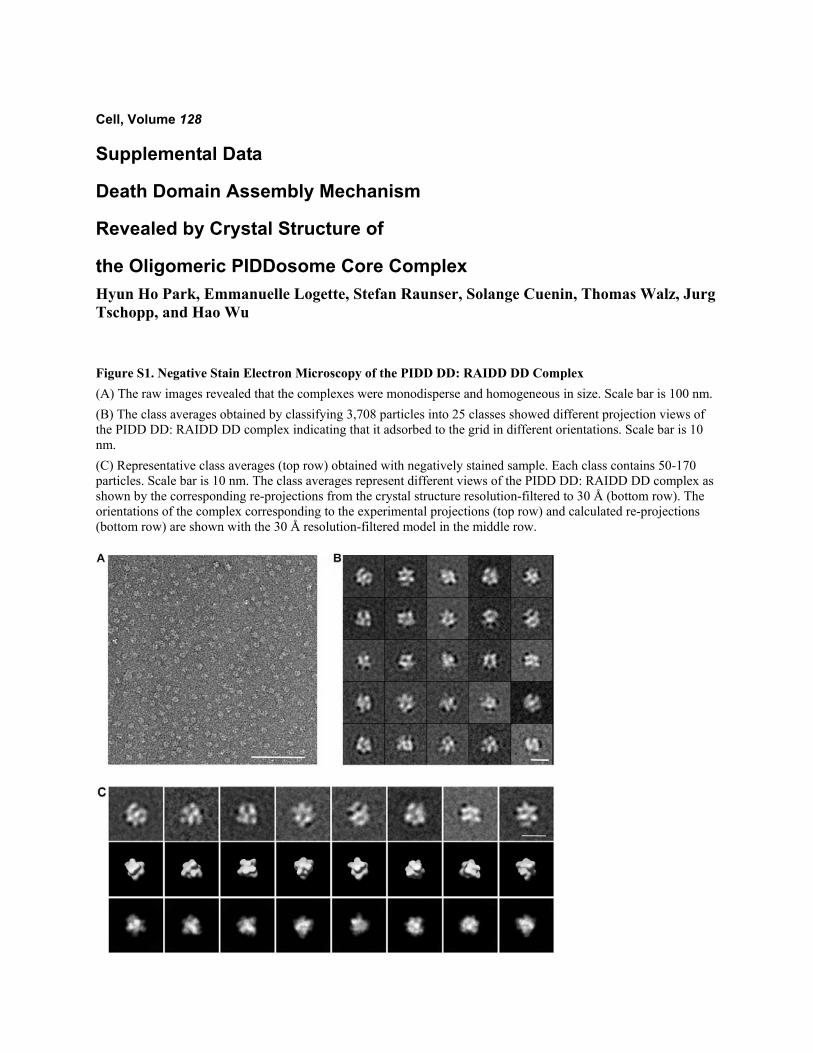
Figure S1. Negative Stain Electron Microscopy of the PIDD DD: RAIDD DD Complex (A) The raw images revealed that the complexes were monodisperse and homogeneous in size. Scale bar is 100 nm. (B) The class averages obtained by classifying 3,708 particles into 25 classes showed different projection views of the PIDD DD: RAIDD DD complex indicating that it adsorbed to the grid in different orientations. Scale bar is 10 nm. (C) Representative class averages (top row) obtained with negatively stained sample. Each class contains 50-170 particles. Scale bar is 10 nm. The class averages represent different views of the PIDD DD: RAIDD DD complex as shown by the corresponding re-projections from the crystal structure resolution-filtered to 30 Å (bottom row). The orientations of the complex corresponding to the experimental projections (top row) and calculated re-projections (bottom row) are shown with the 30 Å resolution-filtered model in the middle row.
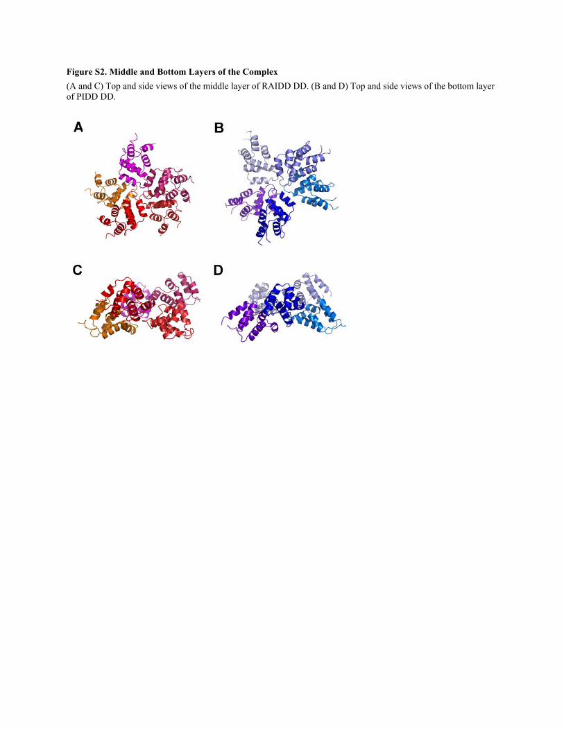
Figure S2. Middle and Bottom Layers of the Complex (A and C) Top and side views of the middle layer of RAIDD DD. (B and D) Top and side views of the bottom layer of PIDD DD.
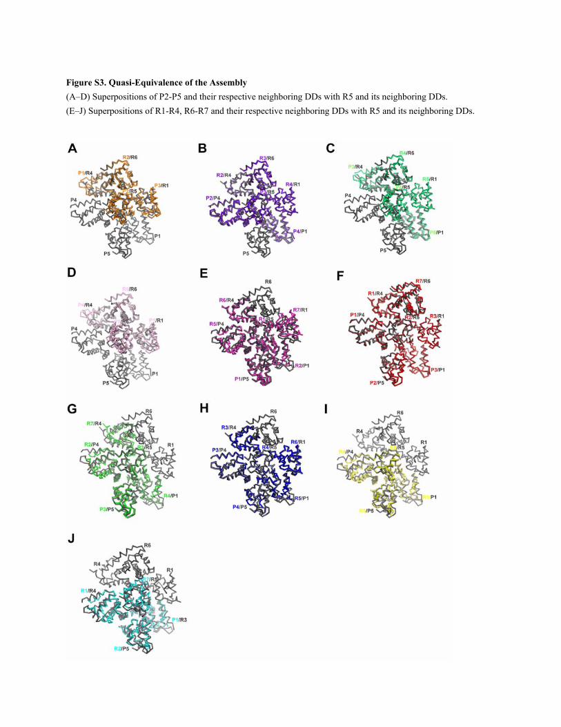
Figure S3. Quasi-Equivalence of the Assembly (A–D) Superpositions of P2-P5 and their respective neighboring DDs with R5 and its neighboring DDs. (E–J) Superpositions of R1-R4, R6-R7 and their respective neighboring DDs with R5 and its neighboring DDs.
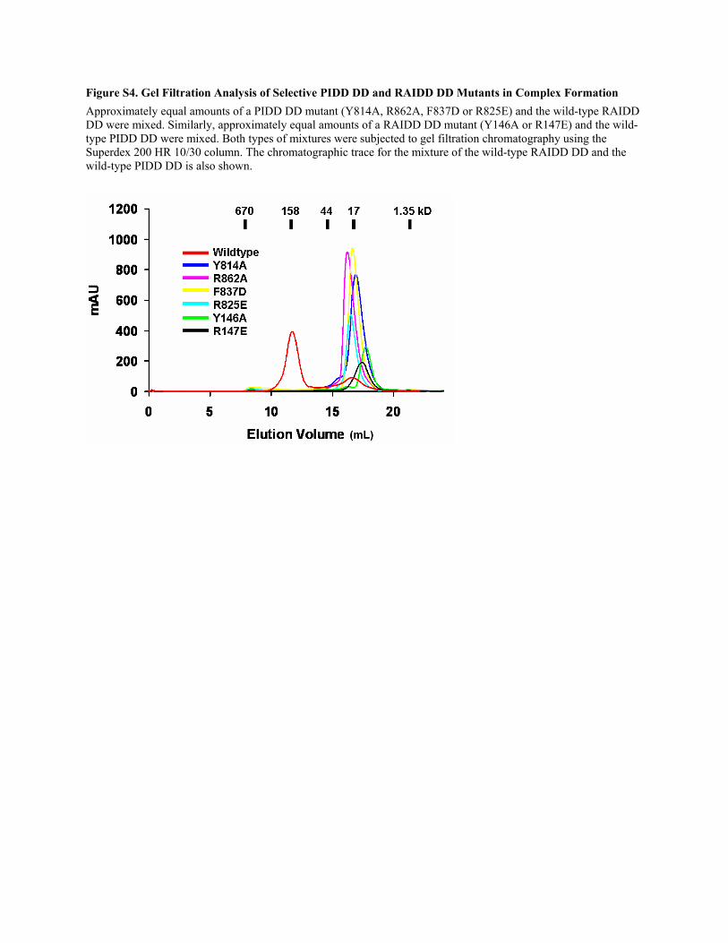
Figure S4. Gel Filtration Analysis of Selective PIDD DD and RAIDD DD Mutants in Complex Formation Approximately equal amounts of a PIDD DD mutant (Y814A, R862A, F837D or R825E) and the wild-type RAIDD DD were mixed. Similarly, approximately equal amounts of a RAIDD DD mutant (Y146A or R147E) and the wild-type PIDD DD were mixed. Both types of mixtures were subjected to gel filtration chromatography using the Superdex 200 HR 10/30 column. The chromatographic trace for the mixture of the wild-type RAIDD DD and the wild-type PIDD DD is also shown.
Related Documents


![Dirigent Protein Mode of Action Revealed by the Crystal ......Dirigent Protein Mode of Action Revealed by the Crystal Structure of AtDIR61[OPEN] Raphael Gasper2, Isabelle Effenberger2,](https://static.cupdf.com/doc/110x72/610900acbd8bfa066b099b99/dirigent-protein-mode-of-action-revealed-by-the-crystal-dirigent-protein.jpg)