De novo self-assembling peptides possessing esterase properties Inaugural-Dissertation to obtain the academic degree Doctor rerum naturalium (Dr. rer. nat.) Submitted to the Department of Biology, Chemistry and Pharmacy of Freie Universität Berlin by Jason L. Heier From Hague, ND, USA October 2015

Welcome message from author
This document is posted to help you gain knowledge. Please leave a comment to let me know what you think about it! Share it to your friends and learn new things together.
Transcript

De novo self-assembling peptides possessing esterase properties
Inaugural-Dissertation to obtain the academic degree
Doctor rerum naturalium (Dr. rer. nat.)
Submitted to the Department of Biology, Chemistry and Pharmacy of Freie Universität Berlin
by
Jason L. Heier
From Hague, ND, USA
October 2015


i
1st Reviewer: Prof. Dr. Beate Koksch
2nd Reviewer: Prof. Dr. Mathias Christmann
Date of defense: June 16, 2016


iii
Declaration
The work presented here was performed in the research group of Prof. Dr. Beate
Koksch from November 2010 until October 2015 at the Institute of Chemistry and
Biochemistry in the Department of Biology, Chemistry and Pharmacy of Freie Universität
Berlin.
I hereby confirm that this dissertation entitled “De novo self-assembling peptides
possessing esterase properties” is exclusively the result of my own autonomous work
based on my research and published literature as cited. I also declare that no part of this
dissertation has been prepared inappropriately or used in any other work in another
higher education or research institution.
Berlin, October 2015 Jason L. Heier


v
Publications
ACS Chem. Biol. DOI: 10.1021/acschembio.5b00435. Tailored Presentation of
Carbohydrates on a Coiled Coil-Based Scaffold for Asialoglycoprotein Receptor Targeting. E. Zacco, J. Hütter, J. L. Heier, J. Mortier, P. H. Seeberger, B. Lepenies & B. Koksch, 2015.
Posters
12th German Peptide Symposium, Darmstadt (Germany), March 18-21, 2015: De novo peptide hexamer cast in new role as zinc metalloenzyme. J. L. Heier & B. Koksch.
Center of Supramolecular Interactions 2nd General Meeting, Berlin (Germany), March 10-11, 2011: Self-assembled metallo-supramolecular squares as templates for α-helical peptide bundles. J. L.Heier, C. A. Schalley & B. Koksch.


vii
For reasons of data security, the curriculum vitae has been omitted from the published
version.

viii
For reasons of data security, the curriculum vitae has been omitted from the published
version.

ix
Acknowledgements
I would like to thank Prof. Dr. Beate Koksch for her support as well as all the patience
she has shown and taught. This project involved many aspects which were new to both
of us. I appreciate her faith in me.
I thank Prof. Dr. Mathias Christmann for agreeing to be my second reviewer.
I thank the mass spectrometry and NMR services of the Chemistry Department at the
FU-Berlin for their efficiency. I also thank the Material Management of Takustr. 3 for their
friendly service.
I am grateful for the help of Dr. Sumati Bhatia and Dr. Manoj Kumar Muthyala.
I thank the entire group of AG Koksch for all their help and support. I thank “My Frient”
Dr. Cosimo Cadicamo who was a mentor to me. Many thanks to Dr. Allison Berger for all
her help and valuable suggestions. Sincere, helpful people can be hard to find. Thank
you, Dr. Elsa Zacco. It was a pleasure working with you. I wish you all the best in your
bright future. Thank you, Valentina and AnaRita. Anytime you need advice or just want
to talk about peptides, call me.
Thanks for everyone in the research group of Jun. Prof. Dr. Annabelle Bertin for all your
support.
I thank my family. I am sorry that I was not there. I owe everything to my parents, Jane
and Tim Heier. I wish there was some form in which I could repay you. Thanks to my
brothers and sisters, Janet, Allen, Randy, Shari, Brenda and Nora and your lovely
families. I love and miss you all.
I thank you, Katie, for being Celina’s mom and always helping me.
And Katja, I hope you understand how much I appreciate all that you do and don’t do for
me. I would be lonely and thin without you.

x
Most of all, I am thankful for you, Celina. I am so proud that you are my daughter.
Thanks for taking care of me. You are the world to me.

xi
Zusammenfassung
Die Fähigkeit von Enzymen, Reaktionen unter Erhalt einer exzellenten Substrat- und Produktspezifität zu
beschleunigen, interessiert Chemiker seit Jahrzehnten. Bei der Adaption von Enzymen für die
Bedingungen der Synthesechemie entstanden jedoch zahlreiche Hindernisse, wie bspw. eine verringerte
Löslichkeit und Thermostabilität sowie eine geringere Substratvielfalt. Für die Bewältigung dieser
Probleme nutzt das protein engineering sowohl Methoden des rationalen Designs und gerichteter
Evolution, als auch moderne computergestützte sowie kombinatorische Ansätze. Obwohl diese
Techniken zur Entwicklung zahlreicher Biokatalysatoren erfolgreich eingesetzt wurden, bleibt das
Verständnis über die Proteinfaltung und Wechselwirkungen, welche für die enzymatische Katalyse
erforderlich sind, weiterhin begrenzt. Dies wird vor allem dadurch belegt, dass es unmöglich ist,
katalytisch aktive Proteine de novo zu generieren, die ähnliche Fähigkeiten aufweisen wie Enzyme.
Um ein besseres Verständnis von der Enzymstruktur und Funktion zu ermöglichen, wird der „bottom-
up“-Ansatz für das de novo Design von katalytisch aktiven Proteinen genutzt. Dieser minimalistische
Ansatz nutzt einfache Peptide mit vorhersagbaren Selbstorganisationseigenschaften zu gut definierten
dreidimensionalen Templaten. In diese Template wird katalytische Aktivität bzw. werden andere
Funktionalitäten eingebaut. Die vereinfachten, dennoch proteinartigen Systeme ermöglichen ein
verbessertes Verständnis darüber, wie einzelne Wechselwirkungen zu bestimmten enzymatischen
Eigenschaften beitragen können.
Unter Einsatz der „bottom-up“-Technik wurde der enzymatische Apparat für eine Esterase-artige
Aktivität in drei Modellpeptide eingeführt. Der Einbau von Histidin in das Carboxylat-reiche Peptid E3
führte zu einem random coil mit einer geringen katalytischen Aktivität. Als das Peptid mit K3 in einem
heterodimeren coiled coil fixiert wurde, nahm die Aktivität ab, aber das Peptid zeigte eine enzymartige
Sensitivität. Außerdem wurde festgestellt, dass Ac-IHIHQI-NH2, welches ein Histidin-koordiniertes Zink
für die Aktivierung von Wasser besitzt, eine höhere Selektivität für hydrophobe L-Aminosäureester
besitzt, wenn die Wechselwirkung mit dem Substrat während des Prozesses der Selbstaggregation
besitzt, als im Vergleich zu vollständig aggregiertem Zustand. Diese Beobachtung deutet auf die
Wechselwirkung von hydrophoben Substratresten mit der hydrophoben Oberfläche der aggregierenden
β-Faltblätter. Zum Schluss wurde Zink in ein hexameres α-helikales coiled coil mit sechs Histidinresten im
hydrophoben Kern eingeführt. Es zeigte sich, dass die Koordination von Zink in einem für Wasser
zugänglichen und Substrat-aufnehmenden Spalt die Esterhydrolyse stark beschleunigte.


xiii
Abstract
The ability of enzymes to accelerate reactions while maintaining excellent substrate
selectivity and product specificity has interested chemists for decades. The adaptation of
enzymes to the reaction conditions of chemical synthesis has, however, been met with
numerous obstacles such as reduced solubility, thermostability and substrate scope. To
overcome these obstacles, protein engineering employs methods of rational design and
directed evolution as well as more modern computational and combinatorial approaches.
Although these techniques have been successfully applied to alter biocatalysts, our
understanding of protein folding and the interactions required for enzyme catalysis
remains limited. This is best evidenced by the inability to design de novo catalytic
proteins comparable to enzymes.
To develop a better understanding of enzyme structure and function, the “bottom-up”
approach to the de novo design of catalytic proteins is applied. This minimalistic
approach uses simple peptides which predictably self-assemble into well-defined three-
dimensional templates, on which catalytic machinery or other functionalities believed to
impart a particular property are introduced. The simplified, yet protein-like, environments
of the self-assembling peptides allow for enhanced clarity of the individual interactions to
assess how such interactions contribute to particular enzymatic properties.
Using the bottom-up technique, catalytic machinery for esterase-like activity was
incorporated into three model peptide systems. The insertion of histidine into the
carboxylate-rich E3 peptide resulted in a random coil with slight catalytic activity. This
activity was muted when fixed in a heterodimeric coiled coil with K3 showing an enzyme-
like sensitivity to its environment. In addition, the fibril-forming Ac-IHIHIQI-NH2, which
uses histidine-coordinated zinc to activate water, was found to be more selective for
hydrophobic, L-amino acid esters while in the process of self-assembly than when
applied as fully-formed fibrils. This finding points to the interaction of hydrophobic
substrate moieties with exposed hydrophobic surfaces of the assembling β-sheets.
Lastly, zinc was introduced to a hexameric α-helical coiled coil featuring six histidine
residues in its hydrophobic interior. The coordination of zinc within a water-accessible,
substrate accommodating cleft was found to greatly accelerate ester hydrolysis.


xv
List of abbreviations
CA Carbonic anhydrase
ZHN Zinc-bound hydroxide nucleophile
NADH Nicotinamide adenine dinucleotide (protonated)
DMSO Dimethyl sulfoxide
DCE Dichloroethane
CALB Candida antarctica lipase B
PPL Porcine pancreas lipase
PCR Polymerase chain reaction
CLECs Cross-linked enzyme crystals
CLEAs Cross-linked enzyme aggregates
Taq Thermus aquaticus
PAL Pseudomonas aeruginosa lipase
ISM Iterative saturation mutagenesis
ORBIT Optimization of Rotamers by Itertive Techniques
pNPA p-Nitrophenyl acetate
NADPH Nicotinamide adenine dinucleotide phosphate (protonated)
bPP Bovine pancreatic polypeptide
Z-Phe-ONp N-Benzyloxycarbonyl-phenylalanine p-nitrophenyl ester
TEM Transmission electron microscopy
CD Circular dichroism
Tris Tris(hydroxymethyl)aminomethane
CT Catalytic triad
hPG Hyper-branched polyglycerol

xvi
List of abbreviations (cont.)
Cu-AAC Copper-catalyzed alkyne-azide cycloaddition
Au-MPCs Gold monolayer-protected clusters
TEG Triethylene glycol
DNPB 2,4-Dinitrophenylbutanoate
Z-Leu-ONp N-Benzyloxycarbonyl-leucine-p-nitrophenyl ester
Z-Gly-ONp N-Benzyloxycarbonyl-glycine -p-nitrophenyl ester
Boc-Asn-ONp N-Butyloxycarbonyl-asparagine -p-nitrophenyl ester
Fmoc 9-Fluorenylmethoxycarbonyl protecting group
SPPS Solid phase peptide synthesis
RP-HPLC Reverse-phase high-pressure liquid chromatography
MS Mass spectrometry
tBu tert-Butyl protecting group
Trt Triphenylmethyl protecting group
Boc Butyloxycarbonyl protecting group
TGA TentaGel-like composite resin for peptide acid synthesis
TGT TentaGel-like composite resin with triphenylmethyl linker
MBHA 4-Methylbenzhydrylamine hydrochloride salt
DMF Dimethylformamide
DCM Dichloromethane
NMP N-Methyl-2-pyrolidone
DBU 1,8-Diazabicyclo[5.4.0]undec-7-ene
DIC N,N’-Diisopropylcarbodiimide
HOAt 1-Hydroxy-7-azabenzotriazole

xvii
List of abbreviations (cont.)
HOBt N-Hydroxybenzotriazole
HCTU O-(6-Chlorobenzotriazol-1-yl)-N,N,N′,N′- tetramethyluronium hexafluorophosphate
TBTU 2-(1H-Benzotriazol-1-yl)-1,1,3,3,-tetramethyluronium tetrafluoroborate
DIPEA N,N-Diisopropylethylamine
TFA Trifluoroacetic acid
TIPS Triisopropylsilane
MeCN Acetonitrile
Abz 2-Aminobenzoic acid
PTFE Polytetrafluoroethylene
ESI-ToF Electrospray ionization-time of flight
PMMA Poly (methyl methacrylate)
Mn Number average molecular weight
PDI Polydispersity index
MWCO Molecular weight cut-off
EDTA Ethylenediaminetetraacetic acid
GPC Gel permeation chromatography
RI Refractive index
DF Dilution factor
UV-Vis Ultraviolet-visible

xviii
List of abbreviations (cont.)
The three- and one-letter abbreviations used in this work to represent L-amino acids are consistent with biochemical nomenclature proposed by the IUPAC-IUB Commission (Eur. J. Biochem. 1984, 138, 9-37).

xix
Table of contents
Page
1. Introduction 1
2. Enzyme catalysis 5
2.1. Catalytic mechanism 8 2.2. Enzyme specificity and selectivity 12 2.3. Enzyme kinetics 13
3. Biocatalysis 21
3.1. The pros and cons of biocatalysis 23 3.2. Enzymes modified for biocatalysis 30
4. De novo catalytic proteins 47
4.1. α-Helical de novo-designed catalytic proteins 50 4.2. β-sheets as scaffolds for de novo-designed catalytic proteins 60 4.3. Histidine as simple, yet versatile catalytic machinery 63
5. Project aim 65
6. Results and discussion 67
6.1. Regulation of random coil peptide catalyst through coiled 69 coil formation
6.2. Amyloid-forming Ac-IHIHIQI-NH2: De novo catalytic proficiency 79 with selectivity
6.3. De novo peptide hexamer recast as metalloenzyme 89
7. Summary 99
8. Materials and methods 101
8.1. Peptide synthesis and verification 101 8.2. Kinetic assays 120
8.3. Structural analysis 125
9. References 133
10. Appendix 149

1
1 Introduction
The number of diverse biochemical reactions required to maintain life are
considerable. Even more staggering are the rates in which such events must proceed
in order to meet biological demands. Nearly all reactions occurring within living
systems are mediated by enzymes. Unlike other classes of protein, enzymes do not
simply function by specifically binding other molecules, but binding them in such a
manner which induces transition.1 By stabilizing a high-energy transition state,
enzymes act as catalysts, accelerating the rate of spontaneous reactions in which
they themselves are not consumed. In contrast to conventional chemical catalysts
(i.e., transition metals, acids and bases), enzymes are generally more efficient and
operate under mild conditions with superior substrate specificity.2,3
Since enzymes catalyze reactions with remarkably high selectivity, their passage
through cellular membranes (and often yet within intact cells) into the chemistry
laboratory as biocatalysts4 only seems natural. Enzymes impose their selectivity
according to the functional groups present on substrates (i.e., chemo-selectivity),
chirality (i.e., stereo-selectivity) and position (i.e., regio-selectivity). When effectively
employed in synthetic chemistry,5,6 this heightened selectivity is ultimately converted
into uncomplicated protection schemes, fewer side reactions and simplified
purification.5,7-8 Moreover, biocatalysts are completely biodegradable and offer a
much greener alternative to classical stoichiometric synthesis and to a lesser extent
traditional chemical catalysis.4,5,7
The catalytic properties of enzymes have traditionally evolved within cells, not flasks
or 96-well microtiter plates. An enzyme is evolved typically for a single substrate, not
a broad class of reactants having a common functional group.5 As a result, the high
specificity which allows enzymes to function so adeptly within living organisms can
be restrictive and less effective in chemical synthesis. Whereas life thrives on
molecular fidelity, biocatalysis often settles for enzymatic promiscuity.10-12
Furthermore, within living organisms, enzymes are responsible for maintaining
chemical balance via cellular regulatory mechanisms known as feedback
inhibition.2,13 In the chemistry lab, this is called product inhibition and results in longer
reaction times or reduced yields.

1. Introduction
2
In their natural form, enzymes are extremely complex molecules possessing intricate
structure-function relationships, which are highly responsive to their environment.
Therefore, the utility of enzymes as biocatalysts outside their familiar settings (e.g., in
organic solvents, at high temperatures and under harsh pH conditions) in chemical
synthesis has been considered limited.5,6 Nonetheless, inspired by a partial, yet ever-
unfolding knowledge of enzyme structure and function, efforts are continually made
to expand the use of biocatalysis in chemical synthesis by engineering tailor-made
enzymes,14,15 and even enzyme mimics16 and artificial enzymes.17-21
Recent developments in biotechnology and protein design have enhanced
biocatalysis by broadening its substrate scope, fine-tuning selectivity, inducing
structural stability under operational conditions and producing biocatalysts that are
more robust and easier to recycle. The end result of such efforts is the production of
catalysts which are not only more practical, but can be used in a cost-effective and
an environmentally friendly fashion. To approach such challenges, protein
engineering typically applies two techniques commonly referred to as rational
design22 and directed evolution.23
The rational design of biocatalysts from model enzymes was traditionally executed
with site-directed mutagenesis,24 a genetic method used to alter specific residues
within a protein’s amino acid sequence. However, in order for this alteration to be
effective, a minimal knowledge of its structural and/or functional outcome is required.
Such studies can be carried out systematically to elucidate the roles of specific
residues within a protein’s sequence. Unfortunately, since enzymes are so complex,
the consequences of site-directed mutagenesis are often difficult to predict as even
the slightest alteration can have a large effect in structure and function. Therefore, a
great deal of time and effort is often required before achieving the properties of the
biocatalyst desired.7
In contrast to rational design, directed evolution relies less on prior structural
knowledge and more on mutagenesis and the screening of mutant libraries. In this
technique, a gene encoding a model enzyme undergoes repetitive cycles of
mutagenesis, expression and selection from mutant libraries. Selection is therefore
based on the properties of the biocatalyst sought and its intended operational
conditions. Surprisingly, the creation (even when random or blind) and screening of a
large number of mutants is often the more effective method for rapidly securing

1. Introduction
3
property-tailored proteins.23 However, extensive deconvolution is needed to evaluate
how each individual mutation contributes to the engineered properties.7
To obtain a less convoluted understanding of residue behavior in its larger protein-
like periphery, de novo design can be advantageously implemented. For this
purpose, peptides, particularly those which self-assemble into protein-like structures,
provide a valuable platform for protein engineering. 25,26 By offering a simplified
environment and, thereby, affording a higher resolution of the interactions involved,
de novo peptide design takes a minimalist or “bottom-up” route27 to understanding
the fundamental aspects required for protein structure and function.28 This bottom-up
approach is, therefore, well suited for unravelling the minimal requirements of
enzyme activity and integral to the de novo design of artificial enzymes.29,30 When
combined with molecular modeling and directed evolution, the once dreamed of
design of novel enzymes from scratch becomes an actual possibility. Moreover, the
application of de novo peptide design in catalysis establishes a logical bridge
connecting the fields of organo- and biocatalysis.27,31,32
The postmillennial explosion of research in the field of organocatalysis33-35 has
revealed what is possible when small organic molecules behave like enzymes in
terms of modes of activation and selectivity. Since amino acids,36-38 amino acid
derivatives39-44 and oligopeptides32,45-48 can serve as organocatalysts, there is little
reason for the fields of organocatalysis and biocatalysis not to meet in the middle
with the application of peptides which self-assemble into higher order structures.
Consolidation via de novo peptide design could thereby result in a better
understanding in biocatalysis and enzyme activity and broaden the practical scope of
organocatalysis.
Hitherto, research in asymmetric organocatalysis has focused largely on bond-
forming reactions involving aldehydes, ketones and ketenes as substrates and much
less on reactions involving esters.49 On the other hand, biocatalyzed stereoselective
esterification and hydrolysis remain widely used in the production of fine-chemicals
and the pharmaceutical synthesis of chiral drugs.8 One advantage larger biocatalysts
offer in comparison to small organic molecules is the provision of a molecular
cavity.50-53 A cavity allows for a multitude of additional interactions required for
substrate recognition and transition state stabilization. In regard to selective ester
hydrolysis, a biocatalyst or an artificial enzyme can differentiate between substrates

1. Introduction
4
according to a property even if that property is well displaced from the point of
hydrolysis. Such selectivity is less likely to be achieved by smaller organocatalysts.
This work delves into the field of designed catalytic proteins for reasons both
fundamental and practical. Particular attention is focused on the “bottom-up”
approach using de novo designed, self-assembling peptides as structural scaffolds to
develop catalysts of ester hydrolysis. In doing so, peptides which adopt predictable α-
helical and β-sheet assemblies provide a structural blueprint for the rational
incorporation of catalytic activity and a clearer view of enzyme catalysis.

2
Enzyme catalysis


2. Enzyme catalysis
7
Enzymes are extremely proficient biological catalysts which greatly accelerate
reaction rates while discriminating between substrates and controlling the reaction
outcome. With the exception of ribozymes,54 catalytic RNA, enzymes are broadly
considered as a class of proteins. As all catalysts, enzymes accelerate reaction rates
by lowering the activation energy (Fig. 2.1). By lowering the energy of activation,
catalysis affects the catalyzed reaction’s kinetic properties without participating in the
reaction’s thermodynamics. Furthermore, as catalysts are not consumed in the
course of the reaction, they can be recycled and are effective at low concentrations.
In order to increase the rate of a reaction, an enzyme like other catalysts must
provide an alternative pathway to the reaction’s highest energy transition state (i.e.
activation energy). Catalytic mechanisms employed by enzymes to reduce energy
barriers are commonly classified as acid-base catalysis, covalent catalysis, metal ion
catalysis, catalysis through proximity and orientation effects, and preferred transition
state binding.3
Free
en
ergy
Reaction coordinate
reactant
product
uncatalyzed
catalyzed
activation energy
activation energy
(catalyzed)
ΔG
Figure 2.1: The effect of catalysis on the activation energy of a reaction.

2. Enzyme catalysis
8
2.1 Catalytic mechanisms
2.1.1 Acid-base catalysis
Via proton transfer, acids and bases can activate electrophilic and nucleophilic
reactants as well as stabilize transition states. The side chains of lysine, tyrosine,
cysteine, histidine, glutamic acid and aspartic acid are capable of proton transfer
under physiological conditions and are typically employed by enzymes in the acid-
base catalysis of biochemical reactions. Due to its structural and functional versatility,
an enzyme can create a local environment within its active site conducive to acid-
base catalysis. Such effects are witnessed with the alteration of a particular residue’s
intrinsic pKa55,56 (Table 2.1) or the arrangement of both acidic and basic residues
around a substrate to promote concerted acid-base catalysis (Fig. 2.2).3
Table 2.1: Effects of local environment on pKa. Local Microenvironment Effect on pKa Hydrophobic Increase Polar Decrease Adjacent to like charge Increase Adjacent to opposite charge Decrease Presence of salt bridges and hydrogen bonds Decrease
Figure 2.2: General acid-base catalysis as performed by triose-phosphate isomerase.

2. Enzyme catalysis
9
2.1.2 Covalent catalysis
In covalent catalysis, a catalyst reduces the energy of a transition state by forming a
transient covalent bond with the substrate. Enzymes perform covalent catalysis using
activated groups within their active sites. Since enzymes typically initiate covalent
catalysis with the nucleophilic side chains of serine, cysteine, lysine and histidine
upon electrophilic substrates or cofactors, this mechanism is often referred to as
nucleophilic catalysis. The transfer of electrons from the nucleophile to the substrate
results in an electron sink, which allows for elimination. Upon elimination, the catalyst
is restored to its original form.3 An example of covalent catalysis is the cleavage of
ester or amide bonds by serine proteases (Fig. 2.3).13 In this example, an activated
serine forms a covalent bond with the substrate resulting in an acyl-enzyme complex.
The oxygen atom of serine serves as an electron sink in the subsequent hydrolysis.
The mechanism presented in Figure 2.3 is an excellent example depicting how an
enzyme may use multiple catalytic mechanisms to accelerate a reaction. In this case,
general base catalysis is required to activate the nucleophile and stabilize the
tetrahedral transition state. It is important to note that if the covalent bond formed by
the catalyst is too stable, catalytic activity will be inhibited.
Figure 2.3: Mechanism of serine protease-catalyzed peptide bond cleavage. Adapted
from Berg et al.13

2. Enzyme catalysis
10
2.1.3 Metal ion catalysis
Many enzymes require metal ions as cofactors for catalytic activity. Metal ions impart
activity either by playing a direct catalytic role in the active site or by stabilizing a
precise three-dimensional structure required for catalysis.57 Ions of transition metals
iron, copper, manganese, and cobalt play important catalytic roles in active sites of
metalloenzymes. Other metals ions such as Na+, K+ and Ca2+ are often required to
stabilize the active structure. Zn2+ and Mg2+ ions serve either structural or catalytic
roles. Metal ions play a direct role in catalysis by binding substrates, mediating
oxidation and reduction reactions and by stabilizing negative charges.3 The
metalloenzyme human carbonic anhydrase II (CA II) is a good example of how a
metal ion can be utilized by an enzyme. CA II employs three histidine residues to
coordinate Zn2+, which in turn polarizes a water molecule (Fig. 2.4).58,59 In CA II, this
interaction lowers the pKa of water from 15.7 to 6.8. Since the lowered pKa value is
associated with catalytic activity, it is a common parameter used to describe other
enzymes or catalyst employing such a mechanism. The zinc-bound hydroxide
nucleophile (ZHN) mechanism60 is widely believed to be responsible for the CA II-
mediated interconversion between carbon dioxide and a bicarbonate ion (Fig.
2.4B).61
Figure 2.4: (A) Crystal structure of human CA II metal-binding motif. Active site shown with Zn2+ (gray) coordinated by three histidine residues and a water molecule (oxygen, red). Crystallization performed by Avvaru et al.58 and rendered with RCSB Protein Workshop.59 (B) Proposed zinc-bound hydroxide nucleophile mechanism of carbonic anhydrase-catalyzed CO2/bicarbonate ion interconversion.
A B.

2. Enzyme catalysis
11
2.1.4 Catalysis through proximity and orientation effects
Like most proteins, enzymes provide particular groups and microenvironments in
particular positions to favorably interact with their substrates. Rather than relying on
random collisions in solution, enzymes lure their substrates into close proximity with
catalytically reactive groups and/or other substrates. Furthermore, unlike traditional
chemical catalysts, enzymes have been designed by evolution to stabilize their
substrates in a precise orientation. By firmly holding its substrate in a reactive
position, enzymes eliminate transitional and rotational motion. In other words,
enzymes are capable of overcoming entropy loss associated with substrate
organization by favorably binding its substrate and, with yet greater affinity, the
reaction’s transition state.
2.1.5 Catalysis though preferred transition state binding
Enzymes specifically bind substrates. However, this alone does not account for the
remarkable turnover in which enzymes transform substrates into products. If a
catalyst were to be so stably bound to its substrate, it would be unlikely for catalysis
to occur. As is shown in Figure 2.1, the degree by which an enzyme enhances the
rate of a reaction depends on its ability to stabilize the transition state in comparison
to the substrate.62 This has been evidenced by studies performed with transition-
state analogs, which show preferential transition state binding.63,64 Due to their
chemical and structural flexibility, it is arguably by this mechanism that enzymes best
exploit their advantage in terms of rate-enhancement over non-enzymatic catalysts.

2. Enzyme catalysis
12
2.2 Enzyme specificity and selectivity
Enzymes not only accelerate kinetically challenged reactions, but do so with
remarkable substrate selectivity and reaction specificity. Once again these properties
are due to the three dimensional structure of enzymes and in particular their active
sites. Enzymes select which substrates to admit into their active sites. Selection can
be limited by something as general as size or can be performed by more scrupulous
chemoselective, regioselective or stereoselective means. In comparison to other
types of catalysts, enzymes have various methods at their disposal to compensate
for substrate desolvation.65 Ultimately, the presence and placement of
complementary hydrogen-bonding, electrostatic and/or hydrophobic groups in
precise locations select which substrates are energetically feasible to desolvate.
Whereas selectivity deals with which substrate the enzyme chooses to interact with,
enzyme specificity dictates which products are formed. Enzymes are stereospecific
when they specifically catalyze the formation of one stereomeric reaction product in
preference to another. An example of enzyme stereospecificity is shown in the
lactate dehydrogenase-catalyzed transfer of a hydride from NADH to pyruvate (Fig.
2.5).65 The orientation of both substrates within the three-dimension space of the
enzyme active site results exclusively in a single enantiomer of lactate.
In addition to directing stereospecific products, enzymes are also reaction specific.
Reactions catalyzed by enzymes in comparison with chemical catalysts generally
occur with fewer side reactions. This arises from an enzyme’s ability to promote a
single pathway by thoroughly stabilizing one transition state.3,65
Figure 2.5: Mechanism of lactate dehydrogenase-mediated hydride transfer. Adapted from Hedstrom.65
2. Enzyme catalysis
13
2.3 Enzyme kinetics
In order to study and assess the catalytic mechanisms enzymes impose upon their
substrates, enzymologists commonly combine available structural knowledge with
the discipline of enzyme kinetics. Enzyme kinetics is an invaluable tool used to
characterize enzyme activity by determining the catalyzed rate of a reaction and how
it responds to changes in experimental conditions. Just as enzyme kinetics is
essential for our understanding of enzyme function and regulation under
physiological conditions, it is also important for adapting enzyme behavior to non-
physiological conditions.66 Because enzyme kinetics is an extension of chemical
kinetics, a brief summary of chemical kinetics and how it might be used to assess
catalytic activity is made prior to a more detailed description of enzyme kinetics.
2.3.1 Chemical kinetics
In the chemical kinetics of a unimolecular or first-order reaction in which reactant S is
converted to product P,
the velocity (v) is directly proportional to the concentration of reactant S
v=-d[S]/dt=k[S] [1]
in which the proportionality constant (k) is referred to as the rate constant. To obtain
the first-order rate equation as a function of time, Equation 1 is rearranged
d[S]/[S]=-kdt [2]
and integrated from its initial concentration ([S]o) to its concentration ([S]) at a
specified time (t) resulting in a linear first-order rate equation.
ln[S]=ln[S]o–kt [3]
A collision of two molecules S and B is a second-order reaction.
Its reaction rate depends on the concentration of both reactants S and B.

2. Enzyme catalysis
14
v=-d[S]/dt=-d[B]/dt=k[S][B] [4]
To experimentally determine the rate constant of such a reaction, it is easier to
employ one of the reactants in large excess, [B] >> [S]. Thus, the concentration of B
would remain relatively constant in comparison to S. The concentration of B can
therefore be treated as a constant and included with the rate constant (k) to become
the apparent rate constant (k ’). In this case, Equation 4 becomes
v=-d[S]/dt=k[S][B]=k’[S] [5]
Since this reaction now resembles Equation 1 of the first-order reaction, it is referred
to as a pseudo-first-order reaction. Following integration, the pseudo-first-order rate
equation is obtained as
ln[S]=ln[S]o–k’t [6]
Although hydrolysis reactions are of the second order (i.e. collision of water with
ester), they are more conveniently studied under pseudo-first-order conditions with a
large excess of water. This will be observed later in Section 8.2.3.
Chemical kinetics may also be applied to simple reactions catalyzed by enzymes.67
In the reaction above, substrate S is converted into product P in the presence of
enzyme E. This reaction can be treated as a first-order reaction as long as the
substrate concentration is so low that it does not saturate the enzyme or catalytic
species.68-70 This limiting condition ensures that the reaction is first-order in [S] and
not of the zeroth-order. In a simplified example of a catalyzed second-order reaction
such as ester hydrolysis in which S represents the ester substrate,
S PE
H2O+
pseudo-first-order conditions ([H20] >> [S]) as shown in Equations 4-6 along with the
limiting substrate concentration can be applied (Section 8.2.3).71-75

2. Enzyme catalysis
15
2.3.2 Enzyme kinetics and the Michaelis-Menten equation
Under biological conditions or conditions applied in synthetic chemistry,
enzymes/catalysts are typically and most efficiently utilized in low concentrations in
comparison to their substrates. When the concentration of substrate is much greater
than enzyme ([S] >> [E]), the rate is zero order with respect to substrate and is
limited by the concentration of enzyme. This clearly suggests that the enzyme and
the substrate must form a complex (ES) as shown in the reaction scheme
The first reaction in which the enzyme-substrate complex ES is formed is
characterized by the forward (k1) and reverse (k-1) rate constants. The rate constant
(k2) is associated with the rate of the second reaction in which the enzyme-substrate
complex is converted to product (P) and enzyme. For simplicity it is assumed that
product formation is not reversible.
The Michaelis-Menten equation is derived to describe the rate of the enzyme-
catalyzed Scheme 7 in terms of substrate concentration. From Equation 7, the
velocity of product formation can be expressed as
v=d[P]/dt=k2[ES] [8]
In turn, the formation of ES must be taken into account. This is performed through
the summation of the individual rates of ES formation and decomposition
d[ES]/dt=k1[E][S]-k-1[ES]-k2[ES] [9]
To integrate Equation 9, Michaelis-Menten kinetics approximates that [ES] remains
constant throughout the reaction. This is what is known as the steady-state
approximation,
d[ES]/dt≈0 [10]
which is only possible when an excess of substrate exists. For the majority of the
catalyzed process in Equation 7, this approximation holds true except when the
reaction is just begun (pre-steady-state) and once the substrate is sufficiently
[7]

2. Enzyme catalysis
16
exhausted (Fig. 2.6).3 Therefore, [ES] is approximately constant and Equation 9
becomes
k1[E][S]=k-1[ES]+k2[ES] [11]
Since neither [E] nor [ES] can be easily measured at any time during the reaction, the
initial amount or total amount of enzyme [E]T
[E]T=[E]+[ES] [12]
is substituted in the form of [E] = [E]T – [ES] into Equation 11, which, following
rearrangement, yields
([�]�–[��])[�]
[��]=
������
�� [13]
At this point, the right side of Equation 13, which is made up of the individual rate
constants of equation 7, can be combined to form the Michaelis constant (KM).
KM = ������
�� [14]
Upon insertion of KM into Equation 13 and algebraic rearrangment, the value of [ES]
is established as
[ES]=[�]�[�]
���[�] [15]
With the insertion of [ES] from Equation 15 into the reaction velocity Equation 8 at t =
0, the equation for the initial velocity (vo) is obtained.
vo=(d[P]/dt)t=0= ��[�]�[�]
���[�] [16]
Earlier, the reaction scheme in Equation 7 was simplified by assuming that the
reversible reaction of product back to substrate did not occur. This is experimentally
possible by measuring the initial velocity. If [S] >> [E], as is likely at an early time
point in the reaction, the steady-state approximation is valid. The initial velocity of an
enzyme-catalyzed reaction should be measured within the first 10% of substrate
conversion while product formation or substrate conversion is linear (zeroth-order)
(Fig. 2.6) to avoid complications which may arise on account of reversible reactions
or product inhibition.
2. Enzyme catalysis
17
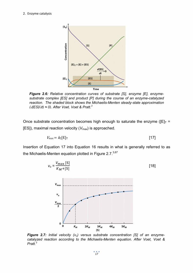
Once substrate concentration becomes high enough to saturate the enzyme ([E]T =
[ES]), maximal reaction velocity (Vmax) is approached.
Vmax=k2[E]T [17]
Insertion of Equation 17 into Equation 16 results in what is generally referred to as
the Michaelis-Menten equation plotted in Figure 2.7.3,67
vo = ����[�]
���[�] [18]
Figure 2.7: Initial velocity (vo) versus substrate concentration [S] of an enzyme-catalyzed reaction according to the Michaelis-Menten equation. After Voet, Voet & Pratt.3
≈
Figure 2.6: Relative concentration curves of substrate [S], enzyme [E], enzyme-substrate complex [ES] and product [P] during the course of an enzyme-catalyzed reaction. The shaded block shows the Michaelis-Menten steady-state approximation (d[ES]/dt) ≈ 0). After Voet, Voet & Pratt.3

2. Enzyme catalysis
18
2.3.3 Michaelis-Menten parameters
The Michaelis-Menten Equation 18 describes the kinetic behavior of enzymes and
enzyme-like catalysts which exhibit saturation kinetics (Fig. 2.7). The parameters
discussed below are commonly used by biochemists when relating and comparing
the activities of enzymes.
The Michaelis constant KM is mathematically defined as the substrate concentration
when the initial velocity of a reaction is half the value of Vmax (Fig. 2.7). KM is
inversely proportional to the fraction of total enzyme ET participating in the enzyme-
substrate complex ES. This relationship has often led to the common misconception
that the value KM is a measure of a substrate affinity to an enzyme. In the simple two-
step process shown in Equation 7, KM can be used as an indicator for substrate
affinity only when k2 << k-1. However, this is often not the case. The Michaelis
constant is best interpreted by how the initial velocity vo of the catalyzed reaction
responds as the concentration of the substrate [S] is increased (i.e. the steepness of
the Michaelis-Menten plot, Fig. 2.7).3,67
As already mentioned in the derivation of the Michaelis-Menten equation, the
maximum velocity Vmax is the initial velocity of a reaction when the total enzyme is
involved in an enzyme-substrate complex. In the simple reaction scheme of Equation
7, Vmax is defined as k2[E]T and is the zeroth-order rate constant.67
The turnover number, kcat, is a more applicable rate constant since it is independent
of the enzyme concentration [E]T. The turnover number is simply the rate at which a
single enzyme active site converts substrate into product. In Equation 7, kcat is equal
to k2. However, in more complicated processes, it is the rate constant of the rate-
limiting step or a more complicated combination of multiple rate-limiting steps. The
turnover number is the first-order rate constant and is computed as Vmax / [E]T.3,66,67
When used alone, kcat indicates only the rate of substrate turnover and little in regard
to the binding interaction between enzyme and substrate or the degree by which the
enzyme accelerates a reaction.65,66 By itself, the value of KM offers no information
about the turnover rate.65 However, when used together, kcat/KM is the most
conclusive constant used to compare the activity of different enzymes with different
substrates. This is best shown by the Michaelis-Menten equation when [S] << KM,
which means that most of the enzyme is unbound, the initial rate becomes

2. Enzyme catalysis
19
vo=����
��[E][S]=
����
��[S] [19]
The parameter kcat/KM is, therefore, the second-order rate constant for the conversion
of free enzyme and substrate into product. Since kcat/KM accounts for both substrate
specificity and turnover, it is often referred to as a measure of catalytic efficiency3,13
or the specificity constant.66,67 The diffusion rate in which enzyme and substrate
encounter one another imposes an upper limit (108-109 M-1 s-1) on values of kcat/KM.
Michaelis-Menten parameters of several enzymes are listed in Table 2.2.76
Parameters Vmax and KM are in practice difficult to ascertain directly from the
Michaelis-Menten Equation 18. This is often due to the high concentration of
substrate required to approach Vmax. Parameters KM, Vmax and kcat can, however, be
determined by transforming the Michaelis-Menten equation into the Lineweaver-Burk
equation.
�
��= (
��
����)
�
[�] +
�
���� [20]
Parameters can be graphically determined by plotting experimental values 1/vo
versus 1/[S]. From the resulting y-intercept and slope of the linear Lineweaver-Burk
plot, paramenters of Vmax and KM are determined as shown in Figure 2.8.77
Table 2.2: Values of KM, kcat and kcat/KM for various enzymes with corresponding substrate. From Voet, Voet & Pratt. 75

2. Enzyme catalysis
20
Figure 2.8: Lineweaver-Burk or double-reciprocal plot. From Chang.77

3
Biocatalysis

3. Biocatalysis
23
Enzymes are highly evolved catalysts with complex structures and remarkable
properties. Regardless of our level of understanding, enzymes will continue to
function with superior activity and selectivity within their natural environment.
However, in order to better exploit the use of enzymes in biocatalysis, more
knowledge is needed.
Biocatalysis is the application of whole cells or enzymes as catalysts in synthetic
chemistry. Although more recently defined, biocatalysis predates recorded history
when it was used to produce beer, wine and cheese by means of fermentation. A
more scientific knowledge of biocatalysis arose in the middle of the 19th century when
Louis Pasteur discovered biological enantioselectivity by resolving racemic tartaric
acid with penicillin.78 Later in the 19th century, Eduard Buchner fermented sugar with
the use of cell-free yeast extracts, which revealed that living cells do not hold a
monopoly on biological transformations.79 Despite advances made in enzyme
purification in the mid-20th century, the use of enzymes in synthetic chemistry lagged
behind breakthroughs in enzyme crystallography and the elucidation of their
mechanisms and involvement in biological pathways.8,80 It was not until the late
1970s that biocatalysis became a trend due to the rapidly increasing accessibility of
enzymes brought about by recombinant DNA methodology. By the 1990s, many
enzymes became commercially available and widely used in industrial processes.81
3.1 The pros and cons of biocatalysis
Enzymes are complex molecules in comparison to traditional catalysts employed in
organic synthesis and many of their means of biotransformation remain unclear and
difficult to study. The synthesis of a product without knowing the mechanism may
seem like mere hand-waving to many chemists. Moreover, enzymes possess
properties that could be seen as both advantageous and disadvantageous when
applied to organic synthesis. It would be unfair to say that many of the perceived
disadvantages are based on misconceptions and prejudice without taking into
account that many advantages are grounded on potential success or studies which
require no shortage of effort. Therefore, this section could have just as fittingly been
called the potential and misconceptions of biocatalysis.

3. Biocatalysis
24
Figure 3.1: Regioselective hydrolysis of aspartic and glutamic acid benzyl esters catalyzed by subtilisin. Adapted from Chen & Wang.82
3.1.1 Advantages of biocatalysis
A) Catalytic efficiency
Enzymes typically enhance reaction rates by factors of 106-1012 and as high as 1020
in comparison to non-catalyzed reactions.3,5 In addition, they often provide effective
catalysis when present in mole percentages of 10-3-10-4.80
B) Substrate selectivity
For the most part, enzymes are chemoselective in that they often catalyze reactions
involving a specific functional group. Furthermore, the three-dimensional structure
and available binding surface within a cleft allow enzymes to regioselectively
differentiate between the same functionality located at different positions on a
substrate molecule. A good example of enzyme regioselectivity is shown in the
subtilisin-catalyzed hydrolysis of benzyl esters of aspartic and glutamic acid (Fig.
3.1).82 Since enzymes are sequences of L-amino acids, they are chiral catalysts and
can distinguish between chiral substrates. Enzymes are enantioselective when they
catalyze one enantiomeric form of a substrate at a higher velocity than the other.
Therefore, enzymes are often applied to kinetically resolve racemic substrate
mixtures. An example of enzymatic kinetic resolution’s utility in synthesis is depicted
in Figure 3.2.83 Another example of enzymatic kinetic resolution is the convenient

3. Biocatalysis
25
one-pot system introduced by Monteiro et al. to resolve racemic secondary alcohols
(Fig. 3.3). In this system, fatty esters are used as both solvent and as an acylating
shuttle between an enantioselective lipase-catalyzed transesterification and
regenerative transesterification. Enantiopure alcohol is distilled after each
transesterification.84 Unfortunately, as described in Section 3.1.2A, superior enzyme
selectivity is often viewed as a double-edged sword when applied in synthesis.
IN
IN
Ethanol
OUT
OUT
Enzyme
Reaction medium: fatty ester
1) Enzymatic transesterification (irreversible process by ethanol removal under vacuum)
2) Distillation
4) Distillation
3) Enzymatic transesterification
(R)
(S)
rac
Figure 3.3: One-pot Candida antarctica lipase C-mediated kinetic resolution of secondary alcohols using fatty esters as solvent and acylating agent. R’ = ethyl. Adapted from Monteiro et al.84
Figure 3.2: Enzymatic kinetic resolution of orthogonally protected D- and L- 4,4,-difluoroglutamic acid. From Li & Miller.83

3. Biocatalysis
26
C) Product specificity
Not only are enzymes regioselective and stereoselective, but the large enzyme to
substrate surface interaction offers a high potential for regio- and stereo-control. In
other words, within the cleft of an enzyme, a prochiral substrate is positioned so the
catalytic machinery can encounter one area (regiocontrol) and one face
(stereocontrol) of the substrate.5,81 A biocatalytic example of product specificity is
asymmetric transfer hydrogenation catalyzed by oxidoreductases (Fig. 2.5).65
D) Catalytic activity under mild conditions
Naturally, enzymes evolved to function under physiological conditions. Therefore,
biocatalysts can be advantageously used on substrates sensitive to heat and harsh
pH conditions. Enzymes function best at temperatures between 20-40oC and at
neutral pH values. However, when enzymes are not robust enough for certain
environments, this property can also be viewed as a shortcoming.
E) Nontoxic and environmentally acceptable
Unlike metal or acid and base catalysts, enzymes are nontoxic and pose few health
hazards. As is the case with all catalysis, biocatalysis greatly reduces the amount
waste products resulting from stoichiometric chemistry, which has a tremendous
impact at the industrial level. In addition, enzyme selectivity decreases the number of
reactions required for orthogonal protection schemes. As enzymes are protein, they
are completely biodegradable.7,85
As expressed above, it is difficult to argue that enzymes do not present potential
advantages when employed in synthetic chemistry. However, these advantages are
based only on the examples of established uses and the inevitable likelihood of
discovering new uses. At present, a synthetic chemist would not rely on the
everyday use of biocatalysts. This does not mean he could not, but simply that the
field of biocatalysis has not approached a point in which it would be practical. As we
begin the next section which focuses on the disadvantages of biocatalysis, it is
important to keep in mind that many of these disadvantages are not directed at
biocatalysis itself, but at the insufficient knowledge we have of enzymes, especially
when removed from physiological conditions.

3. Biocatalysis
27
3.1.2 Disadvantages of biocatalysis
A) Enzymes are too selective
A common misconception about enzymes is that they only catalyze reactions
involving their natural substrates. Since enzyme-functioning has evolved according to
specific metabolites, it is not surprising that a preference for certain substrates exists.
It is true that some enzymes possess much narrower substrate scopes than others.
However, enzymes often have a broad substrate range unless such a property is
biologically detrimental. The catalytic activity of carbonic anhydrase is not limited to
the interconversion of carbon dioxide and bicarbonate (Fig. 2.4), but can also be
extended to ester hydrolysis (Fig. 3.14). The reversibility of enzymes should also not
be ruled out. Under specific laboratory conditions, proteases have been shown to
catalyze peptide synthesis.86,87 Some remarkably promiscuous enzymes have been
found to catalyze reactions in the laboratory with which they have no natural relation.
For instance, Candida antarctica lipase B (CALB), known to catalyze ester hydrolysis
in nature and as a biocatalyst (Fig. 3.3), catalyzes C-C bound forming aldol and
Michael additions.5,12,88 Furthermore, porcine pancreas lipase (PPL) has also been
shown to catalyze asymmetric aldol reactions (Fig. 3.4).89 It is perhaps incorrect to
state that the biocatalytic substrate or reaction scope is limited, but a delicate
balance between substrate selectivity and scope exists and is difficult to generalize
according to the type of enzyme or reaction.
Figure 3.4: Proposed mechanism of lipase-catalyzed asymmetric aldol reaction. From Li et al.89

3. Biocatalysis
28
B) Enzymes are operative under few conditions
Enzymes are developed by nature to operate under physiological conditions. In
biocatalysis, many enzymes are functional under mild conditions. It is well known that
the structure and activity of enzymes and protein in general is greatly affected by its
environment. Enzymes generally become less catalytically active in organic solvents,
at temperatures above 40oC and at pH conditions below 5 and above 9.66,90 A
common misconception is that enzymes are too sensitive or too unstable for use in
synthetic conditions.81 Nonetheless, if handled correctly, enzymes are far less
sensitive than t-butyl lithium or Raney nickel.
Since many organic reactants are poorly dissolved or unstable in water, it would be
beneficial to apply enzyme catalysis in an organic medium. Studies have shown that
enzymes retain a portion of their activity in anhydrous media. Furthermore, in non-
aqueous environments, pH is no longer an issue and enzymes often exhibit
increased thermostability. Interestingly, enzymes have reportedly been used in
organic solvents to impart increased stereocontrol, and even to direct stereocontrol
and alter enantioselectivity according to the selection of organic solvent.91
The loss of enzymatic activity in anhydrous media is said to be largely due to
increased enzyme rigidity. In fascinating studies by Klibanov92 and others,93,94 it was
shown that enzymes have what is called a “molecular memory”95 in anhydrous
solvents (Fig. 3.5).96 This effect is shown when an enzyme in aqueous solution with
Figure 3.5: An enzyme’s “molecular memory” of binding ligand in anhydrous solvent following lyophilization and ligand extraction. Adapted from Budisa.96
+
Lyophilization Binding
Anhydrous ligand extraction
Placement in aqueous media (memory lost)
anhydrous media (memory retained)

3. Biocatalysis
29
particular ligands or a competitive inhibitor is lyophilized. After lyophilization and
removal of the ligand by non-aqueous extraction, the enzyme by remaining rigid in
anhydrous media possesses an increased selectivity for substrates resembling the
initially “imprinted” ligand. Enzymes also maintain their molecular memory in
supercritical carbon dioxide, an effective solvent gathering increased interest in
biocatalysis.96,97 Discoveries such as these greatly widen the operational scope of
enzymes in synthetic chemistry.
C) Substrate and product inhibition
Within the cell, an elaborate network of metabolic pathways is required to maintain a
fine chemical balance. Key components of such pathways are enzymes which do not
operate by simply transforming substrate to product at high speeds, but by
responding to substrate levels and the needs of the cell. Enzymes are, therefore,
prone to both substrate and product inhibition. Substrate inhibition is a mechanism
used to maintain a required metabolic flux.98 In biocatalysis, substrate inhibition can
often be circumvented by keeping the substrate concentration level low with
incremental addition. Since products are in many cases similar to substrates, it is not
surprising that they might produce inhibitory effects on enzymes. Product inhibition,
or negative feedback, results when product concentrations become too high. Such a
mechanism is physiologically important because it helps conserve the substrate
when further conversion is not required. In biocatalysis, product inhibition presents a
more complicated problem. To avoid product inhibition, product must be continually
removed.81
Regardless of the current disadvantages or misconceptions of biocatalysis, it is
becoming increasingly apparent that the use of biocatalysts represents a practical
and efficient alternative in synthetic chemistry. Until this point, while explaining what
biocatalysis is and its perceived advantages and disadvantages, specific examples
have been given in which enzymes have been applied in synthetic chemistry. In
some of the examples, the conditions of the reaction are changed to accommodate
the enzyme. However, the complexity of enzymes offers many opportunities to
optimize such properties as stability, substrate compatibility, enantioselectivity and
stereocontrol. The next section focuses on how enzymes are modified to overcome
limitations and bolster their application in synthetic chemistry.

3. Biocatalysis
30
3.2 Enzymes modified for biocatalysis
The first wave of biocatalysis began with Ludwig Rosenthaler’s application of emulsin
extracted from almonds in the asymmetric conversion of (R)-mandelnitrile from
benzaldehyde and hydrogen cyanide and ended in the mid-1970s with the advent of
recombinant DNA methods. During this initial period, biocatalysts were less
accessible and, therefore, less affordable. In order to save on costs, biocatalysts
were immobilized to make them more stable and reusable.8,14
An increased availability of enzymes due to recombinant DNA techniques ushered in
the second wave of biocatalysis. In the mid-1970s, it became possible to overexpress
desired enzymes and even mutated enzymes in host organisms. As a result, not only
was there an increase in the use of biocatalysts, but also a means to study structure-
function relationships in biocatalysis. The new field of protein engineering was
amplified with the development of the polymerase chain reaction (PCR), which
greatly simplified DNA synthesis and mutagenesis.99,100 By the 1990s, the use of
biocatalysts was no longer limited to industry, but becoming increasingly common in
the production of pharmaceuticals and fine chemicals.8,14
In the early 1990s, mutagenesis of wild-type enzymes began to increase and expand
outward from active-sites. It was soon discovered that a large fraction of residues
were not necessary for general enzyme structure or function.101 Site-directed
mutagenesis is an adequate technique for learning about enzyme structure and
function, but was believed by many to be an ineffective method for quickly adapting
enzymes to synthetic needs (e.g. chiral pharmaceuticals). Before long, the third wave
of biocatalysis14 broke with the creation of large libraries of mutant enzymes,
requiring increased screening for desired properties. This repetitive process is
commonly referred to as directed or in vitro evolution. The engineering of biocatalysts
no longer looked to overcome enzyme limitations, but to fine-tune biocatalytic activity
according to process specifications.

3. Biocatalysis
31
3.2.1 Biocatalyst immobilization
Original efforts to immobilize enzymes were made to save money. Immobilization
prolongs the lifetime and facilitates the reusability of enzymes. Today, enzymes are
commonly immobilized for industrial use not only for cost efficiency and convenient
purification (filtration), but also to reduce wastes. In addition, enzyme immobilization
is becoming increasingly applied with multiple enzymes at different points on solid
support or cross-linked to mirror pathway or compartmentalization processes.
Enzymes are generally immobilized though solid support binding, entrapment or
cross-linking.102,103
Enzymes can be immobilized to a solid support by means of adsorption or covalent
bond. Supports consist of organic polymers (e.g. acrylic resins), biopolymers (e.g.
water-insoluble polysaccharides) and inorganic solids (e.g. silica, zeolites). A
commercially available example of a support-immobilized enzyme is Novozym 435,
C. antarctica lipase B adsorbed to acrylic resin. As opposed to support binding,
entrapment immobilizes enzymes by enclosing them within organic polymers,
gelatinous networks or membrane-like structures. When an inert carrier is unwanted,
immobilization can be performed by crosslinking enzymes. Enzymes can be cross-
linked with an agent such as glutaraldehyde and maintain activity as crystals (cross-
linked enzyme crystals, CLECs) or precipitate (cross-linked enzyme aggregates,
CLEAs).102,103 These strategies of enzyme immobilization along with efforts made in
protein engineering are commonly applied to tailor biocatalysts.
3.2.2 Biocatalysts via traditional rational design
Early efforts to refine enzyme properties for biocatalysis adopted the rational design
approach of protein engineering. This approach, however, is limited by the amount
and quality of structural knowledge available.104 Therefore, much of rational design
research has focused on enzyme active sites,105 although site-directed mutagenesis
beyond the active site has been used successfully to increase stability.106 A good
example of the use of rational design outside of the active site is the 8-fold site-
directed mutation of thermolysin, which maintained proteolytic activity at 100oC as
well as in the presence of denaturing agents.107

3. Biocatalysis
32
Provided with a three-dimensional map of the active site, site-directed mutagenesis
can be effectively used to broaden substrate specificity108 and directly alter the
catalytic mechanism.109,110 To a much lesser extent, site-directed mutagenesis has
also been used to initiate enantioselectivity.111 Rational design can also take a more
piecewise approach.105 Biocatalysts have been designed by incorporating the
catalytic machinery of one enzyme into templates forming a type of hybrid enzyme.
Such a template might provide advantageous solubility, binding or stability. An
example of such modular rational design is the chimeric bacterial-human cytochrome
P450 engineered to combine the high activity of the human enzyme with the
beneficial solubility of the bacterial enzyme.112
As mentioned in Section 2.1.5, enzymes bind best with the catalyzed reaction’s
transition state. In the 1980s, another rational method was developed in which
heptans analogous to a desired reaction’s transition state were recruited to initiate an
immune response.113,114 Although catalytic antibodies, abzymes, actively bind the
transition state, their catalytic activity is generally low due to an absence of
necessary catalytic machinery.8 Convergent rational design in which catalytic
machinery is grafted into antibodies has shown promising results. This can be seen
in research by Lee in which catalytic machinery in the form of glutamate, lysine and
histidine were strategically grafted via site-directed mutagenesis into a structurally
resolved antibody to introduce proteolytic activity (Fig. 3.6).115
Figure 3.6: Antibody converted into a protease. Proposed mechanism resulting from the introduction of residues glutamate (B), lysine (A) and histidine (C) into an antibody via site-directed mutagenesis. In the reaction, glutamate activates a water molecule for nucleophilic attack. Histidine serves as a proton donor for the eliminated amine. Lysine stabilizes the oxyanion intermediate. Courtesy of Liu et al.115

3. Biocatalysis
33
The schematic representation in Figure 3.7 summarizes possible strategies used by
rational design to fine-tune biocatalysts from a template protein of resolved
structure.105 It is commonly said that the use of rational design is restricted by our
knowledge of protein structure. Since design is based on structure, this view is
partially correct. However, it could be more accurate to say that the true limitation of
rational design is how little is known about folding dynamics. Most of the outcomes of
rational design do not match expectations. A shift of a single angstrom within the
active site or an amino acid substitution on a remote loop may make all the difference
in regard to catalytic activity. As Daniel Koshland wrote, “perfecting the molecule
requires such precision in very small changes that our theory and experiments are
strained to make logical predictions.”116 Often it is more effective to make many small
changes at random and select those that work.
(A)
(B)
Figure 3.7: Possible strategies to rationally redesign biocatalysts. (A) Three routes to form a novel biocatalyst from a pre-existing enzyme. (B) Modular approach in which independent enzyme features are assembled to create a novel biocatalyst. The cross and star symbols represent amino acid functionalities required for a specific catalytic activity. Adapted from Cedrone et al.105
3. Biocatalysis
34
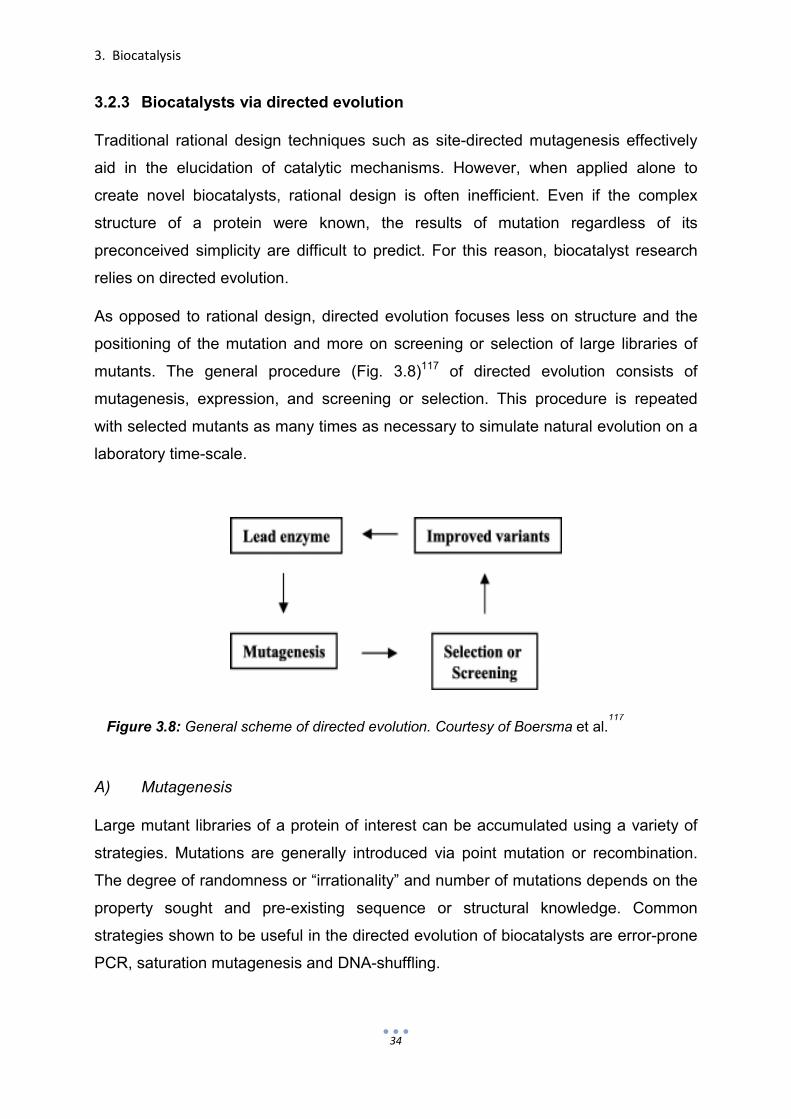
3.2.3 Biocatalysts via directed evolution
Traditional rational design techniques such as site-directed mutagenesis effectively
aid in the elucidation of catalytic mechanisms. However, when applied alone to
create novel biocatalysts, rational design is often inefficient. Even if the complex
structure of a protein were known, the results of mutation regardless of its
preconceived simplicity are difficult to predict. For this reason, biocatalyst research
relies on directed evolution.
As opposed to rational design, directed evolution focuses less on structure and the
positioning of the mutation and more on screening or selection of large libraries of
mutants. The general procedure (Fig. 3.8)117 of directed evolution consists of
mutagenesis, expression, and screening or selection. This procedure is repeated
with selected mutants as many times as necessary to simulate natural evolution on a
laboratory time-scale.
A) Mutagenesis
Large mutant libraries of a protein of interest can be accumulated using a variety of
strategies. Mutations are generally introduced via point mutation or recombination.
The degree of randomness or “irrationality” and number of mutations depends on the
property sought and pre-existing sequence or structural knowledge. Common
strategies shown to be useful in the directed evolution of biocatalysts are error-prone
PCR, saturation mutagenesis and DNA-shuffling.
Figure 3.8: General scheme of directed evolution. Courtesy of Boersma et al.117

3. Biocatalysis
35
Error-prone PCR is the most random method of mutagenesis as it targets the entire
gene for point mutations. The error-rate of Taq DNA polymerase is adjusted by
manipulating the conditions of PCR.118 Since mutations are introduced at random,
error-prone PCR requires no prior knowledge of structure. Error-prone PCR has been
found useful in enhancing biocatalyst solubility in organic solvents101 and
thermostability.119 Furthermore, results obtained from error-prone PCR studies may
also identify potential “hot spots” which contribute to a particular property. Error-
prone PCR was employed by Reetz in the first study120 applying directed evolution to
enhance the enantioselectivity of an enzyme, Pseudomonas aeruginosa lipase
(PAL), for the kinetic resolution of racemic esters (Fig. 3.9A).8 The success of this
experiment and others to follow121 was not only the enhancement of
enantioselectivity (Fig. 3.9B), but also the elucidation of hot spots. These hot spots
were the focal points of later studies by Reetz in which saturation mutagenesis was
employed.122
Unlike error-prone PCR, saturated mutagenesis is an oligonucleotide-based
approach which targets a specific location of expressed protein. Since a certain
structural knowledge is required, this method is considered combinatorial (combines
aspects of rational design and directed evolution). Mutant libraries are produced
using degenerate codons substituted into DNA primers. In addition to enhancing the
substrate scope or selectivity of enzymes by focusing on locations near to the active
(A) (B)
Figure 3.9: (A) Lipase-catalyzed enantioselective hydrolysis of p-nitrophenyl esters and (B) enhancement of enantioselectivity (E) following four successive generations of error-prone PCR starting with wild-type lipase from Pseudomonas aeruginosa. Courtesy of Reetz.8

3. Biocatalysis
36
site, saturation mutagenesis can also be used to develop biocatalysts with improved
stability. Properties such as thermostability, stability in denaturing agents and
solubility in organic solvents are commonly improved by targeting locations shown by
X-ray data to have high flexibility (high average B-factor)123 or on sequences of low
homology.
The benefit of saturation mutagenesis is that it targets specified critical positions,
thereby reducing the size of mutant libraries and the amount of screening/selection,
the bottleneck of directed evolution. In an effort to further curtail screening, Reetz et
al. devised a systematic combinatorial approach known as iterative saturation
mutagenesis (ISM).124 With information obtained from previous error-prone PCR
studies performed to enhance the enantioselectivity of P. aeruginosa lipase (Fig. 3.9)
and X-ray data, three hot spots in the vicinity of the active site were chosen.122 As
shown in Figure 3.10a, a mutation map was created which contained six potential
mutagenic pathways ending with 15 possible mutant libraries. Paths were then
followed until the exceptional result of E = 594 (S) enantioselectivity was achieved
(Fig. 3.10b). Furthermore, follow-up deconvolution studies revealed a strongly non-
additive relationship between the individual mutations, emphasizing the importance
of synergy in protein engineering.125
Another method commonly employed to create mutant libraries is DNA shuffling.
DNA shuffling is a recombinant-based method in which a group of parent genes are
cut into fragments with DNase. These fragments (10-50 base pairs) are then
assembled into full-length genes using primerless PCR. Shuffling or crossover results
when the fragment of one parent gene anneals with a fragment of another gene. In
this method, parent genes can include any number of mutated or homologous
genes.126 DNA shuffling127 and related recombinant methods128,129 have been found
to be effective in the directed evolution of biocatalysts.130

3. Biocatalysis
37
B) Screening or selection
With large mutant libraries made available by biotechnology, the bottle-neck of
directed evolution is passed on to the screening or selection process. Two general
types of approaches are taken to reduce the time and energy spent in this process.
The first involves simply reducing the number of mutants to be screened by
producing what are commonly called smart libraries.131 Smart libraries are normally
created with a preexisting knowledge of structure or homology using combinatorial
methods of mutagenesis. However, protein dynamics can be very unpredictable and,
depending on the starting enzyme and the property desired, this may not be an
option. Therefore, the second approach attacks the bottle-neck head on by
attempting to increase the efficiency of the screening/selection process.
Figure 3.10: Iterative saturation mutagenesis (ISM) as applied by Reetz et al122
to enhance P. aeruginosa lipase enantioselectivity (E). (a) Mutagenic pathway system showing all possible paths of saturated mutagenesis targeting three locations A, B and C. The solid green line represents the best pathway taken, the dotted red lines indicate attempts made with insignificant improvement, and the dotted black line denotes paths which were unnecessary and unexplored. (b) The best path found by ISM corresponding to actual mutations and resulting enantioselectivity.

3. Biocatalysis
38
The main difference between screening and selection is that screening is performed
on each individual mutant, whereas selection can be performed simultaneously on
entire pools of mutants. From this it would seem obvious to avoid screening at all
cost, but this is often not the case. Although processes of selection are less-time
consuming and more closely resemble nature, they tend to be difficult to associate
with or apply to the property desired. Moreover, selection for the property sought
cannot be directly observed as in screening and like the biocatalyst itself, the
selection technique must also be tailored to fit the task at hand.117,132
3.2.4 Biocatalysts via computational design and enzyme redesign
Driven by knowledge obtained from directed evolution and the time lost in screening
mutant libraries, protein engineering is again taking on an increasingly rational form.
Smarter libraries with “higher-quality” mutants are produced by combinatorial or
semi-rational approaches and new and improved software is available for
computational designs, which can subsequently be optimized by directed evolution.
With the growth of structural protein databases and quantum mechanical models,
novel enzymes are created by inserting active sites into previously non-enzymatic
proteins and biocatalysts are designed for reactions that are not in the repertoire of
natural enzymes.
The true meaning of de novo design in regard to proteins and peptides is
debatable.133 This section is written to establish a clearer delineation between current
trends in rational enzyme design, in which catalytic machinery is engineered into
native protein or protein sequences, and true de novo design (Section 4), which
includes the design of not only the active site, but also the entire protein topology. In
doing so, the remainder of this section will serve as a continuation of traditional
rational design (Section 3.2.2) with examples of computational active site design in
native proteins, which is often used in combination with directed
evolution.26,28,29,134,135
In 2001, Bolon and Mayo reported the design of “protozymes.”136 Their strategy
involved the use of the protein design software ORBIT to incorporate an active site
into thioredoxin. This active site was modeled for nucleophilic catalysis (Section
2.1.2) with the histidine-mediated hydrolysis of p-nitrophenyl acetate (pNPA) (Fig.

3. Biocatalysis
39
3.11), a commonly used model substrate for kinetic ester hydrolysis studies.
Although the catalytic results were not spectacular (kcat/KM = 3 M-1s-1), their
computational approach in combination with potential directed evolution represented
wide-ranging possibilities for enzyme design.
By 2008, computational design had gained considerable ground with the use of the
Rosetta software suite developed in the laboratory of David Baker. Biocatalysts
engineered from scaffolds belonging to triose phosphate isomerase were found to
accelerate a retro-aldol reaction (Fig. 3.12) by more than 2 x 10⁴-fold.137 With later
improvements and directed evolution, rate enhancements of almost 6 x 10⁵ were
achieved.138 That same year, the first unnatural reaction, the Kemp elimination of 5-
nitrobenzisoxazole (Fig. 3.12), was also successfully modeled with Rosetta.139 The
best biocatalyst from this study was shown to have a second-order rate constant
(kcat/KM) of 160 M-1s-1 (60,000 M-1s-1 following 13 rounds of directed evolution).140
This finding inspired the use of an alternative scaffold for a more efficient Kemp
eliminase (425 M-1s-1; 230,000 M-1s-1 after 17 rounds of mutagenesis and
screening).141,142
Figure 3.11: A) Histidine-mediated nucleophilic hydroylsis of pNPA B) Transition-state structure with torsion angles varied to generate rotamers used in the design calculations. From Bolon, Mayo et al.136

3. Biocatalysis
40
Until 2010, no enzyme had been shown to catalyze bimolecular Diels–Alder
reactions. The design of a stereoselective Diels-Alderase by Siegel, Baker and
collaborators became a milestone in computational design.143 As opposed to other
biocatalysts developed with Rosetta, this design was the first to catalyze a C-C bond-
forming reaction, which requires the accommodation of two substrates, a diene and a
dienophile (Fig. 3.12). Although not overwhelmed with the activity (kcat/(KMdiene x
KMdienophile) = 6 s-1M-1M-1) of the original designs, a challenge presented to online
enthusiasts of protein structure was answered with the insertion of a helix-turn-helix
and an 18-fold increase in efficiency.144
As is common in biocatalysis research, computational design is often employed to
develop hydrolases. Like Mayo had with ORBIT in 2001, Richter, Baker and
collaborators applied Rosetta in an attempt to engineer an effective nucleophilic
esterase in silico.145 Unlike the previous study by Mayo (Fig. 3.11), this study focused
on the importance of a histidine-cysteine catalytic dyad in the active site (Fig. 3.13).
Following the directed evolution of their most active design, the second-order rate
constant corresponding to catalyzed pNPA hydrolysis was increased from 34 M-1s-1
to 405 M-1s-1.
ester hydrolysis
retro-aldol
Kemp elimination
Diels-Alder
organophosphate hydrolysis
Figure 3.12: Target reactions of computational designed biocatalysts.
Ref. 136, 145, 149
Ref. 137, 138
Ref. 148
Ref. 139-142
Ref. 143, 144

3. Biocatalysis
41
Figure 3.13: Top) Computational designed mechanism of ester hydrolysis by Cys-His dyad with an oxyanion binder. Bottom) Relative substrate (p-nitrophenyl ester) conversion catalyzed by four esterase designs. Blue progress curves correspond to initial designs possessing the complete dyad, red traces correspond to Cys-knockout variants, His-knockouts are shown in black and double Cys and His-knockouts are in green. Courtesy of Richter et al.145

3. Biocatalysis
42
Another route used by enzymes to initiate ester hydrolysis is provided through the
use of metal ion catalysis (Section 2.1.3). This is witnessed with the promiscuous
ester hydrolytic activity of the zinc metalloenzyme carbonic anhydrase (Fig. 3.14).
Zinc in particular plays an important role in numerous hydrolytic enzymes and is,
therefore, commonly exploited in designer enzymes, especially those of de novo
design (Section 4). Zinc serves both structural and catalytic roles in metalloenzymes.
When catalytically active, zinc is bound in an open coordination sphere. This means
that a position for coordination is occupied by solvent/water.58 The geometry of zinc
is most often tetrahedral, but trigonal pyramidal geometries, as is the case in
adenosine deaminase, is also observed (Fig. 3.15).146,147
In yet another accomplishment, Baker and his team set out to repurpose the catalytic
machinery of a functional zinc metalloenzyme.148 Namely, murine adenosine
deaminase was redesigned for organophosphate hydrolysis (Figs. 3.12 and 3.16).
However, all zinc-coordinating residues (Fig. 3.15) were left intact. Following three
rounds of in vitro evolution, the redeployed enzyme acquired a 107-fold gain in kcat/KM
for organophosphate hydrolysis in comparison to the wild-type deaminase. On the
other hand, deaminase activity dropped 50,000 fold.
Figure 3.14: Promiscuous carbonic anhydrase-catalyzed ester hydrolysis via zinc-bound hydroxide mechanism.

3. Biocatalysis
43
Figure 3.15: Trigonal bipyramidal zinc coordination and water activation within the active site of wild-type adenosine deaminase. From Gleeson et al.147
Figure 3.16: Computational adenosine deaminase active site redesign by Khare, Baker et al.148 accounting for the mechanistic parameters: a) Zn-bound hydroxyl nucleophilic attack on the organophosphate, b) stabilization of the developing negative charge, c) simultaneous performance of both a and b; and d) stabilizing H-bond interactions and substrate accommodation. Mutations are shown in italics.
His 15
His 17Asp 295
His 214 OH
P
R2
R1
R3
O
His 15
His 17Asp 295
His 214
OH
P
R2
R1
R3
O
His 15
His 17Asp 295
His 214
OH
P
R2
R1
R3
O
His 15
His 17Asp 295
His 214
OH
P
R2
R1
R3
O
H-bond (E217, L58Q)
Substrate accommodation(F65W, A183I, F61T, E296A)
H-bond (H238)
R1, R
2 = OEt; R
3 = 7-hydroxycoumarin
a b c
d
Zn2+
Zn2+
Zn2+
Zn2+

3. Biocatalysis
44
As the ratio between the size of native protein scaffolds and the degree of
mutagenesis decreases, it becomes increasingly difficult to clearly distinguish true de
novo design from the broader rational design. Inevitably, an example was bound to
arise in which a simple folding motif constructed of a short amino acid sequence was
extracted from a native protein and modified by computational design to possess a
specific function. This is exactly the case presented by the design of Der et al.149 in
which the Rab4-binding domain of rabenosyn150 was used to derive the 46-residue
MID1-zinc (Fig. 3.17). The initial intent behind MID1-zinc, was simply to show how
zinc could induce helix-turn-helix monomers to form dimers (Fig. 3.17 B). However,
the MID1-zinc crystal structure revealed an open coordination sphere, (His)3Zn (Fig.
3.17 C), typical of a zinc catalytic site as opposed to the expected closed (His)4Zn.
Furthermore, the co-crystallization of tartrate (Fig. 3.17 D) from the crystallization
buffer within a cleft of the dimer suggested possible esterase activity. Following
kinetic studies, unoptimized MID1-zinc was found to catalyze the hydrolysis of pNPA
with an astounding rate enhancement of 105 and kcat/KM of 630 M-1s-1. Open site zinc
coordination of water resulted in the lowering of its pKa to 8.2.
The findings of Der et al. highlight key concepts which could be very influential in
future rational design and, more specifically, de novo design. Enzyme-like activity is
not confined to large protein structures. Furthermore, the use of simple, self-
assembling peptides creates protein interfaces resembling clefts of enzyme active
sites. Simpler, well-defined peptide assemblies could, therefore, hold the key to
understanding enzyme function. By applying this bottom-up approach to de novo
design, we could determine the minimal requirements needed for enzyme catalysis.
As knowledge of folding and function continue to grow, so will the complexity of de
novo designs.
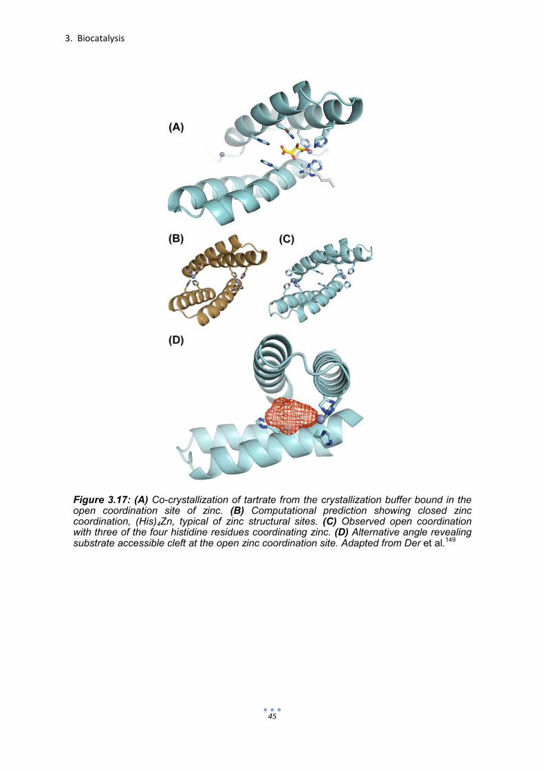
3. Biocatalysis
45
Figure 3.17: (A) Co-crystallization of tartrate from the crystallization buffer bound in the open coordination site of zinc. (B) Computational prediction showing closed zinc coordination, (His)4Zn, typical of zinc structural sites. (C) Observed open coordination with three of the four histidine residues coordinating zinc. (D) Alternative angle revealing substrate accessible cleft at the open zinc coordination site. Adapted from Der et al.149


4
De novo catalytic proteins


4. De novo catalytic proteins
49
If we really understood enzymes, we would be able to design them according to the
reactions or processes desired. Although the field of enzyme design has experienced
much advancement, a designed equivalent has yet to come close to the remarkable
efficiency of native enzymes. The bottom-up or minimalist approach to de novo
design allows researchers to break down complex enzyme interactions into their
individual parts such as the presentation of catalytic machinery and substrate
accommodation.
Through the years, surfactants,151 cyclodextrins51 and more complex supramolecular
arrangements152 have been employed to confine substrates in artificial, enzyme-like
active sites. With a continually growing knowledge of the mechanisms which guide
protein folding, the use of self-assembling, synthetic peptides in the truly de novo
design of catalytic proteins has become a rapidly developing field. This has been
witnessed by the steadily increasing catalytic efficiencies and complexity of designs
achieved. Combined with advancements in computational methods, this trend in the
bottom-up approach is likely to radiate outward to promote indirect interactions in the
active-site periphery.
When using self-assembling peptides as scaffolds to support catalytic machinery,
stability often becomes a major consideration. Functional groups which actively
participate in catalysis are typically polar and prefer being exposed to aqueous
surroundings. The active sites of enzymes commonly exist within clefts or at the
interface of folds. The complex structures of enzymes allow for a multitude of
energetically favorable interactions to stabilize such positioning. The number of
stabilizing interactions of self-assembling peptides is much more limited. Therefore,
the type of assembly and the positioning of catalytic machinery must be carefully
chosen.
De novo peptide designs can take on numerous stable shapes and assemblies.
These assemblies are typically classified according to the resulting secondary
structure (Fig. 4.1). Both α-helical and β-sheet peptide designs are used in the truly
de novo design of catalytic proteins.

4. De novo catalytic proteins
50
4.1 α-Helical de novo-designed catalytic proteins
4.1.1 Coiled coils
Approximately 30% of all protein structure is comprised of short, α-helical amino acid
sequences.153 Many of these same sequences fail to adopt the same structure when
removed from the larger protein environment. 154 Without the hydrophobic support of
the parent protein structure, solitary α-helices are more likely to succumb to entropic
effects.155 However, when such sequences are of the coiled-coil motif, α-helical
structure is more likely to be retained.
(A)
(B) (C)
Figure 4.1: Secondary protein structure. (A) Right-handed α-helix showing central axis (left) and hydrogen bonds (right). (B) Antiparallel β-sheet (C) Parallel β-sheet. Structure-stabilizing hydrogen bonds are designated with dashed blue lines. Adapted from Nelson & Cox.66

4. De novo catalytic proteins
51
α-Helical coiled coils are protein structure domains in which two to six α-helices pack
together and wrap around each other forming a superhelix. They are among the most
ubiquitous folding motifs and are believed to be present in as much as 10% of all
proteins.156 The stability as well as the versatility of this fold, not only in native protein
structures, but also in peptides, makes it an excellent candidate for de novo
design.157-160
Sequences forming coiled-coil structures are characterized by a repeating heptad
pattern (abcdefg)n in which n designates the number of repeats (Fig. 4.2).161 The
heptad’s a and d positions comprise the hydrophobic interface and are therefore
occupied by amino acids possessing hydrophobic side chains.162,163 The stability of
the coiled-coil fold is largely dictated by the ability of these “core” residues to pack
tightly in what is referred to as “knobs into holes.”164,165 In coiled-coil dimers, trimers
and tetramers, the heptad e and g positions are occupied by residues such as
glutamate and lysine which participate in interhelical salt bridges for further
stability.166 The occupancy of b, c and f in dimers, trimers and tetramers is less
restrictive. Since these positions are typically exposed to aqueous media, they are
often occupied by polar residues.167
In addition to stability, α-helical coiled coils can also vary in the degree of
oligomerization (dimer, trimer, etc.), the orientation of the helices relative to one
another (parallel or antiparallel, Fig. 4.2) and specificity of oligomerization
(homomeric or heteromeric). The degree of coiled-coil oligomerization or
oligomerization number is determined by the side-chain geometry of residues and
their packing at the hydrophobic interface. This oligomerization number is typically
between two and six. Table 4.1, based on results collected over numerous studies,
accurately summarizes basic principles in the de novo design of coiled coils. The
orientation and the specificity of oligomerization are largely directed by forces of
attraction or repulsion between charged residues in gn and en+1’ (n + 1 = one heptad
repeat below; the prime signifies the involvement of the interacting helix).168

4. De novo catalytic proteins
52
4.1.2 Examples of α-helical de novo-designed catalytic proteins
Enzymes are large molecules with complex topologies. However, as has been
witnessed by the success of proline and other amino acids in the field of
organocatalysis, complex structure is not a requirement for catalysis. As simple
scaffolds, helical assemblies can predictably present an array of functionalities as
long as stability is maintained.
Figure 4.2: Helical wheel diagrams of coiled coils. (A) Parallel dimer. (B) Antiparallel dimer. (C) Parallel trimer. (D) Parallel tetramer. Curved arrows designate salt bridges and crossed arrows indicate interfacial hydrophobic interactions. Knobs into holes packing of (E) parallel coiled-coil dimers, trimers and tetramers and (F) antiparallel coiled coils. Courtesy of Apostolovic et al.161

4. De novo catalytic proteins
53
Among the original de novo designed catalysts to incorporate α-helices was the
synthetic hemeprotein, ‘helichrome’.169 Interestingly, the main structural scaffold
component of helichrome was not the protein portion, but porphyrin. By covalently
linking porphyrin to four short, amphiphilic peptides, Kaiser created an α-helical
bundle with a hydrophobic core. In the presence of iron and cofactors, 7-acetylflavin
Table 4.1: Basic design principles of α-helical coiled coils.161
Figure 4.3: Proposed structure of Kaiser’s helichrome.169
Courtesy of Lewis.19

4. De novo catalytic proteins
54
and NADPH, aniline hydroxylase activity was observed as aniline was oriented in the
hydrophobic pocket of the bundle, adjacent to the active Fe(III)-porphyrin complex
(Fig 4.3).
Whereas helichrome offered a hydrophobic pocket to draw in substrate, another de
novo design known as ‘oxaldie’ relied on coulombic binding interactions.170 As an
artificial oxaloacetate decarboxylase, oxaldie accelerated the conversion of
oxaloacetate to pyruvate by 4 orders of magnitude. Like helichrome, oxaldie was a
short (14 residues), amphiphilic peptide that assumed an α-helical, tetrameric bundle
in its active state. The key to oxaldie’s catalysis was the presentation of its exposed,
lysine-lined cationic face. Upon luring oxaloacetate with its charged side-chain
amines, the terminal amine, measured pKa of only 7.2, could initiate subsequent
decarboxylation though an imine intermediate (Fig. 4.4). This concept was later
exploited by Allemann with the insertion of the catalytic machinery of oxaldie into
bovine pancreatic polypeptide (bPP) resulting in ‘oxaldie 4’ (Fig. 4.5 A and B).171 The
increased α-helical stability imparted by the linked proline-rich helix was translated
into a three-fold increase in efficiency when compared to the original oxaldie.
Allemann also applied the bPP scaffold to create ‘Art-Est’, an artificial esterase.172
The Art-Est design promotes ester hydrolysis through the interplay of several
residues (Fig. 4.5). Of these residues, two are histidine. One histidine among two
arginine residues serves as a nucleophile with a perturbed pKa of 5.5. The second
histidine is situated next to glutamate and acts as an acid to stabilize the developing
negative charge of the intermediate.
Figure 4.4: Amine-catalyzed decarboxylation of oxaloacetate

4. De novo catalytic proteins
55
Work discussed in the preceding paragraphs emphasizes the flexibility and potential
of de novo peptide design. Essentially, Allemann and collaborators discovered the
decarboxylation mechanism in oxaldie, incorporated it into bPP to form oxaldie 4, and
lastly, repurposed that same scaffold for ester hydrolysis. A very similar cooperative
nucleophilic and general acid mechanism was applied to ester hydrolysis in previous
work performed in the research group of Baltzer.69,173,174 The helix-loop-helix motif,
KO-42, which dimerizes into a four-helix bundle, was shown to possess hydrolytic
activity due to cooperative HisH+-His pairings (Fig. 4.6). Like Allemann, Baltzer later
incorporated a similar i, i + 3 and i + 4 arrangement of histidine, arginine and
arginine, respectively, (Art-Est, Fig. 4.5 C and D) into the same helix-loop-helix motif
to lower the histidine pKa for phosphoester hydrolysis (Fig. 4.7).175,176
bPP APLEPEYPGDNATPEQMAQYAAELRRYINMLTRPRY-NH2
Oxaldie 4 Ac-APLEPEYPGDNATPEQMKQYAKELKRYINML-NH2
Art-Est Ac-APLEPEYPGDNATPEQMHQYAHQLRRYINML-NH2
1 8 14 31
polyproline helix α-helix β-turn
Oxaldie 4
Art-Est
(A) (B)
(D) (C)
Figure 4.5: (A) Structure of oxaldie 4 modeled after bovine pancreatic polypeptide (bPP). (B) Sequence alignment and structures of bPP, oxaldie 4 and Art-Est. (C) Structure of Art-Est. (D) Proposed nucleophilic histidine-catalyzed ester hydrolysis of p-nitrophenyl fumarate by Art-Est. Adapted from Tayler et al.171 and Nicoll et al.172

4. De novo catalytic proteins
56
Ac-N-A-A-D-Nle-E-A-A-I-K-H-L-A-R-R-Nle-A-A-1 11 15 19
Helix
-G-P-V-D- 20 23
Loop
-A-A-Q-Nle-A-E-H-L-A-R-R-F-E-A-F-A-R-A-G-NH2
24 30 34 42 Helix
Ac-N-A-A-D-Nle-E-A-Y-I-K-H-L-A-R-R-Nle-A-A-K- 1 11 15 19
-G-P-V-D- 20 23
-A-A-Y-Nle-A-E-H-L-A-R-R-F-E-A-F-A-R-A-G-NH2
24 30 34 42 26
8
HN1 HJ1
(A) (B)
Figure 4.7: Helix-loop-helix motif as structural scaffolds for de novo-designed catalysts of phosphate diester hydrolysis. Only the monomers of the active four-helix bundles are shown (A) HN1 using histidine in a cooperative nucleophilic-general acid mechanism. (B) HJ1 using histidine in a general acid-base mechanism. Adapted from Razkin et al.175,176
His-11 (pKa = 6.9)
His-15 (pKa = 5.4)
His-19 (pKa = 7.0) His-26 (pKa = 7.2)
His-30 (pKa = 5.3)
His-34 (pKa = 5.2)
Figure 4.6: Helix-loop-helix motif and amino acid sequence of KO-42 monomer with
HisH+-His acid-base pairings. Adapted from Broo et al.
69
Ac-N-A-A-D-Nle-E-A-A-I-K-H-L-A-E-H-Nle-A-A-H- 1 11 15 19
Helix
-G-P-V-D- 20 23
Loop
-A-A-H-Nle-A-E-H-L-A-K-H-F-E-A-F-A-R-A-G-NH2
26 30 34 42 Helix

4. De novo catalytic proteins
57
The most basic barrier of the de novo design of protein catalysts, particularly in the
bottom-up approach, is the ability to introduce catalytic machinery into simple self-
assembling peptide scaffolds without incurring a loss of structural stability. With the
exception of helichrome, which required porphyrin for stability, other examples
including oxaldie variants and the helix-loop-helix scaffolds of Baltzer presented
catalytic functionalities to solvent-exposed environments. However, catalytic activity
of native enzymes is not limited to the surface. A common source of stability and
catalytic activity used by native proteins are metals.
Nearly half of all proteins found in nature are metalloproteins.177 In nature, metal ions
provide proteins with both structural stability and catalytic function. For such reasons,
the incorporation of metal ions is also important for the de novo design of catalytic
proteins. As will be seen in the following examples, the application of metals in the
bottom-up approach of α-helical designs allows active site stabilization and activity
within the hydrophobic core or cleft, a common feature of native metalloenzymes.178
Within the last decade, the increase in the efficiency of truly de novo designed
catalytic proteins directly correlates with the incorporation of metal ions. In 2004,
Kaplan and DeGrado applied their ‘Due Ferri’ concept of metalloproteins to bring
about oxidative catalysis (Fig. 4.8).25 In this study, a heterotetrameric α-helical
bundle, DFtet, was computationally designed to perform phenol oxidase activity by
coordinating two iron (II) ions within a single active site (Fig. 4.9). What was most
interesting about this design was that the two iron ions were coordinated by two
histidine residues and four glutamates within the hydrophobic core of the α-helical
bundle. The design was later optimized to provide quicker oxygen binding (Fig. 4.8
eqn. 1) and more space for substrate accommodation (Fig. 4.8 eqn. 2) by substituting
glycine residues in place of a bulkier leucine and an alanine in two of the heteromers,
resulting in a more active variant G4-DFtet. The heterotetrameric system provides an
effective platform to find the optimal combination of subunits. Later, another design
would join two subunits to form a more stable helix-loop-helix dimer DF3 with due
ferri phenol oxidase activity.179 The general applicability of de novo design was
further demonstrated with single-chain due ferri G4DFsc as well as 3His-G4DFsc
repurposed with an additional histidine for N-hydroxylation catalysis.180

4. De novo catalytic proteins
58
Figure 4.8: Proposed reaction mechanism of 4-aminophenol oxidation to benzoquinone
monoimine catalyzed by diferric protein. Adapted from Kaplan & DeGrado.25
(A)
(D)
(C) (B)
Figure 4.9: (A) Amino acid sequences with heptad register of peptide subunits designed to comprise the heterotetrameric bundle DFtet. Underlined residues of the A subunits were substituted with glycine to create G4-DFtet. (B) Heteromeric subunit composition of DFtet with coordinated iron ions in violet. (C) DFtet active site showing coordinated di-iron (III) bound to 4-aminophenol substrate. (D) Comparative solvent-exposed surfaces showing the increased active site accessibility of G4-DFtet. Adapted from Kaplan & DeGrado.25

4. De novo catalytic proteins
59
Another de novo metalloprotein recently introduced is the Zn(II) TRI system by
Pecoraro.27 The Zn(II) TRI system is an α-helical trimeric coiled coil which positions
three histidine residues within the hydrophobic core for zinc coordination. This open
(His)3Zn alignment is analogous to the active site of carbonic anhydrase (Fig. 2.4)
and MID1-zinc149 (Fig. 3.17). Similar to carbonic anhydrase, the variant
Hg(II)SZn(II)N(TRIL9CL23H)3 was reported to catalyze both ester hydrolysis and the
hydration of carbon dioxide.
The Hg(II)SZn(II)N(TRIL9CL23H)3 variant of the TRI family is composed of a trimeric
coiled-coil assembly of the 30-residue peptide TRIL9CL23H. To compensate for any
destabilization that may result from the inclusion of histidine within the hydrophobic
core, a second metal-binding site, (Cys)3Hg, was designed (Fig. 4.10). This assembly
was reported to possess a catalytic pKa of 8.8 and kcat/KM values of 23 M-1s-1 for p-
nitrophenyl acetate hydrolysis and 1.8 x 10⁵ M-1s-1 for CO2 hydration at pH 9.5. The
efficiency in which Hg(II)SZn(II)N(TRIL9CL23H)3 hydrated CO2 was found to be 70-
fold greater than any reported model and within just 500-fold of the kinetically perfect
CAII. In additional studies, it was shown that altering the position of the metal
coordination sites within the core had an effect on the assembly’s ability to catalyze
ester hydrolysis. However, none of the variants, including one variant with switched
metal coordination sites and another with a more centrally located zinc site, were
Figure 4.10: Trimeric coiled coil structure of Hg(II)SZn(II)N(CSL9PenL23H)3
featuring the
coordination of Hg (II) (gray) by penicillamine (Pen) sulfur (yellow) and Zn (II) (blue) by histidine and water (red). The peptide CSL9PenL23H used in crystallography studies is analogous to TRIL9CL23. The residues which participate in metal coordination are underlined. Adapted from Zastrow et al.27

4. De novo catalytic proteins
60
found to be more efficient.181,182 Interestingly, by simply replacing zinc with redox-
active copper, a very similar assembly, Cu(I/II)(TRIL23H)3+/2+ was found to accelerate
the reduction of nitrite to nitric oxide.183 Returning to carbon dioxide hydration,
Pecoraro introduced the (His)3Zn machinery into the single-stranded three-helix (two-
loop) bundle, α3D modelled by DeGrado.184 The resulting single-chain bundle,
α3DH3, offers the advantage of being more stable and, therefore, requires no
structural mercury coordination site.185 Although, the initial design exhibited 5-fold
less hydration activity as Hg(II)SZn(II)N(TRIL9CL23H)3, it offers greater potential for
future development of the secondary coordination sphere. This stabilization of the
active site or primary metal coordination sphere with secondary shell residues and
the difficulty in channeling water to hydrophobic reaction centers are two obstacles
presently facing de novo hydrolases.
4.2 β-sheets as scaffolds for de novo-designed catalytic proteins
On paper, the design of β-sheet peptides is simple (Fig. 4.1). All that is needed is a
simple alternating sequence of hydrophobic and hydrophilic residues. Since much of
de novo protein design is predicated on the construction of protein interfaces, the
inherent gregarious nature of β-sheet structures would at first seem like a perfect
match. However, a disadvantage of β-sheet structures is that they tend to aggregate
to the point of precipitation.186 The difficulty encountered when designing stable β-
sheet folds has reduced their utility as a platform for de novo catalyst design.30
Nonetheless, the β-sheet blueprint of carbonic anhydrase (Fig. 2.4 A) and heightened
interest in self-assembling nanostructures and hydrogels187 has encouraged an
interest in de novo β-sheet esterases.188,189
Taking a minimalistic route to mimic serine protease activity, Fukushima created
poly(Asp-Leu-His-Leu-Ser-Leu) to obtain a β-sheet structure with its polar
constituents, aspartate, histidine and serine performing as a catalytic triad (Fig. 2.3).
The resulting peptide consisted of approximately ten repeats of the sequence
DLHLSL and folded into a stable, monomeric β-sheet. Although the mechanism was
not clearly revealed, poly(DLHLSL) did show slight esterase activity with a second
order rate constant of 0.5 M-1s-1 (pNPA hydrolysis; pH 8) over the hexapeptide
DLHLSL, which lacked a defined structure, a mixture of the individual amino acids

4. De novo catalytic proteins
61
and imidazole.73 In a separate study, poly(DLHLSL) was also found to exhibit
selectivity (E = 1.5) of N-benzyloxycarbonyl-L-phenylalanine p-nitrophenyl ester (Z-L-
Phe-ONp) over Z-D-Phe-ONp.72 This was attributed to an interaction between the
substrate and hydrophobic portion of the β-sheet.
More recently, Degrado and Korendovych attempted to model the active site of
carbonic anhydrase from a minimalist β-sheet-forming heptapeptide (LKLKLKL).188
Of the variants examined, the peptide of sequence Ac-IHIHIQI-NH2 in the presence
of Zn2+ (Fig. 4.11) was found to most actively catalyze pNPA hydrolysis (kcat/KM = 62
M-1s-1 at pH 8). This incredibly simple design is not only the most efficient truly de
novo esterase to date, but also forms and remains active as amyloid fibrils (Fig.
4.12).
Further characterization revealed that Zn2+ not only participated in catalysis (lowered
the pKa of water to 9.3), but through histidine coordination also played a role in
inducing amyloid formation. Catalytic assays performed after dialysis and again after
filtration did, indeed, show that the fibrils were catalytically active. Furthermore,
removal of blocking N-terminal acetyl group and C-terminal amide led to an inability
of the peptides to form β-sheet structures and an absence of activity. This was likely
the result of charge repulsion, which inhibited formation of the catalytically active
parallel structure.
In a study by Zhang and collaborators, catalytic nanofibers were created from the
self-assembling peptide Q11 (H2N-QQKFQFQFEQQ-OH).189 The aim of this study
was to bring about ester hydrolysis with histidine through either general acid-base or
nucleophilic catalysis. The best catalytic nanofiber was found to consist of a 10:1
ratio of Q11H (H2N-HSGQQKFQFQFEQQ-NH2) and Q11R (H2N-
RSGQQKFQFQFEQQ-NH2). This is likely due to the interaction between arginine
and histidine using a cooperative general acid/nucleophilic mechanism as seen
earlier in the case of Art-Est (Fig. 4.5) and HN1 and HJ1 (Fig. 4.7). Although the
second-order rate constant was only determined at pH 7.4 (pNPA, kcat/KM = 0.15
M-1s-1), these nanofibers were shown to be biocompatible.

4. De novo catalytic proteins
62
Figure 4.11: (A) The crystal structure and active site Zn2+ coordination of carbonic
anhydrase II. (B) Ac-IHIHIQI-NH2 monomer. (C) Overall fold of Ac-IHIHIQI-NH2. (D)
Hydrophobic packing of isoleucine residues and (E) primary Zn2+ coordination sphere of
Ac-IHIHIQI-NH2. Adapted from Rufo et al.188
(A)
(C)
(B)
(D) (E)
(A) (B)
Figure 4.12: TEM images of Ac-IHIHIQI-NH2 in the presence of Zn2+ (A) immediately
following preparation and (B) after 72 hours of incubation. Adapted from Rufo et al.188

4. De novo catalytic proteins
63
To date, all β-sheet-based designs of de novo catalytic proteins have presented their
catalytic machinery to the solvent-exposed surface. Such presentation is likely to
result in less substrate specificity, which is an important aspect to consider in enzyme
mimicry. It is unclear whether the tight packing which occurs at the hydrophobic core
of fibrils can impart favorable or selective interactions with substrate molecules.
Future efforts are required to stabilize β-sheet design in solution and involve them in
designs which are more three-dimensional to provide greater protein-substrate and
secondary protein-protein interactions.
4.3 Histidine as simple, yet versatile catalytic machinery
Histidine plays an important role in the active site of nearly every enzyme and
biocatalyst. This is due to its side-chain imidazole group. By itself, imidazole
catalyzes the hydrolysis of p-nitrophenyl acetate with a second-order rate constant of
0.2 M-1s-1 at pH 8.73 Depending on the environment of the active site, the imidazole of
histidine (pKa ≈ 6.5) can be either protonated or deprotonated under physiological
conditions (Section 2.1.1). When protonated, histidine acts as a general acid by
donating its proton to a substrate or by stabilizing a transition state. When
deprotonated, histidine is an effective base which can activate a nucleophilic species
or serve directly as a nucleophile (Section 2.1.2 and Figs. 3.11, 4.5C and 4.7). As
seen in proceeding sections, histidine is also an important ligand for the coordination
of metal ions within an active site (Section 2.1.3). Histidine can readily participate in
each of the catalytic mechanisms outlined in Section 2.1. However, the most
important and often overlooked characteristic of histidine is perhaps its structural
versatility. The attachment of imidazole C-5 to the amino acid allows histidine, via
rotation, access to a large coverage area to perform its mechanistic roles (Fig.
4.13).190
Many of the studies thus far reported in enzyme redesign and the true de novo
design of catalytic proteins have modeled hydrolase activity. As has been seen in the
examples presented, histidine can play many different roles, which is especially the
case in catalyzed hydrolysis. As far as flexibility is concerned, scaffolds designed or
redesigned are not limited to only one specific substrate, reaction or even group of
reactions. Numerous examples have been reported which show the chemical

4. De novo catalytic proteins
64
versatility of scaffolds employed in enzyme-like catalysis. A scaffold with introduced
catalytic machinery can be applied directly to another reaction or undergo modest
redesign with alternative machinery or other slight adjustments, which have little
effect on scaffold structure.
Figure 4.13: Spatial coverage of histidine upon rotation. Adapted from Rebek.190

65
5 Project aim
In order to gain a clearer understanding of enzymes both in form and function, the
bottom-up approach of de novo protein design is applied. In this case, catalytic
machinery, namely histidine, is incorporated into peptides which self-assemble into
predictable structures. This endeavor is pursued to develop catalytic proteins which
behave in a manner similar to enzymes by enhancing reaction rates while providing
substrate selectivity and the means to regulate activity. In order to gain a full breadth
of possibilities, hydrolytic esterase activity is studied by employing three different
peptide folding motifs, two different catalytic mechanisms and various ester
substrates.
In the first study (Section 6.1), general acid-base catalytic machinery in the form of
histidine, serine and aspartate as well as histidine in a glutamate-rich environment
are to be introduced into one peptide of a heterodimeric coiled-coil. The goal of these
experiments is to create a catalytic system whose activity can be regulated according
to peptide structure.
In another study (Section 6.2), the catalytic ability and substrate selectivity of a
previously reported β-sheet catalyst which aggregates into amyloid fibrils is examined
for catalytic esterase proficiency as a fully-formed fibril and while in the process of
self-assembly. This catalyst, which hydrolyzes esters using the zinc-bound-hydroxide
mechanism, is also tested for substrate selectivity using substrates which varied in
hydrophobicity and chirality. These experiments are performed to grasp the possible
utility of such a β-sheet assembly if it could be integrated into a stable, non-amyloid
forming design.
The final project (Section 6.3) employs a more complicated α-helical coiled-coil
hexamer which also uses the zinc-bound-hydroxide mechanism of ester hydrolysis.
The goal of this project is to create a de novo protein catalyst which offers its active
site within a hydrophobic, yet water-accessible cleft. The main intention in each of
these studies is to uncover minimal elements of enzyme form and function which can
be incorporated into the design of more complex de novo catalytic proteins.


6
Results and discussion


6. Results and discussion
69
6.1 Regulation of random-coil peptide catalyst through coiled coil formation
In addition to providing a simple α-helical scaffold, the heterodimeric version of a
coiled-coil model (Section 4.1.1; Fig. 4.2) offers yet another attractive feature as it
helps clarify the importance of the structure/function relationship. In a heterodimeric
system, the presence of a single peptide sequence forms an undefined secondary
structure or random coil. Therefore, an interacting, partner sequence is required to
induce the α-helical coiled-coil conformation.191 By these means, a test is thereby
established in which a function such as catalytic esterase activity can be attributed
simply to the functional groups involved in the primary structure or to the interplay of
chemical moieties in a higher-order fold. To best stress such applicability of model
peptides, a very simple and well-characterized base-model was selected for these
studies. The IAAL-E3/IAAL-K3 (or simply E3/K3) system meets the necessary
structural requirements of a heterodimeric coiled-coil while consisting of only 21
residues per peptide and 5 different amino acids in the entire system (Fig. 6.1).192
Initial studies focused on the introduction of a catalytic triad (CT),193 aspartate,
histidine and serine, into both E3 and K3 of the E3/K3 scaffold to bring about ester
hydrolysis and to observe the role structure plays in catalytic activity. Analogous to
the studies of Fukushima,72,73 in which serine protease-like activity was intended by
the respective placement of aspartate, histidine and serine in positions i, i + 2 and i +
4 on the hydrophilic face of a β-sheet (Section 4.2), this study inserted the triad into
the i, i + 4 and i + 8 positions of an α-helix. Selection of these positions was based on
their close proximity when the peptide is folded in a coiled-coil motif. Variants E3CT
and K3CT are shown in Figure 6.2. Catalytic assays were performed with peptides
E3, K3, E3CT and K3CT as well as each heterodimeric combination (E3 and K3
variants in 1:1 ratio) using pseudo-first-order kinetics with N-benzyloxycarbonyl-L-
phenylalanine p-nitrophenyl ester (Z-L-Phe-ONp) as substrate.
Surprisingly, the random coil form of E3CT showed the most esterase activity (Table
6.2). The mixture of E3CT/K3CT displayed relatively slight activity, but little coiled-coil
formation according to the measured α-helicity. Although the combinations E3CT/K3
and E3/K3CT formed coiled coils, they exhibited no sign of activity. Unlike its random
coil counterpart E3CT, K3CT was inactive. From these results, it appears that the
formation of a coiled coil actually locks the peptides containing the catalytic triads into

6. Results and discussion
70
an inactive state. When designing the peptides, it was initially assumed that by
locating individual residues of the catalytic triad in the relative pattern i, i + 4, i + 8
that they would be close enough to one another to interact. However, the
directionality of the side chains appears to keep the triad from forming the acid-base
interaction required for charge relay (Fig. 6.3). Furthermore, the catalytic activity
observed in the presence of E3CT was possibly due to the combination of histidine in
a glutamate-rich environment and the allowed flexibility for histidine to interact with
these glutamates. As can be seen in Figure 6.2, K3CT has just two glutamate
residues and one aspartate in comparison to the five glutamates and one aspartate
of E3CT. As the total peptide concentration was the same in all experiments, the
reduced activity of the E3CT/K3CT mixture (25 µM each) was likely due to the
Figure 6.1: Sequence and helical wheel representation of parallel heterodimeric coiled-coil E3/K3 (top) and corresponding CD spectra (bottom). CD spectra taken at pH 7.5 (50 mM Tris/HCl) and 20oC with 100 µM peptide concentration.
-30
-25
-20
-15
-10
-5
0
5
10
195 205 215 225 235
[θ]
X 1
0-3
(de
g•
cm2
• d
mol
-1)
λ (nm)
E3 K3
E3
K3

6. Results and discussion
71
concentration of E3CT present in random-coil form. The involvement of histidine and
glutamate in catalysis is suggestive of two mechanisms. In each of the possible
mechanisms, glutamate acts as a base to activate histidine. In turn, histidine can
directly attack the ester as a nucleophile (Fig. 3.11) or can act as a general base to
deprotonate water or another nucleophilic species (Fig. 6.4).71,74,75,194
K3CT H
2N – Abz-K I A A L K D
7 K I A H
11 L K E
14 S
15 I A A L K E
21 - OH
E3CT H
2N – Abz-E I A A L E D
7 E I A H
11 L E K
14 S
15 I A A L E K
21 -
Figure 6.2: Parallel heterodimeric coiled-coil E3CT/K3CT. Sequence and helical wheel representation (top) and side-view (bottom). Residues corresponding to the catalytic triad are in shown in orange.

6. Results and discussion
72
To further investigate the cause of catalytic activity by E3CT in its random-coil form,
additional E3 variants E3H11 and E3S15 (Fig. 6.5) were assayed (Table 6.2). As
before, each of the mixtures E3H11/K3 and E3S15/K3 formed α-helical structures,
but lacked significant activity. As a random coil, the variant E3H11 was shown to be
as active as E3CT. The variant E3S15 showed only minimal activity. These findings
emphasize the importance of histidine and the less likely participation of serine in the
catalytic activity of E3CT.
Additional tests were performed to better understand the role histidine and
carboxylate-containing residues play in ester hydrolysis in the near neutral pH range.
In doing so, the short peptide EEEHEEE of sequence H2N-Abz-EEEHEEE-OH (same
glutamate/histidine ratio as E3H11) and the peptide E3H11-Ala (Table 6.1) were
synthesized and assayed along with L-histidine for catalytic activity. In E3H11-Ala,
Figure 6.3: Molecular model showing an inadequate charge relay between the residues aspartate, histidine and serine when spaced in positions i, i + 4, i + 8 relative to one another in an α-helix. Molecular modeling performed by J. Mortier.
Asp or Glu His
Ester substrate
Figure 6.4: Cooperativity of carboxylic acid and histidine in general base-catalyzed ester hydrolysis.

6. Results and discussion
73
glutamate and histidine residues were left in the same position relative to each other
as in E3H11. However, all other residues were substituted with alanine. Additional
lysine residues were added to terminal positions to increase the net charge.
E3H11 H
2N – Abz-E I A A L E K
7 E I A H
11 L E K
14 E I A A L E K
21 - OH
E3S15 H
2N – Abz-E I A A L E K
7 E I A A L E K
14 S
15 I A A L E K
21 - OH
Figure 6.5: E3 variants E3H11 and E3S15 of the parallel heterodimeric coiled coil E3/K3.
Table 6.1: Amino acid sequences of peptides E3, E3H11 and E3H11-Ala. Each peptide has unblocked amino and carboxyl termini.

6. Results and discussion
74
Without neighboring carboxylic residues, the second-order rate constant (k2)
associated with Z-L-Phe-ONp in the presence of L-histidine was measured to be
approximately 9-fold less than E3H11. With the positioning of three glutamate
residues on both sides of histidine in the form of EEEHEEE, the second-order rate
constant was elevated from 20 to 50 M-1min-1. The finding that EEEHEEE
corresponds to a second-order rate constant which is just one-third of that belonging
Table 6.2: Determined second-order rate constants of Z-L-Phe-ONp hydrolysis at 20 oC in
50 mM Tris/HCl pH 7.3 and 3.2 % acetonitrile in the presence of 50 µM peptide, 1:1 heteromeric peptide mixtures and L-histidine as well as the calculated α-helical content of peptide under the conditions specified.

6. Results and discussion
75
to E3CT or E3H11 suggests the importance of spacing between glutamate and
histidine residues within an extended sequence length. As expected, the peptide
E3H11-Ala (Table 6.1), which has the same sequence distribution of glutamate and
histidine residues as E3H11, was found to enhance ester hydrolysis with a k2 more
than twice the value corresponding to EEEHEEE. These results point to a
cooperative interaction of histidine with distant, intramolecular glutamate residues in
a flexible random coil. Although such contact between histidine and glutamate within
an unstructured peptide are likely fleeting, they provide a minimal requirement for
cooperative glutamate/histidine-catalyzed ester hydrolysis.
To test for enantioselectivity, the ester substrate Z-D-PheONp was hydrolysed in the
presence of peptides E3CT and E3H11. Remarkably, E3CT and E3H11 showed little
catalytic activity in the hydrolysis of the D-substrate with second-order rate constants
of 2 and 19 M-1min-1, respectively. Unfortunately, these enantioselective results are
difficult to explain without a defined peptide structure.
The activity shown by E3CT and E3H11 is modest, but could be greatly enhanced by
increasing the local peptide concentration, which would induce contact between
intermolecular histidine and glutamate residues. One way to increase the local
concentration of catalytic peptide is to graft the peptide to a polymer or dendrimer.195
In addition, conjugation to such a support would allow for easier recovery of catalyst
from reaction media via dialysis. Due to its high loading capacity and solubility in
water, hyper-branched polyglycerol (hPG) was selected (Fig. 6.6).196,197
Immobilization of peptide E3H11 to hPG was achieved via copper-catalyzed alkyne–
azide cycloaddition (Cu-AAC) (Fig. 6.7).198 Prior to immobilization, alkyne
Figure 6.6: A hyper-branched polyglycerol (hPG) dendrimer

6. Results and discussion
76
functionalized peptides E3, E3H11 and E3H11-Ala, named E3yne, E3H11yne and
E3H11-Alayne, respectively, were synthesized (8.1.1.5). Azide-functionalized hPG
was provided by the research group of Professor Rainer Haag.
Although the grafting of peptide to hPG was achieved, further efforts are required to
obtain a more complete conjugation. With complete or equal conjugation, the hPG-
peptide variants (hPG-E3, hPG-E3H11 and hPG-E3H11-Ala) can then undergo
comparative kinetic examination and structural studies as performed on their non-
conjugated counterparts (Table 6.2). This includes measuring the α-helicity and the
ability of these variants, including hPG-E3H11, to catalyze ester hydrolysis in the
presence of ungrafted peptide K3.
In addition to polymers, the fuctionalization of gold monolayer-protected clusters (Au-
MPC) with peptides such as E3CT or E3H11 could also be particularly effective in
enhancing activity. Not only is the application of Au-MPCs likely to increase effective
catalyst concentration, but is also expected to impart an increased affinity for
hydrophobic substrates (esters) with the provision of a hydrophobic interface. Studies
by Scrimin, Pasquato et al. have made use of HS-C8-TEG-ensconced gold
nanoparticles to encapsulate hydrophobic substrates. 199 Furthermore, the core size
of the nanoparticle plays a role in substrate selectivity as it dictates how tightly the
protective monolayer molecules (HS-C8-TEG), shown in Figure 6.6, pack together as
they radiate from the nanoparticle.200 The end result of these studies, was the
creation of a nanosystem capable of selective solute encapsulation based on solute
size and hydrophobicity. Upon funtionalization with a simple random-coil peptide
containing histidine, the k2 corresponding to the hydrolysis of 2,4-
dinitrophenylbutanoate (DNPB) was heightened by more than 60-fold at pH 8 (Fig.
6.8).74 The increased activity under these conditions was attributed to the
Figure 6.7: Immobilization of peptide to hyper-branched polyglycerol (hPG) via copper-catalyzed alkyne-azide cycloaddition.
hPG hPG
Cu(SO4) · 5H
20,
sodium ascorbate, THF/H
20 (3:1), RT, 72 h

6. Results and discussion
77
interaction (general base; Fig. 6.4) between histidine and the terminal carboxyl group
of the peptide. These “nanozymes” were also shown to be substrate selective.
Although generally more stable, the more lipophilic substrate N-benzyloxycarbonyl-L-
leucine-p-nitrophenyl ester (Z-L-Leu-ONp) was readily hydrolyzed in comparison to
N-benzyloxycarbonyl-glycine-p-nitrophenyl ester (Z-Gly-ONp). It is therefore
conceivable that either of the catalytic E3 varients E3CT or E3H11 presented in this
work when applied in such a Au-MPC system would selectively accelerate ester
hydrolysis. In addition to studies previously performed, the involvement of the simple
heterodimeric E3/K3 coiled-coil system, would allow, in the case of E3CT/K3 or
E3H11/K3 (Table 6.2; Fig. 6.9), activity regulation.
The merging of monolayer-protected gold nanoparticles with self-assembling
peptides presents a promising approach to the construction of artifical enzymes. This
modular approach allows the researcher to incorporate and tailor particular elements
of natural enzymes into nanozymes. Such elements include substrate affinity and/or
selectivity dictated by nanoparticle size and choice of monolayer at the interface and,
Figure 6.8: Peptide-functionalized gold monolayer-protected cluster possessing esterase activity. From Guarise et al.199

6. Results and discussion
78
of course, catalytic activity according to peptide sequence and the insertion of
catalytic machinery therein. The self-assembly of the peptides into specific structures
is not only expected to have an effect on reaction acceleration, but on regulation.
Substrate selectivity, catalytic proficiency and regulation are all key features of
natural enzymes which need better understanding in order to design catalytic
proteins or artificial enzymes. This modular approach aids in the assessment of the
individual elements of catalysis such as substrate affinity or catalytic turnover, a
delineation made more complicated by the folding dynamics of enzymes and
biocatalysts. Peptide-functionalized Au-MPCs could, therefore, be used directly to
catalyze reactions or serve as a tool to help piece together more complicated de
novo protein catalysts.
Figure 6.9: Proposed activity regulation of catalytic E3H11-functionalized monolayer-protected gold clusters via K3-induced heterodimeric coiled coil formation. Glutamate and histidine residues of E3H11 involved in general base-catalyzed ester hydrolysis are shown in gray and green, respectively.

6. Results and discussion
79
6.2 Amyloid-forming Ac-IHIHIQI-NH2: De novo catalytic proficiency
with selectivity
Korendovych and DeGrado showed that a de novo-designed catalytic protein
constructed from a minimal β-sheet sequence can serve as a proficient catalyst of
ester hydrolysis (Section 4.2; Fig. 4.11).188 The peptide Ac-IHIHIQI-NH2 was reported
to possess a second-order rate constant for the hydrolysis of pNPA of 62 M-1s-1 at pH
8 and to lower the pKa of water to 9.3. In conditions which are slightly more basic, its
catalytic efficiency approaches a respectable 360 M-1s-1. Furthermore, Ac-IHIHIQI-
NH2 was reported to catalyze ester hydrolysis via the zinc-bound hydroxide
mechanism. Its active form for the hydrolysis of pNPA was characterized as an
amyloid-like fibril.
The high catalytic activity of Ac-IHIHIQI-NH2 coupled with its interesting structural
aspects and aggregation behavior warrant further examination. As depicted in Figure
4.11, the catalytic machinery (His)3Zn is exposed to the surface. Therefore, it is easy
to expect, with the absence of a three-dimensional cleft, little to no substrate
selectivity. However, it should not be forgotten that the residues are chiral and well-
organized in a tightly-packaged fibril. In addition, Ac-IHIHIQI-NH2 was kept in a stock
solution at pH 2 in which it was stable for more than a week prior to being added to
the reaction mixture (Fig. 6.10). At this point, the question arises as to whether Ac-
IHIHIQI-NH2 would exhibit different catalytic behavior as a fully formed fibril or during
the process of self-assembly and fibrillar growth.
Studies performed by Korendovych and DeGrado on their minimalist design Ac-
IHIHIQI-NH2 also employed the simple model ester pNPA as substrate (Fig. 6.11). As
a model ester substrate, pNPA meets the most basic requirements, an acetyl acyl
component and the standard nitrophenol (actually nitrophenolate, pKa = 7) as
chromophore to monitor reaction progress. Because pNPA is achiral and lacks any
significant functional group or hydrophobic portion, it is unlikely to participate in any
specific interaction with the catalyst other than presenting its ester group for
hydrolysis. To further study substrate selectivity of the catalyst Ac-IHIHIQI-NH2 in its
incubated, fibrillar form and while it is in the process of self-assembly, the work
presented here applied both L- and D-enantiomers of benzyloxycarbonyl-protected
phenylalanine p-nitrophenyl ester (Z-Phe-ONp) and butyloxycarbonyl-protected
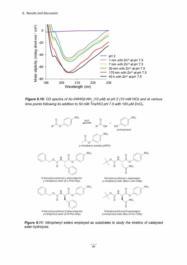
6. Results and discussion
80
Figure 6.10: CD spectra of Ac-IHIHIQI-NH2 (10 µM) at pH 2 (10 mM HCl) and at various
time points following its addition to 50 mM Tris/HCl pH 7.5 with 100 µM ZnCl2.

6. Results and discussion
81
asparagine p-nitrophenyl ester (Boc-Asn-ONp) (Fig. 6.11).
Due to the limited solubility of substrate Z-Phe-ONp in water, pseudo-first-order
kinetic experiments were performed. The production of p-nitrophenolate as mediated
by Ac-IHIHIQI-NH2 in its pre-incubated, fibrillar form and during the process of self-
assembly is shown in Figures 6.12-6.14. Not surprisingly, the hydrolysis of pNPA was
most rapid when treated with fibrils incubated with zinc 24 hours prior to the reaction
(Fig. 6.12). The initial reaction velocity was more than 500 times greater in the
presence of the fibrils than without catalyst. The second-order rate constant at pH 7.5
was determined to be 87 M-1s-1 (Table 6.3), based on the previous finding that two
peptides constitute one active site.188 On the other hand, the second-order rate
constant corresponding to the actively assembling catalyst was just over 40 M-1s-1,
which is in agreement with the results of Korendovych and DeGrado obtained via
saturation kinetics.188 The faster hydrolysis observed in the presence of the
incubated fibrils was due to the greater number of organized and established active
sites as opposed to catalytic peptides still in the process of aggregation.
Figure 6.12: Production of p-nitrophenolate upon Ac-IHIHIQI-NH2-mediated hydrolysis of p-
nitrophenyl acetate (pNPA) in the presence of zinc. Reactions were performed in Tris/HCl
(50 mM with 100 µM ZnCl2) pH 7.5 at 20
oC with 50 µM substrate and 10 µM Ac-IHIHIQI-NH
2.
0
2
4
6
8
10
12
14
0 100 200 300 400 500 600Time (s)
µM
p-n
itro
ph
en
ola
te
pNPA
pNPA with Ac-IHIHIQI-NH2 pre-incubated with Zn
2+
pNPA with actively assembling Ac-IHIHIQI-NH2 and Zn
2+

6. Results and discussion
82
Remarkably, the opposite effect was observed when Z-L-Phe-ONp was employed as
substrate. In this case, Ac-IHIHIQI-NH2 was most active when self-assembly and the
reaction occurred simultaneously (Fig. 6.13). The second-order rate constant of the
actively assembling catalyst acting on Z-L-Phe-ONp at pH 7.5 was found to be 2.2 x
104 M-1s-1, ten-fold higher than the 1.9 x 10³ M-1s-1 rate constant observed with
incubated Ac-IHIHIQI-NH2. The turnover rate (apparent first-order rate constant) of
the freshly prepared variant was more than 70-fold greater than when uncatalyzed.
This finding is perhaps the result of favorable interactions between the hydrophobic
domains of the substrate and hydrophobic portions of the self-assembling catalyst.
As aggregation occurs and the active site is established, the substrate may,
therefore, already be in the vicinity (Fig. 6.16). Such hydrophobic interactions would
be less likely in the fully formed fibril, which has its hydrophobic isoleucine side
chains well buried prior to the approach of substrate. Heightened catalytic efficiency
during self-assembly was not the case when pNPA was employed as a substrate
because it lacks the large hydrophobic groups present in Z-L-Phe-ONp.
Table 6.3: Rate constants corresponding to the hydrolysis of esters pNPA and L- and D-enantiomers of Z-Phe-ONp and Boc-Asn-ONp in the presence of catalytic peptide Ac-IHIHIQI-NH2 in its fully fibrullar form following 24 h incubation with zinc and with zinc in its actively self-assembling form. Reactions were perfomed at 20oC and pH 7.5 (50 mM Tris/HCl with 100 µM ZnCl2) with 50 µM pNPA and 10 µM Z-Phe-ONp and Boc-Asn-ONp. A peptide concentration of 10 µM (2 peptides form 1 active site = 5 µM of catalyst) was used. The apparent k1 is the observed pseudo-first order rate constant and does not account for uncatalyzed background hydolysis

6. Results and discussion
83
The reduced catalysis produced by the fibrillar form of Ac-IHIHIQI-NH2 upon the
substrate Z-L-Phe-ONp may also be due to steric effects. The bulky
benzyloxycarbonyl protecting group and benzyl side-chain of phenylalanine may not
approach the broad surface of fully formed fibrils as easily as it would smaller
assemblies.
Unlike Z-L-Phe-ONp, which was quickly cleaved by the self-assembling form of Ac-
IHIHIQI-NH2, hydrolysis of the more hydrophilic Boc-L-Asn-ONp occurred less rapidly
(Fig. 6.14). In this case, the second-order rate constants corresponding to the
incubated and freshly prepared forms were 1.6 x 10³ M-1s-1 and 2.7 x 10³ M-1s-1,
respectively. Although hydrolysis of Boc-L-Asn-ONp is generally faster than Z-L-Phe-
ONp, kinetic studies showed that Ac-IHIHIQI-NH2, when self-assembling in the
presence of Zn2+, was clearly selective for the more hydrophobic Z-L-Phe-ONp (Fig.
6.15). The ability of Ac-IHIHIQI-NH2 to exhaust the hydrophobic Z-L-Phe-ONp more
than 5 times faster than the more reactive, yet more hydrophilic Boc-L-Asn-ONp
clearly suggests that the hydrophobic nature of the substrate plays a role in the
selectivity of the self-assembling catalytic peptide. Furthermore, substrate selectivity
of Ac-IHIHIQI-NH2 was greatly reduced when the catalyst was allowed to incubate
with divalent zinc 24 hours prior to the initiation of the reaction (Fig. 6.17).68,201
0
2
4
6
8
10
0 50 100 150
µM
p-n
itro
phe
nola
te
Time (s)
Z-L-Phe-ONp with actively assembling Ac-IHIHIQI-NH2 and Zn2+
Z-L-Phe-ONp with Ac-IHIHIQI-NH2 pre-incubated with Zn
2+
Z-L-Phe-ONp
Figure 6.13: Production of p-nitrophenolate upon Ac-IHIHIQI-NH2-mediated hydrolysis of N-benzyloxycarbonyl-L-phenylalanine p-nitrophenyl ester (Z-L-Phe-ONp) in the presence of zinc. Reactions were performed in Tris/HCl (50 mM with 100 µM ZnCl2) pH 7.5 at 20oC with 10 µM substrate and 10 µM Ac-IHIHIQI-NH2.

6. Results and discussion
84
0
2
4
6
8
10
0 50 100 150
Figure 6.15: Production of p-nitrophenolate upon the hydrolysis of Z-L-Phe-ONp and Boc-L-Asn-ONp with Ac-IHIHIQI-NH2 present in the process of fibril formation and growth with zinc. Reactions were performed in Tris/HCl (50 mM with 100 µM ZnCl2) pH 7.5 at 20oC with 10 µM substrate and 10 µM Ac-IHIHIQI-NH2.
µM
p-n
itro
phen
ola
te
Time (s)
0
2
4
6
8
10
0 100 200 300
Figure 6.14: Production of p-nitrophenolate upon Ac-IHIHIQI-NH2-mediated hydrolysis of N-butyloxycarbonyl-L-asparagine p-nitrophenyl ester (Boc-L-Asn-ONp) in the presence of zinc. Reactions were performed in Tris/HCl (50 mM with 100 µM ZnCl2) pH 7.5 at 20oC with 10 µM substrate and 10 µM Ac-IHIHIQI-NH2.
µM
p-n
itro
phe
nola
te
Time (s)
Boc-L-Asn-ONp with actively assembling Ac-IHIHIQI-NH2 and Zn2+
Boc-L-Asn-ONp
Boc-L-Asn-ONp with Ac-IHIHIQI-NH2 pre-incubated with Zn
2+
Boc-L-Asn-ONp with actively assembling Ac-IHIHIQI-NH2 and Zn2+
Z-L-Phe-ONp
Boc-L-Asn-ONp
Z-L-Phe-ONp with actively assembling Ac-IHIHIQI-NH2 and Zn
2+

6. Results and discussion
85
Figure 6.16: Primary Ac-IHIHIQI-NH2 structure (1). Proposed interaction between hydrophobic substrate Z-Phe-ONp and exposed isoleucine residues of the catalytic peptide Ac-IHIHIQI-NH2 during self-assembly (2). Tight packing of the isoleucine residues expels the substrate into the vicinity of the hydrolytic (His)3Zn active site (3).
Z-Phe-ONp
Ac-IHIHIQI-NH2
1. 2. 3.
(His)3Zn active site
0
2
4
6
8
10
0 50 100 150
Time (s)
Figure 6.17: Production of p-nitrophenolate upon the hydrolysis of Z-L-Phe-ONp and Boc-L-Asn-ONp in the presence of fibrillar Ac-IHIHIQI-NH2 incubated with zinc. Reactions were performed in Tris/HCl (50 mM with 100 µM ZnCl2) pH 7.5 at 20oC with 10 µM substrate and 10 µM Ac-IHIHIQI-NH2.
µM
p-n
itro
phen
ola
te Boc-L-Asn-ONp with Ac-IHIHIQI-NH2 pre-incubated with Zn
2+
Z-L-Phe-ONp
Boc-L-Asn-ONp
Z-L-Phe-ONp with Ac-IHIHIQI-NH2 pre-incubated with Zn2+

6. Results and discussion
86
In addition to substrate selectivity, both forms of Ac-IHIHIQI-NH2 in the presence of
zinc were examined for enantioselectivity using Z-D-Phe-ONp and Boc-D-Phe-ONp
as substrates. While actively assembling, Ac-IHIHIQI-NH2 in the presence of zinc
was found to be selective for the L-enantiomer of both substrates (Fig. 6.18 and Fig.
6.19). The ratio of second-order rate constants (k2L:k2D) resulted in enantioselectivity
values (EL/D) of 3.0 and 2.5 for Z-Phe-ONp and Boc-Asn-ONp, respectively. However,
enantioselectivity of Z-L-Phe-ONp (EL/D = 1.1) was greatly diminished when Ac-
IHIHIQI-NH2 was incubated with zinc prior to use (Fig. 6.20). To a lesser extent, this
was also observed with Boc-Asn-ONp (EL/D = 1.7; Fig. 6.21).
Added to its catalytic capability, the simple β-sheet structure of Ac-IHIHIQI-NH2 is
also reasonably selective when carefully applied. The most encountered limitation of
β-sheet structures in de novo design is solubility. The results shown here, hint at
what might be possible, especially in terms of substrate selectivity, if such structures
could be stabilized in solution. Future efforts to stabilize sequences similar to IHIHIQI
between α-helices could afford additional stability and define a three-dimensional
active site enhanced by secondary interactions and additional selectivity.
0
2
4
6
8
10
0 50 100
µM
p-n
itro
phen
ola
te
Time (s)
Figure 6.18: Production of p-nitrophenolate upon the hydrolysis of Z-L-Phe-ONp and Z-D-Phe-ONp with Ac-IHIHIQI-NH2 present in the process of fibril formation and growth with zinc. Reactions were performed in Tris/HCl (50 mM with 100 µM ZnCl2) pH 7.5 at 20oC with 10 µM substrate and 10 µM Ac-IHIHIQI-NH2.
Z-D-Phe-ONp with actively assembling Ac-IHIHIQI-NH2 and Zn
2+
Z-D-Phe-ONp
Z-L-Phe-ONp
Z-L-Phe-ONp with actively assembling Ac-IHIHIQI-NH2 and Zn
2+

6. Results and discussion
87
0
2
4
6
8
10
0 100 200 300
µM
p-n
itro
phe
nola
te
Time (s)
Figure 6.19: Production of p-nitrophenolate upon the hydrolysis of Boc-L-Asn-ONp and Boc-D-Asn-ONp with Ac-IHIHIQI-NH2 present in the process of fibril formation and growth with zinc. Reactions were performed in Tris/HCl (50 mM with 100 µM ZnCl2) pH 7.5 at 20oC with 10 µM substrate and 10 µM Ac-IHIHIQI-NH2.
0
2
4
6
8
10
0 50 100 150
Figure 6.20: Production of p-nitrophenolate upon the hydrolysis of Z-L-Phe-ONp and Z-D-Phe-ONp in the presence of fibrillar Ac-IHIHIQI-NH2 incubated with zinc. Reactions were performed in Tris/HCl (50 mM with 100 µM ZnCl2) pH 7.5 at 20oC with 10 µM substrate and 10 µM Ac-IHIHIQI-NH2.
µM
p-n
itro
phen
ola
te
Time (s)
Boc-D-Asn-ONp with actively assembling Ac-IHIHIQI-NH2 and Zn
2+
Boc-D-Asn-ONp
Boc-L-Asn-ONp
Boc-L-Asn-ONp with actively assembling Ac-IHIHIQI-NH2 and Zn
2+
Z-D-Phe-ONp with Ac-IHIHIQI-NH2 pre-incubated with Zn
2+
Z-D-Phe-ONp
Z-L-Phe-ONp
Z-L-Phe-ONp with Ac-IHIHIQI-NH2 pre-incubated with Zn
2+

6. Results and discussion
88
0
2
4
6
8
10
0 100 200 300
Figure 6.21: Production of p-nitrophenolate upon the hydrolysis of Boc-L-Asn-ONp and Boc-D-Asn-ONp in the presence of fibrillar Ac-IHIHIQI-NH2 incubated with zinc. Reactions were performed in Tris/HCl (50 mM with 100 µM ZnCl2) pH 7.5 at 20oC with 10 µM substrate and 10 µM Ac-IHIHIQI-NH2.
µM
p-n
itro
phe
nola
te
Time (s)
Boc-D-Asn-ONp with Ac-IHIHIQI-NH2 pre-incubated with Zn
2+
Boc-D-Asn-ONp
Boc-L-Asn-ONp
Boc-L-Asn-ONp with Ac-IHIHIQI-NH2 pre-incubated with Zn
2+

6. Results and discussion
89
6.3 De novo peptide hexamer recast as metalloenzyme
Limitations facing the de novo design of catalytic proteins are largely centered on the
inability to position a functional active site within a cleft which also provides sufficient
substrate accommodation. Although peptides offer simple building blocks for bottom-
up design, their small size reduces the number of stabilizing interactions needed to
form and retain a proficiently catalytic species.
While focusing our attention on the de novo design of peptides possessing esterase
activity, we have studied random-coil peptides (Section 6.1) which are just slightly
catalytic as they contain catalytic machinery, but are unable to form a stable structure
or to accommodate substrate. Also, we have studied the catalytic properties of small
β-sheet peptides in the presence of zinc in the process of amyloid formation (Section
6.2). Such assemblies, though reasonably active, tend to lose the ability to
accommodate substrates and, in turn, their substrate selectivity once the fibril is
formed.
When researching possible de novo α-helical designs which to catalyze ester
hydrolysis, particular attention was paid to whether or not the assembly could provide
a cleft for the accommodation of both substrate and water as well as the presentation
of catalytic machinery. To meet these criteria, a coiled-coil assembly featuring
histidine within its hydrophobic core was sought. Since histidine within a hydrophobic
core of a coiled-coil structure is likely to encourage destabilization, particularly when
charged at acidic pH values, the catalytic mechanism employed would need to take
place under basic conditions. Therefore, the use of zinc as applied in the zinc-bound
hydroxide nucleophile mechanism (Figs. 2.4, 3.14) becomes a relevant choice. The
employment of zinc in this case is expected not only to enhance esterase activity, as
was the case in Section 6.2, but might also aid in structural stabilization through the
coordination of histidine within the hydrophobic core at near-neutral pH conditions.
As pointed out by Zastrow et al.,27 an obstacle facing de novo hydrolases based on
the coiled-coil design is the inability to channel water to an active site nestled within
the hydrophobic core. Taking this into account, a coiled-coil structure of a greater
oligomerization state than the trimeric Hg(II)SZn(II)N(TRIL9CL23H)3 (Fig. 4.10)
reported by Zastrow, Pecoraro et al. was envisioned. Ultimately, the de novo peptide
hexamer CC-HexH24 designed in the laboratories of D. N. Woolfson was selected.202

6. Results and discussion
90
The peptide model CC-Hex (Table 6.4) reported by Zaccai et al., is the only parallel
hexameric bundle featuring an interior channel. Although lined exclusively with
hydrophobic leucine and isoleucine residues, the lumen of this coiled coil was shown
by X-ray diffraction to be approximately 6 Å in diameter and water permeable. The
design of this hexamer is based on the same principles discussed in Section 4.1.1.
However, unlike previous models which place charged, salt-bridge-forming residues
at positions e and g, CC-Hex has a neutral alanine at position e while relocating the
salt-bridge-forming residue to position b (Fig. 6.22). This residue rearrangement
results in the conversion of a coiled-coil tetramer into a hexamer. Furthermore,
following the placement of a histidine residue in position a within the lumen, the
peptide was still capable of parallel hexameric coiled-coil formation as shown by its
crystal structure (Fig. 6.23). Unlike CC-Hex, the histidine-containing variant CC-
HexH24 was not found to form an ideal hexameric species.202
Figure 6.22: Helical wheel projection of the parallel hexameric coiled-coil motif CC-Hex. Yellow a and d positions are occupied, respectively, by hydrophobic leucine and isoleucine residues. Interhelical salt bridges occur between positions g (Glu) and b (Lys). Charge neutral alanine residues are located in position e.

6. Results and discussion
91
To study the catalytic esterase activity of CC-HexH24 (Table 6.4), the substrate
pNPA was used. It is important to point out that the substrate pNPA is well-suited for
the assessment of general esterase activity. Unlike many ester substrates, pNPA is
quite soluble in aqueous solutions and its hydrolysis can be easily monitored and
assessed by Michaelis-Menten parameters. However, as already discussed in the
previous section, pNPA offers little information in regard to enzyme substrate affinity.
Initial experiments were performed to test the catalytic ability of CC-HexH24 and the
effect Zn2+ has on the rate of pNPA hydrolysis and secondary peptide structure.
Figure 6.23: Crystal structure of parallel hexameric coiled-coil peptide CC-HexH24 showing the orientation of histidine residues and water (red spheres) within its lumen. Crystallization performed by Zaccai et al.202 Image rendered with RCSB Protein Workshop.58
Table 6.4: Amino acid sequences and heptad register of CC-HexH24, CC-Hex and CC-HexH24 scrambled.

6. Results and discussion
92
Interestingly, at pH 7.5, the effect that varying Zn2+ concentrations had on CC-
HexH24 activity (as shown by initial reaction rate vo) was found to mirror the effect it
had on α-helicity (as shown by molar ellipticity at 222 nm, ϴ222, Fig. 6.24). These
findings indicate that Zn2+ not only directly affects the rate of pNPA hydrolysis when
present within CC-HexH24, but may do so indirectly by increasing the α-helicity of the
peptide structure. In addition, thermal denaturation studies showed that Zn2+
enhances the thermal stability at pH 7.5 of CC-HexH24 (Fig. 6.25).
Under conditions of increased basicity, CC-HexH24 was found to assume an α-
helical structure regardless of zinc present (Fig. 6.26). This is due to the increased
likelihood of histidine being deprotonated or uncharged and, therefore, more stable
within a hydrophobic environment. In other words, zinc coordination is no longer
needed to provide structural stabilization. On the other hand, as the conditions
become more basic, the esterase activity of CC-HexH24 with Zn2+ increases (Fig.
6.27). Furthermore, Figure 6.25 shows that the initial reaction rates begin to plateau
as the Zn2+ concentration approaches 8.3 µM or in this case 1/6 molar equivalent of
CC-HexH24, which suggests that the model’s oligomerization is for the most part
0
0,002
0,004
0,006
0,008
0,01
0,012
0,014
0,016
0 20 40 60 80 100-20
-18
-16
-14
-12
-10
0 20 40 60 80 100
[Θ] 2
22 x
10
-3(d
eg c
m2 d
mol r
es
-1)
Catalytic activity
α-Helicity
Figure 6.24: The mirrored effect of Zn2+ in both increasing the rate of CC-HexH24-catalyzed pNPA hydrolysis and α-helicity at pH 7.5. The initial rates of reaction (vo) were determined in the presence of 50 µM CC-HexH24 and with an initial substrate concentration of 1 mM. All reactions were carried out at 25oC in 50 mM Tris/HCl and 2 % acetonitrile. Values of molar ellipticity at 222 nm, [ϴ]222, were determined under identical conditions.
[Zn2+
] (µM)
vo (
µM
/s)
0.016 0.014 0.012 0.010 0.008

6. Results and discussion
93
maintained in the catalytically active metallo-form. As the mechanism for ester
hydrolysis employs a zinc-bound-hydroxide nucleophile, the pKa of zinc-activated
water was determined to be 9.4 according to catalytic activity as depicted in the
sigmoidal plot of Figure 6.28. This is comparable to lowered water pKa values of 9.3
and 8.8 associated with Ac-IHIHIQI-NH2 and Hg(II)SZn(II)N(TRIL9CL23H)3,
respectively.27,188
0
0,5
1
20 40 60 80 100
CC-Hex-H24 (50 µM) with Zn (100 µM)
CC-Hex-H24 (50 µM) without Zn
Figure 6.25: Thermal denaturation of CC-HexH24 with and without Zn2+ present at pH 7.5 as determined by CD-spectroscopy (θ
222).
rela
tive
denatu
ration
Temperature (oC)
-22
-18
-14
-100 20 40 60 80 100
pH 9.5
pH 8.5
pH 7.5
Figure 6.26: The effect of Zn2+ concentration and pH on α-helicity of CC-HexH24 (50 µM) at 25oC. For pH 8.5 and 7.5, 50 mM Tris/HCl was used as buffer. For pH 9.5, 50 mM carbonate buffer was employed.
[Zn2+
] (µM)
[Θ] 2
22 x
10
-3(d
eg c
m2
dm
ol r
es
-1
)
0.5
CC-HexH24 (50 µM) with Zn2+
(100 µM)
CC-HexH24 (50 µM) without Zn2+

6. Results and discussion
94
In order to show that the esterase activity of CC-HexH24 is due to the positioning of a
functional histidine-coordinated zinc active site within a cleft, two control peptides,
CC-Hex and CC-HexH24 scramble, were synthesized and subjected to kinetic and
structural analysis (CD) at pH 9.5. CC-Hex (Table 6.4) is the model parallel
hexameric coiled coil (Fig. 6.29) and lacks a histidine residue. The control peptide
CC-HexH24 scramble consists of the same amino acids as CC-HexH24, but in a
random sequence. In other words, it contains histidine, but is unlikely to fold into a
vo (
µM
/s)
pH
Figure 6.28: pH-dependency of pNPA (1 mM) hydrolysis at 25oC by CC-HexH24 (50 µM) in the presence of 8.3 µM Zn2+. Reactions at pH 7.5 – 9 were performed in Tris/HCl (50 mM). Reactions above pH 9 were performed in 50 mM carbonate buffer.
Figure 6.27: Effect of Zn2+ concentration and pH on CC-HexH24-mediated (50 µM) ester (pNPA, 1 mM) hydrolysis (25oC). Reactions at pH 7.5 and 8.5 were performed in Tris/HCl (50 mM). Reactions at pH 9.5 were performed in 50 mM carbonate buffer.

6. Results and discussion
95
well-defined structure. As was observed in the case of CC-HexH24 (Fig. 6.26), the
presence of zinc at pH 9.5 did not alter the α-helicity of either CC-Hex or CC-HexH24
scramble (Fig. 6.29). Figure 6.29 clearly shows that the peptide CC-Hex assumes a
more α-helical structure than CC-HexH24. As reported by Zaccai et al.,202 this is the
result of a slight uncoiling (Fig. 6.30) observed at the carboxyl terminus of CC-
HexH24 due to the incorporation of histidine. Furthermore, it has also been pointed
out that unlike the model CC-Hex, CC-HexH24 does not form a single ideal hexamer
as determined from analytical ultracentrifugation studies.202 The presence of more
than one oligomerization state is likely retained in the presence of zinc. Nonetheless,
the CD spectra corresponding to CC-Hex and CC-HexH24 suggest the formation of
α-helical coiled coils, whereas the spectrum for CC-HexH24 scramble corresponds,
as expected, to a random coil.
To offer a clear view of activity, catalytically-cleaved pNPA, that is the concentration
of p-nitrophenolate produced in the reaction with peptide present minus that caused
by the random hydrolysis of an uncatalyzed reaction, was monitored during the
course of the reaction (Fig. 6.31). As predicted, substrate turnover was noticeably
lower (4-5x) in the presence of CC-Hex and CC-HexH24 scramble with and without
zinc, as well as CC-HexH24 without zinc, than CC-HexH24 with zinc. This outcome
clearly shows the catalytic advantage gained when catalytic machinery, particularly
histidine and zinc, is placed within a relatively defined structure.
-40
-30
-20
-10
0
10
20
30
40
195 205 215 225 235
CC-Hex
CC-HexH24
CC-HexH24scramble
-40
-30
-20
-10
0
10
20
30
40
195 205 215 225 235
[Θ] x
10
-3(d
eg c
m2 d
mol r
es
-1)
[Θ] x
10
-3(d
eg c
m2 d
mol r
es
-1)
Figure 6.29: CD spectra of peptides CC-Hex, CC-HexH24 and CC-HexH24 scramble (50 µM) at pH 9.5 (50 mM Na(CO3)2/NaHCO3 with 2% MeCN) and 25oC in the absence of Zn2+ (A) and with Zn2+ (8.3 µM) (B).
λ (nm) λ (nm)
(A) (B)

6. Results and discussion
96
Figure 6.31: pNPA converted into p-nitrophenolate resulting from catalysis during the course of hydrolysis reactions performed in the presence of peptides CC-HexH24, CC-HexH24 scramble and CC-Hex (50 µM) with zinc chloride (8.3 µM) and without at pH 9.5 (50 mM Na(CO3)2/NaHCO3 with 2% acetonitrile) and 25oC. Starting pNPA concentration was 1 mM.
0
2
4
6
8
10
0 10 20 30 40 50
CC-HexH24 with Zn
CC-HexH24
CC-HexH24 scramble with Zn
CC-HexH24 scramble
CC-Hex with Zn
CC-Hex
pN
PA
cata
lyze
d (
µM
)
Time (s)
(B) (A)
Figure 6.30: Cross sections of electrostatic surface of (A) CC-Hex with leucine rendered in heptad position a and (B) CC-HexH24 with histidine 24 occupying a heptad’s a position. C-termini are shown at the top. Adapted from Zaccai et al.202

6. Results and discussion
97
Michaelis-Menten enzyme kinetics was applied to determine the kinetic parameters
of pNPA hydrolysis reactions catalyzed by CC-HexH24 in the presence of zinc. Since
peptide CC-HexH24 does not assemble into a species possessing a single
oligomerization state, quantification of its catalytic parameters becomes more
complicated. However, by using CC-HexH24 without zinc as a blank to account for
both random ester hydrolysis and hydrolysis attributed to histidine residues (general
base or nucleophilic), catalyzed rate constants attributed only to the zinc-bound
hydroxide mechanism can be calculated. As each catalyst using the zinc-bound
hydroxide mechanism has one zinc atom, a limiting zinc concentration was used to
obtain first-order (kcat) and second-order (kcat/KM) rate constants.
In response to increased substrate concentrations, CC-HexH24 in the presence of
zinc adhered to Michaelis-Menten kinetics (Fig. 2.7). The saturation of CC-HexH24
by substrate was observed as the measured values of initial velocity approached a
maximum (VMAX = 0.55 µM/s) at elevated pNPA concentrations (Fig. 6.32). Double-
reciprocal, Lineweaver-Burke plots were used to determine the Michaelis-Menten
parameters kcat, KM and kcat/KM, found to be 0.11 s-1, 3.4 mM and 31.5 ± 0.9 M-1s-1,
respectively. The second-order rate constant of 31.5 M-1s-1 at pH 9.5 compares
favorably to the value reported by Zastrow et al. of 23.3 M-1s-1 corresponding to the
trimeric coiled coil Hg(II)SZn(II)N(TRIL9CL23H)3 (Fig. 4.10).27
Figure 6.32: Esterase activity catalyzed by CC-HexH24
(30 µM) in the presence of 5 µM Zn2+
showing enzyme-like saturation at elevated substrate (pNPA) concentrations. Reactions
were performed at pH 9.5 (50 mM Na(CO3)2/NaHCO
3 with 2% acetonitrile) and 25
oC.
0
0,05
0,1
0,15
0,2
0,25
0,3
0 1000 2000 3000
[pNPA] (µM)
vo (
µM
/s)
0.30 0.25 0.20 0.15 0.10 0.05

6. Results and discussion
98
The catalytic capacity of CC-HexH24 with zinc reported here is only the beginning.
Research which addresses substrate selectivity using substrates of varying size,
hydrophobicity and chirality remains to be performed. It would be especially
interesting to observe the hydrolysis of the enantiomers of substrates such as Z-Leu-
ONp or Z-Ile-ONp as they insert themselves within the substrate cleft rich with L-
leucine and L-isoleucine residues. In addition, the catalyst CC-HexH24 has yet to be
optimized. Mutations including those resulting from the repositioning of histidine
along the inner channel of CC-HexH24 or from the substitution of other residues
which interact with the active site would likely affect properties such as catalytic
proficiency and substrate selectivity. Ultimately, the peptide CC-HexH24 as it
functions with zinc represents a promising candidate for future design ideas in the
progression toward more active and practical de novo protein catalysts.

7. Summary
99
7 Summary
The ability of enzymes to accelerate reactions while maintaining excellent substrate
selectivity and product specificity has interested chemists for decades. The
adaptation of enzymes to the reaction conditions of chemical synthesis has, however,
been met with numerous obstacles such as reduced solubility, thermostability and
substrate scope. To overcome these obstacles, protein engineering employs
methods of rational design and directed evolution as well as more modern
computational and combinatorial approaches. Although these techniques have been
successfully applied to alter biocatalysts, our understanding of protein folding and the
interactions required for enzyme catalysis remains limited. This is best evidenced by
the inability to design de novo catalytic proteins comparable to enzymes.
To develop a better understanding of enzyme structure and function, the “bottom-up”
approach to the de novo design of catalytic proteins is applied. This minimalistic
approach uses simple peptides which predictably self-assemble into well-defined
three-dimensional templates, on which catalytic machinery or other functionalities
believed to impart a particular property are introduced. The simplified, yet protein-
like, environments of the self-assembling peptides allow for enhanced clarity of the
individual interactions to assess how such interactions contribute to particular
enzymatic properties.
In the work presented here, the bottom-up technique was employed to introduce
enzyme-like properties into three model peptide systems. Particular properties of
interest include catalytic activity, substrate selectivity and affinity as well as the ability
to regulate catalytic activity by altering conformation. As a model reaction, ester
hydrolysis was selected due in large part to the ease by which such reactions can be
kinetically monitored as well as the vast amount of studies and comparable data
available in literature.
In an effort to design a catalytic peptide system in which the catalytic activity is
regulated according to conformation, the heterodimeric coiled-coil model system
E3/K3 was applied. The original design incorporated the catalytic triad (serine,
histidine and aspartate) in proximal positions of the coiled coil to initiate a serine
protease-like charge relay resulting in accelerated ester hydrolysis. Initial studies
showed the moderate activity of the unfolded, carboxylate-rich E3CT peptide

7. Summary
100
containing the catalytic triad substitution. Almost no activity was observed in E3CT
when folded into a coiled coil in the presence of K3. Further studies, particularly with
the variant E3H11, in which only histidine is substituted, showed that the activity of
the random coil is due to general base catalysis resulting from the interaction of
histidine with glutamate. Such interactions are greatly reduced when peptides
containing histidine are locked into a coiled coil. To develop the enzyme-like
properties of E3H11, further efforts which include fixing it to a support such as hyper-
branched polyglycerol (hPG) or gold monolayer-protected clusters (Au-MPC) may be
taken.
In a separate study, the catalytically proficient, amyloid-forming peptide Ac-IHIHIQI-
NH2, which features the zinc-bound hydroxide mechanism (ZHN) of hydrolysis, was
examined for properties of substrate selectivity. These studies revealed that Ac-
IHIHIQI-NH2 was especially selective for the hydrophobic substrate Z-L-Phe-ONp
over the more hydrophilic Boc-L-Asn-ONp while in the process of fibril assembly.
Furthermore, Ac-IHIHIQI-NH2 showed increased enantioselectivity for the L-
enantiomer of Z-Phe-ONp in the process of self-assembly as opposed to when it is in
the form of a fully-established fibril. Such findings are likely the result of increased
hydrophobic interactions occurring between hydrophobic moieties of the substrate
and hydrophobic surfaces of the peptide, which experience greater exposure while in
the process of fibril assembly. Future efforts to stabilize this β-sheet active site within
a larger protein structure would provide interesting results.
In the final study, the ZHN mechanism applied in Ac-IHIHIQI-NH2 was incorporated
into the hexameric α-helical coiled coil CC-HexH24, which features six adjacent
histidine residues in its hydrophobic interior. The coordination of zinc within a water-
accessible, substrate accommodating cleft was found to greatly accelerate ester
hydrolysis. However, further structural studies including crystallography should be
pursued to obtain a detailed image of the active-site. In addition, substrate selectivity
studies involving substrates varying in hydrophobicity and size would also contribute
a wealth of information in regard to active-site design.
These studies represent a growing understanding in protein structure and function
through the use of peptide design. As our knowledge of such molecules and their
interactions develop so will the complexity of catalytic proteins designed de novo
from the bottom up.

8
Materials and methods


8. Materials and methods
103
8.1 Peptide synthesis and verification
Peptides were synthesized using the Fmoc (fluorenylmethoxycarbonyl) method of
solid phase peptide synthesis (SPPS),203 purified via reverse-phase high-pressure
liquid chromatography (RP-HPLC) and characterized with analytical HPLC and mass
spectrometry. A list of peptides discussed in this work along with corresponding
sequences and mass analyses is shown in Table 8.2.
8.1.1 Fmoc solid phase peptide synthesis
In solid phase peptide synthesis, amino acids are coupled to a growing peptide chain
attached to a solid support, typically an organic polymer. Following each reaction, the
peptide linked to the insoluble support can be easily purified from soluble reagents by
filtration and repeated washing. Not only does this save time and labor, but allows
the use of excess reagents, which translates into both increased peptide yield and
purity.
Since the carboxyl group of first amino acid in linked to the support, synthesis of
peptides on solid phase starts from the carboxyl terminus and proceeds in the
direction of the amino terminus. The reaction scheme of peptide synthesis involves
repetitive deprotection and coupling reactions (Fig. 8.1) which are separated by
thorough washing.
Figure 8.1: Reaction scheme of solid phase peptide synthesis. Courtesy of Chan & White.203

8. Materials and methods
104
Each of the peptides employed in this work were synthesized using the Fmoc or
Fmoc/tBu method of solid phase peptide synthesis. This strategy uses 9-
fluorenylmethoxycarbonyl (Fmoc) as the temporary amine protecting group (X in
Figure 8.1). Deprotection of Fmoc was carried out under basic conditions. Coupling
reactions are performed upon the formation of active esters (Fig. 8.1 A) of the next
amino acid. Once the final amino acid of the sequence has been coupled, the
permanent side-chain protecting groups (Y in Fig. 8.1) are removed and the peptide
is simultaneously cleaved from the solid support under acidic conditions.
8.1.1.1 Materials for Fmoc SPPS
Fmoc- and side-chain-protected amino acids: Fmoc-L-Ala-OH·H2O, Fmoc-L-
Asp(OtBu)-OH·H2O, Fmoc-L-Glu(OtBu)-OH·H2O, Fmoc-L-Gln(Trt)-OH, Fmoc-L-Gly-
OH, Fmoc-L-His(Trt)-OH, Fmoc-L-Ile-OH, Fmoc-L-Leu-OH, Fmoc-L-Lys(Boc)-
OH, Fmoc-L-Ser(OtBu)-OH and Fmoc-Trp(Boc)-OH were purchased from Orpegen
Pharma.
Solid support resins: Fmoc-Glu(OtBu)-NovaSyn® TGA was used in the syntheses
of K3 variants and peptide EEEHEEE. Fmoc-Lys(Boc)-NovaSyn® TGA was
employed in the syntheses of all E3 variants. Fmoc-Gly-NovaSyn® TGT was used in
each of the CC-Hex variants and Rink Amide MBHA resin (100-200 mesh) for Ac-
IHIHIQI-NH2. Each of the resins was obtained from Novabiochem.
Solvents: Dimethylformamide (DMF, 99.9%; VWR), dichloromethane (DCM, ≥99%;
Fischer) and N-methyl-2-pyrrolidone (NMP, 99%; ACROS) were used without further
purification.
Reagents for Fmoc deprotection: Piperidine (99%; ACROS) and 1,8-
diazabicyclo[5.4.0]undec-7-ene (DBU, ≥ 98%; Merck).
Coupling reagents: N,N’-Diisopropylcarbodiimide (DIC, 99%; Sigma Aldridge), 1-
hydroxy-7-azabenzotriazole (HOAt, 99%; Carbolution), N-hydroxybenzotriazole
(HOBt, 99%; Carbolution), O-(6-chlorobenzotriazol-1-yl)-N,N,N′,N′-
tetramethyluronium hexafluorophosphate (HCTU, 99%; Activotec), 2-(1H-
benzotriazol-1-yl)-1,1,3,3,-tetramethyluronium tetrafluoroborate (TBTU, 99%;

8. Materials and methods
105
Carbolution) and N,N-diisopropylethylamine (DIPEA, ≥ 98%; ACROS) were used as
purchased.
Reagents for resin cleavage and side-chain deprotection: Trifluoroacetic acid
(TFA, 99%; Merck) and triisopropylsilane (TIPS, 98%; Alfa Aesar) were used without
further purification.
HPLC Eluents: Acetonitrile (MeCN, HPLC grade; VWR), trifluoroacetic acid (TFA,
HPLC grade; Merck). Water was obtained from a Milli-Q® Advantage A10 purification
system.
Additional materials: Boc-Abz-OH (Bachem), acetic anhydride (Fischer, distilled
prior to use), 4-pentynoic acid (98%; Alfa Aesar), CuSO4 · 5 H2O (≥ 98%; Sigma-
Aldrich), sodium ascorbate (≥ 98%; Sigma-Aldrich), dialysis tubes (MWCO = 3.5-5
kD; Spectrum Labs®), diethyl ether (VWR, 99%). Water was obtained from a Milli-Q®
Advantage A10 purification system.
8.1.1.2 Manual Fmoc SPPS protocol
Resin (preloaded or not) was added to a polypropylene syringe reaction vessel
furnished with a polytetrafluoroethylene (PTFE) frit and allowed to swell 20 minutes in
DMF. As is always the case before proceeding to the next step, the solution or
solvent was pushed through the frit. Fmoc was removed from the preloaded resin by
shaking in a solution of 2% piperidine, 2% DBU in DMF for 10 min (3x). After
sufficient washing with DMF, coupling was performed by one of the two methods
shown below:
(1) DIC/HOBt or HOAt: Fmoc-amino acid (5 equiv.) and HOBt or HOAt (5 equiv.)
are dissolved in approximately 6 mL DMF. Upon addition of 4.9 equiv. DIC, a
10 min. carboxylic acid activation period was allowed prior to being added to
the resin. The vessel was agitated for at least 40 min.
(2) HCTU/HOBt/DIPEA: Fmoc-amino acid (5 equiv.), HOBt (5 equiv.) and HCTU
(4.9 equiv.) were dissolved in approximately 6 mL DMF. Upon treatment with
DIPEA (10 equiv.), the solution was added immediately to the resin. The
vessel was agitated for at least 40 min.

8. Materials and methods
106
To ensure completion, coupling reactions were performed twice. After coupling, the
resin was again washed with DMF and the Fmoc group from the newly coupled
amino acid was removed. Repeated Fmoc-removal, washing, coupling and washing
cycles were carried out until the last amino acid of the sequence was added.
8.1.1.3 Automated Fmoc SPPS protocol
Automated peptide syntheses of E3 and K3 variant peptides were performed by
SyroXP manufactured by Multi-SynTec GmbH. The process of the automated cycle
was very similar to that of the manual process (Fmoc-deprotection, washing,
coupling, washing). However, the SyroXP is best suited for reaction scales of 0.5 µM
performed in 10 mL PTFE fritted, polypropylene vessels. The reaction cycle of
SyroXP including the reagents used and the time of each step is summarized in
Table 8.1.
Table 8.1: Fmoc-deprotection and coupling procedure employed in a cycle by the fully-automated SyroXP by Multi-SynTech GmbH.

8. Materials and methods
107
Syntheses of peptides CC-Hex and CC-HexH24 scramble were performed using the
fully-automated peptide synthesizer Activo-P11 manufactured by ActivoTec which is
best suited for performing reaction up to 0.1 mmol scale. The reaction cycle of
employed with Activo-P11 is summarized in Table 8.2.
8.1.1.4 N-terminal acetylation
Capping or acetylation is a common method employed to block the reactivity of the
amino-terminus. Acetylation in the preparation of Ac-IHIHIQI-NH2 was performed
following the Fmoc-deprotection of the last isoleucine residue coupled. Thus, resin-
attached peptide was treated with a solution of 10% freshly distilled acetic anhydride
and 10% DIPEA in DMF twice for 10 min.
8.1.1.5 N-terminal alkyne functionalization
In order to perform copper-catalyzed alkyne-azide cycloaddition, peptides E3, E3H11
and E3H11-Ala were each coupled to 4-pentynoic acid. Coupling of pentynoic acid
took place at the N-terminus of the peptide following Fmoc cleavage of the last
residue while the peptide was on solid support with side-chain protecting groups
intact. 4-Pentynoic acid (5 equiv.), HOBt (5 equiv.) and HCTU (4.9 equiv.) were
Table 8.2: Fmoc-deprotection and coupling procedure employed in a cycle by the fully-automated Activo-P11 by ActivoTec.

8. Materials and methods
108
dissolved in approximately 6 mL DMF. Upon treatment with DIPEA (10 equiv.), the
solution was drawn immediately to the resin within the PTFE reaction vessel. The
vessel was agitated for at least 40 min. This procedure was performed twice to
ensure complete coupling. Following sufficient washing with DMF, the alkyne
functionalized E3yne, E3H11yne and E3H11-Alayne were cleaved from the resin.
8.1.1.6 Peptide cleavage from resin and trituration
Prior to cleavage and the deprotection of the side chains, the resin was washed with
DCM in order to remove the less volatile DMF.204 For each peptide, cleavage was
carried out by treating the resin with a cocktail containing 95% TFA, 3% TIS and 2%
H20 (1 mL cocktail per 100 mg resin) for 4 h. The cocktail containing the cleaved
peptide was then collected in a round-bottom flask and concentrated under a stream
of argon. Crude peptide was collected via trituration in ice-cold diethyl ether and
centrifugation. The resulting pellet containing the crude peptide was resuspended
and centrifuged several times with cold ether. After decanting the ether from the final
washing, the remaining ether was removed from the pellet in vacuo (400 mbar).
8.1.2 Peptide analysis and purification via HPLC
Crude peptide was dissolved in aqueous MeCN with 0.1% TFA and purified by RP-
HPLC using a high-pressure gradient system supplied by Knauer. The system was
outfitted with a Smartline Manager 5000 user interface with solvent degasser, two
Smartline 1000 eluent pumps, an adjustable UV-detector 2500, eluent gradient
mixer, and a 5 mL sample injection loop. As eluent, gradients of water and MeCN
each with 0.1% TFA were used at a flowrate of 20 mL/min. Chromatography of
peptide variants E3 and K3 were performed using the column Phenomenex® Gemini-
NX C18 (10 µM; 250 x 21.20 mm) . Peptide variants of CC-Hex were purified with the
column Phenomenex® Luna C8 (10 µM; 250 x 21.20 mm) and peptide Ac-IHIHIQI-
NH2 with Phenomenex® Jupiter C4 (10 µM; 250 x 21.20 mm). Fractions were
collected corresponding to UV-absorption recorded with a Perkin Elmer 56 analog
blotter. Collected fractions were then characterized by analytical HPLC and mass
spectrometry.

8. Materials and methods
109
Peptide purity was assessed using the analytical system Elite LaChrome from VWR-
Hitachi or the system Chromaster from VWR-Hitachi. Elite LaChrome was furnished
with two L-2130 pumps, diode array detector L-2485, autosampler L-2200, solvent
degasser L7612 and 100 µL injection loop. Chromatography proceeded with a
flowrate of 1 mL/min through the column Phenomenex® Kinetix C18 (5 µM; 250 x 4.6
mm). The system Chromaster was outfitted with a single 5160 pump, diode array
detector 5430, autosampler 5260, column oven 5310, solvent degasser and 20 µL
injection loop. Chromatography was carried out with a flowrate of 0.6 mL/min through
the column Purospher® STAR C18 (2 µM; 50 x 2.1 mm) from Merck. The software
interface employed by both analytical systems was EZChrom Elite developed by
Agilent.
Following purification, all peptides were lyophilized with a Christ Alpha 1-2 LD freeze
dryer. All peptides were stored at -20oC when not in use.
8.1.3 Synthesis and purification details of individual peptides
E3: H2N-Abz-EIAALEKEIAALEKEIAALEK-OH
The peptide E3 was synthesized on Fmoc-Lys(Boc)-NovaSyn® TGA resin (0.20
mmol/g loading). Peptide E3 was synthesized on separate occasions, once
employing a 0.1 mmol scale using the manual couplinga method
(8.1.1.2 with DIC and HOBt/HOAt) and twice using the automated SyroXP procedure2
(Table 8.1) with a reaction scale of 0.05 mmol (8.1.1.3). When employing the
automated procedure, the final residue, Boc-protected 2-aminobenzoic acid, was
manually coupled using the standard procedure. The peptide was cleaved from the
resin, deprotected and triturated as described in Section 8.1.1.6. HPLC analysis was
performed as described in Section 8.1.2 using the column Phenomenex® Kinetix C18
(5 µM; 250 x 4.6 mm) with an eluent gradient which starts at 5% MeCN in water and
increases to 70% over 30 minutes. Using this method, the retention time of E3 was
26.0 minutes. The same gradient was employed to purify E3 via preparative HPLC
(8.1.2) using a Phenomenex® Gemini-NX C18 column (10 µM; 250 x 21.20 mm). The
retention time for purification was 24.1 minutes. Individual fractions collected from
1 Performed by Sabrina Lehmann under the author’s supervision
2 Performed by Jennifer Hoffmann under the author’s supervision

8. Materials and methods
110
preparative HPLC were further assessed for purity using analytical HPLC (Appendix:
Section 10). Following HPLC and removal of solvent, yields of 86.9 mg resulting from
manual synthesis as well as 18.3 mg and 24.6 mg from automated synthesis were
obtained.
K3: H2N-Abz-KIAALKEKIAALKEKIAALKE-OH
The peptide K3 was synthesized on Fmoc-Glu(tBu)-NovaSyn® TGA resin (0.24
mmol/g loading). K3 was synthesized on separate occasions, once using a 0.1 mmol
scale using the manual coupling method3 (8.1.1.2 with DIC and HOBt/HOAt) and
twice using the automated SyroXP procedure4 (Table 8.1) with a reaction scale of
0.05 mmol (8.1.1.3). When employing the automated procedure, the final residue,
Boc-protected 2-aminobenzoic acid, was manually coupled using the standard
protocol. The peptide was cleaved from the resin, deprotected and triturated as
described in Section 8.1.1.6. HPLC analysis was performed as described in Section
8.1.2 using the column Phenomenex® Kinetix C18 (5 µM; 250 x 4.6 mm) with an
eluent gradient which starts at 5% MeCN in water and increases to 70% over 30
minutes. Using this method the retention time of K3 was 19.8 minutes. The same
gradient was employed to purify K3 via preparative HPLC (8.1.2) using a
Phenomenex® Gemini-NX C18 column (10 µM; 250 x 21.20 mm). The retention time
for purification was 18.6 minutes. Individual fractions collected from preparative
HPLC were further assessed for purity using analytical HPLC (Appendix: Section 10).
Following HPLC and removal of solvent, yields of 53.3 mg via manual synthesis as
well as 45.4 mg and 32.1 mg by automated synthesis were obtained.
E3CT: H2N-Abz-EIAALEDEIAHLEKSIAALEK-OH
The peptide E3CT was synthesized on Fmoc-Lys(Boc)-NovaSyn® TGA resin (0.20
mmol/g loading). E3CT was synthesized on separate occasions, once employing a
0.1 mmol scale using the manual coupling methodb (8.1.1.2 with DIC and
HOBt/HOAt) and twice using the automated SyroXP procedure (Table 8.1) with a
reaction scale of 0.05 mmol (8.1.1.3). When employing the automated procedure, the
3 Performed by Sabrina Lehmann under the author’s supervision
4 Performed by Jennifer Hoffmann under the author’s supervision

8. Materials and methods
111
final residue, Boc-protected 2-aminobenzoic acid, was manually coupled using the
standard procedure. The peptide was cleaved from the resin, deprotected and
triturated as described in Section 8.1.1.6. HPLC analysis was performed as
described in Section 8.1.2 using the column Phenomenex® Kinetix C18 (5 µM; 250 x
4.6 mm) with an eluent gradient which starts at 5% MeCN in water and increases to
70% over 30 minutes. Using this method the retention time of E3CT was 25.5
minutes. The same gradient was employed to purify E3CT via preparative HPLC
(8.1.2) using the column Phenomenex® Gemini-NX C18 (10 µM; 250 x 21.20 mm).
The retention time for purification was 23.9 minutes. Individual fractions collected
from preparative HPLC were further assessed for purity using analytical HPLC
(Appendix: Section 10). Following HPLC and removal of solvent, yields of 78.0 mg
resulting from manual synthesis and 16.2 mg from automated synthesis were
obtained.
K3CT: H2N-Abz-KIAALKDKIAHLKESIAALKE-OH
The peptide K3CT was synthesized on Fmoc-Glu(tBu)-NovaSyn® TGA resin (0.24
mmol/g loading). K3CT was prepared on separate occasions, once employing a 0.1
mmol scale using the manual coupling method5 (8.1.1.2 with DIC and HOBt/HOAt)
and once using the automated SyroXP procedure (Table 8.1) with a reaction scale of
0.05 mmol (8.1.1.3). When employing the automated procedure, the final residue,
Boc-protected 2-aminobenzoic acid, was manually coupled using the standard
procedure. The peptide was cleaved from the resin, deprotected and triturated as
described in Section 8.1.1.6. HPLC analysis was performed as described in Section
8.1.2 using the column Phenomenex® Kinetix C18 (5 µM; 250 x 4.6 mm) with an
eluent gradient which starts at 5% MeCN in water and increases to 70% over 30
minutes. Using this method the retention time of K3CT was 19.3 minutes. The same
gradient was employed to purify K3CT via preparative HPLC (8.1.2) using the column
Phenomenex® Gemini-NX C18 (10 µM; 250 x 21.20 mm). The retention time for
purification was 18.2 minutes. Individual fractions collected from preparative HPLC
were further assessed for purity using analytical HPLC (Appendix: Section 10).
5 Performed by Sabrina Lehmann under the author’s supervision

8. Materials and methods
112
Following HPLC and removal of solvent, yields of 19.8 mg by manual synthesis and
46.7 mg resulting from automated synthesis were obtained.
E3H11: H2N-Abz-EIAALEKEIAHLEKEIAALEK-OH
The peptide E3H11 was synthesized on Fmoc-Lys(Boc)-NovaSyn® TGA resin (0.20
mmol/g loading). E3H11 was synthesized on separate occasions, once employing a
0.1 mmol scale using the manual coupling method (8.1.1.2 with DIC and HOBt/HOAt)
and twice using the automated SyroXP procedure6 (Table 8.1) with a reaction scale of
0.05 mmol (8.1.1.3). When employing the automated procedure, the final residue,
Boc-protected 2-aminobenzoic acid, was manually coupled using the standard
procedure. The peptide was cleaved from the resin, deprotected and triturated as
described in Section 8.1.1.6. HPLC analysis was performed as described in Section
8.1.2 using the column Phenomenex® Kinetix C18 (5 µM; 250 x 4.6 mm) with an
eluent gradient which starts at 5% MeCN in water and increases to 70% over 30
minutes. Using this method the retention time of E3H11 was 25.1 minutes. The same
gradient was employed to purify E3H11 via preparative HPLC (8.1.2) using the
column Phenomenex® Gemini-NX C18 (10 µM; 250 x 21.20 mm). The retention time
for purification was 23.5 minutes. Individual fractions collected from preparative
HPLC were further assessed for purity using analytical HPLC (Appendix: Section 10).
Following HPLC and the removal of solvent, yields of 86.3 mg by manual synthesis
as well as 12.7 mg and 15.2 mg by automated synthesis were obtained.
E3S15: H2N-Abz-EIAALEKEIAALEKSIAALEK-OH
The peptide E3S15 was synthesized on Fmoc-Lys(Boc)-NovaSyn® TGA resin (0.20
mmol/g loading) using the automated SyroXP proceduref (Table 8.1) with a reaction
scale of 0.05 mmol (8.1.1.3). When employing the automated procedure, the final
residue, Boc-protected 2-aminobenzoic acid, was manually coupled using the
standard procedure. The peptide was cleaved from the resin, deprotected and
triturated as described in Section 8.1.1.6. HPLC analysis was performed as
described in Section 8.1.2 using the column Phenomenex® Kinetix C18 (5 µM; 250 x
6 Performed by Jennifer Hoffmann under the author’s supervision

8. Materials and methods
113
4.6 mm) with an eluent gradient which starts at 5% MeCN in water and increases to
70% over 30 minutes. Using this method the retention time of E3S15 was 25.9
minutes. The same gradient was employed to purify E3S15 via preparative HPLC
(8.1.2) using the column Phenomenex® Gemini-NX C18 (10 µM; 250 x 21.20 mm).
The retention time for purification was 24.0 minutes. Individual fractions collected
from preparative HPLC were further assessed for purity using analytical HPLC
(Appendix: Section 10). Following HPLC and the removal of solvent, a yield of 22.2
mg was obtained.
E3H11-Ala: H2N-Abz-KAEAAAAEAEAAHAEAAAAEAK-OH
The peptide E3H11-Ala was synthesized on Fmoc-Lys(Boc)-NovaSyn® TGA resin
(0.20 mmol/g loading) using the automated SyroXP procedure (Table 8.1) with a
reaction scale of 0.05 mmol (8.1.1.3). When employing the automated procedure, the
final residue, Boc-protected 2-aminobenzoic acid, was manually coupled using the
standard procedure. The peptide was cleaved from the resin, deprotected and
triturated as described in Section 8.1.1.6. HPLC analysis was performed as
described in Section 8.1.2 using the column Phenomenex® Kinetix C18 (5 µM; 250 x
4.6 mm) with an eluent gradient which starts at 5% MeCN in water and increases to
70% over 18 minutes. Using this method the retention time of E3H11-Ala was 12.8
minutes. A gradient of 5 to 70% acetonitrile was employed over 30 minutes to purify
E3H11-Ala via preparative HPLC (8.1.2) using the column Phenomenex® Gemini-NX
C18 (10 µM; 250 x 21.20 mm). The retention time for purification was 22.5 minutes.
Individual fractions collected from preparative HPLC were further assessed for purity
using analytical HPLC (Appendix: Section 10). Following HPLC and solvent removal,
a yield of 15.2 mg was obtained.
EEEHEEE: H2N-Abz-EEEHEEE-OH
The peptide EEEHEEE was synthesized on Fmoc-Glu(tBu)-NovaSyn® TGA resin
(0.24 mmol/g loading) using the manual coupling procedure at a reaction scale of 0.1
mmol (8.1.1.2). The peptide was cleaved from the resin, deprotected and triturated
as described in Section 8.1.1.6. HPLC analysis was performed as described in
Section 8.1.2 using the column Phenomenex® Kinetix C18 (5 µM; 250 x 4.6 mm) with

8. Materials and methods
114
an eluent gradient which starts at 0% MeCN in water and increases to 45% over 22
minutes. Using this method the retention time of EEEHEEE was 13.8 minutes. A
gradient of 0 to 45% acetonitrile was employed over 30 minutes to purify EEEHEEE
via preparative HPLC (8.1.2) using the column Phenomenex® Gemini-NX C18 (10
µM; 250 x 21.20 mm). The retention time for purification was 21.0 minutes. Individual
fractions collected from preparative HPLC were further assessed for purity using
analytical HPLC (Appendix: Section 10). Following HPLC and the removal of solvent,
a yield of 48.4 mg was obtained.
E3yne: Alkyne functionalized E3
The peptide E3yne was synthesized on Fmoc-Lys(Boc)-NovaSyn® TGA resin (0.20
mmol/g loading) using the manual coupling procedure at a reaction scale of 0.2 mmol
(8.1.1.2). Following the Fmoc deprotection of the N-terminus, the coupling of 4-
pentynoic acid was carried out as described in Section 8.1.1.5. The peptide was
cleaved from the resin, deprotected and triturated as described in Section 8.1.1.6.
HPLC analysis was performed as described in Section 8.1.2 using the column
Phenomenex® Kinetix C18 (5 µM; 250 x 4.6 mm) with an eluent gradient which starts
at 5% MeCN in water and increases to 100% over 18 minutes. Using this method the
retention time of E3yne was 14.4 minutes. A gradient of 15 to 100% acetonitrile was
employed over 40 minutes to purify E3yne via preparative HPLC (8.1.2) using the
column Phenomenex® Gemini-NX C18 (10 µM; 250 x 21.20 mm). The retention time
for purification was 33.5 minutes. Individual fractions collected from preparative
HPLC were further assessed for purity using analytical HPLC (Appendix: Section 10).
Following HPLC and the removal of solvent, a yield of 127.4 mg was obtained.
E3H11yne: Alkyne functionalized E3H11
The peptide E3H11yne was synthesized on Fmoc-Lys(Boc)-NovaSyn® TGA resin
(0.20 mmol/g loading) using the manual coupling procedure at a reaction scale of 0.2
mmol (8.1.1.2). Following the Fmoc deprotection of the N-terminus, the coupling of 4-
pentynoic acid was carried out as described in Section 8.1.1.5. The peptide was
cleaved from the resin, deprotected and triturated as described in Section 8.1.1.6.
HPLC analysis was performed as described in Section 8.1.2 using the column

8. Materials and methods
115
Phenomenex® Kinetix C18 (5 µM; 250 x 4.6 mm) with an eluent gradient which starts
at 5% MeCN in water and increases to 100% over 18 minutes. Using this method the
retention time of E3H11yne was 13.8 minutes. A gradient of 15 to 100% acetonitrile
was employed over 40 minutes to purify E3H11yne via preparative HPLC (8.1.2)
using the column Phenomenex® Gemini-NX C18 (10 µM; 250 x 21.20 mm). The
retention time for purification was 32.0 minutes. Individual fractions collected from
preparative HPLC were further assessed for purity using analytical HPLC (Appendix:
Section 10). Following HPLC and solvent removal, a yield of 147.0 mg was obtained.
E3H11-Alayne: Alkyne functionalized E3H11-Ala
The peptide E3H11-Alayne was synthesized on Fmoc-Lys(Boc)-NovaSyn® TGA resin
(0.20 mmol/g loading) using the manual coupling procedure at a reaction scale of 0.2
mmol (8.1.1.2). Following the Fmoc deprotection of the N-terminus, the coupling of 4-
pentynoic acid was carried out as described in Section 8.1.1.5. The peptide was
cleaved from the resin, deprotected and triturated as described in Section 8.1.1.6.
HPLC analysis was performed as described in Section 8.1.2 using the column
Phenomenex® Kinetix C18 (5 µM; 250 x 4.6 mm) with an eluent gradient which starts
at 5% MeCN in water and increases to 100% over 18 minutes. Using this method the
retention time of E3H11-Alayne was 10.7 minutes. A gradient of 15 to 100%
acetonitrile was employed over 40 minutes to purify E3H11-Alayne via preparative
HPLC (8.1.2) using the column Phenomenex® Gemini-NX C18 (10 µM; 250 x 21.20
mm). The retention time for purification was 27.5 minutes. Individual fractions
collected from preparative HPLC were further assessed for purity using analytical
HPLC (Appendix: Section 10). Following HPLC and solvent removal, a yield of 122.5
mg was obtained.
Ac-IHIHIQI-NH2
The peptide Ac-IHIHIQI-NH2 was synthesized on Rink Amide MBHA resin (100-200
mesh) resin (0.64 mmol/g loading) using the manual coupling procedure at a reaction
scale of 0.2 mmol (8.1.1.2). Following the final Fmoc deprotection, the N-terminus
underwent acetylation as mentioned in Section 8.1.1.4. The peptide was cleaved
from the resin, deprotected and triturated as described in Section 8.1.1.6. HPLC

8. Materials and methods
116
analysis was performed as described in Section 8.1.2 using the column
Phenomenex® Kinetix C18 (5 µM; 250 x 4.6 mm) with an eluent gradient which starts
at 10% MeCN in water and increases to 100% over 18 minutes. Using this method
the retention time of Ac-IHIHIQI-NH2 was 10.6 minutes. A gradient of 30 to 100%
acetonitrile was employed over 30 minutes to purify Ac-IHIHIQI-NH2 via preparative
HPLC (8.1.2) using the column Phenomenex® Jupiter C4 (10 µM; 250 x 21.20 mm).
The retention time for purification was 14.0 minutes. Individual fractions collected
from preparative HPLC were further assessed for purity using analytical HPLC
(Appendix: Section 10). Following HPLC and solvent removal, a yield of 95.2 mg was
obtained.
CC-HexH24: H2N-GELKAIAQELKAIAKELKAIAWEHKAIAQGAG-OH
The peptide CC-HexH24 was synthesized on Fmoc-Gly-NovaSyn® TGT resin (0.63
mmol/g loading) using the manual coupling procedure at a reaction scale of 0.2 mmol
(8.1.1.2). The peptide was cleaved from the resin, deprotected and triturated as
described in Section 8.1.1.6. HPLC analysis was performed as described in Section
8.1.2 using the column Phenomenex® Kinetix C18 (5 µM; 250 x 4.6 mm) with an
eluent gradient which starts at 5% MeCN in water and increases to 95% over 18
minutes. Using this method the retention time of CC-HexH24 was 13.3 minutes. A
gradient of 15 to 95% acetonitrile was employed over 30 minutes to purify CC-
HexH24 via preparative HPLC (8.1.2) using the column Phenomenex® Luna C8 (10
µM; 250 x 21.20 mm). The retention time for purification was 22.5 minutes. Individual
fractions collected from preparative HPLC were further assessed for purity using
analytical HPLC (Appendix: Section 10). Following HPLC and solvent removal, a
yield of 156.5 mg was obtained.
CC-HexH24 scramble: H2N-EEKAHKLGAKIGLIQEAIAAKAWEAALQAIKG-OH
The peptide CC-HexH24 scramble was synthesized with Activo-P11 on Fmoc-Gly-
NovaSyn® TGT resin (0.63 mmol/g loading) using the automated ActivoTec
procedure7 (Table 8.2) employing a reaction scale of 0.1 mmol (8.1.1.3). The peptide
7 Performed by Kumar Chaitanya Thota under the supervision of the author.

8. Materials and methods
117
was cleaved from the resin, deprotected and triturated as described in Section
8.1.1.6. HPLC analysis was performed as described in Section 8.1.2 using the
column Phenomenex® Kinetix C18 (5 µM; 250 x 4.6 mm) with an eluent gradient
which starts at 5% MeCN in water and increases to 95% over 18 minutes. Using this
method the retention time of CC-HexH24 scramble was 13.4 minutes. A gradient of
15 to 95% acetonitrile was employed over 30 minutes to purify CC-HexH24 scramble
via preparative HPLC (8.1.2) using the column Phenomenex® Luna C8 (10 µM; 250 x
21.20 mm). The retention time for purification was 22.5 minutes. Individual fractions
collected from preparative HPLC were further assessed for purity using analytical
HPLC (Appendix: Section 10). Following HPLC and solvent removal, a yield of 70.1
mg was obtained.
CC-Hex: H2N-GELKAIAQELKAIAKELKAIAWELKAIAQGAG-OH
The peptide CC-Hex was synthesized with Activo-P11 on Fmoc-Gly-NovaSyn® TGT
resin (0.63 mmol/g loading) using the automated ActivoTec procedure (Table 8.2)
employing a reaction scale of 0.1 mmol (8.1.1.3). The peptide was cleaved from the
resin, deprotected and triturated as described in Section 8.1.1.6. HPLC analysis was
performed as described in Section 8.1.2 using the column Phenomenex® Kinetix C18
(5 µM; 250 x 4.6 mm) with an eluent gradient which starts at 5% MeCN in water and
increases to 95% over 18 minutes. Using this method the retention time of CC-Hex
was 14.1 minutes. A gradient of 15 to 95% acetonitrile was employed over 30
minutes to purify CC-Hex via preparative HPLC (8.1.2) using the column
Phenomenex® Luna C8 (10 µM; 250 x 21.20 mm). The retention time for purification
was 23.5 minutes. Individual fractions collected from preparative HPLC were further
assessed for purity using analytical HPLC (Appendix: Section 10). Following HPLC
and solvent removal, a yield of 62.8 mg was obtained.

8. Materials and methods
118
8.1.4 Peptide characterization via mass spectrometry
Peptides were verified with ESI-ToF mass spectrometry using the spectrometer
Agilent 6210 supplied by Agilent Technologies. Mass data (Table 8.3) was acquired
and analyzed with the software Masshunter developed by Agilent. Mass spectra of
the individual peptides are shown in the Appendix.
Table 8.3: Mass to charge ratios of synthetic peptides measured by ESI-ToF mass spectrometry and corresponding calculated ratios. The residue 1 represents the amide linkage of peptide to 4-pentynoic acid.

8. Materials and methods
119
8.1.5 Copper-mediated cycloaddition of alkyne-functionalized peptide to azide-
functionalized hyper-branched polyglycerol.
Azide-functionalized (15.5%) hyper-branched polyglycerol (Mn = 13.2 kD, PDI = 1.62,
26 azide/molecule PG) was synthesized and purified in the research group of Rainer
Haag. Alkyne-functionalized peptide E3H11yne (35 mg, 14 µmol, 1.5 eq. alkyne) and
azide-hPG (5 mg 0.37 µmol, 1 eq. azide) were dissolved in 3:1 mixture of THF/water,
which was then purged for 10 minutes with argon. Sodium ascorbate (4 mg, 20 µmol,
2 eq.) and CuSO4 · 5H20 (0.5 mg, 2 µmol, 0.2 eq.) were dissolved in water and
added to the mixture which was stirred at room temperature. After 72 h, the solvent
was removed from the reaction mixture and the residue was redissolved in 50 mM
aqueous solution of Tris base to be purified via dialysis (MWCO = 3.5-5 kD). Dialysis
was carried out further with a saturated solution of ethylenediaminetetraacetic acid
followed by a 1:1 mixture or methanol/H2O. RP-HPLC analysis (Phenomenex®
Kinetix C18, 5%-100% MECN in 18 min) showed conjugated hPG-E3H11 product
(retention time = 12.1 min) and remaining E3H11yne. Pure hPG-E3H11yne (6.5 mg)
was obtained by preparative HPLC.
Characterization of the grafted product hPG-E3H11 was performed using gel
permeation chromatography (GPC) coupled to a refractive index (RI) detector. The
obtained hPG-E3H11 was found to possess an Mn of 41010 g/mol. Taking into
account the molecular weight of the peptide and the Mn the hPG, this value
corresponds to an average of 11 peptides per hPG. The resulting DPI of hPG-E3H11
was 1.09. These measurements were carried out under highly dilute conditions (5
mg/mL) with the use of an Agilent 1100 solvent delivery system with pump, manual
injector, and an Agilent differential refractometer. Three 30 cm columns (PPS:
Polymer Standards Service GmbH, Germany; Suprema 100 Å, 1000 Å, 3000 Å with
5 and 10 μm particle size) were used to separate aqueous polymer samples using
water with 0.1 M NaNO3 as the mobile phase at a flow rate of 1 mL/min. The columns
were operated at room temperature with the RI detector at 50 °C.

8. Materials and methods
120
8.2 Kinetic assays
8.2.1 Materials for kinetic assays
Buffers: Tris/HCl was prepared using 2-amino-2-hydroxymethyl-propane-1,3-diol
(Merck) and concentrated hydrochloric acid (37%; Roth). Carbonate buffer was
prepared using sodium carbonate (Merck) and sodium hydrogen carbonate (99.5%;
Roth). Water was obtained from a Milli-Q® Advantage A10 purification system.
Determination of peptide concentration: H2N-Abz-Gly HCl (Bachem), guanidine
hydrochloride (Amresco).
Substrates: p-Nitrophenylacetate (pNPA, ≥98%; Alfa Aesar), N-benzyloxycarbonyl-
L-phenylalanine-p-nitrophenyl ester (Z-L-Phe-ONp; Bachem), N-benzyloxycarbonyl-
D-phenylalanine-p-nitrophenyl ester (Z-D-Phe-ONp; Bachem), N-butyloxycarbonyl-L-
asparagine-p-nitrophenyl ester (Boc-L-Asn-ONp; Bachem), N-butyloxycarbonyl-D-
asparagine-p-nitrophenyl ester (Boc-D-Asn-ONp; Bachem). All substrate stock
solutions were prepared with acetonitrile (HPLC grade; VWR).
Additional materials: Zinc chloride (99%; Grüssing).
8.2.2 Determination of peptide concentration
Peptide concentrations are accurately determined by measuring their absorption of
ultraviolet light. This measurement is greatly simplified with the presence of aromatic
residues such as tryptophan, tyrosine and phenylalanine (ca. 280 nm). If an aromatic
residue is not present, other chromophores such as 2-aminobenzoic acid (Abz; 320
nm) can be incorporated. The preparation of the peptide stock solutions and
concentration determination are described in the following sections.

8. Materials and methods
121
8.2.2.1 Preparation of E3/K3 variant stock solutions
Peptide stock solutions were prepared by dissolving lyophilized peptides in 50 mM
Tris/HCl buffer pH 7.3 (ca. 1 mg/mL). The pH was carefully determined with a WTW
526 pH-meter equipped with an InLab® microelectrode (Mettler Toledo) and adjusted
with 50 mM Tris solution to pH 7.3. To determine the peptide concentration, 100 µL
of the stock was diluted with 900 µL buffer (10-fold dilution factor). The absorbance
(A320) of 2-aminobenzoic acid located on the amino-terminus was measured at 320
nm (Cary 50 Bio, Varion) using 1 cm Plastibrand® PMMA cuvettes. After subtracting
the background absorbance (50 mM Tris/HCl pH 7.3), the concentrations of the
samples measured were calculated using a 2-aminobenzoic extinction coefficient
(ε320) of 2980 Lmol-1cm-1. This extinction coefficient was predetermined with various
concentrations of H2N-Abz-Gly-OH HCl in 50 mM Tris/HCl pH 7.3. The concentration
of the diluted samples measured were then multiplied by the dilution factor (DF = 10)
to obtain the concentration of the peptide stock solution (Cstock) as shown in Equation
21.
Cstock = (A320 · DF)/ε320 [21]
8.2.2.2 Preparation of CC-Hex variant stock solutions
Lyophilized peptides were dissolved in the appropriate buffer (ca. 4 mg/mL) to create
stock peptide solutions. To determine the concentration, 20 µL of a stock solution
was added to 980 µL (dilution factor, DF = 50) 6 M guanidine hydrochloride at pH
6.5. The absorbance (A280) of this mixture was measured at 280 nm (tryptophan) in a
1 cm quartz cuvette. With consideration of background absorbance, the
concentration of the stock solution was determined as shown in Equation 21, in
which the known tryptophan extinction coefficient (ε280) of 5690 Lmol-1cm-1 was
used.205
8.2.2.3 Preparation of Ac-IHIHIQI-NH2 stock solution
Lyophilized peptide was dissolved in 10 mM HCl. To measure the peptide
concentration of this solution, 10 µL were added to 990 µL 10 mM HCl (DF = 100)
and the absorbance was measured at 214 nm in a 1 cm quartz cuvette. This

8. Materials and methods
122
absorbance measured was corrected for background absorbance. The concentration
of the original solution was determined as shown in Equation 21 with a calculated
peptide Ac-IHIHIQI-NH2 molar extinction coefficient of 17,956 Lmol-1cm-1.206 The
peptide solution was then diluted with 10 mM HCl to achieve a concentration of 1.1
mM pH2 stock Ac-IHIHIQI-NH2 solution.
8.2.3 Pseudo-first-order kinetic assays
The substrate N-benzyloxycarbonyl-phenylalanine-p-nitrophenyl ester (Z-Phe-ONp)
was selected for many of the kinetic assays because it provides reasonable
hydrophobic groups for semi-selective peptide interaction. In addition, the amino acid
ester also allows testing for catalytic enantioselectivity. However, due to the low
solubility of Z-Phe-ONp, all assays involving this substrate employed pseudo-first-
order kinetics (Section 2.3.1) as opposed to saturation kinetics. This includes the
kinetic studies performed to determine the catalytic activity of the E3/K3 variants
(Section 6.1) and the peptide Ac-IHIHIQI-NH2 (Section 6.2). A catalyzed reaction can
be treated as a pseudo-first-order reaction when the concentration of substrate is low
in comparison to KM.207 Under such conditions, the rate of the reaction becomes
directly proportional to the substrate concentration (Section 2.3; Equations 18 and
19).
8.2.3.1 Kinetic assays of E3/K3 variants
Ester hydrolysis was monitored by measuring the absorbance of p-nitrophenolate at
400 nm as a function of time (At) using a UV-Vis spectrophotometer (Cary 50 Bio,
Varion) thermostatted at 20oC (Lauda RKS). The reactions were performed in
stoppered 1 cm quartz QS cuvettes (3.5 mL; Hellma Analytics) with a total peptide
concentration of 50 µM prepared from peptide stock solutions and additional buffer
(50 mM Tris/HCl pH 7.3). Prior to adding substrate, the stock substrate solution and
the contents of the reaction cuvette were allowed 30 min. for equilibration to the
proper temperature. Absorbance readings were recorded upon the addition of ester
(Z-L-PheONp or, Z-D-PheONp, dissolved in acetonitrile) to meet initial reaction
conditions of 10 µM substrate and 3.2 % acetonitrile. As a blank, the assay was
performed as just described without peptide. In addition, assays were also performed

8. Materials and methods
123
using histidine (50 µM) in place of peptide. Upon completion of the reaction (>24 h),
the final absorbance (A∞) was measured at 400 nm. The final absorbance reading is
particularly important as it is proportional to the concentration of p-nitrophenolate
when the reaction is finished, which in turn is proportional to the concentration of the
substrate ([S]o) at the beginning of the reaction.
[S]o∼A ∞ [22]
Therefore, the concentration of Z-Phe-ONp at a given time, [S], is
[S]∼A ∞ -At [23]
With Equations 22 and 23 substituted into Equation 6, pseudo-first-order rate
constants (k1) were determined as the negative slope of the first-order plot of linear
Equation 24.
Ln(A∞ -At)=-k1t+LnA∞ [24]
Second-order rate constants (k2) were obtained by subtracting the pseudo-first-order
rate constant of the blank (k1blank) from the apparent first-order rate constants
measured in the presence of peptide or histidine (k1) and dividing by the initial
concentration ([Cat]0) of the catalyst (peptide or histidine) as shown in Equation 25.
k2=(k1–k1blank)/[Cat]0 [25]
8.2.3.2 Kinetic assays of Ac-IHIHIQI-NH2
Ester hydrolysis reactions were performed in 1 cm quartz QS cuvettes (3.5 mL;
Hellma Analytics) equipped with Teflon stopper. All reactions with the exception of
the blanks were performed using 10 µM Ac-IHIHIQI-NH2, which corresponds to 5 µM
of the characterized active catalyst.188 Each of the reactions including the blank took
place at 20oC in 50 mM Tris/HCl buffer pH 7.5 with 100 µM zinc chloride and a
starting substrate concentration of 10 µM for Z-Phe-ONp hydrolysis and 50 µM for
pNPA hydrolysis. Reaction progress was monitored by measuring the formation of p-
nitrophenolate at 405 nm as a function of time (At) using a UV-Vis spectrophotometer
(Cary 50 Bio, Varion) thermostatted at 20oC (Lauda RKS). Upon completion of the
reaction (>24 h), the final absorbance (A∞) was measured at 405 nm. The apparent

8. Materials and methods
124
first-order rate constants k1 of the catalyzed and uncatalyzed blank reaction as well
as second-order rate constants k2 were determined as described in Section 8.2.3.1.
For reactions performed with incubated Ac-IHIHIQI-NH2, peptide was added from the
pH 2 stock solution to the cuvette containing buffer with ZnCl2 24 h prior to the
reaction. These contents were equilibrated at 20oC for 30 min. before the start of the
reaction. The reaction was initiated and the absorbance was recorded with the
addition of substrate dissolved in acetonitrile (2% acetonitrile in reaction mixture).
For reactions performed with actively assembling Ac-IHIHIQI-NH2, the reaction
cuvette containing only buffer with ZnCl2 was allowed to equilibrate 30 min. before
the reaction. The reactions were started with the simultaneous addition of peptide
and substrate from their thermo-equilibrated stock solutions.
8.2.4 Michaelis-Menten kinetic assays
Michaelis-Menten kinetic assays were performed to evaluate the catalytic activity of
the peptide CC-HexH24 in the presence of Zn2+ involved in the hydrolysis of
substrate pNPA. In doing so, the initial reaction rates (vo) of catalyzed reactions over
a range of substrate concentrations were determined. A range of substrate
concentrations was selected in order to show a saturation effect as the maximum
reaction rate (Vmax) is approached (Section 2.3.3). Initial reaction velocities were
determined by recording the absorbance at 405 nm of p-nitrophenolate produced
until 10 % substrate turnover was reached. The absorbance was then converted into
concentration using experimentally determined molar extinction coefficients of p-
nitrophenolate corresponding to the reaction conditions. Molar extinction coefficients
were found to be 12,700 and 16,030 Lmol-1cm-1 at pH 7.5 and 8.5, respectively. A
molar extinction coefficient of 18,700 Lmol-1cm-1 was used corresponding to
conditions in which the pH was 9 or greater. The initial reaction rates of catalyzed
reactions were calculated by plotting the concentration of p-nitrophenolate vs. time
and subtracting the obtained slopes from those corresponding blank reactions
occurring under the same conditions. With values of vo, all Michaelis-Menten
parameters were determined as described in Section 2.3.
All reactions were performed in 1 cm quartz QS cuvettes equipped with Teflon
stoppers (3.5 mL; Hellma Analytics) and monitored using UV-Vis spectrophotometer

8. Materials and methods
125
(Cary 50 Bio, Varion) thermostatted at 25oC (Lauda RKS). The reaction cuvettes
containing buffer, ZnCl2, and peptide and, separately, the stock solution of substrate
in acetonitrile were allowed 30 min. to equilibrate to 25oC prior to the start of the
reaction. The reactions were initiated with the addition of substrate to the cuvette.
The reaction cuvettes contained 2% acetonitrile following substrate addition.
To assess the dependence of CC-HexH24 activity on zinc, assays were performed to
determine vo at pH 7.5, 8.5 (50 mM Tris/HCl) and 9.5 (50 mM Na(CO3)2/NaHCO3).
These tests were carried out with 50 µM peptide and a starting substrate
concentration of 1 mM. Zn2+ concentrations of 0, 2.5, 5.0, 8.3, 12.5, 25, 50, and 100
µM were tested.
In order to ascertain the effect of pH on CC-HexH24/Zn2+ activity, assays were
performed with 50 µM peptide in the presence of 8.3 µM ZnCl2 with a starting
concentration of 1 mM pNPA. Experiments were executed at pH 7.5, 8.5, 9 (50 mM
Tris/HCl) and 9.3, 9.5, 9.8 and 10.5 (50 mM Na(CO3)2/NaHCO3).
To determine Michaelis-Menten parameters, saturation kinetics were performed at
pH 9.5 (50 mM Na(CO3)2/NaHCO3) using 30 µM peptide and 5 µM ZnCl2. Starting
substrate pNPA concentrations of 0.5, 1.0, 1.5, 2.0 and 3.0 mM were selected. As
the values of kcat, KM and kcat/KM were attributed solely to catalysis resulting from the
zinc-bound hydroxide mechanism, reaction mixtures containing CC-HexH24 (30 µM)
in the absence ZnCl2 were employed as blanks.
8.3 Structural analysis
8.3.1 Circular dichroism spectroscopy
Most biological molecules are chiral. This includes 19 of the 20 canonical amino
acids and their arrangement into higher-order protein structures. The specific
behavior of chiral molecules is not limited to their interactions with other chiral
molecules, but is also exhibited in their ability to differently absorb polarized
light.208,209 Because small amounts of analyte can be quickly measured and analyzed
under a wide range of conditions, circular dichroism (CD) spectroscopy is widely
exploited in the structural analysis of biological molecules.210

8. Materials and methods
126
Circularly polarized light arises from the superposition of vertical and horizontal
plane-polarized light waves with a relative phase shift of 90o (Fig. 8.2).211 The
resulting electric field vector maintains a constant amplitude as it rotates about the
axis of propagation. At a particular point of reference, this vector will trace one circle
corresponding to one period. When facing the light source, the electric field vector of
right circularly polarized light is observed tracing the circle in the clockwise direction,
whereas that of the left counterpart will follow the circle in a counterclockwise
manner. In turn, superposition of right and left circularly polarized light of equal
amplitude results in the reformation of plane-polarized light.
Circular dichroism is the differential adsorption of right and left circularly polarized
light by an optically active (in this case, CD-active) medium. This can be expressed
as
ΔA=AL–AR=εL·c·l–εR·c·l=Δε·c·l [26]
In which A represents absorbance, c is the molar concentration of the CD-active
substance, l is the path length of the light through the medium, and ε is the molar
extinction coefficient of the substance for circularly polarized light.212 Values of ε are
dependent on the conditions in which the measurement is taken and on the
wavelength of the light. The value Δε is empirically known as circular dichroism.
Figure 8.2: Right circularly polarized light (blue) resulting from the superposition of vertical plane-polarized (green) and vertical plane-polarized (red) light waves 90o out of phase. (Left) Three-dimension view and (right) front view of circular path traced by the electric field vector of the circularly polarized light (blue). Courtesy of Szilagyi.211

8. Materials and methods
127
Although CD is typically measured according to the difference in absorption, it is
commonly expressed in terms of ellipticity. The reasoning for the use of ellipticity
becomes more obvious when the passage of circularly polarized light is observed
through a CD-active medium (Fig. 8.3).
In Figure 8.3, it can be seen that as the plane-polarized light (blue), which is
composed equally of left (red) and right (green) circularly polarized light, traverses
the CD-active medium (orange box) the right circularly polarized light is absorbed to
a greater extent than the left (Fig. 8.4). As a consequence, the incident plane-
polarized light with an oscillating electric field is transformed into a detected light with
an electric field tracing an elliptical pattern. The degree of the resulting ellipticity is
described using the semimajor and semiminor elliptical axes (Fig. 8.5). The
semimajor axis of an ellipse is the electric field vector of elliptically polarized light
when both the left and right circularly polarized light are in phase, whereas the
semiminor axis corresponds to left and right circularly polarized light when 180o out of
phase. As shown in Figure 8.5, the ratio of the semiminor and semimajor axes is
Figure 8.3: Plane-polarized light (blue) composed of equal amplitudes of right (green) and left (red) circularly polarized light passing through a CD-active medium (orange box). The additive electric field vectors of the circularly polarized light is transformed from a planar oscillation (blue) into an elliptical orbit due to the differential absorption of the left and right circularly polarized light. Courtesy of Szilagyi.211

8. Materials and methods
128
equal to the tangent of the ellipticity ϴ. However, ellipticity is generally so small that
tan ϴ can be approximated as ϴ in radians.
tanϴ≈ϴ(rad)=│��│� │�� │
│��│�│�� │ [27]
Since intensity I is proportional to the square of the electric field vector E, Equation
27 becomes
ϴ(rad)=√��� √��
√���√�� [28]
By substituting I = eAln10 (Beer’s law in natural log form),
ϴ(rad)=�
� ������ � �
� � �����
�� �����
� ��� � ����
�
=�
� ������ � �
�� �����
� ��
[29]
Figure 8.4: (A) Plane-polarized light (blue) composed of equal amplitudes of right (green) and left (red) circularly polarized light prior to passing through a CD-active medium. (B) After passing through a CD-active medium, The additive electric field vectors of the circularly polarized light is transformed from a planar oscillation (A:blue) into an elliptical orbit (B:blue) due to the differential absorption of the left (red) and right (green) circularly polarized light. Courtesy of Szilagyi.211
A B

8. Materials and methods
129
Following exponential expansion and the conversion of radians to degrees
ϴ(deg)=ΔA(����
�)(
���
�) [30]
Furthermore, molar ellipticity is defined as
[ϴ]=ϴ
�� [31]
When applied to peptides, molar ellipticity is commonly normalized according to the
number of residues nr (ellipticity is often reported per residue, but should be per
amide bond).
[ϴ] = ϴ
��·
�
�� [32]
As mentioned, CD spectroscopy is most commonly used to analyze the secondary
structure of peptides and proteins. In doing so, the chiroptic character of the entire
conformation as opposed to the sum of the chiral residues is acquired. Therefore,
protein or peptides of the same residual constitution may possess different CD
properties according to their conformation. Figure 8.6 shows CD spectra of
secondary proteins structures with their signature bands.213
semimajor axis = │EL│ + │E
R│ semiminor axis = │E
L│ - │E
R│
ϴ ϴ ϴ │EL│ + │E
R│
│EL│ - │E
R│
tan ϴ = │E
L│ - │E
R│
│EL│ + │E
R│
Figure 8.5: Circular dichroism characterized by ellipticity ϴ. The blue ellipse represents the path of the electric field vector (blue) of elliptically polarized light resulting from the unequal absorption of left (red) and right (green) circularly polarized light. The tangent of ellipticity is determined by the ratio of the semiminor and semimajor axes.

8. Materials and methods
130
As shown in Figure 8.6, much of the CD-activity of protein and peptides takes place
in the far UV region. It is in this energy range which electronic transitions of the
amide group are observed (Fig. 8.7). Such transitions are the nπ* (ca. 220 nm) and
the πoπ* (ca. 190 nm).5 Characteristic signals of secondary structural elements
shown in Figure 8.6 are summarized according to electronic transition in Table 8.4.
Figure 8.6: Characteristic CD-spectra of protein secondary structure. From Nelson & Cox.213
Figure 8.7: Molecular orbital and electronic transitions of the amide function. From Woody.212

8. Materials and methods
131
8.3.1.1 Applied circular dichroism spectroscopy
Circular dichroism spectroscopy was performed using a Jasco J-810
spectropolarimeter with a Jasco PTC-348WI thermostat under a steady N2 flow of 3
L/min. All samples of predetermined concentration were measured in a Quartz
Suprasil cuvette (l = 0.1 cm) equipped with Teflon stopper from Hellma Analytics.
Measurements on each sample were performed at least three times with the suitable
background correction. Measurements were obtained in ellipticity units of mdeg
which were converted in molar ellipticity (Equation 26) which were in turn normalized
according to the number of amide bonds (Equation 27).
Proportion α-helicity: The proportion α-helicity of a sample was determined with the
normalized, molar ellipticity [ϴ]222 experimentally observed at 222 nm (n→π*; Fig.
8.6, Table 8.3). This calculation is based on the findings by Chen et al.214 in which it
was determined that the signal corresponding to the n→π* transition of peptides
depends on the number of residues.
[ϴ]��� = [ϴ]���� ·(1 −
�
�) [33]
In this equation, [ϴ]���� represents the ellipticity of an infinite chain of lysine residues
corresponding to 100% α-helical content. The symbol k is what is called the chain-
length-dependent factor, which is specific for the size-range of the peptide analyzed,
and n is the number of residues. The value of k employed in this work was 4.6,215
Table 8.4: Characteristic CD signals of protein secondary structural elements corresponding to electron transitions of the amide functionality.

8. Materials and methods
132
which was also used to study the same E3/K3 heterodimeric peptide system applied
here.192
Thermal denaturation: In addition to providing structural information of protein
folding properties under various conditions, CD spectroscopy can also be effectively
used to monitor structural transitions. The structural transition from native fold to
random coil in the presence of increasing denaturing conditions is commonly used to
assess the folding stability of a particular peptide or protein. Thermal stability is easily
ascertained by monitoring the ellipticity in response to increasing temperature as the
protein/peptide “melts” from its native to an undefined fold (random coil, Fig. 8.6).
In this work, ellipticity of the α-helical peptide CC-HexH24 at pH 7.5 was monitored
as the temperature was gradually increased from 20-100oC. This measurement was
performed with and without Zn2+ in order to determine the structure-stabilizing role of
metal coordination.

9
References

9. References
134

9. References
135
1. Pauling, L., Chemical Achievement and Hope for the Future, Am. Sci. 36, 50-58
(1948). 2. Alberts, B., Johnson, A., Lewis, J., Raff, M., Roberts, K. & Walter, P. Molecular
Biology of the Cell (Garland, 2007). 3. Voet, D., Voet, J.G. & Pratt, C.W. Fundamentals of Biochemistry: Life at the Molecular
Level (Wiley, 2013). 4. Schmid, A., Dordick, J. S., Hauer, B., Keiner, A., Wubbolts, M. & Witholt, B. Industrial
biocatalysis today and tomorrow. Nature 409, 258-268 (2001). 5. Clouthier, C. M. & Pelletier, J. N. Expanding the organic toolbox: a guide to integrating
biocatalysis in synthesis. Chem. Soc. Rev. 41, 1585-1605 (2012). 6. Huisman, G. W. & Collier, S. J. On the development of new biocatalytic processes for
practical pharmaceutical synthesis. Curr. Opin. Chem. Biol. 17, 284-292 (2013). 7. Sheldon, R. A. Fundamentals of green chemistry: efficiency in reaction design. Chem.
Soc. Rev. 41, 1437-1451 (2012). 8. Reetz, M. T., Biocatalysis in Organic Chemistry and Biotechnology: Past, Present,
and Future. J. Am. Chem. Soc. 135, 12480-12496 (2013). 9. Johannes, T., Simurdiak, M. R. & Zhao, H. Encyclopedia of Chemical Processing
(Taylor & Francis, 2006). 10. Khersonsky, O. & Tawfik, D. S. Enzyme Promiscuity: A Mechanistic and Evolutionary
Perspective. Annu. Rev. Biochem. 79, 471-505 (2010). 11. Hult, K. & Berglund, P. Enzyme promiscuity: mechanism and applications. Trends
Biotechnol. 25, 231-238 (2007). 12. Busto, E., Gotor-Fernández, V. & Gotor, V. Hydrolases: catalytically promiscuous
enzymes for the non-conventional reactions in organic synthesis. Chem. Soc. Rev. 39, 4504-4523 (2010).
13. Berg, J. M., Tymoczko, J. L. & Stryer, L. Biochemistry (W.H. Freeman, 2006). 14. Bornscheuer, U. T., Huisman, G. W., Kazlauskas, R. J., Lutz, S., Moore, J. C. &
Robins, K. Engineering the third wave of biocatalysis, Nature 485, 185-194 (2012). 15. Arnold, F. H. Combinational and computational challenges for biocatalyst design.
Nature 409, 253-257 (2001). 16. Breslow, R. Biomimetic Chemistry and Artificial Enzymes: Catalysis by Design. Acc.
Chem. Res. 28, 146-153 (1995). 17. Breslow, R. Artificial Enzymes. Science 218, 532-537 (1982). 18. Motherwell, W. B., Bingham, M. J. & Six, Y. Recent progress in the design and
synthesis of artificial enzymes. Tetrahedron 57, 4663-4686 (2001).

9. References
136
19. Lewis, J. C. Artificial Metalloenzymes and Metallopeptide Catalysts for Organic
Synthesis. ACS Catalysis 3, 2954-2975 (2013). 20. Bos, J. & Roelfes, G. Artificial metalloenzymes for enantioselective catalysis. Curr.
Opin. Chem. Biol. 19, 135-143 (2014). 21. Sellergren, B., Karmalkar, R. N. & Shea, K. J. Enantioselective Ester Hydrolysis
Catalyzed by Imprinted Polymers. J. Org. Chem. 65, 4009-4027 (2000). 22. Bommarius, A. S., Blum, J. K. & Abrahamson, M. J. Status of protein engineering for
biocatalysts: how to design an industrially useful biocatalyst. Curr. Opin. Chem. Biol. 15, 194-200 (2011).
23. Reetz, M. T., Laboratory Evolution of Stereoselective Enzymes: A Prolific Source of
Catalysis for Asymmetric Reactions. Angew. Chem. Int. Ed. 50, 138-174 (2011). 24. Rotticci, D., Rotticci-Mulder, J.C., Denman, S., Norin, T. & Hult, K. Improved
Enantioselectivity of a Lipase by Rational Protein Engineering. ChemBioChem 2, 766-770 (2001).
25. Kaplan, J. & DeGrado, W. F. De novo design of catalytic proteins. Proc. Nat. Acad.
Sci. 101, 11566-11570 (2004). 26. Korendovych, I. V. & DeGrado, W. F. Catalytic efficiency of designed catalytic
proteins. Curr. Opin. Struct. Biol. 27, 113-121 (2014). 27. Zastrow, M. L., Peacock, A. F. A., Stuckey, J. A. & Pecoraro, V. L. Hydrolytic catalysis
and structural stabilization in a designed metalloprotein. Nat. Chem. 4, 118-123 (2012).
28. Kries, H., Blomberg, R. & Hilvert, D. De novo enzymes by computational design. Curr.
Opin. Chem. Biol. 17, 221-228 (2013). 29 Nanda, V. Do-it-yourself enzymes. Nat. Chem. Biol. 4, 273-275 (2008) 30. Nanda, V. & Koder, R. L., Designing Artificial Enzymes by Intuition and Computation.
Nat. Chem. 2, 15-24 (2010). 31. Zastrow, M. L. & Pecoraro, V. L. Designing functional metalloproteins: from structural
to catalytic metal sites. Coord. Chem. Rev. 257, 2565-2588 (2013). 32. Davie, E. A. C., Mennen, S. M., Xu, Y. & Miller, S. J. Asymmetric Catalysis Mediated
by Synthetic Peptides. Chem. Rev. 107, 5759-5812 (2007). 33. List, B., Lerner, R. A. & Barbas, C. F. III, Proline-Catalyzed Direct Asymmetric Aldol
Reactions. J. Am. Chem. Soc. 122, 2395-2396 (2000). 34. Ahrendt, K. A., Borths, C. J. & MacMillan, D. W. C. New Strategies for Organic
Catalysis: The First Highly Enantioselective Organocatalytic Diels-Alder Reaction. J. Am. Chem. Soc. 122, 4243-4244 (2000).
35. MacMillan, D. W. C. The advent and development of organocatalysis. Nature 455,
304-308 (2008).

9. References
137
36. Northrup, A. B. & MacMillan, D. W. C. The First Direct and Enantioselective Cross-
Aldol Reaction of Aldehydes. J. Am. Chem. Soc. 124, 6798-6799 (2002). 37. Yang, J. W., Stadler, M. & List, B. Proline-Catalyzed Mannich Reaction of Aldehydes
with N-Boc-Imines. Angew. Chem. Int. Ed. 46, 609-611 (2007). 38. Lam, Y., Houk, K. N., Scheffler, U. & Mahrwald, R. Stereoselectivities of Histidine-
Catalyzed Asymmetric Aldol Additions and Contrasts with Proline Catalysis: A Quantum Mechanical Analysis. J. Am. Chem. Soc. 134, 6286-6295 (2012).
39. Tang, Z., Jiang, F., Cui, X., Gong, L., Mi, A., Jiang, Y. & Wu, Y. Enantioselective
direct aldol reactions catalyed by L-prolinamide derivatives. Proc. Nat. Acad. Sci. 101, 5755-5760, (2004).
40. Mase, N., Nakai, Y., Ohara, N., Yoda, H., Takabe, K., Tanaka, F. and Barbas, C. F.
III. Organocatalytic Direct Asymmetric Aldol Reactions in Water. J. Am. Chem. Soc. 128, 734-735 (2006).
41. Tang, Z. & Marx, A. Proline-Modified DNA as Catalyst of the Aldol Reaction. Angew.
Chem. Int. Ed. 46, 7297-7300 (2007). 42. Mukherjee, S., Yang, J. W., Hoffmann, S. & List, B. Asymmetric Enamine Catalysis.
Chem. Rev. 107, 5471-5569 (2007). 43. Paradowska, J., Stodulski, M. & Mlynarski, J. Catalysts Based on Amino Acids for
Asymmetric Reactions in Water. Angew. Chem. Int. Ed. 48, 4288-4297 (2009). 44. Sutar, R. L. & Joshi, N. N. Systematic evaluation of a few proline derivatives as
catalysts for a direct aldol reaction. Tetrahedron: Asymmetry 24, 43-49 (2013). 45. Tsogoeva, S.B., Jagtap, S.B. & Ardemasova, Z.A. 4-trans-Amino-proline based di-
and tetrapeptides as organic catalysts for asymmetric C-C bond formation reactions. Tetrahedron: Asymmetry 17, 989-992 (2006).
46. Revell, J.D. & Wennemers, H. Peptidic catalysts developed by combinatorial
screening methods. Curr. Opin. Chem. Biol. 11, 269-278 (2007). 47. Jakobsche, C. E., Peris, G. & Miller, S. J. Functional Analysis of an Aspartate-Based
Epoxidation Catalyst with Amide-to-Alkene Peptidomimetic Catalyst Analogues. Angew. Chem. Int. Ed. 47, 6707-6711 (2008).
48. Fowler, B. S., Mikochik, P. J. & Miller, S .J. Peptide-Catalyzed Kinetic Resolution of
Formamides and Thioformamides as an Entry to Nonracemic Amines. J. Am. Chem. Soc. 132, 2870-2871 (2010).
49. Hao. L., Chen, S., Xu, J., Tiwari, B., Fu, Z., Li, T., Lim, J. & Chi, Y. R. Organocatalytic
Activation of Alkylacetic Esters as Enolate Precursors to React with α,β–Unsaturated Imines. Org. Lett. 15, 4956-4959 (2013).
50. Cram, D. J. The Design of Molecular Hosts, Guests, and Their Complexes. Angew.
Chem. Int. Ed. 27, 1009-1020 (1988). 51. Breslow, R. & Dong, S. D. Biomimetic Reactions Catalyzed by Cyclodextrins and
Their Derivatives. Chem. Rev. 98, 1997-2011 (1998).

9. References
138
52. Wulff, G. & Liu, J. Design of Biomimetic Catalysts by Molecular Imprinting in Synthetic
Polymers: The Role of Transition State Stabilization. Acc. Chem. Res. 45, 239-247 (2011).
53. Poznik, M. & König, B. Cooperative hydrolysis of aryl esters on functionalized
membrane sufaces and in micellar solutions. Org. Biolmol. Chem. 12, 3175-3180 (2014).
54. Doherty, E. A. & Doudna, J. A. Ribozyme Structures and Mechanisms. Annul. Rev.
Biophys. Biomol. Struct. 30, 457-475 (2001). 55. Edgcomb, S.P. & Murphy, K.P. Variability in the pKa of Histidine Side-Chains
Correlates With Burial Within Proteins. Proteins 49, 1-6 (2002). 56. Wong, S. S. & Jameson, D. M. Chemistry of Protein and Nucleic Acid Cross-Linking
and Conjugation (CRC Press, 2011). 57. McCall, K. A., Huang, C. & Fierke, C. A. Function and Mechanism of Zinc
Metalloenzymes. J. Nutr. 130, 1437S-1446S (2000). 58. Avvaru, B. S., Kim, C. U., Sippel, K. H., Gruner, S. M, Agbandje-McKenna, M.,
Silverman, D. N. & McKenna, R. A Short, Strong Hydrogen Bond in the Active Site of Human Carbonic Anhydrase II. Biochemistry 49, 249-251 (2010).
59. Moreland, J. L., Gramada, A., Buzko, O. V., Zhang, Q. & Bourne, P. E. The Molecular
Biology Toolkit (MBT): a modular platform for developing molecular visualization applications. BMC Bioinformatics 6, 6-21(2005).
60. Woolley, P. Models for metal ion function in carbonic anhydrase. Nature 258, 677-682
(1975). 61. Vallee, B. L. & Auld, D. S. Active-site zinc ligands and activated H2O of zinc enzymes.
Proc. Natl. Acad. Sci. 87, 220-224 (1990). 62. Pauling, L. Nature of forces between large molecules of biological interest. Nature
161, 707-709 (1948). 63. Wolfenden, R. Analog Approaches to the Structure of the Transition State in Enzyme
Reactions. Accounts Chem. Res. 5, 10-18 (1972). 64. Lienhard, G.E. Enymatic Catalysis and Transition-State Theory. Science 180, 149-
154 (1973). 65. Hedstrom, L. Enzyme Specificity and Selectivity. Encyclopedia of Life Sciences
(Wiley, 2010). 66. Nelson, D. L. & Cox, M. M. Lehninger: Principles of Biochemistry (Freeman, 2013). 67. Pratt, C. & Cornely, K. Essential Biochemistry (Wiley, 2013). 68. Cleij, M. C., Drenth, W. & Nolte, R. J. M. Mechanism of Enantioselective Ester
Cleavage by Histidine-Containing Dipeptides at a Micellar Interface. J. Org. Chem. 56, 3883-3891 (1991).

9. References
139
69. Broo, K. S., Brive, L., Ahlberg, P. & Baltzer, L. Catalysis of Hydrolysis and
Transesterification Reactions of p-Nitrophenyl Esters by a Designed Helix-Loop-Helix Dimer. J. Am. Chem. Soc. 119, 11362-11372 (1997).
70. Zhang, B. & Breslow, R. Ester Hydrolysis by a Catalytic Cyclodextrin Dimer Enzyme
Mimic with a Metallobipyridyl Linking Group. J. Am. Chem. Soc. 119, 1676-1681 (1997).
71. Ueoka, R., Matsumoto, Y., Dŏzono, H., Yano, Y, Hirasa, H., Goto, K. & Kato, Y.
Remarkable Substituent Effects on the Micellar Enantioselective Hydrolysis of Amino Acid Esters. Tet. Lett. 31, 5311-5314 (1990).
72. Fukushima, Y. Enantioselectivity Enhancement of Ester Cleavage by a β-Sheet
Polypeptide Containing Catalytic Triads in a Serine Protease. Bull. Chem. Soc. Jpn. 69, 2269-2274 (1996).
73. Fukushima, Y. Stable β-Sheet Formation and Enhanced Hydrolytic Catalysis of a
Sequential Alternating Amphiphilic Polypeptide Containing Catalytic Triads in a Serine Protease. Bull. Chem. Soc. Jpn. 69, 431-439 (1996).
74. Pengo, P., Baltzer, L., Pasquato, L. & Scrimin, P. Substrate Modulation of the Activity
of an Artificial Nanoesterase Made of Peptide-Functionalized Gold Nanoparticles. Angew. Chem. 119, 404-408 (2007).
75. Zaramella, D., Scrimin, P. & Prins, L. J. Self-Assembly of a Catalytic Multivalent
Peptide-Nanoparticle Complex. J. Am. Chem. Soc. 134, 8396-8399 (2012). 76. Voet, D., Voet, J.G. & Pratt, C.W. Fundamentals of Biochemistry (Wiley, 2008). 77. Chang, R. Physical Chemistry for the Biosciences (Wiley, 2005). 78. Gal. J. The Discovery of Biological Enantioselectivity: Louis Pasteur and the
Fermentation of Tartaric Acid, 1857-Areview and Analysis 150 Yr Later. Chirality 20, 5-19 (2008).
79. Jaenicke, L. Centenary of the Award of a Nobel Prize to Eduard Buchner, the Father
of Biochemistry in a Test Tube and Thus of Experimental Molecular Bioscience. Angew. Chem. Int. Ed. 46, 6776-6782 (2007).
80. Scopes, R. K. Overview of Protein Purification and Characterization, Curr. Protoc.
Protein Sci., 1.1, 1-6 (1995). 81. Faber, K., Biotransformations in Organic Chemistry (Springer, 1992). 82. Chen, S. T. & Wang, K. T. The Synthesis of β-Benzyl L-Aspartate and γ-Benzyl L-
Glutamate by Enzyme-Catalyzed Hydrolysis. Synthesis 1987, 581-582 (1987). 83. Li, Y. & Miller, S. J. Chemoenzymatic Synthesis of Each Enantiomer of Orthogonally
Protected 4,4-Difluoroglutamic Acid: A Candidate Monomer for Chiral Bronsted Acid Peptide-Based Catalysts. J. Org. Chem. 76, 9785-9791 (2011).

9. References
140
84. Monteiro, C. M., Lourenco, N. M. & Afonso, C. A. M. Separation of secondary
alcohols via kinetic resolution using fatty esters as reusable acylating agents. Tetrahedron: Asymmetry 21, 952-956 (2010).
85. Russo, M. E., Scialla, S., De Luca, V., Capasso, C., Olivieri, G. & Marzocchella, A.
Immobilization of Carbonic Anhydrase for Biomimetic CO2 Capture. Chem. Eng. Trans. 32, 1867-1872 (2013).
86. Schellenberger, V., Jakubke, H. D., Zapevalova, N. P. & Mitin, Y. V. Protease-
Catalyzed Peptide Synthesis Using Invers3e Substrates: The Influence of Reaction Conditions on the Trypsin Acyl Transfer Efficiency. Biotechnol. Bioeng. 38, 104-108 (1991).
87. Ulijin, R. V., Baragana, B., Halling, P. J. & Flitsch, S. L. Protease-Catalyzed Peptide
Synthesis on Solid Support. J. Am. Chem. Soc. 124, 10988-10989 (2002). 88. Branneby, C., Carlqvist, P., Magnusson, A., Hult, K., Brinck, T. & Berglund, P.
Carbon-Carbon Bonds by Hydrolytic Enzymes. J. Am. Chem. Soc. 125, 874-875 (2003).
89. Li, C., Feng, X. W., Wang, N., Zhou, Y. J. & Yu, W. Q. Biocatalytic promiscuity: the
first lipase-catalyzed asymmetric aldol reaction. Green Chem. 10, 616-618 (2008). 90. Halgas, J. Biocatalysis in Organic Synthesis (Elsevier, 1992). 91. Klibanov, A. M. Improving enzymes by using them in organic solvents. Nature. 409,
241-246 (2001). 92. Ke, T. & Klibanov, A. M. On Enzymatic Activity in Organic Solvents as a Function of
Enzyme History. Biotechnol. Bioeng. 57, 746-750 (1998). 93. Stahl, M., Jeppsson-Wistrand, U., Mansson, M. O. & Mosbach, K. Induced
Stereoselectivity and Substrate Selectivity of Bio-Imprinted α-Chymotrypsin in Anhydrous Organic Media. J. Am. Chem. Soc. 113, 9366-9368 (1991).
94. Rich, J. O. & Dordick, J. S. Controlling Subtilisin Activity and Selectivity in Organic
Media by Imprinting with Nucleophilic Substrates. J. Am. Chem. Soc. 119, 3245-3252 (1997).
95. Klibanov, A. M. Enzyme Memory: What is remembered and why? Nature 374, 596
(1995). 96. Budisa, N. & Schulze-Makuck, D. Supercritical Carbon Dioxide and Its Potential as a
Life-Sustaining Solvent in a Planetary Environment. Life 4, 331-340 (2014). 97. Matsuda, T. Recent progress in biocatalysis using supercritical carbon dioxide. J.
Biosci. Bioeng. 155, 233-241 (2013). 98. Reed, M. C., Lieb, A. & Nijhout, H. F. The biological significance of substrate
inhibition: A mechanism with diverse functions. BioEssays 32, 422-429 (2010). 99. Glick, B. R., Pasternak, J. J. & Patten, C. L. Molecular Biotechnology: Principles and
Applications of Recombinant DNA (ASM Press, 1998).

9. References
141
100. Zoller, M. J. New recombinant DNA methodology for protein engineering. Curr. Opin.
Biotechnol. 3, 348-354 (1992). 101. Chen. K. & Arnold, F. H. Tuning the activity of an enzyme for unusual environments:
Sequential random mutagenesis of subtilisin E for catalysis in dimethylformamide. Proc. Natl. Acad. Sci. 90, 5618-5622 (1993).
102. Sheldon, R. A. Enzyme Immobilization: The Quest for Optimum Performance. Adv.
Synth. Catal. 349, 1289-1307 (2007). 103. Datta, S., Christena, L. R. & Rajaram, Y. R. S. Enzyme immobilization: an overview
on techniques and support materials. 3 Biotech 3, 1-9 (2013). 104. Woodley, J. M. Protein engineering of enzymes for process applications. Curr. Opin.
Chem. Biol. 17, 310-316 (2013). 105. Cedrone, F., Ménez, A. & Quéméneur. Tailoring new enzyme functions by rational
redesign. Curr. Opin. Struct. Biol. 10, 405-410 (2000). 106. Ó’Fágáin, C. Enzyme stabilization-recent experimental progress. Enzyme Microb.
Technol. 33, 137-149 (2003). 107. Van den Burg, B., Vriend, G., Veltman, O. R., Venema, G. & Eijsink, V. G. H.
Engineering an enzyme to resist boiling. Proc. Natl. Acad. Sci. 95, 2056-2060 (1998). 108. DeSantis, G. Shang, X. & Jones, J.B. Toward Tailoring the Specificity of the S1
Pocket of Subtilisin B. lentus: Chemical Modification of Mutant Enzymes as a Strategy for Removing Specificity Limitations. Biochemistry 38, 13391-13397 (1999).
109. Hong, X., Luo, L., Taylor, K. L. & Dunaway-Mariano, D. Interchange of Catalytic
Activity within the 2-Enoyl-Coenzyme A Hydratase/Isomerase Superfamily Based on a Common Active Site Template. Biochemistry 38, 7638-7652 (1999).
110. Millard, C. B., Lockridge, O. & Broomfield, C. A. Organophosphorus Acid Anhydride
Hydrolase Activity in Human Butyrylcholinesterase: Synergy Results in a Somanase. Biochemistry 37, 237-247 (1998).
111. Ema, T., Kamata, S., Takeda, M., Nakano, Y. & Sakai, T. Rational creation of mutant
enzyme showing remarkable enhancement of catalytic activity and enantioselectivity toward poor substrates. Chem. Commun. 46, 5440-5442 (2010).
112. Shimoji, M., Yin, H., Higgins, L. & Jones, J. P. Design of a Novel P450: A Functional
Bacterial-Human Cytochrome P450 Chimera. Biochemistry 37, 8848-8852 (1998). 113. Lerner, R. A., Benkovic, S. J. & Schultz, P. G. At the Crossroads of Chemistry and
Immunology: Catalytic Antibodies. Science 252, 659-667 (1991). 114. Schultz, P. G. & Lerner, R. A. Completing the circle. Nature 418, 485 (2002). 115. Liu. E., Prasad, L., Delbaere, L. T. J., Waygood, E. B. & Lee, J. S. Conversion of an
antibody into an enzyme which cleaves the protein HPr. Mol. Immunol. 35, 1069-1077 (1998).

9. References
142
116. Koshland, D. E. Jr. Confomational changes: How small is big enough? Nat. Med. 4,
1112-1114 (1998). 117. Boersma, Y. L., Dröge, M. J. & Quax, W. J. Selection strategies for improved
biocatalysts. FEBS J. 274, 2181-2195 (2007). 118. Labrou, N. E. Random Mutagenesis Methods for In Vitro Directed Enzyme Evolution.
Curr. Protein Pept. Sci. 11, 91-100 (2010). 119. Giver, L. Gershenson, A., Freskgard, P-O. & Arnold, F. H. Directed evolution of a
thermostable esterase. Proc. Natl. Acad. Sci. 95, 12809-12813 (1998). 120. Reetz, M. A., Zonta, A., Schimossek, K., Liebeton, K. & Jaeger, K-E. Creation of
Enantioselective Biocatalysts for Organic Chemistry by In Vitro Evolution. Angew. Chem. Int. Ed. 36, 2830-2832 (1997).
121. Liebeton, K., Zonta, A., Schimossek, K., Nardini, M., Lang, D., Dijkstra, B.W., Reetz,
M.T. & Jaeger, K-E. Directed evolution of an enantioselective lipase. Chem. Biol. 7, 709-718 (2000).
122. Reetz, M. T., Prasad, S., Carballeira, J. D., Gumulya, Y. & Bocola, M. Iterative
Saturation Mutagenesis Accelerates Laboratory Evolution of Enzyme Stereoselectivity: Rigorous Comparison with Traditional Methods. J. Am. Chem. Soc. 132, 9144-9152 (2010).
123. Reetz, M. T. & Carballeira, J. D. Iterative saturation mutagenesis (ISM) for rapid
directed evolution of functional enzymes. Nat. Protoc. 2, 891-903 (2007). 124. Reetz, M. T., Wang, L-W. & Bocola, M. Directed Evolution of Enantioselective
Enzymes: Iterative Cycles of CASTing for Probing Protein-Sequence Space. Angew. Chem. Int. Ed. 45, 1236-1241 (2006).
125. Reetz, M. T. The Importance of Additive and Non-Additive Mutational Effects in
Protein Engineering. Angew. Chem. Int. Ed. 52, 2658-2666 (2013). 126. Stemmer, W. P. C. DNA shuffling by random fragmentation and reassembly: In vitro
recombination for molecular evolution. Proc. Natl. Acad. Sci. 91, 10747-10751 (1994). 127. Stemmer, W. P. C. Rapid evolution of a protein in vitro by DNA shuffling. Nature 370,
389-391 (1994). 128. Coco, W. M., Levinson, W. E., Crist, M. J., Hektor, H. J., Darzins, A., Pienkos, P. T.,
Squires, C. H. & Monticello, D. J. DNA shuffling method for generating highly recombined genes and evolved enzymes. Nat. Biotechnol. 19, 354-359 (2001).
129. Sieber, V., Martinez, C. A. & Arnold, F. H. Libraries of hybrid proteins from distantly
related sequences. Nat. Biotechnol. 19, 456-460 (2001). 130. Rubin-Pitel, S. B., Cho, C. M-H., Chen, W. & Zhao, H. Directed Evolution Tools in
Bioproduct and Bioprocess Development. From Bioprocessing for Value-Added Products from Renewable Resources (Elsevier, 2007).

9. References
143
131. Chica, R.A., Doucet, N. & Pelletier, J. N. Semi-rational approaches to engineering
enzyme activity: combining the benefits of directed evolution and rational design. Curr. Opin. Biotechnol. 16, 378-384 (2005),
132. Aharoni, A., Griffiths, A. D. & Tawfik, D. S. High-throughput screens and selections of
enzyme-encoding genes. Curr. Opin. Chem. Biol. 9, 210-216 (2005). 133. DeGrado, W. F., Summa, C. M., Pavone, V., Nastri, F. & Lombardi, A. De Novo
Design and Structural Characterization of Proteins and Metalloproteins. Annu. Rev. Biochem. 68, 779-819 (1999).
134. Lutz, S. Beyond directed evolution – semi-rational protein engineering and design.
Curr. Opin. Biotechnol. 21, 734-743 (2010). 135. Höhne, M. & Bornscheuer, U. T. Protein Engineering from “Scratch” Is Maturing.
Angew. Chem. Int. Ed. 53, 1200-1202 (2014). 136. Bolon, D. N. & Mayo, S. L. Enzyme-like proteins by computational design. Proc. Natl.
Acad. Sci. 98, 14274-14279 (2001). 137. Jiang, L., Althoff, E. A., Clemente, F. R., Doyle, L., Röthlisberger, D., Zanghellini, A.,
Gallaher, J. L., Betker, J. L., Tanaka, F., Barbas, C. F. III, Hilvert, D., Houk, K. N., Stoddard, B. L. & Baker, D. De Novo Computational Design of Retro-Aldol Enzymes. Science 319, 1387-1391 (2008).
138. Althoff, E. A., Wang, L., Jiang, L., Giger, L., Lassila, J. K., Wang, Z., Smith, M., Hari,
Kast, P., Herschlag, D., Hilvert, D. & Baker, D.Robust design and optimization of retroaldol enzymes. Protein Sci. 21, 717-726 (2012).
139. Röthlisberger, D., Khersonsky, O., Wollacott, A. M., Jiang, L., DeChancie, J., Betker,
J., Gallaher, J. L., Althoff, E. A., Zanghellini, A., Dym, O., Albeck, S., Houk, K. N., Tawfik, D. S. & Baker, D. Kemp Elimination catalysts by computational enzyme design. Nature 453, 190-197 (2008).
140. Khersonsky, O., Kiss, G., Röthlisberger, D., Dym, O., Albeck, S. Houk, K. N., Baker,
D. & Tawfik, D. S. Bridging the gaps in design methodologies by evolutionary optimization of the stability and proficiency of designed Kemp eliminase KE59. Proc. Natl. Acad. Sci. 109, 10358-10363 (2012).
141. Privett, H. K., Kiss, G., Lee, T. M., Blomberg, R., Chica, R. A., Thomas, L. M., Hilvert,
D., Houk, K. N., Mayo, S. L. Iterative approach to computational enzyme design. Proc. Natl. Acad. Sci. 109, 3790-3795 (2012).
142. Blomberg, R., Kries, H., Pinkas, D. M., Mittl, P. R. E., Grütter, M. G., Privett, H. K.,
Mayo, S. L. & Hilvert, D. Precision is essential for efficient catalysis in an evolved Kemp eliminase. Nature 503, 418-423 (2013).
143. Siegel, J. B., Zanghellini, A., Lovick, H. M., Kiss, G., Lambert, A. R., St. Clair, J. L.,
Gallaher, J. L., Hilvert, D., Gelb, M. H., Stoddard, B. L., Houk, K. N., Michael, F. E. & Baker, D. Computational Design of an Enzyme Catalyst for a Stereoselective Bimolecular Diels-Alder Reaction. Science 329, 309-313 (2010).

9. References
144
144. Eiben, C. B., Siegel, J. B., Bale, J. B., Cooper, S., Khatib, F., Shen, C. B., Foldit
Players, Stoddard, B. L., Popovic, Z. & Baker, D. Increased Diels-Alder Activity through Foldit Player Guided Backbone Remodeling. Nat. Biotechnol. 30, 190-192 (2013).
145. Richter, F., Blomberg, R., Khare, S. D., Kiss, G., Kuzin, A. P., Smith, A .J. T.,
Gallahar, J., Pianowski, Z., Helgeson, R. C., Grjasnow, A., Xiao, R., Seetharaman, Su, M., Vorobiev, S., Lew, S., Forouhar, F., Kornhaber, G. J., Hunt, J. F., Montelione, G. T., Tong, L., Houk, K. N., Hilvert, D. & Baker, D. Computational Design of Catalytic Dyads and Oxyanion Holes for Ester Hydrolysis. J. Am. Chem. Soc. 134, 16197-16206 (2012).
146. Gleeson, M. P., Burton, N. A. & Hillier, I. H. The mechanism of adenosine deaminase
catalysis studied by QM/MM calculations: The role of histidine 238 and the activity of the alanine 238 mutant. Phys. Chem. Chem. Phys. 5, 4272-4278 (2003).
147. Wilson, D. K., Rudolph, F. B. & Quiocho, F. A. Atomic Structure of Adenosine
Deaminase Complexed with a Transition-State Analog: Understanding Catalysis and Immunodeficiency Mutations. Science 252, 1278-1284 (1991).
148. Khare, S. D., Kipnis, Y., Greisen Jr., P., Takeuchi, R., Ashani, Y., Goldsmith, M.,
Song, Y., Gallaher, J. L., Silman, I., Leader, H., Sussman, J. L., Stoddard, B. L., Tawfik, D. S. & Baker, D. Redesign of a mononuclear zinc metalloenzyme for organophosphate hydrolysis. Nat. Chem. Biol. 8, 294-300 (2014).
149. Der, D. S., Edwards, D. R. & Kuhlman, B. Catalysis by a de novo zinc-mediated
interface: implications for natural enzyme and rational enzyme engineering. Biochemistry 51, 3933-3940 (2012).
150. Eathiraj, S., Pan, X., Ritacco, C. J. & Lambright, D. G. Structural basis of family-wide
Rab GTPase recognition by Rabenosyn-5. Nature 436, 415-419 (2005). 151. Dwars, T., Paetzold, E. & Oehme, G. Reactions in Micellar Systems. Angew. Chem.
Int. Ed. 44, 7174-7199 (2005). 152. Raynal, M., Ballester, P., Vidal-Ferran, A. & van Leeuwen, P. W. N. M.
Supramolecular catalysis. Part 2: artificial enzyme mimics. Chem. Soc. Rev. 43, 1734-1787 (2014).
153. Shepherd, N. E., Hoang, H. N., Abbenante, G. & Fairlie, D. P. Single Turn Peptide
Alpha Helices with Exceptional Stability in Water. J. Am. Chem. Soc. 127, 2974-2983 (2005).
154. Scholtz, J. M. & Baldwin, R. L. The Mechanism of α-Helix Formation by Peptides.
Annu. Rev. Biophys. Biomol. Struct. 21, 95-118 (1992). 155. Zimm, B. H. & Bragg, J. K. Theory of the Phase Transition between Helix and
Random Coil in Polypeptide Chains. J. Chem. Phys. 31, 526-535 (1959). 156. Moutevelis, E. & Woolfson, D. N. A Periodic Table of Coiled-Coil Protein Structures. J.
Mol. Biol. 385, 726-732 (2009).

9. References
145
157. Parry, D. A. D., Fraser, R. D. B. & Squire, J. M. Fifty years of coiled-coils and α-helical
bundles: A close relationship between sequence and structure. J. Struct. Biol. 163, 258-269 (2008).
158. Yu, Y. B. Coiled-coils: stability, specificity, and drug delivery potential. Adv. Drug
Deliv. Rev. 54, 1113-1129 (2002). 159. Burkhard, P., Stetefeld, J. & Strelkov, S. V. Coiled coils: a highly versatile protein
folding motif. Trends Cell Biol. 11, 82-88 (2001). 160. Gáspári, Z. & Nyitray, L. Coiled coils as possible models of protein structure evolution.
BioMol Concepts. 2, 199-210 (2011). 161. Apostolovic, B., Danial, M. & Klok, H-A. Coiled coils: attractive protein folding motifs
for the fabrication of self-assembled, responsive and bioactive materials. Chem. Soc. Rev. 39, 3541-3575 (2010).
162. Monera, O. D., Zhou, N. E., Lavigne, P., Kay, C. M. & Hodges, R. S. Formation of
Parallel and Antiparallel Coiled-coils Controlled by the Relative Positions of Alanine Residues in the Hydrophobic Core. J. Biol. Chem. 271, 3995-4001 (1996).
163. Zhou, N. E., Kay, C. M. & Hodges, R. S. Synthetic Model Proteins: Positional Effects
of Interchain Hydrophobic Interactions on Stability of Two-stranded α-Helical Coiled-coils. J. Biol. Chem. 267, 2664-2670 (1992).
164. Walshaw, J. & Woolfson, D. N. Extended knobs-into-holes packing in the classical
complex coiled-coil assemblies. J. Struct. Biol. 144, 349-361 (2003). 165. Walshaw, J. & Woolfson, D. N. SOCKET: A Program for Identifying and Analysing
Coiled-coil Motifs Within Protein Structures. J. Mol. Biol. 307, 1427-1450 (2003). 166. Lee, D. L., Ivaninskii, S., Bunkhard, P. & Hodges, R. S. Unique stabilizing interactions
identified in the two-stranded α-helical coiled-coil: Crystal structure of a cortexillin I/GCN4 hybrid coiled-coil peptide. Protein Sci. 12, 1395-1405 (2003).
167. Dahiyat, B. I., Gordon, D. B. & Mayo, S. L. Automated design of the surface positions
of protein helices. Protein Sci. 6, 1333-1337 (1997). 168. Mason, J. M. & Arndt, K. M. Coiled Coil Domains: Stability, Specificity, and Biological
Implications. ChemBioChem. 5, 170-176 (2004). 169. Sasaki, T. & Kaiser, E.T. Helichrome: Synthesis and Enzymatic Activity of a Designed
Hemeprotein. J. Am. Chem. Soc. 111, 380-381 (1989). 170. Johnsson, K., Allemann, R.K., Widmer, H. & Benner, S.A. Synthesis, structure and
activity of artificial, rationally designed catalytic polypeptides. Nature 365, 530-532 (1993).
171. Taylor, S. E., Rutherford, T. J. & Allemann, R. K., Design of a folded, conformationally
stable oxaloacetate decarboxylase. J. Chem. Soc., Perkin Trans. 2. 751-755 (2002). 172. Nicoll, A. J. & Allemann, R. K. Nucleophilic and general acid catalysis at physiological
pH by a designed miniature esterase. Org. Biomol. Chem. 2, 2175-2180 (2004).

9. References
146
173. Broo, K. S., Nilsson, H., Nilsson, J., Flodberg, A. & Baltzer, L. Cooperative
Nucleophilic and General-Acid Catalysis by the HisH+-His Pair and Arginine Transition State Binding in Catalysis of Ester Hydrolysis Reactions by Designed Helix-Loop-Helix Motifs. J. Am. Chem. Soc. 120, 4063-4068 (1998).
174. Baltzer, L., Broo, K. S., Nilsson, H. & Nilsson. J. Designed Four-helix Bundle
Catalysts-the Engineering of Reactive Sites for Hydrolysis and Transesterification Reactions of p-Nitrophenyl Esters. Bioorg. Med. Chem. 7, 83-91 (1999).
175. Razkin, J., Nilsson, H. & Baltzer, L. Catalysis of the Cleavage of Uridine 3´-2,2,2-
Trichloroethylphosphate by a Designed Helix-Loop-Helix Motif Peptide. J. Am. Chem. Soc. 129, 14752-14758 (2007).
176. Razkin, J., Lindgren, J., Nilsson, H. & Baltzer, L. Enhanced Complexity and Catalytic
Efficiency in the Hydrolysis of Phosphate Diesters by Rationally Designed Helix-Loop-Helix Motifs. ChemBioChem 9, 1975-1984 (2008).
177. Lu, Y., Yeung, N., Sieracki, N. & Marshall, M. Design of functional metalloproteins.
Nature 460, 855-862 (2009). 178. Yamashita, M. M., Wesson, L., Eisenman, G. & Eisenberg, D. Where metal ions bind
in proteins. Proc. Natl. Acad. Sci. 87, 5648-5652 (1990). 179. Faiella, M., Andreozzi, C., de Rosales, R.T.M., Pavone, V., Maglio, O., Nastri, F.,
DeGrado, W.F. & Lombardi, A. An artificial di-iron oxo-protein with phenol oxidase activity. Nat. Chem. Biol. 5, 882-884 (2009).
180. Reig, A. J., Pires, M. M., Snyder, R. A., Wu, Y., Jo, H., Kulp, D. W., Butch, S. E.,
Calhoun, J. R., Szyperski, T., Soloman, E. I. & DeGrado, W. F. Altering the O2-Dependent Reactivity of de novo Due Ferri Proteins. Nat. Chem. 4, 900-906 (2012).
181. Zastrow, M. L. & Pecoraro, V. L. Influence of Active Site Location on Catalytic Activity
in de Novo-Designed Zinc Metalloenzymes. J. Am. Chem. Soc. 135, 5895-5903 (2013).
182. Zastrow, M. L. & Pecoraro, V. L. Designing Hydrolytic Zinc Metalloenzymes.
Biochemistry 53, 957-978 (2014). 183. Tegoni, M., Yu, F., Bersellini, M., Penner-Hahn, J. E. & Pecoraro, V. L. Designing a
functional type 2 copper center that has nitrite reductase activity within α-helical coiled coils. Proc. Natl. Acad. Sci. 109, 21234-21239 (2012).
184. Bryson, J. W., Desjarlais, J. R., Handel, T. M. & DeGrado, W. F. From coiled coils to
small globular proteins: Design of a native-like three-helix bundle. Protein Sci. 7, 1404-1414 (1998).
185. Cangelosi, V. M., Deb, A., Penner-Hahn, J. E. & Pecoraro, V. L. A De Novo Designed
Metalloenzyme for the Hydration of CO2. Angew. Chem. Int. Ed. 53, 7900-7903 (2014).
186. Hecht, M. H., De novo design of β-sheet proteins. Proc. Natl. Acad. Sci. 91, 8729-
8730 (1994).

9. References
147
187. Kopecek, J. & Yang, J. Smart Self-Assembled Hybrid Hydrogel Biomaterials. Angew.
Chem. Int. Ed. 51, 7396-7417 (2012). 188. Rufo, C. M., Moroz, Y. S., Moroz, O. V., Stöhr, J., Smith, T. A., Hu, X., DeGrado, W.
F. & Korendovych, I. V. Short peptides self-assemble to produce catalytic amyloids. Nat. Chem. 6, 303-309 (2014).
189. Zhang, C., Xue, X., Luo, Q., Li, Y., Yang, K., Zhuang, X., Jiang, Y., Zhang, J., Liu, J.,
Zou, G. & Liang, X-J. Self-Assembled Peptide Nanofibers Designed as Biological Enzymes for Catalyzing Ester Hydrolysis. ACS Nano 8, 11715-11723 (2014).
190. Rebek Jr., J. On the Structure of Histidine and Its Role in Enzyme Active Sites. Struct.
Chem. 1, 129-131 (1989). 191. O’Shea, E. K., Lumb, K. J. & Kim, P. S. Peptide ‘Velcro’: design of a heterodimeric
coiled coil. Curr. Biol. 3, 658-667 (1993). 192. Litowski, J. R. & Hodges, R. S. Designing Heterodimeric Two-stranded α-Helical
Coiled-coils. J. Biol. Chem. 277, 37272-37279 (2002). 193. Hedstrom, L. Serine Protease Mechanism and Specificity. Chem Rev. 102, 4501-
4523 (2002). 194. Guarise, C., Manea, F., Zaupa, F., Pasquato, L., Prins, L. J. & Scrimin, P.
Cooperative nanosystems. J. Peptide Sci. 14, 174-183 (2008). 195. Caminade, A. M., Turrin, C.O., Laurent, R., Ouali, A. & Delavaux-Nicot, B. Eds.
Dendrimers: Towards Catalytic, Material and Biomedical Uses (Wiley, 2011). 196. Pecchioli, T., Muthyala, M. K., Haag, R. & Christmann, M. Multivalent polyglycerol
supported imidazolidin-4-one organocatalysts for enantioselective Friedel–Crafts alkylations. Beilstein J. Org. Chem. 11, 730-738 (2015).
197. Sunder, A., Mülhaupt, R., Haag, R. & Frey, H. Hyperbranched Polyether Polyols: A
Modular Approach to Complex Polymer Architectures. Adv. Mater. 12, 235–239 (2000).
198. Rostovtsev, V. V., Green, L. G., Fokin, V. V. & Sharpless, K. B. A Stepwise Huisgen Cycloaddition Process: Copper(I)-Catalyzed Regioselective “Ligation” of Azides and Terminal Alkynes. Angew. Chem. Int. Ed. 41, 2596-2599 (2002).
199. Lucarini, M., Franchi, P.,Pedulli, G. F., Pengo, P., Scrimin, P., Pasquato, L. EPR
Study of Dialkyl Nitroxides as Probes to Investigate the Exchange of Solutes between the Ligand Shell of Monolayers of Protected Gold Nanoparticles and Aqueous Solutions. J. Am. Chem. Soc. 126, 9326-9329 (2004).
200. Lucarini, M., Franchi, P.,Pedulli, G. F., Gentilini, C., Polizzi, S., Pengo, P., Scrimin, P.,
Pasquato, L. Effect of Core Size on the Partition of Organic Solutes in the Monolayer of Water-Soluble Nanoparticles: An ESR Investigation. J. Am Chem. Soc. 127, 16384-16385 (2005).
201. Ihara, Y. Kunikiyo, N. & Kunimasa, T. Stereoselective Micellar Catalysis, Part 4.
Catalytic Hydrolysis of Enantiomeric Esters by Dipeptide Derivatives containing a Histidyl Residue. J. Chem. Soc. Perkin Trans. II 1741-1745 (1983).

9. References
148
202. Zaccai, N. R., Chi, B., Thomson, A. R., Boyle, A. L., Bartlett, G. J., Bruning, M.,
Linden, N., Sessions, R. B., Booth, P. J., Brady, R. L. & Woolfson, D. N. A de novo peptide hexamer with a mutable channel. Nat. Chem. Biol. 7, 935-941 (2011).
203. Chan, W. C. & White, P. C. Fmoc Solid Phase Peptide Synthesis: A Practical
Approach (Oxford, 2004). 204. Dick, F. Acid Cleavage/Deprotection in Fmoc/tBu Solid-Phase Peptide Synthesis from
Methods in Molecular Biology, vol 35 Peptide Synthesis Protocols (Humana, 1994). 205. Edelhoch, H. Spectroscopic Determination of Tryptophan and Tyrosine in Proteins.
Biochemistry 6, 1948-1954 (1967). 206. Kuipers, B. J. H. & Gruppen, H. Prediction of Molar Extinction Coefficients of Proteins
and Peptides Using UV Absorptions of the Constituent Amino Acids at 214 nm to Enable Quantitative Reverse Phase High-Performance Liquid Chromatography-Mass Spectrometry Analysis. J. Agricult. Food Chem. 55, 5445-5451 (2007).
207. Schnell, S. & Mendoza, C. The condition for pseudo-first-order kinetics in enzymatic
reactions is independent of the initial enzyme concentration. Biophys. Chem. 107, 165-174 (2004).
208. Kelly, S.M. & Price, N.C. The Use of Circular Dichroism in the Investigation of Protein
Structure and Function. Curr. Protein Peptide Sci. 1, 349-384 (2000). 205. Whitmore, L. & Wallace, B. A. Protein Secondary Structure Analysis from Circular
Dichroism Spectroscopy: Methods and Reference Databases. Biopolymers 89, 392-400 (2007).
206. Greenfield, N. J. Using circular dichroism spectra to estimate protein secondary
structure. Nat. Protocol. 1, 2876-2890 (2006). 211. Szilagyi, A. Electromagnetic waves and circular dichroism: an animated tutorial.
Institute of Enzymology of the Research Centre for Natural Sciences, Hungarian Academy of Sciences. Viewed 19 June, 2015. http://cddemo.szialab.org.
212. Fasman, G. D. Ed. Circular Dichroism and the Conformational Analysis of
Biomolecules. (Plenum, 1996). 213. Nelson, D. L. & Cox, M. M. Lehninger Principles of Biochemistry (Freeman, 2008). 214. Chen, Y-H., Yang, J. T. & Chau, K. H. Determination of the Helix and β Form of
Proteins in Aqueous Solution by Circular Dichroism. Biochemistry 13, 3350-3359 (1974).
215. Gans, P. J., Lyu, P. C. Manning, M. C., Woody, R. W. & Kallenbach, N. R. The Helix-
Coil Transition in Heterogeneous Peptides with Specific Side-Chain Interaction: Theory and Comparison with CD Spectral Data. Biopolymers 31, 1605-1614 (1991).

10
Appendix


10. Appendix
151
Appendix: ESI-Tof and RP-HPLC peptide analysis
10.1 E3: H2N-Abz-EIAALEKEIAALEKEIAALEK-OH
App. 10.1: (Top) RP-HPLC of peptide E3 using a linear gradient of solvent A (95% H2O, 5% MeCN, 0.1% TFA) and solvent B (100% MeCN, 0.1% TFA) over 18 min with Phenomenex
® Kinetix C18
column. (Bottom) ESI-ToF mass spectrum of peptide E3.
Minutes1 2 3 4 5 6 7 8 9 10 11 12 13 14 15 16 17 18 19 20 21 22 23 24
mA
U
0
250
500
750
1000
1250
1500
1750
2000
2250
2500
2750
3000
14
,34
7
DAD-234 nm
03.03.2016 17-39-32_E3 2 ROSI C18_5_100 18 min.met
Retention Time

10. Appendix
152
10.2 K3: H2N-Abz-KIAALKEKIAALKEKIAALKE-OH
App. 10.2: (Top) RP-HPLC of peptide K3 using a linear gradient of solvent A (95% H2O, 5% MeCN, 0.1% TFA) and solvent B (70% MeCN, 30% H2O, 0.1% TFA) over 18 min with Phenomenex
® Kinetix
C18 column. (Bottom) ESI-ToF mass spectrum of peptide K3.
Minutes1 2 3 4 5 6 7 8 9 10 11 12 13 14 15 16 17 18 19 20 21 22 23 24 25
mA
U
0
200
400
600
800
1000
1200
1400
1600
14
,46
7DAD-221 nm
25.04.2013 21-26-11run 3 f5
Retention Time

10. Appendix
153
10.3 E3CT: H2N-Abz-EIAALEDEIAHLEKSIAALEK-OH
App. 10.3: (Top) RP-HPLC of peptide E3CT using a linear gradient of solvent A (95% H2O, 5% MeCN, 0.1% TFA) and solvent B (70% MeCN, 30% H2O, 0.1% TFA) over 30 min with Phenomenex
®
Kinetix C18 column. (Bottom) ESI-ToF mass spectrum of peptide E3CT.
Minutes2 4 6 8 10 12 14 16 18 20 22 24 26 28 30 32 34 36 38 40
mA
U
0
200
400
600
800
1000
1200
1400
1600
1800
25
,50
7
DAD-220 nmE3CT1 HPLC run 3 f3.dat
Retention Time

10. Appendix
154
10.4 K3CT: H2N-Abz-KIAALKDKIAHLKESIAALKE-OH
App. 10.4: (Top) RP-HPLC of peptide K3CT using a linear gradient of solvent A (95% H2O, 5% MeCN, 0.1% TFA) and solvent B (70% MeCN, 30% H2O, 0.1% TFA) over 30 min with Phenomenex
®
Kinetix C18 column. (Bottom) ESI-ToF mass spectrum of peptide K3CT.
Minutes2 4 6 8 10 12 14 16 18 20 22 24 26 28 30 32 34 36 38 40
mA
U
0
250
500
750
1000
1250
1500
1750
2000
2250
2500
19
,34
7
DAD-221 nm11.09.2012 13-39-17K3CT 3-9.dat
Retention Time

10. Appendix
155
10.5 E3H11: H2N-Abz-EIAALEKEIAHLEKEIAALEK-OH
App. 10.5: (Top) RP-HPLC of peptide E3H11 using a linear gradient of solvent A (95% H2O, 5%
MeCN, 0.1% TFA) and solvent B (100% MeCN, 0.1% TFA) over 18 min with Phenomenex® Kinetix
C18 column. (Bottom) ESI-ToF mass spectrum of peptide E3H11.
Minutes1 2 3 4 5 6 7 8 9 10 11 12 13 14 15 16 17 18 19 20 21 22 23 24
mA
U
100
150
200
250
300
350
400
450
500
550
600
14,0
00
DAD-320 nm
07.03.2016 09-23-37_E3H11 _5_100 18 min.met
Retention Time

10. Appendix
156
10.6 E3S15: H2N-Abz-EIAALEKEIAALEKSIAALEK-OH
App. 10.6: (Top) RP-HPLC of peptide E3S15 using a linear gradient of solvent A (95% H2O, 5% MeCN, 0.1% TFA) and solvent B (100% MeCN, 0.1% TFA) over 18 min with Phenomenex
® Kinetix
C18 column. (Bottom) ESI-ToF mass spectrum of peptide E3S15.
Minutes1 2 3 4 5 6 7 8 9 10 11 12 13 14 15 16 17 18 19 20 21 22 23 24
mA
U
100
200
300
400
500
600
700
800
900
1000
1100
14
,36
0
DAD-320 nm
07.03.2016 09-49-33_E3S15_5_100 18 min.met
Retention Time
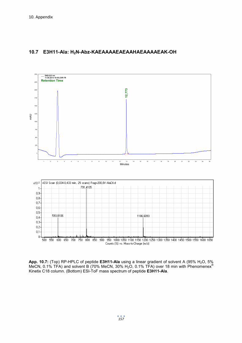
10. Appendix
157
10.7 E3H11-Ala: H2N-Abz-KAEAAAAEAEAAHAEAAAAEAK-OH
App. 10.7: (Top) RP-HPLC of peptide E3H11-Ala using a linear gradient of solvent A (95% H2O, 5% MeCN, 0.1% TFA) and solvent B (70% MeCN, 30% H2O, 0.1% TFA) over 18 min with Phenomenex
®
Kinetix C18 column. (Bottom) ESI-ToF mass spectrum of peptide E3H11-Ala.
Minutes1 2 3 4 5 6 7 8 9 10 11 12 13 14 15 16 17 18 19 20 21 22 23 24 25
mA
U
0
250
500
750
1000
1250
1500
1750
2000
2250
2500
12
,77
3
DAD-222 nm11.08.2014 18-56-23R1 F8
Retention Time

10. Appendix
158
10.8 EEEHEEE: H2N-Abz-EEEHEEE-OH
App. 10.8: RP-HPLC of peptide EEEHEEE using a linear gradient of solvent A (100% H2O, 0.1% TFA) and solvent B (55% H2O, 45% MeCN, 0.1% TFA) over 22 min with Phenomenex
® Kinetix C18
column. (Bottom) ESI-ToF mass spectrum of peptide EEEHEEE.
.
Minutes2 4 6 8 10 12 14 16 18 20 22 24 26 28
mA
U
0
250
500
750
1000
1250
1500
1750
2000
2250
2500
13
,76
0
DAD-222 nm14.04.2014 02-38-04Run 3 Fraction 12
Retention Time

10. Appendix
159
10.9 E3yne: Alkyne functionalized E3
App. 10.9: (Top) RP-HPLC of peptide E3yne using a linear gradient of solvent A (70% H2O, 30% MeCN, 0.1% TFA) and solvent B (70% MeCN, 30% H2O, 0.1% TFA) over 5 min with column Purospher
® STAR C18. (Bottom) ESI-ToF mass spectrum of peptide E3yne.
Minutes0,5 1,0 1,5 2,0 2,5 3,0 3,5 4,0 4,5 5,0 5,5 6,0 6,5 7,0 7,5 8,0 8,5 9,0
mA
U
100
200
300
400
500
600
700
4,1
47
DAD-226 nmE3yneHand R1F14.dat
Retention Time

10. Appendix
160
10.10 E3H11yne: Alkyne functionalized E3H11
App. 10.10: (Top) RP-HPLC of peptide E3H11yne using a linear gradient of solvent A (70% H2O, 30% MeCN, 0.1% TFA) and solvent B (70% MeCN, 30% H2O, 0.1% TFA) over 5 min with column Purospher
® STAR C18. (Bottom) ESI-ToF mass spectrum of peptide E3H11yne.
Minutes0,5 1,0 1,5 2,0 2,5 3,0 3,5 4,0 4,5 5,0 5,5 6,0 6,5 7,0 7,5 8,0 8,5 9,0
mA
U
0
200
400
600
800
1000
1200
1400
1600
4,0
50
DAD-221 nm
23.10.2014 16-32-03E3H11yne Hand 3 F18.dat
Retention Time

10. Appendix
161
10.11 E3H11-Alayne: Alkyne functionalized E3H11-Ala
App. 10.11: (Top) RP-HPLC of peptide E3H11-Alayne using a linear gradient of solvent A (80% H2O, 20% MeCN, 0.1% TFA) and solvent B (70% MeCN, 30% H2O, 0.1% TFA) over 6 min with column Purospher
® STAR C18. (Bottom) ESI-ToF mass spectrum of peptide E3H11-
Alayne.
Minutes0,5 1,0 1,5 2,0 2,5 3,0 3,5 4,0 4,5 5,0 5,5 6,0 6,5 7,0 7,5 8,0 8,5 9,0 9,5 10,0
mA
U
0
200
400
600
800
1000
1200
1400
1600
3,1
97
DAD-222 nm03.11.2014 13-15-22AlaCtlyne2 R2 F10.dat
Retention Time

10. Appendix
162
10.12 hPG-Azide: Azide-functionalized hyper-branched polyglycerol
App. 10.12: RP-HPLC of azide-functionalized (15.5%) hyper-branched polyglycerol (Mn = 13.2 kD) using a linear gradient of solvent A (95% H2O, 5% MeCN, 0.1% TFA) and solvent B (100% MeCN, 0.1% TFA) over 18 min with Phenomenex
® Kinetix C18 column.
Minutes1 2 3 4 5 6 7 8 9 10 11 12 13 14 15 16 17 18 19 20 21 22 23 24 25 26
mA
U
0
200
400
600
800
1000
1200
1400
11
,40
7
DAD-220 nm
07.11.2014 15-25-53PGazide 12600 15,5load
Retention Time

10. Appendix
163
10.13 Immobilization of E3H11yne to hPG-Azide
App. 10.13: (Top) RP-HPLC of hPG-E3H11 and residual E3H11yne following peptide immobilization and dialysis and (bottom) hPG-E3H11 after preparative RP-HPLC. Chromatograms were obtained using a linear gradient of solvent A (95% H2O, 5% MeCN, 0.1% TFA) and solvent B (100% MeCN, 0.1% TFA) over 18 min with Phenomenex
® Kinetix C18 column.
Minutes1 2 3 4 5 6 7 8 9 10 11 12 13 14 15 16 17 18 19 20 21 22 23 24 25 26
mA
U
0
100
200
300
400
500
600
700
800
900
1000
1100
12
,02
0
13
,96
7
DAD-220 nm
06.11.2014 10-09-2815.5 PG E3H11yne 1.dat
Retention Time
Minutes1 2 3 4 5 6 7 8 9 10 11 12 13 14 15 16 17 18 19 20 21 22 23 24 25 26
mA
U
-150
-100
-50
0
50
100
150
200
250
300
350
12
,06
0
DAD-220 nm
28.11.2014 10-59-37PGE3H11 R1 F8.dat
Retention Time
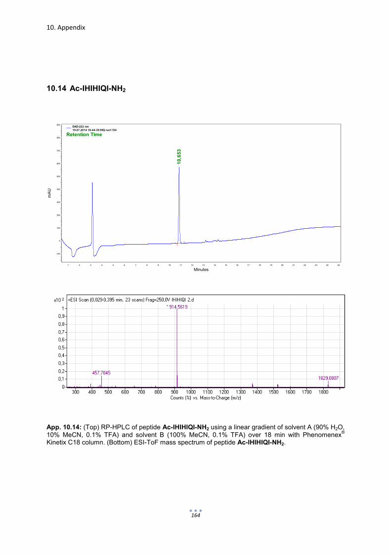
10. Appendix
164
10.14 Ac-IHIHIQI-NH2
App. 10.14: (Top) RP-HPLC of peptide Ac-IHIHIQI-NH2 using a linear gradient of solvent A (90% H2O, 10% MeCN, 0.1% TFA) and solvent B (100% MeCN, 0.1% TFA) over 18 min with Phenomenex
®
Kinetix C18 column. (Bottom) ESI-ToF mass spectrum of peptide Ac-IHIHIQI-NH2.
Minutes1 2 3 4 5 6 7 8 9 10 11 12 13 14 15 16 17 18 19 20 21 22 23 24 25
mA
U
-100
0
100
200
300
400
500
600
700
800
900
10
,65
3
DAD-222 nm19.07.2014 16-44-39 IHQ run1 f34
Retention Time

10. Appendix
165
10.15 CC-HexH24: H2N-GELKAIAQELKAIAKELKAIAWEHKAIAQGAG-OH
App. 10.15: (Top) RP-HPLC of peptide CC-HexH24 using a linear gradient of solvent A (95% H2O, 5% MeCN, 0.1% TFA) and solvent B (95% MeCN, 5% H2O, 0.1% TFA) over 18 min with Phenomenex
® Kinetix C18 column. (Bottom) ESI-ToF mass spectrum of peptide CC-HexH24.
Minutes1 2 3 4 5 6 7 8 9 10 11 12 13 14 15 16 17 18 19 20 21 22 23 24 25
mA
U
0
200
400
600
800
1000
1200
1400
1600
1800
2000
2200
13,4
00
DAD-233 nm03.10.2014 23-49-01h4r5
Retention Time
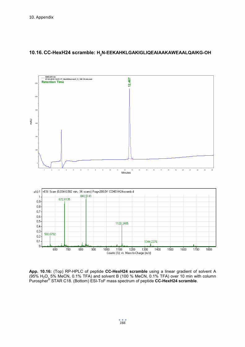
10. Appendix
166
10.16. CC-HexH24 scramble: H2N-EEKAHKLGAKIGLIQEAIAAKAWEAALQAIKG-OH
App. 10.16: (Top) RP-HPLC of peptide CC-HexH24 scramble using a linear gradient of solvent A (95% H2O, 5% MeCN, 0.1% TFA) and solvent B (100 % MeCN, 0.1% TFA) over 10 min with column Purospher
® STAR C18. (Bottom) ESI-ToF mass spectrum of peptide CC-HexH24 scramble.
Minutes1 2 3 4 5 6 7 8 9 10 11 12 13 14 15 16 17 18 19 20 21 22 23 24
mA
U
0
200
400
600
800
1000
1200
12
,46
7
DAD-225 nm
07.03.2016 12-01-17_HexH24scram3_5_100 18 min.met
Retention Time

10. Appendix
167
10.17 CC-Hex: H2N-GELKAIAQELKAIAKELKAIAWELKAIAQGAG-OH
App. 10.17: (Top) RP-HPLC of peptide CC-Hex using a linear gradient of solvent A (60% H2O, 40% MeCN, 0.1% TFA) and solvent B (100% MeCN, 0.1% TFA) over 6 min with column Purospher
® STAR
C18. (Bottom) ESI-ToF mass spectrum of peptide CC-Hex.
Minutes0,5 1,0 1,5 2,0 2,5 3,0 3,5 4,0 4,5 5,0 5,5 6,0 6,5 7,0 7,5 8,0 8,5 9,0 9,5
mA
U
0
250
500
750
1000
1250
1500
1750
2000
2250
2500
3,2
07
DAD-221 nm08.07.2015 18-31-16CC-HEX RUN1 F8.dat
Retention Time
Related Documents