Letter to the Editor De novo homozygous mutation of the C1 inhibitor gene in a patient with hereditary angioedema To the Editor: Hereditary angioedema (HAE; OMIM #106100) is a rare autosomal-dominant disease resulting from congenital deficiency of C1 esterase inhibitor protein (C1-INH) that controls comple- ment, contact-kinins, coagulation, and fibrinolytic cascades. 1 Symptoms relate to acute edema of subcutaneous tissues, viscera, and the upper airways (submucosal tissue). Edema of the larynx is the most fearsome feature of this disorder and is often life- threatening. 2 HAE due to C1-INH deficiency occurs as type I (85%), characterized by functional and antigenic C1-INH deficiency, or type II (15%), characterized by normal C1-INH antigenic levels and reduced functional activity. 1,2 Individuals affected by type I and II HAE carry a mutation in the C1-INH gene (C1NH, SERPING1; OMIM #606860). 3,4 The C1NH gene maps onto chromosome 11q12-q13.1 and is organized into 8 exons and 7 in- trons, particularly rich in repetitive Alu sequences. 5 More than 300 deficiency-causing mutations have been identified, and approximately 25% of them occur de novo. 6 Patients with HAE are generally heterozygous for the C1NH gene mutation; only recently, 3 subjects from 2 different families with a homozygous genetic defect have been identified. 7,8 We describe here the molecular genetic analysis of a 21-year- old woman who showed clinical and laboratory findings typical of HAE; we also provide evidence for a de novo homozygous muta- tion in the C1NH locus. The proband presented with recurrent peripheral and abdominal attacks of angioedema, starting at the age of 16 years. Both parents and 1 sibling did not manifest a his- tory of angioedema attacks. The patient experienced about 8 to 10 attacks per year, with about 50% of them as abdominal attacks and 50% at cutaneous or facial sites. There was no history of laryngeal attack. On-demand treatment of acute attacks with icatibant led to prompt resolution of symptoms in the majority of cases. The laboratory assessment showed very low antigenic and nondetectable functional levels of C1 INH and reduced C4, with normal levels of C3 and C1q. Neither the parents nor the sibling showed alterations in complement parameters (Table I). Western blot analysis of plasma protein with a specific rabbit antihuman C1 INH antibody revealed the presence of the 2 bands (105 and 96 kD, native and cleaved form of C1 INH protein, respectively) both in parents and in the sibling, while no signif- icant bands were evident in the proband plasma (Fig 1, A). Mutational screening of C1NH in the proband allowed us to identify a novel genetic defect, the insertion/deletion insTCAGTGTCGTGdelA at position 646 in exon 4, p.Lys216- Serfs*4 (Fig 1, B). Interestingly, the proband was homozygous for the mutation. This genetic defect has been not previously re- ported and was absent in 100 healthy and unrelated subjects used as control. The complete sequencing of the C1NH revealed 2 additional single nucleotide polymorphisms (SNPs), that is, Val458Met in exon 8 and IVS7-20A>G, that were previously re- ported not to affect C1 INH activity. Genetic analysis of C1NH of both parents showed that they did not carry the mutation of the affected daughter; thus, the genetic defect of the proband was a de novo mutation. To rule out that a small deletion of exon 4 could simulate a homozygosity in the patient, we used different proce- dures. First, we performed a long-range PCR by amplifying a seg- ment of C1NH that encompasses the repetitive Alu sequences 1 to 8 surrounding exon 4. As shown in Fig 1, C, only a fragment of 3.9 kb was observed in the proband and her parents, as expected by amplifying the wild-type allele with no deletion. Second, we per- formed RsaI restriction analysis of genomic DNA by amplifying a 2.9-kb fragment encompassing exon 4 (Fig 1, D). PCR products from the proband were completely digested into subfragments of 1301, 1053, and 528 bp, consistent with the absence of a small deletion. Finally, quantitative real-time PCR analysis was performed by amplifying a 202-bp fragment carrying the homo- zygous mutation. The signal for the C1NH product was normal- ized to that of the transferrin receptor gene, as reported in the Methods section (see the Online Repository at www.jacionline. org). We found that the proband showed a DDCt ratio of 0.99; her father and mother had a ratio of 1.05 and 1.21, respectively, in agreement with the DDCt ratio measured in 10 healthy subjects (1.16 6 0.1). This result suggests a normal amount of the exon 4 PCR product in the proband and her parents. Having excluded the deletion of exon 4, we investigated by means of intragenic polymorphisms whether uniparental isodis- omy or gene conversion could have occurred. A panel of 9 SNPs was evaluated in the family. Among these SNPs, 8 were fully informative and showed typical Mendelian inheritance with paternal and maternal alleles that were detected in the proband (see Fig E1 in this article’s Online Repository at www.jacionline. org). However, because the 2 informative SNPs closer to the mu- tation are located 251 bp upstream and 1070 bp downstream of the homozygous ins/del, a small gene conversion within this region cannot be completely excluded, possibly mediated by Alu sequences 1 to 8. If the ins/del mutation had occurred postzygoti- cally, one could expect a mosaic pattern in the DNA of the patient. Then, we performed genetic analysis on DNA isolated from buc- cal cells, which have ectodermal origin, a convenient alternative for collecting genetic material. The inspection of the sequence chromatogram confirmed the presence of the mutation in the proband (Fig 1, B). Blanch et al 7 reported the first 2 siblings with the I440S mis- sense mutation occurring in homozygosis. Analysis of the family demonstrated consanguinity and the presence of multiple hetero- zygous relatives who were free of symptoms. In 2010, L opez- Lera et al 8 described a new case of homozygous deficiency in a patient carrying the R378C substitution. Five asymptomatic relatives of the proband were heterozygous for the mutation. TABLE I. Clinical and laboratory features of the propositus and relatives Parameter Propositus Father Mother Sibling Clinical HAE Yes No No No C1 INH antigenic (NV 21–32 mg/dL) 4 28 30 32 C1 INH functional (NV 68%-132%) ND 108 102 104 C1q (NV 70%-130%) 83 105 93 110 C4 (NV 10-40 mg/dL) 6 18 21 19 ND, Not detectable; NV , normal values. 1

Welcome message from author
This document is posted to help you gain knowledge. Please leave a comment to let me know what you think about it! Share it to your friends and learn new things together.
Transcript

Letter to the Editor
TABLE I. Clinical and laboratory features of the propositus and
relatives
Parameter Propositus Father Mother Sibling
Clinical HAE Yes No No No
C1 INH antigenic (NV 21–32 mg/dL) 4 28 30 32
C1 INH functional (NV 68%-132%) ND 108 102 104
C1q (NV 70%-130%) 83 105 93 110
C4 (NV 10-40 mg/dL) 6 18 21 19
ND, Not detectable; NV, normal values.
De novo homozygous mutation of the C1inhibitor gene in a patient with hereditaryangioedema
To the Editor:Hereditary angioedema (HAE; OMIM #106100) is a rare
autosomal-dominant disease resulting from congenital deficiencyof C1 esterase inhibitor protein (C1-INH) that controls comple-ment, contact-kinins, coagulation, and fibrinolytic cascades.1
Symptoms relate to acute edema of subcutaneous tissues, viscera,and the upper airways (submucosal tissue). Edema of the larynx isthe most fearsome feature of this disorder and is often life-threatening.2
HAE due to C1-INH deficiency occurs as type I (85%),characterized by functional and antigenic C1-INH deficiency, ortype II (15%), characterized by normal C1-INH antigenic levelsand reduced functional activity.1,2 Individuals affected by typeI and II HAE carry a mutation in the C1-INH gene (C1NH,SERPING1; OMIM #606860).3,4 The C1NH gene maps ontochromosome 11q12-q13.1 and is organized into 8 exons and 7 in-trons, particularly rich in repetitive Alu sequences.5 More than300 deficiency-causing mutations have been identified, andapproximately 25% of them occur de novo.6 Patients with HAEare generally heterozygous for the C1NH gene mutation; onlyrecently, 3 subjects from 2 different families with a homozygousgenetic defect have been identified.7,8
We describe here the molecular genetic analysis of a 21-year-old womanwho showed clinical and laboratory findings typical ofHAE; we also provide evidence for a de novo homozygous muta-tion in the C1NH locus. The proband presented with recurrentperipheral and abdominal attacks of angioedema, starting at theage of 16 years. Both parents and 1 sibling did not manifest a his-tory of angioedema attacks. The patient experienced about 8 to 10attacks per year, with about 50% of them as abdominal attacks and50% at cutaneous or facial sites. Therewas no history of laryngealattack. On-demand treatment of acute attacks with icatibant led toprompt resolution of symptoms in the majority of cases.
The laboratory assessment showed very low antigenic andnondetectable functional levels of C1 INH and reduced C4, withnormal levels of C3 and C1q. Neither the parents nor the siblingshowed alterations in complement parameters (Table I).
Western blot analysis of plasma protein with a specific rabbitantihuman C1 INH antibody revealed the presence of the 2 bands(105 and 96 kD, native and cleaved form of C1 INH protein,respectively) both in parents and in the sibling, while no signif-icant bands were evident in the proband plasma (Fig 1, A).
Mutational screening of C1NH in the proband allowed usto identify a novel genetic defect, the insertion/deletioninsTCAGTGTCGTGdelA at position 646 in exon 4, p.Lys216-Serfs*4 (Fig 1, B). Interestingly, the proband was homozygousfor the mutation. This genetic defect has been not previously re-ported and was absent in 100 healthy and unrelated subjectsused as control. The complete sequencing of the C1NH revealed2 additional single nucleotide polymorphisms (SNPs), that is,Val458Met in exon 8 and IVS7-20A>G, that were previously re-ported not to affect C1 INH activity. Genetic analysis of C1NH ofboth parents showed that they did not carry the mutation of theaffected daughter; thus, the genetic defect of the proband was a
de novomutation. To rule out that a small deletion of exon 4 couldsimulate a homozygosity in the patient, we used different proce-dures. First, we performed a long-range PCR by amplifying a seg-ment of C1NH that encompasses the repetitive Alu sequences 1 to8 surrounding exon 4. As shown in Fig 1,C, only a fragment of 3.9kb was observed in the proband and her parents, as expected byamplifying the wild-type allele with no deletion. Second, we per-formedRsaI restriction analysis of genomic DNAby amplifying a2.9-kb fragment encompassing exon 4 (Fig 1, D). PCR productsfrom the proband were completely digested into subfragmentsof 1301, 1053, and 528 bp, consistent with the absence of a smalldeletion. Finally, quantitative real-time PCR analysis wasperformed by amplifying a 202-bp fragment carrying the homo-zygous mutation. The signal for the C1NH product was normal-ized to that of the transferrin receptor gene, as reported in theMethods section (see the Online Repository at www.jacionline.org). We found that the proband showed a DDCt ratio of 0.99;her father and mother had a ratio of 1.05 and 1.21, respectively,in agreement with theDDCt ratio measured in 10 healthy subjects(1.166 0.1). This result suggests a normal amount of the exon 4PCR product in the proband and her parents.
Having excluded the deletion of exon 4, we investigated bymeans of intragenic polymorphisms whether uniparental isodis-omy or gene conversion could have occurred. A panel of 9 SNPswas evaluated in the family. Among these SNPs, 8 were fullyinformative and showed typical Mendelian inheritance withpaternal and maternal alleles that were detected in the proband(see Fig E1 in this article’s Online Repository at www.jacionline.org). However, because the 2 informative SNPs closer to the mu-tation are located 251 bp upstream and 1070 bp downstream of thehomozygous ins/del, a small gene conversion within this regioncannot be completely excluded, possibly mediated by Alusequences 1 to 8. If the ins/del mutation had occurred postzygoti-cally, one could expect a mosaic pattern in the DNA of the patient.Then, we performed genetic analysis on DNA isolated from buc-cal cells, which have ectodermal origin, a convenient alternativefor collecting genetic material. The inspection of the sequencechromatogram confirmed the presence of the mutation in theproband (Fig 1, B).
Blanch et al7 reported the first 2 siblings with the I440S mis-sense mutation occurring in homozygosis. Analysis of the familydemonstrated consanguinity and the presence of multiple hetero-zygous relatives who were free of symptoms. In 2010, L�opez-Lera et al8 described a new case of homozygous deficiency in apatient carrying the R378C substitution. Five asymptomaticrelatives of the proband were heterozygous for the mutation.
1
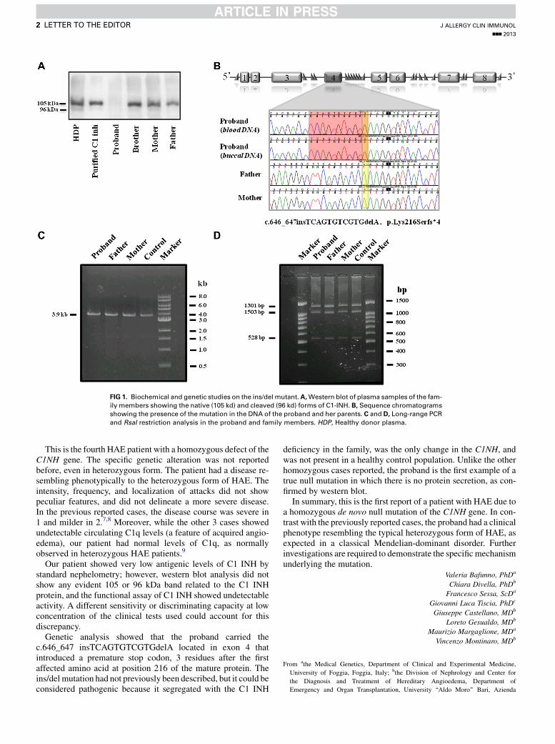
FIG 1. Biochemical and genetic studies on the ins/del mutant.A,Western blot of plasma samples of the fam-
ily members showing the native (105 kd) and cleaved (96 kd) forms of C1-INH. B, Sequence chromatograms
showing the presence of the mutation in the DNA of the proband and her parents. C and D, Long-range PCR
and RsaI restriction analysis in the proband and family members. HDP, Healthy donor plasma.
J ALLERGY CLIN IMMUNOL
nnn 2013
2 LETTER TO THE EDITOR
This is the fourth HAE patient with a homozygous defect of theC1NH gene. The specific genetic alteration was not reportedbefore, even in heterozygous form. The patient had a disease re-sembling phenotypically to the heterozygous form of HAE. Theintensity, frequency, and localization of attacks did not showpeculiar features, and did not delineate a more severe disease.In the previous reported cases, the disease course was severe in1 and milder in 2.7,8 Moreover, while the other 3 cases showedundetectable circulating C1q levels (a feature of acquired angio-edema), our patient had normal levels of C1q, as normallyobserved in heterozygous HAE patients.9
Our patient showed very low antigenic levels of C1 INH bystandard nephelometry; however, western blot analysis did notshow any evident 105 or 96 kDa band related to the C1 INHprotein, and the functional assay of C1 INH showed undetectableactivity. A different sensitivity or discriminating capacity at lowconcentration of the clinical tests used could account for thisdiscrepancy.
Genetic analysis showed that the proband carried thec.646_647 insTCAGTGTCGTGdelA located in exon 4 thatintroduced a premature stop codon, 3 residues after the firstaffected amino acid at position 216 of the mature protein. Theins/del mutation had not previously been described, but it could beconsidered pathogenic because it segregated with the C1 INH
deficiency in the family, was the only change in the C1NH, andwas not present in a healthy control population. Unlike the otherhomozygous cases reported, the proband is the first example of atrue null mutation in which there is no protein secretion, as con-firmed by western blot.
In summary, this is the first report of a patient with HAE due toa homozygous de novo null mutation of the C1NH gene. In con-trast with the previously reported cases, the proband had a clinicalphenotype resembling the typical heterozygous form of HAE, asexpected in a classical Mendelian-dominant disorder. Furtherinvestigations are required to demonstrate the specificmechanismunderlying the mutation.
Valeria Bafunno, PhDa
Chiara Divella, PhDb
Francesco Sessa, ScDa
Giovanni Luca Tiscia, PhDc
Giuseppe Castellano, MDb
Loreto Gesualdo, MDb
Maurizio Margaglione, MDa
Vincenzo Montinaro, MDb
From athe Medical Genetics, Department of Clinical and Experimental Medicine,
University of Foggia, Foggia, Italy; bthe Division of Nephrology and Center for
the Diagnosis and Treatment of Hereditary Angioedema, Department of
Emergency and Organ Transplantation, University ‘‘Aldo Moro’’ Bari, Azienda

J ALLERGY CLIN IMMUNOL
VOLUME nnn, NUMBER nn
LETTER TO THE EDITOR 3
Ospedaliero-Universitaria ‘‘Consorziale Policlinico,’’ Bari, Italy; and cthe Thrombosis
& Haemostasis Unit, IRCCS Casa Sollievo della Sofferenza, S. Giovanni Rotondo,
Foggia, Italy. E-mail: [email protected].
Disclosure of potential conflict of interest: V. Montinaro has consultant arrangements
with Shire HGT and Viropharma; has grants/grants pending with CSL Behring; has
received payment for lectures, including service on speakers’ bureaus, from CSL
Behring and Shire HGT; has received payment for development of educational pre-
sentations from Shire HGT; and has received travel/accomodations/meeting expenses
from Shire HGT, CSL Behring, and SOBI. The rest of the authors declare that they
have no relevant conflicts of interest.
REFERENCES
1. Longhurst H, Cicardi M. Hereditary angio-oedema. Lancet 2012;379:474-8.
2. Nagy N, Grattan CE, McGrath JA. New insights into hereditary angio-oedema:
molecular diagnosis and therapy. Australasian J Derm 2010;51:157-62.
3. Tosi M. Molecular genetics of C1 inhibitor. Immunobiology 1998;199:358-65.
4. Davis AE III, Whitehead AS, Harrison RA, Dauphinais A, Bruns GA, Cicardi M,
et al. Human inhibitor of the first component of complement, C1: characterization
of cDNA clones and localization of the gene to chromosome 11. Proc Natl Acad
Sci USA 1986;83:3161-5.
5. Carter PE, Duponchel C, Tosi M, Fothergill JE. Complete nucleotide sequence of
the gene for human C1 inhibitor with an unusually high density of Alu elements.
Eur J Biochem 1991;97:301-8.
6. Pappalardo E, Cicardi M, Duponchel C, Carugati A, Choquet S, Agostoni A, et al.
Frequent de novo mutations and exon deletions in the C1 inhibitor gene of patients
with angioedema. J Allergy Clin Immunol 2000;106:1147-54.
7. Blanch A, Roche O, Urrutia I, Gamboa P, Font�an G, L�opez-Trascasa M. First case
of homozygous C1 inhibitor deficiency. J Allergy Clin Immunol 2006;118:1330-5.
8. L�opez-Lera A, Favier B, Mena de la Cruz R, Garrido S, Drouet C, L�opez-Trascasa
M. A new case of homozygous C1-inhibitor deficiency suggests a role for Arg378
in the control of kinin pathway activation. J Allergy Clin Immunol 2010;126:
1307-10.
9. Cicardi M, Zingale LC, Pappalardo E, Folcioni A, Agostoni A. Autoantibodies and
lymphoproliferative diseases in acquired C1-inhibitor deficiencies. Medicine
(Baltimore) 2003;82:274-81.
http://dx.doi.org/10.1016/j.jaci.2013.04.006

REFERENCES
E1. Miller SA, Dykes DD, Polesky HF. A simple salting out procedure for extracting
DNA from human nucleated cells. Nucleic Acids Res 1988;16:1215.
E2. Roche O, Blanch A, Duponchel C, Font�an G, Tosi M, L�opez-Trascasa M. Hered-
itary angioedema: the mutation spectrum of SERPING1/C1NH in a large Spanish
cohort. Hum Mutat 2005;26:135-44.
J ALLERGY CLIN IMMUNOL
nnn 2013
3.e1 LETTER TO THE EDITOR
METHODS
Study subjectsWe report the clinical and laboratory features of a 21-year-old Caucasian
womanwhoshowed recurrent attacks of angioedema.Bothparents and 1 sibling
of the probandwere also investigated for circulating levels of C1 INH,while the
genetic analysis was performed in the proband and her parents. The study was
carried out in accordancewith the Principles of theDeclaration of Helsinki, and
a written informed consent was obtained from the proband and her parents.
Laboratory diagnosisSerumandplasmaEDTAsampleswereobtainedfromall the familymembers.
An aliquot of serum was directly used to measure the complement levels. The
remaining serum and plasma samples were frozen at2808C until used. C1-INH
and C4 antigenic levels were assessed by using nephelometry (Siemens, Milano,
Italy), while C1 INH functional activity was evaluated by using an EIA test
(MicroVue C1 inhibitor Enzyme Immune Assay, Quidel Co, San Diego, Calif).
C1q levelsweremeasuredbyusing theHumanComplementC1qBindaridRadial
Immunodiffusion Kit (The Binding Site Group Ltd, Birmingham, United
Kingdom).
Western blot analysis of C1-INHPlasma samples from the proband and relatives were run on 7.5%
SDS-PAGE under nonreducing conditions; 0.25 to 2.0 mL of plasma was
analyzed in different experiments. Fifty nanograms of purified C1-INH
(Berinert; CSL Behring, Marburg, Germany) were run in each experiment.
After the electrophoretic run, proteins were transferred onto a polyvinylidene
fluoride membrane (Millipore, Badford, Mass) at 250 mA and 48C overnight.
The filter was blocked with 5% nonfat dry milk powder in PBS that contained
0.1% tween-20 (TBS) and incubated with a rabbit anti-human C1-INH
antibody (provided by Prof. M. Daha, University of Leiden) 1:1000 dilution
in TBS for 2 hours at room temperature. The membrane was washed twice in
TBS and incubated with horseradish peroxidase–conjugated sheep anti-rabbit
IgG (Santa Cruz Biotechnology; Santa Cruz, Calif) 1:10,000 dilution in TBS.
Immune complexes were detected by using the ECL enhanced chemilumines-
cence system (Amersham, GE Healthcare, Milano, Italy), as recommended by
the manufacturer. Bands were quantified by using the Image J 1.34 Software.
Molecular diagnosisGenomic DNAwas isolated from peripheral blood leukocytes according to
standard protocols.E1 Genetic analysis was performed by direct DNA sequenc-
ing. Proband DNA was also isolated from buccal mucosa cells by using the
QIAamp DNA Mini Kit (Quiagen, Hilden, Germany) according to the manu-
facturer’s instructions. DNA eluates were kept at 48C while undergoing gen-
otyping assays and kept at 2808C for long-term storage.
We have standardized the PCR conditions by using primers designed with
Primer 3 software (www.genome.wi.mit.edu/cgibin/primer/primer3_www.
cgi) on the basis of known sequences of C1NH as reported in ENSEMBL da-
tabase (Wellcome Trust Sanger Institute, Cambridge, United Kingdom):
SERPING1 ENSG00000149131.
Briefly, PCRwas carried out on 50-mL samples in a Bio-Rad thermal cycler
(Bio-Rad Laboratories, Inc, Hercules, Calif). Each sample contained 0.15 mg
of genomic DNA, 0.3 mM of each primer, 200 mM of deoxyribonucleotide
triphosphates, 1X PCR buffer (with 1.5 mM MgCl2), and 1.5 U of AmpliTaq
gold polymerase with appropriate 1X buffer (Applied Biosystems, Inc, Foster
City, Calif). PCR products were purified and subjected to direct-cycle se-
quence analysis by using the Taq dye-deoxy terminator method and an ABI
Prism 3100 genetic analyzer (Applied Biosystems) according to the manufac-
turer’s instructions. Primers and PCR conditions for DNA analysis are
reported in Table E1. We used samples from 100 unrelated healthy subjects
as the control group to distinguish novel mutations from rare polymorphisms.
E3. Iwamoto K, Tanaka A, Kawai M, Ishii K, Mihara S, Hide M. A large heterozy-gous deletion including the entire C1 inhibitor gene in a sporadic case of hered-
itary angio-oedema. Clin Exp Dermatol 2012;37:20-3.
E4. Hayat Nosaeid M, Mahdian R, Jamali S, Maryami F, Babashah S, Maryami F,
et al. Validation and comparison of two quantitative real-time PCR assays for
direct detection of DMD/BMD carriers. Clin Biochem 2009;42:1291-9.
Deletion testsTo investigate the presence of a deletion of exon 4 in C1NH gene, we used
different techniques reported below.
d Long-Range PCR
Long-range PCR was performed as reported by Roche et al.E2 The selected
primers amplified a segment of C1NH encompassing the repetitive Alu se-
quences 1 to 8 that surround exon 4. The expected 3.9-kb fragment will be
barely detected in the patients with deletion because the PCR preferentially
will amplify the shorter bands corresponding to the alleles carrying the
mutation.
The PCRwas performed in a final volume of 50mL, using 1.5 U of TaKaRa
LaTaqDNApolymerase (TakaraBio, Inc,Otsu, Shiga, Japan), 15 pmol of each
primer, 200mMof deoxyribonucleotide triphosphate, and 0.15mg of genomic
DNA. The cycling conditions were as follows: 30 cycles at 988C for 30
seconds, 568C for 30 seconds, and 728C for 5 minutes. The final extension was
728C for 5 minutes. PCR products were separated in a 0.9% agarose-ethidium
bromidegel. To confirm accuracy of the results, long-rangePCRproductswere
also sequenced by conventional DNA sequencing (data not shown).
d PCR-RFLP
PCR-RFLP was conducted in a 15-mL reaction volume containing Rsa I
(New England Biolabs, Beverly, Mass) restriction enzyme and genomic
DNA amplified by PCR by using the following primers: int3.3fw
59TTGTCCCAGCTACTTGGGAGG39 and int4.2rw 59GCTCATTTTTGGGTCAAAGG3 9. The restriction enzyme cleaved the PCR product of
2.9 kb in 3 fragments of 1301, 1053, and 528 bp, respectively. Upon digestion
performed at 378C overnight, the resulting DNA fragments were visualized in
4% agarose gel.
d Quantitative real-time PCR
The quantitative real-time PCR assay to measure gene dosage was
performed by using the 7500 Fast Real Time PCR system (PE, Applied
Biosystems) according to the manufacturer’s protocol. Primers to amplify the
exon 4 of C1NH were RTex4fw: 59-TCTTTCATCTCTGCCCTTTGTTG-39and RTex4rw: 59-AGTGTCTGCAGAGGAGGGTCC-39. As a reference
gene, we used the transferrin receptor (TFRC): TFRC forward primer is 59-ACGGAATATGAAGATGATCTCAGCAAGG-39; TFRC reverse primer is
59-GCGCAGATCACGAGATGGA-39.E3 PCR was performed in a total vol-
ume of 20 mL in each well, containing 10 mL of SYBR Green PCR Master
mix (PE, Applied Biosystems), 20 ng of genomic DNA, and 15 pmol of
C1NH and TFRC primers. Each sample was run in triplicate. To make sure
that the amplification efficiencies for the 2 segments of the target and reference
gene were similar, standard curves were generated with serial dilutions of
DNA from a normal individual. The relative gene copy numbers were
calculated by using the comparative Ct method, using the ratio formula
(ratio 5 22DDCt). This ratio was expected to be about 1 in normal control
and 0.5 in heterozygous patient.E4
d SNP genotyping
Nine polymorphisms (1 in 59UTR, 5 in intron 3, 1 in intron 4, 1 in intron 7,and 1 in exon 8) throughout C1NH were used for haplotyping of the mutant
allele, that is, c.-21C>T (rs28362944), c.5501748A>G (rs2432809),
c.5501794C>A (rs1812005), c.551-500C>G (rs28362947), c.551-495A>C
(rs28362948), c.551-155A >G (rs2936694), c.68511100C>T(rs78364821), c.1030-20A>G (rs2511988) and c.1438G>A (rs4926). Genetic
analysis was performed by direct DNA sequencing. Primers and PCR condi-
tions for the analysis of SNPs are reported in Table E1.

FIG E1. Pedigree of the family. Results of the C1NHmutation analysis and the haplotypes using 8 intragenic
different SNPs are presented.
J ALLERGY CLIN IMMUNOL
VOLUME nnn, NUMBER nn
LETTER TO THE EDITOR 3.e2

TABLE E1. Primers and PCR conditions for the genetic analysis of C1NH
Exons Primer forward Primer reverse T annealing (8C)
1 59AGCTGAGAACTGCACCCAAG 39 59CGGGTCTCTGTCCATCTGTT 39 57
2 59 CTCTGGCCTCATTGTTTGGT 39 59 CGGAGCCTGAAGGGTTAAT39 56
3 59 CCACACCTTCTCTTCCTGCT 39 59 CCAGAGGCATGGCTTTGTAA 39 54
4 59AACCCTCATTCCCAAGGAAG 39 59 TTCCCTCTGTCCTTTCTTTCC 39 56
5-6 59 GCATGCTCACTCTCAAATCG 39 59 CAAAATGATGGGACTACAGCA 39 54
7 59 ACTGCAGGACAGCATTGTGA 39 59 CTAGAGAATGCTAACTACATCTTAGGG 39 56
8 59 GGACAAAGGTCTCCATCAGC 39 59 TAGCCTGGGTGACAGATTGA39 62
Int3.1 59GCCTCTGGGAAGAAGCTGTA 39 59CTGCCTCAGCCTCCTGAGTA 39 59
Int3.2 59 TGAACACGGGAGGCAGAG 39 59 CAGGTGAAGTCCTTGGGGTA 39 58
Int4 59 ATGGTGGTGTGCACCTGTA 39 59 GCTCATTTTTGGGTCAAAGG 39 57
J ALLERGY CLIN IMMUNOL
nnn 2013
3.e3 LETTER TO THE EDITOR
Related Documents