THE HISTOLOGIC STRUCTURE OF LOWER ALIMENTARY TRACT S R I W I R Y A W A N LARGE INTESTINE SMALL INTESTINE What is the region of small intesti ne ? What is the region of large intesti ne ?

Day 2 Histo-lower Alimentary
Dec 14, 2015
Histology of Lower Alimentary
Welcome message from author
This document is posted to help you gain knowledge. Please leave a comment to let me know what you think about it! Share it to your friends and learn new things together.
Transcript

THE HISTOLOGIC STRUCTURE OF
LOWER ALIMENTARY TRACT
SRI
WIRYAWA
N
LARGE INTESTINESMALL INTESTINE
What is the region of small intestine ?
What is the region of large intestine ?

Medical Faculty of Udayana University.
GENERAL STRUCTURE OF ALIMENTARY TRACT
• Mucosa– Epithelium– Lamina
Propia– Mucularis
mucosa
• Submucosa• Muscularis
externa• Adventitia/
SerosaWhat is the difference of adventitia & serosa ?

Medical Faculty of Udayana University.
THE HISTOLOGIC STRUCTURE OF SMALL INTESTINE

Medical Faculty of Udayana University.
THE HISTOLOGIC STRUCTURE OF SMALL INTESTINE

Medical Faculty of Udayana University.
THE HISTOLOGIC STRUCTURE OF SMALL INTESTINE

Medical Faculty of Udayana University.
`
What are the functions of Brunner’s glands ?

Medical Faculty of Udayana University.
SMALL INTESTINE : JEJUNUM

Medical Faculty of Udayana University.
SMALL INTESTINE : ILEUM

Structural Modifications of The Small Structural Modifications of The Small Intestine wall increase surface area :Intestine wall increase surface area :
Plicae circulares: deep circular folds of the Plicae circulares: deep circular folds of the mucosa and submucosamucosa and submucosa
Villi – fingerlike extensions of the mucosaVilli – fingerlike extensions of the mucosa
Microvilli – tiny projections of absorptive Microvilli – tiny projections of absorptive mucosal cells’ plasma membranesmucosal cells’ plasma membranes

Copyright © 2004 Pearson Education, Inc., publishing as Benjamin Cummings
Small Intestine: Microscopic Anatomy
Figure 23.21


GLYCOCALYX
What are the functions of glycocalyx ?

Medical Faculty of Udayana University.
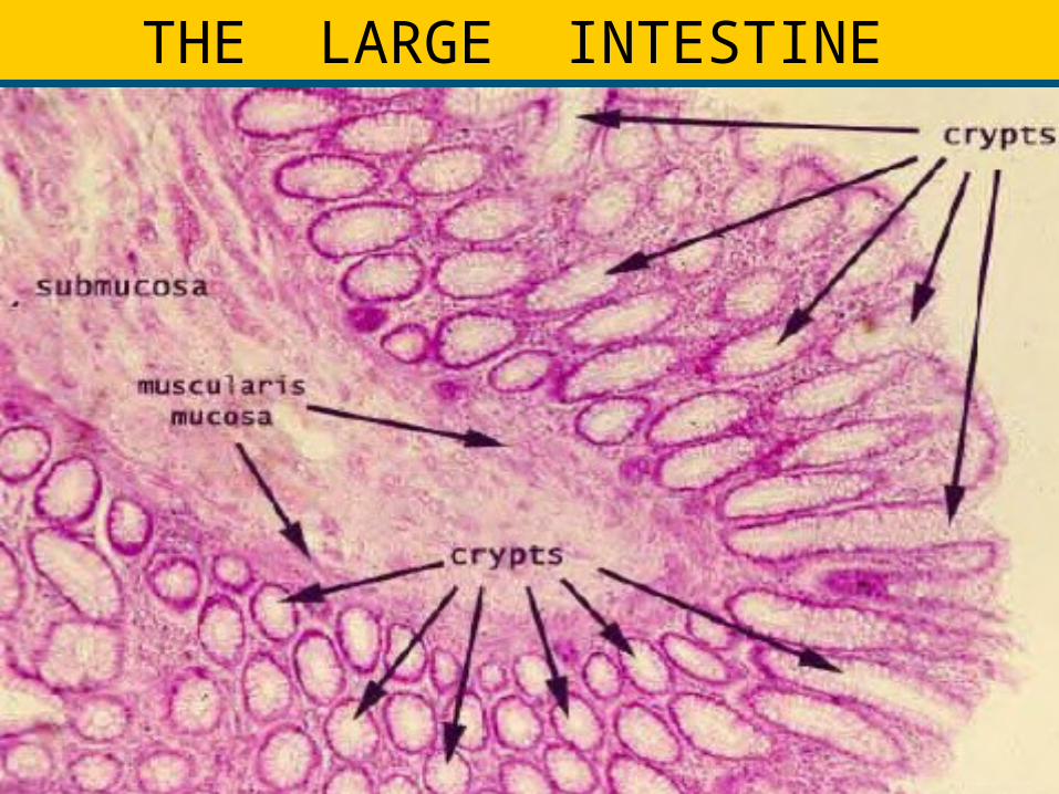
HISTOLOGY OF LARGE INTESTINE


GlandsLamina propria
Tunica serosa (some adventitia)
}Tunica mucosa
LARGE INTESTINE
Muscularis mucosae
Tunica submucosa
Tunica muscularis externa
with Tenia coli - 3 bands of
longitudinal muscle - absent from the rectum & v. appendix
No villi

Medical Faculty of Udayana University.
Medical Faculty of Udayana University.
THE LARGE INTESTINE
Lamina propria
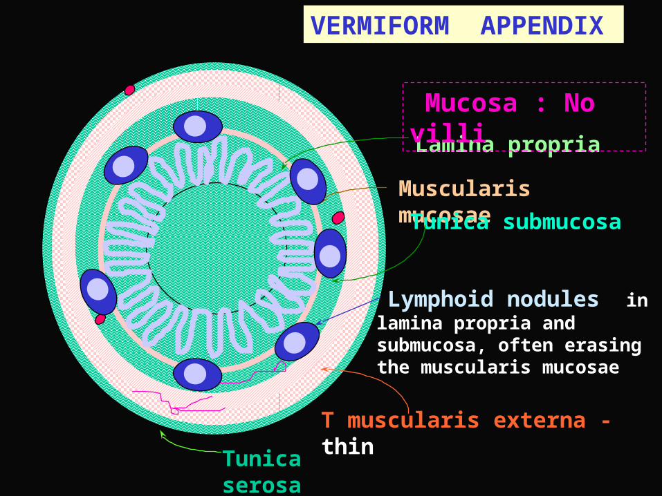
Tunica serosa
VERMIFORM APPENDIX
Muscularis mucosae
Tunica submucosa
T muscularis externa - thin
Lymphoid nodules in lamina propria and submucosa, often erasing the muscularis mucosae
Mucosa : No villi


THANK YOU
Related Documents