Davis, A. P. (2020). Biomimetic carbohydrate recognition. Chemical Society Reviews, (9), 2531-2545. https://doi.org/10.1039/C9CS00391F Peer reviewed version Link to published version (if available): 10.1039/C9CS00391F Link to publication record in Explore Bristol Research PDF-document This is the author accepted manuscript (AAM). The final published version (version of record) is available online via Royal Society of Chemistry at https://pubs.rsc.org/en/content/articlelanding/2020/cs/c9cs00391f#!divAbstract . Please refer to any applicable terms of use of the publisher. University of Bristol - Explore Bristol Research General rights This document is made available in accordance with publisher policies. Please cite only the published version using the reference above. Full terms of use are available: http://www.bristol.ac.uk/red/research-policy/pure/user-guides/ebr-terms/

Welcome message from author
This document is posted to help you gain knowledge. Please leave a comment to let me know what you think about it! Share it to your friends and learn new things together.
Transcript

Davis, A. P. (2020). Biomimetic carbohydrate recognition. ChemicalSociety Reviews, (9), 2531-2545.https://doi.org/10.1039/C9CS00391F
Peer reviewed version
Link to published version (if available):10.1039/C9CS00391F
Link to publication record in Explore Bristol ResearchPDF-document
This is the author accepted manuscript (AAM). The final published version (version of record) is available onlinevia Royal Society of Chemistry at https://pubs.rsc.org/en/content/articlelanding/2020/cs/c9cs00391f#!divAbstract. Please refer to any applicable terms of use of the publisher.
University of Bristol - Explore Bristol ResearchGeneral rights
This document is made available in accordance with publisher policies. Please cite only thepublished version using the reference above. Full terms of use are available:http://www.bristol.ac.uk/red/research-policy/pure/user-guides/ebr-terms/

Received 00th January 20xx,
Accepted 00th January 20xx
DOI: 10.1039/x0xx00000x
Biomimetic Carbohydrate Recognition
Anthony P. Davis
Carbohydrates are important but challenging targets for supramolecular chemists. They possess complex, irregular and
variable structures, and are strongly attracted to water, their natural environment. This tutorial review describes work on
synthetic receptors which bind carbohydrates through non-covalent interactions, mimicking the strategies used in biology.
Emphasis is placed on systems which operate in purely aqueous solution, without involvement of organic solvents. Although
the problem is difficult, the careful design of complementary cavities can lead to surprisingly good results. In particular, a
receptor for glucose has achieved performance which generally matches biology, and augurs well for real-world applications.
Key learning points
(1) Principles of carbohydrate recognition through non-covalent interactions; requirement for full host-guest complementarity, especially in
water.
(2) Successful approaches using preorganised hydrogen bonding groups in organic solvents.
(3) Receptors for operation in aqueous solution, especially the “temple” family for all-equatorial substrates.
(4) High levels of complementarity are possible, at least for glucose, leading to remakable affinity and selectivity.
School of Chemistry, University of Bristol, Cantock’s Close, Bristol BS8 1TS,
UK. E-mail: [email protected]
Electronic Supplementary Information (ESI) available: [details of any
supplementary information available should be included here]. See
DOI: 10.1039/x0xx00000x

1. Introduction
The design of carbohydrate receptors is a major challenge for supramolecular chemists. Carbohydrates are clearly important
substrates. They are the most abundant molecules in biology, and are highly versatile. They serve as fuels (e.g. starch), b uilding
materials (e.g. cellulose), and form the backbone for the genetic code (ribose in DNA). They show exceptional structural variability,
which is widely exploited in labelling systems for cells and proteins.1,2 Agents which bind to saccharides could serve many medical
purposes. Perhaps the most obvious relate to diabetes, where selective glucose receptors could be exploited in blood glucose
monitors3 and glucose-responsive insulin.4 However, there are many other possibilities, including synthetic antibodies to cancer
cells, anti-infectives, anti-inflammatories, and many diagnostic applications.1,2,5
From a theoretical viewpoint, carbohydrate recognition is representative of a wider issue – how do we bind polar molecules in water,
where solvent already binds strongly to both substrate and complementary (polar) binding site? The problem is especially acu te for
saccharides, where hydroxyl and ether groups are predominant and most examples are quite similar to clusters of water molecules.
Distinguishing these “hydromimetic” targets from solvent seems an almost impossible task. Indeed, biological carbohydrate
recognition is generally quite weak. Lectins, the major class of carbohydrate-binding proteins, often bind monosaccharides with Ka
< 103 M-1.6 Nonetheless, higher affinities are possible, especially for the bacterial periplasmic proteins which help guide the
organisms towards food sources.7 Biology thus encourages and focuses our research. It confirms that strong binding is feasible,
while posing questions about the principles which underlie success.
Research into carbohydrate receptors has a considerable history, going back at least to the 1980s. Two main approaches have been
taken. One exploits the reversible reaction between boronic acids and diols to give cyclic boronates, allowing the design of binding
agents which are simple, accessible, and effective in the natural medium of water. These systems have been covered in depth by
others2,3 and, as they are not biomimetic, fall outside the present scope. The alternative relies on noncovalent bonding and more
closely mimics the strategy used by biology.5,8 This article will discuss how receptors based on this approach have been developed,
starting in unnatural media, moving to truly biomimetic systems which operate in aqueous solution, and finally to a system wh ich
both mimics and matches biology. Particular emphasis is placed on water as solvent, complementing earlier accounts which provide
good coverage of organic media.5,9 The progression illustrates the potential of supramolecular chemistry to achieve quite surprising
goals, given sufficient focus and persistence.
2. General Principles
As illustrated in Fig. 1, carbohydrates are polyols with occasional presence of other polar functionality (e.g. NHAc, CO2H). The
monosaccharide units are cyclic, possessing distinct conformations, so the structures tend to be geometrically well-defined. In terms
of noncovalent interactions, the polar groups can act as H-bond donors or acceptors, the oxygen atoms can serve as electron pair
donors to metal ions, and the CH groups in unfunctionalised areas can also act as weak H-bond donors. Reducing sugars, with C1-
OH, occur as a mixture of anomers (illustrated for glucose 1).

With so many polar groups, carbohydrates are quite easy targets in non-polar organic media. A solvent such as chloroform does not
depress interactions such as H-bonding, so preorganised arrangements of polar binding groups should be effective (Fig. 2a). As
implied by Fig. 2a, complementarity with apolar surfaces may not be important. The interactions between scaffold and substra te
CH are likely to be weak, and not so different from contacts with solvent, so the exact framework design may be less critical.
For carbohydrate recognition in water, the situation is very different. When a saccharide -OH binds to a polar group in a receptor it
must displace a water molecule, so the process is nearly isoenergetic. Polar interactions should not be worthless because an array of
H-bonding groups which perfectly complements the target should not be ideal for solvent water. Nonetheless, they are severely
weakened. On the other hand, compensation is available if the apolar CH groups in the carbohydrate make contact with apolar
surfaces in the receptor. “High energy” water molecules are displaced from both and are free to move into bulk, lowering the free
energy. To take advantage of this, the receptor must be fully complementary to the substrate, matching both polar and apolar moieties
(Fig. 2b). There is particular benefit if the receptor’s apolar surfaces are aromatic, allowing the formation of CH-π bonds (Fig. 2c).
Although it is not obvious that this interaction will translate to water (OH-π bonds are also possible), there is good evidence that it
can be effective.10
Fig. 1 Selected biologically important carbohydrates.
Fig. 2 (a) Design strategy for carbohydrate recognition in an organic solvent such as chloroform, focusing on complementarity between polar binding groups. (b) Moving to
water, where matching apolar surfaces may be equally important. (c) CH-π interactions between an aromatic surface and axial carbohydrate CH groups. (d) X-ray crystal
structure of β-D-fucose in the binding site of Aleuria aurantia lectin (see ref. 11). Residues contributing to apolar interactions are shown as thicker tubes with standard
colouring. Selected polar hydrogens are shown, the rest are concealed. (e) Crystal structure of β-D-glucose complexed to the periplasmic galactose-binding protein from E.
coli (see ref. 7). Display conventions as for (d).
a)
b) complementary polar and apolar surfaces
d) e)
polar groups
spacer scaffold
complementary polar units
c)
CHCl3
CHCl3
CHCl3
CHCl3
CHCl3
CHCl3CHCl3
H2O
H2O
H2O
H2O H2O H2OH2O
H2O
H2O
H2O
The principle of full complementarity is well supported by crystallography of carbohydrate-binding proteins. Two examples are
illustrated in Figs. 2d/e. The former shows fucose 4 in the binding site of a lectin from the fungus Aleuria aurantia.11 A tryptophan
provides hydrophobic/CH-π interactions to the upper face of the fucose, while other apolar amino acids help to form an apolar pocket
for the methyl group. Five hydrogen bonds are formed involving arginine, aspartic and tryptophan side-chains. The fucose C1 and
C2-OH groups are exposed to solvent and not directly involved in binding. Ka for the complex is 6000 M-1, fairly typical for lectin-
monosaccharide interactions. Fig. 2e shows β-D-glucose 1b complexed to a monosaccharide-binding protein found in the
periplasmic space of E. coli.7 Axial CH groups again form hydrophobic/CH-π interactions to a tryptophan, with a phenylalanine
performing the same role on the other side of the substrate. In this case the carbohydrate is fully enclosed, with ten hydrogen bonds
to polar residues and two more to an immobilised water molecule. This is reflected in a Ka of 5 106 M-1, among the largest known
for protein monosaccharide interactions. The message appears to be that high affinities are possible, but only with optimum
complementarity.
Finally, it was mentioned earlier that polar groups preorganised for carbohydrates may not be ideal for solvent water. A fur ther
possibility was raised by Lemieux – that the amphiphilic surfaces required to bind saccharides may interact surprisingly poorly with
water, despite the presence of strongly polar groups.12 This “hydraphobic” effect is difficult to discuss quantitatively, but could help
to explain natural saccharide binding and would presumably assist supramolecular chemists. One might further speculate that the
effect could be optimised, i.e. that binding sites could be designed both for complementarity to carbohydrates and, to some extent
independently, for hostility to water. However, for the time being it is probably enough to match the target, and this is the focus of
the remaining sections.
3. Carbohydrate Recognition in Organic Solvents
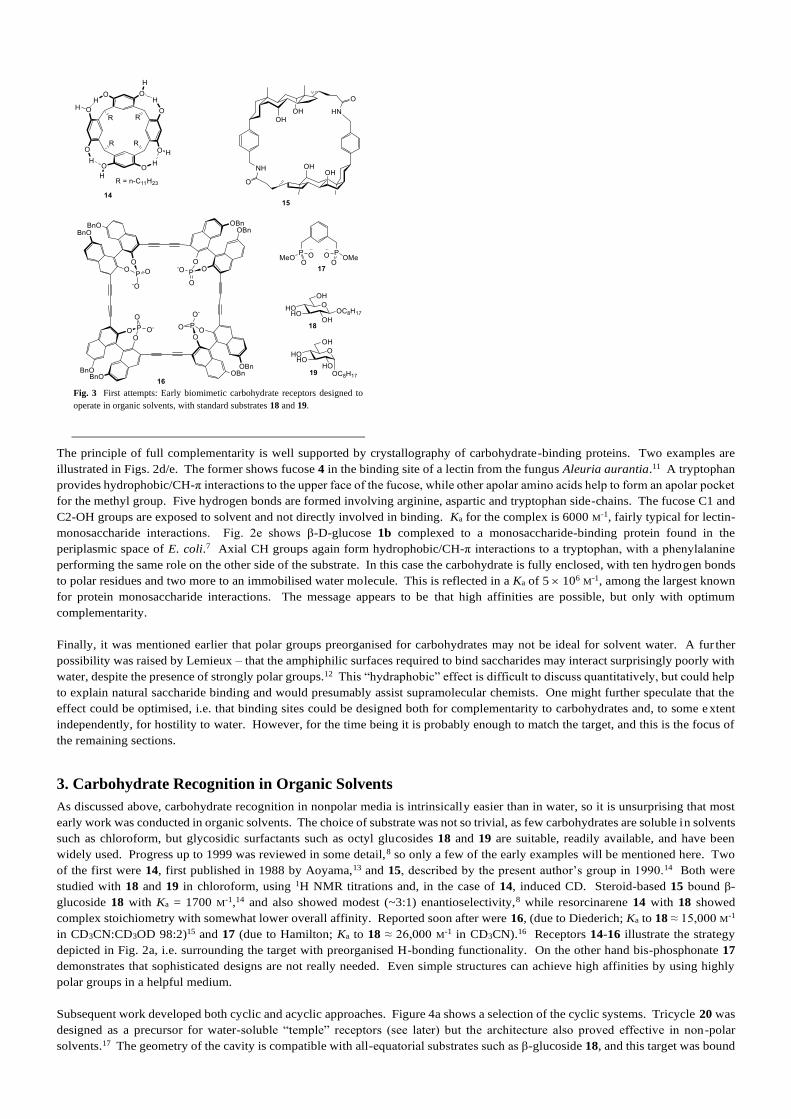
As discussed above, carbohydrate recognition in nonpolar media is intrinsically easier than in water, so it is unsurprising that most
early work was conducted in organic solvents. The choice of substrate was not so trivial, as few carbohydrates are soluble in solvents
such as chloroform, but glycosidic surfactants such as octyl glucosides 18 and 19 are suitable, readily available, and have been
widely used. Progress up to 1999 was reviewed in some detail,8 so only a few of the early examples will be mentioned here. Two
of the first were 14, first published in 1988 by Aoyama,13 and 15, described by the present author’s group in 1990.14 Both were
studied with 18 and 19 in chloroform, using 1H NMR titrations and, in the case of 14, induced CD. Steroid-based 15 bound β-
glucoside 18 with Ka = 1700 M-1,14 and also showed modest (~3:1) enantioselectivity,8 while resorcinarene 14 with 18 showed
complex stoichiometry with somewhat lower overall affinity. Reported soon after were 16, (due to Diederich; Ka to 18 ≈ 15,000 M-1
in CD3CN:CD3OD 98:2)15 and 17 (due to Hamilton; Ka to 18 ≈ 26,000 M-1 in CD3CN).16 Receptors 14-16 illustrate the strategy
depicted in Fig. 2a, i.e. surrounding the target with preorganised H-bonding functionality. On the other hand bis-phosphonate 17
demonstrates that sophisticated designs are not really needed. Even simple structures can achieve high affinities by using highly
polar groups in a helpful medium.
Subsequent work developed both cyclic and acyclic approaches. Figure 4a shows a selection of the cyclic systems. Tricycle 20 was
designed as a precursor for water-soluble “temple” receptors (see later) but the architecture also proved effective in non-polar
solvents.17 The geometry of the cavity is compatible with all-equatorial substrates such as β-glucoside 18, and this target was bound
Fig. 3 First attempts: Early biomimetic carbohydrate receptors designed to
operate in organic solvents, with standard substrates 18 and 19.

with Ka = 300,000 M-1 in chloroform. α-Glucoside 19 was complexed >20 times less strongly. A derivative related to 20 was able
to extract glucose into chloroform from water.18 Bicyclic cage 21, due to Roelens, possesses a similar geometry and also bound 18
in chloroform (Ka ~50,000 M-1).19 In this case binding to α anomer 19 was undetectable. Martinez and Dutasta have studied a range
of bicyclic cryptophanes, typified by 22, with several octyl glycosides as substrates.20 These receptors are less powerful (Ka up to
2500 M-1 in chloroform) but are readily varied (including sense of chirality) and show tuneable selectivities. Macrocycles 23, due
to Abe and Inouye, bind octyl glycosides with Ka up to 5 106 M-1 in DCE, but modest selectivity.21
Among acyclic systems, the 1,3,5-triethyl-2,4,6-tris(aminomethyl)benzene scaffold has proved especially popular. This unit, which
is also present in 21, is nicely sized to encompass a monosaccharide. The parent triamine is highly accessible and can be derivatised
in a variety of ways, which may include loss of C3 symmetry. Figure 4b shows two examples. Bis-phenanthroline 24 is one of many
that have been studied by Mazik,9 and was shown to bind β-glucoside 18 with Ka >105 M-1 in CDCl3, even when 5% DMSO was
added. Hexa-amine 25 belongs to a series made by Roelens and colleagues.5 This podand binds octyl α-D-mannoside 26 quite
strongly (Ka ~104 M-1) in CD3CN, a fairly competitive solvent.22 α-Mannosides are interesting targets as they appear on the surface
of various pathogenic organisms, including HIV-1. Indeed, 26 was tested for inhibition of this virus and showed significant activity.
This highlights an important point; while fully biomimetic carbohydrate receptors should operate in aqueous media, water-solubility
is not a prerequisite for biological activity. Provided a receptor can work in the presence of water, perhaps across a phase boundary,
it has the potential to be useful.
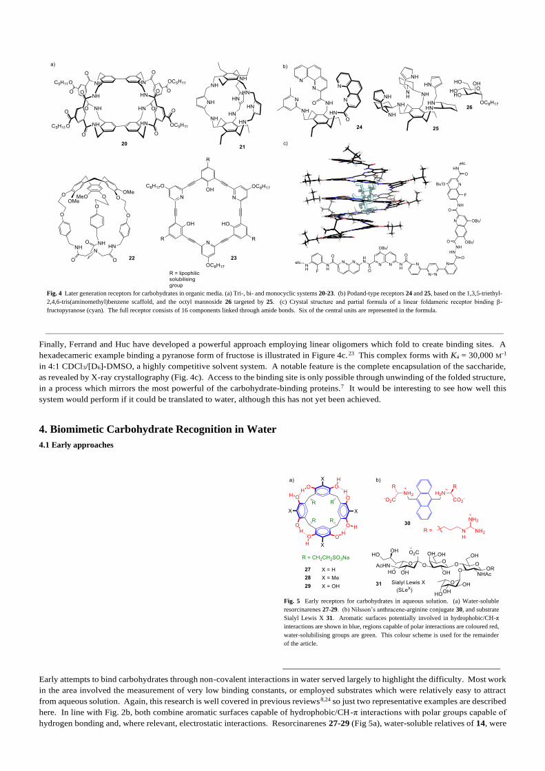
Finally, Ferrand and Huc have developed a powerful approach employing linear oligomers which fold to create binding sites. A
hexadecameric example binding a pyranose form of fructose is illustrated in Figure 4c.23 This complex forms with Ka = 30,000 M-1
in 4:1 CDCl3/[D6]-DMSO, a highly competitive solvent system. A notable feature is the complete encapsulation of the saccharide,
as revealed by X-ray crystallography (Fig. 4c). Access to the binding site is only possible through unwinding of the folded structure,
in a process which mirrors the most powerful of the carbohydrate-binding proteins.7 It would be interesting to see how well this
system would perform if it could be translated to water, although this has not yet been achieved.
4. Biomimetic Carbohydrate Recognition in Water
4.1 Early approaches
Early attempts to bind carbohydrates through non-covalent interactions in water served largely to highlight the difficulty. Most work
in the area involved the measurement of very low binding constants, or employed substrates which were relatively easy to attract
from aqueous solution. Again, this research is well covered in previous reviews8,24 so just two representative examples are described
here. In line with Fig. 2b, both combine aromatic surfaces capable of hydrophobic/CH-π interactions with polar groups capable of
hydrogen bonding and, where relevant, electrostatic interactions. Resorcinarenes 27-29 (Fig 5a), water-soluble relatives of 14, were
Fig. 4 Later generation receptors for carbohydrates in organic media. (a) Tri-, bi- and monocyclic systems 20-23. (b) Podand-type receptors 24 and 25, based on the 1,3,5-triethyl-
2,4,6-tris(aminomethyl)benzene scaffold, and the octyl mannoside 26 targeted by 25. (c) Crystal structure and partial formula of a linear foldameric receptor binding β-
fructopyranose (cyan). The full receptor consists of 16 components linked through amide bonds. Six of the central units are represented in the formula.
Fig. 5 Early receptors for carbohydrates in aqueous solution. (a) Water-soluble
resorcinarenes 27-29. (b) Nilsson’s anthracene-arginine conjugate 30, and substrate
Sialyl Lewis X 31. Aromatic surfaces potentially involved in hydrophobic/CH-π
interactions are shown in blue, regions capable of polar interactions are coloured red,
water-solubilising groups are green. This colour scheme is used for the remainder
of the article.
reported by Aoyama in 1992. No binding was detected to glucose 1, galactose 2 or mannose 3, but fucose 4 formed weak complexes
to the three macrocycles with Ka = 2, 6 and 8 M-1 respectively.25 While fucose is not exactly an easy substrate, it is more hydrophobic
than 1-3 and is presumably more readily separated from water. Higher affinities of ~100 M-1 were achieved by the acyclic receptor
30, due to Nilsson, targeting important oligosaccharides such as Sialyl Lewis X (31).26 However, despite their complexity, these
substrates are probably less challenging than simple monosaccharides (provided selectivity is not required). Large targets p resent
more surface area for non-covalent interactions, and the presence of functional groups other than hydroxyl (e.g. CO2-, NHAc in 31)
provides further opportunities for binding.
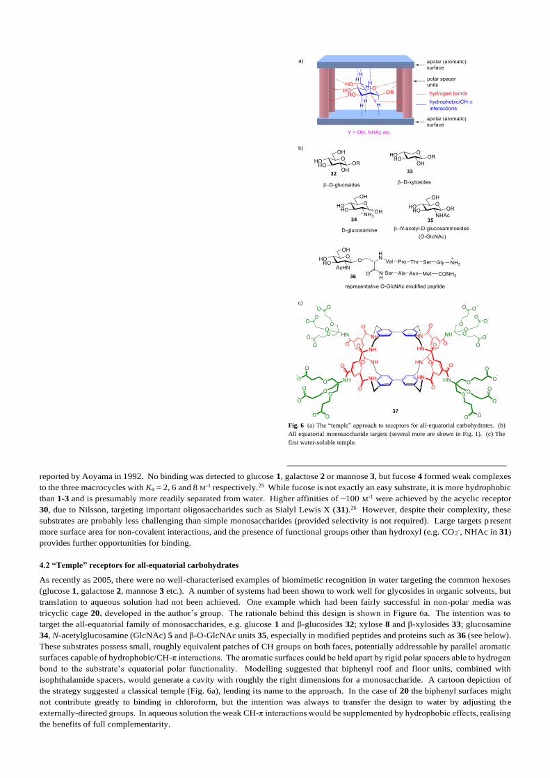
4.2 “Temple” receptors for all-equatorial carbohydrates
As recently as 2005, there were no well-characterised examples of biomimetic recognition in water targeting the common hexoses
(glucose 1, galactose 2, mannose 3 etc.). A number of systems had been shown to work well for glycosides in organic solvents, but
translation to aqueous solution had not been achieved. One example which had been fairly successful in non-polar media was
tricyclic cage 20, developed in the author’s group. The rationale behind this design is shown in Figure 6a. The intention was to
target the all-equatorial family of monosaccharides, e.g. glucose 1 and β-glucosides 32; xylose 8 and β-xylosides 33; glucosamine
34, N-acetylglucosamine (GlcNAc) 5 and β-O-GlcNAc units 35, especially in modified peptides and proteins such as 36 (see below).
These substrates possess small, roughly equivalent patches of CH groups on both faces, potentially addressable by parallel aromatic
surfaces capable of hydrophobic/CH-π interactions. The aromatic surfaces could be held apart by rigid polar spacers able to hydrogen
bond to the substrate’s equatorial polar functionality. Modelling suggested that biphenyl roof and floor units, combined with
isophthalamide spacers, would generate a cavity with roughly the right dimensions for a monosaccharide. A cartoon depiction of
the strategy suggested a classical temple (Fig. 6a), lending its name to the approach. In the case of 20 the biphenyl surfaces might
not contribute greatly to binding in chloroform, but the intention was always to transfer the design to water by adjusting th e
externally-directed groups. In aqueous solution the weak CH-π interactions would be supplemented by hydrophobic effects, realising
the benefits of full complementarity.
Fig. 6 (a) The “temple” approach to receptors for all-equatorial carbohydrates. (b)
All equatorial monosaccharide targets (several more are shown in Fig. 1). (c) The
first water-soluble temple.

The first water-soluble temple receptor was 37 (Fig. 6c), reported in early 2005.27 The tricarboxylate solubilising groups, derived
from dendrimer chemistry, were sufficient to ensure that the cage was monomeric in water and readily studied by NMR. The affinity
of 35 for glucose was low, at Ka = 9 M-1, but selectivity was fair (e.g. 5:1 for glucose 1 vs. galactose 2), and this was the first clear
example of biomimetic recognition of these substrates in water. Moreover, it was later found that 37 performed far better with the
β-O-GlcNAc unit 35. This monosaccharide has special importance as a dynamic post-translational modification of proteins, which
is thought to play roles in diabetes, cancer and neurodenegerative diseases.28 Glycoside 35 (R = Me) was bound with Ka = 630 M-1,
and modified peptide 36 with Ka = 1100 M-1.29 Usefully, the complex between 37 and 35 (R = Me) formed with slow kinetics so
that its 1H NMR spectrum could be observed directly. A full assignment was possible, 48 NOESY signals could be measured, and
the structure shown in Figure 7 could be deduced. A significant feature is the variable geometry of the isophthaloyl spac ers.
Although these are fairly rigid, rotation around C-CO and N-CH2 bonds is possible and this allows the cavity to breathe. As shown
in Figure 7, the orientation with both NH groups directed inwards places the aromatic surfaces 7.3 Å apart, while rotat ing one NH
outwards increases the spacing to ~8.7 Å. In the complex, three of the spacers adopt the NH-in,out conformation while just one
exists as NH-in,in.
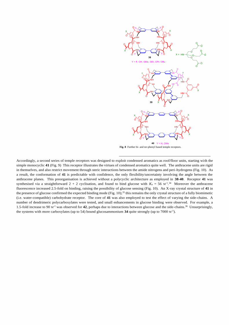
Meanwhile, the biphenyl-based design of 37 was developed in two ways; firstly by adding substituents to moderate binding
properties (as in 38), and secondly by extending the roof/floor units to terphenyls so that disaccharides could be addressed (as in 39
and 40) (see Figure 8). In receptors 38, a series of groups were added to the biphenyl 4,4’-positions, moderating the properties of
the temple roof/floor without substantially changing the conformations.30,31 CH-π interactions are greater for electron-rich π-surfaces
so, for example, it was expected that 38 (Y = F) would be less effective than 37 or than 38 (Y = OR). In the event all variants of 38
bound glucose more strongly than 37, suggesting that electronic effects are not so easy to manipulate.31 On the other hand the highest
affinity of 60 M-1, for 38 (Y = OPr), represented a useful improvement on 37.30 In tetracycle 39, m-terphenyl surfaces were used to
create a binding site for cellobiose 10 and other all-equatorial disaccharides.32 Receptor 39 bound cellobiose with Ka = 600 M-1 and
50-fold selectivity vs. lactose 11, a disaccharide with just one axial OH. The design of 39 was based on the notion that five spacers
would be required to maintain an open cavity. Modelling of the tricyclic alternative 40 (R = H) had suggested that the cavity should
close via a twisting motion. When receptors 40 were prepared somewhat later, the results were unexpected; affinities for cellobiose
10 were raised to 3000 M-1, despite the simpler, less connected framework.33 The success of these receptors highlights the danger of
trusting too much in modelling, especially when predictions are negative.
Although the bi/ter-phenyl units were clearly effective as hydrophobic surfaces, they are probably not ideal. The tendency for Ar-
Ar bonds to twist means that extended planar structures are disfavoured, and this restricts the potential for simultaneous CH-π
interactions as in Fig 2c. It is notable that biology often uses a condensed aromatic, the tryptophan side-chain, to connect with
carbohydrate CH groups (see Figs. 2d,e). Condensed aromatics present large continuously planar surfaces which can allow substrates
to move without losing contact. They also confer greater rigidity and predictability on macrocyclic archi tectures, and possess optical
properties which may be useful in sensing.
Fig. 7 (a) The structure of 37 complexed to GlcNAc-β-OMe (35, R = Me) in water,
as determined by NOESY. (b) Conformational options of the isophthalamide spacer.
The structure in (a) contains one NH-in,in spacer (back left), the remainder adopting
the NH-in,out conformation.
Accordingly, a second series of temple receptors was designed to exploit condensed aromatics as roof/floor units, starting wi th the
simple monocyclic 41 (Fig. 9) This receptor illustrates the virtues of condensed aromatics quite well. The anthracene units are rigid
in themselves, and also restrict movement through steric interactions between the amide nitrogens and peri-hydrogens (Fig. 10). As
a result, the conformation of 41 is predictable with confidence, the only flexibility/uncertainty involving the angle between the
anthracene planes. This preorganisation is achieved without a polycyclic architecture as employed in 38-40. Receptor 41 was
synthesised via a straightforward 2 + 2 cyclisation, and found to bind glucose with Ka = 56 M-1.34 Moreover the anthracene
fluorescence increased 2.5-fold on binding, raising the possibility of glucose sensing (Fig. 10). An X-ray crystal structure of 41 in
the presence of glucose confirmed the expected binding mode (Fig. 10);35 this remains the only crystal structure of a fully biomimetic
(i.e. water-compatible) carbohydrate receptor. The core of 41 was also employed to test the effect of varying the side-chains. A
number of dendrimeric polycarboxylates were tested, and small enhancements in glucose binding were observed. For example, a
1.5-fold increase to 90 M-1 was observed for 42, perhaps due to interactions between glucose and the side-chains.36 Unsurprisingly,
the systems with more carboxylates (up to 54) bound glucosammonium 34 quite strongly (up to 7000 M-1).
Fig. 8 Further bi- and ter-phenyl based temple receptors.

Given the success of biphenyl surfaces in 37 and 38, mutation of these units to pyrenes was a natural development. The pyrene unit
is geometrically similar to biphenyl, but rigidified and with a larger surface area. The first pyrene-based temple was bicyclic 43,
chosen in part because a straightforward (if lengthy) directed synthesis was feasible.37 Dispersing the pyrene surfaces in water was
challenging, and the second-generation dendrimer from 42 was required for full solubility. Performance with monosaccharides was
modest (e.g. Ka to glucose = 120 M-1), but better results were obtained with all-equatorial oligosaccharides such as the cellodextrins
Fig. 10 Top: X-ray crystal structure of the complex between D-glucose and receptor
41.35 Side chains are omitted for clarity, intermolecular H-bonds are shown as green.
Steric clashes between lactam N and peri-H atoms help to control flexibility.
Bottom: Model of receptor 41 in absence of glucose, and variation in fluorescence
emission as glucose is added. The empty receptor retains an open cavity although
the anthracenes can tilt so that they meet at one end. When the glucose enters the
cavity the angle between the anthracenes changes, presumably causing the increase
in fluorescence.
Fig. 9 Temple receptors based on condensed aromatic roof/floor units.
Fig. 11 Oligo- and polysaccharide substrates for receptor 43, and model of the
pseudorotaxane formed from 43 and cellopentaose 47 (n = 3).
peri-Hlactam N
450 500 550 600 650
0
50
100
150
200
250
300
Flu
ore
sce
nce
In
ten
sity
wavelength / nm
added
glucose

(cellulose fragments) 47 (Fig 11). For example cellotetraose 47 (n = 2) was bound with Ka = 12,000 M-1.37 The portals in 43 are
wider than in previous temples, and NMR studies supported the formation of threaded pseudorotaxane complexes with the
oligosaccharides (Fig. 11). Moreover, AFM evidence was obtained for binding of 43 to cellulose 48, forming multiply threaded
polypseudorotaxane complexes. This raises the possibility that threading receptors like 43 could be used to modify the properties
of cellulose, for example promoting solubility in water. Cellulose is the most abundant organic material on earth, so any development
which helps its exploitation could be very valuable.
Tricyclic cage 44 is directly analogous to prototype temple 37, and might seem especially promising as a monosaccharide receptor.
The directed synthesis of 44 proved unrealistic but, by creating all three rings in a single step reaction between 49 and 50 (Fig. 12),
it was possible to prepare both 44 and isomer 45 in acceptable yields.38 The isomers were challenging to separate and identify, but
both had interesting binding properties. Affinities to glucose were moderate (120 and 190 M-1 for 44 and 45 respectively), but
binding to the β-O-GlcNAc unit 35 was promising. For example tricycle 45 bound GlcNAc-β-OMe 35 (R = Me) with Ka ~18,000
M-1, while the glycopeptide 36 (Fig. 6) was complexed to 44 with Ka = 67,000 M-1. These affinities are high enough to suggest that
applications in glycobiology, for example the detection of O-GlcNAc in specific protein environments, may soon be feasible.
Finally, intermediate 49 was also combined with triamine 51 (Fig. 12) to give the chiral bicyclic receptors -46.39 Although the
enantiomers could not be separated, they could be studied independently in the racemate. This system showed the highest affinities
yet for simple underivatised monosaccharides; glucose 1 and GlcNAc 5 were bound with Ka = 250 and 1280 M-1 respectively (in
each case by one unidentified enantiomer). In the case of GlcNAc 5, the affinity for the second enantiomer could also be measured,
at 80 M-1, implying 16:1 enantioselectivity.
In summary, a range of temple receptors were reported between 2005 and 2017, featuring various roof/floor units separated by the
ubiquitous isophthalamide spacer groups. They were not all targeted at glucose, and indeed some performed quite well in other
respects. However, for those with glucose-sized cavities it is interesting to survey their performance against this important substrate.
Table 1 gives a list of binding constants, as well as selectivities vs. galactose, a benchmark competitor. There is a clear sense of
progression as affinities rise from very low to a level which might be termed respectable, considering the challenge. Although the
culminating value of 250 M-1 is still very small by general biological standards, it is worth noting that common lectins are not so far
ahead. For example the lectin used to bind glucose, Concanavalin A, shows Ka of just 520 M-1,6 around double the synthetic system.
For GlcNAc the temples’ performance is arguably quite impressive. The standard lectin for β-GlcNAc is Wheat Germ Agglutinin,
which binds 35 (R = Me) with Ka = 730 M-1.38 In comparison, the affinity of 45 for the same substrate is 18,000 M-1, twenty-five
times higher.
Table 1. Binding of glucose to temple monosaccharide receptors; association
constants (Ka) and selectivities vs. galactose where available.
Receptor Ka (M-1) to D-
glucose in water
Selectivity vs. D-
galactose
37 9 4.5
38 (R = OPr) 60 20
41 56 14
42 90 13
43 120 7
44 120
45 190
46a 250
a Unidentified enantiomer
Fig. 12 Key synthetic steps leading to temple receptors 44 – 46.
Fig. 13 Model of uric acid monoanion 52 bound to receptor 41. Receptor side-
chains are omitted for clarity. The four receptor NH groups form hydrogen bonds to
the urate N3 and 6CO. The anthracene surfaces are nearly parallel, in contrast to the
glucose complex in Fig. 10, implying a good fit between host and guest.
This said, if the aim is to create useful carbohydrate binding agents, these temples are not quite satisfactory. For most ap plications
one would want higher affinities, and accessibility is a problem in many cases. Moreover there is a general issue which remains
hidden during studies of carbohydrate recognition, but becomes apparent when applications are considered. To illustrate, we return
to the anthracene-based monocyclic receptors such as 41. Of all the systems described above, these appear to have the most real-
world potential. They are easy to synthesise, feature optical reporting, and bind glucose with affinities which are quite su itable for
monitoring in biological samples such as blood. Blood glucose levels are around 6 mM, and more powerful receptors would be
saturated and insensitive to variations around this concentration. Unfortunately, while these molecules can be used to monitor
glucose in water, they give no signal variation in blood serum. They clearly bind something else and, considering the dimensions
of the isophthalamide spacer (Fig. 7b), this may not be surprising. In the NH-in,in conformation, which pertains in 41, the aromatic
surfaces are 7.3 Å apart. This seems to be slightly too small for a carbohydrate. In the NMR structure of 37∙GlcNAc-β-OMe (Fig.
7a) most spacers adopted the wider NH-in,out conformation, while in 41∙glucose (Fig. 10) the guest sits towards one end of the
cavity, pushing the anthracene surfaces apart and seemingly unable to reach the centre. However, 7.3 Å is very close to the inter-
base spacing in DNA and almost ideal for an aromatic substrate. A polar aromatic which can form H-bonds to the lactams is
potentially an excellent substrate, and a number of such molecules do in fact bind the bis-anthracene macrocycles with Ka as high as
107 M-1. Uric acid, present as monoanion 52, occurs in blood at quite high levels and is probably the main interferent (see Fig. 13).
The isophthalamide spacer is ubiquitous in all the temples discussed thus far, so this issue is likely to be general whether or not it
has been investigated. Binding saccharides in water is difficult enough, but it is still more challenging to ignore everything else that
might be present.
Although these systems may not find practical applications, they have contributed theoretically. Unlike proteins, synthetic receptors
can be used in a wide range of solvents, especially when peripheral modifications can be made. This allows studies of solven t
effects, which can throw light on the driving force for binding. Receptors 37 and 39 were studied with glucose 1 and cellobiose 10
respectively, in a series of polar solvent mixtures (water + MeOH/DMSO/MeCN).40 The corresponding organic-soluble precursor
macrocycles were also investigated with octyl glycosides in chloroform + MeOH mixtures. In the latter experiments, H-bonding is
dominant and the addition of methanol to the chloroform (increasing solvent polarity) reduced affinities as expected. However, in
the polar solvent mixtures, addition of organic solvents to water (decreasing polarity) also depressed binding. This implies that
water is less comfortable than the organic solvents in the amphiphilic receptor cavities, and that hydrophobic/hydraphobic ef fects
do make a contribution to binding. If 37 and 39 are accepted as models of proteins, this conclusion extends to natural carbohydrate
recognition and helps to elucidate an important biological phenomenon.
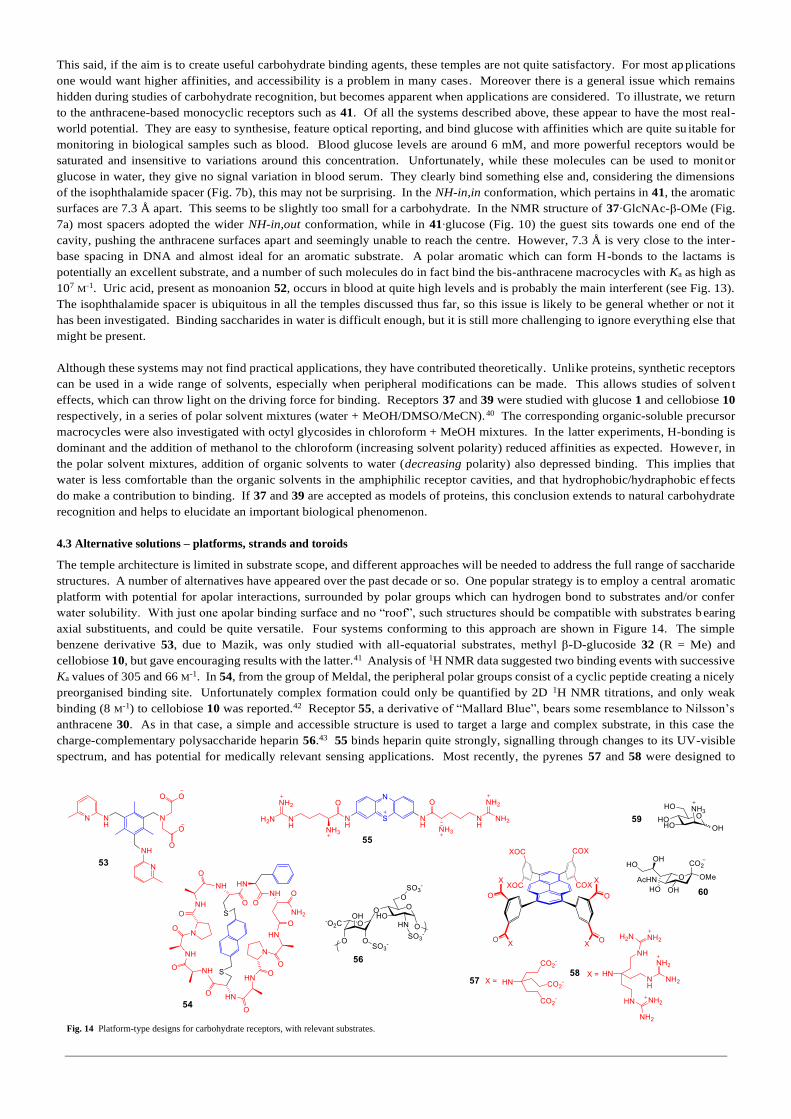
4.3 Alternative solutions – platforms, strands and toroids
The temple architecture is limited in substrate scope, and different approaches will be needed to address the full range of saccharide
structures. A number of alternatives have appeared over the past decade or so. One popular strategy is to employ a central aromatic
platform with potential for apolar interactions, surrounded by polar groups which can hydrogen bond to substrates and/or confer
water solubility. With just one apolar binding surface and no “roof”, such structures should be compatible with substrates b earing
axial substituents, and could be quite versatile. Four systems conforming to this approach are shown in Figure 14. The simple
benzene derivative 53, due to Mazik, was only studied with all-equatorial substrates, methyl β-D-glucoside 32 (R = Me) and
cellobiose 10, but gave encouraging results with the latter.41 Analysis of 1H NMR data suggested two binding events with successive
Ka values of 305 and 66 M-1. In 54, from the group of Meldal, the peripheral polar groups consist of a cyclic peptide creating a nicely
preorganised binding site. Unfortunately complex formation could only be quantified by 2D 1H NMR titrations, and only weak
binding (8 M-1) to cellobiose 10 was reported.42 Receptor 55, a derivative of “Mallard Blue”, bears some resemblance to Nilsson’s
anthracene 30. As in that case, a simple and accessible structure is used to target a large and complex substrate, in this case the
charge-complementary polysaccharide heparin 56.43 55 binds heparin quite strongly, signalling through changes to its UV-visible
spectrum, and has potential for medically relevant sensing applications. Most recently, the pyrenes 57 and 58 were designed to
Fig. 14 Platform-type designs for carbohydrate receptors, with relevant substrates.

present two identical binding sites, aimed primarily at substrates with charged axial groups. Accordingly, anionic 57 bound
mannosamine 59, and cationic 58 bound sialoside 60, with first (1:1) Ka values of 3000 M-1 and 1300 M-1 respectively.44
Complexation on both faces was detected, mimicking the multivalency shown by many lectins.
A second approach involves linear oligomeric structures containing aromatic and polar units. Three examples are shown in Figure
15. Nonaphenyl 61 forms a homo-double helix in water, but in the presence of heptasaccharides this unwinds and binding to the
carbohydrate may be detected by CD. However, the effect is difficult to quantify because of complex stoichiometries.45 The phenol-
pyridine oligomer 62, an acyclic water-soluble relative of 23, showed evidence of very weak binding to glucose, but was more
successful with aminosugars; its affinity for glucosamine 34 was measured as Ka = 2000 M-1.46 Both 61 and 62 are composed of
rigid units so that conformational freedom is limited. It may be difficult to predict exactly how they will fold, but the chances of
clefts or cavities may be relatively high. The third example, peptide 63, is more flexible and emerged from a different concept. In
a study of borylated peptides as oligosaccharide receptors, Hall and coworkers made a library of pentapeptides each containing two
-B(OR)2 groups, and screened for binding to the Thomsen-Friedenreich antigen 64.47 Disaccharide 64 is a tumour-associated antigen
and so an important target. A diborylated analogue of 63 emerged from the screening and was found to bind 64 with Ka = 2000 M-1
(presumably in non-biomimetic fashion, involving B-O bonds). More importantly, from the viewpoint of this article, 63 itself was
prepared as a control and retained much of the affinity; Ka was lowered, but only as far as 400 M-1. The result suggests that short
peptides with aromatic components could be quite effective as biomimetic carbohydrate receptors, and might be discoverable by
combinatorial methods.
Macrocyclic, toroidal structures have also been successful in some cases. The coordination capsule 65 (Fig. 16), due to Yoshizawa,
possesses an apolar interior composed of extended aromatic surfaces.48 Although this might not seem promising for carbohydrate
binding, in that some polar interactions would usually be expected, macrocycle 65 is an effective and selective receptor for sucrose
66. The affinity for sucrose was measured at 1170 M-1, and the complex was formed in the presence of several disaccharide
competitors (e.g. cellobiose 10, lactose 11, maltose 12). Although it is interesting that such a hydrophobic cavity can succeed,
sucrose is probably a special case, capable of adopting a conformation with a substantially apolar exterior. Cucurbiturils such as 67
also possess hydrophobic interiors, in this case supplemented by externally directed polar carbonyl oxygens. The group of Kim have
shown that 67 is a good receptor for aminosugars, binding protonated glucosamine 34, galactosamine 68 and mannosamine 59 with
impressive affinities of 4400, 16000 and 1900 M-1.49 At first sight these results may seem surprising, as host and guests are not
obviously complementary. However, they are less remarkable when one considers that other ammonium cations are bound far more
strongly; for example cyclo-octylammonium with Ka = 3 1011 M-1. The affinities for aminosugars probably reflect the extreme
hydrophobicity of 67 more than its compatibility with carbohydrates.
Finally the macrocycle 69, due to Francesconi, Roelens, Nativi and colleagues, possesses an amphiphilic cavity with anthracenyl
aromatic surfaces and diaminocarbazoles as polar spacers.50 This receptor bears similarities to the temple family discussed in Section
Fig. 15 Linear oligomeric receptors for carbohydrate recognition in water.
Fig. 16 Macrocyclic carbohydrate receptors.

4.2, especially the bis-anthracenes 41 and 42. Its behaviour is complicated by dimerisation leading to multiple binding
stoichiometries, but could be quantified using “median binding concentration” (BC050), a parameter designed by the authors to reflect
overall binding ability. For all-equatorial methyl β-D-glucoside 32 (R = Me), BC050 values equivalent to Ka ~1000 M-1 were
measured, comparing well with the temples discussed in section 4.2. Interestingly, however, the highest affinities of Ka ~2000 M-1
were measured for methyl α-L-fucoside 70, with two axial groups. Molecular modelling guided by NOESY suggested the structure
for 69∙70 shown in Figure 17. Complex formation appears to be driven by formation of five H-bonds as well as hydrophobic/CH-π
interactions. The distance between anthracene units is ~8 Å, slightly larger than for 41, and this may be advantageous (see next
section). The structure also shows how some axial substitutions may be compatible with parallel aromatic surfaces, suggesting that
temple and related architectures may be more versatile than originally supposed.
4.4 The Hexaurea Temple – hitting the sweet spot for glucose
As described in Section 4.2, the temple architecture was designed to address all-equatorial targets, of which the most important is
glucose. On the positive side, moderate affinities (up to 250 M-1) were achieved, enough for some applications, while selectivities
against most other carbohydrates were fairly good (see Table 1). Unfortunately the most promising candidate 41 suffered from
strong binding to non-carbohydrate substrates, and it seemed likely that this issue would affect the whole family of temple receptors.
The problem lies with the isophthalamide spacer, which allows a spacing of 7.3 Å between roof and floor surfaces (Fig 7b), an d in
some designs (e.g. 41) enforces this distance. 7.3 Å is excellent for binding aromatics, but at least 1 Å too small for carbohydrates.
Fig. 17 Anthracene-carbazole macrocycle 69, methyl α-L-fucoside 70, and proposed
structure of 69∙70. 69 is modelled as the neutral tetraphosphonic acid. Five NH···O
hydrogen bonds, between 1.9 and 2.1 Å, are formed between host and guest. The
spacing between anthracene units is ~8 Å. We thank Dr. O. Francesconi for
providing the coordinates of the modelled complex.
Fig. 18 o-Phenylene-bis-urea, an alternative spacer for temple receptors.
Considering alternatives which might expand the cavity, the bis-urea unit 71 seemed interesting (Fig. 18). One bond longer than the
isophthalamide, it places the aromatic surfaces up to 8.9 Å apart. When presented with vicinal OR groups it is able to make four
hydrogen bonds, as in 72, while the spacing contracts slightly to ~8.4 Å. Disadvantages are its ability to access conformation 73
with an intramolecular H-bond, and the tendency of oligoureas to insolubility. However, its potential is especially clear when it is
used to separate 1,3,5-trisubstituted benzene units. Although such small roof/floor components could not be used with the
isophthalamide spacers, the extra length of the bis-ureas allows shrinkage of the apolar surfaces while maintaining cavity volume.
Hexaurea 7451 (Fig. 19), designed to prototype this new family of temples, serves to illustrate its advantages. The interior of 74 can
accept a monosaccharide and, being C3 symmetric, is roughly congruent with a six-membered pyranose ring. When β-D-glucose is
placed inside the cavity, the complementarity is remarkable (Fig. 19). All the glucose oxygens bar one (the anomeric OH), and all
the ureas bar one, are involved in intermolecular hydrogen bonds, and all the axial CH make contact with the cavity roof/floo r.
Altogether there are ten H-bonds and five CH-π interactions to drive complex formation. In the absence of glucose, modelling
indicates that the geometry of the cage changes very little, maintaining an open cavity. The contrast with earlier temples (Section
4.2) is stark. These systems provided plenty of π-surface for apolar interactions, but polar interactions were essentially left to chance
and certainly not optimal. The hexaurea temple was the first to feature a rationally conceived, almost comprehensive pattern of polar
interactions.
In addition to the core hexaurea cage, 74 incorporates two further design elements with important roles. Three nonacarboxylate
solubilising groups, previously used for many of the earlier temples, guarantee water-solubility, and six ethyl groups constrain the
conformational freedom of the core and promote an open binding site. Both these features also assist with the synthesis, shown in
Figure 20.51 Triethyltriamine 51 is readily available,52 and has featured in several earlier carbohydrate receptors.5,19,9,22,39 Its
derivatives favour an up-down alternating conformation which presumably assists the last step. The solubilising groups are O-
protected until the final step and help to maintain solubility in organic media throughout the synthesis. It is notable that yields in
the cyclisation step were greatly improved by addition of octyl β-D-glucoside 18. The synthesis in Fig. 20 is remarkably short,
especially in comparison to some of the earlier temples discussed in Section 4.2.

Receptor 74 was expected to bind strongly and selectively to β-glucosyl units, and did not disappoint. Results from binding studies
are summarised in Figure 21. Glucose itself was bound with Ka ~18,000 M-1 in aqueous solution, determined by both 1H NMR and
ITC. This represents a seventy-fold increase over the best of the earlier temples (see Table 1), and is higher than most lectin-
monosaccharide interactions (cf. Fig. 2d).6. Only the very strongest natural glucose receptors show greater affinities (Fig. 2e).7
Methyl β-D-glucoside 32 (R = Me) was also bound, with Ka = ~8000 M-1; this is consistent with the model in Fig. 19 which places
the anomeric OH in a portal, such that some modifications should be allowed. A few other all-equatorial substrates were also bound
fairly well: xylose 8 at 5,800 M-1; glucuronic acid 76 at 5,300 M-1; 2-deoxyglucose 77 at 725 M-1 (though this represents a 25-fold
decrease from glucose, resulting from loss of just one OH group). Among other carbohydrates, galactose 2, mannose 3, ribose 9,
fructose 78 and cellobiose 10 are bound to measurable extents (Fig. 21). Curiously, 74 is probably the strongest biomimetic galactose
receptor yet reported (Ka = 180 M-1), yet shows the highest selectivity for glucose against galactose (100:1).
Perhaps equally important is the list of compounds for which no binding could be detected. Based on ITC data for cellobiose (above)
it is likely that affinities down to ~30 M-1 should have been measurable. Titrations were performed for several other carbohydrates
(fucose 4, N-acetylglucosamine 5, methyl α-D-glucoside 79, maltose 12, ascorbic acid 80), linear polyols (mannitol 81, D-gluconic
acid 82) and a range of biologically relevant aromatic compounds (83 – 89, see Fig. 21). All gave negative results. Finally 74 was
tested in serum, where it was found to bind glucose with Ka = 11,000 M-1. Although this is slightly lower than in water, it should not
be enough to preclude applications in biological media.
Fig. 19 Hexaurea temple 74, and two representations of its complex with β-D-glucose 1b (side-chains omitted for clarity). Ten hydrogen bonds (1.95 – 2.48 Å) are formed
between host and guest. The spacing between roof/floor benzene units is ~8.4 Å.
Fig. 20 Synthetic route to hexaurea temple 74.

Clearly the hexaurea temple represents a major step forward for biomimetic carbohydrate recognition. At the outset of this research
three decades ago, achieving any measurable binding to simple monosaccharides in water seemed enough of a challenge. Just a few
years ago, an affinity of ~100 M-1 was considered significant (and was not always accompanied by good selectivity, especially against
non-carbohydrates). Receptor 74 operates on a different level, with affinities and selectivities which might be considered high for
proteins. Its selectivity is perhaps especially remarkable. According to SciFinder there are ~2,000,000 molecules with the same
molecular weight as glucose, or less. Of these, the hexaurea temple binds glucose, xylose, deoxyglucose (less well) and probably a
few other closely related structures (e.g. other deoxy- and fluorinated glucoses are likely substrates). There can be few other products
of supramolecular chemistry which perform an important and difficult task so well.
Fig. 21 Substrates and results for binding studies to 74 in 10 mM aqueous phosphate
buffer (pH = 7.4). Numbers in red are binding constants (Ka, M-1) obtained from 1H
NMR and/or ITC titrations. (a) All-equatorial substrates showing moderate to strong
binding. (b)Weakly bound substrates. (c) Binding undetectable by ITC.

5. Conclusions
The story told in this article ends on a positive note. At least for glucose, probably the most important substrate, we have a receptor
that is fully biomimetic. The hexaurea temple 74 employs the same strategy and interactions as biology, works in biological
environments, and performs to biological standards. Moreover, the prospects for real-world applications seem good. The receptor
was commercialised through Ziylo, a company which had been spun out of the University of Bristol a few years previously. In
August 2018 Ziylo (with rights to 74) was sold to Novo Nordisk for a sum which could exceed $800 million depending on future
developments. Novo Nordisk is the world’s major supplier of insulin, and plans to exploit 74 in glucose-responsive variants of their
products.4 A new company, Carbometrics, was created to replace Ziylo in Bristol, and is collaborating with Novo Nordisk in this
enterprise. At time of writing the research is still in its early stages, but the size of the deal attests to the confidence, of all concerned,
that the venture can be successful. Meanwhile, Carbometrics has retained the right to exploit the hexaurea temple in non-therapeutic
applications, and is also working towards this goal.
On the scientific side we can conclude, firstly, that the principles outlined in Fig 2 seem to be correct; complementing both polar
and apolar substrate surfaces is the key to biomimetic carbohydrate recognition. This is no surprise given the evidence from protein
structures, but it is welcome to find support from synthetic systems. Secondly, persistent efforts can lead to remarkably good
performance. In at least one case there is a fairly simple and accessible receptor structure which works as well as we could hope.
The question remains whether this will be an exception. We may find that other substrates are more difficult to accommodate, either
because designs cannot be found or because promising structures are inaccessible. This said, optimal performance may not be
needed for all applications, and useful affinities may be easier to achieve for some substrates. For example, tricycles 44 and 45 bind
β-O-GlcNAc derivatives with Ka up to 70,000 M-1, yet are surely not ideal for these targets.
Another issue is the role of combinatorial chemistry in carbohydrate recognition. For small monosaccharide targets, where a
precisely defined cavity is needed, one feels that rational design will be more effective. However the balance may be different for
larger substrates such as oligosaccharide antigens. Here it should be easier to achieve a useful degree of binding but more difficult
to make rational predictions, especially as selectivity will be critical. Hall’s discovery47 that oligomer 63 binds disaccharide 64
suggests that a combinatorial approach could be successful. Libraries which incorporate rationally designed cavities and modular,
variable regions may represent the best of both worlds, and it will be interesting to see if such strategies can be developed .
Conflicts of interest The author was a director and shareholder of Ziylo at the time of the sale to Novo Nordisk, and is now a director and shareho lder of
Carbometrics.
Acknowledgements
The author thanks the many colleagues, co-workers and collaborators who contributed to research described in this article. Funding
for the work has been provided by many organisations including Enterprise Ireland, the European Union, EPSRC, the Royal Society,
and Novo Nordisk.
Notes and references
References 1. A guide into glycosciences: How chemistry, biochemistry and biology cooperate to crack the sugar code, D. Solis, N. V. Bovin, A. P.
Davis, J. Jiménez-Barbero, A. Romero, R. Roy, K. Smetana and H. J. Gabius, Biochim. Biophys. Acta-Gen. Subj., 2015, 1850, 186-235. 2. The challenges of glycan recognition with natural and artificial receptors, S. Tommasone, F. Allabush, Y. K. Tagger, J. Norman, M. Kopf,
J. H. R. Tucker and P. M. Mendes, Chem. Soc. Rev., 2019, 48, 5488-5505. 3. Glucose Sensing in Supramolecular Chemistry, X. L. Sun and T. D. James, Chem. Rev., 2015, 115, 8001-8037. 4. Glucose-responsive insulin by molecular and physical design, N. A. Bakh, A. B. Cortinas, M. A. Weiss, R. S. Langer, D. G. Anderson, Z.
Gu, S. Dutta and M. S. Strano, Nat Chem, 2017, 9, 937-943. 5. Biomimetic Carbohydrate-Binding Agents (CBAs): Binding Affinities and Biological Activities, O. Francesconi and S. Roelens,
ChemBioChem, 2019, 20, 1329-1346. 6. Structure and energetics of protein-carbohydrate complexes, E. J. Toone, Curr. Opin. Struct. Biol., 1994, 4, 719-728. 7. Protein-Carbohydrate interactions: basic molecular features, F. A. Quiocho, Pure & Appl. Chem., 1989, 61, 1293-1306. 8. Carbohydrate recognition through noncovalent interactions: A challenge for biomimetic and supramolecular chemistry, A. P. Davis and R.
S. Wareham, Angew. Chem., Int. Ed., 1999, 38, 2978-2996. 9. Recent developments in the molecular recognition of carbohydrates by artificial receptors, M. Mazik, RSC Advances, 2012, 2, 2630-2642. 10. Carbohydrate-Aromatic Interactions, J. L. Asensio, A. Arda, F. J. Canada and J. Jimenez-Barbero, Acc. Chem. Res., 2013, 46, 946-954. 11. Crystal structure of fungal lectin - Six-bladed beta-propeller fold and novel fucose recognition mode for Aleuria aurantia lectin, M.
Wimmerova, E. Mitchell, J. F. Sanchez, C. Gautier and A. Imberty, J. Biol. Chem., 2003, 278, 27059-27067. 12. How water provides the impetus for molecular recognition in aqueous solution, R. U. Lemieux, Acc. Chem. Res., 1996, 29, 373-380. 13. Highly cooperative binding of alkyl glucopyranosides to the resorcinol cyclic tetramer due to intracomplex guest-guest hydrogen bonding:
solvophobicity/solvophilicity control by an alkyl group of the geometry, stoichiometry, stereoselectivity, and cooperativity, Y. Kikuchi,
Y. Tanaka, S. Sutaro, K. Kobayashi, H. Toi and Y. Aoyama, J. Am. Chem. Soc., 1992, 114, 10302-10306, and references cited therein.

14. Artificial Receptors for Carbohydrate Derivatives, R. P. Bonar-Law, A. P. Davis and B. A. Murray, Angew. Chem., Int. Ed. Engl., 1990,
29, 1407. 15. A new family of chiral binaphthyl-derived cyclophane receptors: complexation of pyranosides, S. Anderson, U. Neidlein, V. Gramlich
and F. Diederich, Angew. Chem., Int. Ed. Engl., 1995, 34, 1596-1600. 16. Molecular recognition of carbohydrates: strong binding of alkyl glycosides by phosphonate derivatives, G. Das and A. D. Hamilton, J.
Am. Chem. Soc., 1994, 116, 11139. 17. A tricyclic polyamide receptor for carbohydrates in organic media, A. P. Davis and R. S. Wareham, Angew. Chem., Int. Ed., 1998, 37,
2270-2273. 18. Phase Transfer of Monosaccharides Through Non-Covalent Interactions; Selective Extraction of Glucose by a Lipophilic Cage Receptor,
T. J. Ryan, G. Lecollinet, T. Velasco and A. P. Davis, Proc. Natl. Acad. Sci. USA, 2002, 99, 4863-4866. 19. A self-assembled pyrrolic cage receptor specifically recognizes beta-glucopyranosides, O. Francesconi, A. Ienco, G. Moneti, C. Nativi and
S. Roelens, Angew. Chem., Int. Ed., 2006, 45, 6693-6696. 20. Emergence of Hemicryptophanes: From Synthesis to Applications for Recognition, Molecular Machines, and Supramolecular Catalysis,
D. W. Zhang, A. Martinez and J. P. Dutasta, Chem. Rev., 2017, 117, 4900-4942. 21. D-3h-Symmetrical Shape-Persistent Macrocycles Consisting of Pyridine-Acetylene-Phenol Conjugates as an Efficient Host Architecture
for Saccharide Recognition, H. Abe, T. Yoneda, Y. Ohishi and M. Inouye, Chem. Eur. J., 2016, 22, 18944-18952. 22. Antiviral Activity of Synthetic Aminopyrrolic Carbohydrate Binding Agents: Targeting the Glycans of Viral gp120 to Inhibit HIV Entry,
O. Francesconi, C. Nativi, G. Gabrielli, I. De Simone, S. Noppen, J. Balzarini, S. Liekens and S. Roelens, Chem. Eur. J., 2015, 21, 10089-
10093. 23. Iterative design of a helically folded aromatic oligoamide sequence for the selective encapsulation of fructose, N. Chandramouli, Y.
Ferrand, G. Lautrette, B. Kauffmann, C. D. Mackereth, M. Laguerre, D. Dubreuil and I. Huc, Nature Chem., 2015, 7, 334-341. 24. Progress in biomimetic carbohydrate recognition, D. B. Walker, G. Joshi and A. P. Davis, Cell. Mol. Life Sci., 2009, 66, 3177-3191. 25. Complexation of hydrophobic sugars and nucleosides in water with tetrasulfonate derivatives of resorcinol cyclic tetramer having a
polyhydroxy aromatic cavity: importance of guest-host CH-pi interactions, K. Kobayashi, Y. Asakawa, Y. Kato and Y. Aoyama, J. Am.
Chem. Soc., 1992, 114, 10307-10313. 26. Amphiphilic anthracene-amino acid conjugates as simple carbohydrate receptors in water, J. Billing, H. Grundberg and U. J. Nilsson,
Supramol. Chem., 2002, 14, 367-372. 27. Carbohydrate recognition in water by a tricyclic polyamide receptor, E. Klein, M. P. Crump and A. P. Davis, Angew. Chem., Int. Ed., 2005,
44, 298-302. 28. Protein O-GlcNAcylation: emerging mechanisms and functions, X. Y. Yang and K. V. Qian, Nature Reviews Molecular Cell Biology,
2017, 18, 452-465. 29. A synthetic lectin for O-linked beta-N-acetylglucosamine, Y. Ferrand, E. Klein, N. P. Barwell, M. P. Crump, J. Jimenez-Barbero, C. Vicent,
G. J. Boons, S. Ingale and A. P. Davis, Angew. Chem., Int. Ed., 2009, 48, 1775-1779. 30. A synthetic lectin for beta-glucosyl, N. P. Barwell, M. P. Crump and A. P. Davis, Angew. Chem., Int. Ed., 2009, 48, 7673-7676. 31. Substituent effects in synthetic lectins - exploring the role of CH-pi interactions in carbohydrate recognition, N. P. Barwell and A. P. Davis,
J. Org. Chem., 2011, 76, 6548-6557. 32. A synthetic lectin analog for biomimetic disaccharide recognition, Y. Ferrand, M. P. Crump and A. P. Davis, Science, 2007, 318, 619-622. 33. High-Affinity Disaccharide Binding by Tricyclic Synthetic Lectins, B. Sookcharoenpinyo, E. Klein, Y. Ferrand, D. B. Walker, P. R.
Brotherhood, C. Ke, M. P. Crump and A. P. Davis, Angew. Chem., Int. Ed., 2012, 51, 4586-4590. 34. A simple and accessible synthetic lectin for glucose recognition and sensing, C. Ke, H. Destecroix, M. P. Crump and A. P. Davis, Nature
Chem., 2012, 4, 718-723. 35. Crystal structure of a complex between beta-glucopyranose and a macrocyclic receptor with dendritic multicharged water solubilizing
chains, P. K. Mandal, B. Kauffmann, H. Destecroix, Y. Ferrand, A. P. Davis and I. Huc, Chem. Commun., 2016, 52, 9355-9358. 36. Affinity Enhancement by Dendritic Side Chains in Synthetic Carbohydrate Receptors, H. Destecroix, C. M. Renney, T. J. Mooibroek, T.
S. Carter, P. F. N. Stewart, M. P. Crump and A. P. Davis, Angew. Chem., Int. Ed., 2015, 54, 2057-2061. 37. A threading receptor for polysaccharides, T. J. Mooibroek, J. M. Casas-Solvas, R. L. Harniman, C. M. Renney, T. S. Carter, M. P. Crump
and A. P. Davis, Nature Chem., 2016, 8, 69-74. 38. Synthetic Receptors for the High-Affinity Recognition of O-GlcNAc Derivatives, P. Rios, T. S. Carter, T. J. Mooibroek, M. P. Crump, M.
Lisbjerg, M. Pittelkow, N. T. Supekar, G.-J. Boons and A. P. Davis, Angew. Chem., Int. Ed., 2016, 55, 3387-3392. 39. Enantioselective carbohydrate recognition by synthetic lectins in water, P. Rios, T. J. Mooibroek, T. S. Carter, C. Williams, M. R. Wilson,
M. P. Crump and A. P. Davis, Chem. Sci., 2017, 8, 4056-4061. 40. Solvent effects in carbohydrate binding by synthetic receptors: Implications for the role of water in natural carbohydrate recognition, E.
Klein, Y. Ferrand, N. P. Barwell and A. P. Davis, Angew. Chem., Int. Ed., 2008, 47, 2693-2696. 41. Carboxylate-based receptors for the recognition of carbohydrates in organic and aqueous media, M. Mazik and H. Cavga, J. Org. Chem.,
2006, 71, 2957-2963. 42. Specific recognition of disaccharides in water by an artificial bicyclic carbohydrate receptor, T. Reenberg, N. Nyberg, J. O. Duus, J. L. J.
van Dongen and M. Meldal, Eur. J. Org. Chem., 2007, 5003-5009. 43. Mallard Blue: A High-Affinity Selective Heparin Sensor That Operates in Highly Competitive Media, S. M. Bromfield, A. Barnard, P.
Posocco, M. Fermeglia, S. Pricl and D. K. Smith, J. Am. Chem. Soc., 2013, 135, 2911-2914. 44. Platform Synthetic Lectins for Divalent Carbohydrate Recognition in Water, T. S. Carter, T. J. Mooibroek, P. F. N. Stewart, M. P. Crump,
M. C. Galan and A. P. Davis, Angew. Chem., Int. Ed., 2016, 55, 9311-9315. 45. Double helical oligoresorcinols specifically recognize oligosaccharides via heteroduplex formation through noncovalent interactions in
water, H. Goto, Y. Furusho and E. Yashima, J. Am. Chem. Soc., 2007, 129, 9168-9174. 46. Saccharide Recognition and Helix Formation in Water with an Amphiphilic Pyridine-Phenol Alternating Oligomer, Y. Ohishi, H. Abe and
M. Inouye, Eur. J. Org. Chem., 2017, 6975-6979. 47. Synthesis, and screening of a library of peptidyl bis(boroxoles) as oligosaccharide receptors in water: Identification of a receptor for the
tumor marker TF-antigen disaccharide, A. Pal, M. Berube and D. G. Hall, Angew. Chem., Int. Ed., 2010, 49, 1492-1495. 48. A polyaromatic nanocapsule as a sucrose receptor in water, M. Yamashina, M. Akita, T. Hasegawa, S. Hayashi and M. Yoshizawa, Science
Advances, 2017, 3, e1701126. 49. Cucurbit 7 uril: A High-Affinity Host for Encapsulation of Amino Saccharides and Supramolecular Stabilization of Their alpha-Anomers
in Water, Y. Jang, R. Natarajan, Y. H. Ko and K. Kim, Angew. Chem., Int. Ed., 2014, 53, 1003-1007. 50. A Biomimetic Synthetic Receptor Selectively Recognising Fucose in Water, O. Francesconi, M. Martinucci, L. Badii, C. Nativi and S.
Roelens, Chem. Eur. J., 2018, 24, 6828-6836.
51. A biomimetic receptor for glucose, R. A. Tromans, T. S. Carter, L. Chabanne, M. P. Crump, H. Li, J. V. Matlock, M. G. Orchard and A.
P. Davis, Nature Chem., 2019, 11, 52-56. 52. 1,3,5-2,4,6-functionalized, facially segregated benzenes - Exploitation of sterically predisposed systems in supramolecular chemistry, G.
Hennrich and E. V. Anslyn, Chem. Eur. J., 2002, 8, 2218-2224.
Related Documents