▪ Damage to multipotent stem cell in bone marrow ▪ Bone marrow becomes depleted of hematopoietic cells ▪ Peripheral blood pancytopenia ▪ Low reticulocytes

Welcome message from author
This document is posted to help you gain knowledge. Please leave a comment to let me know what you think about it! Share it to your friends and learn new things together.
Transcript

▪ Damage to multipotent stem cell in bone marrow
▪ Bone marrow becomes depleted of hematopoietic cells
▪ Peripheral blood pancytopenia
▪ Low reticulocytes
▪ Extrinsic factor
▪ Antigen cross reactivity with stem cells (drug, virus, environmental factor)
▪ Activated T-lymphocytes destroys stem cells
▪ Evidence: immunesuppressive drug restores bone marrow in 70% of cases
▪ Most cases are idiopathic
▪ Associated factors: chloramphenicol, gold injections, NSAID, preg nancy, some hepatitis viruses

▪ Intrinsic factor
▪ 10% of aplastic anemia patients have inherited defects in telomerase (stability of chromosomes)
▪ Stem cells die early
▪ These genetically altered stem cells might express abnormal antigen?? Attracting T-cells
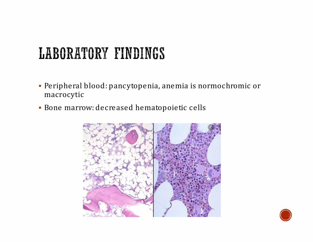
▪ Peripheral blood: pancytopenia, anemia is normochromic or macrocytic
▪ Bone marrow: decreased hematopoietic cells

▪ Fanconi anemia: rare, inherited form of AA, defect in DNA repair proteins, patients develop AA and acute leukemia in early life
▪ Pure red cell aplasia: only erythroid cells are absent in bone marrow, can be congenital (Diamond-Blackfan anemia) or acquired (autoimmune, Parvovirus B19 infection)

▪ Infiltration of bone marrow causing physical damage to hematopoietic cells
▪ Cancer: most commonly in acute leukemia, advanced lymphoma, metastatic cancer
▪ G ranulomatous disease: TB
▪ Storage diseases: G aucher
▪ Immature granulocytic and erythroid precursors commonly appear in peripheral blood

▪ Peripheral blood: leucoerythroblastic anemia (shift to left + nucleated RBCs)
▪ Insidious but accelerated symptoms of anemia
▪ Thrombocytopenia manifests as skin bleeding
▪ Neutropenia may results in serious infections and death
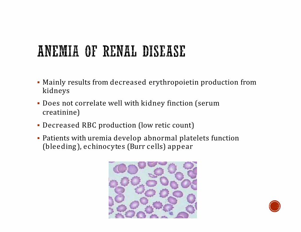
▪ Mainly results from decreased erythropoietin production from kidneys
▪ Does not correlate well with kidney finction (serumcreatinine)
▪ Decreased RBC production (low retic count)
▪ Patients with uremia develop abnormal platelets function (bleeding ), echinocytes (Burr cells) appear

▪ Multiple factors causing anemia
▪ Decreased synthesis of clotting factors (bleeding)
▪ Bleeding from varices
▪ Decreased synthesis of transferrin
▪ Acanthocyte (spur cell) appears

▪ Thyroid hormones stimulate erythropoiesis
▪ Also stimulates erythropoietin production
▪ Anemia is most commonly normocytic, but can be marcocytic
▪ Acquired, relatively common disease
▪ Primarily disease of old age
▪ Mutations in BM stem cell, results in prolonged survival and defective maturation
▪ Most patients have anemia, refractory to treatment
▪ RBCs are macrocytes
▪ RBC life span < 120 days
▪ Hypoxia triggers release of erythropoietin
▪ Erythroid hyperplasia in bone marrow
▪ Peripheral blood reticulocytosis
▪ Extramedullary hematopoiesis in severe cases
▪ Hemoglobin is released in from damaged RBCs
▪ Serum haptoglobin: decreased (binds free Hg)

Main site of hemolysis:
▪ 1) Extravascular: occurs primarily in spleen (RBCs have abnormal shape or coated with antibodies, removed by macrophages, patients have jaundice, pigmented gall bladder stones, splenomegaly)
▪ 2) Intravascular: inside blood stream (sudden release of Hg, patients have hemog lobinemia, hemog lobinurea, hemosiderinurea, iron deficiency)
According to cause of hemolysis
▪ Extracorpuscular vs intracorpuscular

▪ Group of inherited disorders that result in decreased productionof either α/β chains
▪ Amount of synthesized Hg is below normal
▪ The deficiency in one of globin chains results in a relative increase in the other one, excessive unpaired chains will cause instability and hemolysis
▪ Mode of inheritance: autosomal recessive
▪ Common in Middle East, Africa and South East Asia
▪ Resistant to infection by malaria falciparum
▪ Normal Hg types in adults: Hg A, Hg A2, Hg F

▪ α-chain is encoded by 2 genes on chromosome 16
▪ Most mutations in α-thalassemia are deletion
▪ Deletion in 1,2 gene(s) results in a silent carrier
▪ Deletion of 4 genes results in hydrops fetalis
▪ Deletion of 3 genes results in Hemoglobin H disease (extra β-chains binds each other to a tetramer called Hg-H, extra γ-chains form Hg -Barts). Both have hig h affinity to oxyg en
▪ Β-chain is encoded by a single gene of chromosome 11
▪ Most mutations in β-thal are point mutations
▪ β0: no production of β-chain
▪ β+: decreased production of β-chain
▪ β/β+:silent carrier or mild anemia (thal-minor)
▪ β+/β+ : thalassemia intermedia
▪ β0/β0 or β0/β+: thalassemia major (Cooley anemia)
▪ Extra α-chains remain uncoupled, causing hemolysis of RBCs and precursors (ineffective erythropoiesis)
▪ Hypochromic microcytic anemia
▪ Target cells
▪ Basophilic stippling
▪ Reticulocytosis
▪ In thalassemia major:
▪ Peripheral blood: + poikelocytosis, nucleated RBC s
▪ Bone marrow: ↑↑ normoblasts, filling BM spaces and expanding into bone, hemosiderosis

Basophilic stippling of RBCs

Thalassemia major blood film

▪ Thalassemia traits are asymptomatic, normal life span, premarital test is important
▪ Thalassemia major: symptoms begin after age of 6 months, persistent symptoms of anemia, growth retardation, skeletal abnormalities, both are ameliorated by reg ular blood transfusion
▪ Systemic hemochromatosis and related organ damage occurs in 2nd or 3rd decade of life
▪ Thalassemia intermedia and HgH disease have moderate anemia, do not require regular blood transfusion

▪ Hemoglobin electrophoresis test
▪ In all types of β-thal, there is increase in HgA2 and HgF percentages
▪ In β-thal major, HgA is absent or markedly decreased
▪ In HgH disease, HgH and Hg Barts bands appear
▪ In α-thal carrier and minor, no abnormality is found. Genetic testing is available
Related Documents

![JOURNAL OF Veterinary Science Isolation and ...€¦ · potential while embryonic stem cells are totipotent. Multipotent stem cells were first isolated from adult bone marrow [17].](https://static.cupdf.com/doc/110x72/5ec360e5b4a67233333e80da/journal-of-veterinary-science-isolation-and-potential-while-embryonic-stem-cells.jpg)